HAL Id: hal-03207565 https://hal.archives-ouvertes.fr/hal-03207565 Submitted on 25 Apr 2021 HAL is a multi-disciplinary open access archive for the deposit and dissemination of sci- entific research documents, whether they are pub- lished or not. The documents may come from teaching and research institutions in France or abroad, or from public or private research centers. L’archive ouverte pluridisciplinaire HAL, est destinée au dépôt et à la diffusion de documents scientifiques de niveau recherche, publiés ou non, émanant des établissements d’enseignement et de recherche français ou étrangers, des laboratoires publics ou privés. Speciation Analysis of Gadolinium in the Water-Insoluble Rat Brain Fraction After Administration of Gadolinium-Based Contrast Agents Izabela Strzeminska, Cécile Factor, Philippe Robert, Joanna Szpunar, Claire Corot, Ryszard Lobinski To cite this version: Izabela Strzeminska, Cécile Factor, Philippe Robert, Joanna Szpunar, Claire Corot, et al.. Spe- ciation Analysis of Gadolinium in the Water-Insoluble Rat Brain Fraction After Administration of Gadolinium-Based Contrast Agents. Investigative Radiology, Lippincott, Williams & Wilkins, 2021, 56 (9), pp.535-544. 10.1097/rli.0000000000000774. hal-03207565
Transcript
HAL Id: hal-03207565https://hal.archives-ouvertes.fr/hal-03207565
Submitted on 25 Apr 2021
HAL is a multi-disciplinary open accessarchive for the deposit and dissemination of sci-entific research documents, whether they are pub-lished or not. The documents may come fromteaching and research institutions in France orabroad, or from public or private research centers.
L’archive ouverte pluridisciplinaire HAL, estdestinée au dépôt et à la diffusion de documentsscientifiques de niveau recherche, publiés ou non,émanant des établissements d’enseignement et derecherche français ou étrangers, des laboratoirespublics ou privés.
Speciation Analysis of Gadolinium in theWater-Insoluble Rat Brain Fraction After
Administration of Gadolinium-Based Contrast AgentsIzabela Strzeminska, Cécile Factor, Philippe Robert, Joanna Szpunar, Claire
Corot, Ryszard Lobinski
To cite this version:Izabela Strzeminska, Cécile Factor, Philippe Robert, Joanna Szpunar, Claire Corot, et al.. Spe-ciation Analysis of Gadolinium in the Water-Insoluble Rat Brain Fraction After Administration ofGadolinium-Based Contrast Agents. Investigative Radiology, Lippincott, Williams & Wilkins, 2021,56 (9), pp.535-544. �10.1097/rli.0000000000000774�. �hal-03207565�
Claire Corot, PharmD, PhD,* and Ryszard Lobinski, PhD, DSc†‡
Purpose: To date, the analysis of gadolinium (Gd) speciation in the brain of an-imals administered with macrocyclic and linear Gd-based contrast agents(GBCAs) has been limited to Gd soluble in mild buffers. Under such conditions,less than 30% of the brain tissue was solubilized and the extraction recoveries ofGBCAs into the aqueous phase were poor, especially in the case of the linearGBCAs. The aim of this study was to find the conditions to solubilize the braintissue (quasi-)completely while preserving the Gd species present. The subse-quent analysis using size exclusion chromatography–inductively coupledplasma–mass spectrometry (SEC-ICP-MS) was intended to shed the light onthe speciation of the additionally recovered Gd.Methods: Four groups of healthy female Sprague Dawley rats (SPF/OFA rats;Charles River, L'Arbresle, France) received randomly 5 intravenous injections(1 injection per week during 5 consecutive weeks) of either gadoterate meglumine,gadobenate dimeglumine, gadodiamide (cumulated dose of 12 mmol/kg), or no in-jection (control group). The animals were sacrifice 1 week (W1) after the last injec-tion. Brain tissues were solubilized with urea solution, whereas tissues extractedwith water served as controls. Total Gd concentrations were determined in theoriginal brain tissue and its soluble and insoluble fractions by inductively coupledplasma–mass spectrometry (ICP-MS) to calculate the Gd accumulation and ex-traction efficiency. Size exclusion chromatography coupled to ICP-MS was usedto monitor the speciation of Gd in the soluble fractions. The stability of GBCAsin the optimum conditions was monitored by spiking the brain samples from theuntreated animals. The column recoveries were precisely determined in the purposeof the discrimination of weakly and strongly bound Gd complexes. The identity ofthe eluted specieswas explored by the evaluation of themolecular size and retentiontime matching with Gd chelates and ferritin standard. The speciation analyseswere carried out for 2 different brain structures, cortex and cerebellum.Results: The combination of water and urea extractions (sequential extraction)managed to solubilize efficiently the brain tissue (97% ± 1%) while preserving
the stability of the initially injected form of GBCA. For macrocyclic gadoterate,97% ± 1% and 102% ± 3% of Gd initially present in the cortex and cerebellumwere extracted to the soluble fraction. For gadobenate, similar amounts of Gd(49% ± 1% and 46% ± 4%) were recovered from cortex and cerebellum. Forgadodiamide, 48% ± 2% of Gd was extracted from cortex and 34% ± 1% fromcerebellum. These extraction efficiencies were higher than reported elsewhere.The SEC-ICP-MS and the column recovery determination proved that Gd presentat low nmol/g levels in brain tissuewas exclusively in the intact GBCA form in allthe fractions of brain from the animals treated with gadoterate. For the linearGBCAs (gadobenate and gadodiamide), 3 Gd species of different hydrodynamicvolumes were detected in the urea-soluble fraction: (1) larger than 660 kDa, (2)approximately 440 kDa, and (3) intact GBCAs. The species of 440 kDacorresponded, on the basis of the elution volume, to a Gd3+ complex with ferritin.Gd3+was also demonstrated by SEC-ICP-MS to react with the ferritin standard in100 mM ammonium acetate (pH 7.4). In contrast to macrocyclic gadoterate, forlinear GBCAs, the column recovery was largely incomplete, suggesting the pres-ence of free, hydrolyzed, or weakly bound Gd3+ with endogenous ligands.Conclusions: The sequential extraction of rat brain tissue with water and urea so-lution resulted in quasi-complete solubilization of the tissue and a considerable in-crease in the recoveries of Gd species in comparison with previous reports. Themacrocyclic gadoterate was demonstrated to remain intact in the brain 1 week afteradministration to rats. The linear GBCAs gadobenate and gadodiamide underwentligand exchange reactions resulting in the presence of a series of Gd3+ complexes ofdifferent strength with endogenous ligands. Ferritin was identified as one of themacromolecules reacting with Gd3+. For the linear GBCAs, 3% of the insolublebrain tissue was found to contain more than 50% of Gd in unidentified form(s).
C ontrast enhancement in magnetic resonance imaging is usuallyachieved by intravenous administration of chelates of the paramag-
netic gadolinium ion (Gd3+) with polyaminocarboxylic acids in chargeof reducing its acute and chronic toxicities.1 These chelates, referred toas Gd-based contrast agents (GBCAs), vary in terms of thermodynamic(logKtherm16.6–25.6) andkinetic stabilities (T1/2 at pH1and25°C<5sec-onds to 338 hours) and can be divided, on the basis of the molecularstructure, into linear (L-GBCAs) and macrocyclic (M-GBCAs), eitherclass of which can be further subdivided into ionic and nonionic com-pounds.2 Under physiological conditions, the M-GBCAs are more sta-ble than the linear type and ionic L-GBCAs more than nonionic L-GBCAs.3
Historically, the relatively hydrophilic, small-sized moleculesof GBCAs were designed not to enter the cells or to penetrate theblood-brain barrier, and to be completely eliminated in the intact form.4
However, in the last decade, it has becomewell established that a trace amountof Gd does accumulate for a prolonged time in multiple organs, includingthe brain, after single5,6 and repeated GBCAs administration.7–10
Based on the animal studies, it can be estimated that eventually less than0.0005% of the injected dose is found in the brain.11 Although the
Received for publication November 18, 2020; and accepted for publication, after revi-sion, January 31, 2021.
From the *Guerbet Research and Innovation Department, Aulnay-sous-Bois; †Instituteof Analytical Sciences and Physico-Chemistry for Environment and Materials,UMR 5254, CNRS-UPPA, Pau, France; and ‡Department of Chemistry, WarsawUniversity of Technology, Poland.
Correspondence to: Izabela Strzeminska, PhD, Guerbet Research and Innovation De-partment, BP57400, 95943 Roissy CDG Cedex, France. E-mail: [email protected].
Conflicts of interest and sources of funding: I.S., C.F., P.R., and C.C. are (or were)Guerbet employees at the time of the study. For J.S. and R.L., Institute ofAnalytical Sciences and Physico-Chemistry for Environment and Materialsreceived funding from Guerbet within a common research project. R.L. is theacademic tutor of PhD thesis of I.S. The study was funded by Guerbet.
Supplemental digital contents are available for this article. Direct URL citations appearin the printed text and are provided in the HTML and PDF versions of this articleon the journal’s Web site (www.investigativeradiology.com).
amount of retained Gd is at trace level, the local concentrations may beelevated, achieving as much as 64 nmol/g (10.1 ppm).12 Albeit theseobservations have not been associated with any clinical effects or histo-logical alterations, the understanding of the mechanisms of the Gd reten-tion, including exhaustive characterization of the Gd species involved, isessential for the long-term GBCAs safety. Consequently, it has beenattracting the attention of regulatory agencies such as the European Medi-cal Agency and the Food and Drug Administration.13,14
Numerous studies in patients15 and in animals16 provided evi-dence for the relationship between the structure of GBCAs and theamount of total Gd accumulated in tissue. Significantly higher concentra-tions were found in brain tissues after L-GBCAs administration in com-parison with M-GBCAs. Several localization experiments demonstratedthat the Gd distribution in the brain was not uniform; the elements accu-mulated preferentially in the dentate nucleus or globus pallidus, which arerich in endogenousmetals such as iron.17,18 This finding suggests that theL-GBCAs may partially undergo a dechelation process through thetransmetalation with endogenous metals such as iron, producing newGd species. Transmission electron microscopy (TEM) associated withnanoSIMS (nanoscale secondary ion mass spectrometry) demonstratedthe presence of Gd deposits containing phosphorus in the deep cerebel-lar nuclei (DCN), strongly suggesting formation of GdPO4.
19
Complementary data on the speciation of the residual Gd can beobtained by chromatography coupled with Gd-specific or GBCA-specificdetection by inductively coupled plasma–mass spectrometry (ICP-MS)11,20
or electrospray mass spectrometry,21 respectively. Size exclusion chro-matography (SEC), offering the separation according to the hydrody-namic volume, has been favored as the separation technique, allowingthe preservation of the Gd ligand species.16 Even if low, the separationefficiency was sufficient to discriminate between the intact GBCA spe-cies and the potential complexes of Gd with other (macromolecular) li-gands present in the sample.11,20 Indeed, high-molecular-weight (HMW)species (66 to 250 kDa, depending on the type of the column and cali-bration markers used) were detected but were not formally identi-fied.11,20 An alternative to SEC is hydrophilic interaction liquidchromatography. It offers a better resolution for low-molecular-weight(LMW) compounds for the identification or quantification of intactGBCAs in water fraction, but it is unable to detect HMW species.
The coupled techniques have 2 significant drawbacks. First, theGd species have to be present in solution, which raises questions abouttheir recovery, in the original chemical form, from the tissue duringsample treatment and chromatography. A typical water extraction pro-cedure, based on the tissue homogenization with 10 mM Tris-HClbuffer (pH 7.4) and the recovery of the subsequently analyzed superna-tant, shows relatively poor extraction efficiencies of Gd, which meansthat only a small fraction of the Gd present in the tissue is actuallyaccounted for. Indeed, Frenzel et al11 could recover approximatively17% to 37% of Gd present in the brain tissue of rats administered withL-GBCAs. To date, no effort has been made to analyze the Gd retainedin the brain tissue fraction insoluble with mild buffer, because of thelack of methods able to solubilize it without compromising the natureof the contained Gd compounds. The speciation of the to-date unac-counted for Gd species remains therefore unknown. It is unclearwhether GBCA is retained in the pellet as intact GBCA or whether ithad undergone dissociation. In contrast to L-GBCAs, the presence ofthe kinetically very stable M-GBCAs in the pellet was hypothesizedto be due to incomplete washout of entrapped GBCA inside cell de-bris. However, the fact that 3 washings of the remaining pellet wereinsufficient suggests that the mechanism of the Gd retention can bemore complex.11
The second drawback of the to-date reported procedures is thespecies-dependent nonquantitative recovery of Gd from the chromato-graphic column. The stationary phase is not entirely inert and may reactwith the Gd species depending on their stability. This concerns not onlythe GBCAs but also the species potentially present in the tissue, and
specially Gd3+. The mass balance of the introduced and recovered Gdhas seldom been reported.21 To the best of our knowledge, the recover-ies of Gd species from the SEC column have never been reported.11,20
Finally, even if some evidence of the presence of Gd complexeswith macromolecules in water-soluble fractions exists, the identity ofthis species remains unknown.
The aims of this study were to develop a method able to solubilizethe brain tissue completely and to obtain an insight into the speciationof the additionally recovered Gd, as a function of GBCA. Rats admin-istered with 3 GBCAs (1 macrocyclic and ionic GBCA gadoteratemeglumine and 2 linear [1 ionic gadobenate dimeglumine and 1 non-ionic gadodiamide]) were investigated. Size exclusion chromatographywas revisited with an emphasis of the compound-dependent column re-covery of Gd and insight into the identity of the observed Gd species.
Materials and MethodsAll animal experiments were conducted in full compliance with
the European Union Directive 2010/63/EU on the protection of animalsused for scientific purposes.
Analytical reagent grade chemicals were used. They were ob-tained from Merck (Darmstadt, Germany) unless stated otherwise.Dionized water (18 MΩ cm) was obtained from a Milli-Q water purifi-cation system (Merck, Darmstadt, Germany).
Animal Model and Gadolinium-Based ContrastAgent Administration
To cover the different GBCA classes available on the market, 3compounds were selected: 1 macrocyclic and ionic GBCA (gadoteratemeglumine, Dotarem; Guerbet, Villepinte, France) and 2 linear—1ionic (gadobenate dimeglumine, MultiHance; Bracco, Milan, Italy) and1 nonionic (gadodiamide, Omniscan; GEHealthcare, Boston,MA). TheseGBCAs were used in the form of the commercially available solutionfor injection (0.5 M solutions). Four groups of healthy female SpragueDawley rats (SPF/OFA rats; Charles River, L'Arbresle, France) ran-domly received an intravenous injection of 2.4 mmol/kg of GBCA eachweek during 5 consecutive weeks (cumulated dose of 12 mmol/kg) orno injection (control group). A total of 2.4 mmol/kg corresponds to0.4 mmol/kg in humans according to the Food and Drug Administra-tion guidance, after adjustment to the body surface area of the rat.22 In-travenous injections were performed in 1 tail vein under isofluraneanesthesia (IsoFlo; Axience, Pantin, France). The animals were sacrificed1 week (W1) after the last injection. The GBCA dose and the sacrificetime were chosen as a compromise between the analysis of the residualGd species and the sufficient Gd concentration for the sample preparationmethod optimization. At the completion of the washout period, the ratswere euthanized under isoflurane anesthesia by exsanguination. Thebrains were removed and dissected to sample a half cerebellum, half cor-tex, and half subcortical brain. The cerebral tissues were stored at −80°Cbefore sample preparation.
Sample PreparationThe cerebral tissue was homogenized using an Ultra-Turrax dis-
perser (IKA-Werke, Staufen, Germany) in 100 mM ammonium acetate(pH 7.4; dilution factor of 6) in an ice bath. Then, the homogenate wasdivided into 400 μL or 650 μL aliquots and stored at −20°C.
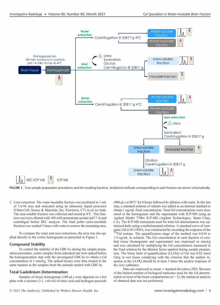
The sequential extraction procedure, schematically presented inFigure 1, was based on the following:
1. Water extraction: The tissue homogenate was centrifuged using aCentrifuge 5804 R model (Eppendorf, Hamburg, Germany) for30 minutes at 20,800g at 4°C. The water-soluble fraction was sepa-rated from the water-insoluble fraction and stored in −20°C. Thewater-insoluble fraction was washed with 0.5 mL of 100 mM ammo-nium acetate.
Strzeminska et al Investigative Radiology • Volume 00, Number 00, Month 2021
2. Urea extraction: The water-insoluble fraction was incubated in 1 mLof 5.6-M urea and sonicated using an ultrasonic liquid processor(Vibra-Cell; Sonics & Materials, Inc, Newtown, CT) in an ice bath.The urea-soluble fraction was collected and stored at 4°C. This frac-tion was twice diluted with 100 mMammonium acetate (pH 7.4) andcentrifuged before SEC analysis. The final pellet (urea-insolublefraction) waswashed 3 timeswith water to remove the remaining urea.
To compare the water and urea extractions, the urea was also ap-plied directly to the cortex homogenate as presented in Figure 1.
Compound StabilityTo control the stability of the GBCAs during the sample prepa-
ration procedure, cortex samples from untreated rats were spiked beforethe homogenization step with the investigated GBCAs to obtain a Gdconcentration of 1 nmol/g. The spiked tissues were then treated in thesame manner as the samples from the animals treated with GBCAs.
Total Gadolinium DeterminationSamples of tissue homogenate (100 μL) were digested on a hot
plate with a mixture (3:1, vol/vol) of nitric acid and hydrogen peroxide
(400 μL) at 80°C for 8 hours followed by dilution with water. In this laststep, a standard solution of indium was added as an internal standard toobtain 1 ng/mL final concentration. Total Gd concentrations were mea-sured in the homogenate and the supernatant with ICP-MS using anAgilent Model 7700x ICP-MS (Agilent Technologies, Santa Clara,CA). The ICP-MS instrument used for total Gd determination was op-timized daily using a multielemental solution. A standard curve of inor-ganic Gd in 4%HNO3was constructed by recording the response of the158Gd isotope. The quantification range of the method was 0.010 to1.0 ng/mL in solution. The Gd concentration in each fraction of cere-bral tissue (homogenate and supernatant) was expressed as nmol/gand was calculated by multiplying the Gd concentration measured inthe final solution by the dilution factor applied during sample prepara-tion. The lower limit of quantification (LLOQ) of Gd was 0.02 nmolGd/g in wet tissue complying with the criterion that the analyte re-sponse at the LLOQ should be at least 5 times the analyte response ofthe zero calibrator.
Data are expressed as mean ± standard deviation (SD). Becauseof the limited number of biological replicates used for the Gd determi-nation in most of the brain compartments (n = 2), the statistical analysisof obtained data was not performed.
FIGURE 1. Two sample preparation procedures and the resulting fractions. Analytical methods corresponding to each fraction are shown schematically.
Investigative Radiology • Volume 00, Number 00, Month 2021 Gd Speciation in Water-Insoluble Brain Fraction
Extraction Efficiency MeasurementTo evaluate the amount of Gd extracted from the cerebral tissue
to the soluble fraction(s), the extraction efficiency (EE) was calculatedusing the following equation: EE ¼ nGd supernatant
nGd homogenate� 100%; where nGd is the
number of moles of Gd. For sequential extraction, the amount of Gdin 2 water-soluble supernatants (first extraction and washing step) andin the 2.8-M urea-soluble fraction was considered.
Solubilization of Cerebral TissueThe percentage of solubilization of a cerebral tissue (BS) was es-
timated using the following equation: BS %ð Þ ¼ mdry homogenate−mdry pellet
mdry homogenate� 100%.
For this purpose, all the pellets were washed twice with 1 mL of water,lyophilized and weighed (mdry pellet). The mass of dry homogenate wascalculated using the following equation: mdry homogenate = mwet homoge-
nate · fdry tissue, where the proportion of a dry tissue in a wet homogenatefdry tissue was estimated for 6 representative homogenates, which werelyophilized: fdry tissue ¼ mdry homogenate
mwet homogenate.
Enrichment FactorThe enrichment factor (EF) is a widely used metric for determin-
ing the number of times the analyte concentration has increased relativeto its concentration without enrichment. The water, urea, and sequentialextraction procedures were compared in terms of the EFs for Gdcontained in the final insoluble residue. The EF was calculated as theratio of the initial dry mass of the homogenate and the reduced finaldry mass of the insoluble residue.
Gadolinium Speciation AnalysisThe SEC-ICP-MS analysis was carried out using an high perfor-
mance liquid chromatography 1260 system (Agilent Technologies)coupled to ICP-MS 7700x (Agilent Technologies). Gadolinium specieswere isocratically eluted either from a Superdex 75 or a Superdex 200column (300 � 10 mm, GE Healthcare) with 100 mM ammonium ac-etate (pH 7.4) over 45 minutes at a flow rate of 0.7 mL/min. Becauseof the limited separation range of the Superdex 75 column (3–80 kDa),the Superdex 200 (separation range, 10–660 kDa) column was used tobetter estimate the molecular weight of the detected Gd species. The in-jection volumewas 100 μL. The LLOQ of SEC-ICP-MS was estimatedusing standard GBCA solutions and set at 0.32 pmol/mL for all3 GBCAs using the signal-to-noise > 5 criterion. To control the back-ground level of Gd, the supernatants obtained from the samples of thecontrol group were analyzed systematically at the beginning and atthe end of the sequence as the blanks. The average blank chromatogram
per run was calculated and subtracted from the chromatograms unlessspecified otherwise.
The column recovery was verified for Gd in spiked samplesfrom untreated animals and in the real samples from treated animals,comparing the total area of the peaks detected using the chromatographiccolumnwith the area of the peak obtained in flow injection mode (with-out column).
RESULTS
Total Gadolinium DeterminationThe total Gd concentrations determined are summarized in Figure 2.
The concentration in the cerebellum obtained after injections of rats withgadoterate (0.63 ± 0.12 nmol/g) was lower than that obtained after injec-tions with either linear contrast agents: gadobenate (1.52 ± 0.16 nmol/g)and gadodiamide (2.74 ± 0.10 nmol/g). In addition, the Gd concentrationafter injection of gadobenate was lower than that measured after injectionof gadodiamide. The same trend was found for the subcortical brain andthe cortex tissues.
Brain Solubilization and Extraction RecoveriesTable 1 summarizes the percentage of the brain solubilization,
the Gd extraction efficiency data, and the EFs obtained using water,urea, and the sequential extraction for the samples from the treatedgroups. The extraction with the ammonium acetate buffer (water extrac-tion) allowed the recovery of 68% to 72% of Gd initially present in thetissue for macrocyclic gadoterate. For L-GBCAs, the extracted amountsof Gd were much lower: 13% to 16% for gadobenate and 12% to 13%for gadodiamide. Water extraction managed to solubilize only 33% ofthe brain tissue matrix components. The cortex was better solubilized(65%–72%) using a chaotropic agent urea. It also resulted in the highestGd extraction efficiencies of 47% ± 2% and 44% ± 7% for gadobenateand gadodiamide, respectively. The extraction for macrocyclic gadoteratewas also improved (88% ± 3%).
The combination of water and urea extraction maximized the sol-ubilization to reach 97% of brain tissue. Washing of the water-insolublefraction allowed the recovery of an additional amount of Gd, similar forboth brain structures, 12% to 15% for gadoterate but only 2% to 3%for gadobenate and gadodiamide (data not shown). In the second step,for gadoterate-containing samples, 15% to 19% of original Gd were ex-tracted to urea-soluble fraction regardless of the brain matrix; conse-quently, almost no Gd (1%–2%) was found in the final, insolubleresidue. In total, similar amounts of Gd (97% ± 1% and 102% ± 3%)were extracted from the cortex and cerebellum for macrocyclic
FIGURE 2. Accumulation of gadolinium (Gd) in the cerebellum, subcortical brain, and cortex 1 week after the fifth injection of gadolinium-basedcontrast agents.
Strzeminska et al Investigative Radiology • Volume 00, Number 00, Month 2021
gadoterate. For gadobenate and gadodiamide, the total amount of Gdrecovered from the cortex was 49% ± 1% and 48% ± 2% and fromthe cerebellum was 46% ± 4% and 34% ± 1%, respectively. In additionto a lower recovery in comparison with the macrocyclic gadoterate, 1significant difference was observed between the cortex and the cerebel-lum tissues for these L-GBCAs. Dilution of the 5.6-M urea-solublefraction with ammonium acetate buffer (pH 7.4) decreased the amountof Gd for the cerebellum but not for cortex samples. Indeed, approx-imately half of the amount of Gd species extracted from thewater-insoluble fraction to 5.6-M urea fraction was not soluble in 2.8-Murea (data not shown).
Table S1 (Supplemental Digital Content, http://links.lww.com/RLI/A617) shows high extraction efficiencies calculated for the brainsamples from the control group spiked with either gadoterate,gadobenate, or gadodiamide, which suggests that no significant degra-dation of GBCAs occurred during sample preparation. Indeed, 85% to96% of total Gd was extracted for all 3 GBCAs with urea.Water extrac-tion resulted in slightly less efficient extraction (64% to 95%).
The Gd contained in the final, insoluble residuewas enriched themost efficiently using sequential extraction procedure (EFof 28 ± 7 and34 ± 14 for the cortex and cerebellum, respectively) compared with wa-ter (1.4 ± 0.1) and urea (3.5 ± 0.2) extractions.
Speciation of Gadolinium Using SEC-ICP-MSRepresentative SEC-ICP-MS and SEC-UV chromatograms
(Superdex 75 column) of the water- and urea-soluble fractions fromthe treated rat group are shown in Figure 3. The chromatograms forthe brain samples from the control group spiked with 1 of the GBCAsare shown in Figure S1 (Supplemental Digital Content, http://links.lww.com/RLI/A617). A different number of Gd species was detected for lin-ear and macrocyclic GBCAs.
Water ExtractionFor macrocyclic gadoterate, Gd was exclusively detected as 1
large peak with a similar retention time as the intact GBCA (RT,26.3 minutes). On the other hand, for L-GBCAs, 3 distinct peaks werepresent at the retention times of RT1 = 11.7minutes, RT2 = 13.4minutes,and RT3 = 26.2 minutes (for gadodiamide) or RT3 = 26.8 minutes (forgadobenate). According to the column calibration, which is shown inFigure S2 (Supplemental Digital Content, http://links.lww.com/RLI/A617), the Gd species eluting at 11.7 minutes should be larger than81 kDa, the 1 eluting at 13.4 minutes should be of approximately66 kDa, and the last one, eluting between 20.1 and 29.3 minutes, shouldbe smaller than 12 kDa. Therefore, the signal observed approximately26 to 27 minutes corresponds to LMW molecules, such as the intactGBCA, whereas the peaks at 11.7 minutes and 13.4minutes correspond
to HMWGd species with the size of approximately of 66 kDa or larger.The column recoveries obtained for the water-soluble fraction of thecontrol group samples spiked with standard GBCAs solutions and fromthe samples of treated rats are presented in Figure 4. The column recov-eries for the spiked samples confirmed that intact GBCAs are elutedcompletely from the chromatographic column regardless of their stabil-ity with no degradation of GBCAs in the chromatographic conditionsused for the analysis. Likewise, the column recovery obtained for thereal samples after administration of gadoterate was 111% ± 2%, whichproved that all Gd extracted to the water-soluble fraction of the brainwas present solely as intact GBCA form. However, for the L-GBCAsgadodiamide and gadobenate, the column recovery was approxi-mately 82% ± 2%, suggesting that a no negligible amount of Gdpresent in the water-soluble fraction was retained on the stationaryphase of the chromatographic column, which would happen, for in-stance, for Gd present as weakly bound Gd3+ with endogenous li-gands present in the sample.
Urea ExtractionNo change in the peak intensity was observed for the samples
from the control group spiked with either gadoterate, gadobenate, orgadodiamide (see Fig. S1, Supplemental Digital Content, http://links.lww.com/RLI/A617). Moreover, the examination of column recoveryshowed complete elution of Gd from the chromatographic column(103%–114%) for all 3 GBCAs, proving their stability in these condi-tions. A representative SEC-ICP-MS chromatogram of the supernatant(cortex W1) obtained for gadoterate sample (Fig. 3B) shows that simi-larly towater extractionGdwas eluted completely as 1 peak at the reten-tion time RT1 = 26.4 minutes. Typical SEC-ICP-MS chromatograms ofthe urea-soluble fraction of cortex (W1) for gadobenate andgadodiamide are presented in Figures 3C and D. At least 3 Gd peakswere observed for these L-GBCAs. Two of the peaks were eluted veryclose to the void volume of Superdex 75 column in the HMW elutionrange (RT1 = 11.1 minutes, RT2 = 11.7 minutes), whereas 1 peak wasobserved in the LMW range at 26.7 minutes. The 2 peaks in theHMW fraction were not well separated, and there may be another broadpeak of smaller intensity, eluting in the void volume of the column(10.3 minutes) but covered by the peak at 11.1 minutes. Because ofthe low resolution and the limited separation range of the chromato-graphic column used for this analysis, the hydrodynamic volume ofthe Gd species present in the HMW fraction could only be estimatedas larger than 80 kDa. Also, the Gd species detected by SEC-ICP-MSdid not account for all the Gd present in the supernatant. The columnrecoveries for both L-GBCAs showed that 38% of extracted Gd wasretained on the chromatographic column strongly, suggesting the pres-ence of other Gd species (see Fig. 4).
TABLE 1. Brain Solubilization and Gadolinium Extraction Efficiencies in the Different Analytical Protocols Developed
Brain solubilization was calculated based on the different number of data points for each analytical procedure, for cortex samples: water extraction n = 8, urea extrac-tion n = 6, and water + urea extraction n = 6; for cerebellum: water + urea extraction n = 7. Extraction efficiency was calculated based on n = 2 data points except waterextraction for cerebellum n = 5 (gadobenate n = 4).
*Brain solubilization after water extraction was determined only for cortex; no significant differences between cortex and cerebellum were expected.
Investigative Radiology • Volume 00, Number 00, Month 2021 Gd Speciation in Water-Insoluble Brain Fraction
FIGURE 4. Gd recovery from the Superdex 75 and 200 columns. Data obtained for the samples from the untreated animals spiked withgadolinium-based contrast agent standards and from the treated animals. Column recoveries determined for Gd species present in the water- andurea-soluble cortex fractions after extraction from either the homogenate or the water-insoluble fraction.
FIGURE 3. Molecular size distribution of Gd species recovered to water- and urea-soluble fractions from the cortex homogenate. Typical size exclusionchromatography UV (A) and size exclusion chromatography–inductively coupled plasma–mass spectrometry (B–D) chromatograms obtained usingSuperdex 75 column.
Strzeminska et al Investigative Radiology • Volume 00, Number 00, Month 2021
Sequential ExtractionRepresentative SEC-ICP-MS chromatograms obtained using
Superdex 200 column of the 2.8-M urea-soluble fractions extractedfrom the water-insoluble fractions are shown in Figure 5. For gadoterate,only 1 species was detected in the LMW fraction at RT1 = 28.1 minutes.For L-GBCAs, 3 Gd peaks were observed at the following retentiontimes: RT1 = 11.0 minutes, RT2 = 20.1 minutes, and RT3 = 28.3 minutes,which, according to the calibration of the Superdex 200 column (Fig. S3,Supplemental Digital Content, http://links.lww.com/RLI/A617), cor-responds to the MW1 larger than 660 kDa, MW2 of approximatelyof 440 kDa, and MW3 inferior to 44 kDa. The column recoveriesof water-soluble fractions are in good agreement with previouslyshown data (gadoterate 111% ± 2%, gadobenate 83% ± 1%, andgadodiamide 82% ± 2%) and are shown in Figure S4 (SupplementalDigital Content, http://links.lww.com/RLI/A617) together with thedata obtained for the cerebellum samples. The column recoveries ob-tained for 2.8-M urea-soluble cortex fractions are presented in Figure 4.Regarding the urea-soluble fractions, quantitative elution of Gd fromthe chromatographic column (85%–98%) was observed only forgadoterate. For gadobenate and gadodiamide, half of the extractedGd was retained on the stationary phase for cortex and 60% for cere-bellum samples.
Insight Into the Nature of the Extracted GadoliniumLigand Species
Figures 6A and B demonstrate SEC-ICP-MS chromatograms ofwater-soluble fraction of cortex sample (W1) from gadodiamide groupspikedwith 10, 50, or 100 ppb of either Gd3+ or gadodiamide standards.The chromatograms of the sample spiked with a gadodiamide standardshowed that only the intensity of the peak eluting at 27.9 minutes in-creased. In contrast, when the same sample was spiked with Gd3+, theintensity of the peak eluting at 10.7 minutes increased significantly, anda slight increase in the height of 2 peaks eluting at 19.6 and 27.9 minuteswas observed. Therefore, only the ionic Gd but not the GBCAshowed a strong affinity toward the HMW ligands. Figure 6C showsSEC-UV-ICP-MS chromatograms obtained for a ferritin standard so-lution before and after spiking with ionic Gd to the final Gd concen-tration of 100 ppb. Two different peaks are present at the retentiontimes: RT1 = 17.2 minutes and RT2 = 20.1 minutes, proving that ferritinbound some ionic Gd both from the chromatographic column and fromthe spike.
FIGURE 5. Molecular size distribution of Gd species recovered to 2.8-Murea-soluble fraction from the cortex and cerebellum water-insolublefractions.
FIGURE 6. Insight into the nature of the extracted Gd species. Sizeexclusion chromatography–inductively coupled plasma–massspectrometry chromatograms of water-soluble fraction of cortex samplesof gadodiamide spiked with either 10, 50, or 100 ppb of gadodiamide(A) or ionic gadolinium (B). Size exclusion chromatography–inductivelycoupled plasma–mass spectrometry of ferritin standard alone andspiked with 100 ppb of ionic gadolinium (C).
Investigative Radiology • Volume 00, Number 00, Month 2021 Gd Speciation in Water-Insoluble Brain Fraction
Total Gadolinium ConcentrationsThe total Gd concentrations measured by ICP-MS in the cerebel-
lum, subcortical brain, and in the cortex confirmed the class effectresulting in the higher Gd concentrations accumulated after L-GBCAsadministrations.16
Extraction Efficiency
Water ExtractionIn the previous bioanalytical studies, water extraction providing
mild conditions during sample processing (absence of detergents,chaotropic agents, and organic solvents) combined with pH close tothe physiological value (7.4) was typically used to minimize the riskof changes of the original Gd speciation. Our data obtained for the sam-ples from the animals treated with GBCAs confirmed that this extrac-tion approach failed to recover all the Gd to the water-soluble fractionfrom the tissue, which was further analyzed by high performanceliquid chromatography-ICP-MS. These results are consistent withprevious findings, although the availability of precisely calculatedextraction efficiency values for the brain tissue is limited in the litera-ture.11,20 In this study, 68% to 72% of Gd from the macrocyclicgadoterate and only 12% to 16% from the linear gadobenate andgadodiamide were found in the water-soluble brain fraction. Such lowrecoveries limit significantly the investigation of Gd species by hyphen-ated techniques and leave unanswered the question of the nature of Gdspecies present in the pellet after administration of GBCAs. Indeed, itshould be determined whether Gd remained in the water-insoluble frac-tion because of the unsuitable extraction conditions or rather because ofthe insoluble character of Gd species. In the case of L-GBCAs, the mostlikely explanation is that, because of their lower kinetic stability, they grad-ually dissociate in tissues and form nonextractable in water Gd species,such as, for example, inorganic salts or strong complexes with biomole-cules forming the pellet. Interestingly, Kahakachchi et al23 reported lowextraction efficiency (5%) similar to L-GBCAs from this study (8%–18%) when bovine muscle powder was spiked with ionic Gd3+ and ex-tracted with water. Moreover, the insoluble deposits of Gd associatedwith phosphorus were observed using TEM-Energy-dispersive X-rayspectroscopy in skin tissues and using TEM and nanoSIMS in the cer-ebellum.19 However, it is unlikely that Gd is present in the pellet only insolid binary forms such as GdPO4, Gd2(CO3)3, and Gd(OH)3 because itcould not generate the high signal intensity observed on T1-weightedmagnetic resonance images. Indeed, the relaxivity values (60 MHz,37°C) measured for Gd phosphate, carbonate, and hydroxide in waterand in skin samples are very low (0.01–0.29 mM−1 s−1).24
Optimization of Brain Solubilization andExtraction Conditions
Water extraction was found to solubilize only 33% of brain tis-sue, which may explain the poor extraction recoveries of Gd species.Therefore, it was decided to develop a more efficient brain tissue solu-bilization procedure. The brain is composed of approximately 77% to78% of water, 10% to 12% of lipids, 8% of proteins, 1% of carbohy-drates, 2% of soluble organic substances, and 1% of inorganic salts.25
A dedicated approach based on the use of chaotropic agent ureawas op-timized to assure its solubilization in conditions preserving the analytespecies. Indeed, urea appeared to have higher capacity to solubilize thebrain tissue than water, allowing the dissolution of 72% of the cortexmatrix while preserving the initially injected form of GBCAs (seeFig. S1, Supplemental Digital Content, http://links.lww.com/RLI/A617). More importantly, it improved Gd extraction. Almost half ofGd was recovered from the brain tissue for L-GBCAs (47% ± 2% and44% ± 7% for gadobenate and gadodiamide, respectively). Nevertheless,approximately half of total Gd remained in the insoluble residue. It is
consistent with the imaging experiments showing the electron-dense de-posits containing Gd in the brain.8,19 Indeed, the solid forms cannot beextracted using the procedures developed in this work. In the case ofmacrocyclic gadoterate, all the Gd was extracted using urea.
Total Brain Solubilization: The Sequential ExtractionTo solubilize brain tissue completely, a combination of water and
urea extraction was applied in sequence. For each step of this solubili-zation process, the extraction efficiency was calculated showing thatin total 97% to 102% for gadoterate and 34% to 49% of Gd speciesfor L-GBCAs could be solubilized in the conditions guaranteeing thestability of initially injected GBCA form. Note that sequential extrac-tion did not maximize the extraction efficiency in comparison to ureaextraction alone, even if it solubilized almost completely the brain tis-sue. However, by reducing the mass of the final insoluble residue, theconcentration of Gd species in that fraction was considerably increasedcompared with the initial concentration in the homogenate. Indeed, theGd EFs in the insoluble fraction reached 28 ± 7 and 34 ± 14 for the cor-tex and cerebellum, respectively, which might be an important improve-ment for future studies of these insoluble Gd species.
Speciation of Gadolinium in the Water-Soluble Part ofBrain Tissue
In the case of macrocyclic gadoterate, the extracted Gd was de-tected exclusively as the intact GBCA in water-soluble brain fraction.For the L-GBCAs, apart from the dominating intact GBCA form, thetiny amount of Gd bound to macromolecules was observed as alreadyreported by Frenzel et al11 and Robert et al.20 Water allows the extrac-tion of highly soluble Gd species such as intact GBCA or macromole-cules that can be proteins, carbohydrates, or some fragments of morecomplex endogenous molecules. However, based on the determinationof the column recovery, approximately 18% ± 2% of Gd was present inthe water-soluble fraction in other than previously reported form(s) thatwas retained on the stationary phase (see Fig. 4).
Speciation of Gadolinium in the Urea-Soluble Part ofBrain Tissue
All Gd was extracted from the brain homogenate of rats treatedwith gadoterate using urea procedure, and SEC-ICP-MS demonstratedthat it was present exclusively as intact GBCA form in the cortex (Fig.3B). This result confirmed that, in the case of M-GBCA, partial loss ofGd in the water-insoluble fraction was not due to the formation ofnonextractable Gd species but rather due to mechanical capture ofGBCA form in tissue or during sample processing. This part cannotbe totally extracted with water, but it is rapidly released when the tissueis better solubilized with urea. The Gd extraction was also improved forthe L-GBCAs. Interestingly, 2 forms of Gd were better extracted in ureacompared with water-soluble fraction: Gd bound to macromoleculesand weak complexes of Gd3+ with endogenous ligands, in which pres-ence was deduced from the incomplete elution of Gd from the chromato-graphic column (see Fig. 4). Indeed, the column recoveries were 82% and62% in water- and urea-soluble fractions, respectively. The HMW Gdspecies detected in the urea-soluble fraction were approximately 5-foldmore abundant than those obtained after water extraction. At high con-centrations, urea is commonly used to solubilize more hydrophobic,membrane-associated protein, and also proteoglycans,26 by breakingthe intermolecular and intramolecular noncovalent interactions (suchas hydrogen bonds, dipole-dipole, and hydrophobic interactions). Ap-proximately 50% of brain proteins are insoluble in water, acid, and al-kaline25; thus, it is possible that urea allowed to solubilize some ofthem without disrupting their interaction with Gd. The SEC-UV chro-matograms obtained for water- and urea-soluble fractions (cf Fig. 3A)support this hypothesis. Indeed, 1 relatively intense peak was detectedusing a UV detector at 280 nm at RT1 = 11.2 minutes in urea-soluble
Strzeminska et al Investigative Radiology • Volume 00, Number 00, Month 2021
fraction but was absent in the water one. This peak is eluted very closeto or in the void volume of the column, which strongly suggests thepresence of very large molecules or aggregates. Indeed, by breakingthe secondary protein structure, urea increases the number of hydropho-bic fragments exposed to the solvent, and thus the possibilities of pro-tein aggregation.27
Speciation of Gadolinium in the Water-Insoluble(Urea-Soluble) Part of Brain Tissue
The Gd extracted with urea from the water-insoluble brain frac-tion was demonstrated to be entirely present in the GBCA form in thegadoterate-treated rats. For L-GBCAs (gadobenate and gadodiamide),not only the intact GBCA formwas detected but also 2 different Gd spe-cies in high-molecular fraction. To estimate more precisely the size ofthese Gd species, a column (Superdex 200) with a much larger separa-tion range (10–660 kDa) than that of Superdex 75 was used. Conse-quently, the separation of Gd species eluting in the HMW fractionwas improved (cf Fig. 5). Two different Gd species with molar massesof (1) approximately 440 kDa and (2) larger than 660 kDa were ob-served. The Gd bound to macromolecules of approximately 440 kDais likely to correspond to the Gd species detected previously in thewater-soluble fraction of the brain by Frenzel et al11 and Robert et al20
who reported sizes of 250 kDa or superior to 66 kDa, respectively. Ourresult obtained using the Superdex 200 column (440 kDa) fits betterwith the estimation of Frenzel et al11 who used a similar column witha stationary phase made of cross-linked agarose and dextran. The Gdspecies with hydrodynamic size larger than 660 kDa were not detectedpreviously in water-soluble fractions. Also, the presence of another, notyet defined, Gd species was detected by controlling the column recovery.Indeed, for L-GBCAs, the column recovery was complete neither in wa-ter nor in urea extracted fractions (see Fig. 4; Fig. S4, Supplemental Dig-ital Content, http://links.lww.com/RLI/A617). As SEC is known for theincomplete recovery of ionic forms of metals or weakmetal complexes,28
our results suggest that a significant amount of Gd was present in theform of weak or in other terms labile complexes of Gd3+ with some en-dogenous molecules. The released Gd3+ is retained on the stationaryphase, and the corresponding ligand elutes from the analytical systemwithout being detected by ICP-MS. The identification of in vivo Gd3+
complexes with weakly bound ligands remains to be a challenge.
Insight Into the Nature of the Extracted GadoliniumLigand Species
Another piece of argument in favor of the presence of HMWex-cess ligand strongly binding Gd3+ was demonstrated by the rapid com-plexation of ionic Gd3+ added to the water-soluble fraction. Thisfinding, corroborating the transmetalation hypothesis, represents sub-stantial progress toward a better understanding of the processes in-volved in the residual Gd presence in the brain and its putativebiological consequences. It is also important to highlight that the inter-action between Gd and macromolecules turned out to be rather strongbecause it was not disrupted by denaturing conditions (concentratedurea) combined with probe sonication. Consequently, the presence ofrelatively specific Gd binding sites, and thereby its affinity toward anendogenous molecule, might be hypothesized. Several potential Gd tar-gets have been proposed such as Ca2+-binding proteins, apotransferrin,29
ferritin,30,31 neuromelanins,32 polysialic acid,21 or proteoglycans33 inthe literature. Our attention was directed to the iron-storing protein fer-ritin, because we observed that ferritin was able to interact with Gdretained on the chromatographic column and also with Gd3+ added toferritin solution (cf Fig. 6C). Ferritin is also known to bind in vitroand in vivo other metals, such as Cu, Zn, Cd, and also Tb.34 Interest-ingly, the nonferrous metal ions can bind to the iron oxyhydroxide core,which is stored in the central cavity of the ferritin35 that can include upto 4500 iron atoms with variable amounts of phosphate. Gadolinium's
high affinity to the phosphate ion is well known.36 Recently, it wasshown that nanomolar concentrations of Gd3+ ions bind to the ferritinmineral core.30 In our study, the ferritin spiked with ionic Gd produceda 158Gd peak at the same retention time as the HMWGd species presentboth inwater- and urea-soluble fractions. Figure S5 (Supplemental Dig-ital Content, http://links.lww.com/RLI/A617) represents the chromato-grams monitored not only for 158Gd but also for 54Fe confirming thecoelution of the iron and Gd peak at the retention time of 20.1 minutes(440 kDa) in the urea-soluble fraction of the cerebellum. However, be-cause of the low resolution of SEC, the unambiguous identification ofthe nature of these Gd-containing macromolecules based on the stan-dard retention time matching is not sufficient. Unfortunately, consider-ing the matrix complexity, the size of the molecule, and the lowconcentrations involved, alternative techniques to corroborate the pro-posed identity of the complex seem to be lacking. Nevertheless, the re-cent in vitro study demonstrated the capacity of the Gd3+-ferritinnanoparticles to shorten the proton relaxation times.30 Taken together,it is very likely that ferritin is 1 of the macromolecules interacting withdissociated Gd3+ and is responsible for the T1 signal intensity enhance-ment in iron-rich brain structures observed in patients after repeated ad-ministrations with L-GBCAs.
ConclusionsThe combination of water and urea extraction allowed almost
complete solubilization of the brain tissue and gave a valuable insightinto the identity of the Gd species, 1 week after the last injection thathad so far escaped the analysis. The macrocyclic gadoterate was foundto be accumulated exclusively as intact in the brain. For both L-GBCAsgadobenate and gadodiamide, it was demonstrated that the amount ofGd bound to macromolecules and other endogenous ligands have beenlargely underestimated in the literature. Moreover, spiking experimentsshowed that the macromolecules present in the brain interacted onlywith the ionic Gd3+ rather than with the intact GBCA form, and the de-termination of the column recovery indicated the presence of labilecomplexes of Gd3+. Ferritin was identified as a potential endogenousmacromolecule interacting with the dissociated Gd3+. Our results em-phasize the validity of the dechelation hypothesis for L-GBCAs, suchas gadobenate and gadodiamide, and the stability of a macrocyclicGBCA in the brain. For the linear GBCAs, the insoluble brain fractioncomposed of 3% of the initial tissue was found to contain more than50% of Gd in unidentified form(s). Further investigations of these spe-cies would be interesting for a thorough evaluation of their potentiallong-term impact.
REFERENCES
1. Idée J-M, Port M, Dencausse A, et al. Involvement of gadolinium chelates in themechanism of nephrogenic systemic fibrosis: an update. Radiol Clin North Am.2009;47:855–869.
2. Port M, Idée J-M,Medina C, et al. Efficiency, thermodynamic and kinetic stabilityof marketed gadolinium chelates and their possible clinical consequences: a criti-cal review. Biometals. 2008;21:469–490.
3. Frenzel T, Lengsfeld P, Schirmer H, et al. Stability of gadolinium-based magneticresonance imaging contrast agents in human serum at 37 degrees C. Invest Radiol.2008;43:817–828.
4. Lancelot E, Raynaud J-S, Desché P. Current and future MR contrast agents: seek-ing a better chemical stability and relaxivity for optimal safety and efficacy. InvestRadiol. 2020;55:578–588.
5. Radbruch A, Richter H, Fingerhut S, et al. Gadolinium deposition in the brain in alarge animal model: comparison of linear and macrocyclic gadolinium-based con-trast agents. Invest Radiol. 2019;54:531–536.
6. Strzeminska I, Factor C, Robert P, et al. Long-term evaluation of gadolinium retentionin rat brain after single injection of a clinically relevant dose of gadolinium-basedcontrast agents. Invest Radiol. 2020;55:138–143.
7. Kanda T, Ishii K, Kawaguchi H, et al. High signal intensity in the dentate nucleusand globus pallidus on unenhanced T1-weightedMR images: relationship with in-creasing cumulative dose of a gadolinium-based contrast material. Radiology.2014;270:834–841.
Investigative Radiology • Volume 00, Number 00, Month 2021 Gd Speciation in Water-Insoluble Brain Fraction
8. McDonald RJ, McDonald JS, Kallmes DF, et al. Intracranial gadolinium deposi-tion after contrast-enhanced MR imaging. Radiology. 2015;275:772–782.
9. Le Fur M, Caravan P. The biological fate of gadolinium-based MRI contrastagents: a call to action for bioinorganic chemists.Metallomics. 2019;11:240–254.
10. Gianolio E, Gregorio ED, Aime S. Chemical insights into the issues of Gd reten-tion in the brain and other tissues upon the administration of Gd-containing MRIcontrast agents. Eur J Inorg Chem. 2019;2019:137–151.
11. Frenzel T, Apte C, Jost G, et al. Quantification and assessment of the chemicalform of residual gadolinium in the brain after repeated administration ofgadolinium-based contrast agents: comparative study in rats. Invest Radiol.2017;52:396–404.
12. El Hamrani D, Vives V, Buchholz R, et al. Effect of long-term retention of gado-linium on metabolism of deep cerebellar nuclei after repeated injections ofgadodiamide in rats. Invest Radiol. 2020;55:120–128.
13. Food and DrugAdministration. FDA drug safety communication: FDAwarns thatgadolinium-based contrast agents (GBCAs) are retained in the body; requires newclass warnings. Available at: https://www.fda.gov/drugs/drugsafety/ucm589213.htm. Accessed March 5, 2019.
14. Lancelot E, Desché P. Gadolinium retention as a safety signal: experience of amanufacturer. Invest Radiol. 2020;55:20–24.
15. RadbruchA. Are some agents less likely to deposit gadolinium in the brain?MagnReson Imaging. 2016;34:1351–1354.
16. Robert P, Frenzel T, Factor C, et al. Methodological aspects for preclinical evalu-ation of gadolinium presence in brain tissue: critical appraisal and suggestions forharmonization—a joint initiative. Invest Radiol. 2018;53:499–517.
17. Zhang Y, Cao Y, Shih GL, et al. Extent of signal hyperintensity on unenhancedT1-weighted brain MR images after more than 35 administrations of lineargadolinium-based contrast agents. Radiology. 2017;282:516–525.
18. Hallgren B, Sourander P. The effect of age on the non-haemin iron in the humanbrain. J Neurochem. 1958;3:41–51.
19. Rasschaert M, Schroeder JA,Wu T-D, et al. Multimodal imaging study of gadolin-ium presence in rat cerebellum: differences between Gd chelates, presence in theVirchow-Robin space, association with lipofuscin, and hypotheses about distribu-tion pathway. Invest Radiol. 2018;53:518–528.
20. Robert P, Fingerhut S, Factor C, et al. One-year retention of gadolinium in thebrain: comparison of gadodiamide and gadoterate meglumine in a rodent model.Radiology. 2018;288:424–433.
21. Gianolio E, Bardini P, Arena F, et al. Gadolinium retention in the rat brain: assess-ment of the amounts of insoluble gadolinium-containing species and intact gado-linium complexes after repeated administration of gadolinium-based contrastagents. Radiology. 2017;285:839–849.
22. US Food and Drug Administration. Guidance for industry: Estimating the maxi-mum safe starting dose in adult healthy volunteer. 2005. Available at: https://
www.fda.gov/regulatory-information/search-fda-guidance-documents/estimating-maximum-safe-starting-dose-initial-clinical-trials-therapeutics-adult-healthy-volunteers. Accessed April 6, 2020.
23. Kahakachchi CL, Moore DA. Identification and characterization of gadolinium(III) complexes in biological tissue extracts.Metallomics. 2010;2:490.
24. Fretellier N. Role of gadolinium complexes in the mechanism of nephrogenic sys-temic fibrosis. 2013. Available at: https://tel.archives-ouvertes.fr/tel-00843108/document. Accessed March 2, 2021.
25. Turner A. Biochemistry and the central nervous system (fifth edition). BiochemEduc. 1986;14:46.
26. Karamanos NK, Aletras AJ, Antonopoulos CA, et al. Extraction and frac-tionation of proteoglycans from squid skin. Biochim Biophys Acta. 1988;966:36–43.
27. Yanagishita M, Hascall VC. Proteoglycans synthesized by rat ovarian granulosacells in culture. Isolation, fractionation, and characterization of proteoglycans as-sociated with the cell layer. J Biol Chem. 1984;259:10260–10269.
28. Schaumlöffel D, Ouerdane L, Bouyssiere B, et al. Speciation analysis of nickelin the latex of a hyperaccumulating tree Sebertia acuminata by HPLC and CZEwith ICP MS and electrospray MS-MS detection. J Anal At Spectrom. 2003;18:120–127.
29. Du X, Zhang T, Yuan L, et al. Complexation of ytterbium to human transferrinand its uptake by K562 cells: Ytterbium transferrin. Eur J Biochem. 2002;269:6082–6090.
30. Neburkova J, Rulseh AM, Chang SLY, et al. Formation of gadolinium–ferritinfrom clinical magnetic resonance contrast agents. Nanoscale Adv. 2020;10:1039.C9NA00567F.
31. Rasschaert M, Weller RO, Schroeder JA, et al. Retention of gadolinium in brainparenchyma: pathways for speciation, access, and distribution. A critical review.J Magn Reson Imaging. 2020;52:1293–1305.
32. Zecca L, Bellei C, Costi P, et al. New melanic pigments in the human brain thataccumulate in aging and block environmental toxic metals. Proc Natl Acad Sci.2008;105:17567–17572.
33. Taupitz M, Stolzenburg N, Ebert M, et al. Gadolinium-containing magneticresonance contrast media: investigation on the possible transchelation ofGd3+ to the glycosaminoglycan heparin. Contrast Media Mol Imaging. 2013;8:108–116.
34. Joshi JG, Zimmerman A. Ferritin: an expanded role in metabolic regulation. Tox-icology. 1988;48:21–29.
35. Pead S, Durrant E,Webb B, et al. Metal ion binding to apo, holo, and reconstitutedhorse spleen ferritin. J Inorg Biochem. 1995;59:15–27.
36. Sherry AD, Caravan P, Lenkinski RE. Primer on gadolinium chemistry. J MagnReson Imaging. 2009;30:1240–1248.
Strzeminska et al Investigative Radiology • Volume 00, Number 00, Month 2021