950 Volume 57, Number 8, 2003 APPLIED SPECTROSCOPY 0003-7028 / 03 / 5708-0950$2.00 / 0 q 2003 Society for Applied Spectroscopy Speciation of New Tri- and Tetra-Glucoconjugated Tetrapyrrolic Macrocycles (Porphyrins and Chlorins): An Electronic Molecular Spectroscopy Study MARIE-CATHERINE DESROCHES, SANDRINE LAYAC, PATRICE PROGNON, PHILIPPE MAILLARD, DAVID S. GRIERSON, EMMANUEL CURIS, IOANNIS NICOLIS, and ATHENA KASSELOURI* Laboratoire de Chimie Analytique, UPRES EA 3343, Faculte ´ de Pharmacie Paris XI, 5 rue J.-B. Cle ´ment, F-92296 Chatenay- Malabry, France (M.-C.D., S.L., P.P., A.K.); UMR 176 CNRS/Institut Curie, Laboratoire Raymond Latarjet, Ba ˆt. 110-112, Faculte ´ des Sciences, F-91405 Orsay, France (P.M., D.S.G.); and Laboratoire de Biomathe ´matiques et Informatique, Faculte ´ de Pharmacie, Paris V, 4, avenue de l’Observatoire, F-75006 Paris, France (E.C., I.N.) In this work, we study the physicochemical properties of some new- ly developed glycoconjugated photosensitizers that can be used in photodynamic therapy (PDT) of cancers: meso -tri- and tetra-(meta - O-b-D-glucosyloxyphenyl)porphyrins and meso- , tri-, and tetra- (meta -O-b-D-glucosyloxyphenyl)chlorins. Their properties are com- pared to the non-glycosylated hydroxylated parent compounds meso -tetra-(meta -hydroxyphenyl) porphyrin and meso -tetra-(meta - hydroxyphenyl)chlorin. It was found that at the ground state, all porphyrins present, independent of the substitution, have the same mean ionization constant (pKa 5 2.7), corresponding to two indis- tiguishable steps of protonation of tetrapyrrolic nitrogens. On the other hand, in the case of chlorins, one proton process can be ob- served and the corresponding nitrogen exhibits a slightly superior basicity (pKa 5 3.0) with respect to porphyrins. Hydroxylated com- pounds present a second transition at high pH corresponding to the ionization of phenol groups (pKa 5 10.5). Consequently, all pho- tosensitizers are not charged at physiological pH (< 7.4), and so the ionization process does not in uence their activity in biological me- dia. Ionization induces very important variations in photosensitizer absorption and emission spectra. For example, absorption in the red region (band V), one of the most important characteristics of a good photosensitizer, is only important for diprotonated porphyrins and neutral chlorins. As far as uorescence emission is concerned, neutral chlorins are almost six times more uorescent than the cor- responding neutral porphyrins (F chlorin /F porphyrin < 6). It should be emphasized that the spectra modi cations induced by pH variations can nd interesting applications in the optimization of visible and uorescence detection in high-performance liquid chromatography (HPLC) as well as in the development of direct, rapid uorimetric analytical methods. Index Headings: Absorption spectroscopy; Fluorescence emission; Ionization; pKa; Glycoconjugated porphyrin; Glycoconjugated chlorin. INTRODUCTION Photodynamic therapy (PDT) is a promising treatment for small cancers, accessible by endoscopy. It is based on the accumulation of a photosensitizer, having a high af- nity for the neoplasic tissues, followed by irradiation of the tumor by light of appropriate wavelength. A photo- chemical reaction is generated that results in tissue de- struction. 1 Photofrint was the rst photosensitizer used in clinics, Received 14 November 2002; accepted 20 March 2003. * Author to whom correspondence should be sent. E-mail: athena. [email protected]. but it has some major adverse effects, for example, it induces prolonged cutaneous sensitivity. 2 In recent years, numerous new photosensitizers have been developed, such as purpurins, benzoporphyrins, texaphyrins, and chlorins, that have an enhanced absorption in the red re- gion and a high quantum yield of singlet oxygen pro- duction. 3 Meso -tetraarylporphyrins are the most easily accessible synthetic porphyrins. 4 Derivatives can be converted to meso-tetraarylchlorins 5 having favorable photophysical properties. Among these analogues, 5,10,15,20-meso -tet- ra-( meta hydroxyphenyl) chlorin (m -THPC, Foscant ) seems to be a promising candidate, 6 and it received reg- ulatory approval in 2002 in the European Union for hu- man palliative treatment. 7 Although the mechanism of tumor retention of the sen- sitizer is not yet clear, there is evidence that the amphi- philic character of the photosensitizer is an important fac- tor. 8 Many amphiphilic carbohydrate derivatives of tetra- pyrrolic macrocycles have been synthesized in order to improve water solubility and cellular recognition, 8,9 as lectines are over-expressed in malignant cells, thus facil- itating the penetration of glycoconjugated compounds. Recently, a methodology was developed for the synthesis of glycosylated derivatives of tetraphenylporphins 10 and tetraphenylchlorins. 11 A study of the physicochemical characteristics and photochemical activity of these new compounds has been undertaken. In this work, steady-state absorption and uorescence spectroscopies and spectra deconvolution are used to study the pH speciation of three meso -tetraphenylpor- phyrins (tri, tetra, or not glycoconjugated) and their cor- responding chlorins (Fig. 1), particularly in order to de- ne a possible structure–ionization relationship for mem- brane penetration. The knowledge of the ionization con- stants can contribute to a better understanding of the differences in physicochemical characteristics and pho- tochemical activity between these compounds as well as their biodistribution characteristics. The equilibrium constants of various families of por- phyrins have been studied by other authors. First com- parisons of ionization constants were made by Falk in 1964, who tabulated the basicity properties of some deu- teroporphyrin derivatives. 12 Recently equilibrium data for protonation and deprotonation of free base porphyrins were reviewed by Tabata and Nishimoto. 13 As far as te-
Transcript
950 Volume 57, Number 8, 2003 APPLIED SPECTROSCOPY0003-7028 / 03 / 5708-0950$2.00 / 0q 2003 Society for Applied Spectroscopy
Speciation of New Tri- and Tetra-GlucoconjugatedTetrapyrrolic Macrocycles (Porphyrins and Chlorins): AnElectronic Molecular Spectroscopy Study
MARIE-CATHERINE DESROCHES, SANDRINE LAYAC, PATRICE PROGNON,PHILIPPE MAILLARD, DAVID S. GRIERSON, EMMANUEL CURIS, IOANNISNICOLIS, and ATHENA KASSELOURI*Laboratoire de Chimie Analytique, UPRES EA 3343, Faculte de Pharmacie Paris XI, 5 rue J.-B. Clement, F-92296 Chatenay-Malabry, France (M.-C.D., S.L., P.P., A.K.); UMR 176 CNRS/Institut Curie, Laboratoire Raymond Latarjet, Bat. 110-112, Facultedes Sciences, F-91405 Orsay, France (P.M., D.S.G.); and Laboratoire de Biomathematiques et Informatique, Faculte de Pharmacie,Paris V, 4, avenue de l’Observatoire, F-75006 Paris, France (E.C., I.N.)
In this work, we study the physicochemical properties of some new-ly developed glycoconjugated photosensitizers that can be used inphotodynamic therapy (PDT) of cancers: meso -tri- and tetra-(meta -O-b-D-glucosyloxyphenyl)porphyrins and meso- , tri-, and tetra-(meta -O-b-D-glucosyloxyphenyl)chlorins. Their properties are com-pared to the non-glycosylated hydroxylated parent compoundsmeso -tetra-(meta -hydroxyphenyl) porphyrin and meso -tetra-(meta -hydroxyphenyl)chlorin. It was found that at the ground state, allporphyrins present, independent of the substitution, have the samemean ionization constant (pKa 5 2.7), corresponding to two indis-tiguishable steps of protonation of tetrapyrrolic nitrogens. On theother hand, in the case of chlorins, one proton process can be ob-served and the corresponding nitrogen exhibits a slightly superiorbasicity (pKa 5 3.0) with respect to porphyrins. Hydroxylated com-pounds present a second transition at high pH corresponding to theionization of phenol groups (pKa 5 10.5). Consequently, all pho-tosensitizers are not charged at physiological pH ( < 7.4), and so theionization process does not in� uence their activity in biological me-dia. Ionization induces very important variations in photosensitizerabsorption and emission spectra. For example, absorption in thered region (band V), one of the most important characteristics of agood photosensitizer, is only important for diprotonated porphyrinsand neutral chlorins. As far as � uorescence emission is concerned,neutral chlorins are almost six times more � uorescent than the cor-responding neutral porphyrins (Fchlorin/Fporphyrin < 6). It should beemphasized that the spectra modi� cations induced by pH variationscan � nd interesting applications in the optimization of visible and� uorescence detection in high-performance liquid chromatography(HPLC) as well as in the development of direct, rapid � uorimetricanalytical methods.
Index Headings: Absorption spectroscopy; Fluorescence emission;Ionization; pKa; Glycoconjugated porphyrin; Glycoconjugatedchlorin.
INTRODUCTION
Photodynamic therapy (PDT) is a promising treatmentfor small cancers, accessible by endoscopy. It is based onthe accumulation of a photosensitizer, having a high af-� nity for the neoplasic tissues, followed by irradiation ofthe tumor by light of appropriate wavelength. A photo-chemical reaction is generated that results in tissue de-struction.1
Photofrin t was the � rst photosensitizer used in clinics,
Received 14 November 2002; accepted 20 March 2003.* Author to whom correspondence should be sent. E-mail: athena.
but it has some major adverse effects, for example, itinduces prolonged cutaneous sensitivity.2 In recent years,numerous new photosensitizers have been developed,such as purpurins, benzoporphyrins, texaphyrins, andchlorins, that have an enhanced absorption in the red re-gion and a high quantum yield of singlet oxygen pro-duction.3
Meso-tetraarylporphyrins are the most easily accessiblesynthetic porphyrins.4 Derivatives can be converted tomeso-tetraarylchlorins5 having favorable photophysicalproperties. Among these analogues, 5,10,15,20-meso-tet-ra-(meta hydroxyphenyl) chlorin (m-THPC, Foscan t )seems to be a promising candidate,6 and it received reg-ulatory approval in 2002 in the European Union for hu-man palliative treatment.7
Although the mechanism of tumor retention of the sen-sitizer is not yet clear, there is evidence that the amphi-philic character of the photosensitizer is an important fac-tor.8 Many amphiphilic carbohydrate derivatives of tetra-pyrrolic macrocycles have been synthesized in order toimprove water solubility and cellular recognition,8,9 aslectines are over-expressed in malignant cells, thus facil-itating the penetration of glycoconjugated compounds.Recently, a methodology was developed for the synthesisof glycosylated derivatives of tetraphenylporphins10 andtetraphenylchlorins.11 A study of the physicochemicalcharacteristics and photochemical activity of these newcompounds has been undertaken.
In this work, steady-state absorption and � uorescencespectroscopies and spectra deconvolution are used tostudy the pH speciation of three meso-tetraphenylpor-phyrins (tri, tetra, or not glycoconjugated) and their cor-responding chlorins (Fig. 1), particularly in order to de-� ne a possible structure–ionization relationship for mem-brane penetration. The knowledge of the ionization con-stants can contribute to a better understanding of thedifferences in physicochemical characteristics and pho-tochemical activity between these compounds as well astheir biodistribution characteristics.
The equilibrium constants of various families of por-phyrins have been studied by other authors. First com-parisons of ionization constants were made by Falk in1964, who tabulated the basicity properties of some deu-teroporphyrin derivatives.12 Recently equilibrium data forprotonation and deprotonation of free base porphyrinswere reviewed by Tabata and Nishimoto.13 As far as te-
APPLIED SPECTROSCOPY 951
FIG. 1. Molecular structures of the studied porphyrins and chlorins.
traphenylporphins are concerned, various studies havebeen performed on tetra-sulfonatophenyl porphyrins,14
pyridyl porphyrins,15 and on amino phenyl porphyrins.16
All these compounds present the advantage of being wa-ter soluble, but ionization of peripheral groups in� uencethe protonation of tetrapyrrolic hydrogens. On the otherhand, the non-ionic tetrapyrrolic compounds like meso-5,10,15,20-tetraphenylporphyrin (TPP) and meso-5,10,15,20-tetraphenylchlorin (TPC) are not water solu-ble, so their ionization processes have been studied onlyin non-aqueous solvents, i.e., nitrobenzene17 or dimethyl-formamide.18 In our study, a methanol/water mixture (50/50 v/v) was chosen (a 50% methanol content is necessaryin order to maintain photosensitizer solubility): in suchan environment the ionization process of amines is closeto that occuring in 100% water19 and thus in biologicalmedia.
Although the spectroscopic characteristics of some gly-cosylated tetrapyrrolic macrocycles have been partiallystudied,20,21 the ionization process and its in� uence on thespectral properties have not yet been determined for thesecompounds.
EXPERIMENTAL
Chemicals. Porphyrins, 5,10,15,20-meso-tetra-(meta-hydroxyphenyl) porphyrin [abbreviated m-THPP (1)],5,10,15-meso-tri-(meta-O-b-D-glucosyloxyphenyl)-20-phenyl porphyrin [abbreviated TPP(glu)3 (2)], and5,10,15,20-meso-tetra-(meta-O-b-D-glucosyloxyphenyl)porphyrin [abbreviated TPP(glu)4 (3)]; and chlorins,5,10,15,20-meso-tetra-(meta hydroxyphenyl) chlorin [ab-breviated m-THPC (4)], 5,10,15-meso-tri-(meta-O-b-D-glucosyloxyphenyl)-20-phenyl chlorin [abbreviatedTPC(glu)3, (5)], and 5,10,15,20-meso-tetra-(meta-O-b-D-glucosyloxyphenyl) chlorin [abbreviated TPC(glu)4 (6)](Fig. 1) were prepared according to previous works.10,11,22
TPC(glu)3 (5) was obtained as an inseparable mixture oftwo isomeric forms (50/50).11
All other chemical reagents were of analytical grade.Hydrochloric acid (37% min), orthoboric acid, and so-dium hydroxide were purchased from Prolabo (France).Citric acid and phosphoric acid were Merck products(Germany). Methanol (Merck) was of spectroscopic
grade and was checked for the absence of � uorescentimpurities prior to use. Ultrapure water was provided bya UHQ t USF-Elga system (France).
Solutions. Stock solutions of porphyrins m-THPP (1),TPP(glu)3 (2), TPP(glu)4 (3), and corresponding chlorinsm-THPC (4), TPC(glu)3 (5), and TPC(glu)4 (6) were pre-pared in methanol (0.1 g/L) and kept at 14 8C in the darkfor one month. Working solutions all have the same mo-lar concentration (3 3 1026 M) and were prepared in amethanol/aqueous buffer 50/50 (v/v) media immediatelybefore use. Preliminary studies have demonstrated that a50% methanol content (v/v) is necessary in order tomaintain photosensitizer solubility and to prevent self-assembly.20
Buffers of various pH (from 2 to 11) were preparedby mixing adequate volumes of an alkaline solution A(100 mL 0.33 M citric acid monohydrate 1 100 mL 0.33M phosphoric acid 1 3.54 g orthoboric acid 1 343 mL1 M NaOH per liter) and an acidic solution B (0.1 MHCl).23 Low pH values (from 0.5 to 2) were obtainedusing aqueous HCl solutions at adequate concentrations.In order to obtain high pH values (from 11 to 13), pureNaOH solutions were used.
Apparatus. UV-visible absorption spectroscopic mea-surements were performed at room temperature (23 6 18C) with a Hewlett Packard 8453 spectrophotometer, us-ing a 1-cm-path-length quartz cell. Absorption spectrawere recorded between 300 and 700 nm.
Fluorescence measurements were performed at roomtemperature (23 6 1 8C) using a Perkin-Elmer LS50Bspectro� uorimeter, equipped with a red sensitive R6872photomultiplier. Excitation wavelength was set at 423nm, and emission spectra were recorded between 550 and800 nm. The excitation slit was set at 2.5 nm in all cases;emission slits were set at 4 nm in the case of chlorinsand at 15 and 4 nm in the case of porphyrins.
Chemical Equilibrium. Free base tetrapyrrolic macro-cycles (P) present two nitrogen atoms (5N–) capable ofaccepting protons and therefore acting as basic centers:12
1 1P 1 nH > P(H )n
species 1 species 2 (equilibrium 1)
where P is the photosensitizer studied (PP in the case of
952 Volume 57, Number 8, 2003
porphyrins and PC in the case of chlorins) and n is thenumber of protons exchanged. Thus, n can be equal to 1or 2 since the two equilibrium steps are often indistin-guishable.15,18 When n 5 1, only the � rst ionization pro-cess takes place, described by pKa1, but in the case of atwo-proton process (n 5 2), separate pKa1 and pKa2 val-ues cannot be calculated and the overall equilibrium con-stant b should be used. b is de� ned by b 5 Ka1 x Ka2 ormore generally speaking:
n1 n 1b 5 Ka 5 [P][H ] /[P(H ) ]P i n
i51
Consequently, the pH evolution relationship is written as:
pH 5 [pb 1 log([P]/[P(H1)n])]/n
or generally:
pH 5 [pb 1 log([basic form]/[acidic form])]/n (1)
where:n
pb 5 pKaO ii51
Thus, the average pKa ionization constant is computedfrom pb/n.
Moreover, non-glycosylated compounds m-THPC andm-THPP have phenol groups that can act as weak acidsaccording to the following equilibrium:
2 1P–(OH) > P–(O ) 1 nHn n
species 1 species 3 (equilibrium 2)
where n is the number of ionized phenol groups. n canbe 1, 2, 3, or 4, and the pH evolution relationship issimilar to Eq. 1.
Calculations. Deconvolution of absorption and emis-sion spectra was performed using the LASE (Logicield’Analyse des Spectres EXAFS) software developed byone of us24 initially for the treatment of EXAFS spectra.The software determines the relative abundance of theevolving species by a least-squares algorithm.25
R t software 26 was used in order to adjust the speciesdistribution diagrams to usual pH evolution relationships(Eq. 1), via a nonlinear regression algorithm.
RESULTS
pKa Determination using Absorption Spectroscopy.Ground-State Ionization of the Porphyrins. Absorptionspectra of compounds m-THPP (1), TPP(glu)3 (2), andTPP(glu)4 (3) were recorded over a wide pH scale (from0.5 to 13).
As an example, the absorbance bands of the tri-glu-coconjugated derivative [TPP(glu)3 (2)] are shown in Fig.2a. As can be seen, one isobestic point is observed, at424 nm. At low pH values (pH , 2) the main band [Soretband (I)] is located at 440 nm. In neutral and alkalineenvironments, a blue shift is noted and this band is dis-placed down to 415 nm. Hence, these variations stronglysuggest a protic equilibrium involving the nitrogens ofthe tetrapyrrolic moieties (equilibrium 1). The absorptionspectra were thus decomposed using the LASE programas the sum of the absorption of two species (Fig. 2b): thenon-charged PP species and the protonated one, PP(H1)n.
The relative distribution (speciation diagram) of the twospecies is also presented in the corresponding box. Theapparent pKa value (52.65) is calculated from the inter-section of the two curves. Adjustment of speciation dia-gram to Eq. 1 (via the R t software) con� rms the foundpKa value and demonstrates that n is equal to 2, so atwo-proton process takes place. As we already men-tioned, the Soret band of the protonated form PP(H1)2 islocated at 440 nm instead of 415 nm for the neutral por-phyrin PP (Fig. 2b). As far as the secondary bands areconcerned, bands II and III disappear with the proton-ation, contrary to band V, important only for the ionizedform. Thus, the absorption in the red region (band V),one of the most important factors in PDT treatment, isimportant only for the protonated porphyrin, i.e., at acidicpH.
The spectrum of tetra-g lucoconjugated porphyrin[TPP(glu)4 (3)] exhibits exactly the same pH behavior(data not shown) and the calculated mean pKa value (52.64) is very close to that of the tri-glucoconjugated de-rivative (Table I).
Spectral analysis of the non-glycoconjugated com-pound m-THPP (1) demonstrates a similar ionization pro-cess in the low pH region (pKa 5 2.65, n 5 2). Moreover,at high pH values (.8), the presence of the third species[PP–(O2)n] is detected, having a main band at 422 nm,obviously corresponding to the ionization of one or morephenol groups according to equilibrium 2. The spectralcharacteristics of the PP and [PP–(O2)n] forms are toosimilar to permit distinguishing an intermediate species(if it is exists). An apparent pKa value of 10.52 (TableII) was extracted from the species distribution diagram.
The spectroscopic characteristics of ionized and neutralforms are summarized in Table III.
Ground-State Ionization of the Chlorins. Componentsm-THPC (4), TPC(glu)3 (5), and TPC(glu)4 (6) have asimilar structure to m-THPP (1), TPP(glu)3 (2), andTPP(glu)4 (3) except that one of the double bonds of thetetrapyrrolic ring is saturated (Fig. 1). As a consequence,the chlorin macrocycle becomes asymmetric. The tran-sitions induced by pH modi� cation are illustrated in Fig.3a, taking the tri-glycoconjugated compound [TPC(glu)3
(5)] as an example. The blue shift of the Soret band issmaller (from 425 to 416 nm) than in the case of por-phyrins and the two bands partially overlap. Spectral de-convolution demonstrates the presence of two species ina neutral and acidic microenvironment: namely non-charged (PC) and protonated [PC(H1)n] (Fig. 3b). pKavalues were calculated (Table I) for the three compoundsusing the species distribution diagram. Nonlinear regres-sion adjustment to Eq. 1 demonstrated that n 5 1, incontrast to porphyrins. It can be seen that the pH effecton band V (650 nm) is also the opposite of that observedin the case of porphyrins: absorption at 650 nm is highonly in the case of neutral chlorin. Indeed, all secondarybands are signi� cant for the PC form (neutral pH region)but at low pH, bands II and III disappear and band Vsigni� cantly diminishes ([PC(H1)] Fig. 3b).
It should be noted that in both cases (porphyrins andchlorins) the spectroscopic characteristics observed in aneutral microenvironment (PP and PC) are in agreementwith those reported in previous studies in organic me-dia.21,27
APPLIED SPECTROSCOPY 953
FIG. 2. (a) Variation of the absorption spectra of the TPP(glu)3 (2), as a function of the pH. (b) Absorption spectra of non-charged PP andprotonated PP(H1)2 species and their distribution (box) according to the LASE algorithm. [TPP(glu)3] 5 3 3 1026 M, solvent: methanol/buffer, 50/50 (v/v).
TABLE I. pKa values (60.05) for protonation of the tetrapyrrolic ring (T 5 23 6 1 8C).
Mean pKa (n 5 2)
m-THPP TPP(glu)3 TPP(glu)4
pKa1 (n 5 1)
m-THPC TPC(glu)3 TPC(glu)4
AbsorptionFluorescence
2.652.89
2.652.70
2.642.64
2.983.24
3.023.35
2.943.13
The calculated pKa values of chlorins are higher thanthose of corresponding porphyrins (DpKa ø 0.3, TableI). The phenolic ionization constant, calculated using theabundance distribution diagram, is the same for m-THPC(4) and m-THPP (1) (Table II).
It is of interest to note that, in the case of porphyrins,as the shift of the Soret band is quite large, ionizationconstants can also be evaluated using absorbance varia-tion at 415 nm (band I). However, in the cases of chlor-ins, Soret bands of non-charged and charged speciesoverlap (Fig. 3b) and protonation constants cannot be ac-curately calculated without spectral deconvolution. For
the same reasons, spectral deconvolution is also essentialfor the determination of the pKa values of the phenolicgroups.
As will be seen in the next section, pH variations in-duce very important modi� cations on the emission spec-tra of the porphyrins and (especially) the chlorins. Thus,all ionization processes were examined by � uorescencespectroscopy.
pKa Determination using Fluorescence Emission.Ionization of the Porphyrins. Figure 4a, shows, as anexample, the variation of the � uorescence spectrum ofTPP(glu)3 (2) as a function of pH. At low pH values (,2)
954 Volume 57, Number 8, 2003
FIG. 3. (a) Variation of the absorption spectra of the TPC(glu)3 (5), as a function of the pH. (b) Absorption spectra of non-charged PC andprotonated PC(H1) species and their distribution (box) according to the LASE algorithm. [TPC(glu)3] 5 3 3 1026 M, solvent: methanol/buffer, 50/50 (v/v).
TABLE II. Apparent pKa values (60.05) corresponding to the ion-ization of the phenolic group (n 5 1) (T 5 23 6 1 8C).
m-THPP m-THPC
AbsorptionFluorescence
10.529.54
10.469.45
the protonated form [PP(H 1)2, Fig. 4b] presents only oneemission band, located at 680 nm (F0). This band dis-appears as the pH increases and two others bands appear,characteristic of the neutral form (PP): a dominant one,located at 650 nm (F1), and another of lower intensity at720 nm (F2). The relative abundances of the involvedspecies PP and PP(H1)2 were calculated as a function ofthe pH and are presented in Fig. 4b. Adjustment to Eq.1 con� rms that n 5 2. The same spectral variations wereobserved in the case of m-THPP (1) and TPP(glu)4 (3).
The relative abundance diagrams were used to calculatethe ionization constants and the number of protons ineach case. The resultant mean pKa for equilibrium 1(mean pKa ø 2.7 and n 5 2) (Table I) is the same forTPP(glu)3 (2) and TPP(glu)4 (3), in agreement with theresults obtained from absorption measurements. On theother hand, the calculated pKa value of m-THPP (1) isslightly higher than that obtained from absorption mea-surements, most likely due to ionization in the excitedstate, in accordance with the Forster cycle.28 For the samederivative, a third species is present at alkaline pH; thepKa of this transition is calculated to be 9.54 (Table II)and Eq. 1 best � ts the experimental data for n 5 1.
Table III summarizes the relative � uorescence quantumyields for the neutral and ionized porphyrins. Tri- andtetra-glucoconjugated tetraphenyl porphyrins show ex-actly the same behavior: the � uorescence yield is high
APPLIED SPECTROSCOPY 955
FIG. 4. (a) Fluorescence emission spectra of the TPP(glu)3 (2), as a function of the pH. (b) Fluorescence emission spectra of non-charged PP andprotonated PP(H1)2 species and distribution diagram (box) obtained from the emission spectra as a function of the pH. [TPP(glu)3] 5 3 3 1026 M,solvent: methanol/buffer, 50/50 (v/v). lexc 5 423 nm, Dlexc 5 2.5 nm, Dlem 5 15 nm.
TABLE III. Comparison of the spectroscopic characteristics of the m-THPP, TPP(glu)3, and TPP(glu)4. Conditions: [photosensitizer] 5 33 1026 M, solvent: methanol/buffer (see text): 50/50 (v/v). lexc 5 423 nm, Dlexc 5 2.5 nm, Dlem 5 15 nm.a
PP
m-THPP TPP(glu)3 TPP(glu)4
PP(H1)2
m-THPP TPP(glu)3 TPP(glu)4
PP–(O2)m-THPP
AbsorptionlI (nm)Log(«I)
/«I«Ia
lV (nm)«V/«I
4155.490.24
6450.02
4155.530.23
6410.01
4155.450.21
6410.01
4425.27–
6520.17
4405.45–
6510.12
4414.36–
6520.12
4225.21–
6490.03
Fluorescencelmax (nm)F2/F1
F/Fmax
6500.310.11
6500.340.16
6500.370.17
680–
0.03
680–
0.41
680–
0.40
6500.23
,0.01
a lI and lV are the maximum of the Soret band (band I) and band V, respectively; «I, , and «V are the extinction coef� cients of the I, Ia, and V«Ia
bands, respectively (see Fig. 2a); lmax is the wavelength of the maximum � uorescence emission; F1 and F2 are the � uorescence intensity of themajor and the minor emission bands; F/Fmax is the relative � uorescence yield with respect to the maximum quantum yield Fmax observed duringour experiments (Fmax 5 at the neutral form); (–) not available for this compound.FTPC(glu) 3
956 Volume 57, Number 8, 2003
FIG. 5. (a) Fluorescence emission spectra of the TPC(glu)3, (5) as a function of the pH. (b) Fluorescence emission spectra of non-charged PCand protonated PC(H1) species and distribution diagram (box) obtained from the emission spectra as a function of the pH. [TPC(glu)3] 5 3 3 1026
when the macrocycle is protonated and the neutral formsare obviously less � uorescent. The presence of free –OHgroups in m-THPP (1) drastically in� uences the quantumyield, which is signi� cant only for the neutral species PP:the ionized forms [PP–(O2) and PP(H1)2] are not or areonly very weakly � uorescent, respectively (Table III).
Thus, it can be concluded that the tri- and tetra-(meta-glucoconjugated phenyl) porphyrins (2 and 3) have thesame mean ionization constant as that of the tetrapyrrolicmacrocycle (pKa ø 2.7), which is not affected by thesubstitution of the phenyl group. It is also evident thatthe degree of glucosylation in� uenced neither the ioni-zation process nor the spectroscopic characteristics. Incontrast, the presence of one or more –OH substituentssigni� cantly modi� es the � uorescence spectra of the ion-ized species, and the ionization constant of m-THPP (1)appears slightly higher (pKa 5 2.89).
Ionization of the Chlorins. The same experimentswere carried out for the chlorins [Fig. 5; TPC(glu)3 (5)].
Fluorescence extinction is observed for the protonatedspecies PC(H1). The uncharged chlorin (species PC) hastwo emission bands (F1 and F2), as in the case of theporphyrins. The speciation diagram was used in order toestablish the ionization constants and the number of pro-tons (Table I). The pKa1 values (corresponding to themono-protonation of the macrocycle) are slightly differ-ent for the three derivatives, presenting the highest value(and thus the highest basicity) for the tri-glucoconjugatedcompound (5) (pKa 5 3.35, Table I). It should be notedthat pKa values calculated using absorption measure-ments appeared quasi-independent of substitution. ThepKa values obtained by � uorescence emission are slight-ly, but signi� cantly, higher, obviously due to the ioniza-tion of the chlorin moiety in the excited state. This is inperfect agreement with the increase of basicity due to theintramolecular bond stretching associated with the excit-ed state of organic chromophores (Forster cycle), as hasalready been observed for pyrridinic nitrogen.28 m-THPC
APPLIED SPECTROSCOPY 957
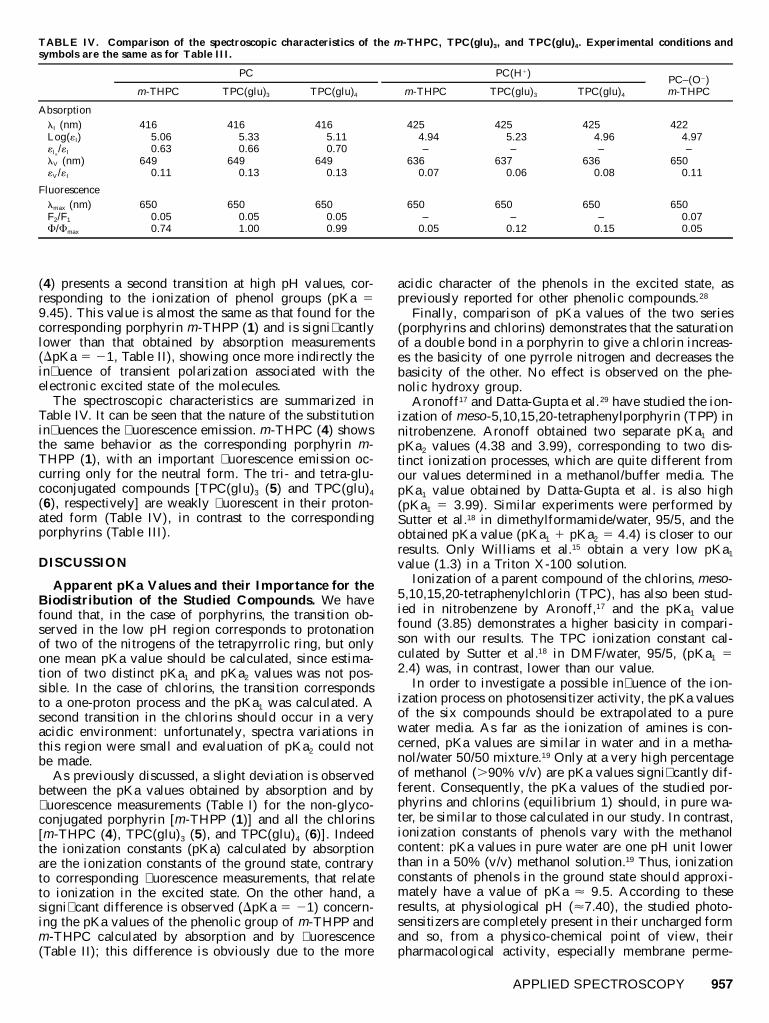
TABLE IV. Comparison of the spectroscopic characteristics of the m-THPC, TPC(glu)3, and TPC(glu)4. Experimental conditions andsymbols are the same as for Table III.
PC
m-THPC TPC(glu)3 TPC(glu)4
PC(H1)
m-THPC TPC(glu)3 TPC(glu)4
PC–(O2)m-THPC
AbsorptionlI (nm)Log(«I)
/«I«Ia
lV (nm)«V/«I
4165.060.63
6490.11
4165.330.66
6490.13
4165.110.70
6490.13
4254.94–
6360.07
4255.23–
6370.06
4254.96–
6360.08
4224.97–
6500.11
Fluorescencelmax (nm)F2/F1
F/Fmax
6500.050.74
6500.051.00
6500.050.99
650–
0.05
650–
0.12
650–
0.15
6500.070.05
(4) presents a second transition at high pH values, cor-responding to the ionization of phenol groups (pKa 59.45). This value is almost the same as that found for thecorresponding porphyrin m-THPP (1) and is signi� cantlylower than that obtained by absorption measurements(DpKa 5 21, Table II), showing once more indirectly thein� uence of transient polarization associated with theelectronic excited state of the molecules.
The spectroscopic characteristics are summarized inTable IV. It can be seen that the nature of the substitutionin� uences the � uorescence emission. m-THPC (4) showsthe same behavior as the corresponding porphyrin m-THPP (1), with an important � uorescence emission oc-curring only for the neutral form. The tri- and tetra-glu-coconjugated compounds [TPC(glu)3 (5) and TPC(glu)4
(6), respectively] are weakly � uorescent in their proton-ated form (Table IV), in contrast to the correspondingporphyrins (Table III).
DISCUSSION
Apparent pKa Values and their Importance for theBiodistribution of the Studied Compounds. We havefound that, in the case of porphyrins, the transition ob-served in the low pH region corresponds to protonationof two of the nitrogens of the tetrapyrrolic ring, but onlyone mean pKa value should be calculated, since estima-tion of two distinct pKa1 and pKa2 values was not pos-sible. In the case of chlorins, the transition correspondsto a one-proton process and the pKa1 was calculated. Asecond transition in the chlorins should occur in a veryacidic environment: unfortunately, spectra variations inthis region were small and evaluation of pKa2 could notbe made.
As previously discussed, a slight deviation is observedbetween the pKa values obtained by absorption and by� uorescence measurements (Table I) for the non-glyco-conjugated porphyrin [m-THPP (1)] and all the chlorins[m-THPC (4), TPC(glu)3 (5), and TPC(glu)4 (6)]. Indeedthe ionization constants (pKa) calculated by absorptionare the ionization constants of the ground state, contraryto corresponding � uorescence measurements, that relateto ionization in the excited state. On the other hand, asigni� cant difference is observed (DpKa 5 21) concern-ing the pKa values of the phenolic group of m-THPP andm-THPC calculated by absorption and by � uorescence(Table II); this difference is obviously due to the more
acidic character of the phenols in the excited state, aspreviously reported for other phenolic compounds.28
Finally, comparison of pKa values of the two series(porphyrins and chlorins) demonstrates that the saturationof a double bond in a porphyrin to give a chlorin increas-es the basicity of one pyrrole nitrogen and decreases thebasicity of the other. No effect is observed on the phe-nolic hydroxy group.
Aronoff17 and Datta-Gupta et al.29 have studied the ion-ization of meso-5,10,15,20-tetraphenylporphyrin (TPP) innitrobenzene. Aronoff obtained two separate pKa1 andpKa2 values (4.38 and 3.99), corresponding to two dis-tinct ionization processes, which are quite different fromour values determined in a methanol/buffer media. ThepKa1 value obtained by Datta-Gupta et al. is also high(pKa1 5 3.99). Similar experiments were performed bySutter et al.18 in dimethylformamide/water, 95/5, and theobtained pKa value (pKa1 1 pKa2 5 4.4) is closer to ourresults. Only Williams et al.15 obtain a very low pKa1
value (1.3) in a Triton X-100 solution.Ionization of a parent compound of the chlorins, meso-
5,10,15,20-tetraphenylchlorin (TPC), has also been stud-ied in nitrobenzene by Aronoff,17 and the pKa1 valuefound (3.85) demonstrates a higher basicity in compari-son with our results. The TPC ionization constant cal-culated by Sutter et al.18 in DMF/water, 95/5, (pKa1 52.4) was, in contrast, lower than our value.
In order to investigate a possible in� uence of the ion-ization process on photosensitizer activity, the pKa valuesof the six compounds should be extrapolated to a purewater media. As far as the ionization of amines is con-cerned, pKa values are similar in water and in a metha-nol/water 50/50 mixture.19 Only at a very high percentageof methanol (.90% v/v) are pKa values signi� cantly dif-ferent. Consequently, the pKa values of the studied por-phyrins and chlorins (equilibrium 1) should, in pure wa-ter, be similar to those calculated in our study. In contrast,ionization constants of phenols vary with the methanolcontent: pKa values in pure water are one pH unit lowerthan in a 50% (v/v) methanol solution.19 Thus, ionizationconstants of phenols in the ground state should approxi-mately have a value of pKa ø 9.5. According to theseresults, at physiological pH (ø7.40), the studied photo-sensitizers are completely present in their uncharged formand so, from a physico-chemical point of view, theirpharmacological activity, especially membrane perme-
958 Volume 57, Number 8, 2003
ation, is more likely to be controlled by parameters suchas partition coef� cient (LogP).
Spectroscopic Characteristics of the Speciated Por-phyrins and Chlorins and their Relevance to Photo-dynamic Therapy. The spectral characteristics of theneutral and protonated compounds are summarized in Ta-bles III and IV. It can be seen that protonation causes thedisplacement of the major band (Soret band) and of bandV in the case of porphyrins by DlI ; 25 nm and by DlV
; 10 nm, and in the case of chlorines by DlI ; 9 nmand by DlV ; 13 nm. The Soret band shows a shoulder(labeled Ia in Fig. 2a) only for the neutral form. This ismuch more in evidence in the spectra of chlorins. Asalready mentioned, absorption in the red region (band V)(one of the more important photosensitizer characteris-tics) is high only for neutral chlorins and protonated por-phyrins. Two emission bands (F1, F2) are observed forneutral species. The secondary emission band (F2) is al-most negligible in the case of chlorins (F2 /F1 ø 0) but issigni� cant for porphyrins (F2 /F1 5 0.31–0.37). Neutralporphyrins are six times less � uorescent than neutralchlorins (i.e., FTPC(glu)3 /FTPP(glu)3 5 6.25). It is also inter-esting to note that protonated porphyrins are at least threetimes more � uorescent than the corresponding chlorins(i.e., FTPP(glu)3 /FTPC(glu)3 5 3.40). Finally, hydroxylated de-rivatives m-THPP (1) and m-THPC (4) are not � uorescentin an alkaline environment, contrary to the glycosylatedderivatives, which present the same emission yield as atneutral pH.
Analytical Perspectives. The determination of absorp-tion and emission characteristics of the six studied com-pounds in various environments reveals the experimentalconditions that optimize their absorption and � uorescencedetection and so could signi� cantly contribute to the de-velopment of sensitive analytical methods.
For example, the � uorescence mode of detection wasreported to be less sensitive than absorption in high-per-formance liquid chromatography (HPLC) analysis of m-THPC30 when tri� uoroacetic acid was used in the mobilephase. Indeed, our study demonstrates that chlorin pro-tonation in acidic media induces quenching of the � uo-rescence emission [Table IV, PC(H1)]. The effect is muchless pronounced in absorbance measurements. As a con-sequence, when a � uorescence detection mode is used,chlorins should be detected in a neutral microenviron-ment, contrary to porphyrins that are more � uorescent intheir protonated form [PP(H1)2, Table III].
Another interesting perspective is the development ofselective analytical methods for the analysis of the stud-ied compounds using direct � uorescence spectroscopy.The simultaneous analysis of porphyrins and chlorins us-ing direct emission spectroscopy without HPLC separa-tion could be envisaged. At neutral pH, the componentsexhibit very similar emission spectra and simultaneousanalysis is not possible, but they present different spectradistributions in their protonated forms (Figs. 4a and 5a).Thus, simultaneous analysis becomes possible in an acid-ic environment. This kind of direct technique could bevery useful due to its simplicity and rapidity in stabilitystudies of chlorines, where the corresponding porphyrinis also present as a photodegradation product.31
Lastly, a recent study has shown that deglycosylationof some porphyrins can take place in cells, introducing
phenol functions.32 As our results have demonstrated thationization of phenol functions induces extinction of the� uorescence emission, deglycosylation can be studied(using direct � uorescence spectroscopy) in a high pH en-vironment, where phenolic derivatives do not � uoresce,contrary to glycosylated compounds.
CONCLUSION
The present study has demonstrated for the � rst timethat porphyrins m -THPP (1), TPP(glu)3 (2 ), andTPP(glu)4 (3) exhibit, in their ground states, essentiallythe same mean pKa value (ø2.7) for protonation of twotetrapyrrolic nitrogens, which is independent of the sub-stitution in peripheral meso-phenyl groups. The pKa1 val-ues of the chlorins [m-THPC (4), TPC(glu)3 (5), andTPC(glu)4 (6)] are slightly, but signi� cantly, higher(DpKa1ø0.3) and are also independent of substitution.Thus, both the new porphyrins and chlorins are extremelyweak bases, and this is con� rmed by the � uorescencemeasurements, even taking into account a slight increaseof basicity due to the corresponding excited singlet states.We observe that our pKa values for porphyrin proton-ation are two pH units lower with respect to the valuesobtained by other authors in nitrobenzene for a parentderivative (TPP), but quite near the pKa obtained in aDMF/water media (b 5 4.4). In the case of chlorins, thevalues observed for the corresponding derivative (TPC)are also higher in nitrobenzene (pKa1 5 3.85) but lowerin DMF/water (pKa1 5 2.4) with respect to our results.As our experiments have been conducted in a methanol/aqueous 50/50 (v/v) mixture, the obtained pKa values forthe pyrrole nitrogens can be reasonably extrapolated to a100% aqueous media. Thus, it can be concluded that thestudied photosensitizers are, at physiological pH, un-charged species, and the ionization process should notin� uence their activity in vivo.
Finally, the � uorescence emission characteristics of thestudied compounds show a strong pH dependence. Thishas potential applications in the development of sensitiveanalytical methods for their detection and quantitation inbiological media. We can in this way optimize detectionafter HPLC separation, or develop new methods of directanalysis, applied to photodegradation and metabolismstudies. All these perspectives will be the subject of ournext work.
ACKNOWLEDGMENT
This work was � nancially supported by the ‘‘Association de la re-cherche contre le cancer’’ grant N8 5334.
1. M. Ochsner, J. Photochem. Photobiol., B 39, 1 (1997).2. R. Boyle and D. Dolphin, Photochem. Photobiol. 64, 469 (1996).3. (a) T. J. Dougherty, C. J. Gomer, B. W. Henderson, G. Jori, D.
Kessel, M. Korbelik, J. Moan, and Q. Peng, J. Natl. Cancer Inst.90, 889 (1998); (b) T. J Dougherty, Photodynamic News 3 (1),(2000).
4. R. K. Pandey and G. Zheng, ‘‘Porphyrins as Photosensitizers inPhotodynamic Therapy’’ in Applications: Past, Present and Future,The Porphyrin Handbook, K. M. Kadish, K. M. Smith, and R.Guilard, Eds. (Academic Press, New York, 2000), vol. 6, Chap. 43,p. 162.
5. H. W. Whitlock, R. Hanauer, M. Y. Oester, and B. K. Bower, J.Am. Chem. Soc. 91, 7485 (1969).
6. R. Bonnett, in ‘‘Chemical Aspects of Photodynamic Therapy’’, in
APPLIED SPECTROSCOPY 959
Advanced Chemistry Texts (Gordon and Breach Science Publishers,London, 2000), Chap. 14, pp. 274, 281.
7. (a) J. F. Savary, Ph. Monnier, C. Fontolliet, J. Mizeret, G. Wagni-eres, D. Braichotte, and H. van den Bergh, Arch. Otolaryngol. HeadNeck Surg. 123, 162 (1997); (b) A. Radu, G. Wagnieres, H. vanden Bergh, and Ph. Monnier, Gastrointest. Endosc. Clin. N. Am.10, 439 (2000).
8. K. R. Adams, M. C. Berenbaum, R. Bonnet, A. N. Nizhnik A.Salgado, and M. A. Valles, J. Chem. Soc. Perkin Trans. 1, 1465(1992).
9. (a) G. Fulling, D. Schroder, and B. Franck, Angew. Chem. Int. Ed.Engl. 28, 1519 (1989); (b) Ph. Maillard, J.-L. Guerquin-Kern, M.Momenteau, and S. Gaspard, J. Am. Chem. Soc. 111, 9125 (1989);(c) Ph. Maillard, J.-L. Guerquin-Kern, C. Huel, and M. Momenteau,J. Org. Chem. 58, 2774 (1993); (d) M. Momenteau, D. Oulmi, Ph.Maillard, and A. Croisy, ’’In vitro photobiology of a new series ofporphyrins: the glycoconjugated porphyrins’’, SPIE Photodynamictherapy of cancer II, 2325, 13 (1994); (e) M. Momenteau, Ph. Mail-lard, M.-A. de Belinay, D. Carrez, and A. Croisy, J. Biomed. Opt.4, 1 (1999); (f) V. Sol, J.-C. Blais, V. Carre , R. Granet, M. Guil-loton, M. Spiro, and P. Krausz, J. Org. Chem. 64, 4431 (1999); (g)G. Zheng, A. Graham, M. Shibata, J. Missert, A. R. Oseroff, T. J.Dougherty, and R. K. Pandey, J. Org. Chem. 66, 8709 (2001); (h)I. Sylvain, R. Zerrouki, R. Granet, Y. M. Huang, J.-F. Lagorce, M.Guilloton, J.-C. Blais, and P. Krausz, Bioorg. Med. Chem. 10, 57(2002).
10. D. Oulmi, Ph. Maillard, J.-L. Guerquin-Kern, C. Huel, and M. Mo-menteau, J. Org. Chem. 60, 1554 (1995).
11. I. Laville, T. Figueiredo, B. Loock, S. Pigalio, Ph. Maillard, D. S.Grierson, D. Carrez, A. Croisy, and J. Blais, ‘‘Synthesis, cellularinternalization and photodynamic activity of glucoconjugated de-rivatives of tri and tetra (meta-hydroxyphenyl)chlorins’’, Bioorg.Med. Chem. 11, 1643 (2003).
12. J. E. Falk, in Porphyrins and Metalloporphyrins (Elsevier Publish-ing Company, New York, 1964), vol. 2, p. 28.
13. M. Tabata and J. Nishimoto, ‘‘Equilibrium data of porphyrins andmetalloporphyrins’’, in ‘‘Database of Redox Potentials and BindingConstants’’ The Porphyrin Handbook, K. M. Kadish, K. M. Smith,and R. Guilard, Eds. (Academic Press, New York, 2000), vol. 6,Chap. 60, pp. 222–416.
14. S. C. M. Gandini, V. E. Yushmanov, I. E. Borissevitch, and M.Tabak, Langmuir 15, 6233 (1999).
15. G. N. Williams, R. F. X. Williams, A. Lewis, and P. Hambright, J.Inorg. Nucl. Chem. 41, 41 (1979).
16. D. L. Lavallee, Z. Xu, and R. Pina, J. Org. Chem. 58, 6000 (1993).17. S. Aronoff, J. Phys. Chem. 62, 428 (1958).18. T. P. G. Sutter and P. Hambright, Inorg. Chem. 31, 5089 (1992).19. (a) G. Charlot, in Chimie Analytique Generale, Solutions aqueuses
et non aqueuses (Masson et Cie Edition, Paris, 1967), vol. I, Chap.V, pp. 219 and 237–241; (b) G. Charlot and B. Tremillon, in Lesreactions chimiques dans les solvants et les sels fondus (Gauthier-Villars Editeur, Paris, 1963), Chap. IX, pp. 301–317.
20. D. Oulmi, Ph. Maillard, C. Vever-Bizet, M. Momenteau, and D.Brault, Photochem. Photobiol. 65, 511 (1998).
21. G. Csik, E. Balog, I. Voszka, F. Togyesi, D. Oulmi, Ph. Maillard,and M. Momenteau, J. Photochem. Photobiol., B 44, 216 (1998).
22. (a) R. Bonnett, A. N. Nizhnik, S. G. White, and M. C. Berembaum,J. Photochem. Photobiol., B 6, 29 (1990); (b) Y. Mikata, Y. Oncji,M. Shibata, T. Kakuchi, H. Hono, S.-I. Ogura, I. Okura, and S.Yano, Biorg. Med. Chem. Lett. 8, 3543 (1998).
24. E. Curis, ‘‘Developpement d’outils informatiques et statistiquespour l’analyse des spectres E.X.A.F.S.—Application aux systemesbioinorganiques’’, Ph.D. Thesis, University Paris XI, Paris, France(2000).
25. LASE software is available for download from http://xlase.free.fr/.26. R. Ihaka and R. Gentleman, J. Computational Graphical Statistics
5, 299 (1996).27. M.-C. Desroches, O. Bourdon, Y. Morokro, P. Chaminade, J. Blais,
P. Prognon, and A. Kasselouri, Luminescence 16, 1 (2001).28. S. G. Schulman, Molecular Luminescence Spectroscopy, Methods
and Applications, Part 2 (John Wiley and Sons, New York, 1988),Chap. 6, pp. 461–471.
29. N. Datta-Gupta, D. Malakar, V. O. Jones, and T. Wright, Chem.Lett., 1659 (1986).
30. R. Whelpon, A. T. Michael-Titus, S. S. Basra, and M. Grahn, Pho-tochem. Photobiol. 61, 397 (1995).
31. A. Kasselouri, G. Mahuzier, O. Bourdon, D. Demore, J.-C. Blais,P. Prognon, G. Bourg-Heckly, and J. Blais, Photochem. Photobiol.70, 275 (1999).
32. J. Blais, I. Laville, and Ph. Maillard, personal communication(2002).


![V. SPECIATION A. Allopatric Speciation B. Parapatric Speciation (aka Local or Progenitor - Derivative) C. Adaptive Radiation D. Sympatric Speciation [Polyploidy]](https://static.documents.pub/doc/80x56/56649d3f5503460f94a186e2/v-speciation-a-allopatric-speciation-b-parapatric-speciation-aka-local.jpg)

