Spectroscopic properties and photodynamic effects of new lipophilic porphyrin derivatives: Efficacy, localisation and cell death pathways Gabriela Kramer-Marek a,b, * , Carlos Serpa c , Agnieszka Szurko a,d , Maria Widel d , Aleksander Sochanik e , Miroslaw Snietura f , Piotr Kus g , Rui M.D. Nunes c , Luis G. Arnaut c , Alicja Ratuszna a a A.Chelkowski Institute of Physics, University of Silesia, Uniwersytecka 4, 40-007 Katowice, Poland b Department of Tumour Biology, Maria Sklodowska-Curie Memorial Cancer Centre and Institute of Oncology, 44-100 Gliwice, Poland c Chemistry Department, University of Coimbra, 3004-535 Coimbra, Portugal d Department of Clinical and Experimental Radiobiology, Maria Sklodowska-Curie Memorial Cancer Centre and Institute of Oncology, 44-100 Gliwice, Poland e Department of Molecular Biology, Maria Sklodowska-Curie Memorial Cancer Centre and Institute of Oncology, 44-100 Gliwice, Poland f Department of Tumour Pathology, Maria Sklodowska-Curie Memorial Cancer Centre and Institute of Oncology, 44-100 Gliwice, Poland g Institute of Chemistry, University of Silesia, 40-007 Katowice, Poland Received 26 July 2005; received in revised form 7 December 2005; accepted 11 December 2005 Available online 21 February 2006 Abstract Photodynamic therapy (PDT) and photodynamic diagnostics (PDD) of cancer are based on the use of non-toxic dyes (photosensi- tisers) in combination with harmless visible light. This paper reports physicochemical properties, cell uptake, localisation as well as pho- todynamic efficiency of two novel lipophilic porphyrin derivatives, suitable for use as PDT sensitisers. Both compounds are characterised by high quantum yield of singlet oxygen generation which was measured by time-resolved phosphorescence. Photodynamic in vitro stud- ies were conducted on three cancer cell lines. Results of cell survival tests showed negligible dark cytotoxicity but high phototoxicity. The results also indicate that cell death is dependent on energy dose and time following light exposure. Using confocal laser scanning micros- copy both compounds were found to localise in the cytoplasm around the nucleus of the tumour cells. The mode of cell death was eval- uated based on the morphological changes after differential staining. In summary, good photostability, high quantum yield of singlet oxygen and biological effectiveness indicate that the examined lipo- philic porphyrin derivatives offer quite interesting prospects of photodynamic therapy application. Ó 2006 Elsevier B.V. All rights reserved. Keywords: Photodynamic therapy; Singlet oxygen; Photosensitisation; Apoptosis; Necrosis 1. Introduction Photodynamic therapy (PDT) is a relatively novel and promising anticancer therapy based on combined use of a photosensitising agent and visible or near-infrared light [1]. It can be very effective in destroying tumour cells and side effects in healthy tissues are relatively low [2,3]. Gener- ally, PDT is based on photoactivation of a sensitiser taken up by targeted (i.e., cancer) cells. Two types of reaction can occur after photoactivation of the photosensitiser. The first involves generation of free radicals like superoxide anion, O 2 (type I); the second produces singlet molecular oxygen 1 O 2 ( 1 D g ) (type II), possibly the main species responsible for oxidising neighbouring molecules [4,5]. Highly reactive oxygen species formed within cell membrane, cytoplasm or organelles lead to peroxidative reactions causing dam- age to DNA and other molecules and finally resulting in 1011-1344/$ - see front matter Ó 2006 Elsevier B.V. All rights reserved. doi:10.1016/j.jphotobiol.2005.12.011 * Corresponding author. Present address: Instytut Fizyki, Uniwersytet S ´ la ˛ ski, Uniwersytecka 4, 40-007 Katowice, Poland. Tel.: +48 32 359 21 05; fax: +48 32 258 84 31. E-mail address: [email protected](G. Kramer-Marek). www.elsevier.com/locate/jphotobiol Journal of Photochemistry and Photobiology B: Biology 84 (2006) 1–14
Transcript
www.elsevier.com/locate/jphotobiol
Journal of Photochemistry and Photobiology B: Biology 84 (2006) 1–14
Spectroscopic properties and photodynamic effects of new lipophilicporphyrin derivatives: Efficacy, localisation and cell death pathways
Gabriela Kramer-Marek a,b,*, Carlos Serpa c, Agnieszka Szurko a,d, Maria Widel d,Aleksander Sochanik e, Miroslaw Snietura f, Piotr Kus g, Rui M.D. Nunes c,
Luis G. Arnaut c, Alicja Ratuszna a
a A.Chelkowski Institute of Physics, University of Silesia, Uniwersytecka 4, 40-007 Katowice, Polandb Department of Tumour Biology, Maria Sklodowska-Curie Memorial Cancer Centre and Institute of Oncology, 44-100 Gliwice, Poland
c Chemistry Department, University of Coimbra, 3004-535 Coimbra, Portugald Department of Clinical and Experimental Radiobiology, Maria Sklodowska-Curie Memorial Cancer Centre and Institute of Oncology,
44-100 Gliwice, Polande Department of Molecular Biology, Maria Sklodowska-Curie Memorial Cancer Centre and Institute of Oncology, 44-100 Gliwice, Polandf Department of Tumour Pathology, Maria Sklodowska-Curie Memorial Cancer Centre and Institute of Oncology, 44-100 Gliwice, Poland
g Institute of Chemistry, University of Silesia, 40-007 Katowice, Poland
Received 26 July 2005; received in revised form 7 December 2005; accepted 11 December 2005Available online 21 February 2006
Abstract
Photodynamic therapy (PDT) and photodynamic diagnostics (PDD) of cancer are based on the use of non-toxic dyes (photosensi-tisers) in combination with harmless visible light. This paper reports physicochemical properties, cell uptake, localisation as well as pho-todynamic efficiency of two novel lipophilic porphyrin derivatives, suitable for use as PDT sensitisers. Both compounds are characterisedby high quantum yield of singlet oxygen generation which was measured by time-resolved phosphorescence. Photodynamic in vitro stud-ies were conducted on three cancer cell lines. Results of cell survival tests showed negligible dark cytotoxicity but high phototoxicity. Theresults also indicate that cell death is dependent on energy dose and time following light exposure. Using confocal laser scanning micros-copy both compounds were found to localise in the cytoplasm around the nucleus of the tumour cells. The mode of cell death was eval-uated based on the morphological changes after differential staining.
In summary, good photostability, high quantum yield of singlet oxygen and biological effectiveness indicate that the examined lipo-philic porphyrin derivatives offer quite interesting prospects of photodynamic therapy application.� 2006 Elsevier B.V. All rights reserved.
Keywords: Photodynamic therapy; Singlet oxygen; Photosensitisation; Apoptosis; Necrosis
1. Introduction
Photodynamic therapy (PDT) is a relatively novel andpromising anticancer therapy based on combined use of aphotosensitising agent and visible or near-infrared light[1]. It can be very effective in destroying tumour cells and
1011-1344/$ - see front matter � 2006 Elsevier B.V. All rights reserved.
side effects in healthy tissues are relatively low [2,3]. Gener-ally, PDT is based on photoactivation of a sensitiser takenup by targeted (i.e., cancer) cells. Two types of reaction canoccur after photoactivation of the photosensitiser. The firstinvolves generation of free radicals like superoxide anion,O�2 (type I); the second produces singlet molecular oxygen1O2 (1Dg) (type II), possibly the main species responsible foroxidising neighbouring molecules [4,5]. Highly reactiveoxygen species formed within cell membrane, cytoplasmor organelles lead to peroxidative reactions causing dam-age to DNA and other molecules and finally resulting in
2 G. Kramer-Marek et al. / Journal of Photochemistry and Photobiology B: Biology 84 (2006) 1–14
cell death [6]. PDT can rapidly induce apoptosis of targetedcells although precise mechanisms underlying this processare not completely characterised [7,8]. A second mode ofcell death observed in PDT is necrosis [9].
Porphyrins and porphyrin-related macrocycles areamong the sensitisers most frequently used in PDT. Onceinside the cell, they bind to apolar endocellular matricessuch as lysosomes, mitochondria or/and plasma mem-branes. The site of porphyrin action depends on its polarityand, to some extent, is cell-line dependent [7,10].
Among the first-generation photosensitisers the mostpopular is Photofrin II, widely used in clinical practice[11,12]. However, its applications remain limited becausePhotofrin II is a complex mixture with low extinctioncoefficient in the red spectral region. This implies admin-istration of relatively large amounts of this drug in orderto obtain satisfactory phototherapeutic response. In con-sequence, due to high drug accumulation, patients haveto stay several days out of sunlight to avoid sunburnreactions. These drawbacks have stimulated the searchfor photosensitising agents with improved optical andpharmacokinetic characteristics.
Two synthetic porphyrin derivatives: 5-(4-hydroxyphe-nyl)-10,15,20-tritolylporphyrin and 5-(4-hexadecyloxyphe-nyl)-10,15,20-tri-pyridylporphyrin were synthesised in ourlaboratory. The structures of these compounds are shownin Fig. 1 and for convenience they will be referred to asC16-TTP and TPYR-PP, respectively. They have beenshown to possess suitable chemical purity, relatively highabsorption coefficient in the phototherapeutic windows(600–800 nm), as well as high quantum yield of singlet oxy-gen generation that favour their use as potential photody-namic agents.
In this paper, we describe the synthesis and photochem-ical properties of these porphyrins. Particular attention isgiven to the time-resolved singlet oxygen phosphorescenceas a measure of C16-TTP and TPYR-PP singlet oxygenquantum yield (UD). The results of cytotoxicity and photo-toxicity studies as well as cellular localisation and the effectof radiation dose for cell death induction in differenttumour cell lines are also presented.
HH
N
N
N
N
N
NN
O
HH
N
N
N
N
H3C
H3C
TPYR-PP
Fig. 1. The structure of 5-(4-hexadecyloxyphenyl)-10,15,20-tri-pyridylporphyrTTP).
2. Materials and methods
2.1. Materials
5-(4-Hydroxyphenyl)-10,15,20-tritolylporphyrin (C16-TTP) was synthesised as described elsewhere [13]. Briefly,50 mg of hydroxyphenylporphyrin, 50 mg of cetyl bromide,2 g of anhydrous potassium carbonate and 20 ml ofdimethyl formamide (DMF) were placed in a flask and stir-red at room temperature (RT) for 5 h. The reaction pro-gress was checked using TLC (silica gel/chloroform). Onehundred millilitre of water was added to the reaction andthe resulting mixture was extracted three times using ethylacetate (3 · 20 ml). The extracts were washed five timeswith water, dried over anhydrous MgSO4 and evaporated.The raw product was washed with 30 ml of methanol. Pre-cipitated porphyrin was separated and air-dried. The prod-uct was purified twice on a silica gel column using hexane(to remove excess cetyl bromide) and eluted withchloroform.
5-(4-Hexadecyloxyphenyl)-10,15,20-tri-pyridylporphyrin(TPYR-PP) was synthesised using the procedures partlydescribed elsewhere [14]. Briefly, 2.12 g of p-hexadecyloxy-benzaldehyde, 1.82 g of 4-pyridylcarboxyaldehyde and1.25 g of pyrrole were heated for 30 min in boiling propi-onic acid. After 24 h, an excess of acid was distilled outand the mixture was extracted with ethyl acetate. Then,extract was dried with anhydrous MgSO4. The end-prod-ucts were separated by column chromatography and thecompound of interest eluted.
Photofrin II was purchased from Axcan Pharma Com-pany. For in vitro confocal microscopy studies it was dis-solved in distilled water at a maximum concentration of2 mg/ml and diluted using DMEM medium without phenolred. A pure sample of 5,10,15,20-tetraphenylporphyrin(TPP) was available from previous studies [15].
2.2. Photophysical studies
In the photophysical characterisation of the sensitiserstoluene was used as a solvent. The results obtained shouldbe indicative of sensitiser behaviour in cellular environ-ment, although absolute values should be looked with cau-tion as deviations may arise due to specific interactions.The ground-state absorption and luminescence spectrawere recorded at room temperature with a ShimadzuUV-2100 spectrophotometer and SPEX Fluorolog 111spectrophotometer, respectively. Transient triplet–tripletabsorption was measured with an Applied PhotophysicsLKS.60 flash kinetic spectrometer; excitation was achievedwith the third harmonic of a Nd/YAG laser (Spectra-Phys-ics Quanta Ray GCR 130).
Singlet oxygen phosphorescence was detected at roomtemperature using an adaptation of the LKS.60 spectrom-eter. The singlet oxygen emission was detected using aHamamatsu R5509-42 photomultiplier, cooled to 193 Kin a liquid nitrogen chamber (Products for Research, modelPC176TSCE005), following Nd/YAG laser excitation ofaerated toluene solutions containing the sensitiser at a con-centration necessary to produce absorbance in the 0.15–0.30 range at the excitation wavelength (355 nm). Underthese conditions there was no indications of porphyrins’aggregation.
The modification of the spectrometer involved the inter-position of a Melles Griot dielectric mirror (08MLQ005/345), that reflects more than 99.5% of the incident lightin the 610–860 nm range and a Scotch RG665 filter. A600 line diffraction grating was mounted in place of a stan-dard one. This equipment allows spectral identification ofthe singlet oxygen phosphorescence and measurement ofsinglet oxygen lifetime in the nanosecond and microsecondranges [16]. The filters employed are essential in eliminatingfrom the infrared signal all first-harmonic contributions ofthe sensitiser emission in the 500–800 nm range.
2.3. In vitro studies
2.3.1. Cell cultures
Human malignant melanoma (Me45) cell line (derivedfrom a lymph node metastasis of skin melanoma in a 35-year-old male) was established in 1997 at the RadiobiologyDepartment of the Centre of Oncology in Gliwice. Identityof melanoma cells was confirmed by immunocytochemicalreaction with DAKO monoclonal antibodies HMB 50,
S-100 and Melan A (Widel, unpublished). Human colonadenocarcinoma (Hct116) was obtained from AmericanType Culture Collection. Murine melanoma (B16(F10))was obtained from Wistar Institute, Philadelphia, PA,USA. Cells were grown as monolayer cultures in 75 cm2
flasks (Nunc)/37 �C, humidified atmosphere, 5% CO2/using the following media: DMEM (Me45), McCoy(Hct116) and RPMI (B16(F10)) from Sigma–Aldrich, sup-plemented with 10% foetal bovine serum (Gibco-BRL) and100 lg/ml of gentamycin (Polfa).
2.3.2. Liposome preparation
Liposomes used in this study contained a cationic com-ponent, cetylated polyethylenimine (CT-PEI), obtained bypartially substituting amine groups of low-molecular-weight polyetylenimine with cetyl residues. Synthesis ofCT-PEI was performed according to Yamazaki et al. [17].In brief, Epomin (low-molecular-weight polyethylenimine600, Nippon Shokubai Ltd., Japan) was refluxed with cetylbromide (Sigma–Aldrich) in the presence of triethylaminefor 12 h at 63 �C. The product was dialysed against 40%ethanol, then against water, and finally lyophilised.
The final product was 1H NMR-analysed using UnityInova-300 spectrometer operating at 300 MHz frequency(Institute of Organic Chemistry and Technology, SilesianTechnical University in Gliwice, Poland) with deuteratedchloroform as solvent and tetramethylsilane as reference.The liposomes were prepared by mixing aliquots of chloro-form solutions (10 mg/ml) of CT-PEI, cholesterol, andTPYR-PP or C16-TTP at appropriate molar ratios andspin-evaporating the resulting preparations. Dry lipid filmswere rehydrated at 4 �C for 1 h and sonicated with tipprobe until emulsions became clear (two or three 3-mincycles, medium power, Branson sonifier).
2.3.3. In vitro transfection studies
Transfection studies with cationic liposome-plasmidDNA complexes (lipoplexes) at various stoichiometricratios were performed in order to check cellular internalisa-tion of constructs containing the synthesised compounds.Transfection calibration was achieved by employing com-plexes containing plasmid DNA (Promega) with luciferasereporter gene insert. The resulting luminescence ofexpressed luciferase protein was quantified using a Bert-hold luminometer. One day prior to transfection, cells wereseeded onto 24-well plates at 5 · 104 cells/well. When cul-tures reached about 80% confluence, appropriate dilutionsof liposome emulsions were prepared in 100 ll final volumeusing polystyrene tubes (Sarstedt). Separately, appropriatedilutions of 1 lg plasmid DNA were prepared in polysty-rene tubes in 100 ll final volume. DNA solutions wereadded drop wise to liposomal emulsions with gently mix-ing. Following 15-min incubation at room temperature2.8 ml of suitable medium (Opti-MEM, Gibco-BRL) wasadded. After removal of growth medium the transfectionmixtures were transferred to the culture wells. Cells wereincubated for 4 h at 37 �C and 5% CO2. At the end of
4 G. Kramer-Marek et al. / Journal of Photochemistry and Photobiology B: Biology 84 (2006) 1–14
incubation period the transfection medium was replacedwith regular growth medium. After 24 h cells in each wellwere lysed using a kit-included lysis buffer (Promega).The lysates were centrifuged for 30 min at 15,000 rpmand 4 �C. The supernatants (300 ll) were transferred intofresh test tubes. Luciferase activity was determined usinga Berthold luminometer, in supernatant aliquots (20 ll orless), after addition of 100 ll of enzyme substrate. Lucifer-ase activity was expressed as relative luminescence units(RLU) per milligram of protein in cell culture homoge-nates. Lysate protein content was measured by Bradfordmethod using a BioRad kit and BSA calibration.
2.3.4. Photostability studies of the compoundsPhotostability of the photosensitisers was determined by
irradiating 1 lM solutions of the phorphyrins incorporatedinto liposomes. Two different light doses were used (7.5 and15 J/cm2). During such irradiation the solutions were mag-netically stirred and kept at room temperature. Afterappropriate time intervals concentration of the porphyrinswas determined using absorption spectrophotometer. Thephotostability was calculated as the ratio of residual absor-bance after determined irradiation time and the absorbancevalue measured before irradiation.
2.3.5. Cytotoxicity and phototoxicity
For irradiation experiments, exponentially growing cellswere harvested by trypsinisation of subconfluent cultures.Cells were then seeded at 8 · 104 cells/35 mm dish (Nunc).After 18 h incubation, the growth medium was exchangedfor medium containing preselected (i.e., the best in termsof liposome stability and transfection efficiency) formula-tion of lipoplexes (6:1 lipid to DNA, w/w, final concentra-tion of porphyrins 1 lM) and cells were incubated 4 h inthe dark. A representative experiment calibrating liposometo DNA ratio is shown in Fig. 2, for details see Section2.3.3. Prior to light exposure, cells were washed three times
0,00E+00
5,00E+06
1,00E+07
1,50E+07
2,00E+07
2,50E+07
3,00E+07
1:1 2:1 3:1 4
liposom
leR
ait
evL
umin
esc
neec
U n
tis
p er
mi
llig
ram
rptoei
nU
LR
/mg
Fig. 2. Luciferase expression in Hct116 cells following transfection with ppolyethylenimine CT-PEI, cholesterol (1:1 mol/mol) and C16-TTP.
with phenol red-free medium (DMEM). The dishes werethen exposed to different light doses (2.5–15 J/cm2). Firstcontrol group (no photosensitiser) was also treated withred light. The second control group, containing photosen-sitiser, was not exposed to light (dark control) and servedfor cytotoxicity assay. The light source was a halogen lamp(250 W, 24 V) with heat isolation filter and 610 nm long-pass filter. The total power output (measured at the culturesolution surface) was 8.5 mW/cm2, as indicated by Radi-ometer Laser Mate-Q, Coherent. Immediately after irradi-ation the medium was replaced with fresh aliquotsupplemented with 10% FBS and cells were further incu-bated for 12 or 24 h under standard culture conditions.
2.3.6. Cell viability
Cell viability was monitored by MTS-tetrazoliumreduction assay (Promega) which relies on the ability ofviable cells to reduce the colourless blue-tetrazolium salt(3-(4,5-dimethylthiazol-2-yl)-5-(3-carboxymethoxyphenyl)-2-(4-sulfophenyl)-2H-tetrazol) to colour formazan. MTS,which measures mitochondrial dehydrogenase activity,was added to cell cultures 12 and 24 h after treatmentwith photosensitisers and/or light, according to the man-ufacturer’s instructions. Absorbance measurements at490 nm were performed using Elx800 universal platereader (Biotek Instruments, Inc.). A standard solutioncontaining 100 ll of DMEM without phenol red and20 ll of MTS solution was used to determine ‘‘blank’’absorbance. Survival of PDT-treated cells was normalisedagainst control cells that were only incubated (not irradi-ated). The results are reported as mean ± SD of three sep-arate experiments.
2.3.7. Clonogenic survival assay
Clonogenic potential of cells subjected to PDT treat-ment following transfer of C16-TTP and TPYR-PP con-taining constructs was assessed as described by
:1 5:1 6:1 7:1 8:1
es/DNA [w/w]
lasmid DNA (pVR1255). Complexed to liposomes containing cetylated
G. Kramer-Marek et al. / Journal of Photochemistry and Photobiology B: Biology 84 (2006) 1–14 5
Carmichael [18]. Exponentially growing cells seeded in 35-mm dishes (Nunc) were subjected to PDT, as described inSection 2.3.4. After irradiation with different light doses,cells were trypsinised and seeded, at different densities, in60-mm dishes. Cells were then incubated for 9 days at37 �C in humidified atmosphere. The colonies were thenfixed and stained with 0.5% methylene blue in 50% ethanoland the number of colonies containing more than 50 cellswas counted. The surviving fraction was calculated as therelative plating efficiency of PDT-treated vs. non-PDT-treated cells (no drug, no light).
To visualise the sensitisers accumulation within the cells,3 · 104 Me45 cells in 800 ll growth medium were platedinto 4-well chambered coverglass (Nunc) and incubatedfor 18 h. Then, cells were transfected with liposome com-plexes containing C16-TTP or TPYR-PP (final concentra-tion 1 lM), as described in Section 2.3.3. Following 4 hincubation cells were washed three times in phosphate-buf-fered saline (PBS, pH 7.2) and covered with 800 ll of freshgrowth medium. Thirty minutes before analysis cells werelabelled with 5 lM CellTracker Green CMFDA (5-chlo-romethylfluorescein diacetate; Molecular Probes). Thisreagent passes freely through cell membrane but, onceinside the cell, its chloromethyl group reacts with thiolsand the reagent is transformed into membrane-impermeantthioether adduct, fluorescing after esterases-mediatedcleavage of acetate moiety. It allows localising targetedcells and assessing their viability. After 4 and 12 h incuba-tion intracellular localisation of porphyrin derivatives wasmonitored using LSM 510 confocal laser scanning micro-scope (Carl Zeiss GmbH). CellTracker Green CMFDAand porphyrins were excited using argon–ion laser488 nm line and helium–neon laser 543 nm line, respec-tively. The fluorescence was filtered with 505 nm band-passand 560 nm long-pass emission filters. To reduce inter-channel cross-talk a multitracking technique was used.Image acquisition was performed using 40 · 1.30 NA oilimmersion objective lens (resolution: 512 · 512 or1024 · 1024 pixels; optical slice thickness: 1 lm). Imageswere stored in TIF format and analysed using KS400 soft-ware (Carl Zeiss GmbH). Five images were taken for eachkind of lipoplex-associated porphyrins and controls cellcultures (without lipoplexes added). Every image featured5–7 cells. Using green image channel the mask for defini-tion of ROI was generated. In such defined regions meandensitometric signal value was calculated after backgroundfluorescence subtraction.
To determine exact intracellular localisation of drugs 3Dreconstructions of specified cells were generated. Oursource image stacks consisted of 55 (two-channel, 1024 ·1024 pixels) images taken every 0.4 lm. Final resolutionof this reconstruction was 0.16 lm/pixel horizontally and0.4 lm/pixel vertically. Acquisition time was 1.17 s perimage (65 s for entire stack). Using these stacks horizontaland vertical projections were generated.
2.3.9. Determination of apoptotic cells
To study the mode of cell death following PDT we useddifferential staining of apoptotic and necrotic cells withacridine orange/ethidium bromide (AO/EB) [19,20].Tumour cells (Me45) growing on 35 mm culture disheswere transfected with porphyrin-carrying liposomes for4 h as described in Section 2.3.3 and irradiated with twodifferent light energy doses: 7.5 and 15 J/cm2. Next, cellswere washed with fresh medium and incubated 0–24 h.At times 0, 3, 6, 12 and 24 h cells were trypsinised, pelleted,suspended in 25 ll of phosphate buffered saline (PBS) and1 ll of AO/EB (1:1, 10 lM) solution was added. A drop ofcell suspension was placed on microscope slide and ana-lysed at 400· magnification under fluorescence microscope(AXIOPHOT, Zeiss-Opton) equipped with band-pass filterfor 400–500 nm wavelength. One thousand cells werescored and percentage of live, apoptotic and necrotic cellswas calculated. Live cells contain green fluorescent regularnuclei with orange granules in the cytoplasm; apoptoticcells present nuclei with intense green fluorescence fromcondensed chromatin (early apoptosis) and bright orangefluorescence from highly condensed and/or fragmentedchromatin (late apoptosis); necrotic cells possess regularorange-stained nuclei and often disrupted cell membranewhich is clearly seen using phase-contrast objective [20].
3. Results
3.1. Photophysics and photochemistry
C16-TTP and TPYR-PP show typical spectroscopic fea-tures of free-base porphyrins (intense Soret band) in theviolet range of the visible region (e > 105 M�1 cm�1) andfour wide Q-bands localised in the 500–650 nm region(e 6 104 M�1 cm�1); the data are summarised in Table 1.The band absorption coefficient was calculated from Lam-bert–Beer law using solutions in the 10�4–10�6 M concen-tration range. The relative intensity of the Q-bandsindicates an etio-type spectrum (eIV > eIII > eII > eI), [21].Good linear plots (r P 0.999) that pass through the originwere obtained, indicating that there is no compound aggre-gation under these conditions. However, this does notexclude the possibility of aggregation inside cells in vitro,which is often the case for this kind of compounds.
The steady-state fluorescence emission spectra of theseporphyrins present two maxima which have been assignedto Q(0–0) and Q(0–1) transitions, see Table 1. A very small(5 nm) Stokes shift between last band of absorptionQx(0–0) and first emission band Q(0–0), characteristic formonomeric porphyrins, was also observed indicating thatthe spectroscopic energy is nearly identical to the relaxa-tion energy of the singlet state. The fluorescence quantumyield (UF) was calculated by steady-state comparativemethod using TPP as reference. The spectroscopic sin-glet-state energies (ES) were obtained from the intersectionof the normalised absorption and fluorescence spectra. Nophosphorescence was observed at 77 K in toluene glasses.
Table 1Absorption and luminescence data for free bases in deaerated toluene solution
6 G. Kramer-Marek et al. / Journal of Photochemistry and Photobiology B: Biology 84 (2006) 1–14
All results are consistent with those obtained for TPP andrelated porphyrins [15,22,23].
Triplet–triplet absorption spectra of both porphyrinspresented typical intense absorption band at �460 nmand the ground state bleaching bands. Triplet state life-times in presence (air-equilibrated samples) and absenceof oxygen were obtained. Triplet quantum yields (UT) werecalculated using a published procedure [24] (see Table 2).
3.2. Quantum yield of singlet oxygen generation
Singlet oxygen quantum yields (UD) were obtained bycomparing the intensity of singlet oxygen emission at1270 nm in an air-equilibrated sample containing a givensensitiser against the intensity obtained from an opticallymatched sample containing a reference sensitiser. The laserpower employed was varied until reaching the lowest emis-sion signal limit (Fig. 3). We employed 1H-phenalen-1-oneas a reference. The literature value for the singlet oxygenquantum yield in toluene, obtained for this sensitiser is0.95 [25].
The studied molecules generate a species emitting at1270 nm with the lifetime between 32 and 35 ls (Table 2).These values are in good agreement with lifetime of singletoxygen phosphorescence in the solvent used, confirmingthe origin of the emission measured [26]. Also, the shapeof emission spectra taken between 1200 and 1350 nm corre-sponds to that emission, with a maximum at 1270 nm [27].
The oxygen quantum yield values obtained for TPP andPhotofrin II (0.62 and 0.32, respectively) are in full agree-ment with those found in the literature [28,29], which vali-dates the method used. The value of 0.32 obtained for thecomplex mixture Photofrin II in toluene is within the rangeof the values obtained for pure hematoporphyrins in this
Table 2Triplet state characterisation, singlet oxygen lifetimes and quantum yields
solvent [30]. C16-TTP has a singlet oxygen quantum yield(0.66) comparable to that of TPP, twice the value forPhotofrin II under the same measuring conditions.TPYR-PP yield of singlet oxygen generation reaches thevalue of 0.82 which is quite enhanced, compared to thosefor Photofrin II or even C16-TPP and TPP (Table 2).The value obtained for TPYR-PP can be compared withthe value obtained by Postigo et al. [31] for tetra-pyr-idylporphyrin in the same solvent (0.85).
3.3. Photostability of compounds
In all cases, exposure of liposome-incorporated porphy-rins to light doses used in phototoxicity studies caused anegligible decrease in the intensity of the visible absorptionbands. This process, generally defined as photobleaching, isusually related to irreversible destruction of the tetrapyrr-olic macrocycle. The data in Table 3 evidently suggest thatTPYR-PP and C16-TTP have shown high photostabilityunder the conditions used for PDT tests.
3.4. PDT effects on the cell growth and survival
The purpose of this group of experiments was to deter-mine the degree of dark toxicity and phototoxicity of C16-TTP and TPYR-PP for various transfected cell linesfollowing exposure to different doses of light. The use ofcationic liposomes as photosensitiser vehicles transportinghydrophobic constitutes an advantage for their use inPDT, since liposomes optimise the release of lipophilic sen-sitisers to low density lipoproteins LDL. The percentage ofcell survival was measured by normalising the results ofMTS assay with cells that were neither exposed to porphy-rins nor to light.
Fig. 3. 1270 nm emission intensity as a function of relative laser intensity: (squares) 1H-phenalen-1-one; (circles) A, Photofrin II; B, C16-TPP; C, TPYR-PP; D, emission intensity as a function of wavelength for C16-TPP.
Table 3Photostability percentage of the photosensitisers after irradiation withdifferent light doses
Compound Energy dose/J cm�2
0 7.5 15
C16-TTP 100 99 98TPYR-PP 100 98 94
G. Kramer-Marek et al. / Journal of Photochemistry and Photobiology B: Biology 84 (2006) 1–14 7
Figs. 4 and 5 show the characteristics of C16-TTP andTPYR-PP phototoxicity after exposure to five doses oflight irradiation, respectively. Cell viability was measured12 h (A) and 24 h (B) post-irradiation in three different celllines. When the cells were exposed to light in the absence ofthe compounds no toxicity was recorded (data not pre-sented). Only negligible proliferative change was observedwhen the treatment with drug was conducted in the absenceof light (0 energy dose in Figs. 4 and 5). This proves thatneither light nor photosensitiser are toxic separately. Whenincubating cells with studied agents and irradiating themwith various energy doses cells demonstrated inhibited pro-liferation compared to control. As expected, cell survivalwas dependent on the dose of energy used and was signif-icantly decreased when the dose was raised. The survivaleffect was also proportional to the time period elapsed from
conclusion of light exposure. Comparing the effectivenessof the studied compounds TPYR-PP derivative turnedout to be more efficient than C16-TTP, demonstratingresults after 24 h from light irradiation.
Human melanoma (Me45) appears to be the most resis-tant cell line. In contrast, human colon adenocarcinoma(Hct116) shows greatest sensitivity to irradiation after24 h post-light exposure. We have also calculated theLD50, i.e., the light dose, required for killing 50% of thecells at a given drug concentration (Figs. 4 and 5). ForHct116 line cells treated with C16-TTP after 12 and 24 hfrom the conclusion of light exposure the required energydose to observe this effect was 5 and 3 J/cm2, respectively.LD50 figured out for Me45 cell line after 12 h was12.5 J/cm2 and after 24 h it was 7.5 J/cm2. The most resis-tant to C16-TTP-mediated PDT treatment was B16(F10)melanoma which required 15 J/cm2 after 12 h and nearly10 J/cm2 after 24 h, respectively. In case of using TPYR-PP the energy dose leading to 50% B16(F10) cell deathwas 7.5 J/cm2 after 12 h. For the two remaining cell linesthe dose was nearly twice higher. After 24 h the LD50 dosewas similar for all cell lines studied, ca. 6-7.5 J/cm2. At alight dose of 15 J/cm2 after 24 h from using TPYR-PPthe survival of Hct116 cells was less than 10%. These resultsindicate that both compounds show PDT effect.
0
20
40
60
80
100
LD50
15107.552.50
Hct116 Me45 B16(F10)
ces llu
[ lavivr%
]
energy dose [J/cm2]
0
20
40
60
80
100
energy dose [J/cm2]
15107.552.50
LD50
Hct116 Me45 B16(F10)
vrus llecvi
]%[ la
A
B
Fig. 4. Cytotoxicity of C16-TTP determined by MTS colorimetric assay at12 h (A) and 24 h (B) after PDT treatment. Three cell lines were exposedto different light doses (2.5–15 J/cm2). Data are expressed as mean ± SDfrom at least three independent experiments.
0
20
40
60
80
100
15107.552.50
LD50
Hct116 Me45 B16(F10)
vrus llecvi
]%[ la
energy dose [J/cm2]
0
20
40
60
80
100
LD50
15107.552.50
Hct116 Me45 B16(F10)
vrus llecvi
]% [ la
energy dose [J/cm2]
B
A
Fig. 5. Cytotoxicity of TPYR-PP determined by MTS colorimetric assayat 12 h (A) and 24 h (B) after PDT treatment. Three cell lines were exposedto different light doses (2.5–15 J/cm2). Data are expressed as mean ± SDfrom at least three independent experiments.
0.01
0.1
1
10
100
7.5 151050
- control- 1 μM C16-TTP- 1 μM TPYR-PP
vrusiv
laitcarf
%( no)
energy dose (J/cm2)
Fig. 6. Cell survival measured by clonogenic assay for Hct116 cellsfollowing PDT treatment with C16-TTP and TPYR-PP. After treatmentcells were grown for additional 9 days in culture medium. Cell survival wasexpressed as a ratio of plating efficiency of treated cells to that of untreatedcells. Each experiments were performed tree times (mean ± SD).
8 G. Kramer-Marek et al. / Journal of Photochemistry and Photobiology B: Biology 84 (2006) 1–14
3.5. Clonogenic survival assay
MTS test was used to generally characterise the antipro-liferative effect of the tested compounds following lightirradiation of cell cultures. This test does not allow to stateunequivocally if the observed proliferation inhibition isreversible or not since it only gives information about thebehaviour of certain metabolic functions (mitochondrialactivity). The final result is additionally influenced by therate of cellular divisions and by the rate of cell death. Thisis why, in order to confirm the results obtained using MTStest, also clonogenic assays were performed using chosencell lines (Hct116 and Me45) and varying doses of lightenergy.
The results in Figs. 6 and 7 were obtained by compari-son with the colony survival of untreated cells (100%).They are in agreement with MTS data. As can be seen,light alone at doses 5–15 J/cm2 was non-toxic for either cellline. The application of 1 lM of porphyrin derivativeswithout irradiation slightly diminished the survivingfractions (SF = 85–97%) of cells, indicating relatively lowdark toxicity. In both cell lines the photodynamic effect
increased with light energy applied. However, after expo-sure of both cell lines to combined effect of sensitiser andlight, higher efficiency of TPYR-PP, compared to
1
10
100
- control- 1 μM C16-TTP- 1 μM TPYR-PP
15107.550
usvr
vioitcarf la
n%(
)
energy dose (J/cm2)
Fig. 7. Cell survival measured by clonogenic assay for Me45 cellsfollowing PDT treatment with C16-TTP and TPYR-PP. After treatmentcells were grown for additional 9 days in culture medium. Cell survival wasexpressed as a ratio of plating efficiency of treated cells to that of untreatedcells. Each experiments were performed tree times (mean ± SD).
G. Kramer-Marek et al. / Journal of Photochemistry and Photobiology B: Biology 84 (2006) 1–14 9
C16-TTP, was clearly seen. Furthermore, it was observedthat human colon adenocarcinoma cells were more sensi-tive to PDT treatment (especially with pyridyl-porphyrinderivative, TPYR-PP), than malignant melanoma cells.The surviving fraction of Hct116 cells after 15 J/cm2 dose(Fig. 6) was two order of magnitude lower than that ofmalignant melanoma Me45 cells (Fig. 7).
3.6. Intracellular localisation of C16-TTP and TPYR-PP in
Me45 cells
For PDT to be effective, the localisation of photosensi-tiser inside the cell is of substantial importance. It deter-mines the mechanisms set in motion in the wake ofphotodynamic reactions. The manner according to whichsensitisers are taken up by cells is related to their chemicalnature and so is their localisation in intracellular microen-vironment. This is why intracellular localisation of thestudied porphyrin derivatives was analysed using confocalmicroscopy. The experiments were carried out usinghuman melanoma cells Me45 which appeared relativelyrefractory to PDT therapy with the studied compounds.
The photosensitiser localisation was assessed after 4 and12 h following conclusion of transfection. In both cases itappeared similar. After 4 h accumulation was stronger inthe plasma membrane and in peripheral regions of thecytoplasm (Fig. 8B). In no case fluorescence was nucleus-localised. After 12 h condensation of both derivatives wasseen mainly in organelles around cell nucleus (Fig. 8Cand D). More precise determination of their localisationrequires further studies involving specific organelle mark-ers. For comparative purposes also intracellular localisa-tion of Photofrin II (5 lg/ml) was checked after 12 hincubation. In this case intensive fluorescence diffusedthroughout the cytoplasm was seen (Fig. 8E). Using imageanalysis software fluorescence intensity was also deter-
mined by computing mean optical density correspondingto horizontal projections of the cells treated with com-pounds and control. The results are shown in Table 4. Asexpected, the fluorescence intensity of TPYR-PP in cellswas far greater than that of C16-TTP. The ratio of opticaldensity measured in cells treated with TPYR-PP (after sub-tracting the background) to the optical density of cellsexposed to C16-TTP is 1.6. TPYR-PP turned out to be amore effective PDT photosensitiser in studied cell lines.
3.7. Determination of apoptotic cells
Cell death is induced via two mechanisms: apoptosis ornecrosis. Apoptosis is characterised by changes in cellularmorphology, such as shrinkage, cell surface blebbing, chro-matin condensation and DNA fragmentation. This type ofmorphology is different from the well-known necrotic type,characterised by swelling organelles, clumping of chroma-tin, breakdown of plasma membrane and, finally, total celldisintegration [32].
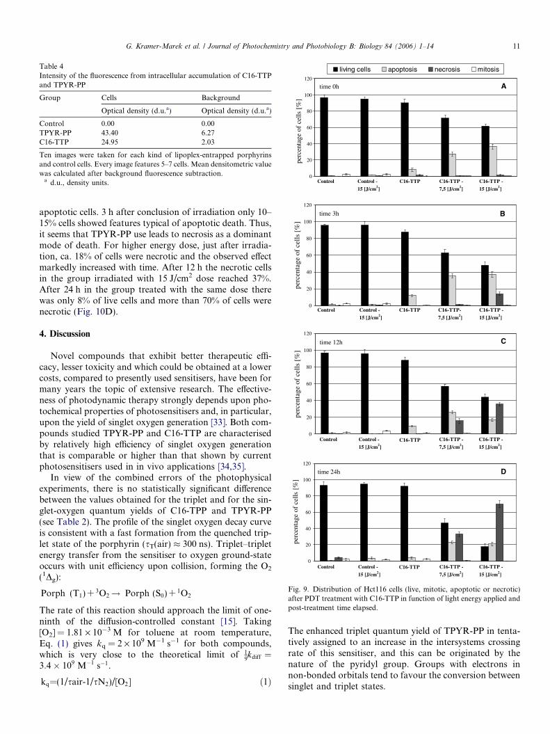
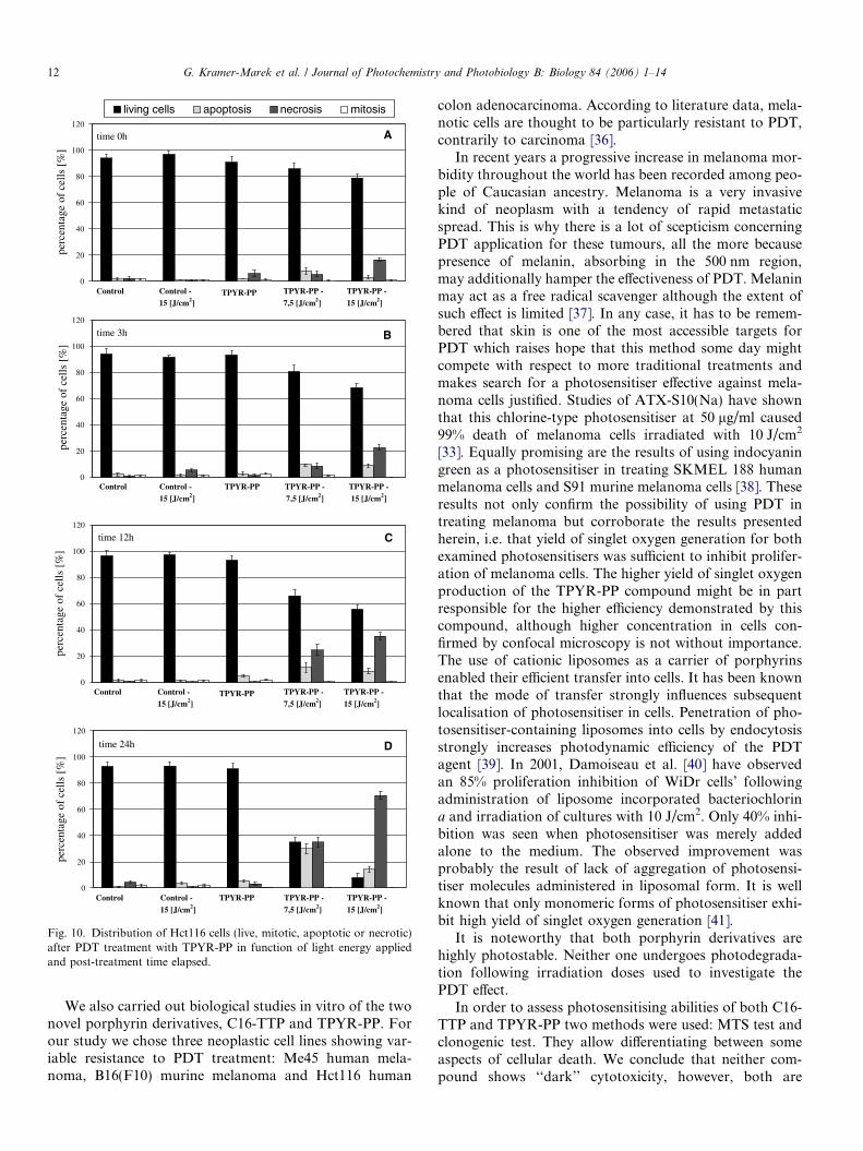
In our study PDT-treated and untreated controls werestained with AO/EB in order to verify the modes of celldeath based on their morphological changes. The investiga-tions were carried out on Hct116 cell line which showed tobe the most sensitive to PDT. Two energy doses were used:7.5 and 15 J/cm2. Analysis was done right after irradiationand after 3, 6, 12, and 24 h. One thousand cells were eval-uated under fluorescent microscope (objective 40·), simul-taneously using phase contrast. Percentage of undamagedcells, those in apoptosis and necrosis and cells undergoingmitosis were calculated.
Fig. 9A–D shows the results for C16-TTP derivative.Treatment of Hct116 cells only with light doses 7.5 and15 J/cm2 is almost non-toxic (90–95% live cells, and veryfew apoptotic and necrotic cells). Differentiation ofHct116 cell response treated with C16-TTP is clearlydependent on energy dose used. Cells quickly undergoapoptosis, which is expected after PDT. Even shortly afterirradiation (0–3 h, Fig. 9A–B) the percentage of apoptoticcells was ca. 25–37% for both low and high energy dose.For the higher dose after 3 h there were 14% of necroticcells. After longer time periods the percentage of necroticcells significantly increased for cultures treated with thisenergy dose, reaching 70% after 24 h (Fig. 9D). In the caseof cells irradiated with 7.5 J/cm2 dose after 3 h there wasonly a minor percentage of necrotic cells. At further timepoints their number slowly raised reaching ca. 35% after24 h (Fig. 9D). Apoptotic cells in this group show initialrise, but the percentage decreases with time, resulting inthe microscopic picture being finally dominated by necroticcells.
The results obtained for TPYR-PP (Fig. 10A–D) mark-edly differ from the results obtained for C16-TTP, althoughhere one can notice as well differentiation of cellularresponse depending on light energy dose used. In this case,however, cells treated with both lower and higher energydoses exhibited a substantially lower percentage of
Fig. 8. Intracellular distribution of TPYR-PP, C16-TTP and PH in Me45 cells evaluated by confocal microscopy. 5-Chloromethylfluorescein diacetate(CMFDA) was used as a marker molecule. Porphyrins were excited by argon ion laser at 488 nm (red fluorescence image, channel I) and CMFDA by neonlaser at 543 nm (green fluorescence image, channel II); combined multitruck system image (Ch I + Ch II) presents both type of fluorescence.
10 G. Kramer-Marek et al. / Journal of Photochemistry and Photobiology B: Biology 84 (2006) 1–14
0
20
40
60
80
100
120
perc
enta
ge o
f ce
lls
[%]
C16-TTP C16-TTP-
7,5 [J/cm2]
C16-TTP -
15 [J/cm2]
Control -
15 [J/cm2]
Control
time 3h
0
20
40
60
80
100
120
perc
enta
ge o
f ce
lls
[%]
C16-TTP C16-TTP -
7,5 [J/cm2]
C16-TTP -
15 [J/cm2]
Control -
15 [J/cm2]
Control
time 12h
0
20
40
60
80
100
120
perc
enta
ge o
fce
lls [
%]
living cells apoptosis necrosis mitosis
C16-TTP C16-TTP -
7,5 [J/cm2]
C16-TTP -
15 [J/cm2]
Control -
15 [J/cm2]
Control
time 0h
0
20
40
60
80
100
120
perc
enta
ge o
f ce
lls [
%]
C16-TTP C16-TTP -
7,5 [J/cm2]
C16-TTP -
15 [J/cm2]
Control -
15 [J/cm2]
Control
time 24h D
C
B
A
Fig. 9. Distribution of Hct116 cells (live, mitotic, apoptotic or necrotic)after PDT treatment with C16-TTP in function of light energy applied andpost-treatment time elapsed.
Table 4Intensity of the fluorescence from intracellular accumulation of C16-TTPand TPYR-PP
Group Cells Background
Optical density (d.u.a) Optical density (d.u.a)
Control 0.00 0.00TPYR-PP 43.40 6.27C16-TTP 24.95 2.03
Ten images were taken for each kind of lipoplex-entrapped porphyrinsand control cells. Every image features 5–7 cells. Mean densitometric valuewas calculated after background fluorescence subtraction.
a d.u., density units.
G. Kramer-Marek et al. / Journal of Photochemistry and Photobiology B: Biology 84 (2006) 1–14 11
apoptotic cells. 3 h after conclusion of irradiation only 10–15% cells showed features typical of apoptotic death. Thus,it seems that TPYR-PP use leads to necrosis as a dominantmode of death. For higher energy dose, just after irradia-tion, ca. 18% of cells were necrotic and the observed effectmarkedly increased with time. After 12 h the necrotic cellsin the group irradiated with 15 J/cm2 dose reached 37%.After 24 h in the group treated with the same dose therewas only 8% of live cells and more than 70% of cells werenecrotic (Fig. 10D).
4. Discussion
Novel compounds that exhibit better therapeutic effi-cacy, lesser toxicity and which could be obtained at a lowercosts, compared to presently used sensitisers, have been formany years the topic of extensive research. The effective-ness of photodynamic therapy strongly depends upon pho-tochemical properties of photosensitisers and, in particular,upon the yield of singlet oxygen generation [33]. Both com-pounds studied TPYR-PP and C16-TTP are characterisedby relatively high efficiency of singlet oxygen generationthat is comparable or higher than that shown by currentphotosensitisers used in in vivo applications [34,35].
In view of the combined errors of the photophysicalexperiments, there is no statistically significant differencebetween the values obtained for the triplet and for the sin-glet-oxygen quantum yields of C16-TPP and TPYR-PP(see Table 2). The profile of the singlet oxygen decay curveis consistent with a fast formation from the quenched trip-let state of the porphyrin (sT(air) � 300 ns). Triplet–tripletenergy transfer from the sensitiser to oxygen ground-stateoccurs with unit efficiency upon collision, forming the O2
(1Dg):
Porph (T1) + 3O2! Porph (S0) + 1O2
The rate of this reaction should approach the limit of one-ninth of the diffusion-controlled constant [15]. Taking[O2] = 1.81 · 10�3 M for toluene at room temperature,Eq. (1) gives kq = 2 · 109 M�1 s�1 for both compounds,which is very close to the theoretical limit of 1
9kdiff ¼
3:4� 109 M�1 s�1.
kq=(1/sair-1/sN2)/[O2] ð1Þ
The enhanced triplet quantum yield of TPYR-PP in tenta-tively assigned to an increase in the intersystems crossingrate of this sensitiser, and this can be originated by thenature of the pyridyl group. Groups with electrons innon-bonded orbitals tend to favour the conversion betweensinglet and triplet states.
0
20
40
60
80
100
120
perc
enta
geof
cel
ls [
%]
TPYR-PP TPYR-PP -
7,5 [J/cm2]
TPYR-PP -
15 [J/cm2]
Control -
15 [J/cm2]
Control
time 24h
0
20
40
60
80
100
120
perc
enta
ge o
f ce
lls
[%]
living cells apoptosis necrosis mitosis
TPYR-PP TPYR-PP -
7,5 [J/cm2]
TPYR-PP -
15 [J/cm2]
Control -
15 [J/cm2]
Control
time 0h
0
20
40
60
80
100
120
perc
enta
geof
cel
ls [
%]
TPYR-PP TPYR-PP -
7,5 [J/cm2]
TPYR-PP -
15 [J/cm2]
Control -
15 [J/cm2]
Control
time 3h
0
20
40
60
80
100
120
perc
enta
ge o
f ce
lls
[%]
TPYR-PP TPYR-PP -
7,5 [J/cm2]
TPYR-PP -
15 [J/cm2]
Control -
15 [J/cm2]
Control
time 12h
B
D
C
A
Fig. 10. Distribution of Hct116 cells (live, mitotic, apoptotic or necrotic)after PDT treatment with TPYR-PP in function of light energy appliedand post-treatment time elapsed.
12 G. Kramer-Marek et al. / Journal of Photochemistry and Photobiology B: Biology 84 (2006) 1–14
We also carried out biological studies in vitro of the twonovel porphyrin derivatives, C16-TTP and TPYR-PP. Forour study we chose three neoplastic cell lines showing var-iable resistance to PDT treatment: Me45 human mela-noma, B16(F10) murine melanoma and Hct116 human
colon adenocarcinoma. According to literature data, mela-notic cells are thought to be particularly resistant to PDT,contrarily to carcinoma [36].
In recent years a progressive increase in melanoma mor-bidity throughout the world has been recorded among peo-ple of Caucasian ancestry. Melanoma is a very invasivekind of neoplasm with a tendency of rapid metastaticspread. This is why there is a lot of scepticism concerningPDT application for these tumours, all the more becausepresence of melanin, absorbing in the 500 nm region,may additionally hamper the effectiveness of PDT. Melaninmay act as a free radical scavenger although the extent ofsuch effect is limited [37]. In any case, it has to be remem-bered that skin is one of the most accessible targets forPDT which raises hope that this method some day mightcompete with respect to more traditional treatments andmakes search for a photosensitiser effective against mela-noma cells justified. Studies of ATX-S10(Na) have shownthat this chlorine-type photosensitiser at 50 lg/ml caused99% death of melanoma cells irradiated with 10 J/cm2
[33]. Equally promising are the results of using indocyaningreen as a photosensitiser in treating SKMEL 188 humanmelanoma cells and S91 murine melanoma cells [38]. Theseresults not only confirm the possibility of using PDT intreating melanoma but corroborate the results presentedherein, i.e. that yield of singlet oxygen generation for bothexamined photosensitisers was sufficient to inhibit prolifer-ation of melanoma cells. The higher yield of singlet oxygenproduction of the TPYR-PP compound might be in partresponsible for the higher efficiency demonstrated by thiscompound, although higher concentration in cells con-firmed by confocal microscopy is not without importance.The use of cationic liposomes as a carrier of porphyrinsenabled their efficient transfer into cells. It has been knownthat the mode of transfer strongly influences subsequentlocalisation of photosensitiser in cells. Penetration of pho-tosensitiser-containing liposomes into cells by endocytosisstrongly increases photodynamic efficiency of the PDTagent [39]. In 2001, Damoiseau et al. [40] have observedan 85% proliferation inhibition of WiDr cells’ followingadministration of liposome incorporated bacteriochlorina and irradiation of cultures with 10 J/cm2. Only 40% inhi-bition was seen when photosensitiser was merely addedalone to the medium. The observed improvement wasprobably the result of lack of aggregation of photosensi-tiser molecules administered in liposomal form. It is wellknown that only monomeric forms of photosensitiser exhi-bit high yield of singlet oxygen generation [41].
It is noteworthy that both porphyrin derivatives arehighly photostable. Neither one undergoes photodegrada-tion following irradiation doses used to investigate thePDT effect.
In order to assess photosensitising abilities of both C16-TTP and TPYR-PP two methods were used: MTS test andclonogenic test. They allow differentiating between someaspects of cellular death. We conclude that neither com-pound shows ‘‘dark’’ cytotoxicity, however, both are
G. Kramer-Marek et al. / Journal of Photochemistry and Photobiology B: Biology 84 (2006) 1–14 13
strongly toxic for the transfected cell lines studied uponlight irradiation. The progressive inhibition of proliferativecapabilities was proportional to irradiation dose and timeelapsed thereafter. It is difficult, however, to compare theseresults with literature data due to different origin of celllines or the nature of photosensitisers.
Intracellular localisation study of C16-TTP and TPYR-PP, performed using confocal microscopy, has shown thatfluorescence appears initially in regions adjacent to cyto-plasmic membrane, later becoming more pronounced inareas around nuclear membrane. These changes suggestthat this is the result of translocation of either derivativetowards nucleus and adjacent areas, involving mitochon-dria and endoplasmatic reticulum. In case of PhotofrinII, after 4 h of incubation evenly distributed fluorescencecan be seen throughout the cytoplasm. Differences inintracellular localisation between investigated compoundsand Photofrin II are due to different chemical propertiesand a different translocation mechanism (diffusion vs.endocytosis).
Our results show that the mechanism of action for thestudied compounds affects the coexisting processes ofapoptosis and necrosis. The presented data are based onof morphological differentiation criteria between apoptoticand necrotic cells, as established by Lelli et al. [20]. Theyrequire confirmation by independent techniques such asflow cytometry, immunocytochemistry as well as biochem-ical studies. Nonetheless, analysis of the gathered data sug-gests that the manner of cell death following application ofeither one of the examined compounds depends on lightirradiation dose used. In either case, when high dose(15 J/cm2) was used the dominating effect was necrosis.Almost total inhibition of cellular proliferation was thenobserved.
5. Conclusion
Both studied porphyrin derivatives TPYR-PP (5-(4-hydroxyphenyl)-10,15,20-tritolylpor) and C16-TTP (5-(4-hexadecyloxyphenyl)-10,15,20-tri-pyridylporphyrin) werechemically well characterised. They are photodynamiclyactive, effectively inducing cell death when light activated,presumably due to the efficient generation of singlet oxy-gen. Singlet oxygen is produced with unit efficiency uponcollision of the triplet of the compounds with ground-stateoxygen. The phototoxicity towards cancer cells showedlight-dose and cell line dependent characteristics. Such typeof porphyrin derivatives seem to be promising agents forPDT treatment of neoplasms. Further quantitative dataabout stability, concentration, and retention in cells needto be gathered. In vivo use would probably require applica-tion of more sophisticated (for example: targeted) carriers.
idylporphyrinNA numerical apertureRT room temperature
Acknowledgements
This work has been supported by Internal StatutoryGrant of Department of Solid State Physics from The Uni-versity of Silesia. We are gratefully acknowledged to AnnaPasewicz for taking part in the synthesis of TPYR-PP andCarlos J.P. Monteiro for the careful sample purification.One author (C.S.) acknowledges FCT (Portugal) for GrantSFRH/BPD/13297/2003.
References
[1] T.J. Dougherty, C.J. Gomer, B.W. Henderson, G. Jori, D. Kessel, M.Korbelik, J. Moan, Q. Peng, Photodynamic therapy, J. Natl. CancerInst. 90 (1998) 889–905.
[3] Z. Luksiene, Photodynamic therapy: mechanism of action and waysto improve the efficiency of treatment, Medicina 12 (2003) 1137–1150.
[4] M.E. Milanesio, F.S. Moran, E.I. Yslas, M.G. Alvarez, V. Rivarola,E.N. Durantini, Synthesis and biological evaluation of methoxyphe-nyl porphyrin derivatives as potential photodynamic agents, Bioorg.Med. Chem. 8 (2001) 1943–1949.
[5] D.E. Dolmans, D. Fukumura, R.K. Jain, Photodynamic therapy forcancer, Nat. Rev. Cancer 3 (2003) 380–387.
[6] A.C. Moor, Signaling pathways in cell death and survival afterphotodynamic therapy, J. Photochem. Photobiol. B 57 (2000) 1–13.
[7] J. Morgan, A.R. Oseroff, Mitochondria-based photodynamic anti-cancer therapy, Adv. Drug Deliv. Rev. 49 (2001) 71–86.
[8] N.L. Oleinick, R.L. Morris, I. Belichenko, The role of apoptosis inresponse to photodynamic therapy: What, where, why, and how?Photochem. Photobiol. Sci. 1 (2002) 1–21.
[9] C.N. Zhou, Mechanisms of tumor necrosis induced by photodynamictherapy, J. Photochem. Photobiol. B 3 (1989) 299–318.
[10] E.S. Nyman, P.H. Hynninen, Research advances in the use oftetrapyrrolic photosensitisers for photodynamic therapy, J. Photo-chem. Photobiol. B 73 (2004) 1–28.
[11] R. Bonnett, Photodynamic therapy in historical perspective, Rev.Contemp. Pharmacother. 10 (1999) 1–17.
[13] X.D. Wang, B.W. Zhang, J.W. Bai, Y. Cao, X.R. Xiao, J.M. Xu,Light-induced electron transfer of porphyrin triad photoelectricconversion, J. Phys. Chem. 96 (1992) 2886–2891.
[14] S. Takagi, T. Yamamura, M. Nakajima, K. Ishiguro, Y. Kawanishi,S. Nihojima, H. Tsuchiya, Synthesis of amphiphilic porphyrins, Bull.Chem. Soc. Jpn. 54 (1981) 3879–3880.
14 G. Kramer-Marek et al. / Journal of Photochemistry and Photobiology B: Biology 84 (2006) 1–14
[15] M. Pineiro, A.L. Carvalho, M.M. Pereira, A.M.d’A. Rocha Gonsal-ves, L.G. Arnaut, S.J. Formosinho, Photoacoustic measurements ofporphyrin triplet-state quantum yields and singlet-oxygen efficiencies,Chem. Eur. J. 4 (1998) 2299–2307.
[16] C.J.P. Monteiro, M.M. Pereira, M.E. Azenha, H.D. Burrows, C.Serpa, L.G. Arnaut, M.J. Tapia, M. Sarakha, P.W.-W.-Chung S.Nararatnam, A comparative study of water soluble 5,10,15,20-tetrakis (2,6-dichloro-3-sulfophenyl)porphyrin and its metal com-plexes as efficient sensitizers for photodegradation of phenols,Photochem. Photobiol. Sci. 4 (2005) 617–624.
[17] Y. Yamazaki, M. Nango, M. Matsuura, Y. Hasegawa, M. Hasegawa,N. Oku, Polycation liposomes, a novel nonvirial gene transfer system,constructed from cetylatedolyethylenimine, Gene Ther. 7 (2000)1148–1155.
[18] J. Carmichael, W.G. DeGraff, A.F. Gazdar, J.D. Minna, J.B.Mitchel, Evaluation of a tetrazolium-based semiautomated colori-metric assay: assessment of chemosensitivity testing, Cancer Res. 47(1987) 936–942.
[19] R.C. Duke, J.J. Cohen, Morphology and biochemical assays ofapoptosis, Curr. Protein Immunol. 17 (Suppl. 3) (1992) 1–16.
[21] R. BonnettChemical Aspect of Photodynamic Therapy, vol. 68–69,Gordon and Breach Science Publishers, New York, 2000, pp. 150–151.
[22] R. Bonnett, Photosensitisers of the porphyrin and phtalocyanineseries for photodynamic therapy, Chem. Soc. Rev. 24 (1995) 19–33.
[23] M.C. DeRosa, R.J. Crutchley, Photosensitised singlet oxygen and itsapplications, Coord. Chem. Rev. 233–234 (2002) 351–371.
[24] H.D. Burrows, L.G. Arnaut, J. Pina, J. Seixas de Melo, N.Chattopadhyay, L. Alcacer, A. Charas, J. Morgado, Characterisationof the triplet state of a fluorene–terthiophene alternating copolymer,Chem. Phys. Lett. 402 (2005) 197–201.
[25] R. Schmidt, C. Tanielian, R. Dunsbach, C. Wolff, Phenalenone, auniversal reference compound for the determination of quantumyields of singlet oxygen O2(1Dg) sensitization, J. Photochem. Photo-biol. A 79 (1994) 11–17.
[28] F. Wilkinson, W.P. Helman, A.B. Ross, Quantum yields for thephotosensitized formation of the lowest electronically excited singletstate of molecular oxygen in solution, J. Phys. Chem. Ref. Data 22(1993) 113–262.
[29] R. Bonnett, D.J. McGarvey, A. Harriman, E.J. Land, T.G. Truscott,U.-J. Winfield, Photophysical properties of meso-tetraphenylporph-yrin and some meso-tetra(hydroxypheny)porphyrins, Photochem.Photobiol. 48 (1988) 271–276.
[30] S. Nonell, P.F. Aramendia, K. Heihoff, R.M. Negri, S. Braslavsky, J.Phys. Chem. 94 (1990) 5879–5883.
[31] F. Postigo, M. Mora, M.A. De Madariaga, S. Nonell, M.L. Sagrista,Incorporation of hidrofobic porphyrins into lipossomes: character-ization and structural requirements, Int. J. Pharm. 278 (2004) 239–254.
[32] W.N. Leung, X. Sun, N.K. Mak, C.M.N. Yow, Photodynamic effectsof mTHPC on human colon adenocarcinoma cells: photocytotoxicity,subcellular localisation and apoptosis, Photochem. Photobiol. 75(2002) 406–411.
[33] G. Jori, Tumour photosensitisers: approaches to enhance the selec-tivity and efficiency of photodynamic therapy, J. Photochem. Photo-biol. B 36 (1996) 87–93.
[34] R.W. Redmond, J.N. Gamlin, A compilation of singlet oxygen yieldsfrom biologically relevant molecules, Photochem. Photobiol. 70(1999) 391–475.
[35] L. Bourre, G. Simonneaux, Y. Ferrand, S. Thibaut, Y. Lajat, T.Patrice, Synthesis, and in vitro and in vivo evaluation of adiphenylchlorin sensitiser for photodynamic therapy, J. Photochem.Photobiol. B 69 (2003) 179–192.
[36] S. Banfi, E. Caruso, S. Caprioli, L. Mazzagatti, G. Canti, R. Ravizza,M. Gariboldi, E. Monti, Photodynamic effects of porphyrin andchlorin photosensitisers in human colon adenocarcinoma cells,Bioorg. Med. Chem. 12 (2004) 4853–4860.
[37] S. Nagata, A. Obana, Y. Gohto, S. Nakajima, Necrotic and apoptoticcell death of human malignant melanoma cells following photody-namic therapy using an amphiphilic photosensitiser, ATX-S10(Na)Lasers, Surg. Med. 33 (2003) 64–70.
[38] K. Urbanska, B. Romanowska-Dixon, Z. Matuszak, J. Oszajca, P.Nowak-Sliwinsk, G. Stochel, Indocyanine green as a prospectivesensitizer for photodynamic therapy of melanomas, Acta Biochim.Pol. 49 (2002) 387–391.
[39] Y. Takeuchi, K. Ichikawa, S. Yonezawa, K. Kurohane, T. Koishi, M.Nango, Y. Namba, N. Oku, Intracellular target for photosensitiza-tion in cancer antiangiogenic photodynamic therapy mediated bypolycation liposome, J. Control. Release 97 (2004) 231–240.
[40] X. Damoiseau, H.J. Schuitmaker, J.W. Lagerberg, M. Hoebeke,Increase of the photosensitizing efficiency of the Bacteriochlorin a byliposome-incorporation, J. Photochem. Photobiol. B 60 (2001) 50–60.
[41] L.E. Bennett, K.P. Ghiggino, R.W. Henderson, Singlet oxygenformation in monomeric and aggregated porphyrin c, J. Photochem.Photobiol. B 3 (1989) 81–89.