Contents lists available at SciVerse ScienceDirect
NeuroImage
j ourna l homepage: www.e lsev ie r .com/ locate /yn img
Speech-induced striatal dopamine release is left lateralized and coupled to functionalstriatal circuits in healthy humans: A combined PET, fMRI and DTI study
Kristina Simonyan a,b,c,⁎, Peter Herscovitch d, Barry Horwitz e
a Department of Neurology, Mount Sinai School of Medicine, New York, NY, USAb Department of Otolaryngology, Mount Sinai School of Medicine, New York, NY, USAc Laryngeal and Speech Section, National Institutes of Neurological Disorders and Stroke, National Institutes of Health, USAd PET Department, Clinical Center, National Institutes of Health, Bethesda, MD, USAe Brain Imaging and Modeling Section, National Institute on Deafness and Other Communication Disorders, National Institutes of Health, Bethesda, MD, USA
⁎ Corresponding author at: Department of Neurology, MOne Gustave L. Levy Place, Box 1137, New York, NY 1002
Considerable progress has been recently made in understanding the brain mechanisms underlying speechand language control. However, the neurochemical underpinnings of normal speech production remainlargely unknown. We investigated the extent of striatal endogenous dopamine release and its influenceson the organization of functional striatal speech networks during production of meaningful English sentencesusing a combination of positron emission tomography (PET) with the dopamine D2/D3 receptor radioligand[11C]raclopride and functional MRI (fMRI). In addition, we used diffusion tensor tractography (DTI) to exam-ine the extent of dopaminergic modulatory influences on striatal structural network organization. We foundthat, during sentence production, endogenous dopamine was released in the ventromedial portion of thedorsal striatum, in both its associative and sensorimotor functional divisions. In the associative striatum,speech-induced dopamine release established a significant relationship with neural activity and influencedthe left-hemispheric lateralization of striatal functional networks. In contrast, there were no significanteffects of endogenous dopamine release on the lateralization of striatal structural networks. Our data providethe first evidence for endogenous dopamine release in the dorsal striatum during normal speaking and pointto the possible mechanisms behind the modulatory influences of dopamine on the organization of functionalbrain circuits controlling normal human speech.
Human speech, one of the most complex and rapid motor behaviors,relies on multi-level integration between brain networks regulatingspeech perception, processing and production. Although our knowledgeof the organization and function of these sensorimotor and higher-ordercognitive speech controlling systems has evolved considerably over thelast decade (Price, 2012), the neuromodulatory bases of normal humanspeech and language control remain poorly understood (Simonyan etal., 2012).
Since the discovery of dopamine as a major neurotransmitter in thebrain in 1950s (Carlsson, 1959), itsmodulatory effects onneural activityand networks have been shown to influence a wide range of humanbehaviors (Montague et al., 2004; Redgrave et al., 2010; Salimpoor etal., 2011; van Schouwenburg et al., 2010). In the speech control system,recent studies have shown that dopaminergic transmission may influ-ence phonological speech processing (Tettamanti et al., 2005), verbal
ount Sinai School of Medicine,9, USA. Fax: +1 646 537 8628.monyan).
rights reserved.
episodic memory and word generation (Cervenka et al., 2008). Onthe other hand, the role of dopaminergic neuromodulation in normalspeechmotor control remains unknown and is based largely on indirectclinical evidence. Case reports in patients treated with dopamine D2
receptor antagonists have demonstrated that drug-induced alter-ations of dopaminergic transmission may lead to the developmentof uncontrolled laryngealmuscle spasmsdue to the transient disruptionof sensorimotor control of speech production (Newton-John, 1988;Warren and Thompson, 1998). The other well-known clinical illustra-tions of dopaminergic deficits affecting speech and language controlin patients with neurological and psychiatric problems range from afundamental inability to control motor aspects of speech production(e.g., monotone, hypotonic speech with reduced loudness and pitch,decreased accuracy of articulation and a raspy voice in Parkinson'sdisease; involuntary voice breaks in spasmodic dysphonia; frequent rep-etitions of syllables in stuttering; and vocal tics in Tourette's syndrome) toprofound speech-associated cognitive impairments (e.g., difficulties withphonological processing, syntactic complexity and language comprehen-sion in Parkinson's disease; and auditory verbal hallucinations in schizo-phrenia) (Heinz et al., 1998; Kataoka and Ueno, 2010; Ludlow et al.,2008; Thompson et al., 2009; Walsh and Smith, 2011; Wu et al., 1997).Interestingly, in Parkinson's disease, in contrast to the main motor
22 K. Simonyan et al. / NeuroImage 70 (2013) 21–32
impairments of the extremities, speech motor changes, occurring innearly 90% of all patients (Aronson, 1980), do not respond to traditionaldopamine replacement therapy and usually require additional treat-ment protocols, such as behavioral therapy, in order to manage thesymptoms (Ciucci et al., 2010; Ramig et al., 2001). In addition, patientswith advanced Parkinson's disease may occasionally develop compul-sive involuntary singing and humming while receiving high-dosedopamine replacement therapy (Bonvin et al., 2007; Friedman, 1993;Kataoka and Ueno, 2010). Moreover, while deep brain stimulation ofthe subthalamic nucleus (STN DBS) is used to successfully improvethe motor symptoms affecting extremities in Parkinson's disease(Fasano et al., 2012), it is less effective, if at all, in the management ofspeech impairments (Karlsson et al., 2012; Tripoliti et al., 2011; VanLancker Sidtis et al., 2010; Xie et al., 2011). Such differential outcomeof pharmacological and surgical treatments may, in part, be dependenton different mechanisms of dopaminergic transmission for speechcompared to limb control. Thus, despite the lack of direct knowledgeabout the dopaminergic function underlying normal speech production,clinical evidence strongly suggests a particular role for central dopa-mine modulation of both speech motor commands and higher-ordercognitive processes during speaking.
In this study, we investigated the mechanisms of dopaminergicinfluences on the central control of normal human speech productionby examining the extent of speech-induced striatal endogenousdopamine release and its relationship with striatal neural activity andthe organization of functional and structural speech networks. We useda combination of three different neuroimaging methodologies (positronemission tomography with the dopamine D2/D3 receptor radioligand[11C]raclopride (RAC PET), event-related sparse-sampling functionalMRI (fMRI) with striatal functional connectivity analysis, and diffusiontensor imaging (DTI) with striatal probabilistic tractography) in 20healthy subjects. The experimental condition during RAC PET and fMRIstudies involved production of meaningful English sentences withcorrect grammatical and lexical structure and resting as a baseline.The experimental condition (i.e., sentence production) was chosen torepresent human speech and language as complex behaviors used ineveryday communication as closely as possible. RAC PET enableddetection of changes in striatal dopamine release between speaking andresting due to the binding competition for D2/D3 dopamine receptorsbetween endogenous dopamine and the radiolabeled D2/D3 dopaminereceptor antagonist, [11C]raclopride, with a decrease in RAC binding dur-ing speaking being interpreted as a speech-induced increase in dopaminerelease (Laruelle, 2000). In addition, the use of fMRI in the same subjectsundergoing RAC PET allowed for characterization of striatal activity asso-ciated with dopamine release during speech production and mappingof striatal functional networks, while DTI tractography determined theunderlying striatal structural connections.
Based on the previous neuroanatomical tract tracing studies(Jurgens, 1976; Simonyan and Jurgens, 2003), we hypothesized thatsignificant dopamine release during speech production would befound in the ventromedial portion of the dorsal striatum consistentwith the striatal projection fields of the laryngeal motor cortex. Wealso predicted that speech-induced striatal dopaminergic releasewould be associated with BOLD signal change during speech produc-tion to influence the organization of striatal functional networks con-trolling normal speech production. We expected to find minimalmodulatory effects of dopamine release on the striatal structuralnetwork involved in speech control.
Materials and methods
Subjects
Twenty healthy subjects participated in each of RAC PET, fMRI andDTI study. The same 20 subjects (age 53.2±10.1 years, mean±s.d.,13 females/7 males) underwent both the PET and fMRI studies.
Eight subjects from this group participated in the DTI study, while 12additional age- and gender-matched subjects (age 53.8±10.9 years,mean±s.d., 13 females/7 males) were scanned with the same DTIsequence to match the total number of 20 subjects, who participatedin the PET and fMRI studies.
All subjects were right-handed on the Edinburgh HandednessInventory (Oldfield, 1971) and monolingual native English speakers.No participant had any neurological, psychiatric or laryngeal problemsbased on history and physical examination. All subjects underwentfiberoptic nasolaryngoscopy to confirm normal anatomy and functionof the larynx during rest and different laryngeal tasks. Routine neurora-diological evaluation of subjects' high-resolution MRI found normalbrain structure without any gross abnormalities.
All participants provided written informed consent before partici-pation in the study, which was approved by the Institutional ReviewBoards of the Mount Sinai School of Medicine, National Institute ofNeurological Disorders and Stroke, and the NIH Radiation SafetyCommittee.
Data acquisition
The scanning sessions were randomized between the subjects. Allsubjects completed the fMRI/DTI scans in the afternoon (between2:00 PM and 5:00 PM) and the PET scan in the morning or earlyafternoon (18 subjects between 8:30 AM and 11:30 AM, 2 subjectsbetween 12:45 PM and 2:00 PM) to control for possible diurnalvariations in dopamine transmission. Participants were instructed toabstain from alcohol one week prior to the PET scanning, not todrink any beverages containing caffeine within 24 h and not to eat3 h before the PET study.
RAC PET data were acquired on a GE Advance PET scanner(GE Medical Systems, Milwaukee, WI). An 8-min transmission scanwas first obtained using a 68Ge source for attenuation correction ofemission data. Through a catheter placed in the antecubital vein,RAC was administered as a 1-min bolus followed by a constant infu-sion over 99 min (bolus-plus-infusion (B/I) method) (Watabe et al.,2000) using a computer-operated pump (Harvard Instruments,Natick, MA) (Fig. 1A). Accounting for decay, the effectively injectedRAC activity over 100-min PET was 19.7±1.4 mCi (i.e., 728.9±51.8 MBq) (mean±s.d.) with specific activity at the time of injection2484.5±1164.7 mCi/μmol (i.e., 91.9±43.1 GBq/μmol) (mean±s.d.).The practical advantages of the B/I method included the requirementfor only a single radiochemical synthesis and a single administrationof the radiotracer, which significantly minimized the levels of radiationexposure to the study participant. Importantly, the B/I method allowedfor the avoidance of potential physiological effects of different scanningsessions, helped the maintenance of RAC equilibrium throughout theentire study (Carson et al., 1997; Slifstein and Laruelle, 2001; Watabeet al., 2000), and avoided the confounding effects of behavior-inducedregional cerebral blood flow (rCBF) changes on specific binding values(Carson, 2000; Carson et al., 1993, 1997; Endres and Carson, 1998;Endres et al., 1997).
A dynamic RAC PET scan in the 3D scanning mode with septaretracted was initiated at the start of RAC injection, generating 27time frames of 30 s to 5 min epochs over 100 min (FOV 148 mm,reconstructed resolution 6 mm in all direction). The 100-min PETscan included two conditions: an initial 50-min resting state, duringwhich the subjects were asked to relax with their eyes closed in anenvironment with dimmed light and reduced ambient noise; and50-min speaking, during which the subjects listened to the auditorysample of meaningful English sentences with correct grammaticaland lexical structure (e.g., “Tom is in the army”, “We are alwaysaway”) and repeated it at a convenient conversational speech level.Subjects produced different sentences for 4 min and rested for1 min in order to avoid boredom (Fig. 1A). Ten different sentenceswere presented one at a time and were pseudorandomized between
Fig. 1. Schematic illustration of the experimental design during RAC PET (A) and speech-production fMRI (B) studies.
23K. Simonyan et al. / NeuroImage 70 (2013) 21–32
subjects. Auditory stimuli were recorded from a native English femalespeaker for the purpose of this study. No auditory stimulus waspresented during the resting condition. Subjects were trained tospeak without head movements, which were additionally minimizedby using a comfortably tight thermoplastic mask molded around thesubject's head.
Functional MRIWhole-brain functional images were acquired with a gradient-
weighted EPI pulse sequence using BOLD contrast and a sparse-sampling event-related design on a 3.0 Tesla GE scanner equippedwith a quadrature birdcage radio frequency head coil (Fig. 1B). TheEPI sequence included TE 30 ms, TR 10.6 s with 8.6 s for task produc-tion and 2 s for image acquisition, FA 90 degrees, FOV 240×240 mm,matrix 64×64 mm, in-plane resolution 3.75 mm, 33 sagittal slices,and slice thickness 4.0 mm. Experimental tasks included 1) produc-tion of the same sentences as during the RAC PET study (one sentencewas acoustically presented and reproduced by the subjects per acqui-sition volume); 2) whimper (not reported here), and 3) rest as silentfixation at a cross that appeared on the screen in front of subjects'eyes. During scanning, subjects were asked first to listen to theauditory sample of the sentence delivered through MR-compatibleheadphones (SilentScan Audio System, Avotec Inc., Stuart, FL) for a3.6-s period (Fig. 1B). A visual cue (arrow) then instructed thesubjects to speak the sentence within the 5-s interval followed by a2-s images acquisition. All tasks were pseudorandomized betweenscanning sessions and subjects. Five scanning sessions were acquiredwith a total of 36 trials per task.
The use of a sparse-sampling event-related fMRI design helpedreduce the effect of self-speech perception on data acquisition duringspeech production; this design, however, was impossible to implementduring the PET scan. Thus, while fMRI acquisition may have been lessinfluenced by perception of each subject's own speech, both fMRI andPET contained, to some extent, the effects of self-produced sentenceson brain activity and neurotransmission, respectively. This, however,did not present a major problem for our current study because ouraim was to examine speech production as a complex human behavior,in which speech perception for production represents one of theintegral components.
DTIWhole-brain diffusion-weighted images were acquired using a
single-shot spin-echo EPI sequence and an array spatial sensitivityencoding (ASSET) factor of 2 (TE 73.4 ms, TR 13 s, FOV 240 mm, ma-trix 256×256 mm, in-plane resolution 0.9375 mm, 54 contiguousaxial slices, slice thickness 2.4 mm) on the same scanner. Diffusionwas measured along 33 noncollinear directions (b 1000 s/mm2);three reference images were collected with no diffusion gradientsapplied.
High-resolution MRIIn each subject, a high-resolution T1-weighted image was collected
as an anatomical reference for the PET and fMRI data using 3Dmagnetization prepared rapid acquisition gradient echo (MPRAGE:TI 450 ms; TE 3.0 ms; FA 10 degrees; bandwidth 31.25 mm; FOV240 mm; matrix 256×256 mm; 128 contiguous axial slices; slicethickness 1.2 mm).
Data analysis
PETData analysis was performed to determine the extent of dopamine
release during sentence production. Initially, correction for subjectmotion during the dynamic PET scan was performed using the regis-tered attenuation correctionmethod. After reconstruction of emissionimages with filtered backprojection with no attenuation correction,all emission frames were registered with mutual information tothe first emission frame with sufficient counts (i.e., prime emissionimage) (FLIRT, FSL, FMRIB Software Library). The transmissionimage was registered to the same prime emission image with thesame algorithm. For each emission frame, the transmission imagewas resliced taking into account the motion between transmissionand each frame. The emission frame was reconstructed with filteredbackprojection, and the newly synthesized transmission scan wasused for attenuation correction, after which the emission image wasresliced back to the transmission position, thus correcting for motion.
Taking into the account the pharmacokinetics of the RAC (Laruelle,2000), the motion- and decay-corrected PET frames (Woods et al.,1993) were averaged over 40–50 min of baseline resting and over60–100 min of speech production once equilibrium was achieved(PMOD Technologies, Zurich, Switzerland), as reported earlier in asimilar experimental design (Garraux et al., 2007). The two PETdatasets per subject (i.e., resting and speech production) werealigned to each individual's T1-weighted MR image using Hellingerdistance and two-pass alignment strategy, normalized to the stan-dard Talairach space and spatially smoothed with a 6-mm Gaussianfilter (PMOD Technologies and AFNI software (Cox, 1996)). Alltransformed images were visually inspected for alignment errors.
Detection of dopamine release was based on estimation ofchanges in the concentration of available receptor sites (Bavail) inresponse to the associated changes in dopamine concentration. Thekinetic behavior of RAC is known to be linear at tracer concentrationsand dependent on Bavail, which enables determination of bindingpotential (BP) (Laruelle, 2000). BP equals the equilibrium ratio ofbound ligand to free and non-specifically bound tracer under theassumption that non-specific binding is uniform throughout the brain(Innis et al., 2007). The parametric voxelwise RAC BP maps duringeach condition (i.e., resting and speech production) were calculated ineach subject using an equation BP=(C−C′)/C′ based on the radioac-tivity concentration in the striatum (C) as a region with the highest
24 K. Simonyan et al. / NeuroImage 70 (2013) 21–32
density of dopamineD2/D3 receptors and the cerebellum (C′) as a regiondevoid of D2/3 receptors (Hall et al., 1996). The striatal and cerebellarregions were defined using the probabilistic macrolables atlas of theAnatomy Toolbox (Eickhoff et al., 2005) similar to previously describedmethods (Del Campo et al., 2011; Salimpoor et al., 2011). Striatalregions-of-interest (ROIs) were sampled at the entire rostro-caudaland dorso-ventral extents; the cerebellar ROI included the gray matterdefined on the five consecutive slices of both hemispheres. Radioactivityin the cerebellar gray matter did not show significant differences(two-sample independent t-test, p=0.81) between resting (BP=9.6±2.1 nCi/ml/mCi injected) and speech production (BP=9.5±1.9 nCi/ml/mCi injected).
Next, derived statistical parametric t maps of RAC BP for eachcondition were used to calculate percentage change in RAC BP (ΔBP)caused by speech production in each subject (ΔBP=(BPspeech−BPresting) /BPresting×100% (Watabe et al., 2000)). The groupstatistical significance of speech-induced changes on RAC BP wasassessed with a voxelwise paired t-test at a corrected p≤0.01. Theanatomical location of significant striatal RAC ΔBP in striatal functionaldivisions was determined as described earlier (Mawlawi et al., 2001).
fMRITo estimate the extent and intensity of brain activity during
sentence production, we conducted univariate analysis of functionalimaging data using AFNI software. Each subject's first two volumes ineach series, collected before magnetization equilibrium was reached,were discarded. The EPI volumes from all runs were registered tothe single EPI volume collected closest in time to the high-resolutionanatomical scan using heptic polynomial interpolation and spatiallysmoothed with a 6-mm Gaussian filter. Voxelwise normalization topercent signal change was applied to each voxel in the whole brain.The task-related responses were analyzed using multiple linear regres-sion with a single regressor for each task convolved with a canonicalhemodynamic response function. Baseline drifts were modeled usingquadratic polynomials in time for each separate imaging run, andmotion parameter estimates were used as additional regressors of nointerest in the multiple regression analysis. The correction for multiplecomparisonswas performed usingMonte-Carlo simulations that identi-fied a minimum cluster size of 808 mm3 at a voxelwise threshold of0.001 to achieve overall significance level of a corrected p≤0.01. Forgroup analysis, the anatomical datasets of each subject were spatiallynormalized and converted to the standard Talairach space. The resultingnormalization parameters were applied to the 4-D time series datasets,which were transformed into the standard space. To estimate the maineffect of speech production, the group analysis was carried out using atwo-way within-subject mixed effect design analysis of variance(ANOVA) with subject as a random factor and the task as the fixedfactor (p≤0.01, corrected).
Relationships between striatal dopamine release and neural activationUsing AFNI software, voxelwise striatal Spearman's rank correlation
coefficients were computed to estimate the statistical dependencebetween speech-induced dopamine release and striatal neural activity.The normalized PET and fMRI datasets were first resampled to matchthe same orientation and grid spacing. To create a single 3D+subjectsvolume for each of the fMRI and PET datasets, the respective imageswere concatenated across all subjects. Voxelwise Spearman's correla-tion coefficients were computed between the corresponding voxels ofRAC ΔBP and speech-induced BOLD signal change. The resultant mapswere thresholded at a corrected p≤0.05.
To account for possible influences on correlation coefficients by afew points, a jackknife procedure was performed to eliminate any cor-relation coefficients that are statistically significant due to the presenceof an outlier. Further, a bootstrap procedure with replacement in 1000samples was performed to obtain the estimates of statistical accuracyof correlation coefficients (Horwitz et al., 1986, 1991).
Striatal network analysesTo examine the influences of speech-induced dopamine release on
striatal functional networks, we used psychophysiological interac-tions (PPI) analysis (Friston et al., 1997) of fMRI data during sentenceproduction. A 4-mm spherical seed region was placed in the leftdorsal anterior putamen (APU) (x,y,z=±23,4,6), which showed astatistically significant relationship between speech-related BOLDsignal and RAC ΔBP derived from Spearman correlation analysis(see the section on Relationships between striatal dopamine releaseand neural activation). Although coupling between speech-inducedBOLD signal and RAC ΔBP was found only in the left APU, we alsoconducted functional connectivity analysis using the correspondingseed region in the right APU in order to test our prediction thatspeech-induced dopaminergic function has lesser modulatory effectson the right APU networks. Seed time series in each subject wereextracted during speech production and silent fixation, multipliedby the task vector, regressed with the time series from the entirebrain in each subject, and submitted to group analysis using atwo-sample t-test at a corrected p≤0.01 (minimum cluster size of40 mm3 at a voxelwise threshold of 0.001).
To examine striatal structural circuits underlying functional net-works of speech control, diffusion tensor modeling with probabilistictractography from the same seeds in the bilateral APU as used forfunctional network analysis was performed using the FSL softwarepackage (FDT Diffusion Toolbox (Behrens et al., 2007)). After correc-tion of diffusion-weighted images for eddy current and headmotion artifacts, the voxelwise diffusion tensor was calculated usingmultivariate fitting and diagonalization. A probabilistic streamlineand the connectivity distribution were constructed based on thedistribution probabilities of each fiber direction sampled in each voxel.The APU structural connections were identified using unconstrainedprobabilistic tractography in each subject. The probabilistic tractographyparameters were set at 5000 streamline samples, 2000 steps with0.5 mm step length and 0.1-curvature threshold. The group maps ofleft/right APU structural connectivitywere created by averaging resultantprobabilistic tractographymaps in all subjects normalized to the standardTalairach space and thresholded at a corrected p≤0.01.
Assessment of hemispheric lateralityTo examine the lateralization of striatal activation and functional/
structural networks, whole-hemispheric ROIs were used to obtain thenumber of significantly activated and connected voxels, respectively.Statistical significance of lateralization was assessed across all subjectsusing paired t-test at p≤0.01 to correct for multiple comparisons.
Additionally, a laterality index (LI) was used to examinethe lateralization of functional activation and networks, definedas: LIactivity=(# active voxels in LH−# active voxels in RH)/(# activevoxels in LH+# active voxels in RH) and LInetwork=(# connectedvoxels of LS in LH−# connected voxels of RS in RH)/(# connectedvoxels of LS in LH+# connected voxels of RS in RH), where LH — lefthemisphere, RH — right hemisphere, LS — left seed, and RS — rightseed. A positive LI indicated a left-hemispheric lateralization, and anegative LI denoted right-hemispheric lateralization of activation ornetwork (Seghier, 2008). Statistical significance of LI was determinedusing paired t-tests at p≤0.01.
Methodological limitationsThe combined use of PET, fMRI and DTI methodologies as well as a
combination of univariate data analyses and network analyses wereessential for obtaining unique information about different properties(i.e., neurotransmitter, functional, and structural) of the striatalsystem involved in the control of normal speech production withinthe same study. Specifically, such a multimodal approach allowedus to not only map the extent of striatal dopamine release and brainactivation during speech production but also to integrate informationabout speech-induced dopaminergic transmission (derived from
25K. Simonyan et al. / NeuroImage 70 (2013) 21–32
RAC PET) with neural function (derived from fMRI) and anatomicalconnectivity (derived from DTI tractography) for identification ofpotential mechanisms of dopamine action on the speech control-ling system. However, our study had some limitations.
As an experimental task, we intentionally chose production ofsentences with correct grammatical and lexical structure to reflectnormal ‘everyday’ speech and language. We recognize that such atask carried both sensorimotor and a number of linguistic and cogni-tive components, for example, working memory due to multiplerepetitions of the same sentences throughout the scanning session. Onthe other hand, our task did not have obvious reward and emotionalcomponents, frequently present in human speech. These are importantaspects of human speech and language control, which should beaddressed in the future studies. In the present study, the conduct ofmultiple PET scanning sessions separating each speech-forming factorwas technically not feasible due to limitations in PET radiation exposureto subjects. Moreover, choosing to focus on one of the components orhaving a different baseline task would have required making a specifichypothesis about dopaminergic mechanisms during speaking prior toknowing the fundamentals of dopamine action on the speech produc-tion system. As our study identifiedwhere and how dopamine interactswith the speech production system, the future studies may be under-taken to dissect its contribution to the control of separate componentsof speech and language production.
Results
Striatal dopamine release during speech production
We found that endogenous dopamine release during sentence pro-duction led to predominantly left-sided RAC displacement (measuredby a decrease in RAC BP between resting and speech conditions) inthe ventromedial portion of the associative (AST) and sensorimotor(SMST) functional divisions of the dorsal striatum (Fig. 2A). Whileboth striatal divisions receive dopaminergic input through thenigro-striatal pathway, functionally, the AST is known to play a role incognition and includes the APU, dorsal anterior caudate nucleus(ACA), and dorsal posterior caudate nucleus (PCA), whereas the SMSTis involved in movement coordination and is comprised of the dorsalposterior putamen (PPU) (Mawlawi et al., 2001).Within the AST, statis-tically significant decreases in RAC BP during speaking were found inthe left APU (ΔBP=−6.32%, p=0.001) and left PCA (ΔBP=−9.54%,p=0.002), whereas the ACA showed a bilateral RAC BP reduction (leftΔBP=−8.55%, t19=−3.30; right ΔBP=−3.96%, p=0.002) (Fig. 2B,Table 1). A follow-up paired t-test found no lateralization effect indopamine release within the ACA (t19=−0.53, p=0.70) (Fig. 2C). Aspeech-induced RAC BP decrease in the SMST was also found only inthe left PPU (ΔBP=−6.25%, p=0.007).
No significant changes in RAC BP from baseline were found in theventral (limbic) striatum, which receives dopaminergic input throughthe mesolimbic pathway. The lack of dopamine release in the ventralstriatum was consistent with the fact that our speech task did notcontain a reward, penalty, or emotional component. It may alsopoint to minimal, if any, involvement of episodic memory associatedwith sentence production in our experimental setting (Cervenka etal., 2008).
Dopamine release is coupled with neural activity in the left APU
In the same subjects, the univariate analysis of fMRI data during pro-duction of the same sentences as during the PET study found widelyspread bilateral activation in the entire striatum(Fig. 3A)without hemi-spheric lateralization of either its extent (t19=1.34, p=0.19, meanlaterality index (LI)=−0.09) or intensity (t19=0.62, p=0.54; meanLI=0.007) (Figs. 3B–E).
However, despite thewhole-striatal activation during speaking, onlythe left APU (part of AST) showed a significant linear relationshipbetween speech-induced RAC ΔBP and BOLD signal change duringspeaking (jackknife estimated rs=−0.59, p=0.006) (Fig. 4).
Dopamine influences the organization of APU functional but not structuralspeech networks
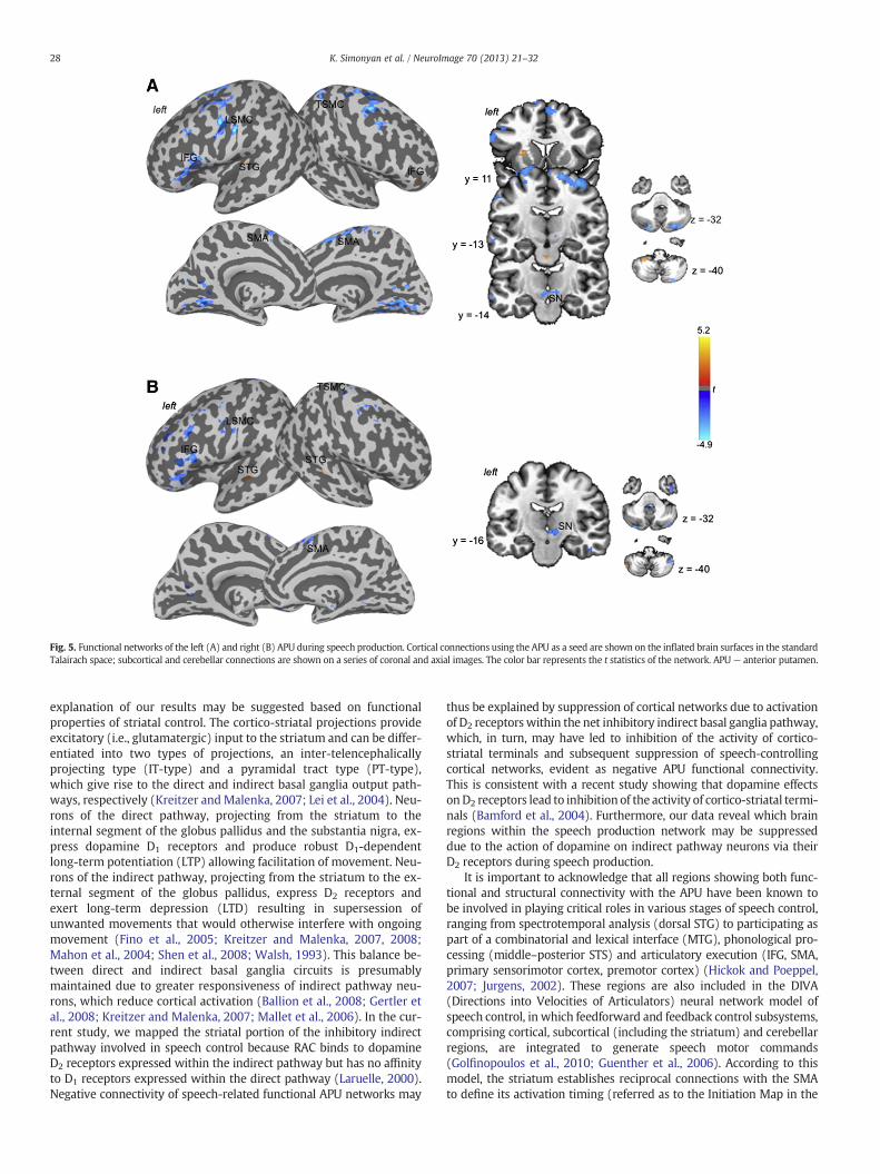
Functional and structural striatal networks were examined fromthe APU region, which showed significant relationship between thespeech-related endogenous dopamine release and neural activity.During speech production, the left APU showed positive functionalconnections with the right inferior frontal gyrus (IFG), left superiortemporal gyrus (STG), including Heschl's gyrus, cerebellum (lobuleVIII), pons, and the APU itself. Negative functional connections ofthe left APU were observed with the left laryngeal sensorimotorcortex (LSMC), right trunkal sensorimotor cortex (TSMC), bilateralpremotor cortex, precuneus, supplementary motor area (SMA), mid-brain including the substantia nigra (SN), and cerebellum (Crus 1–2)(Fig. 5A). The right APU showed positive functional connections onlywith the bilateral STG and left cerebellum (Crus 2). The negativeconnections were similar to those from the left APU (Fig. 5B).
Structurally, both left and right APU networks had the heaviestconnections (>51–100% probability) with frontal cortical regions,including the IFG, dorsal and ventral premotor cortex, dorsolateralprefrontal cortex (dlPFC), ventrolateral prefrontal cortex (vlPFC), insula,SMA, and anterior/middle cingulate cortex (ACC/MCC) (Fig. 6). Some-what weaker striatal connections (>15–50% probability) were foundwith the primary sensorimotor cortex, including the LSMC and TSMC, aswell as with the STG, middle temporal gyrus (MTG), supramarginalgyrus (SMG), midbrain, and cerebellum.
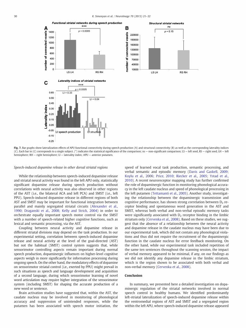
Similar to the left-striatal RAC displacement during speechproduction and despite the bilateral striatal neural activity, speech-related APU functional networks exhibited left-hemispheric laterali-zation (LI=0.33±0.10; p=0.0005) (Fig. 7A). In contrast, APU struc-tural networks were bilaterally distributed without hemisphericlateralization (LI=0.04±0.14, p=0.15) (Fig. 7B). Left-hemisphericlateralization of speech production functional networks was signifi-cantly greater compared to structural networks (all p=0.0005)(Fig. 7C).
Discussion
Despite the long recognized role of dopamine as a neuromodulatorof striatal functions (Albin et al., 1989; Schultz, 2006; Wickens et al.,2003), information on dopaminergic regulation of human speech andlanguage has remained limited. Using a multi-modal neuroimagingapproach, we present here the first evidence of striatal dopaminergictransmission during normal speech production in healthy humansand define the extent of dopaminergic effects on the organization ofstriatal networks involved in speech control.
Dopamine release and lateralization of striatal speech networks
We demonstrated that sentence production was associated withsignificant release and binding of endogenous dopamine to theD2/D3 receptors in the left putamen and bilateral caudate nucleuswithin the AST and SMST divisions of the striatum. We found thatthese striatal regions were similar to those known to receive directprojections from the laryngeal motor cortex in non-human primates,that is the ventromedial portions of both AST and SMST (Jurgens,1976; Simonyan and Jurgens, 2003). While the laryngeal motor corti-cal input into the SMST has been shown to overlap with the inputfrom the facial motor cortex, the input into the AST appears to be seg-regated from other descending motor cortical projections, thusforming a ‘larynx’ striatal representation (Jurgens, 1976; Kunzle,
Fig. 2. Striatal dopamine release during speech production. A speech-induced decrease of RAC BP was found in the left APU, PPU, PCA and in the bilateral ACA. (A) Significantchanges in RAC BP during speech production compared to resting are shown on a series of brain images in the coronal plane in the standard Talairach space. The color bar representsthe t statistic. (B) The graphs display the RAC BP values and the corresponding RAC ΔBP values in each statistically significant striatal region during resting and speech production in20 subjects. (C) The graph compares right and left RAC BP values of 20 subjects in the ACA during speech production. ns— non-significant. APU— anterior putamen; PPU— posteriorputamen; PCA — posterior caudate nucleus; ACA — anterior caudate nucleus; RAC BP — binding potential of the [11C]raclopride radiotracer.
26 K. Simonyan et al. / NeuroImage 70 (2013) 21–32
1975; Simonyan and Jurgens, 2003). Based on our findings of couplingbetween the speech-related dopamine release and neural activity in thesame APU region, which receives descending laryngeal motor corticalprojections,wemay suggest that functional importance of APUdopami-nergic transmission in speech control may, in part, be in its modulatoryeffects on descending laryngeal cortico-striatal terminal activity andspeech cortical network organization. Earlier studies have shown thatthe D2 receptors are localized not only on the striatal medium spinyneurons (MSNs) but also on subpopulations of cortico-striatal terminals(Fisher et al., 1994; Hersch et al., 1995; Sesack et al., 1994; Wang andPickel, 2002), excitability of which is also modulated by dopaminerelease (Bamford et al., 2004; Garris et al., 1994; Gonon, 1997; Nicola
Table 1Striatal regions of significant [11C]raclopride displacement during speech production.
Coordinates are given in the Talairach–Tournoux standard space. Statistical significance isset at p≤0. 013, corrected for multiple comparisons. AST — associative striatum; SMST —
sensorimotor striatum; ACA— anterior caudate; PCA— posterior caudate; APU— anteriorputamen; PPU — posterior putamen; RAC BP — binding potential of the [11C]racloprideradiotracer.
and Malenka, 1998). In line with this assumption, significant left-hemispheric lateralization of APU functional networks found in ourstudy may possibly be due to modulatory influences of dopamine onneural activity in the left but not right APU. On the other hand, themore stable APU structural networks showed a bilateral hemisphericdistribution, suggesting that they may represent an underlyingframework for functional networks controlling different types of vocalbehaviors, and are less receptive to dopaminergic influences.
Lateralization of dopaminergic function has been previouslyreported to be dependent on the direction of motor behavior andtype of reinforcement task in rodents (Glick et al., 1981; Morice etal., 2005; Szostak et al., 1986; Zimmerberg et al., 1974). However,results of studies in songbirds, whose singing is considered to be anal-ogous to human speaking, give no indication of striatal lateralizationof dopamine release during vocal learning and song production(Hara et al., 2007; Huang and Hessler, 2008; Jarvis and Nottebohm,1997; Sasaki et al., 2006; Yanagihara and Hessler, 2006), despite thespecies-specific hemispheric lateralization of avian forebrain vocallearning pathways (Nottebohm et al., 1976; Williams et al., 1992).In humans, recent studies have reported a range of task effects onthe levels of nigro-striatal dopamine release. While no lateralizationof striatal dopamine levels was found in postmortem human braintissue (Rossor et al., 1980), more recent PET studies have establishedthat the levels of striatal dopamine release may depend on the taskperformed. Specifically, the degree of right hand preference duringself-paced freely chosen movements was found to be correlatedwith increased dopaminergic function in the left putamen (de laFuente-Fernandez et al., 2000), whereas performance of a button
Fig. 3. (A) Striatal activation during speech production is shown on a series of coronal images in standard Talairach space. Color bar represents the Z value. Graphs display the+number of active voxels (B) and BOLD percent signal change (D) in the left and right striatum in 20 subjects, showing no significant difference between hemispheres. Bar graphsdemonstrate the corresponding laterality indices of activation extent (C) and activation intensity (E).
27K. Simonyan et al. / NeuroImage 70 (2013) 21–32
press task was associated with bilateral striatal dopamine releasecompared to right-sided striatal lateralization of dopaminergic trans-mission in response to unpredictable reward (Martin-Soelch et al.,2011). The functional importance of left-striatal dopamine releaseduring speech production appears to be in its modulatory influencescontributing to hemispheric lateralization of speech controllingnetworks. Our finding of left-striatal dopaminergic transmission isin line with left-hemispheric lateralization of brain activation duringspeech production, which is known since the time of Broca, andmay point to unique higher-order behavior-specific integration andadaptation of the striatal modulatory dopaminergic system forhuman speech and language control. The significance of left-striataldopaminergic transmission is further supported by clinical evidencethat suggests that abnormalities in lateralization of dopamine release,such as higher uptake of a dopamine precursor in the left but not right
Fig. 4. The left APU shows coupling between dopamine release and brain activation. Relationshshown on a series of coronal images (A) and on a bar graph and correlation plot (B). The colobinding potential of the [11C]raclopride radiotracer.
striatum in persons who stutter (Wu et al., 1997), and the presence ofleft putaminal lesions may contribute to the pathophysiology of vari-ous voice and speech disturbances, ranging from stuttering to aphasiaand auditory agnosia (Ciabarra et al., 2000; D'Esposito and Alexander,1995; Heuer et al., 1996; Lee et al., 1996; Metter et al., 1986; Taniwakiet al., 2000). Collectively, our findings provide evidence for neuro-chemical basis of a still elusive physiological concept of hemisphericdominance of human speech and language production.
Organization of striatal speech networks
An interesting feature of the APU functional speech-relatednetworks was its predominantly negative connectivity. Although themechanisms underlying negative correlations between brain regionswithin a network remain unclear (Bandettini, 2009), a possible
ip between speech-induced RAC ΔBP and BOLD percent signal change during speaking isr bars depict Spearman's correlation coefficients (rs). APU — anterior putamen; RAC BP —
Fig. 5. Functional networks of the left (A) and right (B) APU during speech production. Cortical connections using the APU as a seed are shown on the inflated brain surfaces in the standardTalairach space; subcortical and cerebellar connections are shown on a series of coronal and axial images. The color bar represents the t statistics of the network. APU— anterior putamen.
28 K. Simonyan et al. / NeuroImage 70 (2013) 21–32
explanation of our results may be suggested based on functionalproperties of striatal control. The cortico-striatal projections provideexcitatory (i.e., glutamatergic) input to the striatum and can be differ-entiated into two types of projections, an inter-telencephalicallyprojecting type (IT-type) and a pyramidal tract type (PT-type),which give rise to the direct and indirect basal ganglia output path-ways, respectively (Kreitzer and Malenka, 2007; Lei et al., 2004). Neu-rons of the direct pathway, projecting from the striatum to theinternal segment of the globus pallidus and the substantia nigra, ex-press dopamine D1 receptors and produce robust D1-dependentlong-term potentiation (LTP) allowing facilitation of movement. Neu-rons of the indirect pathway, projecting from the striatum to the ex-ternal segment of the globus pallidus, express D2 receptors andexert long-term depression (LTD) resulting in supersession ofunwanted movements that would otherwise interfere with ongoingmovement (Fino et al., 2005; Kreitzer and Malenka, 2007, 2008;Mahon et al., 2004; Shen et al., 2008; Walsh, 1993). This balance be-tween direct and indirect basal ganglia circuits is presumablymaintained due to greater responsiveness of indirect pathway neu-rons, which reduce cortical activation (Ballion et al., 2008; Gertler etal., 2008; Kreitzer and Malenka, 2007; Mallet et al., 2006). In the cur-rent study, we mapped the striatal portion of the inhibitory indirectpathway involved in speech control because RAC binds to dopamineD2 receptors expressed within the indirect pathway but has no affinityto D1 receptors expressed within the direct pathway (Laruelle, 2000).Negative connectivity of speech-related functional APU networks may
thus be explained by suppression of cortical networks due to activationof D2 receptors within the net inhibitory indirect basal ganglia pathway,which, in turn, may have led to inhibition of the activity of cortico-striatal terminals and subsequent suppression of speech-controllingcortical networks, evident as negative APU functional connectivity.This is consistent with a recent study showing that dopamine effectsonD2 receptors lead to inhibition of the activity of cortico-striatal termi-nals (Bamford et al., 2004). Furthermore, our data reveal which brainregions within the speech production network may be suppresseddue to the action of dopamine on indirect pathway neurons via theirD2 receptors during speech production.
It is important to acknowledge that all regions showing both func-tional and structural connectivity with the APU have been known tobe involved in playing critical roles in various stages of speech control,ranging from spectrotemporal analysis (dorsal STG) to participating aspart of a combinatorial and lexical interface (MTG), phonological pro-cessing (middle–posterior STS) and articulatory execution (IFG, SMA,primary sensorimotor cortex, premotor cortex) (Hickok and Poeppel,2007; Jurgens, 2002). These regions are also included in the DIVA(Directions into Velocities of Articulators) neural network model ofspeech control, in which feedforward and feedback control subsystems,comprising cortical, subcortical (including the striatum) and cerebellarregions, are integrated to generate speech motor commands(Golfinopoulos et al., 2010; Guenther et al., 2006). According to thismodel, the striatum establishes reciprocal connections with the SMAto define its activation timing (referred as to the Initiation Map in the
Fig. 6. Probabilistic structural networks of the left (A) and right (B) APU. Cortical connections determined by probabilistic tractography with APU as a seed are shown on the inflatedbrain surfaces in the standard Talairach space; subcortical and cerebellar connections are shown on a series of coronal and axial images. The color bar represents the degree ofprojection overlap across 20 subjects. APU — anterior putamen.
29K. Simonyan et al. / NeuroImage 70 (2013) 21–32
DIVA model), which subsequently releases the motor commands(referred to as Articulator Velocity and Position Maps) associated withthe selected speech motor programs (referred as to Speech SoundMap). Our current findings on the extent of APU functional and struc-tural connectivity thus fit well with both data from human and animalresearch as well as neurocomputational simulations of speech motorcontrol system. Thewide array of APU connectionswith cortical regionsfound here may be related to the integral role of language in speechformation, which requires not only pure motor control of the laryngealmuscles for voice production but also a number of higher cognitivefunctions, such as construction of grammatically correct sentences,listening to one's own speech, implicit processing of the meaning ofthe words, accurate phonetic processing, and verbal working memory.
We found that APU structural networksweremorewidely distributedcompared to more segregated speech production-related networks. Thedifferences in the connectivity extent were most notable in thetemporo-parietal (STG, MTG, SMG), prefrontal (dlPFC, vlPFC) and cingu-late (MCC/PCC) regions. It is plausible to suggest that striatal structuralnetwork likely represents a shared framework, which may be adaptedto task production (i.e., speaking) by enhancing or reducing the strengthof connectivity with selected brain regions and by changing the degree of
hemispheric lateralization. Therefore, it is not surprising that thiscommon structural network is likely to show connectivity with a largerset of brain regions compared to functional networks underlyingperformance of a specific task.
It is also possible that nigro-striatal dopaminergic input facilitatesmore than suppresses speech-related activity in some temporo-parietal, prefrontal and cingulate regions (e.g., STG, SMG, dlPFC,vlPFC, MCC/PCC). Indeed, one of the few brain regions showingpositive connectivity with the APU during speech production wasHeschl's gyrus, which is the first cortical region to processes anauditory input. This connectivity may have been influenced by the pres-ence of the subject's own speech perception during sentence production.The Hesch's region has been shown to decrease the functional value of itscortical and subcortical re-entrant inputs in the presence of syntactic andprosodic violations (David et al., 2011). We may assume that the activityof D1-family receptors within the net excitatory direct basal gangliacircuitry, as oppose to activity of D2-family receptorswithin the net inhib-itory indirect basal ganglia circuitry, might be more important for modu-lation of striatal connectivity with the temporo-parietal, prefrontal andcingulate brain regions. This, however, remains to be explored in futurestudies with the use of D1-specific radioligands.
Fig. 7. Bar graphs show lateralization effects of APU functional connectivity during speech production (A) and structural connectivity (B) as well as the corresponding laterality indices(C). Each bar in (C) corresponds to a single subject. (*) indicates the statistical significance of the comparison; ns— non-significant comparison; LS— left seed, RS— right seed, LH— lefthemisphere; RH — right hemisphere; LI— laterality index; APU — anterior putamen.
30 K. Simonyan et al. / NeuroImage 70 (2013) 21–32
Speech-induced dopamine release in other dorsal striatal regions
While the relationship between speech-induced dopamine releaseand striatal neural activity was found in the left APU only, statisticallysignificant dopamine release during speech production withoutcorrelations with neural activity was also observed in other regionsof the AST (i.e., the bilateral ACA and left PCA) and SMST (i.e., leftPPU). Speech-induced dopamine release in different regions of bothAST and SMST may be important for functional integration betweenparallel and mainly segregated striatal circuits (Alexander et al.,1990; Draganski et al., 2008; Kelly and Strick, 2004) in order toorchestrate equally important speech motor control via the SMSTwith a number of speech-related higher cognitive functions, such aslexical and semantic processing, via the AST.
Coupling between neural activity and dopamine release indifferent striatal divisions may depend on the task production. In ourexperimental setting, correlation between speech-induced dopaminerelease and neural activity at the level of the goal-directed (AST)but not the habitual (SMST) control system suggests that, whilesensorimotor controlling aspects remain important during normalspeech production, dopaminergic influences on higher-level cognitiveaspects weigh in more significantly for information processing duringongoing speech. On the other hand, themodulatory effects of dopamineon sensorimotor striatal control (i.e., exerted by PPU) might prevail insuch situations as speech and language development and acquisitionof a second language, during which sensorimotor learning of novelword articulation may require higher integration of the sensorimotorsystem (including SMST) for shaping the accurate production of anew word or sentence.
Brain activation studies have suggested that, within the AST, thecaudate nucleus may be involved in monitoring of phonologicalaccuracy and suppression of unintended responses, while theputamen has been associated with speech motor initiation, the
speed of learned vocal task production, semantic processing, andverbal semantic and episodic memory (Davis and Gaskell, 2009;Koylu et al., 2006; Price, 2010; Riecker et al., 2005; Ystad et al.,2010). A recent neuroreceptor mapping study has further confirmedthe role of dopaminergic function in monitoring phonological accura-cy in the left caudate nucleus and speed of phonological processing inthe left putamen (Tettamanti et al., 2005). Another study, investigat-ing the relationship between the dopaminergic transmission andcognitive performance, has shown strong correlation between D2 re-ceptor binding and spontaneous word generation in the AST andSMST, whereas both verbal and non-verbal episodic memory taskswere significantly associated with D2 receptor binding in the limbicstriatum only (Cervenka et al., 2008). Based on these studies, we sug-gest that the absence of a relationship between the neural activityand dopamine release in the caudate nucleus may have been due toour experimental task, which did not contain any phonological viola-tions and thus did not require the recruitment of the dopaminergicfunction in the caudate nucleus for error feedback monitoring. Onthe other hand, while our experimental task included repetition ofthe same ten sentences throughout the scanning session, the impactof verbal memory appeared to be minimal, if any, on our findings aswe did not identify any dopamine release in the limbic striatum,which is the region shown to be associated with both verbal andnon-verbal memory (Cervenka et al., 2008).
Conclusion
In summary, we presented here a detailed investigation on dopa-minergic regulation of the striatal networks involved in normalspeech control in healthy humans. We identified predominantlyleft-striatal lateralization of speech-induced dopamine release withinthe ventromedial regions of AST and SMST and a segregated regionwithin the left APU, where speech-induced dopamine release appeared
to modulate the neural activity to influence the lateralization offunctional but not structural speech networks. As such, our studyprovides the first evidence for the neurochemical underpinnings ofhemispheric dominance of human speech and language control. Thesefindings further lay a foundation for future studies directed towardthe identification of the pathophysiological mechanisms underlyingdopaminergic dysfunction in patients with neurological and psychiatricspeech and language problems, such as hypophonia in Parkinson'sdisease, stuttering, spasmodic dysphonia, vocal tics in Tourette'ssyndrome, and auditory visual hallucinations in schizophrenia.
Acknowledgments
We would like to thank the PET Department of the NIH ClinicalCenter for assistance with PET data acquisition, Pamela Kearney, M.D.,for subject evaluation, and Richard Reynolds, M.S., for help with dataprocessing. Supported by DC009629 grant to KS, the IntramuralPrograms of the National Institute of Neurological Disorders and Stroke,the National Institute on Deafness andOther Communication Disorders,and the NIH Clinical Center.
References
Albin, R.L., Young, A.B., Penney, J.B., 1989. The functional anatomy of basal gangliadisorders. Trends Neurosci. 12, 366–375.
Bandettini, P.A., 2009. What's new in neuroimaging methods? Ann. N. Y. Acad. Sci.1156, 260–293.
Behrens, T.E., Berg, H.J., Jbabdi, S., Rushworth, M.F., Woolrich, M.W., 2007. Probabilisticdiffusion tractography with multiple fibre orientations: what can we gain?Neuroimage 34, 144–155.
Bonvin, C., Horvath, J., Christe, B., Landis, T., Burkhard, P.R., 2007. Compulsive singing:another aspect of punding in Parkinson's disease. Ann. Neurol. 62, 525–528.
Carlsson, A., 1959. The occurrence, distribution and physiological role of catecholaminesin the nervous system. Pharmacol. Rev. 11, 490–493.
Carson, R.E., 2000. PET physiological measurements using constant infusion. Nucl. Med.Biol. 27, 657–660.
Carson, R.E., Channing, M.A., Blasberg, R.G., Dunn, B.B., Cohen, R.M., Rice, K.C.,Herscovitch, P., 1993. Comparison of bolus and infusion methods for receptorquantitation: application to [18F]cyclofoxy and positron emission tomography.J. Cereb. Blood Flow Metab. 13, 24–42.
Carson, R.E., Breier, A., de Bartolomeis, A., Saunders, R.C., Su, T.P., Schmall, B., Der, M.G.,Pickar, D., Eckelman, W.C., 1997. Quantification of amphetamine-induced changesin [11C]raclopride binding with continuous infusion. J. Cereb. Blood Flow Metab.17, 437–447.
Cervenka, S., Backman, L., Cselenyi, Z., Halldin, C., Farde, L., 2008. Associations betweendopamine D2-receptor binding and cognitive performance indicate functionalcompartmentalization of the human striatum. Neuroimage 40, 1287–1295.
Ciabarra, A.M., Elkind, M.S., Roberts, J.K., Marshall, R.S., 2000. Subcortical infarctionresulting in acquired stuttering. J. Neurol. Neurosurg. Psychiatry 69, 546–549.
Ciucci, M.R., Vinney, L., Wahoske, E.J., Connor, N.P., 2010. A translational approach tovocalization deficits and neural recovery after behavioral treatment in Parkinsondisease. J. Commun. Disord. 43, 319–326.
Cox, R.W., 1996. AFNI: software for analysis and visualization of functional magneticresonance neuroimages. Comput. Biomed. Res. 29, 162–173.
David, O., Maess, B., Eckstein, K., Friederici, A.D., 2011. Dynamic causal modeling ofsubcortical connectivity of language. J. Neurosci. 31, 2712–2717.
Davis, M.H., Gaskell, M.G., 2009. A complementary systems account of word learning:neural and behavioural evidence. Philos. Trans. R. Soc. Lond. B Biol. Sci. 364,3773–3800.
de la Fuente-Fernandez, R., Kishore, A., Calne, D.B., Ruth, T.J., Stoessl, A.J., 2000. Nigrostriataldopamine system and motor lateralization. Behav. Brain Res. 112, 63–68.
Del Campo, N., Tait, R.J., Acosta-Cabronero, J., Hong, Y.T., Izquierdo-Garcia, D., Smith, R.,Aigbirhio, F.I., Sahakian, B.J., Muller, U., Robbins, T.W., Fryer, T.D., 2011. Quantificationof receptor-ligand binding potential in sub-striatal domains using probabilistic andtemplate regions of interest. Neuroimage 55 (1), 101–112.
Draganski, B., Kherif, F., Kloppel, S., Cook, P.A., Alexander, D.C., Parker, G.J.,Deichmann, R., Ashburner, J., Frackowiak, R.S., 2008. Evidence for segregatedand integrative connectivity patterns in the human Basal Ganglia. J. Neurosci.28, 7143–7152.
Eickhoff, S.B., Stephan, K.E., Mohlberg, H., Grefkes, C., Fink, G.R., Amunts, K., Zilles, K.,2005. A new SPM toolbox for combining probabilistic cytoarchitectonic maps andfunctional imaging data. Neuroimage 25, 1325–1335.
Endres, C.J., Carson, R.E., 1998. Assessment of dynamic neurotransmitter changes withbolus or infusion delivery of neuroreceptor ligands. J. Cereb. Blood Flow Metab. 18,1196–1210.
Endres, C.J., Kolachana, B.S., Saunders, R.C., Su, T., Weinberger, D., Breier, A.,Eckelman, W.C., Carson, R.E., 1997. Kinetic modeling of [11C]raclopride:combined PET-microdialysis studies. J. Cereb. Blood Flow Metab. 17, 932–942.
Fasano, A., Daniele, A., Albanese, A., 2012. Treatment of motor and non-motor featuresof Parkinson's disease with deep brain stimulation. Lancet Neurol. 11, 429–442.
Fino, E., Glowinski, J., Venance, L., 2005. Bidirectional activity-dependent plasticity atcorticostriatal synapses. J. Neurosci. 25, 11279–11287.
Fisher, R.S., Levine, M.S., Sibley, D.R., Ariano, M.A., 1994. D2 dopamine receptor proteinlocation: golgi impregnation-gold toned and ultrastructural analysis of the ratneostriatum. J. Neurosci. Res. 38, 551–564.
Friston, K.J., Buechel, C., Fink, G.R., Morris, J., Rolls, E., Dolan, R.J., 1997. Psycho-physiological and modulatory interactions in neuroimaging. Neuroimage 6,218–229.
Garraux, G., Peigneux, P., Carson, R.E., Hallett, M., 2007. Task-related interaction betweenbasal ganglia and cortical dopamine release. J. Neurosci. 27, 14434–14441.
Garris, P.A., Ciolkowski, E.L., Pastore, P., Wightman, R.M., 1994. Efflux of dopamine fromthe synaptic cleft in the nucleus accumbens of the rat brain. J. Neurosci. 14,6084–6093.
Gertler, T.S., Chan, C.S., Surmeier, D.J., 2008. Dichotomous anatomical properties ofadult striatal medium spiny neurons. J. Neurosci. 28, 10814–10824.
Golfinopoulos, E., Tourville, J.A., Guenther, F.H., 2010. The integration of large-scaleneural network modeling and functional brain imaging in speech motor control.Neuroimage 52, 862–874.
Gonon, F., 1997. Prolonged and extrasynaptic excitatory action of dopamine mediatedby D1 receptors in the rat striatum in vivo. J. Neurosci. 17, 5972–5978.
Guenther, F.H., Ghosh, S.S., Tourville, J.A., 2006. Neural modeling and imagingof the cortical interactions underlying syllable production. Brain Lang. 96,280–301.
Hall, H., Farde, L., Halldin, C., Hurd, Y.L., Pauli, S., Sedvall, G., 1996. Autoradiographiclocalization of extrastriatal D2-dopamine receptors in the human brain using[125I]epidepride. Synapse 23, 115–123.
Hara, E., Kubikova, L., Hessler, N.A., Jarvis, E.D., 2007. Role of the midbrain dopaminergicsystem in modulation of vocal brain activation by social context. Eur. J. Neurosci. 25,3406–3416.
Hersch, S.M., Ciliax, B.J., Gutekunst, C.A., Rees, H.D., Heilman, C.J., Yung, K.K., Bolam, J.P.,Ince, E., Yi, H., Levey, A.I., 1995. Electron microscopic analysis of D1 and D2 dopaminereceptor proteins in the dorsal striatum and their synaptic relationships with motorcorticostriatal afferents. J. Neurosci. 15, 5222–5237.
Heuer, R.J., Sataloff, R.T., Mandel, S., Travers, N., 1996. Neurogenic stuttering: furthercorroboration of site of lesion. Ear Nose Throat J. 75, 161–168.
Hickok, G., Poeppel, D., 2007. The cortical organization of speech processing. Nat. Rev.Neurosci. 8, 393–402.
Horwitz, B., Duara, R., Rapoport, S.I., 1986. Age differences in intercorrelations betweenregional cerebral metabolic rates for glucose. Ann. Neurol. 19, 60–67.
Horwitz, B., Swedo, S.E., Grady, C.L., Pietrini, P., Schapiro, M.B., Rapoport, J.L., Rapoport,S.I., 1991. Cerebral metabolic pattern in obsessive-compulsive disorder: alteredintercorrelations between regional rates of glucose utilization. Psychiatry Res. 40,221–237.
Huang, Y.C., Hessler, N.A., 2008. Social modulation during songbird courtship potentiatesmidbrain dopaminergic neurons. PLoS One 3, e3281.
Innis, R.B., Cunningham, V.J., Delforge, J., Fujita, M., Gjedde, A., Gunn, R.N., Holden, J.,Houle, S., Huang, S.C., Ichise, M., Iida, H., Ito, H., Kimura, Y., Koeppe, R.A.,Knudsen, G.M., Knuuti, J., Lammertsma, A.A., Laruelle, M., Logan, J., Maguire, R.P.,Mintun, M.A., Morris, E.D., Parsey, R., Price, J.C., Slifstein, M., Sossi, V., Suhara, T.,Votaw, J.R., Wong, D.F., Carson, R.E., 2007. Consensus nomenclature for in vivoimaging of reversibly binding radioligands. J. Cereb. Blood Flow Metab. 27,1533–1539.
Jarvis, E.D., Nottebohm, F., 1997. Motor-driven gene expression. Proc. Natl. Acad. Sci. U. S. A.94, 4097–4102.
Jurgens, U., 1976. Projections from the cortical larynx area in the squirrel monkey. Exp.Brain Res. 25, 401–411.
Karlsson, F., Blomstedt, P., Olofsson, K., Linder, J., Nordh, E., van Doorn, J., 2012. Control ofphonatory onset and offset in Parkinson patients following deep brain stimulation ofthe subthalamic nucleus and caudal zona incerta. Parkinsonism Relat. Disord. 18,824–827.
Kataoka, H., Ueno, S., 2010. Compulsive singing associated with a dopamine agonist inParkinson disease. Cogn. Behav. Neurol. 23, 140–141.
32 K. Simonyan et al. / NeuroImage 70 (2013) 21–32
Kelly, R.M., Strick, P.L., 2004. Macro-architecture of basal ganglia loops with the cerebralcortex: use of rabies virus to reveal multisynaptic circuits. Prog. Brain Res. 143,449–459.
Koylu, B., Trinka, E., Ischebeck, A., Visani, P., Trieb, T., Kremser, C., Bartha, L., Schocke,M., Benke, T., 2006. Neural correlates of verbal semantic memory in patientswith temporal lobe epilepsy. Epilepsy Res. 72, 178–191.
Kreitzer, A.C., Malenka, R.C., 2007. Endocannabinoid-mediated rescue of striatal LTDand motor deficits in Parkinson's disease models. Nature 445, 643–647.
Kunzle, H., 1975. Bilateral projections from precentral motor cortex to the putamenand other parts of the basal ganglia. An autoradiographic study in Macacafascicularis. Brain Res. 88, 195–209.
Laruelle, M., 2000. Imaging synaptic neurotransmission with in vivo binding competitiontechniques: a critical review. J. Cereb. Blood Flow Metab. 20, 423–451.
Lee, M.S., Lee, S.B., Kim,W.C., 1996. Spasmodic dysphonia associatedwith a left ventrolateralputaminal lesion. Neurology 47, 827–828.
Lei, W., Jiao, Y., Del Mar, N., Reiner, A., 2004. Evidence for differential cortical input todirect pathway versus indirect pathway striatal projection neurons in rats. J. Neurosci.24, 8289–8299.
Ludlow, C.L., Adler, C.H., Berke, G.S., Bielamowicz, S.A., Blitzer, A., Bressman, S.B.,Hallett, M., Jinnah, H.A., Juergens, U., Martin, S.B., Perlmutter, J.S., Sapienza, C.,Singleton, A., Tanner, C.M., Woodson, G.E., 2008. Research priorities in spasmodicdysphonia. Otolaryngol. Head Neck Surg. 139, 495–505.
Mahon, S., Deniau, J.M., Charpier, S., 2004. Corticostriatal plasticity: life after thedepression. Trends Neurosci. 27, 460–467.
Mallet, N., Ballion, B., Le Moine, C., Gonon, F., 2006. Cortical inputs and GABA interneuronsimbalance projection neurons in the striatum of parkinsonian rats. J. Neurosci. 26,3875–3884.
Martin-Soelch, C., Szczepanik, J., Nugent, A., Barhaghi, K., Rallis, D., Herscovitch, P.,Carson, R.E., Drevets, W.C., 2011. Lateralization and gender differences in thedopaminergic response to unpredictable reward in the human ventral striatum.Eur. J. Neurosci. 33, 1706–1715.
Mawlawi, O., Martinez, D., Slifstein, M., Broft, A., Chatterjee, R., Hwang, D.R., Huang, Y.,Simpson, N., Ngo, K., Van Heertum, R., Laruelle, M., 2001. Imaging humanmesolimbic dopamine transmission with positron emission tomography: I.Accuracy and precision of D(2) receptor parameter measurements in ventralstriatum. J. Cereb. Blood Flow Metab. 21, 1034–1057.
Metter, E.J., Jackson, C., Kempler, D., Riege, W.H., Hanson, W.R., Mazziotta, J.C., Phelps,M.E., 1986. Left hemisphere intracerebral hemorrhages studied by (F-18)-fluorodeoxyglucose PET. Neurology 36, 1155–1162.
Morice, E., Denis, C., Macario, A., Giros, B., Nosten-Bertrand, M., 2005. Constitutivehyperdopaminergia is functionally associated with reduced behavioral lateralization.Neuropsychopharmacology 30, 575–581.
Newton-John, H., 1988. Acute upper airway obstruction due to supraglottic dystoniainduced by a neuroleptic. BMJ 297, 964–965.
Nicola, S.M., Malenka, R.C., 1998. Modulation of synaptic transmission by dopamine andnorepinephrine in ventral but not dorsal striatum. J. Neurophysiol. 79, 1768–1776.
Nottebohm, F., Stokes, T.M., Leonard, C.M., 1976. Central control of song in the canary,Serinus canarius. J. Comp. Neurol. 165, 457–486.
Oldfield, R.C., 1971. The assessment and analysis of handedness: the Edinburgh inventory.Neuropsychologia 9, 97–113.
Price, C.J., 2010. The anatomy of language: a review of 100 fMRI studies published in2009. Ann. N. Y. Acad. Sci. 1191, 62–88.
Price, C.J., 2012. A review and synthesis of the first 20 years of PET and fMRI studies ofheard speech, spoken language and reading. Neuroimage 62, 816–847.
Ramig, L.O., Sapir, S., Fox, C., Countryman, S., 2001. Changes in vocal loudness followingintensive voice treatment (LSVT (R)) in individuals with Parkinson's disease: acomparison with untreated patients and normal age-matched controls. Mov.Disord. 16, 79–83.
Redgrave, P., Rodriguez, M., Smith, Y., Rodriguez-Oroz, M.C., Lehericy, S., Bergman, H.,Agid, Y., DeLong, M.R., Obeso, J.A., 2010. Goal-directed and habitual control in thebasal ganglia: implications for Parkinson's disease. Nat. Rev. Neurosci. 11, 760–772.
Riecker, A., Mathiak, K., Wildgruber, D., Erb, M., Hertrich, I., Grodd, W., Ackermann, H.,2005. fMRI reveals two distinct cerebral networks subserving speech motorcontrol. Neurology 64, 700–706.
Rossor, M., Garrett, N., Iversen, L., 1980. No evidence for lateral asymmetry ofneurotransmitters in post-mortem human brain. J. Neurochem. 35, 743–745.
Salimpoor, V.N., Benovoy, M., Larcher, K., Dagher, A., Zatorre, R.J., 2011. Anatomicallydistinct dopamine release during anticipation and experience of peak emotion tomusic. Nat. Neurosci. 14 (2), 257–262.
Sasaki, A., Sotnikova, T.D., Gainetdinov, R.R., Jarvis, E.D., 2006. Social context-dependent singing-regulated dopamine. J. Neurosci. 26, 9010–9014.
Schultz, W., 2006. Behavioral theories and the neurophysiology of reward. Annu. Rev.Psychol. 57, 87–115.
Seghier, M.L., 2008. Laterality index in functional MRI: methodological issues. Magn.Reson. Imaging 26, 594–601.
Sesack, S.R., Aoki, C., Pickel, V.M., 1994. Ultrastructural localization of D2 receptor-likeimmunoreactivity in midbrain dopamine neurons and their striatal targets.J. Neurosci. 14, 88–106.
Shen, W., Flajolet, M., Greengard, P., Surmeier, D.J., 2008. Dichotomous dopaminergiccontrol of striatal synaptic plasticity. Science 321, 848–851.
Simonyan, K., Jurgens, U., 2003. Efferent subcortical projections of the laryngealmotorcortex in the rhesus monkey. Brain Res. 974, 43–59.
Simonyan, K., Horwitz, B., Jarvis, E.D., 2012. Dopaminergic regulation of human speechand bird song: a criticial review. Brain Lang. 122 (3), 142–150.
Slifstein, M., Laruelle, M., 2001. Models and methods for derivation of in vivoneuroreceptor parameters with PET and SPECT reversible radiotracers. Nucl.Med. Biol. 28, 595–608.
Szostak, C., Jakubovic, A., Phillips, A.G., Fibiger, H.C., 1986. Bilateral augmentation ofdopaminergic and serotonergic activity in the striatum and nucleus accumbensinduced by conditioned circling. J. Neurosci. 6, 2037–2044.
Taniwaki, T., Tagawa, K., Sato, F., Iino, K., 2000. Auditory agnosia restricted to environmentalsounds following cortical deafness and generalized auditory agnosia. Clin. Neurol.Neurosurg. 102, 156–162.
Tettamanti, M., Moro, A., Messa, C., Moresco, R.M., Rizzo, G., Carpinelli, A., Matarrese,M., Fazio, F., Perani, D., 2005. Basal ganglia and language: phonology modulatesdopaminergic release. Neuroreport 16, 397–401.
Thompson, J.L., Urban, N., Abi-Dargham, A., 2009. How have developments in molecularimaging techniques furthered schizophrenia research? Imaging Med. 1, 135–153.
Tripoliti, E., Zrinzo, L., Martinez-Torres, I., Frost, E., Pinto, S., Foltynie, T., Holl, E.,Petersen, E., Roughton, M., Hariz, M.I., Limousin, P., 2011. Effects of subthalamicstimulation on speech of consecutive patients with Parkinson disease. Neurology76, 80–86.
Van Lancker Sidtis, D., Rogers, T., Godier, V., Tagliati, M., Sidtis, J.J., 2010. Voice andfluency changes as a function of speech task and deep brain stimulation. J. SpeechLang. Hear. Res. 53, 1167–1177.
van Schouwenburg, M., Aarts, E., Cools, R., 2010. Dopaminergic modulation of cognitivecontrol: distinct roles for the prefrontal cortex and the basal ganglia. Curr. Pharm.Des. 16, 2026–2032.
Walsh, J.P., 1993. Depression of excitatory synaptic input in rat striatal neurons. BrainRes. 608, 123–128.
Walsh, B., Smith, A., 2011. Linguistic complexity, speech production, and comprehensionin Parkinson's disease: behavioral and physiological indices. J. Speech Lang. Hear. Res.54, 787–802.
Wang, H., Pickel, V.M., 2002. Dopamine D2 receptors are present in prefrontal corticalafferents and their targets in patches of the rat caudate-putamen nucleus. J. Comp.Neurol. 442, 392–404.
Watabe, H., Endres, C.J., Breier, A., Schmall, B., Eckelman, W.C., Carson, R.E., 2000.Measurement of dopamine release with continuous infusion of [11C]raclopride: optimization and signal-to-noise considerations. J. Nucl. Med. 41,522–530.
Williams, H., Crane, L.A., Hale, T.K., Esposito, M.A., Nottebohm, F., 1992. Right-sidedominance for song control in the zebra finch. J. Neurobiol. 23, 1006–1020.
Woods, R.P., Mazziotta, J.C., Cherry, S.R., 1993. MRI-PET registration with automatedalgorithm. J. Comput. Assist. Tomogr. 17, 536–546.
Wu, J.C., Maguire, G., Riley, G., Lee, A., Keator, D., Tang, C., Fallon, J., Najafi, A., 1997.Increased dopamine activity associated with stuttering. Neuroreport 8,767–770.
Xie, Y., Zhang, Y., Zheng, Z., Liu, A., Wang, X., Zhuang, P., Li, Y., 2011. Changes in speechcharacters of patients with Parkinson's disease after bilateral subthalamic nucleusstimulation. J. Voice 25, 751–758.
Yanagihara, S., Hessler, N.A., 2006. Modulation of singing-related activity in thesongbird ventral tegmental area by social context. Eur. J. Neurosci. 24,3619–3627.
Ystad, M., Eichele, T., Lundervold, A.J., Lundervold, A., 2010. Subcortical functionalconnectivity and verbal episodic memory in healthy elderly — a resting statefMRI study. Neuroimage 52, 379–388.
Zimmerberg, B., Glick, S.D., Jerussi, T.P., 1974. Neurochemical correlate of a spatialpreference in rats. Science 185, 623–625.


![STRIATAL BINDING OF 2-AMINO-6,7-[3H]DIHYDROXY-1,2,3 ...for two to five individual experiments. Binding isotherms and Scatchard analyses were done using [3H]ADTN, [3H]dopamine, and](https://static.documents.pub/doc/80x56/60ce3f179cc6562dfb79dada/striatal-binding-of-2-amino-67-3hdihydroxy-123-for-two-to-five-individual.jpg)


![Regulation of dopamine neurotransmission from serotonergic ...€¦ · DA by serotonergic terminals [45, 47, 64, 65, 77]. This overwhelming exposure of the DA-depleted striatal MSNs](https://static.documents.pub/doc/80x56/608c24dabdeeb6661d2d50cd/regulation-of-dopamine-neurotransmission-from-serotonergic-da-by-serotonergic.jpg)
