Cell Stem Cell Clinical Progress Spermatogonial Stem Cell Transplantation into Rhesus Testes Regenerates Spermatogenesis Producing Functional Sperm Brian P. Hermann, 1,4,7,15 Meena Sukhwani, 7 Felicity Winkler, 7 Julia N. Pascarella, 7 Karen A. Peters, 7 Yi Sheng, 1,7 Hanna Valli, 6,7 Mario Rodriguez, 7 Mohamed Ezzelarab, 5 Gina Dargo, 13 Kim Peterson, 13 Keith Masterson, 8 Cathy Ramsey, 8 Thea Ward, 12 Maura Lienesch, 14 Angie Volk, 14 David K. Cooper, 5 Angus W. Thomson, 5 Joseph E. Kiss, 3,13 Maria Cecilia T. Penedo, 12 Gerald P. Schatten, 1,7 Shoukhrat Mitalipov, 8,9,10,11 and Kyle E. Orwig 1,2,4,7, * 1 Department of Obstetrics, Gynecology, and Reproductive Sciences 2 Department of Developmental Biology 3 Department of Medicine 4 Center for Research in Reproductive Physiology 5 Thomas E. Starzl Transplantation Institute 6 Molecular Genetics and Developmental Biology Graduate Program 7 Magee-Womens Research Institute University of Pittsburgh School of Medicine, Pittsburgh, PA 15260 8 Oregon National Primate Research Center 9 Oregon Stem Cell Center 10 Department of Obstetrics and Gynecology 11 Department of Molecular and Medical Genetics Oregon Health and Science University, Beaverton, OR 97006, USA 12 Veterinary Genetics Laboratory, University of California, Davis, Davis, CA 95616, USA 13 Hemapheresis Department, ITxM Diagnostics Pittsburgh, PA 15213, USA 14 CaridianBCT, Incorporated, Lakewood, CO 80401, USA 15 Present address: Department of Biology, The University of Texas at San Antonio, One UTSA Circle, San Antonio, TX 78249, USA *Correspondence: [email protected]http://dx.doi.org/10.1016/j.stem.2012.07.017 SUMMARY Spermatogonial stem cells (SSCs) maintain sper- matogenesis throughout a man’s life and may have application for treating some cases of male infertility, including those caused by chemotherapy before puberty. We performed autologous and allogeneic SSC transplantations into the testes of 18 adult and 5 prepubertal recipient macaques that were rendered infertile with alkylating chemo- therapy. After autologous transplant, the donor genotype from lentivirus-marked SSCs was evi- dent in the ejaculated sperm of 9/12 adult and 3/5 prepubertal recipients after they reached matu- rity. Allogeneic transplant led to donor-recipient chimerism in sperm from 2/6 adult recipients. Ejaculated sperm from one recipient transplanted with allogeneic donor SSCs were injected into 85 rhesus oocytes via intracytoplasmic sperm injec- tion. Eighty-one oocytes were fertilized, producing embryos ranging from four-cell to blastocyst with donor paternal origin confirmed in 7/81 embryos. This demonstration of functional donor spermato- genesis following SSC transplantation in primates is an important milestone for informed clinical translation. INTRODUCTION In 1994, Ralph Brinster and colleagues transplanted mouse spermatogonial stem cells (SSCs) into the seminiferous tubules of infertile recipient mice and observed donor-derived spermato- genesis that was competent to produce viable progeny (Brinster and Avarbock, 1994; Brinster and Zimmermann, 1994). SSC transplantation has since become the gold standard bioassay for experimental assessment of SSC activity (Phillips et al., 2010) and may also have application in the human fertility clinic. One potential clinical application of SSC transplantation is to preserve and restore the fertility of male cancer patients (Kubota and Brinster, 2006; Geens et al., 2008; Schlatt et al., 2009; Wyns et al., 2010; Hermann and Orwig, 2011). Chemotherapy and radiation treatments for cancer or other conditions can permanently damage fertility (Mitchell et al., 2009). Adult male patients have the option to preserve their future fertility by cryopreserving sperm. Unfortunately, there are no standard-of-care options to preserve the fertility of prepu- bertal boys who are not yet producing mature sperm. For these patients, it may be possible to isolate and freeze SSCs obtained via testicular biopsy prior to gonadotoxic therapy and have these cells reintroduced into their testes after cure (Brinster, 2007; Clark et al., 2011). If results in animal models translate to the clinic, this autologous transplantation paradigm may perma- nently restore natural fertility. The feasibility of this approach is supported by observations in lower animal models that SSCs from donors of all ages, newborn to adult, can regenerate Cell Stem Cell 11, 715–726, November 2, 2012 ª2012 Elsevier Inc. 715
Transcript
Cell Stem Cell
Clinical Progress
Spermatogonial Stem Cell Transplantationinto Rhesus Testes Regenerates SpermatogenesisProducing Functional SpermBrian P. Hermann,1,4,7,15 Meena Sukhwani,7 Felicity Winkler,7 Julia N. Pascarella,7 Karen A. Peters,7 Yi Sheng,1,7
Hanna Valli,6,7 Mario Rodriguez,7MohamedEzzelarab,5 GinaDargo,13 KimPeterson,13 KeithMasterson,8 Cathy Ramsey,8
Thea Ward,12 Maura Lienesch,14 Angie Volk,14 David K. Cooper,5 Angus W. Thomson,5 Joseph E. Kiss,3,13
Maria Cecilia T. Penedo,12 Gerald P. Schatten,1,7 Shoukhrat Mitalipov,8,9,10,11 and Kyle E. Orwig1,2,4,7,*1Department of Obstetrics, Gynecology, and Reproductive Sciences2Department of Developmental Biology3Department of Medicine4Center for Research in Reproductive Physiology5Thomas E. Starzl Transplantation Institute6Molecular Genetics and Developmental Biology Graduate Program7Magee-Womens Research Institute
University of Pittsburgh School of Medicine, Pittsburgh, PA 152608Oregon National Primate Research Center9Oregon Stem Cell Center10Department of Obstetrics and Gynecology11Department of Molecular and Medical GeneticsOregon Health and Science University, Beaverton, OR 97006, USA12Veterinary Genetics Laboratory, University of California, Davis, Davis, CA 95616, USA13Hemapheresis Department, ITxM Diagnostics Pittsburgh, PA 15213, USA14CaridianBCT, Incorporated, Lakewood, CO 80401, USA15Present address: Department of Biology, The University of Texas at San Antonio, One UTSA Circle, San Antonio, TX 78249, USA
Spermatogonial stem cells (SSCs) maintain sper-matogenesis throughout a man’s life and mayhave application for treating some cases of maleinfertility, including those caused by chemotherapybefore puberty. We performed autologous andallogeneic SSC transplantations into the testes of18 adult and 5 prepubertal recipient macaquesthat were rendered infertile with alkylating chemo-therapy. After autologous transplant, the donorgenotype from lentivirus-marked SSCs was evi-dent in the ejaculated sperm of 9/12 adult and3/5 prepubertal recipients after they reached matu-rity. Allogeneic transplant led to donor-recipientchimerism in sperm from 2/6 adult recipients.Ejaculated sperm from one recipient transplantedwith allogeneic donor SSCs were injected into 85rhesus oocytes via intracytoplasmic sperm injec-tion. Eighty-one oocytes were fertilized, producingembryos ranging from four-cell to blastocyst withdonor paternal origin confirmed in 7/81 embryos.This demonstration of functional donor spermato-genesis following SSC transplantation in primatesis an important milestone for informed clinicaltranslation.
Cell
INTRODUCTION
In 1994, Ralph Brinster and colleagues transplanted mouse
spermatogonial stem cells (SSCs) into the seminiferous tubules
of infertile recipientmice and observed donor-derived spermato-
genesis that was competent to produce viable progeny (Brinster
and Avarbock, 1994; Brinster and Zimmermann, 1994). SSC
transplantation has since become the gold standard bioassay
for experimental assessment of SSC activity (Phillips et al.,
2010) and may also have application in the human fertility clinic.
One potential clinical application of SSC transplantation is to
preserve and restore the fertility of male cancer patients
(Kubota and Brinster, 2006; Geens et al., 2008; Schlatt et al.,
2009; Wyns et al., 2010; Hermann and Orwig, 2011).
Chemotherapy and radiation treatments for cancer or other
conditions can permanently damage fertility (Mitchell et al.,
2009). Adult male patients have the option to preserve their
future fertility by cryopreserving sperm. Unfortunately, there
are no standard-of-care options to preserve the fertility of prepu-
bertal boys who are not yet producing mature sperm. For these
patients, it may be possible to isolate and freeze SSCs obtained
via testicular biopsy prior to gonadotoxic therapy and have these
cells reintroduced into their testes after cure (Brinster, 2007;
Clark et al., 2011). If results in animal models translate to the
clinic, this autologous transplantation paradigm may perma-
nently restore natural fertility. The feasibility of this approach is
supported by observations in lower animal models that SSCs
from donors of all ages, newborn to adult, can regenerate
Stem Cell 11, 715–726, November 2, 2012 ª2012 Elsevier Inc. 715
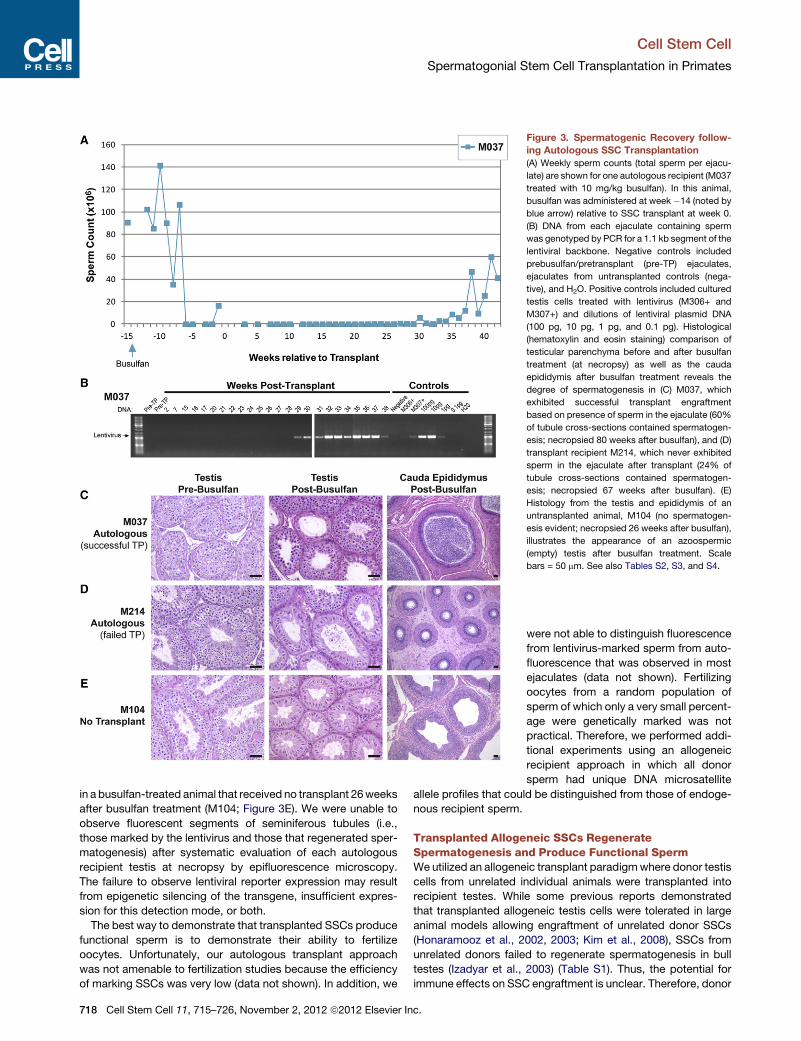
Donor signal was not detectable in ejaculated sperm from
M212 likely due to inflammation in the left epididymis evident
in the posttransplant period and confirmed at necropsy, prevent-
ing donor sperm transit to the ejaculate (data not shown). A
second allogeneic recipient, M027, produced ejaculated sperm
exhibiting donor signal (donor M092; 187 bp allele at locus
D3S1768; 263 bp allele at locus D17S1300) for more than
17 months (50 total samples) (Figure 4E–4G). Thus, these data
show that transplanted allogeneic SSCs produce sperm in
recipient testes. To quantify the degree of donor sperm produc-
tion as a function of time, we identified single nucleotide poly-
morphisms (SNPs) that distinguish donor sperm from recipient
sperm essentially as described previously (Alizadeh et al.,
2002; Kim et al., 2008). We screened 23 SNPs reported in the
Monkey SNP database at Oregon National Primate Research
Center (Khouangsathiene et al., 2008), but none distinguished
donor and recipient and/or were suitable for qPCR. How-
ever, while screening one reported SNP (rs4543622) within the
class II major histocompatibility complex transactivator (CIITA)
locus, we identified a previously unreported SNP for which the
recipient (M027) was homozygous for one allele (G) and the
donor (M092) heterozygous both alleles (A/G). Standard
curves for the relative abundance of each allele as previously
described were used to determine the percent of donor
chimerism in DNA isolated from recipient M027 following
monthly semen samples collected between 3 and 17 months
after transplant (Figure 4H). We observed a consistent level of
donor (M092) chimerism (ranging from 1.7 to 17.2%) in M027
sperm samples for the duration of the 14 months analyzed
(Figure 4H).
To assess function of donor (M092) sperm, ejaculated sperm
from recipient M027 (collected 30 weeks after transplant) were
used to fertilize rhesus oocytes by intracytoplasmic sperm injec-
tion (ICSI) (Hewitson et al., 1999; Mitalipov et al., 2006). Of 85
oocytes injected, 81 (95%) were fertilized (formed male and
female pronuclei) and subsequently cleaved (Figure 5 and Table
S5). Upon in vitro culture, 23% of embryos reached the blasto-
cyst stage with normal morphology (Table S5). To determine
sire by microsatellite DNA fingerprinting, all blastocysts and
arrested embryos were individually harvested and used for
Cell
whole-genome DNA amplification. Genotyping was done for
the gender marker AME to determine sex of embryo and for eight
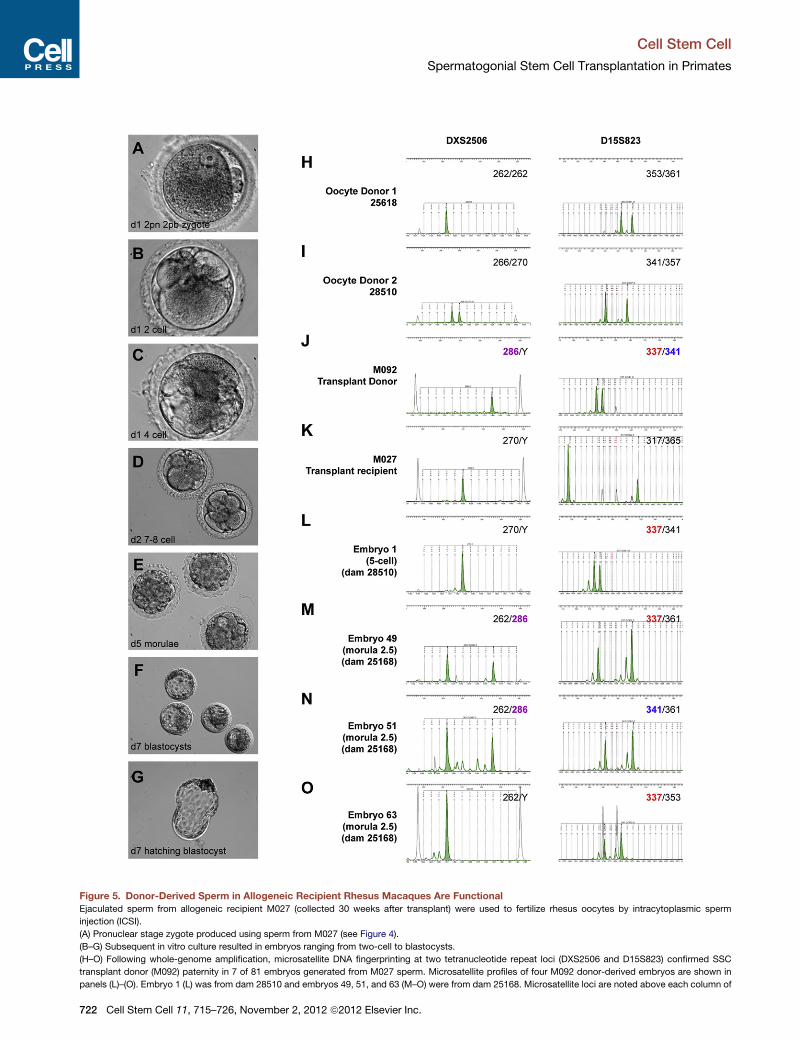
microsatellite loci, two of which (DXS2506 and D15S823) defin-
itively discriminate the genotype of the SSC donor (M092) from
the transplant recipient (M027) and oocyte donors (Figure 5
and Table S6). In this genotyping paradigm, the 286 bp allele
at the X-linked locus DXS2506 and the 337 bp allele at locus
D15S823 were both unique to M092 and their presence in an
embryo could only arise from M092 paternal contribution (Fig-
ure 5 and Table S6). Of the 81 embryos genotyped, 7 exhibited
definitive donor (M092) sire, 3 of which advanced to the morula
stage of preimplantation development (Figure 5 and Table S6).
Since DXS2506 is an X-linked marker, male (XY) embryos,
including three XY M092-sired embryos (embryos 1, 8 and 63;
Figures 5L and 5O and Table S6), displayed only the maternal
allele at this locus. M092 donor paternal contribution in these
embryos was confirmed by the presence of the 337 bp allele at
locus D15S823. These results indicate that sperm generated
from transplanted primate SSCs are competent for fertilization
and preimplantation embryo development.
DISCUSSION
Adult stem cell transplantation for homologous tissue regenera-
tion was first described for primates in the 1950s when bone
marrow stem cells were used to reconstitute the hematopoietic
systems of monkeys and humans treated with chemotherapy
or radiation (Crouch and Overman, 1957; Thomas et al.,
1957). Large animals, primarily the dog andmonkey, were instru-
mental for establishing the safety, feasibility, and range of
applications for bone marrow transplantation. Today, approxi-
mately 50,000 bone marrow or HSC transplant procedures are
performed worldwide each year for diseases ranging from
cancer to thalassemia, sickle cell anemia, and autoimmune
and immune-deficiency disorders (Appelbaum, 2007; Powell
et al., 2009).
Like hematopoiesis, spermatogenesis is a highly productive
stem-cell-based system that produces millions of sperm per
gram of tissue each day (Sharpe, 1994). This productivity is
possible because a relatively small stem cell pool generates
progeny that undergo several rounds of transit-amplifying
divisions before producing the terminally differentiated sperm
(Potten, 1992). Two sequelae of highly productive stem-cell-
based systems are (1) that they can become targets of chemo-
therapy or radiation treatments that damage rapidly dividing
cells (Potten, 1995; Meistrich, 1993; Mauch et al., 1995) and (2)
that transplantation of a small number of stem cells is adequate
to functionally reconstitute the dependent systems (e.g., hema-
topoiesis and spermatogenesis) (Potten et al., 1979; Potten,
1992; Osawa et al., 1996; Ogawa et al., 2000; Shinohara et al.,
2001; Copelan, 2006). Here we demonstrate the feasibility of
SSC transplantation in a nonhuman primatemodel that is infertile
due to alkylating chemotherapy (busulfan) and suggest that this
technique has application for restoring the fertility of cancer
survivors or bone marrow transplant recipients.
SSC transplantation has now been reported in mice, rats,
monkeys, goats, bulls, pigs, sheep, and dogs (Brinster and
Avarbock, 1994; Brinster and Zimmermann, 1994; Ogawa
et al., 1999; Schlatt et al., 2002; Honaramooz et al., 2003; Izadyar
Stem Cell 11, 715–726, November 2, 2012 ª2012 Elsevier Inc. 719
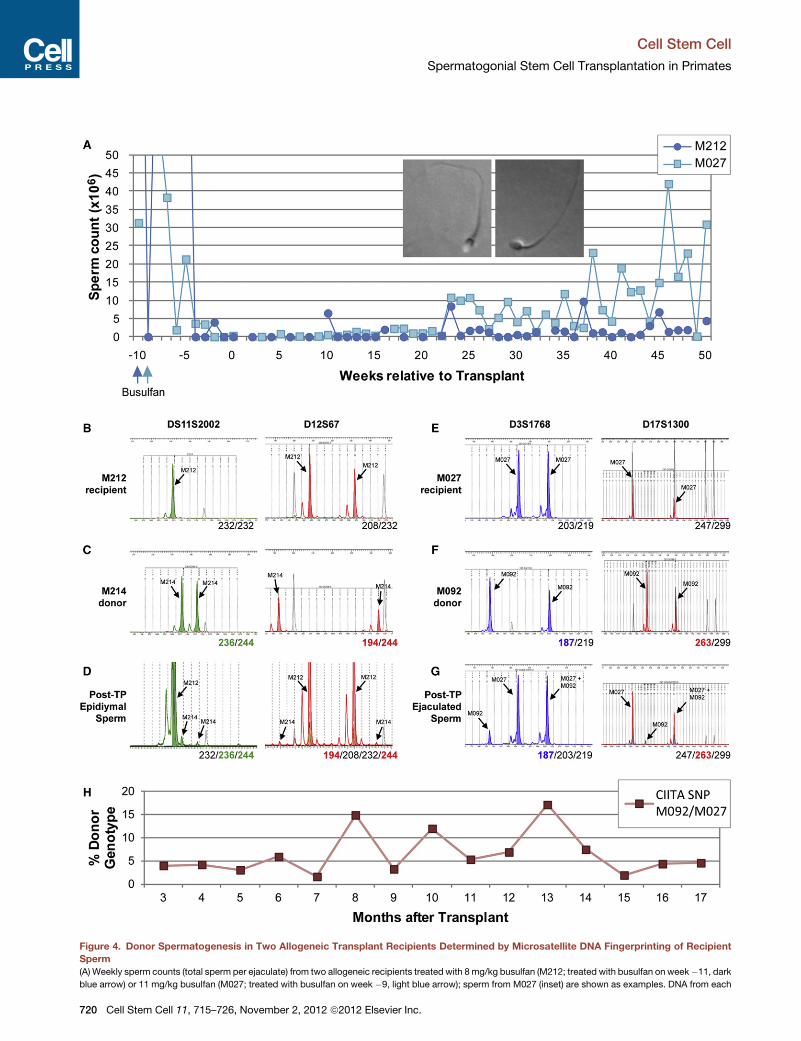
Figure 4. Donor Spermatogenesis in Two Allogeneic Transplant Recipients Determined by Microsatellite DNA Fingerprinting of Recipient
Sperm
(A) Weekly sperm counts (total sperm per ejaculate) from two allogeneic recipients treated with 8mg/kg busulfan (M212; treated with busulfan on week�11, dark
blue arrow) or 11 mg/kg busulfan (M027; treated with busulfan on week �9, light blue arrow); sperm from M027 (inset) are shown as examples. DNA from each
Cell Stem Cell
Spermatogonial Stem Cell Transplantation in Primates
720 Cell Stem Cell 11, 715–726, November 2, 2012 ª2012 Elsevier Inc.
Cell Stem Cell
Spermatogonial Stem Cell Transplantation in Primates
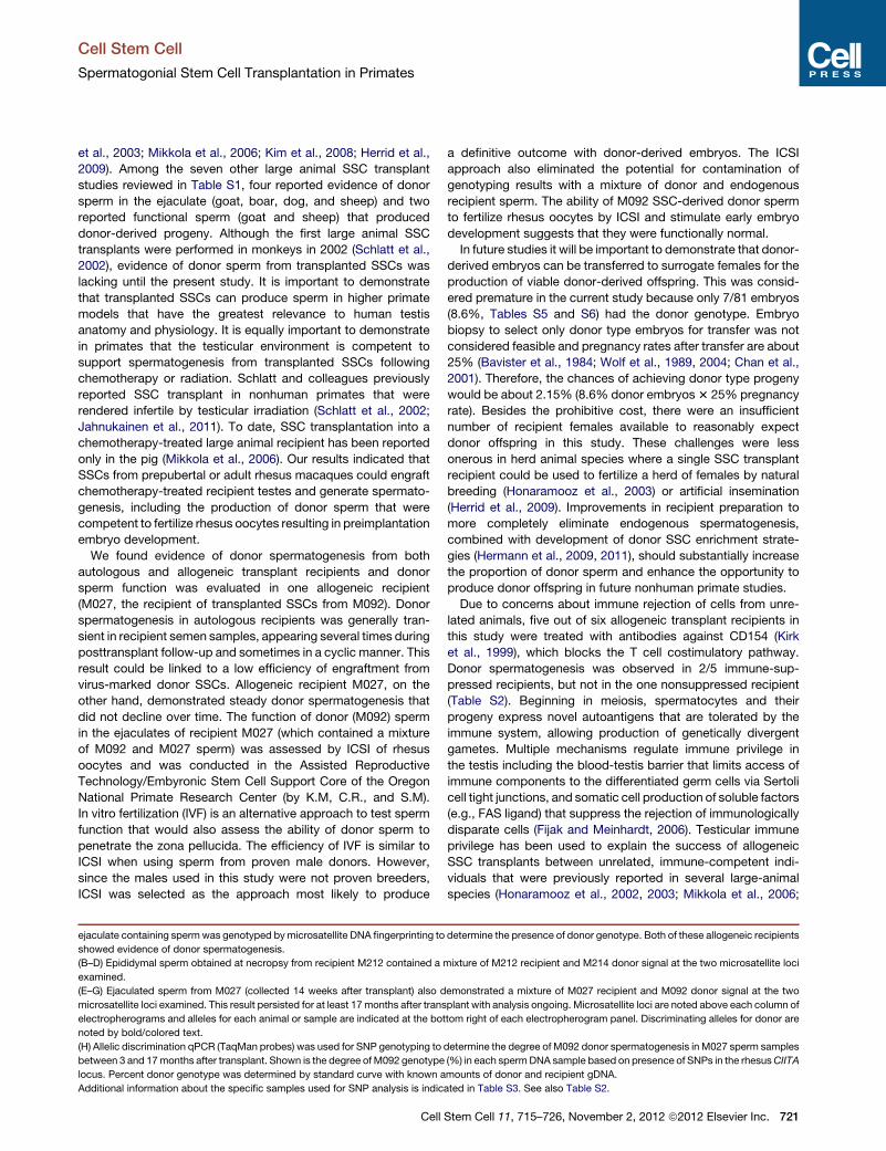
et al., 2003; Mikkola et al., 2006; Kim et al., 2008; Herrid et al.,
2009). Among the seven other large animal SSC transplant
studies reviewed in Table S1, four reported evidence of donor
sperm in the ejaculate (goat, boar, dog, and sheep) and two
reported functional sperm (goat and sheep) that produced
donor-derived progeny. Although the first large animal SSC
transplants were performed in monkeys in 2002 (Schlatt et al.,
2002), evidence of donor sperm from transplanted SSCs was
lacking until the present study. It is important to demonstrate
that transplanted SSCs can produce sperm in higher primate
models that have the greatest relevance to human testis
anatomy and physiology. It is equally important to demonstrate
in primates that the testicular environment is competent to
support spermatogenesis from transplanted SSCs following
chemotherapy or radiation. Schlatt and colleagues previously
reported SSC transplant in nonhuman primates that were
rendered infertile by testicular irradiation (Schlatt et al., 2002;
Jahnukainen et al., 2011). To date, SSC transplantation into a
chemotherapy-treated large animal recipient has been reported
only in the pig (Mikkola et al., 2006). Our results indicated that
SSCs from prepubertal or adult rhesus macaques could engraft
chemotherapy-treated recipient testes and generate spermato-
genesis, including the production of donor sperm that were
competent to fertilize rhesus oocytes resulting in preimplantation
embryo development.
We found evidence of donor spermatogenesis from both
autologous and allogeneic transplant recipients and donor
sperm function was evaluated in one allogeneic recipient
(M027, the recipient of transplanted SSCs from M092). Donor
spermatogenesis in autologous recipients was generally tran-
sient in recipient semen samples, appearing several times during
posttransplant follow-up and sometimes in a cyclic manner. This
result could be linked to a low efficiency of engraftment from
virus-marked donor SSCs. Allogeneic recipient M027, on the
other hand, demonstrated steady donor spermatogenesis that
did not decline over time. The function of donor (M092) sperm
in the ejaculates of recipient M027 (which contained a mixture
of M092 and M027 sperm) was assessed by ICSI of rhesus
oocytes and was conducted in the Assisted Reproductive
Technology/Embyronic Stem Cell Support Core of the Oregon
National Primate Research Center (by K.M, C.R., and S.M).
In vitro fertilization (IVF) is an alternative approach to test sperm
function that would also assess the ability of donor sperm to
penetrate the zona pellucida. The efficiency of IVF is similar to
ICSI when using sperm from proven male donors. However,
since the males used in this study were not proven breeders,
ICSI was selected as the approach most likely to produce
ejaculate containing spermwas genotyped bymicrosatellite DNA fingerprinting to
showed evidence of donor spermatogenesis.
(B–D) Epididymal sperm obtained at necropsy from recipient M212 contained a
examined.
(E–G) Ejaculated sperm from M027 (collected 14 weeks after transplant) also d
microsatellite loci examined. This result persisted for at least 17months after trans
electropherograms and alleles for each animal or sample are indicated at the bot
noted by bold/colored text.
(H) Allelic discrimination qPCR (TaqMan probes) was used for SNP genotyping to
between 3 and 17months after transplant. Shown is the degree of M092 genotype
locus. Percent donor genotype was determined by standard curve with known a
Additional information about the specific samples used for SNP analysis is indic
Cell
a definitive outcome with donor-derived embryos. The ICSI
approach also eliminated the potential for contamination of
genotyping results with a mixture of donor and endogenous
recipient sperm. The ability of M092 SSC-derived donor sperm
to fertilize rhesus oocytes by ICSI and stimulate early embryo
development suggests that they were functionally normal.
In future studies it will be important to demonstrate that donor-
derived embryos can be transferred to surrogate females for the
production of viable donor-derived offspring. This was consid-
ered premature in the current study because only 7/81 embryos
(8.6%, Tables S5 and S6) had the donor genotype. Embryo
biopsy to select only donor type embryos for transfer was not
considered feasible and pregnancy rates after transfer are about
25% (Bavister et al., 1984; Wolf et al., 1989, 2004; Chan et al.,
2001). Therefore, the chances of achieving donor type progeny
would be about 2.15% (8.6% donor embryos3 25% pregnancy
rate). Besides the prohibitive cost, there were an insufficient
number of recipient females available to reasonably expect
donor offspring in this study. These challenges were less
onerous in herd animal species where a single SSC transplant
recipient could be used to fertilize a herd of females by natural
breeding (Honaramooz et al., 2003) or artificial insemination
(Herrid et al., 2009). Improvements in recipient preparation to
more completely eliminate endogenous spermatogenesis,
combined with development of donor SSC enrichment strate-
gies (Hermann et al., 2009, 2011), should substantially increase
the proportion of donor sperm and enhance the opportunity to
produce donor offspring in future nonhuman primate studies.
Due to concerns about immune rejection of cells from unre-
lated animals, five out of six allogeneic transplant recipients in
this study were treated with antibodies against CD154 (Kirk
et al., 1999), which blocks the T cell costimulatory pathway.
Donor spermatogenesis was observed in 2/5 immune-sup-
pressed recipients, but not in the one nonsuppressed recipient
(Table S2). Beginning in meiosis, spermatocytes and their
progeny express novel autoantigens that are tolerated by the
immune system, allowing production of genetically divergent
gametes. Multiple mechanisms regulate immune privilege in
the testis including the blood-testis barrier that limits access of
immune components to the differentiated germ cells via Sertoli
cell tight junctions, and somatic cell production of soluble factors
(e.g., FAS ligand) that suppress the rejection of immunologically
disparate cells (Fijak and Meinhardt, 2006). Testicular immune
privilege has been used to explain the success of allogeneic
SSC transplants between unrelated, immune-competent indi-
viduals that were previously reported in several large-animal
species (Honaramooz et al., 2002, 2003; Mikkola et al., 2006;
determine the presence of donor genotype. Both of these allogeneic recipients
mixture of M212 recipient and M214 donor signal at the two microsatellite loci
emonstrated a mixture of M027 recipient and M092 donor signal at the two
plant with analysis ongoing. Microsatellite loci are noted above each column of
tom right of each electropherogram panel. Discriminating alleles for donor are
determine the degree of M092 donor spermatogenesis in M027 sperm samples
(%) in each spermDNA sample based on presence of SNPs in the rhesusCIITA
mounts of donor and recipient gDNA.
ated in Table S3. See also Table S2.
Stem Cell 11, 715–726, November 2, 2012 ª2012 Elsevier Inc. 721
Figure 5. Donor-Derived Sperm in Allogeneic Recipient Rhesus Macaques Are Functional
Ejaculated sperm from allogeneic recipient M027 (collected 30 weeks after transplant) were used to fertilize rhesus oocytes by intracytoplasmic sperm
injection (ICSI).
(A) Pronuclear stage zygote produced using sperm from M027 (see Figure 4).
(B–G) Subsequent in vitro culture resulted in embryos ranging from two-cell to blastocysts.
(H–O) Following whole-genome amplification, microsatellite DNA fingerprinting at two tetranucleotide repeat loci (DXS2506 and D15S823) confirmed SSC
transplant donor (M092) paternity in 7 of 81 embryos generated from M027 sperm. Microsatellite profiles of four M092 donor-derived embryos are shown in
panels (L)–(O). Embryo 1 (L) was from dam 28510 and embryos 49, 51, and 63 (M–O) were from dam 25168. Microsatellite loci are noted above each column of
Cell Stem Cell
Spermatogonial Stem Cell Transplantation in Primates
722 Cell Stem Cell 11, 715–726, November 2, 2012 ª2012 Elsevier Inc.
Cell Stem Cell
Spermatogonial Stem Cell Transplantation in Primates
Kim et al., 2008). Although animal numbers in this study were not
sufficient to demonstrate that immune suppression was re-
quired, our data clearly indicated that cells from unrelated donor
animals were tolerated in immune-suppressed nonhuman
primates.
Several promising techniques are in the research pipeline (i.e.,
SSC transplantation, testicular tissue grafting or xenografting,
and in vitro development of gametes) that may allow patients
receiving gonadotoxic therapies to preserve their future fertility
(Brinster, 2007; Rodriguez-Sosa and Dobrinski, 2009; Sato
et al., 2011). SSC transplantation has the unique potential to
regenerate spermatogenesis in the autologous environment of
the seminiferous tubules, enabling the recipient male to father
his own genetic children, possibly through normal coitus. As
with hematopoiesis, large animal models that are relevant to
human anatomy and physiology will be important for translating
the SSC transplantation technique to the human fertility clinic.
Considering the successful regeneration of spermatogenesis in
the nonhuman primate model reported here and the fact that
patients are already preserving testicular tissue and/or cells,
clinical translation of the SSC transplantation technique appears
imminent. Responsible development of the technology in a
clinically relevant nonhuman primate system will help to address
issues of safety and feasibility. As with hematopoiesis, the
clinical significance and breadth of applications for SSC trans-
plantation will ultimately be established in human patients.
EXPERIMENTAL PROCEDURES
Animals
All experiments utilizing animals were approved by the responsible Institutional
Animal Care and Use Committees of Magee-Womens Research Institute and
the University of Pittsburgh (Assurance #A3654-01) and the Oregon National
Primate Research Center, Oregon Health and Sciences University (Assurance
#A3304-01) and were performed in accordance with the National Institutes of
Health Guide for the Care and Use of Laboratory Animals.
Preparation of Donor Rhesus Macaque Testis Cell Suspensions
Testis tissue was collected from rhesus macaques by hemicastration or
subcapsular biopsy. For biopsies, less than 30% of the testicular parenchyma
was removed (3.8g–8.7g) through a transverse incision in the tunica albuginea
on the lateral side of the right testis. In one case (M036), the biopsied testis was
later removed by hemicastration due to formation of an abscess. Cells were
recovered from testicular parenchyma using a two-step enzymatic digestion
procedure, cryopreserved, and stored in liquid nitrogen, as described
(Hermann et al., 2007, 2009).
Busulfan Treatment
Recipient animals were treated with the alkylating chemotherapeutic agent
busulfan (Busulfex IV; PDL BioPharma, Fremont, CA), at doses of 8, 10, 11,
or 12mg/kg (Table S2). Busulfex was diluted in physiological saline and admin-
istered intravenously at 0.6 mg/ml over 10–20 min.
PBSC Transplants
Autologous transplants of PBSCswere employed to restore the hematopoietic
system after busulfan treatment. Briefly, PBSCs were mobilized with six, daily
subcutaneous injections with the cytokines G-CSF (10 mg/kg/day, Neupogen;
Amgen; Thousand Oaks, CA) and SCF (200 mg/kg/day; Amgen) or G-CSF
electropherograms and alleles for each animal or sample are indicated in the up
noted by bold/colored text. In cases where embryos were male (i.e., XY; panels L
simply noted by Y. In both cases M092 paternal origin could be confirmed by th
See also Tables S5 and S6.
Cell
alone (20 mg/kg/day), essentially as described (Figure 2) (Donahue et al.,
2005). PBSC collections were performed by apheresis using either a Spectra
or Spectra Optia apheresis device (Caridian BCT; Lakewood, CO). Twenty-
four hours after apheresis, animals were treated with busulfan, and 18 hr
later animals received autologous PBSC transfusions (Figure 2). Two days
later, animals received one subcutaneous injection of long-acting G-CSF
(300 mg/kg; Neulasta, Amgen). Additional details are available in the Supple-
mental Experimental Procedures.
Histology
Portions of testicular parenchyma and epididymis collected above and
at necropsy were fixed with Bouin’s solution (Accustain; Sigma-Aldrich,
St. Louis, MO), paraffin embedded, sectioned (5 mm), and stained with hema-
toxylin and eosin.
SSC Transplant
Spermatogonial stem cell transplants were performed 9–15 weeks after
busulfan treatment (autologous: unilateral; allogeneic: bilateral). In biopsied
animals, autologous transplants were performed into the contralateral testis.
Cryopreserved donor cells were recovered for transplant from storage in liquid
nitrogen, as described (Hermann et al., 2007, 2009). In some cases, donor cells
were enriched for spermatogonia, including SSCs, on a 24% Percoll cushion
(GE Healthcare Life Sciences, Piscataway, NJ) prior to transplant (see Fig-
ure S1 and Table S2). Cells were then suspended at approximately 100 3
106 cells/ml in MEMalpha medium (Invitrogen) containing 10% FBS, 20%
trypan blue, 20% Optison (ultrasound contrast agent; GE Healthcare, Wauke-
sha, WI) and 0.7 mg/ml DNase I in a total volume of %1ml, depending on
recipient testis size and available cells. SSC transplants were performed using
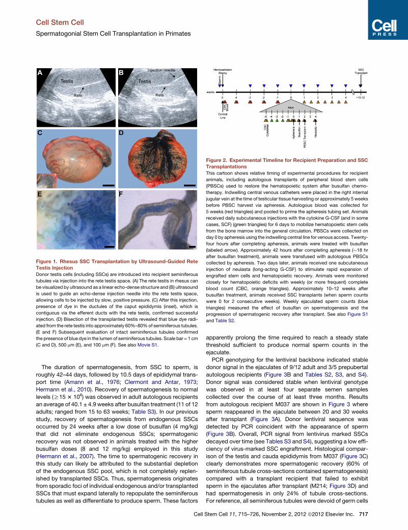
ultrasound-guided rete testis injections (Figure 1 and Movie S1). For this
purpose, a 13 MHz linear superficial probe was used to visualize the rete testis
space on a MicroMaxx ultrasound machine (Sonosite, Bothell, WA) and guide
a 25G 2’’ spinal needle into the rete testis. Cells were injected under slow
constant pressure and chased with saline.
Lentiviral Treatment of Donor Testis Cells
For autologous transplants, donor cells were treated with lentiviral vectors
modified from the FUGWconstruct originally described by Lois and coworkers
(Lois et al., 2002). Details of virus constructs and viral treatments are available
in the Supplemental Experimental Procedures.
Immune Suppression
Five of six allogeneic transplant recipients were treated with human/mouse