Spider Web Glue: Two Proteins Expressed from Opposite Strands of the Same DNA Sequence Omer Choresh,* Battuya Bayarmagnai, and Randolph V. Lewis Department of Molecular Biology, University of Wyoming, Laramie, Wyoming 82071-3944 Received June 15, 2009; Revised Manuscript Received August 14, 2009 The various silks that make up the web of the orb web spiders have been studied extensively. However, success in prey capture depends as much on the web glue as on the fibers. Spider silk glue, which is considered one of the strongest and most effective biological glues, is an aqueous solution secreted from the orb weaving spider’s aggregate glands and coats the spiral prey capturing threads of their webs. Studies identified the major component of the glue as microscopic nodules made of a glycoprotein. This study describes two newly discovered proteins that form the glue-glycoprotein of the golden orb weaving spider Nephila claVipes. Our results demonstrate that both proteins contain unique 110 amino acid repetitive domains that are encoded by opposite strands of the same DNA sequence. Thus, the genome of the spider encodes two distinct yet functionally related genes by using both strands of an identical DNA sequence. Moreover, the closest match for the nonrepetitive region of one of the proteins is chitin binding proteins. The web glue appears to have evolved a substantial level of sophistication matching that of the spider silk fibers. Introduction Spider silk has attracted scientists to study its unique mechanical properties and its potential to provide new biobased materials for numerous applications ranging from protective clothing to medical products. 1 However, one of the most intriguing spider biomaterials, yet one of the least explored, is the aqueous glue that coats the sticky-spiral threads of orb weaving spiders to retain prey in the web. It is considered one of the strongest and most effective biological glues. 2–5 The aqueous coat is secreted from the orb weaving spider’s aggregate glands, and the gland’s contents have been studied by several research groups. 3–5 Chemical analysis of this complex aqueous solution shows relatively high concentrations of water-soluble organic compounds related to neurotransmitters, free amino acids, small peptides, low concentrations of various inorganic salts, and glycoproteins. It has been suggested that the contents of this solution generate hygroscopic forces that may contribute to the thread stickiness, however, studies identified the actual glue of ecribellate spiders as microscopic nodules made of a glycoprotein. 5 To date, almost nothing is known about the molecular structure and function of this glycoprotein. In this study, we describe two newly discovered genes that encode two subunits of the glue-glycoprotein of the golden orb weaving spider Nephila claVipes. We show that both putative glycoprotein subunits have unique repetitive domains that are individually expressed from opposite strands of an identical DNA sequence. Cloning of these glycoprotein genes may enable large-scale production, studying their biochemical traits and developing new biobased glue for numerous purposes. Materials and Methods cDNA Construction and Gene Cloning. Adult females of the golden orb weaving spider Nephila claVipes were collected in Florida and dissected to isolate their aggregate glands. Glands were immediately frozen in liquid nitrogen and kept in a -80 °C freezer until use. Intact mRNA (mRNA) was extracted from those glands, using Tri Reagent (#TR118, MRC, OH, U.S.A.) and oligo-T Dynabeads (#610.05, Dynal Biotech ASA, Oslo, Norway). This mRNA was promptly used to construct a directional cDNA (cDNA) library, using ZAP Express cDNA synthesis kit and ZAP Express cDNA Gigapack III Gold Cloning kit (#200403, #200451, Stratagene, CA). The cDNA library was mass- excised to produce pBK-CMV vectors that were transformed into Escherichia coli host strain cells (as described by manufacturer, Stratagene, CA). The library was then screened by colony lifting. Plasmids were extracted from randomly selected colonies (QIAprep, 27104, Qiagen, CA) and inserts were sequenced. One clone was isolated at relatively high frequency. This clone showed two possible reading frames on opposite DNA strands that translated into two highly repetitive and potentially glycosylated proteins. Oligonucleotides were individually designed toward the C-terminus of each potential gene (reading frame), based on this clone. The oligonucleotides are 5′CA- GAAGAACCCGAAACACCGAGTCCAGAAAC3′ toward the 3 prime end of asg1 and 5′GTTTCTGGACTCGGTGTTTCGGGTTCTTCTG3′ toward the 3′ end of asg2. The oligonucleotides were used as primers to clone the C-terminus of each putative gene by Rapid Amplification of cDNA Ends Polymerase Chain Reaction (RACE-PCR), utilizing the cDNA library as a DNA template and a vector primer adjacent to the C-terminus of the inserted clone. PCR was carried out in a thermal cycler (MyCycler, Bio-Rad). The reactions were performed in a volume of 50 µL of PCR solution containing 2 µL of template, 10 pMol of each primer, and 45 µL of Platinum PCR SuperMix (#11306, Invitrogen, CA). The thermal cycler was programmed to 95 °C (5 min) followed by 35 cycles of 95 °C (15 s), 55 °C (45 s), and 72 °C (5 min), followed by 72 °C (30 min). RACE-PCR products were purified (QIAquick Gel Extraction kit, 28704, Qiagen, CA) and served as DNA templates in a further nested-PCR, utilizing the same primers and same conditions used in RACE-PCR. This strategy helped to amplify gene specific fragments and to avoid low concentration undetectable DNA fragments. Certain nested-PCR products were subcloned and sequenced. The resulting products were visualized on 1% TAE-agarose gel, cleaned (QIAquick Gel Extraction kit, 28704, Qiagen, CA), cloned into a pCR 4-TOPO vector (TOPO TA Cloning kit for sequencing, #K4530-20, Invitrogen, CA), and sequenced. New oligonucleotides were generated toward the N-terminus of each putative gene, based on the newly * To whom correspondence should be addressed. Tel.: 307-766-5534. Fax: 307-766-5098. E-mail: [email protected]. Biomacromolecules 2009, 10, 2852–2856 2852 10.1021/bm900681w CCC: $40.75 2009 American Chemical Society Published on Web 09/04/2009
Transcript
Spider Web Glue: Two Proteins Expressed from OppositeStrands of the Same DNA Sequence
Omer Choresh,* Battuya Bayarmagnai, and Randolph V. Lewis
Department of Molecular Biology, University of Wyoming, Laramie, Wyoming 82071-3944
Received June 15, 2009; Revised Manuscript Received August 14, 2009
The various silks that make up the web of the orb web spiders have been studied extensively. However, successin prey capture depends as much on the web glue as on the fibers. Spider silk glue, which is considered one ofthe strongest and most effective biological glues, is an aqueous solution secreted from the orb weaving spider’saggregate glands and coats the spiral prey capturing threads of their webs. Studies identified the major componentof the glue as microscopic nodules made of a glycoprotein. This study describes two newly discovered proteinsthat form the glue-glycoprotein of the golden orb weaving spider Nephila claVipes. Our results demonstrate thatboth proteins contain unique 110 amino acid repetitive domains that are encoded by opposite strands of the sameDNA sequence. Thus, the genome of the spider encodes two distinct yet functionally related genes by using bothstrands of an identical DNA sequence. Moreover, the closest match for the nonrepetitive region of one of theproteins is chitin binding proteins. The web glue appears to have evolved a substantial level of sophisticationmatching that of the spider silk fibers.
Introduction
Spider silk has attracted scientists to study its uniquemechanical properties and its potential to provide new biobasedmaterials for numerous applications ranging from protectiveclothing to medical products.1 However, one of the mostintriguing spider biomaterials, yet one of the least explored, isthe aqueous glue that coats the sticky-spiral threads of orbweaving spiders to retain prey in the web. It is considered oneof the strongest and most effective biological glues.2–5 Theaqueous coat is secreted from the orb weaving spider’s aggregateglands, and the gland’s contents have been studied by severalresearch groups.3–5 Chemical analysis of this complex aqueoussolution shows relatively high concentrations of water-solubleorganic compounds related to neurotransmitters, free aminoacids, small peptides, low concentrations of various inorganicsalts, and glycoproteins. It has been suggested that the contentsof this solution generate hygroscopic forces that may contributeto the thread stickiness, however, studies identified the actualglue of ecribellate spiders as microscopic nodules made of aglycoprotein.5 To date, almost nothing is known about themolecular structure and function of this glycoprotein.
In this study, we describe two newly discovered genes thatencode two subunits of the glue-glycoprotein of the golden orbweaving spider Nephila claVipes. We show that both putativeglycoprotein subunits have unique repetitive domains that areindividually expressed from opposite strands of an identicalDNA sequence. Cloning of these glycoprotein genes may enablelarge-scale production, studying their biochemical traits anddeveloping new biobased glue for numerous purposes.
Materials and Methods
cDNA Construction and Gene Cloning. Adult females of thegolden orb weaving spider Nephila claVipes were collected in Floridaand dissected to isolate their aggregate glands. Glands were immediately
frozen in liquid nitrogen and kept in a -80 °C freezer until use. IntactmRNA (mRNA) was extracted from those glands, using Tri Reagent(#TR118, MRC, OH, U.S.A.) and oligo-T Dynabeads (#610.05, DynalBiotech ASA, Oslo, Norway). This mRNA was promptly used toconstruct a directional cDNA (cDNA) library, using ZAP ExpresscDNA synthesis kit and ZAP Express cDNA Gigapack III Gold Cloningkit (#200403, #200451, Stratagene, CA). The cDNA library was mass-excised to produce pBK-CMV vectors that were transformed intoEscherichia coli host strain cells (as described by manufacturer,Stratagene, CA). The library was then screened by colony lifting.Plasmids were extracted from randomly selected colonies (QIAprep,27104, Qiagen, CA) and inserts were sequenced. One clone was isolatedat relatively high frequency. This clone showed two possible readingframes on opposite DNA strands that translated into two highlyrepetitive and potentially glycosylated proteins. Oligonucleotides wereindividually designed toward the C-terminus of each potential gene(reading frame), based on this clone. The oligonucleotides are 5′CA-GAAGAACCCGAAACACCGAGTCCAGAAAC3′ toward the 3 primeend of asg1 and 5′GTTTCTGGACTCGGTGTTTCGGGTTCTTCTG3′toward the 3′ end of asg2. The oligonucleotides were used as primersto clone the C-terminus of each putative gene by Rapid Amplificationof cDNA Ends Polymerase Chain Reaction (RACE-PCR), utilizing thecDNA library as a DNA template and a vector primer adjacent to theC-terminus of the inserted clone. PCR was carried out in a thermalcycler (MyCycler, Bio-Rad). The reactions were performed in a volumeof 50 µL of PCR solution containing 2 µL of template, 10 pMol ofeach primer, and 45 µL of Platinum PCR SuperMix (#11306,Invitrogen, CA). The thermal cycler was programmed to 95 °C (5 min)followed by 35 cycles of 95 °C (15 s), 55 °C (45 s), and 72 °C (5min), followed by 72 °C (30 min). RACE-PCR products were purified(QIAquick Gel Extraction kit, 28704, Qiagen, CA) and served as DNAtemplates in a further nested-PCR, utilizing the same primers and sameconditions used in RACE-PCR. This strategy helped to amplify genespecific fragments and to avoid low concentration undetectable DNAfragments. Certain nested-PCR products were subcloned and sequenced.The resulting products were visualized on 1% TAE-agarose gel, cleaned(QIAquick Gel Extraction kit, 28704, Qiagen, CA), cloned into a pCR4-TOPO vector (TOPO TA Cloning kit for sequencing, #K4530-20,Invitrogen, CA), and sequenced. New oligonucleotides were generatedtoward the N-terminus of each putative gene, based on the newly
* To whom correspondence should be addressed. Tel.: 307-766-5534.Fax: 307-766-5098. E-mail: [email protected].
Biomacromolecules 2009, 10, 2852–28562852
10.1021/bm900681w CCC: $40.75 2009 American Chemical SocietyPublished on Web 09/04/2009
discovered C-terminus sequences. Those were used as primers forRACE-PCR and nested PCR as described above. One oligonucleotide,5′GGTGTAGTTGTTGGTTCTTCCG3′, was designed to clone the 5′end of asg1. Two oligonucleotides, 5′CCACTAAACCTTCCAT-TAAG3′ and 5′CCTAACATCTTCCGGTAATTCATCG3′, were de-signed to clone the 5′ end of asg2. All samples were sequenced at theUniversity of Wyoming sequencing facility. Sequence analysis wascarried out using NCBI-BLAST (http://blast.ncbi.nlm.nih.gov/Blast.cgi)and EXPASY tools (http://ca.expasy.org/tools/).
Northern Blots. The oligonucleotides that were designed towardthe C-terminus of each gene (complementary to asg1 and complemen-tary to asg2) were DIG-labeled (DIG oligonucleotide 3′-end labelingkit, second generation, #03353575910, Roche Applied Science, IN,U.S.A.) and used as probes in Northern blot analysis, utilizing totalRNA and mRNA that were purified from different spider silk glandsand tissues of N. claVipes (using intact glands and RNA extractionmethods as described above). Northern blots were carried out accordingto DIG manufacturer protocols and reagents (Roche Applied Science,IN, U.S.A.). Specific reagents we used include: 2% agarose gel in 1×MOPS buffer containing 2% formaldehyde, positively charged nylonmembranes (#11209272001), hybridization solution “DIG Easy Hyb”(#1603558), DIG wash and block buffer set (#11585762001), Anti-Digoxigenin-AP Fab fragments (#11093274910), and CDP-Star(#11685627001).
Biochemical Analysis of Glycoprotein. Uncontaminated fresh orbwebs were obtained by maintaining spiders individually in cages forup to 8 months. Webs were collected daily on sterile glass rods andstored at -20 °C until analyzed. Each extraction included 60-70 webs.Collected rods coated with webs were ground by mortar and pestleand incubated in a minimal volume of extraction buffer (6 M guanidineHCl, 0.7 M �-mercaptoethanol, 0.2 M Na2HPO4) with protease inhibitorcocktail (#P2714,, Sigma, MO, U.S.A.) for 24 h at 4 °C with gentleagitation. Extracts were then centrifuged for 30 min at 4000 rpm (J-6M, Beckman Coulter, Germany). Supernatants were recentrifuged for5 min at full speed (Microfuge18, Beckman Coulter, Germany) todiscard insoluble web contents. Protein extraction samples weredissolved in sample buffer (without �-mercaptoethanol as it is alreadypresent in the extraction buffer), boiled for 10 min, and then resolvedon a 12% mini-SDS-PAGE. Approximately 200 µL was loaded perwell in a concentration of 0.15 µg of soluble protein extract/µL (30µg/lane). Gels were run for 5 h at 60 V in Tris-glycine buffer with0.01 M of sodium thioglycolate. The gels were stained with glycopro-tein-specific staining techniques (Pro-Q Emerald 300, Molecular Probes,OR, U.S.A.). Gels were also stained using total protein stains such asSYPRORuby (Molecular Probes, OR, U.S.A.) to ensure the purity ofthe glycoprotein band. Positive bands (using glycoprotein-specific stain)were cut out from the gel and homogenized in 20% acetonitrile. Sampleswere then vortexed for 15 min, and acetonitrile was added to 50% ofthe solution after vortexing. Samples were centrifuged for 5 min andthe supernatants were collected. The gel pellet was subjected to 3-4cycles of extraction as described above (supernatants were combined).Gel purified glycoprotein solution was dialyzed against double distilledwater at 4 °C for 18 h, then lyophilized, resuspended in 0.75% TritonX-100, 0.4% �-mercaptoethanol, and 0.1% SDS, and homogenizedultrasonically using a frequency of 20 kHz/sec for 5 min (Sonicator3000, Misonix, NY) on ice. Samples were boiled for 15 min beforebeing subjected to deglycosylation.
We have developed protocols for the deglycosylation of purifiedglycoprotein solutions, adapted to glycoprotein from spider silk.Glycoprotein solutions, purified from SDS-PAGE bands, were usedfor deglycosylation. Protocols include enzymatic analyses based onE-DEGLY and PP0200 deglycosylation kits (Sigma, MO, U.S.A.) thatcontain the N-linked glycoprotein cleavage enzyme PNGase F and theO-linked glycoprotein cleavage enzymes endo-O-glycosidase, R-2(3,6,8.9)-neuraminidase (Sialidase A), �-1,4-galactosidase, and �-N-acetylglucosaminidase. Approximately 100 µg of gel-extracted glyco-protein were used in each reaction. Enzymes were directly added to
the solution and the reaction was incubated for 24 h at 37 °C. Samplesof deglycosylation reactions were then resolved on 12% mini-SDS-PAGE. The gels were stained with glycoprotein-specific stainingtechniques as described above. Gels were also stained by total proteinstain. Differential and distinct bands stained by total protein stainingwere cut out from the gel and the glycoprotein was then extracted fromthe gel as described above. Gel purified glycoprotein solution wasdialyzed and lyophilized (as described above). Proteins were subjectedto trypsin digestion. Samples were then analyzed by mass spectrometrycarried out by Thermo Electron Corporation (San Jose, CA) to obtainpeptide sequences.
Results
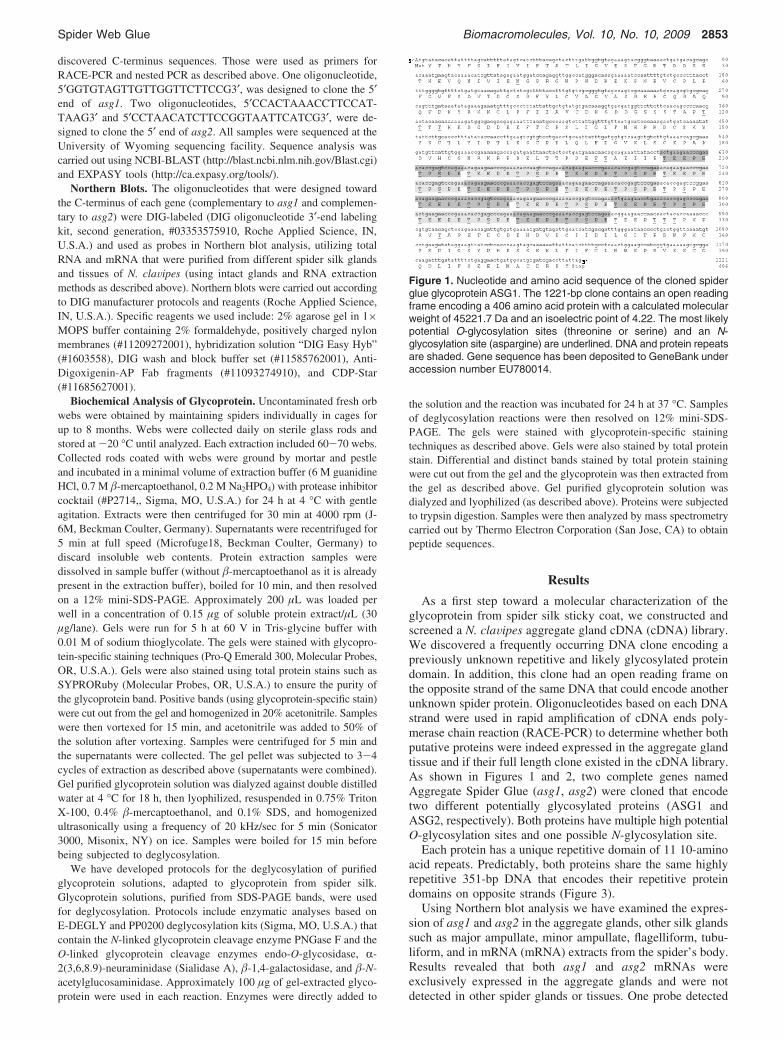
As a first step toward a molecular characterization of theglycoprotein from spider silk sticky coat, we constructed andscreened a N. claVipes aggregate gland cDNA (cDNA) library.We discovered a frequently occurring DNA clone encoding apreviously unknown repetitive and likely glycosylated proteindomain. In addition, this clone had an open reading frame onthe opposite strand of the same DNA that could encode anotherunknown spider protein. Oligonucleotides based on each DNAstrand were used in rapid amplification of cDNA ends poly-merase chain reaction (RACE-PCR) to determine whether bothputative proteins were indeed expressed in the aggregate glandtissue and if their full length clone existed in the cDNA library.As shown in Figures 1 and 2, two complete genes namedAggregate Spider Glue (asg1, asg2) were cloned that encodetwo different potentially glycosylated proteins (ASG1 andASG2, respectively). Both proteins have multiple high potentialO-glycosylation sites and one possible N-glycosylation site.
Each protein has a unique repetitive domain of 11 10-aminoacid repeats. Predictably, both proteins share the same highlyrepetitive 351-bp DNA that encodes their repetitive proteindomains on opposite strands (Figure 3).
Using Northern blot analysis we have examined the expres-sion of asg1 and asg2 in the aggregate glands, other silk glandssuch as major ampullate, minor ampullate, flagelliform, tubu-liform, and in mRNA (mRNA) extracts from the spider’s body.Results revealed that both asg1 and asg2 mRNAs wereexclusively expressed in the aggregate glands and were notdetected in other spider glands or tissues. One probe detected
Figure 1. Nucleotide and amino acid sequence of the cloned spiderglue glycoprotein ASG1. The 1221-bp clone contains an open readingframe encoding a 406 amino acid protein with a calculated molecularweight of 45221.7 Da and an isoelectric point of 4.22. The most likelypotential O-glycosylation sites (threonine or serine) and an N-glycosylation site (aspargine) are underlined. DNA and protein repeatsare shaded. Gene sequence has been deposited to GeneBank underaccession number EU780014.
Spider Web Glue Biomacromolecules, Vol. 10, No. 10, 2009 2853
an mRNA of 1.2 kb (Figure 4a) and the other detected an mRNAof 2.2 kb (Figure 4b).
In our biochemical studies we extracted and purified aglycoprotein directly from spider orb webs. Using specificglycoprotein detection methods, we found this protein to be the
only glycoprotein associated with the aqueous coat of the spidersilk web after SDS-PAGE. This purified glycoprotein from theinitial SDA-PAGE was deglycosylated and rerun on SDS-PAGE, which showed two bands of approximate sizes of 65and 38 kDa (Figure 5).
Staining with highly specific glycoprotein stain clearly showsthat no glycoprotein band can be detected at this stage, whichindicates that no glycosylated glycoprotein remained in thesolution after the enzymatic deglycosylation. Staining with totalprotein dye shows the two deglycosylated glycoprotein bands.Higher molecular weight bands on the gel were shown to beremains of the deglycosylation enzymes in the solution that wereloaded on the gel and are not related to any of the spider’sproteins. Additionally, the larger protein (65 kDa), detected afterdeglycosylation, was cut out of the gel, purified, and subjectedto mass spectrometry analysis. Peptides derived from thisanalysis show identities to the translated protein ASG2. Twoof the peptides found were Gly-Ser-Ser-Val-Ser and Gly-Leu-Gly-Val, which appear as one repeating sequence (GSSVS-GLGV) in ASG2, and the Ala-Gly-Pro-Gly-Thr peptide alsoappears in ASG2.
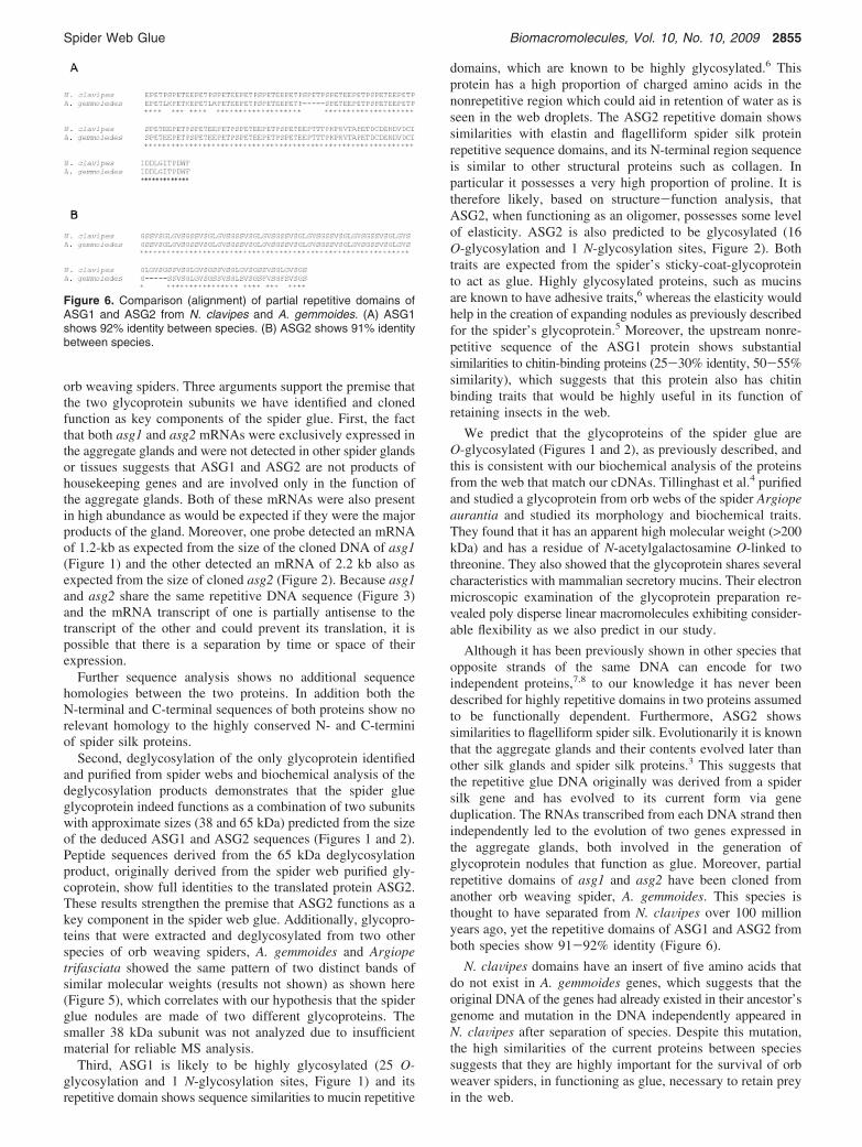
We identified the spider glue glycoproteins in another orb-weaving spider, Araneus gemmoides. We were able to clone aportion of the repetitive domain from this species. Comparisonof sequences from both species shows 92% identity for ASG1and 91% identity for ASG2. As shown in Figure 6, both ASG1and ASG2 from N. claVipes have an insert of five amino acidsthat does not appear in A. gemmoides.
Discussion
This study was designed to determine the molecular com-ponents of the glue that coats the spiral capture prey threads of
Figure 2. Nucleotide and amino acid sequence of the cloned spiderglue glycoprotein ASG2. The 2145-bp clone contains an open readingframe encoding a 714 amino acid protein with calculated size of71548.3 Da and an isoelectric point of 4.10. The most likelyO-glycosylation sites (threonine or serine) and an N-glycosylation site(aspargine) are underlined. The DNA and protein repeat is redunderlined and that region is in red. Gene sequences have beendeposited to GeneBank under accession number EU780015.
Figure 3. DNA and protein sequences of the repetitive regions ofboth proteins. The repetitive domains of ASG1 (amino acids 208-324)and ASG2 (amino acids 545-660) are transcribed and translated ontwo different open reading frames from opposite strands of the sameDNA sequence.
Figure 4. Northern blot analysis shows the transcripts of asg1 andasg2 in the aggregate glands. Oligonucleotides were designed to becomplementary to the repetitive DNA of each gene and were DIG-labeled. For each probe, two samples of aggregate glands of RNAwere used: total RNA (lane 1) and mRNA (lane 2). Mr: DIG-labeledRNA marker indicating sizes in bp (RNA1 #11526529910, RocheApplied Science, IN). (A) Hybridization with a probe complementaryto asg1 detected an approximate 1200 bp transcript. (B) Hybridizationwith a probe complementary to asg2 detected an approximate 2200bp transcript.
Figure 5. SDS PAGE gel of web glue glycoproteins. The 12% SDS-PAGE shows results of deglycosylation of the spider’s glycoproteinpurified from orb webs. Mw: Molecular weight marker indicating sizesin kDa (CandyCane, Molecular Probes, OR, U.S.A.), ASG: deglyco-sylated spider’s glycoprotein. (A) Staining with highly specific glyco-protein stain clearly shows that no glycoprotein band can be detectedwhich indicates that no glycosylated glycoprotein remained in thesolution after the enzymatic treatment. (B) Staining with total proteindye shows two apparent deglycosylated glycoprotein bands (ap-proximately 65 kDa upper band and 38 kDa lower band, arrowed).Higher molecular size bands were shown to be remains of thedeglycosylation enzymes in the solution that was loaded on the gel.
2854 Biomacromolecules, Vol. 10, No. 10, 2009 Choresh et al.
orb weaving spiders. Three arguments support the premise thatthe two glycoprotein subunits we have identified and clonedfunction as key components of the spider glue. First, the factthat both asg1 and asg2 mRNAs were exclusively expressed inthe aggregate glands and were not detected in other spider glandsor tissues suggests that ASG1 and ASG2 are not products ofhousekeeping genes and are involved only in the function ofthe aggregate glands. Both of these mRNAs were also presentin high abundance as would be expected if they were the majorproducts of the gland. Moreover, one probe detected an mRNAof 1.2-kb as expected from the size of the cloned DNA of asg1(Figure 1) and the other detected an mRNA of 2.2 kb also asexpected from the size of cloned asg2 (Figure 2). Because asg1and asg2 share the same repetitive DNA sequence (Figure 3)and the mRNA transcript of one is partially antisense to thetranscript of the other and could prevent its translation, it ispossible that there is a separation by time or space of theirexpression.
Further sequence analysis shows no additional sequencehomologies between the two proteins. In addition both theN-terminal and C-terminal sequences of both proteins show norelevant homology to the highly conserved N- and C-terminiof spider silk proteins.
Second, deglycosylation of the only glycoprotein identifiedand purified from spider webs and biochemical analysis of thedeglycosylation products demonstrates that the spider glueglycoprotein indeed functions as a combination of two subunitswith approximate sizes (38 and 65 kDa) predicted from the sizeof the deduced ASG1 and ASG2 sequences (Figures 1 and 2).Peptide sequences derived from the 65 kDa deglycosylationproduct, originally derived from the spider web purified gly-coprotein, show full identities to the translated protein ASG2.These results strengthen the premise that ASG2 functions as akey component in the spider web glue. Additionally, glycopro-teins that were extracted and deglycosylated from two otherspecies of orb weaving spiders, A. gemmoides and Argiopetrifasciata showed the same pattern of two distinct bands ofsimilar molecular weights (results not shown) as shown here(Figure 5), which correlates with our hypothesis that the spiderglue nodules are made of two different glycoproteins. Thesmaller 38 kDa subunit was not analyzed due to insufficientmaterial for reliable MS analysis.
Third, ASG1 is likely to be highly glycosylated (25 O-glycosylation and 1 N-glycosylation sites, Figure 1) and itsrepetitive domain shows sequence similarities to mucin repetitive
domains, which are known to be highly glycosylated.6 Thisprotein has a high proportion of charged amino acids in thenonrepetitive region which could aid in retention of water as isseen in the web droplets. The ASG2 repetitive domain showssimilarities with elastin and flagelliform spider silk proteinrepetitive sequence domains, and its N-terminal region sequenceis similar to other structural proteins such as collagen. Inparticular it possesses a very high proportion of proline. It istherefore likely, based on structure-function analysis, thatASG2, when functioning as an oligomer, possesses some levelof elasticity. ASG2 is also predicted to be glycosylated (16O-glycosylation and 1 N-glycosylation sites, Figure 2). Bothtraits are expected from the spider’s sticky-coat-glycoproteinto act as glue. Highly glycosylated proteins, such as mucinsare known to have adhesive traits,6 whereas the elasticity wouldhelp in the creation of expanding nodules as previously describedfor the spider’s glycoprotein.5 Moreover, the upstream nonre-petitive sequence of the ASG1 protein shows substantialsimilarities to chitin-binding proteins (25-30% identity, 50-55%similarity), which suggests that this protein also has chitinbinding traits that would be highly useful in its function ofretaining insects in the web.
We predict that the glycoproteins of the spider glue areO-glycosylated (Figures 1 and 2), as previously described, andthis is consistent with our biochemical analysis of the proteinsfrom the web that match our cDNAs. Tillinghast et al.4 purifiedand studied a glycoprotein from orb webs of the spider Argiopeaurantia and studied its morphology and biochemical traits.They found that it has an apparent high molecular weight (>200kDa) and has a residue of N-acetylgalactosamine O-linked tothreonine. They also showed that the glycoprotein shares severalcharacteristics with mammalian secretory mucins. Their electronmicroscopic examination of the glycoprotein preparation re-vealed poly disperse linear macromolecules exhibiting consider-able flexibility as we also predict in our study.
Although it has been previously shown in other species thatopposite strands of the same DNA can encode for twoindependent proteins,7,8 to our knowledge it has never beendescribed for highly repetitive domains in two proteins assumedto be functionally dependent. Furthermore, ASG2 showssimilarities to flagelliform spider silk. Evolutionarily it is knownthat the aggregate glands and their contents evolved later thanother silk glands and spider silk proteins.3 This suggests thatthe repetitive glue DNA originally was derived from a spidersilk gene and has evolved to its current form via geneduplication. The RNAs transcribed from each DNA strand thenindependently led to the evolution of two genes expressed inthe aggregate glands, both involved in the generation ofglycoprotein nodules that function as glue. Moreover, partialrepetitive domains of asg1 and asg2 have been cloned fromanother orb weaving spider, A. gemmoides. This species isthought to have separated from N. claVipes over 100 millionyears ago, yet the repetitive domains of ASG1 and ASG2 fromboth species show 91-92% identity (Figure 6).
N. claVipes domains have an insert of five amino acids thatdo not exist in A. gemmoides genes, which suggests that theoriginal DNA of the genes had already existed in their ancestor’sgenome and mutation in the DNA independently appeared inN. claVipes after separation of species. Despite this mutation,the high similarities of the current proteins between speciessuggests that they are highly important for the survival of orbweaver spiders, in functioning as glue, necessary to retain preyin the web.
Figure 6. Comparison (alignment) of partial repetitive domains ofASG1 and ASG2 from N. clavipes and A. gemmoides. (A) ASG1shows 92% identity between species. (B) ASG2 shows 91% identitybetween species.
Spider Web Glue Biomacromolecules, Vol. 10, No. 10, 2009 2855
The reason for the perfect DNA identity between the twogenes in their repetitive regions is unclear. However the factthat both species examined have maintained the identity of thesesequences in the two genes despite more than 100 million yearsof separation indicates its importance. This is particularly truebecause the five amino acid insertion or deletion between thetwo species is seen in both genes of that species.
The results of this study, together with comparisons toprevious studies of the putative spider glue glycoprotein,contribute novel biological and evolutionary knowledge of thesespecific genes and their partner proteins. Such knowledge isessential for further biological and chemical studies on thestructure and function of these proteins as well as to furtherstudies on their carbohydrate moieties. Once the cloned genesare overexpressed in systems such as insect or bacterial cellcultures, large-scale production of the glycoprotein can be usedto develop a new biobased glue for a variety of purposes.
Acknowledgment. This research was supported by NSF andAFoSR grants to R.V.L. We thank Ms. Kali Wong and Mr.James Obuya for assistance with molecular experiments;
University of Wyoming sequencing facility; Thermo ElectronCorporation (San Jose, CA) for MS analysis; and Prof. PamelaLanger and Dr. Michael Hinman for critically reading thismanuscript.
References and Notes(1) Lewis, R. V. Chem. ReV. 2006, 106, 3762–3774.(2) Peters, H. M. Naturwissenschaften 1995, 82, 380–382.(3) Vollrath, F.; Fairbrother, W. J.; Williams, R. J. P.; Tillinghast, E. K.;
Bernstein, D. T.; Gallagher, K. S.; Townley, M. A. Nature 1990, 345,526–527.
(4) Tillinghast, E. K.; Townley, M. A.; Wight, T. N.; Uhlenbruck, G.;Janssen, E. Mater. Res. Soc. Symp. Proc. 1993, 292, 9–23.
(5) Vollrath, F.; Tillinghast, E. K. Naturwissenschaften 1991, 78, 557–559.
(6) Beeley J. G. In Glycoprotein and Proteoglycan Techniques; BurdonR. H., van Knippenberg, Eds.; Elsevier Amsterdam: New York, Oxford,1985; Vol. 16.
(7) Adelman, P. Science 1987, 235, 1514–1517.(8) Shintani, S.; O’hUigin, C.; Toyosawa, S.; Michalova, V.; Klein, J.
Genetics 1999, 152, 743–754.
BM900681W
2856 Biomacromolecules, Vol. 10, No. 10, 2009 Choresh et al.


![76004 Spider-Man: Spider-Cycle Chase [Marvel]](https://static.documents.pub/doc/80x56/577cc35c1a28aba71195cd3a/76004-spider-man-spider-cycle-chase-marvel.jpg)


