Spin-Label Electron Spin Resonance Studies on the Interactions of Lysine Peptides with Phospholipid Membranes J6rg H. Kleinschmidt and Derek Marsh Max-Planck-lnstitut fOr biophysikalische Chemie, Abteilung Spektroskopie, D-37077 Gottingen, Germany ABSTRACT The interactions of lysine oligopeptides with dimyristoyl phosphatidylglycerol (DMPG) bilayer membranes were studied using spin-labeled lipids and electron spin resonance spectroscopy. Tetralysine and pentalysine were chosen as models for the basic amino acid clusters found in a variety of cytoplasmic membrane-associating proteins, and polylysine was chosen as representative of highly basic peripherally bound proteins. A greater motional restriction of the lipid chains was found with increasing length of the peptide, while the saturation ratio of lipids per peptide was lower for the shorter peptides. In DMPG and dimyristoylphosphatidylserine host membranes, the perturbation of the lipid chain mobility by polylysine was greater for negatively charged spin-labeled lipids than for zwitterionic lipids, but for the shorter lysine peptides these differences were smaller. In mixed bilayers composed of DMPG and dimyristoylphosphatidylcholine, little difference was found in selectivity between spin-labeled phospholipid species on binding pentalysine. Surface binding of the basic lysine peptides strongly reduced the interfacial pK of spin-labeled fatty acid incorporated into the DMPG bilayers, to a greater extent for polylysine than for tetralysine or pentalysine at saturation. The results are consistent with a predominantly electrostatic interaction with the shorter lysine peptides, but with a closer surface association with the longer polylysine peptide. INTRODUCTION The binding of peripheral proteins to biomembranes plays an important role in many cellular processes. Protein kinase C, for example, may be activated by binding to acidic lipids (House and Kemp, 1987; House et al., 1989). In conse- quence, studies have been undertaken to investigate the effects of this type of lipid-protein interaction on the overall structure and properties of the lipid bilayer core of the membrane. Proteins bound to the membrane surface signif- icantly alter the lipid mobility and exhibit specific prefer- ences for certain lipid species (see, for example, Gorrissen et al., 1986; Sankaram et al., 1989a,b; Marsh, 1990a). It was shown that the binding of peripheral proteins may also induce lipid phase separations in certain model membranes (Haverstick and Glaser, 1989; Yang and Glaser, 1995). Clusters of basic amino acid residues, or a generally high content of basic amino acids, are required for proteins to bind to the surface of a biological membrane with a negative electrostatic potential (Kim et al., 1991; Mosior and McLaughlin, 1992). The putative pseudo-substrate region of protein kinase C, for example, consists of 12 amino acids, of which 8 bear a positive charge (Mosior and McLaughlin, 1991), which is predicted to be required for the binding of the protein to the membrane surface, a step necessary for its subsequent activation. A further, somewhat similar example is afforded by the autoinhibitory region at the C-terminus of Received for publication 20 March 1997 and in final form I August 1997. Address reprint requests to Dr. Derek Marsh, Max-Planck-Institut ftir biophysikalische Chemie, Abteilung 010 Spektroskopie, Am Fassberg 11, D-37077 Gottingen, Germany. Tel.: +49-551-201-1285; Fax: +49-551- 201-1501; E-mail: [email protected]. C 1997 by the Biophysical Society 0006-3495/97/11/2546/10 $2.00 the plasma membrane P-type Ca2+-ATPase (Filoteo et al., 1992). For a systematic biophysical approach to characterizing the nature of the interaction of basic proteins with biomem- branes, it is useful to study the interaction of oligopeptides composed of a single basic amino acid type. These consti- tute a simple model for specific basic amino acid clusters in proteins. The interaction of polymers of lysine (molecular masses ranging from 4 to 150 kDa) with negatively charged dipalmitoyl phosphatidylglycerol (DPPG) bilayers was al- ready studied, with a focus on the secondary structure of the polypeptide (Carrier and Pezolet, 1984) and on the effect of polylysine on the cooperative gel-to-liquid crystalline phase transition of DPPG bilayers (Carrier et al., 1985; Carrier and Pezolet, 1986). More recently, the binding of short lysine peptides (Lysn, n = 1-5) and their effects on the surface electrostatics were studied by electrophoretic mobility mea- surements (Kim et al., 1991; Mosior and McLaughlin, 1992). In the present work, we focus on the interactions of oligolysine peptides with lipid bilayers of dimyristoylphos- phatidylglycerol (DMPG) and mixtures with dimyris- toylphosphatidylcholine. Using spin-label electron spin res- onance (ESR) spectroscopy, we characterized the interactions and their lipid specificity by determining the perturbation of the lipid chain mobility on binding the peptide. Tetralysine and pentalysine were used as models for the basic amino acid clusters found in cytoplasmic proteins, and polylysine with 320 residues was chosen for comparison as representative of highly basic peripheral membrane proteins. The studies encompass conventional binding experiments and studies of lipid selectivity and of interfacial interactions via the protonation equilibria of membrane-bound fatty acids, in addition to those on lipid distribution in mixed lipid membranes with bound peptide. 2546
Transcript
Spin-Label Electron Spin Resonance Studies on the Interactions ofLysine Peptides with Phospholipid Membranes
J6rg H. Kleinschmidt and Derek MarshMax-Planck-lnstitut fOr biophysikalische Chemie, Abteilung Spektroskopie, D-37077 Gottingen, Germany
ABSTRACT The interactions of lysine oligopeptides with dimyristoyl phosphatidylglycerol (DMPG) bilayer membranes were
studied using spin-labeled lipids and electron spin resonance spectroscopy. Tetralysine and pentalysine were chosen as
models for the basic amino acid clusters found in a variety of cytoplasmic membrane-associating proteins, and polylysine waschosen as representative of highly basic peripherally bound proteins. A greater motional restriction of the lipid chains wasfound with increasing length of the peptide, while the saturation ratio of lipids per peptide was lower for the shorter peptides.In DMPG and dimyristoylphosphatidylserine host membranes, the perturbation of the lipid chain mobility by polylysine wasgreater for negatively charged spin-labeled lipids than for zwitterionic lipids, but for the shorter lysine peptides thesedifferences were smaller. In mixed bilayers composed of DMPG and dimyristoylphosphatidylcholine, little difference was
found in selectivity between spin-labeled phospholipid species on binding pentalysine. Surface binding of the basic lysinepeptides strongly reduced the interfacial pK of spin-labeled fatty acid incorporated into the DMPG bilayers, to a greater extentfor polylysine than for tetralysine or pentalysine at saturation. The results are consistent with a predominantly electrostaticinteraction with the shorter lysine peptides, but with a closer surface association with the longer polylysine peptide.
INTRODUCTION
The binding of peripheral proteins to biomembranes playsan important role in many cellular processes. Protein kinaseC, for example, may be activated by binding to acidic lipids(House and Kemp, 1987; House et al., 1989). In conse-quence, studies have been undertaken to investigate theeffects of this type of lipid-protein interaction on the overallstructure and properties of the lipid bilayer core of themembrane. Proteins bound to the membrane surface signif-icantly alter the lipid mobility and exhibit specific prefer-ences for certain lipid species (see, for example, Gorrissenet al., 1986; Sankaram et al., 1989a,b; Marsh, 1990a). It wasshown that the binding of peripheral proteins may alsoinduce lipid phase separations in certain model membranes(Haverstick and Glaser, 1989; Yang and Glaser, 1995).Clusters of basic amino acid residues, or a generally highcontent of basic amino acids, are required for proteins tobind to the surface of a biological membrane with a negativeelectrostatic potential (Kim et al., 1991; Mosior andMcLaughlin, 1992). The putative pseudo-substrate region ofprotein kinase C, for example, consists of 12 amino acids, ofwhich 8 bear a positive charge (Mosior and McLaughlin,1991), which is predicted to be required for the binding ofthe protein to the membrane surface, a step necessary for itssubsequent activation. A further, somewhat similar exampleis afforded by the autoinhibitory region at the C-terminus of
Received for publication 20 March 1997 and in finalform I August 1997.
Address reprint requests to Dr. Derek Marsh, Max-Planck-Institut ftirbiophysikalische Chemie, Abteilung 010 Spektroskopie, Am Fassberg 11,D-37077 Gottingen, Germany. Tel.: +49-551-201-1285; Fax: +49-551-201-1501; E-mail: [email protected] 1997 by the Biophysical Society0006-3495/97/11/2546/10 $2.00
the plasma membrane P-type Ca2+-ATPase (Filoteo et al.,1992).For a systematic biophysical approach to characterizing
the nature of the interaction of basic proteins with biomem-branes, it is useful to study the interaction of oligopeptidescomposed of a single basic amino acid type. These consti-tute a simple model for specific basic amino acid clusters inproteins. The interaction of polymers of lysine (molecularmasses ranging from 4 to 150 kDa) with negatively chargeddipalmitoyl phosphatidylglycerol (DPPG) bilayers was al-ready studied, with a focus on the secondary structure of thepolypeptide (Carrier and Pezolet, 1984) and on the effect ofpolylysine on the cooperative gel-to-liquid crystalline phasetransition of DPPG bilayers (Carrier et al., 1985; Carrier andPezolet, 1986). More recently, the binding of short lysinepeptides (Lysn, n = 1-5) and their effects on the surfaceelectrostatics were studied by electrophoretic mobility mea-surements (Kim et al., 1991; Mosior and McLaughlin,1992).
In the present work, we focus on the interactions ofoligolysine peptides with lipid bilayers of dimyristoylphos-phatidylglycerol (DMPG) and mixtures with dimyris-toylphosphatidylcholine. Using spin-label electron spin res-onance (ESR) spectroscopy, we characterized theinteractions and their lipid specificity by determining theperturbation of the lipid chain mobility on binding thepeptide. Tetralysine and pentalysine were used as modelsfor the basic amino acid clusters found in cytoplasmicproteins, and polylysine with 320 residues was chosen forcomparison as representative of highly basic peripheralmembrane proteins. The studies encompass conventionalbinding experiments and studies of lipid selectivity and ofinterfacial interactions via the protonation equilibria ofmembrane-bound fatty acids, in addition to those on lipiddistribution in mixed lipid membranes with bound peptide.
DMPC (1,2-dimyristoyl-sn-glycero-3-phosphocholine), DMPG (1,2-dimyristoyl-sn-glycero-3-phosphoglycerol), and DMPS (1,2-dimyristoyl-sn-glycero-3-phosphoserine) were obtained from Avanti Polar Lipids (Al-abaster, AL) and were checked for purity by thin-layer chromatography.3-(N-Morpholino)propanesulfonic acid (MOPS) was obtained from Sigma(St. Louis, MO). 5-(4,4-dimethyloxazolidine-N-oxyl)stearic acid (5-SASL)was synthesized as described (Hubbell and McConnell, 1971). Phosphati-dylcholine, phosphatidylethanolamine, phosphatidylglycerol, phosphati-dylserine, and phosphatidic acid spin labels with the nitroxyl group on theC-5 atom of the sn-2 chain (5-PCSL, 5-PESL, 5-PGSL, 5-PSSL, and5-PASL, respectively) were synthesized from the corresponding spin-labeled stearic acid (5-SASL) as described by Marsh and Watts (1982).Spin-labeled diacylglycerol (5-DGSL) was synthesized from 5-PCSL as
described by Heimburg et al. (1992). All spin labels were checked bythin-layer chromatography before their use with the solvent systemsCHCl3/CH3OH/ammonia (65/30/3, v/v/v) and hexane ether (1/1, v/v).Furthermore, all spin labels were checked in ethanol solution by ESRspectroscopy to determine the consistency of their concentrations. Tetral-ysine (Miles Labs, Elkhart, IN) and polylysine (320 residues; Sigma) were
used without further purification. Pentalysine was obtained by customsynthesis from Research Plus (Bayonne, NJ) and from Multiple PeptideSystems (San Diego, CA) and used without further purification.
Sample preparation
Lipid dispersions were prepared by first codissolving the lipids (0.5 mg)with 1 mol% spin label in a chloroform/methanol solution (1:1, v/v), andthen evaporating the solvent with a nitrogen gas flow and drying thesample under vacuum overnight. The dried lipid films were dispersed invarious buffer solutions (10 mM buffer with 5 mM EDTA: MOPS for pHrange 6.5-7.5, citric acid for pH range 3-6, sodium borate/NaOH for pHabove 7.6) to a final concentration of 25 mg/ml. The samples were
incubated for 1 h at 35°C. Peptide-containing samples were prepared byhydrating the dry lipid film in buffer and adding the desired amount ofpeptide dissolved in an equivalent volume of the same buffer. Alterna-tively, dried lipid films were hydrated directly with buffer containing thepeptide to give the same total volume and lipid/peptide ratios. No differ-ence in the ESR spectra was observed between the two different methodsof preparation for the peptide-containing samples. Both hydrated lipid andhydrated peptide solutions were adjusted to the desired pH before theywere mixed together. The pH adjustment was done so as not to exceed an
ionic strength of 0.03. Peptide-containing samples were also incubated at
35°C for 1 h.After incubation, samples were centrifuged in a tabletop centrifuge. The
pH of the supernatant did not change by more than ±0.1 pH unitscompared with the separate solutions before mixing. The membrane pelletswere then transferred to ESR capillaries. After the spectra were recorded,the samples were analyzed by protein (Lowry et al., 1951) and phosphate(Rouser et al., 1970) assays. The Lowry assay was performed with bovineserum albumin as a standard. This was calibrated with solutions of knownconcentrations of the lysine peptides used here.
scans were accumulated, depending on the sample. The modulation fre-quency was 100 kHz, with a modulation amplitude of 1.25 Gauss. A timeconstant of 0.25 s was chosen for 4-min scans. The outer hyperfinesplitting, 2Amax, was used to characterize the spectra of the spin labels.Hyperfine splittings were determined by fitting the maxima and minima inthe outer wings of the spectrum empirically to Gaussian curves. The outerhyperfine splittings are sensitive to both the amplitude and rate of the lipidchain motions, and can be used to characterize the strength of interactionof the various spin-labeled lipids with peptides or proteins (Lange et al.,1985; Sankaram et al., 1989a).
RESULTS
Membrane binding of different lysine peptides
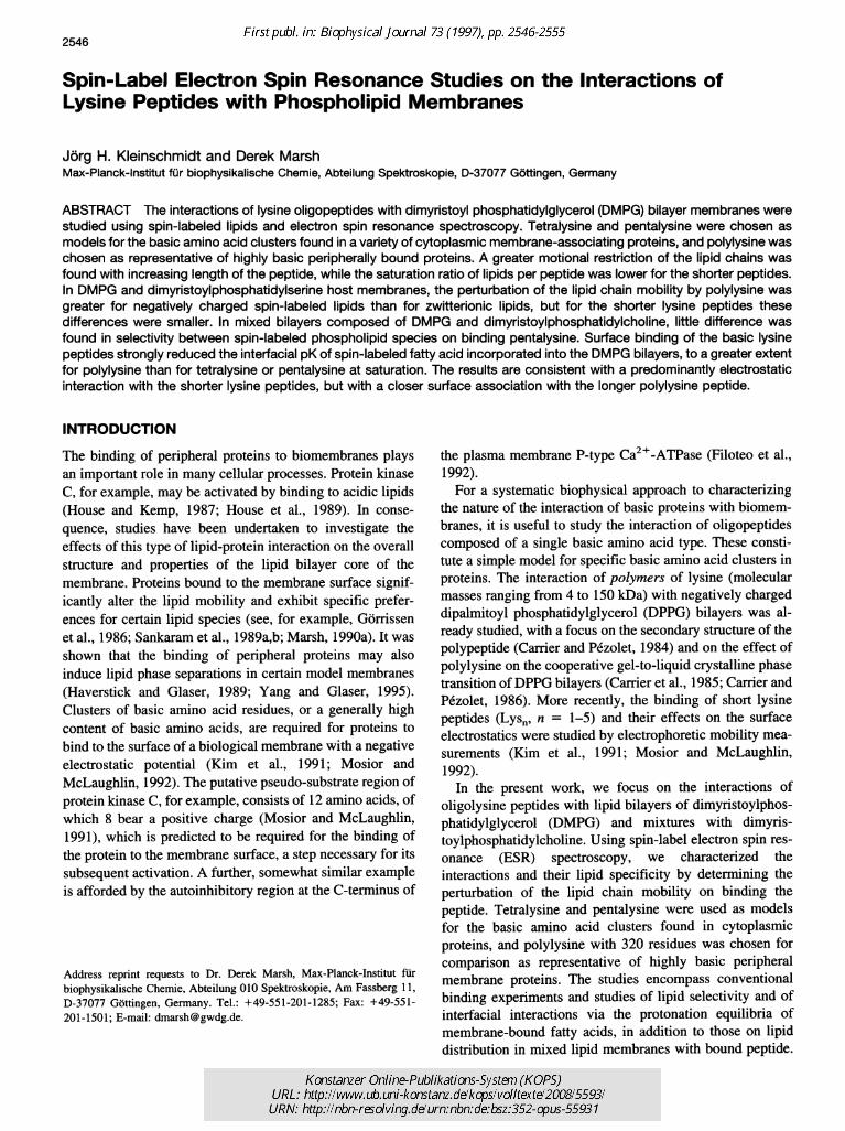
The binding of tetralysine and pentalysine to anionic lipidbilayer membranes was determined by centrifugation assayswith conventional chemical lipid and protein determina-tions. In addition, binding was studied from the dependenceof the outer hyperfine splittings in the ESR spectra of 5C-atom spin-labeled lipids on the lysine peptide/lipid ratio.The ESR spectra of the 5 C-atom spin labels in lipiddispersions correspond to axially symmetrical, partially mo-tional-averaged, anisotropic systems. Binding of the peptidedecreases the degree of motional averaging in the spectrafrom the spin-labeled lipid chains and thus increases themaximum outer hyperfine splitting in the ESR spectra.
Fig. 1 shows the binding behavior of the tetralysine andpentalysine peptides obtained by these two methods. In the
0
000
0.0
ESR spectroscopy
ESR spectra were acquired with an E- 12 Century Line 9-GHz spectrometer(Varian, Sunnyvale, CA) equipped with a TE102 rectangular cavity (Var-ian). The temperature was controlled to within ±0.1°C, by using nitrogengas flow temperature regulation with a thin-wire thermocouple that wasplaced close to the sample at the top of the microwave cavity. A customsample holder allowed positioning of the sample in the cavity with anaccuracy of 0.1 mm. The spectrometer was interfaced to an IBM personalcomputer using a Tecmar Labmaster A/D converter for digitizing andstoring the ESR spectral data. To improve the signal-to-noise ratio, 2-16
P/L added (mol/mol)
1.0
P/L added (mol/mol)
FIGURE 1 Dependence of the outer hyperfine splitting constants, Ama,of the 5-PGSL spin label in DMPG bilayers on the amount of peptideadded to the lipid for (a) tetralysine and (b) pentalysine. The correspondingratios of bound peptide/lipid (PAL) as a function of the added peptide/lipidratio for (c) tetralysine and (d) pentalysine are shown below. In b the valuesof Amax for 5-PGSL in a mixed DMPC:DMPG (1:1 mol/mol) bilayer (0)are shown as a function of the added pentalysine/lipid ratio in the inset.Temperature: 30°C. Buffer: 10 mM MOPS, 5 mM EDTA, and 10 mMNaCl (pH 7.0).
Kleinschmidt and Marsh 2547
Volume 73 November 1997
case of tetralysine, the DMPG bilayers become saturatedwith peptide at a ratio of - 1.6-1.7 lipids/peptide. Chemicalanalysis of the membrane complexes and the point at whichAmax achieves its limiting level yield consistent values forthis binding stoichiometry. In the case of pentalysine, thelipid/peptide binding stoichiometry is increased to -1.9-2.5 mol/mol. The range quoted corresponds to results fromthe chemical binding assays and the peptide/lipid depen-dence of Amax, respectively. For polylysine, a lipid/proteinsaturation binding ratio of 70 mol DMPG/320-residuepolypeptide was estimated (data not shown).ESR data for binding of pentalysine to a mixed mem-
brane containing zwitterionic and anionic lipids are alsogiven in Fig. 1 b. The perturbation of the overall lipidmobility is strongly reduced by admixture of the unchargedDMPC lipid component with DMPG, as seen from thelower limiting value of Amax. This is a result of the reducedbinding of pentalysine to the mixed lipid membrane, wherethe saturation lipid/peptide stoichiometry is -5 lipids/peptide.
Influence on the bilayer phase transition
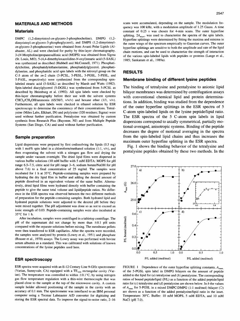
The effect of pentalysine and polylysine on the thermotropiclipid phase behavior was investigated for DMPG bilayerswith saturating amounts of peptide bound. Fig. 2 shows thetemperature dependence of the outer hyperfine splittings inthe ESR spectra of 5-PASL spin label in DMPG bilayers inthe presence and absence of pentalysine or polylysine. Thesharp cooperative phase transition found for the peptide-freebilayer at 23°C is significantly broadened by binding pen-talysine without appreciable shift (maximally -1°) in themidpoint of the transition. The lipid chain mobility is de-creased over the entire temperature range, as reflected bythe increased ESR outer hyperfine splittings at any giventemperature. Similar results were obtained both with otherlipid spin labels and for pentalysine bound to DMPS bilay-ers (data not shown). These trends are further pronouncedon binding polylysine rather than the smaller pentalysineoligomer. The lipid chain-melting transition is broadenedeven more, although any shift in the transition midpoint stillremains small (maximally + 1). The outer hyperfine split-tings are increased more than for pentalysine at all temper-atures, with the effect being stronger in the fluid phase. Thedecrease in lipid chain mobility is therefore particularlypronounced on binding polylysine.
Interaction with different spin-labeledlipid species
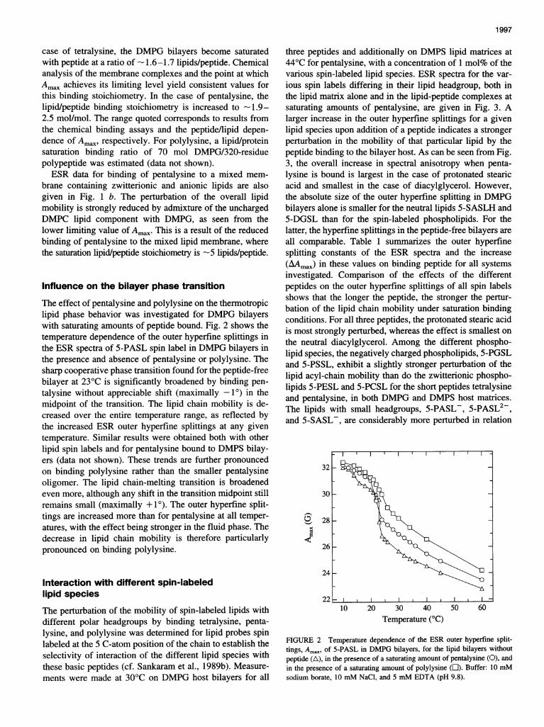
The perturbation of the mobility of spin-labeled lipids withdifferent polar headgroups by binding tetralysine, penta-lysine, and polylysine was determined for lipid probes spinlabeled at the 5 C-atom position of the chain to establish theselectivity of interaction of the different lipid species withthese basic peptides (cf. Sankaram et al., 1989b). Measure-ments were made at 30°C on DMPG host bilayers for all
three peptides and additionally on DMPS lipid matrices at44'C for pentalysine, with a concentration of 1 mol% of thevarious spin-labeled lipid species. ESR spectra for the var-ious spin labels differing in their lipid headgroup, both inthe lipid matrix alone and in the lipid-peptide complexes atsaturating amounts of pentalysine, are given in Fig. 3. Alarger increase in the outer hyperfine splittings for a givenlipid species upon addition of a peptide indicates a strongerperturbation in the mobility of that particular lipid by thepeptide binding to the bilayer host. As can be seen from Fig.3, the overall increase in spectral anisotropy when penta-lysine is bound is largest in the case of protonated stearicacid and smallest in the case of diacylglycerol. However,the absolute size of the outer hyperfine splitting in DMPGbilayers alone is smaller for the neutral lipids 5-SASLH and5-DGSL than for the spin-labeled phospholipids. For thelatter, the hyperfine splittings in the peptide-free bilayers areall comparable. Table 1 summarizes the outer hyperfinesplitting constants of the ESR spectra and the increase(AAmax) in these values on binding peptide for all systemsinvestigated. Comparison of the effects of the differentpeptides on the outer hyperfine splittings of all spin labelsshows that the longer the peptide, the stronger the pertur-bation of the lipid chain mobility under saturation bindingconditions. For all three peptides, the protonated stearic acidis most strongly perturbed, whereas the effect is smallest onthe neutral diacylglycerol. Among the different phospho-lipid species, the negatively charged phospholipids, 5-PGSLand 5-PSSL, exhibit a slightly stronger perturbation of thelipid acyl-chain mobility than do the zwitterionic phospho-lipids 5-PESL and 5-PCSL for the short peptides tetralysineand pentalysine, in both DMPG and DMPS host matrices.The lipids with small headgroups, 5-PASL-, 5-PASL2,and 5-SASL-, are considerably more perturbed in relation
32
30 _
28 k
26 k
24 k
10 20 30 40Temperature (°C)
50 60
FIGURE 2 Temperature dependence of the ESR outer hyperfine split-tings, Ama, of 5-PASL in DMPG bilayers, for the lipid bilayers without
peptide (A), in the presence of a saturating amount of pentalysine (0), and
in the presence of a saturating amount of polylysine (L1). Buffer: 10 mMsodium borate, 10 mM NaCl, and 5 mM EDTA (pH 9.8).
_- -l EI
2548 Biophysical Journal
Lysine Peptide-Lipid Interactions 2549
TABLE 1 Outer hyperfine splitting constants, Amax, of theESR spectra from lipids spin-labeled on the 5 C-atom of thesn-2 acyl chain, or fatty acid spin-labeled at C-atom 5, inDMPG bilayers in the presence and absence of saturatingamounts of tetralysine, pentalysine, and polylysine at 30°C
FIGURE 3 ESR spectra of different lipid species spin-labeled at the fifthposition of the sn-2 acyl-chain, 5-XSL, in DMPG bilayers recorded at 30°Cin the absence (. ) and in the presence ( ) of saturating amounts ofpentalysine. Total scan width: 100 G. Buffers: 10 mM MOPS, 10 mMNaCl, and 5 mM EDTA, pH 7.2, for the spin labels 5-PSSL, 5-PGSL,5-PCSL, 5-PESL, and 5-DGSL; 10 mM citric acid, 10 mM NaCl, and S
mM EDTA, pH 4.7, for spin label 5-SASLH; 10 mM sodium borate, 10mM NaCl, and 5 mM EDTA, pH 9.8, for spin label 5-PASL2- and at pH10.2 for spin label 5-SASL-.
to the lipids with larger headgroups when the long poly-lysine is bound instead of pentalysine.
Selectivity in mixed lipid systems
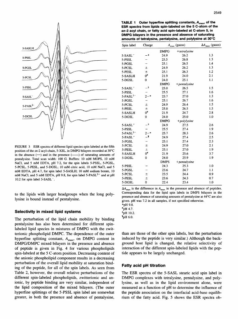
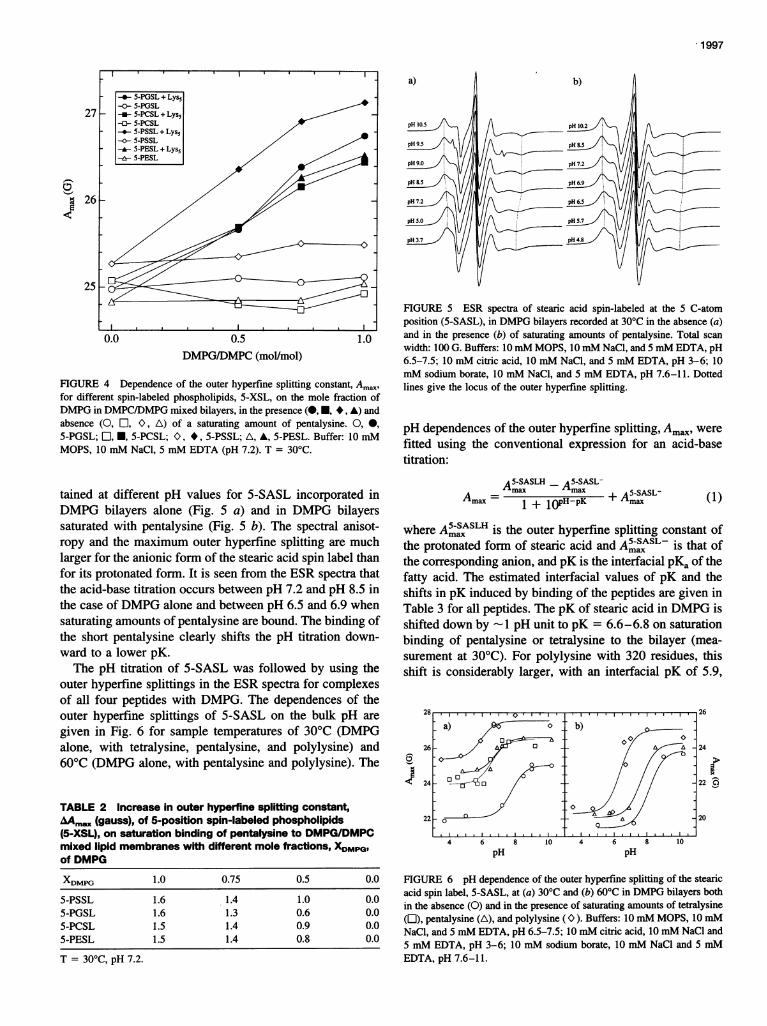
The perturbation of the lipid chain mobility by bindingpentalysine has also been determined for different spin-labeled lipid species in mixtures of DMPG with the zwit-terionic phospholipid DMPC. The dependence of the outerhyperfine splitting constant, Amax, on DMPG content inDMPG/DMPC mixed bilayers in the presence and absenceof peptide is given in Fig. 4 for various phospholipidsspin-labeled at the 5 C-atom position. Decreasing content ofthe anionic phospholipid component results in a decreasingperturbation of the overall lipid mobility at saturation bind-ing of the peptide, for all of the spin labels. As seen fromTable 2, however, the overall relative perturbations of thedifferent spin-labeled phospholipids, zwitterionic and an-
ionic, by peptide binding are very similar, independent ofthe lipid composition of the mixed bilayers. (The outerhyperfine splittings of the 5-PSSL spin label are somewhatgreater, in both the presence and absence of pentalysine,
AAmax is the difference in Amax in the presence and absence of peptides.Corresponding data for the lipid spin labels in DMPS bilayers in thepresence and absence of saturating amounts of pentalysine at 44°C are alsogiven. pH was 7.2 in all samples, if not specified otherwise.*pH 9.8.#pH 4.7.§pH 10.2.IpH 6.0.
than are those of the other spin labels, but the perturbationinduced by the peptide is very similar.) Although the back-ground host lipid is changed, the relative selectivity ofinteraction of the different spin-labeled lipids with the pep-tide appears to be largely unchanged.
Fatty acid pH titration
The ESR spectra of the 5-SASL stearic acid spin label inDMPG complexes with tetralysine, pentalysine, and poly-lysine, as well as in the lipid environment alone, weremeasured as a function of pH to determine the influence ofthe peptide association on the interfacial acid-base equilib-rium of the fatty acid. Fig. 5 shows the ESR spectra ob-
Kleinschmidt and Marsh
5-SASLH
5-PSSL -
5-PGSL
5-PCSL
5-PESL
5-SA5AL -
5-PASL2
5-DGSL
1.I
Volume 73 November 1997
0.0 0.5 1.0DMPG/DMPC (mol/mol)
FIGURE 4 Dependence of the outer hyperfine splitting constant, Am.,for different spin-labeled phospholipids, 5-XSL, on the mole fraction ofDMPG in DMPC/DMPG mixed bilayers, in the presence (-H, *, A) andabsence (0, E, O, A) of a saturating amount of pentalysine. 0, 0,
5-PGSL; l, *, 5-PCSL; O, *, 5-PSSL; A, A, 5-PESL. Buffer: 10 mMMOPS, 10 mM NaCl, 5 mM EDTA (pH 7.2). T = 30°C.
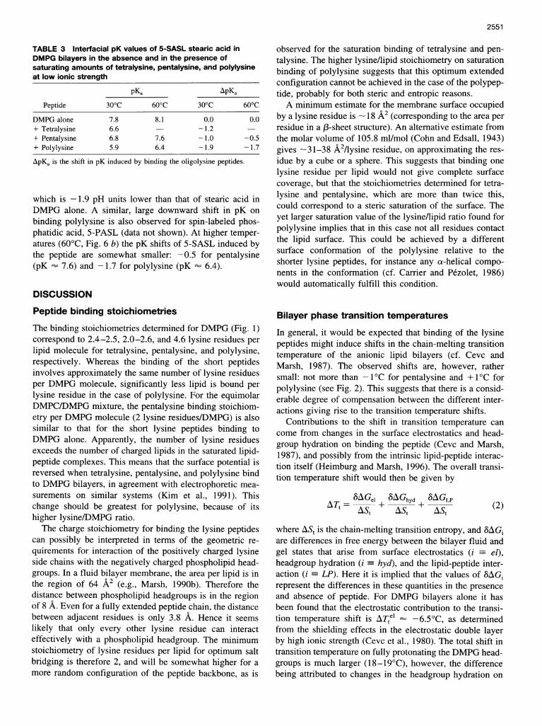
tained at different pH values for 5-SASL incorporated inDMPG bilayers alone (Fig. 5 a) and in DMPG bilayerssaturated with pentalysine (Fig. 5 b). The spectral anisot-ropy and the maximum outer hyperfine splitting are muchlarger for the anionic form of the stearic acid spin label thanfor its protonated form. It is seen from the ESR spectra thatthe acid-base titration occurs between pH 7.2 and pH 8.5 inthe case of DMPG alone and between pH 6.5 and 6.9 whensaturating amounts of pentalysine are bound. The binding ofthe short pentalysine clearly shifts the pH titration down-ward to a lower pK.The pH titration of 5-SASL was followed by using the
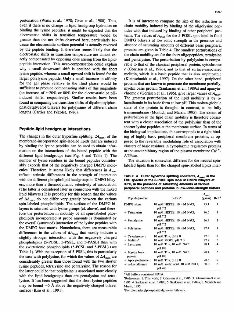
outer hyperfine splittings in the ESR spectra for complexesof all four peptides with DMPG. The dependences of theouter hyperfine splittings of 5-SASL on the bulk pH are
given in Fig. 6 for sample temperatures of 30°C (DMPGalone, with tetralysine, pentalysine, and polylysine) and60°C (DMPG alone, with pentalysine and polylysine). The
TABLE 2 Increase in outer hyperfine splitting constant,AAm,x (gauss), of 5-position spin-labeled phospholipids(5-XSL), on saturation binding of pentalysine to DMPG/DMPCmixed lipid membranes with different mole fractions, XDMPG,of DMPG
FIGURE 5 ESR spectra of stearic acid spin-labeled at the 5 C-atomposition (5-SASL), in DMPG bilayers recorded at 30°C in the absence (a)and in the presence (b) of saturating amounts of pentalysine. Total scan
width: 100 G. Buffers: 10mM MOPS, 10mM NaCl, and 5 mM EDTA, pH6.5-7.5; 10 mM citric acid, 10 mM NaCl, and 5 mM EDTA, pH 3-6; 10mM sodium borate, 10 mM NaCl, and 5 mM EDTA, pH 7.6-11. Dottedlines give the locus of the outer hyperfine splitting.
pH dependences of the outer hyperfilne splitting, Amn,,, werefitted using the conventional expression for an acid-basetitration:
A5-SASLH A5-SASL-=max max +A5-SASL-Amaxc 1 + 1OPHPK Amax
where A5-SASLH is the outer hyperfine splitting constant ofthe protonated form of stearic acid and A5-SASL- is that ofthe corresponding anion, and pK is the interfacial pKa of thefatty acid. The estimated interfacial values of pK and theshifts in pK induced by binding of the peptides are given inTable 3 for all peptides. The pK of stearic acid in DMPG isshifted down by 1 pH unit to pK = 6.6-6.8 on saturationbinding of pentalysine or tetralysine to the bilayer (mea-surement at 30°C). For polylysine with 320 residues, thisshift is considerably larger, with an interfacial pK of 5.9,
4 6 8 10 4 6 8 10
pH pH
FIGURE 6 pH dependence of the outer hyperfine splitting of the stearic
acid spin label, 5-SASL, at (a) 30°C and (b) 60°C in DMPG bilayers both
in the absence (0) and in the presence of saturating amounts of tetralysine(]), pentalysine (A), and polylysine (C>). Buffers: 10 mM MOPS, 10 mM
NaCl, and 5 mM EDTA, pH 6.5-7.5; 10 mM citric acid, 10 mM NaCl and5 mM EDTA, pH 3-6; 10 mM sodium borate, 10 mM NaCl and 5 mMEDTA, pH 7.6-11.
I
a) / b)
a)~~~~~~~~~~~~~~~~~~~~~~~~~~~~~~~~~~~~~
0. C. ,,,'),26
1
A 24
24 -22
22~ A 20
2550 Biophysical Journal
(1)
, a
Lysine Peptide-Lipid Interactions
TABLE 3 Interfacial pK values of 5-SASL stearic acid inDMPG bilayers in the absence and in the presence ofsaturating amounts of tetralysine, pentalysine, and polylysineat low ionic strength
ApKa is the shift in pK induced by binding the oligolysine peptides.
which is -1.9 pH units lower than that of stearic acid inDMPG alone. A similar, large downward shift in pK onbinding polylysine is also observed for spin-labeled phos-phatidic acid, 5-PASL (data not shown). At higher temper-atures (600C, Fig. 6 b) the pK shifts of 5-SASL induced bythe peptide are somewhat smaller: -0.5 for pentalysine(pK 7.6) and -1.7 for polylysine (pK 6.4).
DISCUSSION
Peptide binding stoichiometries
The binding stoichiometries determined for DMPG (Fig. 1)correspond to 2.4-2.5, 2.0-2.6, and 4.6 lysine residues perlipid molecule for tetralysine, pentalysine, and polylysine,respectively. Whereas the binding of the short peptidesinvolves approximately the same number of lysine residuesper DMPG molecule, significantly less lipid is bound perlysine residue in the case of polylysine. For the equimolarDMPC/DMPG mixture, the pentalysine binding stoichiom-etry per DMPG molecule (2 lysine residues/DMPG) is alsosimilar to that for the short lysine peptides binding toDMPG alone. Apparently, the number of lysine residuesexceeds the number of charged lipids in the saturated lipid-peptide complexes. This means that the surface potential isreversed when tetralysine, pentalysine, and polylysine bindto DMPG bilayers, in agreement with electrophoretic mea-surements on similar systems (Kim et al., 1991). Thischange should be greatest for polylysine, because of itshigher lysine/DMPG ratio.The charge stoichiometry for binding the lysine peptides
can possibly be interpreted in terms of the geometric re-quirements for interaction of the positively charged lysineside chains with the negatively charged phospholipid head-groups. In a fluid bilayer membrane, the area per lipid is inthe region of 64 A2 (e.g., Marsh, 1990b). Therefore thedistance between phospholipid headgroups is in the regionof 8 A. Even for a fully extended peptide chain, the distancebetween adjacent residues is only 3.8 A. Hence it seemslikely that only every other lysine residue can interacteffectively with a phospholipid headgroup. The minimumstoichiometry of lysine residues per lipid for optimum saltbridging is therefore 2, and will be somewhat higher for amore random configuration of the peptide backbone, as is
observed for the saturation binding of tetralysine and pen-talysine. The higher lysine/lipid stoichiometry on saturationbinding of polylysine suggests that this optimum extendedconfiguration cannot be achieved in the case of the polypep-tide, probably for both steric and entropic reasons.A minimum estimate for the membrane surface occupied
by a lysine residue is - 18 A2 (corresponding to the area perresidue in a (3-sheet structure). An alternative estimate fromthe molar volume of 105.8 ml/mol (Cohn and Edsall, 1943)gives -31-38 A2/lysine residue, on approximating the res-idue by a cube or a sphere. This suggests that binding onelysine residue per lipid would not give complete surfacecoverage, but that the stoichiometries determined for tetra-lysine and pentalysine, which are more than twice this,could correspond to a steric saturation of the surface. Theyet larger saturation value of the lysine/lipid ratio found forpolylysine implies that in this case not all residues contactthe lipid surface. This could be achieved by a differentsurface conformation of the polylysine relative to theshorter lysine peptides, for instance any a-helical compo-nents in the conformation (cf. Carrier and Pezolet, 1986)would automatically fulfill this condition.
Bilayer phase transition temperatures
In general, it would be expected that binding of the lysinepeptides might induce shifts in the chain-melting transitiontemperature of the anionic lipid bilayers (cf. Cevc andMarsh, 1987). The observed shifts are, however, rathersmall: not more than 1- C for pentalysine and +1°C forpolylysine (see Fig. 2). This suggests that there is a consid-erable degree of compensation between the different inter-actions giving rise to the transition temperature shifts.
Contributions to the shift in transition temperature cancome from changes in the surface electrostatics and head-group hydration on binding the peptide (Cevc and Marsh,1987), and possibly from the intrinsic lipid-peptide interac-tion itself (Heimburg and Marsh, 1996). The overall transi-tion temperature shift would then be given by
AT = AGel 6AGhYd 8AGLPt±As ±At (2)
where ASt is the chain-melting transition entropy, and 6AGiare differences in free energy between the bilayer fluid andgel states that arise from surface electrostatics (i = el),headgroup hydration (i hyd), and the lipid-peptide inter-action (i LP). Here it is implied that the values of 6AGirepresent the differences in these quantities in the presenceand absence of peptide. For DMPG bilayers alone it hasbeen found that the electrostatic contribution to the transi-tion temperature shift is ATt' -6.5° C, as determinedfrom the shielding effects in the electrostatic double layerby high ionic strength (Cevc et al., 1980). The total shift intransition temperature on fully protonating the DMPG head-groups is much larger (18-19°C), however, the differencebeing attributed to changes in the headgroup hydration on
Kleinschmidt and Marsh 2551
Volume 73 November 1997
protonation (Watts et al., 1978; Cevc et al., 1980). Thus,even if there is no change in lipid headgroup hydration onbinding the lysine peptides, it might be expected that theelectrostatic shifts in transition temperature would begreater than the net shifts observed here, particularly be-cause the electrostatic surface potential is actually reversedby the peptide binding. It therefore seems likely that theelectrostatic shifts in transition temperature are almost ex-actly compensated by opposing ones arising from the lipid-peptide interaction. This near-compensation could explainwhy a small downward shift is obtained with the shortlysine peptide, whereas a small upward shift is found for thelarger polylysine peptide. Only a small increase in affinityfor the gel phase relative to the fluid phase would besufficient to produce compensating shifts of this magnitude(an increase of -20% or 80% for the electrostatic or pH-induced shifts, respectively). Similar results have beenfound in comparing the transition shifts of dipalmitoylphos-phatidylglycerol bilayers for polylysines of different chainlengths (Carrier and Pezolet, 1986).
Peptide-lipid headgroup interactions
The changes in the outer hyperfine splitting, 2Ama,, of themembrane-incorporated spin-labeled lipids that are inducedby binding the lysine peptides can be used to obtain infor-mation on the interactions of the bound peptide with thedifferent lipid headgroups (see Fig. 3 and Table 1). Thenumber of lysine residues in the bound peptides consider-ably exceeds that of the negatively charged DMPG mole-cules. Therefore, it seems likely that differences in Amaxreflect intrinsic differences in the strength of interactionwith the different phospholipid headgroups in DMPG bilay-ers, more than a thermodynamic selectivity of association.(The latter is considered later in connection with the mixedlipid bilayers.) It is probably for this reason that the valuesof AAmax do not differ very greatly between the variousspin-labeled phospholipids. The surface of the DMPG bi-layers is saturated with lysine groups (cf. above), and there-fore the perturbation in mobility of all spin-labeled phos-pholipids incorporated at probe amounts is dominated bythe overall (saturated) interaction of the lysine peptides withthe DMPG host matrix. Nonetheless, there are measurabledifferences in the values of AAmax that mostly indicate aslightly stronger interaction with the negatively chargedphospholipids (5-PGSL, 5-PSSL, and 5-PASL) than withthe zwitterionic phospholipids (5-PCSL and 5-PESL) (seeTable 1). With the exception of 5-PSSL, this is particularlythe case with polylysine, for which the values of AAmax areconsiderably greater than those found with the two shorterlysine peptides, tetralysine and pentalysine. The reason forthe latter could be that polylysine is associated more closelywith the lipid headgroups than are pentalysine and tetra-lysine. It has been suggested that the short lysine peptidesmay be bound -5 A above the negatively charged bilayer
It is of interest to compare the size of the reduction inchain mobility induced by binding of the oligolysine pep-tides with that induced by binding of other peripheral pro-teins. The values of Ama for the 5-PGSL spin label in fluidDMPG bilayers at low ionic strength in the presence andabsence of saturating amounts of different basic peripheralproteins are given in Table 4. The smallest perturbations ofthe chain mobility are for the short oligopeptides, tetralysineand pentalysine. The perturbation by polylysine is compa-rable to that of the classical peripheral protein, cytochromec (Gorrisen et al., 1986), and to that of surface-associatedmelittin, which is a basic peptide that is also amphipathic(Kleinschmidt et al., 1997). On the other hand, peripheralproteins that are known to penetrate the membrane partially,myelin basic protein (Sankaram et al., 1989a) and apocyto-chrome c (Gorrisen et al., 1986), give larger values of Amx..The greatest perturbation of the lipid mobility is by a-lactalbumin in its basic form at low pH. This molten-globulestate of the protein is thought, in contrast, to be fullytransmembrane (Montich and Marsh, 1995). The extent ofperturbation in the lipid chain mobility is therefore consis-tent with a closer association of the polylysine than of theshorter lysine peptides at the membrane surface. In terms ofthe biological implications, this corresponds to a tight bind-ing of highly basic peripheral membrane proteins, as op-posed to the reversible modulating role of association withclusters of basic residues in cytoplasmic regulatory proteinsor the autoinhibitory region of the plasma membrane Ca2+-ATPase.The situation is somewhat different for the neutral spin-
labeled lipids than for the charged spin-labeled lipids inter-
TABLE 4 Outer hyperfine splitting constants, Amax, in theESR spectra of the 5-PGSL spin label in DMPG bilayers at300C, in the presence of saturating amounts of variousperipheral peptides and proteins in low-ionic-strength buffers
AmxPeptide/protein Buffer* (gauss) Ref.#
DMPG alone 10 mM HEPES, 10 mM NaCl, 25.1 1pH 7.2
+ Tetralysine 10 mM HEPES, 10 mM NaCl, 26.5 1pH 7.2
+ Pentalysine 10 mM HEPES, 10 mM NaCl, 26.7 1pH 7.2
+ Polylysine 10 mM HEPES, 10 mM NaCl, 27.4 1pH 7.2
+ Cytochrome c 10 mM Tris, pH 8.0 27.0 2+ Melittin§ 10 mM MOPS, pH 7.0 27.7 3+ Lysozyme 10 mM Tris, 10 mM NaCl, 28.1 4
pH 8.0+ Myelin basic 10 mM Tris, 10 mM NaCl, 28.4 5
protein pH 8.0+ Apocytochrome c 10 mM Tris, pH 8.0 28.6 2+ a-Lactalbumin 10 mM acetic acid, 10 mM NaCl, 30.0 6
pH 4.0
*All buffers contained EDTA.#References: 1. This work; 2. Gorissen et al., 1986; 3. Kleinschmidt et al.,1997; 4. Sankaram et al., 1989b; 5. Sankaram et al., 1989a; 6. Montich andMarsh, 1995.
surface (Kim et al., 1991). *For ditetradecylphosphatidylglyceroI bilayers.
2552 Biophysical Journal
Lysine Peptide-Lipid Interactions
acting with the oligolysine peptides. Both the protonatedform of stearic acid and diacylglycerol have lower absolutevalues of Amax in lipid bilayers without peptide (see Table1). This is because their chains are situated deeper in thehydrophobic core of the membrane than are those of thediacyl phospholipids (Sankaram et al., 1990; Schorn andMarsh, 1996). A consequence of this is that the largestvalues of AAmax are found for the protonated fatty acid,5-SASLH, presumably because the overall interaction of thelysine peptides with the DMPG host bilayers causes thesingle-chain fatty acid to move upward in the bilayer into aregion of reduced segmental chain mobility. The two-chaindiacylglycerol presumably does not have this freedom ofmovement and records the smallest values of AAmax becauseits minimal polar group is unable to interact specificallywith the lysine side chains. The ionized form of the fattyacid is situated more or less in register with the diacylphospholipid chains and senses an interaction of a strengthcomparable to that of the anionic phospholipids.
In DMPS bilayers, the perturbations in chain mobility ofthe charged phospholipid spin labels relative to the zwitte-rionic ones are similar to those in DMPG bilayers. Theoverall changes in Amax induced by pentalysine are smallerin DMPS than in DMPG. This may be due partly to thehigher temperature of measurement (approximately equalreduced temperatures) and to the tripolar nature of thephosphatidylserine headgroup, which may modify the elec-trostatic interactions somewhat.
Selectivity of interaction in mixed lipid bilayers
The extent of binding of pentalysine to an equimolarDMPC/DMPG mixture is considerably reduced relative tobinding to bilayers consisting wholly of DMPG (Fig. 1 b).This corresponds to significantly less than complete surfacecoverage (cf. above) and therefore would allow competitionbetween specific spin-labeled lipids and the backgroundhost lipids for association with the peptide. This is unlikethe situation with bilayers consisting only of DMPG, wherethe peptide binding corresponds to saturation of the lipidheadgroups. Nevertheless, the selectivity between the vari-ous spin-labeled lipids for interaction with pentalysine is notgreater than that found in the fully saturated system (see Fig.4 and Table 2). This result is different from that foundpreviously for the interaction of spin-labeled lipids with themyelin basic protein (Sankaram et al., 1989a). In the lattercase, a selectivity of interaction of spin-labeled phosphati-dylglycerol over that of spin-labeled phosphatidylcholinewas found in protein-saturated DMPC/DMPG mixtures upto DMPC mole fractions of 0.25. A possible reason for thedifference may be that pentalysine is not so closely associ-ated with the lipid surface as is the myelin basic protein andtherefore exerts its action by longer range electrostatic ef-fects. This suggestion is in line with recent theoreticalelectrostatics calculations (Ben-Tal et al., 1996) and the factthat large-scale domains containing short basic peptides
may be seen in bilayers of which the negatively chargedlipid component represents a much smaller area fraction ofthe entire vesicle surface (e.g., Yang and Glaser, 1995).
Interfacial effects
The pK of spin-labeled stearic acid in zwitterionic phos-phatidylcholine bilayers is shifted upward to approximatelyneutral pH because of the lower polarity at the membranesurface (Esmann and Marsh, 1985; Horvath et al., 1988). Inanionic lipids, such as phosphatidylglycerol, this upwardshift is further increased to pK -8 (Fig. 6) because thenegative surface potential enhances the interfacial protonconcentration (e.g., Sankaram et al., 1990). The net shift ininterfacial pK of the fatty acid on peptide binding can beexpressed as
ApKnt = ApK°l + ApKel + ApKLP (3)
where ApKPOl is the shift due to the change in interfacialpolarity, ApKel is that due to the change in the surfaceelectrostatics, and ApKLP is the contribution arising fromthe different strengths of interaction of the two titratingstearic acid forms with the peptide. Binding the small lysinepeptides to DMPG shifts the acid-base equilibrium of thefatty acid downward by - 1 pH unit to pK -6.6 for tetra-lysine and pK -6.8 for pentalysine at 30°C (Fig. 6 andTable 3). Saturation of DMPG bilayers with polylysineresults in an even stronger downward shift to pK 5.9 at300C.A strong downward shift in pK on binding the basic
peptide is expected for electrostatic reasons. This contribu-tion ApKel should reduce the interfacial pK considerablybelow that for zwitterionic bilayers (pK -7.0) because thesurface potential, as seen from the bulk solution, is notsimply reduced but reversed by the peptide binding. Anyinterfacial dehydration induced by peptide binding wouldreduce the surface polarity and give rise to an upward shiftApKPOl in interfacial pK. However, as discussed above,surface dehydration is not expected for the short lysinepeptides (see Kim et al., 1991; Ben-Tal et al., 1996). Theinteraction with a basic peptide will tend to stabilize theanionic form of the fatty acid and therefore will give rise toa downward shift in ApKLP. However, the contribution fromdirect lipid-peptide interaction most probably is relativelyminor because there is little selectivity in the perturbation ofspin-labeled lipids with different polar headgroups, andsalt-bridge formation is not expected at a separation of 5 Afrom the lipid surface (Kim et al., 1991) for tetra- andpentalysine. The contribution made by ApKLP is thereforesmaller than the downward shift due to electrostatics,ApKel, but augments the latter, resulting in a net downwardshift that is slightly greater than that arising from the rever-sal of the surface electrostatics. The shift for polylysine isgreater than that for the short lysine peptides because theexcess positive surface charge is greater (cf. above).
2553Kleinschmidt and Marsh
2554 Biophysical Journal Volume 73 November 1997
The reduction in the observed pK shifts at higher tem-perature (i.e., 60°C; see Table 3) is interesting because thetemperature coefficients of the polarity-induced pK shiftsApKPOl are known to be positive for fatty acids (Bonnet etal., 1990). It is likely, however, that part of the observedshift may also be contributed by weaker binding of thepeptide at higher temperature.
CONCLUSIONS
The results of this spin-label study indicate consistently thatthe interactions of the short oligopeptides tetralysine andpentalysine with anionic lipid membranes are principallyelectrostatic in nature. This conclusion is in agreement withprevious electrophoretic and monolayer surface potentialmeasurements (Kim et al., 1991) and with recent theoreticalstudies of the peptide-lipid association (Ben-Tal et al.,1996). The lipid redistribution that takes place on domainformation by such short basic peptides in mixed lipid mem-branes (Glaser et al., 1996) is presumably of a long-rangeelectrostatic nature, because the selective perturbation of thelipid mobility that has a more localized origin and is foundby spin-label ESR with highly basic peripheral membraneproteins (Sankaram et al., 1989a,b) is not observed to anappreciable extent with the short lysine peptides. It is pos-sible that this different form of lipid-peptide associationmay typify the interaction with the plasma membrane of thebasic residue clusters that occur in certain cytoplasmicproteins, such as the pseudo-substrate region of proteinkinase C.
We thank Dr. S. McLaughlin for the gift of the pentalysine and Frau B.Angerstein for synthesis of the spin-labeled lipids.
REFERENCES
Ben-Tal, N., B. Honig, R. M. Peitzsch, G. Denisov, and S. McLaughlin.1996. Binding of small basic peptides to membranes containing acidiclipids: theoretical models and experimental results. Biophys. J. 71:561-575.
Bonnet, P.-A., V. Roman, M. Fatome, and F. Berleur. 1990. Carboxylicacid or primary amine titration at the lipid-water interface: on the role ofelectric charges and phospholipid acyl chain composition. A spin label-ing experiment. Chem. Phys. Lipids. 55:133-143.
Carrier, D., and M. Pezolet. 1984. Raman spectroscopic study of theinteraction of poly-L-lysine with dipalmitoylphosphatidylglycerol bilay-ers. Biophys. J. 46:497-506.
Carrier, D., and M. Pezolet. 1986. Investigation of polylysine-dipalmitoylphosphatidyl glycerol interactions in model membranes. Biochemistry.25:4167-4174.
Carrier, D., J. Dufourcq, J.-F. Faucon, and M. Pezolet. 1985. A fluores-cence investigation of the effects of polylysine on dipalmitoylphosphati-dylglycerol bilayers. Biochim. Biophys. Acta. 820:131-139.
Cevc, G., and D. Marsh. 1987. Phospholipid Bilayers. Physical Principlesand Models. Wiley-Interscience, New York.
Cevc, G., A. Watts, and D. Marsh. 1980. Non-electrostatic contribution tothe titation of the ordered-fluid phase transition of phosphatidylglycerolbilayers. FEBS Lett. 120:267-270.
Cohn, E. J., and J. T. Edsall. 1943. Proteins, Amino Acids and Peptides.Reinhold, New York.
Esmann, M., and D. Marsh. 1985. Spin-label studies on the origin of thespecificity of lipid-protein interactions in Na+,K+-ATPase membranesfrom Squalus acanthias. Biochemistry. 24:3572-3578.
Filoteo, A. G., A. Enyedi, and J. T. Penniston. 1992. The lipid-bindingpeptide from the plasma membrane Ca21 pump binds calmodulin, andthe primary calmodulin-binding domain interacts with lipid. J. Bio.Chem. 267:11800-11805.
Glaser, M., S. Wanaski, C. A. Buser, V. Boguslavsky, W. Rashidzada, A.Morris, M. Rebecchi, S. F. Scarlata, L. W. Runnels, G. D. Prestwich, J.Chen, A. Aderem, J. Ahn, and S. McLaughlin. 1996. Myristoylatedalanine-rich C kinase substrate (MARCKS) produces reversible inhibi-tion of phospholipase C by sequestering phosphatidylinositol 4,5-bisphosphate in lateral domains. J. Bio. Chem. 271:26187-26193.
G6rrissen, H., D. Marsh, A. Rietveld, and B. de Kruijff. 1986. Apocyto-chrome c binding to negatively charged lipid dispersions studied byspin-label electron spin resonance. Biochemistry. 25:2904-2910.
Haverstick, D. M., and M. Glaser. 1989. Influence of proteins on thereorganization of phospholipid bilayers into large domains. Biophys. J.55:677-682.
Heimburg, T., and D. Marsh. 1996. Thermodynamics of the interaction ofproteins with lipid membranes. In Biological Membranes. A MolecularPerspective from Computation and Experiment. K. M. Merz, Jr., and B.Roux, editors. Birkhauser, Boston. 405-462.
Heimburg, T., U. Wuirz, and D. Marsh. 1992. Binary phase diagram ofhydrated dimyristoylglycerol-dimyristoyl phosphatidylcholine mixtures.Biophys. J. 63:1369-1378.
Horvath, L. I., P. J. Brophy, and D. Marsh. 1988. Influence of lipidheadgroup on the specificity and exchange dynamics in lipid-proteininteractions. A spin label study of myelin proteolipid apoprotein-phospholipid complexes. Biochemistry. 27:5296-5304.
House, C., and B. E. Kemp. 1987. Protein kinase C contains a pseudosubstrate prototype in its regulatory domain. Science. 238:1726-1728.
House, C., P. J. Robinson, and B. E. Kemp. 1989. A synthetic peptideanalog of the putative-substrate-binding motif activates protein kinase C.FEBS Lett. 249:243-247.
Hubbell, W. L., and H. M. McConnell. 1971. Molecular motion in spin-labelled phospholipids and membranes. J. Am. Chem. Soc. 93:314-326.
Kim, J., M. Mosior, L. Chung, H. Wu, and S. McLaughlin. 1991. Bindingof peptides with basic residues to membranes containing acidic phos-pholipids. Biophys. J. 60:135-148.
Kleinschmidt, J. H., J. E. Mahaney, D. D. Thomas, and D. Marsh. 1997.Interaction of bee venom melittin with zwitterionic and negativelycharged phospholipid bilayers: a spin-label electron spin resonancestudy. Biophys. J. 72:767-778.
Lange, A., D. Marsh, K.-H. Wassmer, P. Meier, and G. Kothe. 1985.Electron spin resonance study of phospholipid membranes employing acomprehensive line-shape model. Biochemistry. 24:4383-4392.
Lowry, 0. H., N. J. Rosebrough, A. L. Farr, and R. J. Randall. 1951.Protein measurement with the Folin phenol reagent. J. Biol. Chem.193:265-275.
Marsh, D. 1990a. Lipid-protein interactions in membranes. FEBS Lett.268:371-375.
Marsh, D. 1990b. Handbook of Lipid Bilayers. CRC Press, Boca Raton,FL.
Marsh, D., and A. Watts. 1982. Spin-labeling and lipid-protein interactionsin membranes. In Lipid-Protein Interactions, Vol. 2. P. C. Jost and 0. H.Griffith, editors. John Wiley and Sons, New York. 53-126.
Montich, G. G., and D. Marsh. 1995. Interaction of a-lactalbumin withphosphatidylglycerol. Influence of protein binding on the lipid phasetransition and lipid acyl chain mobility. Biochemistry. 34:13139-13145.
Mosior, M., and S. McLaughlin. 1991. Peptides that mimic the pseudosub-strate region of protein kinase C bind to acidic lipids in membranes.Biophys. J. 60:149-159.
Mosior, M., and S. McLaughlin. 1992. Electrostatics and reduction ofdimensionality produce apparent cooperativity when basic peptides bindto acidic lipids in membranes. Biochim. Biophys. Acta. 1105:185-187.
Kleinschmidt and Marsh Lysine Peptide-Lipid Interactions 2555
Rouser, G., S. Fleischer, and A. Yamamoto. 1970. Two dimensional thinlayer chromatographic separation of polar lipids and determination ofphospholipids by phosphorus analysis of spots. Lipids. 5:494-496.
Sankaram, M. B., P. J. Brophy, W. Jordi, and D. Marsh. 1990. Fatty acidpH titration and the selectivity of interaction with extrinsic proteins indimyristoylphosphatidylglycerol dispersions. Spin label ESR studies.Biochim. Biophys. Acta. 1021:63-69.
Sankaram, M. B., P. J. Brophy, and D. Marsh. 1989a. Selectivity ofinteraction of phospholipids with bovine spinal cord myelin basic pro-tein studied by spin-label electron spin resonance. Biochemistry. 28:9699-9707.
Sankaram, M. B., B. de Kruijff, and D. Marsh. 1989b. Selectivity ofinteraction of spin-labelled lipids with peripheral proteins bound to
dimyristoylphosphatidylglycerol bilayers, as determined by ESR spec-troscopy. Biochim. Biophys. Acta. 986:315-320.
Schom, K., and D. Marsh. 1996. Lipid chain dynamics and molecularlocation of diacylglycerol in hydrated binary mixtures withphosphatidylcholine: spin label ESR studies. Biochemistry. 35:3831-3836.
Watts, A., K. Harlos, W. Maschke, and D. Marsh. 1978. Control of thestructure and fluidity of phosphatidylglycerol bilayers by pH titration.Biochim. Biophys. Acta. 510:63-74.
Yang, L., and M. Glaser. 1995. Membrane domains containing phospha-tidylserine and substrate can be important for the activation of proteinkinase C. Biochemistry. 34:1500-1506.