Authors: Finkelstein, E; Rosen, G; and Rauckman E. Year: 1980.Journal: Archives of Biochemistry and Biophysics.
16
ARCHIVES OF BUXHEMISTRY AND BIOPHYSICS Vol. 200, No. 1, March, pp. 1-16, 1980 Invited Paper Spin Trapping of Superoxide and Hydroxyl Radical: Practical Aspects’ ELI FINKELSTEIN, GERALD M. ROSEN, AND ELMER J. RAUCKMAN Departments of Pharmacology and Surgery, Duke University Medical Center, Durham, North Carolina 27710 Received August 28, 19’79; revised October 9, 1979 RATIONALE OF SPIN TRAPPING A free radical is by definition a species containing unpaired electrons, and is there- fore paramagnetic. This property of para- magnetism forms the basis for the detection of free radicals by electron paramagnetic resonance spectrometry (EPR), whereby thk magnetic moment exerted by the un- paired electron is directly detected. A corol- lary to this property is that only paramag- netic species are detectable by EPR. This high degree of selectivity renders EPR use- ful in complex biological systems. A theorstical lower limit of free radical detection by EPR has been discussed by Bolton (1). For detecting radicals in aqueous solutions using existing instruments, the limit of detection is about 10m8 M (2). In many instances, in order to resolve hyper- fine splitting constants, a practical limit of detection is about 10e6 M (2). Thus it is only possible to detect stable free radicals, radicals which accumulate to measurable concentration, or unstable radicals which reach a sufficiently high steady state con- centration. There are many examples of such radicals being directly detected in bio- logical systems, including flavin radicals (3), quinone radicals (4), phenothiazine cation radicals (5), aryloxyl radicals (6), and nitroxide radicals (‘7). Since these studies have been recently reviewed by Mason (8), they will not be discussed further. Many free radicals of biological interest, however, are highly reactive and never reach a concentration high enough to be detected by EPR. An example of this is the * This work was supported in part by NIH Grants GM-25188 and GM-25753. hydroxyl radical, which reacts with itself or with most organic molecules at diffusion controlled rates (9). Its rate of reaction is limited mainly by the frequency which it col- lides with other species. Thus the direct detection of hydroxyl radical by EPR in a biologic system would be almost impossible. For, short-lived radicals of lesser reac- tivity compared to hydroxyl radical, there are various means of detection using EPR. A simple method is to slow the rate of dis- appearance of the radical by rapidly freez- ing the sample. This has the disadvantage that the radical is no longer in a fluid en- vironment, and the resultant anisotropic ef- fects can obscure the identification of the radical. This technique is further limited by the concentration of the radical present before freezing and by the length of time required to freeze the sample, which is about 5 to 10 ms (1). One can improve the sensitivity of free radical detection in biological samples by lyophilization; this decreases microwave absorption by water and thus increases signal intensity. Arti- factual radicals, however, such as that due to ascorbate, are often seen in lyophilized samples exposed to air (2). Continuous flow EPR studies, in conjunction with signal averaging techniques, enabled Yamazaki and Piette to detect the ascorbate semi- quinone free radical in the EPR studies of ascorbate oxidase (10). However, such stud- ies are time consuming and require large quantities of enzyme. In theory, the method of spin trapping can overcome many of these difficulties. This method consists of using a spin trap, i.e., a compound which forms a stable free radical by reacting covalently with an un- stable radical. Thus the radical species is 1 0003-9861/80/030001-16$02.00/O Copyright 0 1980 by Academic Press, Inc. All rights of reproduction in any form reserved.
Transcript
ARCHIVES OF BUXHEMISTRY AND BIOPHYSICS
Vol. 200, No. 1, March, pp. 1-16, 1980
Invited Paper Spin Trapping of Superoxide and Hydroxyl Radical: Practical Aspects’
ELI FINKELSTEIN, GERALD M. ROSEN, AND ELMER J. RAUCKMAN
Departments of Pharmacology and Surgery, Duke University Medical Center, Durham, North Carolina 27710
Received August 28, 19’79; revised October 9, 1979
RATIONALE OF SPIN TRAPPING
A free radical is by definition a species containing unpaired electrons, and is there- fore paramagnetic. This property of para- magnetism forms the basis for the detection of free radicals by electron paramagnetic resonance spectrometry (EPR), whereby thk magnetic moment exerted by the un- paired electron is directly detected. A corol- lary to this property is that only paramag- netic species are detectable by EPR. This high degree of selectivity renders EPR use- ful in complex biological systems.
A theorstical lower limit of free radical detection by EPR has been discussed by Bolton (1). For detecting radicals in aqueous solutions using existing instruments, the limit of detection is about 10m8 M (2). In many instances, in order to resolve hyper- fine splitting constants, a practical limit of detection is about 10e6 M (2). Thus it is only possible to detect stable free radicals, radicals which accumulate to measurable concentration, or unstable radicals which reach a sufficiently high steady state con- centration. There are many examples of such radicals being directly detected in bio- logical systems, including flavin radicals (3), quinone radicals (4), phenothiazine cation radicals (5), aryloxyl radicals (6), and nitroxide radicals (‘7). Since these studies have been recently reviewed by Mason (8), they will not be discussed further.
Many free radicals of biological interest, however, are highly reactive and never reach a concentration high enough to be detected by EPR. An example of this is the
* This work was supported in part by NIH Grants GM-25188 and GM-25753.
hydroxyl radical, which reacts with itself or with most organic molecules at diffusion controlled rates (9). Its rate of reaction is limited mainly by the frequency which it col- lides with other species. Thus the direct detection of hydroxyl radical by EPR in a biologic system would be almost impossible.
For, short-lived radicals of lesser reac- tivity compared to hydroxyl radical, there are various means of detection using EPR. A simple method is to slow the rate of dis- appearance of the radical by rapidly freez- ing the sample. This has the disadvantage that the radical is no longer in a fluid en- vironment, and the resultant anisotropic ef- fects can obscure the identification of the radical. This technique is further limited by the concentration of the radical present before freezing and by the length of time required to freeze the sample, which is about 5 to 10 ms (1). One can improve the sensitivity of free radical detection in biological samples by lyophilization; this decreases microwave absorption by water and thus increases signal intensity. Arti- factual radicals, however, such as that due to ascorbate, are often seen in lyophilized samples exposed to air (2). Continuous flow EPR studies, in conjunction with signal averaging techniques, enabled Yamazaki and Piette to detect the ascorbate semi- quinone free radical in the EPR studies of ascorbate oxidase (10). However, such stud- ies are time consuming and require large quantities of enzyme.
In theory, the method of spin trapping can overcome many of these difficulties. This method consists of using a spin trap, i.e., a compound which forms a stable free radical by reacting covalently with an un- stable radical. Thus the radical species is
1 0003-9861/80/030001-16$02.00/O Copyright 0 1980 by Academic Press, Inc. All rights of reproduction in any form reserved.
2 FINKELSTEIN, ROSEN, AND RAUCKMAN
“trapped” in a “long-lived form,” which can be observed at room temperature using con- ventional EPR equipment. The hyperfine splitting of the adduct provides information which can aid in the identification and quantification of the original radical. Since the stable free radical accumulates, spin trapping is an integrative method of meas- uring free radicals and is inherently more sensitive than procedures which measure only instantaneous or steady state levels of free radicals.
Nitrones and nitroso compounds are the spin traps most commonly used. With both of these traps, the adduct is a nitroxide free radical, formed by covalent reaction of the original radical with the spin trap. Nuclei having a magnetic moment can inter- act with the nitroxide unpaired electron, causing further hyperfme splitting. The magnitude and nature of this interaction is dependent upon the nuclear quantum spin number as well as resonance, inductive, and steric effects (11). Unless a conjugated system is present, magnetic nuclei farther than three bond lengths away from the nitroxide, where the unpaired electron is localized, will not cause further resolvable splitting. With nitroso spin traps, the ad- duct is directly bonded to the nitroxide nitrogen; thus, nitroso spin trap adducts have comparatively large and well-resolved splitting constants. With nitrone spin traps, the trapped radical is bonded to the Q(- carbon, and magnetic nuclei present in the trapped radical are farther away from the nitroxide nitrogen as compared to nitroso spin traps. Thus, hyperfine splitting due to the original radical is less readily resolved. Spin traps possessing a b-hydrogen, such as 5,5-dimethyl-1-pyrroline-N-oxide (DMP0),2 will yield adducts with hyper-
fine splitting due both to the P-hydrogen and the nitroxide nitrogen. In these spin-trapped adducts the magnitudes of AN and AH are very sensitive to the nature of the trapped radical, and this can serve as a means to help identify the trapped species (11-13) (see Fig. 1). These nitrones can trap a large number of different radicals including carbon, hydrogen, oxygen, nitrogen, and halogens (11); whereas nitroso spin traps are more selective and react mainly with carbon and, to a lesser extent, nitrogen and oxygen radicals (11). A series of articles discussing certain aspects of the chemistry of spin traps, their reactivity, and the mechanism of their reaction in organic sol- vents has been presented by Janzen et al. (11-15) and by Evans (16).
This discussion of spin traps will be limited to nitrones since these compounds are the only spin traps currently suitable for detection of hydroxyl and superoxide radicals. Disadvantages of nitroso spin traps in the trapping of hydroxyl radical have been discussed by several authors (15, 17, 18). There has been, however, one report to the contrary (19). The main draw- back to the use of nitroso spin traps is the instability of their hydroxyl radical adducts; for example, the hydroxyl adduct of 2- methyl-2-nitrosopropane decomposes to yield t-butyl radicals (17) and dispropor- tionates to give 2-nitro-2-methylpropane (15). In general, alkoxyl and hydroperoxyl radical adducts of nitroso spin traps are un- stable at room temperature (20, 21).
CHEMISTRY OF NITRONE SPIN TRAPS
Nitrones are highly reactive compounds, which can participate in a wide variety of reactions other than radical trapping. In- deed, prior to the use of nitrones in spin trapping, their main use was as synthetic intermediates (22). It should be noted that nitroxides can be generated from nitrones by methods other than radical trapping. Thus a familiarity with nitrone chemistry is essential in understanding how artifactual radicals may be generated from nitrones.
Nitrones can be reduced or oxidized into a variety of products (22). Inter-conversions between nitrones, hydroxylamines, oximes,
SPIN TRAPPING OF SUPEROXIDE AND HYDROXYL RADICAL
FIG. 1. Computer-simulated spectra (from Ref. (26)). (A) DMPO-OOH, AN = 14.3 G, AHp = 11.7 G, A& = 1.25 G. (B) DMPO-OH, A, = 14.87 G, AH = 14.81 G. (C) Iron-EDTA plus DMPO, AN
= 15.31, A, = 22.0 G.
imines, hydroxamic acids, nitroxides, and nitroso compounds are possible, depending upon the conditions and the reagents used. Metal ions commonly encountered in bio- logical systems, such as iron and copper, can often carry out or catalyze such reactions. For example, aqueous ferric chloride is known to oxidize DMPO and related ni- trones into the corresponding hydroxamic acids (23).
I- I
&LO OH
4-A [ol )
N 0 -lJ N 0
OH c!
4 FINKELSTEIN, ROSEN, AND RAUCKMAN
These hydroxamic acids form tight 1:l complexes with Fe3+ and have a visible light absorbance at approximately 540 nm. (For the above Fe3+-hydroxamic acid com- plex, l s44 = 1075 M-’ cm-‘). Oxidation of the hydroxamic acid would produce the cor- responding nitroxide as shown above. The production of this nitroxide, DMPOX, has also been reported in a biochemical sys- tem containing hematin and cumene hydro- peroxide (24). However, in this case, DMPOX arises by spin trapping followed by rearrangement (25)3 (see Fig. 2). In prac- tice, we find that DMPOX can be produced from DMPO by the action of oxidizing agents such as lead dioxide. Thus, the pro- duction of DMPOX from DMPO is un- doubtedly a common artifact in many oxidiz- ing systems.
Chelated iron can also produce radicals from nitrones. For example, in phosphate buffer iron-EDTA oxidizes DMPO into a nitroxide, A, = 15.3 A, = 22.0 (26). The spectrum is due to an oxidation product of DMPO itself, as the same signal is observed in Tris buffer containing iron, in the ab- sence of EDTA (27). Only trace amounts of iron are required to generate this spectrum. The iron present in phosphate buffer as an impurity is usually sufficient to produce a detectable signal.4 We suggest that the spectrum is due to formation of a DMPO dimer.5
All of the nitrones we have examined were oxidized by iron-EDTA, producing an EPR signal. The mechanism of this oxida- tion is under further investigation, and will be presented in greater detail elsewhere.
3 For a detailed discussion of mechanism see Rosen and Rauckman (25).
4 Phosphate buffer, 50 mM usually contains ap- proximately 1 pM iron. Virtually all laboratory re- agents, such as NaOH and KCI, contain significant amounts of iron.
5 Dimerization of DMPO in the presence of strong base has previously been described (28).
Copper ions can also give rise to artifacts in spin-trapping experiments. For example, the air oxidation of hydroxylamines is greatly accelerated by cupric salts (22). Therefore, we recommend that buffers used in spin-trapping experiments be passed through a Chelex-100 column to remove polyvalent metal ion impurities. The use of DETAPAC, a chelating agent which renders iron incapable of oxidizing DMPO, is also recommended, unless the iron is being used as a reactant in the system (26, 29).
Nitrones are also prone to hydrolysis in aqueous solution, to form an aldehyde and a
hydroxylamine (22). The hydrolysis of nitrones is pH dependent, being more rapid at low pH (22). The susceptibility of nitrones to hydrolysis is also dependent upon their structure. Acyclic nitrones are very sus- ceptible to hydrolysis, while aryl nitrones are less so, and cyclic nitrones are re- portedly very resistant to hydrolysis (22). For example, one report claimed that there was little decomposition of an aqueous DMPO solution stored in the dark for 5 months (23), as measured by its uv ab- sorbance. In contrast, the half-life of the aryl nitrone 4-POBN was reported to be 13.8 min at pH 2, although it was stable for 32 h at neutral pH (30).
Hydrolysis of nitrones can also give rise to nitroxides. For example, as Janzen and associates have shown, the addition of water across the double bond of 4-POBN, followed by H,Oz oxidation, produces 4- POBN-OH. This is the same species as pro- duced by hydroxyl radical trapping (30).
o-44’ \ 3-
L-t.. tddb’
Thus, hydrolytic reactions can also be a source of artifactual radicals, and may lead to the erroneous assumption that hydroxyl radical is being trapped.
SPIN TRAPPING OF SUPEROXIDE AND HYDROXYL RADICAL
FIG. 2. The spectrum of DMPOX showing solvent effect, from Ref. (25). In water AN = 7.2, A&
= AK = 4.1 G.
Nitrones are susceptible to nucleophilic attack. This can also lead to the genera- tion of radical products. Evans has reported that acetoxyl ion can add to PBN. Air oxidation of the resultant hydroxylamine produces the corresponding nitroxide (16).
Nitrones undergo photochemical rear- rangement to yield the isomeric oxaziranes. The formation of an oxazirane from UV- irradiated DMPO has previously been de- scribed (31). It has also been reported that H,Oz converts DMPO into the same oxa- zirane (31).
--LJ hv or H,O,
ti I_ ‘--u
N. 0
By analogy to the ring opening of epox- ides, hydrolysis of the oxazirane may occur. Oxidation of the resultant hydroxylamine yields DMPO-OH, which is the same nitroxide as formed by hydroxyl radical trapping.
JJ N '0
H20 QO” ypO” Ali 0
The oxazirane would also be expected to be more susceptible to nucleophilic attack than DMPO. Thus, photochemical systems containing nitrones can potentially give rise to artifactual EPR signals. We have found that DMPO-OH is produced by a hydroxyl radical-independent mechanism in a light- riboflavin-DMPO system (32).
Therefore, it is obvious that the detec- tion of a nitroxide in a spin-trapping ex- periment is no guarantee that a radical
6 FINKELSTEIN, ROSEN, AND RAUCKMAN
has been trapped. Further evidence is re- quired. Fortunately, in the case of hydroxyl or superoxide radicals, relatively straight- forward procedures exist to determine whether or not these radicals have actually been trapped. These will be discussed in greater detail elsewhere in this paper.
PURIFICATION OF SPIN TRAPS
Nitrones used in spin-trapping experi- ments should be of the highest purity, and should especially be free from nitroxide or hydroxylamine impurities. Commercially available aryl nitrones such as PBN or 4- POBN are usually of sufficient purity and appear to be stable for a long period of time; however, DMPO and related spin traps, are more susceptible to decomposi- tion by light and oxygen, and thus have shorter shelf lives. Storage of spin traps should always be at -2O”C, under nitrogen and away from light.
Commercially available DMPO usually requires further purification. The method of choice, especially for large quantities, is fractional vacuum distillation. DMPO purified by this method is a colorless solid, with a melting point of 25°C. (In contrast, we have found some lots of commercially obtained DMPO are liquid at -2OYZ). An alternate method of purification is column chromatography using charcoal-Celite, or filtration of an aqueous DMPO solution through charcoal, as described by Buettner and Oberley (33). In place of water, a polar solvent such as methanol can be used, as we have employed in the purification of 2-cyano DMPO (27). Charcoal-Celite behaves as a true reverse phase chromatographic me- dium, in that polar solvents elute only the nitrone, while nonpolar solvents will elute both nitrone and impurities (27). Elution of DMPO can be conveniently monitored by its uv absorbance (DMPO, l 234 = 7’700 M-’
cm-’ (22)).
EXPERIMENTAL CONSIDERATIONS
As discussed in the previous section, the detection of a hydroxynitroxide such as DMPO-OH, does not necessariZy mean that hydroxyl radical has been trapped. One method of verifying that hydroxyl radical
trapping has occurred is to utilize the ability of spin trapping to distinguish be- tween different radical species. For example, hydroxyl radicals react with ethanol to pro- duce a-hydroxyethyl radicals (34). These secondary radicals can then react with the spin trap to produce an adduct with an EPR spectrum distinguishable from that of the hydroxyl adduct as shown.
‘OH DMPO
Mad-OH DMPO .,
H
$ bH
Thus if the production of DMPO-OH is due to the spin trapping of hydroxyl radicals, the addition of ethanol should both inhibit the production of DMPO-OH and result in the appearance of a new signal due to trap- ping of the a-hydroxyethyl radical. This is demonstrated in Fig. 3. Splitting constants for hydroxyl and other radical adducts of various spin traps are listed in Table I.
The EPR spectrum of DMPO-OH con- sists of a characteristic 1:2:2:1 quartet, A, = AH = 14.9 G. However, this quartet spectrum is not unique to DMPO-OH, since any nitroxide with hyperfine splitting con- stants such that A, = AH, will exhibit a quartet spectrum. For example, the EPR spectrum of t-butylhydronitroxide is a 1:2:2:1 quartet with AN = AH = 14.4 G (17, 18).
Trapping of oxygen-centered radicals other than superoxide can likewise result in spectra similar to DMPO-OOH. For exam- ple, the spectrum of the benzyloxy radical adduct of DMPO is similar to that of DMPO- OOH (Fig. 4). Unlike th.e benzyloxy radical adduct, however, DMPO-OOH is unstable and decomposes into a nonradical species and DMPO-OH (26). The stability of DMPO- OOH is pH dependent (33) and is greater at acid pH. DMPO-OOH can also be con- verted into DMPO-OH by the action of cer- tain sulfhydryl reductants, such as diethyl- dithiocarbamate (27). Thus the chemical properties of DMPO-OOH can serve to distinguish it from other species. Most im-
SPIN TRAPPING OF SUPEROXIDE AND HYDROXYL RADICAL
-
FIG. 3. The effect of ethanol on hydroxyl radical trapping by DMPO, from Ref. (32). (A) Hydroxyl radical adduct generated by uv photolysis of H,Oz, A, = An = 14.9 G. (B) Combination of hydroxyl and a-hydroxyethyl radical adducts generated by uv photolysis of H,O, in the presence of ethanol, A, = 15.8, An = 22.8 G.
TABLE I
HYPERFINE SPLITTING CONSTANTS FOR AQUEOUS HYDROXYL AND SUPEROXIDE RADICAL ADDUCTS AND THE EFFECTS OF COMPETITIVE INHIBITORS ON HYDROXYL RADICAL TRAPPING”
Spin trap System Inhibitor Radical trapped AN & AA Reference
DMPO Photolysis of HZ02 OJHO, 14.3 11.7 1.25 53 DMPO Photolysis of aqueous
chlorophyll OH. 14.9 14.9 70 DMPO Photolysis of aqueous
PBN Photolysis of H,O, OJHOz . 14.8 2.75 53 PBN Photolysis of HZ02 OH. 15.3 2.75 53
PBN Photolysis of H,O, CH,OH .CHZOH 16.2 3.60 58 PBN Photolysis of H,O, CH,CH,OH CH,CH.OH 16.2 3.34 58 4-POBN Xanthine-xanthine
oxidase Oc/HOl. 14.16 1.75 26
4-POBN Photolysis of HZOZ OH. 14.97 1.68 0.34 30
I-POBN Photolysis of H,Os CH&H20H CH,CH .OH 15.56 2.59 27 2-MePyBN Photolysis of H,O, OH. 14.95 3.90 52
3-MePyBN Photolysis of H202 OH. 14.80 1.42 0.32 52 I-MePyBN Photolysis of H,O, OH. 14.70 1.45 0.38 52 TMPO Xanthine-xanthine
oxidase 0, 15.6 26 Photolysis of H,O, OH. 15.7 27
2-Carboxy
DMPO Photolysis of H,O, OH. 14.8 27
a Further examples are given in Ref. (58). Splitting constants will vary depending on solvent concentration, when an inhibitor is used.
8 FINKELSTEIN, ROSEN, AND RAUCKMAN
A 1
FIG. 4. Similarity between superoxide and ben- zyloxy radical adducts of DMPO, in aqueous solution.
Spectra were recorded with a modulation amplitude
of 1 G, time constant 0.3 s, scan time = 4 min. (A) DMPO-OOH produced by reaction of DMPO with
superoxide generated by a system containing rat liver microsomes, CN-, NADPH, and methyl viologen. (B)
Spectrum of benzyloxy radical adduct of DMPO, generated by thermal decomposition of benzoyl
peroxide in a 1:l mixture of ethanol and DMPO, which was then dissolved in water.
portantly, the production of DMPO-OOH should be prevented by superoxide dismutuse.
A potential problem with spin trapping in biological systems is the reduction, either enzymatically or chemically, of the nitrox- ide into its hydroxylamine. Since hy- droxylamines cannot be detected by EPR, the result will be either an apparent de- creased formation of the nitroxide, or the failure to detect any of the nitroxide. Nitroxides can be reduced by various biological systems such as ascorbic acid (35), sulfhydryl compounds (36), the mito- chondrial electron transport chain (37), cytochrome P-450 (38, 39), an NADPH- dependent nitroxide reductase present in liver cytosol (40), and bacterial electron transport systems (41). The enzymatic reduction of nitroxides by enzyme systems is dependent upon the nitroxide’s structure.
For example, it was found that phenobar- bital-induced rat liver microsomes could reduce TEMPO (2,2,6,6-tetramethyl- piperidinoxy) and OXANO (2-ethyl-2,5,5- trimethyl-3-oxazolidinyloxy), whereas only TEMPO was reduced by control or 3- methylcholanthrane-induced rat liver microsomes (42). Both TEMPO and OXANO have identical half-cell reduction potentials (42). Thus even if redox potentials are known, it is difficult to predict whether or not a particular spin-trap adduct will be re- duced by an enzyme system. In the studies undertaken to date, biological reduction of the spin-trapped adducts has not appeared to be a problem. Spin trapping has been used in hepatic microsomal systems to de- tect superoxide (43), hydroxyl radical (44), trichloromethyl(45) or lipid derived radicals (46), and halothane radicals (47). However, none of these studies considered whether or not reduction actually occurred. The situation is further complicated by the find- ing that superoxide-generating systems, such as xanthine oxidase or mixed func- tion amine oxidase, readily reoxidize hydroxylamines into nitroxides (48). Thus, it is conceivable that redox cycling of spin- trap adducts may occur. The reduction of a nitroso spin trap by microsomes and NADPH has also been described (18).
Most of the nitrone spin traps in current use contain a p-hydrogen. Nitroxides formed from these traps will be susceptible to H * abstraction, or disproportionation, or both. The disproportionation of nitroxides containing a p-hydrogen has been exten- sively studied (49-51) and involves the intermediate formation of a dimer, which then decomposes. In contrast, nitrones lacking a p-hydrogen, such as TMPO (2,5,5-trimethyl-1-pyrroline-N-oxide), form more stable adducts (26). The usefulness of TMPO is limited since one can only obtain AN values, which do not vary greatly be- tween different trapped radicals. There- fore, using the spin traps currently avail- able, there exists a trade-off between spin- adduct stability (using TMPO) and ease of identification of the trapped radical species (using DMPO).
In general, the hydroxyl radical adducts of aryl nitrones are less stable than the
SPIN TRAPPING OF SUPEROXIDE AND HYDROXYL RADICAL 9
hydroxyl radical adducts of cyclic nitrones. For example, the half-lives of DMPO-OH and 4-POBN-OH were 2.6 h and 23 s, respectively, at 0.6% hydrogen peroxide and pH 7.4. The decay of 4-POBN-OH was first order (32), indicating that a decay route other than disporportionation had occurred. The low stability of 4-POBN-OH makes it less suitable for hydroxyl radical trapping experiments; however, Janzen has indicated that the analogous n-alkyl py- ridinium nitrones form spin adducts of greater stability (52). The stability of DMPO-OH is a function of hydrogen perox- ide concentration and decreases with in- creasing hydrogen peroxide concentration (32). Therefore, the stability of the hydroxyl radical adduct is highly dependent upon re- action conditions.
SUPEROXIDE AND HYDROXYL RADICAL
TRAPPING: MECHANISTIC ASPECTS
The use of spin trapping to detect super- oxide and hydroxyl radicals was first de- scribed by Harbour and associates in 1974 (53). These original experiments employed the spin traps PBN and DMPO, and a uv photolysis system. Photolysis of a 1% H,Oz solution containing DMPO produced a four- line, 1:2:2:1, spectrum, AN = AH = 15.3 G, whereas at a higher Hz02 concentration (30%) a spectrum with A, = 14.3, A$, = 11.7 and AK = 1.25 G was observed. These spectra were attributed to DMPO- OH and DMPO-OOH, formed by trapping hydroxyl and superoxide radicals, respec- tively. The mechanism described was:
H,Oz 2 20H.
OH * + H,O, + HO; + H,O.
When photolysis was carried out in 30% H202, the ratio of the spectra attributed to DMPO-OH and DMPO-OOH varied with DMPO concentration. At high DMPO con- centrations, the spectrum of DMPO-OH predominated, whereas at low DMPO con- centrations the spectrum of DMPO-OOH predominated. This indicates that hydroxyl is the primary (original) radical formed in the photolysis of H202, and is consistent with the above mechanism. Other than
these concentration effects, no other evidence was presented for the structural assignments of the hydroxyl and super- oxide radical adducts.
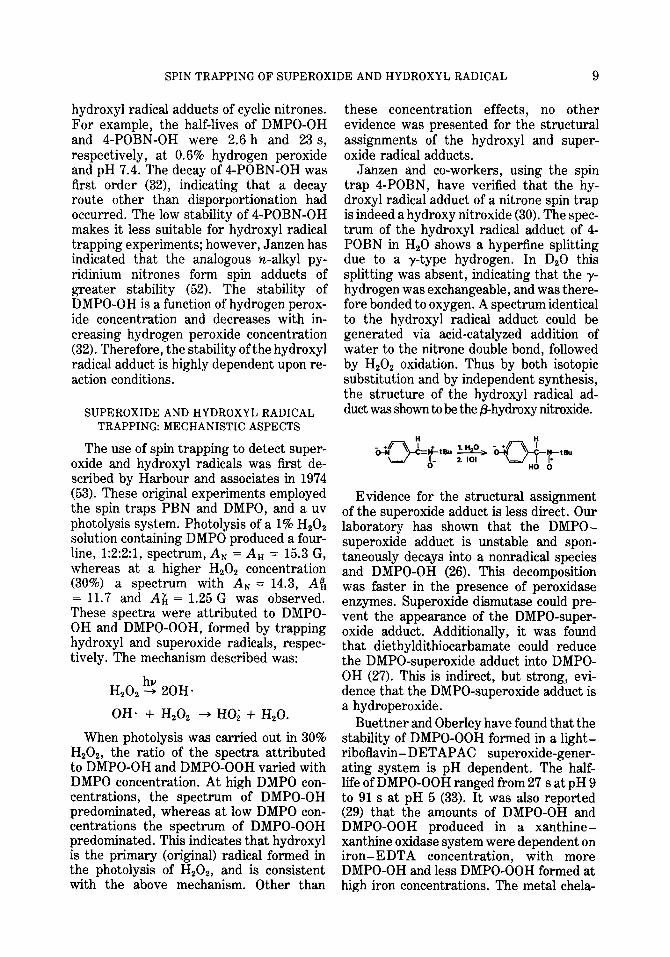
Janzen and co-workers, using the spin trap 4-POBN, have verified that the hy- droxyl radical adduct of a nitrone spin trap is indeed a hydroxy nitroxide (30). The spec- trum of the hydroxyl radical adduct of 4- POBN in H,O shows a hypeffine splitting due to a y-type hydrogen. In DzO this splitting was absent, indicating that the y- hydrogen was exchangeable, and was there- fore bonded to oxygen. A spectrum identical to the hydroxyl radical adduct could be generated via acid-catalyzed addition of water to the nitrone double bond, followed by H,O, oxidation. Thus by both isotopic substitution and by independent synthesis, the structure of the hydroxyl radical ad- duct was shown to be the phydroxy n&oxide.
HO 0
Evidence for the structural assignment of the superoxide adduct is less direct. Our laboratory has shown that the DMPO- superoxide adduct is unstable and spon- taneously decays into a nonradical species and DMPO-OH (26). This decomposition was faster in the presence of peroxidase enzymes. Superoxide dismutase could pre- vent the appearance of the DMPO-super- oxide adduct. Additionally, it was found that diethyldithiocarbamate could reduce the DMPO-superoxide adduct into DMPO- OH (27). This is indirect, but strong, evi- dence that the DMPO-superoxide adduct is a hydroperoxide.
Buettner and Oberley have found that the stability of DMPO-OOH formed in a light- riboflavin-DETAPAC superoxide-gener- ating system is pH dependent. The half- life of DMPO-OOH ranged from 27 s at pH 9 to 91 s at pH 5 (33). It was also reported (29) that the amounts of DMPO-OH and DMPO-OOH produced in a xanthine- xanthine oxidase system were dependent on iron-EDTA concentration, with more DMPO-OH and less DMPO-OOH formed at high iron concentrations. The metal chela-
10 FINKELSTEIN, ROSEN, AND RAUCKMAN
TABLE II
APPARENT RATE CONSTANTS FOR SUPEROXIDE TRAPPING”
Spin trap Rate constant
M-l s-L Conditions Reference
TMPO 7
TMPO 1.44
DMPO 10
DMPO 15.7
pH 7.8 xanthine-xanthine oxidase, competition with SOD
pH 8.1 xanthine-xanthine oxidase, competition with spontaneous dis- mutation of superoxide
pH 7.8 xanthine-xanthine oxidase, competition with TMPO
pHEl.Olight-riboflavin-DETAPAC, competition with SOD
26
32
26
32
a Rates were determined by kinetic competition.
tor DETAPAC could prevent the effects of iron. Based on these data, it was suggested that DETAPAC prevents the formation of hydroxyl radical in this system. This has since been confirmed by Halliwell (54), using the hydroxylation of salicylate as a method for detecting hydroxyl radical.
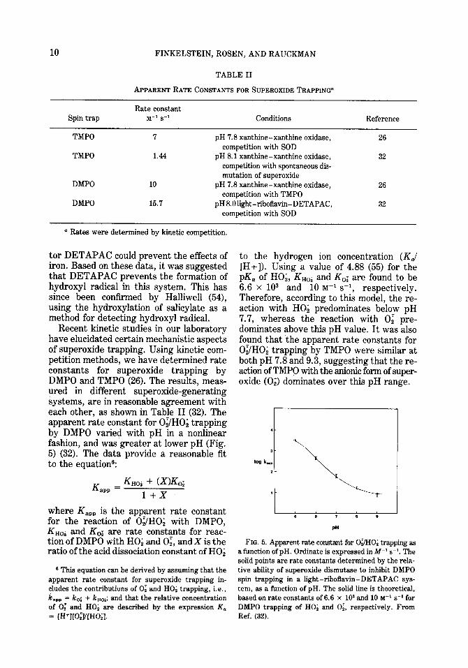
Recent kinetic studies in our laboratory have elucidated certain mechanistic aspects of superoxide trapping. Using kinetic com- petition methods, we have determined rate constants for superoxide trapping by DMPO and TMPO (26). The results, meas- ured in different superoxide-generating systems, are in reasonable agreement with each other, as shown in Table II (32). The apparent rate constant for Oi/HO, trapping by DMPO varied with pH in a nonlinear fashion, and was greater at lower pH (Fig. 5) (32). The data provide a reasonable fit to the equation?
K am = K~oi + UK,;
1+x
where K,,, is the apparent rate constant for the reaction of Oi/HO, with DMPO, K nOi and K,; are rate constants for reac- tion of DMPO with HO; and O& and X is the ratio of the acid dissociation constant of HO;
8 This equation can be derived by assuming that the apparent rate constant for superoxide trapping in- cludes the contributions of 0; and HO; trapping, i.e., km = k,,; + kHOi; and that the relative concentration of 0: and HO; are described by the expression K,
to the hydrogen ion concentration (K,/ [H+]). Using a value of 4.88 (55) for the pKa of HO,, KHoi and K,; are found to be 6.6 x lo3 and 10 M-’ s-l, respectively. Therefore, according to this model, the re- action with HO, predominates below pH 7.7, whereas the reaction with 02 pre- dominates above this pH value. It was also found that the apparent rate constants for Oi/HO, trapping by TMPO were similar at both pH 7.8 and 9.3, suggesting that the re- action of TMPO with the anionic form of super- oxide (0,) dominates over this pH range.
FIG. 5. Apparent rate constant for OBO; trapping as a function of pH. Ordinate is expressed in M-l s-l. The solid points are rate constants determined by the rela- tive ability of superoxide dismutase to inhibit DMPO spin trapping in a light-riboflavin-DETAPAC sys- tem, as a function of pH. The solid line is theoretical, baaed on rate constants of 6.6 x lo3 and 10 M-’ s-* for DMPO trapping of HO; and O;, respectively. From Kef. (32). = [H+J[O;J/[HO;l.
SPIN TRAPPING OF SUPEROXIDE AND HYDROXYL RADICAL 11
The rate constants for 0, trapping are quite low compared to the rate constants of other molecules used to detect 0; (56). For example, the rate constants for reaction of 0; with cytochrome c and tetranitromethane are about 6 x lo5 and 2 x log M-’ s,
respectively (57,56). The low rate constants for 0, trapping means that relatively high spin-trap concentrations must be used to trap superoxide before it can decompose via spontaneous dismutation.
Rate constants have also been deter- mined for reaction of hydroxyl radical with nitrone spin traps, using kinetic competi- tion methods. In contrast to the low rate constants found for superoxide trapping, spin traps are efficient detectors of hydroxyl radical, as shown by the data in Table III.
RECENT STUDIES ON TRAPPING OF SUPEROXIDE AND HYDROXYL
RADICALS IN VARIOUS
SYSTEMS
The use of nitrone spin traps for detecting superoxide and hydroxyl radicals is be- coming increasingly popular, with new studies appearing regularly. Since many of these studies have been reviewed in detail elsewhere (15), the following discussion mainly represents a brief summary of this literature.
Microsomal Systems
Work in Piette’s laboratory has been con- cerned with elucidating reactive inter- mediates in iron-catalyzed lipid peroxida- tion. In a study by Saprin and Piette (58, the formation of PBN adducts by a liver microsomal system containing NADPH and iron pyrophosphate was investigated. The authors found that the adducts obtained were dependent upon the “substrate” added to the system. For example, when Tris buf- fer was used in place of phosphate, a Tris radical adduct was trapped. When ethanol was added to the system, an ethanol radical adduct was trapped. The production of “substrate” radicals was attributed to the initial formation of hydroxyl radical, fol- lowed by the reaction of this radical with the substrate to form a substrate-derived radical. This was verified by independent
TABLE III
RATE CONSTANTSFOR HYDROXYL RADICALTRAPPING"
Spin trap
DMPO
TMPO 4-POBN
a From Ref. (32).
Rate constant (M-' S-l)
3.4 x 10⁹
3.8 x lo9 1.9 x 10⁹
generation of substrate radicals via uv photolysis of aqueous hydrogen peroxide containing these substrates and PBN. The same adducts which were observed in the photolysis system were seen in micro- somes. Although PBN-OH production by microsomes was not detected in this study, it was detected in later studies by Lai and Piette, when the incubation conditions were changed (44). The production of DMPO-OH and PBN-OH was attributed to the trapping of hydroxyl radical produced by the Fenton reaction, since catalase (59), but not super- oxide dismutase (60), could prevent the formation of DMPO-OH. It was also shown that a system consisting of Fe*+-EDTA, purified NADPH cytochrome P-450 re- ductase, and NADPH could produce DMPO-OH (60). Linolenic acid could inhibit the production of DMPO-OH. The produc- tion of DMPO-OH was dependent on pH, Fez+-EDTA, and KC1 concentrations, and was inhibited by p-chloromercuribenzoate, but not by metyrapone (59, 60). The inhibi- tion of MDA production by DMPO was at- tributed to trapping of hydroxyl radical (59). It may alternatively be due to the oxidation of DMPO into its hydroxamic acid. The hydroxamic acid binds iron tightly, thus the inhibition of MDA formation could be due to there being less iron avail- able to catalyze lipid peroxidation.
Floyd and co-workers (61) have detected DMPO-OH and another adduct in a system consisting of nitrosamine carcinogens, and either microsomes or nuclei. DMPO-OH was seen in all instances, but the other adduct varied with the nitrosamine, sug- gesting that it was due to a nitrosamine- derived radical. Oxygen was necessary for
12 FINKELSTEIN, ROSEN, AND RAUCKMAN
the production of DMPO-OH, but it de- creased the yield of the trapped nitrosamine radical. More controls need to be performed to ascertain whether DMPO-OH production is indeed due to hydroxyl radical trapping in this system.
Sealy et al. have used DMPO spin trap- ping to detect superoxide formed during the aerobic microsomal reduction of aromatic nitro compounds (43). This is consistent with previous studies by Mason and Holtz- man who have proposed that superoxide is formed during such reactions (62-64).
Photochemical Systems
Harbour and associates have used spin trapping to detect superoxide and hydroxyl radical production in diverse photochemical systems. Harbour and Bolton (65) could de- tect superoxide production by light-ir- radiated chloroplasts using DMPO. Methyl viologen increased the production of the superoxide adduct. Based on this, it was suggested that methyl viologen accepts electrons from the primary electron accep- tor of photosystem I.
Harbour and Hair have used spin trap- ping to detect superoxide production by light irradiation of CdS (66,67) and phthalo- cyanine (67) pigments in both aqueous and nonaqueous systems. The production of DMPO-OOH by irradiation of CdS was enhanced by EDTA, and by a cationic surfactant, but not an anionic surfactant. Although the effect of EDTA was at- tributed to its acting as an electron donor, an alternate explanation is the ability of EDTA to render Cu2+ impurities ineffec- tive as superoxide dimutases (68). Solvent effects on the hyperfme splitting of the DMPO-OOH adduct spectrum were also studied (67). (These solvent effects are also discussed in detail by Janzen in Refs. (15) and (69).)
A study on the production of hydroxyl radical and its involvement in the destruc- tive photooxidation of chlorophylls has been reported by Harbour and Bolton (70). DMPO-OH was produced during the il- lumination of both chloroplast suspensions, and detergent-solubilized chlorophyll preparations. The production of hydroxyl
radical was verified by the ability of formate to inhibit DMPO-OH production, with the resultant production of the CO, adduct of DMPO, i.e.:
‘OH + H-t’ - t$O + ‘q ‘0-
DMFU + ‘Cq- - OMPO-co;
The photodestruction of chlorophyll A could be monitored optically, and was inhibited by DMPO, as expected.
Buettner and Oberley have demonstrated that superoxide can be produced by light irradiation of protoporphyrin, using DMPO as a spin trap. The production of DMPO- OOH was enchanced by prior bubbling with oxygen, and was prevented by superoxide dismutase (71). The relationship between superoxide production in this system and the cutaneous photosensitivity of indi- viduals with protoporphyria was discussed.
An excellent study by Sargent and Gardy investigated the ability of the spin traps PBN, DMPO, and NtB to trap H ., solvated electrons, and hydroxyl radical formed by 3(MeV) electron radiolysis of water (17). Structural assignments of the spectra were verified by isotopic substitution, inde- pendent synthesis, computer simulation, or the effects of various competitive inhibitors. It was concluded that “DMPO was an excel- lent spin trap for such studies.” This paper should prove useful both for the spectra shown, and for its methodology.
Hydroxyl Radical Production by Antibiotics
Studies by Buettner and Oberley have used spin trapping in an attempt to demon- strate hydroxyl radical production in Fe2+- bleomycin (72) and Cu2+-tallysomycin (73) systems. In both of these studies the pro- duction of DMPO-OH required both the metal ion and the antibiotic. However, these studies did not take into account that DMPO-OH may arise by mechanisms other than hydroxyl radical trapping. Thus the studies are inconclusive, in themselves.
Lown and co-workers (74) have used PBN in an attempt to detect hydroxyl radical produced by a system containing a sodium borohydride-reduced aminoquinone anti-
SPIN TRAPPING OF SUPEROXIDE AND HYDROXYL RADICAL 13
biotic (either mitomycin B, mitomycin C, or streptonigrin), 0.06 to 0.2 M PBN, and 10% DMSO. The authors could detect a PBN ad- duct,A, = 16.OA,, = 3.4 G, whoseproduc- tion was inhibited by either catalase, superoxide dismutase, or EDTA. The identical PBN adduct could be detected in a Fez+-H,Oz system containing 0.06 M
PBN, and 10% DMSO. The authors as- signed this adduct to PBN-OH produced by hydroxyl radical trapping. Although hy- droxyl radical is likely produced in their system, their assignment of the PBN ad- duct is probably incorrect for the follow- ing reasons:
(i) Since both the concentration of DMSO (1.4 M) and its rate of reaction with hy- droxyl radical (7 x log M-’ s-l (75)) were much greater than the PBN concentration and rate constant, most of the hydroxyl radical would have reacted with DMSO. Thus the adduct observed is possibly due to a secondary radical derived from DMSO, such as CH,. (76).
(ii) The direction of the “solvent effect” on the spectrum of PBN-OH due to 10% DMSO, which the authors described, is in- correct. AN and A, should be decreased in a less polar solvent, and not increased as described (35). The change in hyperfine splitting is due to a different radical (i.e., from DMSO) being trapped.
(iii) PBN-OH is less stable than alkyl radical adducts of PBN, thus if any PBN- OH were formed, it would likely have de- cayed during the extended incubation used. (See the discussion in Refs. (44, 58). The spectrum shown by the authors is probably the same as “DMSO signal A” described by Saprin and Piette (58).
Another study has been published de- scribing the detection of PBN-OH and PBN-OOH produced by the interaction of Fez+-bleomycin with PBN (77). Both Fe*+ and oxygen were required to produce an EPR signal. When cobalt was used instead of iron, a weak PBN-OH signal was de- tected; however, neither Cu2+ nor Zn2+ were effective in producing an EPR signal. PBN-OOH could only be detected at low Fe2+-bleomycin concentrations; at higher Fe2+-bleomycin concentrations the spec- trum of PBN-OH predominated.
Miscellaneous Studies
The first use of spin trapping in an intact living cell has been described by Green and co-workers in a study on superoxide and hydroxyl radical production by stimulated human polymorphonuclear neutrophils (78). Both DMPO-OH and DMPO-OOH could be detected, depending upon the stimulator used. The production of both species was prevented by superoxide dismutase, but not by catalase, and was increased by cyanide or azide. The authors suggested that DMPO-OH production is due to hy- droxyl radical trapping, and that the hy- droxyl radical is generated by a mechanism which does not involve “free” hydrogen peroxide. Based on our observations con- cerning spin trapping in the xanthine-xan- thine oxidase system (26), an alternate ex- planation is that DMPO-OH production is due largely to the decomposition of DMPO- OOH formed by superoxide trapping. The effects of cyanide and azide may therefore be due to their inhibition of endogenous superoxide dismutase (79) or superoxide dismutase present as an impurity from erythrocytes. Even small amounts of super- oxide dismutase can cause a large inhibition of spin trapping, due to the low rate con- stant for superoxide trapping by DMPO (26, 32).
A report by Floyd and Wiseman (80) has described the production of DMPO-OH dur- ing the autoxidation of 6-hydroxydopamine; SOD, and catalase only partially inhibited DMPO-OH formation. Oxygen was re- quired for DMPO-OH formation. The authors were also able to detect the 6- hydroxydopamine semiquinone radical. The chelators DETAPAC and deferoxa- mine could inhibit both DMPO-OH and 6- hydroxydopamine semiquinone formation, suggesting the involvement of iron in their formation.
SUMMARY
Reports are regularly appearing in the literature describing spin trapping of super- oxide and hydroxyl radicals from various sources. Careful scrutiny of these reports will often reveal that insufficient controls have been run to properly validate the
14 FINKELSTEIN, ROSEN, AND RAUCKMAN
results. Nitrones are highly reactive com- pounds which can form nitroxides by mech- anisms other than radical trapping. Thus, investigators using spin trapping should be cognizant of the many artifacts which accompany this technique, and take care to validate their results with satisfactory con- trols. We have described straightforward procedures to determine whether super- oxide or hydroxyl radical trapping have occurred, and which can help verify the assignment of the radical adduct.
Nitrones are the only spin traps cur- rently suitable for detection of hydroxyl and superoxide radicals. The various nitrone spin traps in current use each have advantages and disadvantages. In general, the cyclic nitrone traps such as DMPO have greater reactivity with superoxide and hydroxyl radicals, are less readily hy- drolyzed, but are more susceptible to oxy- gen and light, and thus have lesser shelf lives. Aryl nitrones such as 4-POBN or PBN have lesser reactivity with superoxide and hydroxyl radicals, are more readily hy- drolyzed, but have greater shelf lives. The stability of DMPO-OH is also greater than that of 4-POBN-OH. Thus in general, DMPO appears to be the most versatile spin trap currently available.
Spin trapping is an inefficient means of detecting superoxide, due to the low rate constants for spin trapping. Spin traps pos- sessing a p-hydrogen will also form unstable superoxide adducts. Spin trapping will, however, undoubtedly prove useful in de- tecting superoxide under conditions where more conventional methods, such as cyto- chrome c reduction, cannot be used.
ACKNOWLEDGMENT
The authors are grateful for the helpful discus- sions with Dr. Irwin Fridovich on superoxide and superoxide dismutsse.
REFERENCES
1. BOLTON, J. R., BORG, D. C., AND SWARTZ, H. M. (1972) in Biological Applicaltions of Electron Spin Resonance (Swartz, H. M., Bolton, J. R., and Borg, D. C., eds.), pp. 63-118, Wiley- Interscience, New York.
2. BORG, D. C. (19’76) in Free Radicals in Biology
(Pryor, W. A. ed.), Vol. 1, pp. 69-147, Aca- demic Press, New York.
3. BEINERT, H. (1972) in Biological Applications of Electron Spin Resonance (Swartz, H. M., Bol- ton, J. R., and Borg, D. C., eds.), pp. 351-410, Wiley-Interscience, New York.
4. YAMAZAKI, I., MASON, H. S., AND PIETTE, L. (1960) J. Biol. Chem. 235, 2444-2449.
5. BORG, D. C. (1972) in Biological Applications of Electron Spin Resonance (Swartz, H. M., Bolton, J. R., and Borg, D. C., eds.), pp. 265-350, Wiley-Interscience, New York.
6. OHNISHI, T., YAMAZAKI, H., IYANAGI, T., NAKAMURA, T., AND YAMAZAKI, I. (1969) Biochim. Biophys. Acta. 172, 357-369.
7. STONE, T., BUCKMAN, T., NORDIO, P., MCCON- NELL, H. (1965) Proc. Nat. Acad. Sci. USA 54,
1010-1017. 8. MASON, R. P. (1979) in Reviews in Biochemical
Toxicology (Hodgson, E., Bend, J. R., and Phil- pot, R. N., eds.), p. 151, ElsevieriNorth- Holland, Amsterdam.
9. DORFMAN, L. M., AND ADAMS, G. E. (1973) Re- activity of the Hydroxyl Radical in Aqueous solutions, NSRDS, National Bureau of Stand- ards, 46, Washington, D. C.
10. YAMAZAKI, I., AND PIETTE, L. H. (1961) Bio- chim. Biophys. Acta 50, 62-69.
11. JANZEN, E. G. (1971) Accounts Chem. Res. 4, 31-40.
12. JANZEN, E. G., AND LIU, J. I. P. (1973) J. Mug- netic Resonance 9, 510-512.
13. JANZEN, E. G., EVANS, C. A., AND LIU, J. I. P., (1973) J. Magnetic Resonance 9, 513-516.
14. JANZEN, E. G., EVANS, C. A., AND DAVIS, E. R. (1978) in Organic Free Radicals (Pryor, W. A., ed.), ACS Symposium Series 69, pp. 433-446, Amer. Chem. Sot., Washington, D. C.
15. JANZEN, E. G. in Free Radicals in Biology (Pryor, W. A., ed.), Vol. IV, Academic Press, New York, in press.
16. EVANS, C. A. (1979) Aldrichim. Acta 12, 23-29.
17. SARGENT, F. P., AND GARDY, E. M. (1976) Canad. J. Chem. 54, 275-279.
18. KALYANARAMAN, B., PEREZ-REYES, E., AND MASON, R. P., submitted for publication.
19. LAI, C. S., AND PIE=E, L. H. (1979) Tetrahedron Lett. 9, 775.
20. WARGON, J. A., AND WILLIAMS, F. (1972) J. Amer. Chem. Sot. 94, 7917-7918.
21. MACKOR, A., WAJER, TH. A. J., DEBOER TH. J., AND VAN VOORST, J. D. W., (1967) Tetm- hedron. Lett., 385.
22. HAMER, J., AND MACALUSO, A. (1964) Chm. Rev. 64, 473-495.
23. ELSWORTH, J. F., AND LAMCHEN, M. (1971) J. S. Afr. Chem. Inst. 24, X86-204.
SPIN TRAPPING OF SUPEROXIDE AND HYDROXYL RADICAL 15
24. FLOYD, R. A., AND SOONG, L. A. (1977) B&hem.
Biophys. Res. Commun. 74, 79-84.
25. ROSEN, G. M., AND RAUCKMAN, E. J. (1980) Mol.
Pharmacol. 17, in press.
26. FINKELSTEIN, E., ROSEN, G. M., RAUCKMAN, E. J., AND PAXTON, J. (1979) Mol. Pharmacol.,
16, 676-685. 27. FINKELSTEIN, E., ROSEN, G. M., AND RAUCK-
MAN, E. J., unpublished observations.
28. BROWN, R. F. C., CLARK, V. M., LAMCHEN, M., AND TODD, A. (1959) J. Chem. Sot. Pt. II,
2116-2122. 29. BUETTNER, G. R., OBERLEY, L. W., AND
LEUTHAUSER, S. W. H. G. (1978) Photo&m.
Photobiol. 28, 693-695. 30. JANZEN, E. G., WANG, Y. Y., AND SHETTY, R. V.
(1978) J. Amer. C&m. Sot. 100, 2923-2925.
31. BONNETT, R., CLARK, V. M., AND TODD, A.
(1959) J. Chem. Sot., 2102-2104. 32. FINKELSTEIN, E., ROSEN, G. M., AND RAUCK-
MAN, E. J., submitted for publication.
33. BUETTNER, G. R., AND OBERLEY, L. W. (1978)
Biochem. Biophys. Res. Commun. 83, 69-74.
34. ADAMS, E. G., AND WARDMAN, P. (1977) in Free
Radicals in Biology (Pryor, W. A., ed.), Vol. III,
pp. 53-95, Academic Press, New York.
35. SMITH, I. C. P. (1972) in Biological Applications
of Electron Spin Resonance (Swartz, H. M., Bolton, J. R., and Borg, D. C., eds.), pp.
483-539, Wiley-Interscience, New York.
36. GAFFNEY, B. J. (1976) in Spin Labeling Theory and Application (Berliner, L. J., ed.), pp. 183- 238, Academic Press, New York.
37. LIKHTENSHTEIN, G. T. (1976) in Spin Labeling Methods in Molecular Biology, pp. 190-232, Wiley-Interscience, New York.
38. STIER, A., AND SACKMAN, E. (1973) Biochim.
Biophys. Acta 311, 400-408.
39. ROSEN, G. M., AND RAUCKMAN, E. J. (1977)
Biochem. Pharvnacol. 26, 675-678.
40. SHORR, R. I., RAUCKMAN, E. J., AND ROSEN,
G. M., unpublished observations.
41. GOLDBERG, J. S., RAUCKMAN, E. J., AND ROSEN, G. M. (1977) Biockm. Biophys. Res. Commun.