19
SPINAL PAIN Mr. Yagnesh Vellore FRACS Neurosurgeon and Spine Surgeon
SPINAL PAIN
Mr. Yagnesh Vellore FRACS Neurosurgeon and Spine Surgeon
Mr. Yagnesh Vellore FRACSNeurosurgeon & Spine SurgeonProvider ° 242453RXP: 03 9429 7888F: 03 9429 [email protected] Erin Street, Richmond, Vic 3121www.advancedneurosurgery.com.au
Mr. Yagnesh Vellore FRACSNeurosurgeon & Spine SurgeonProvider ° 242453RXP: 03 9429 7888F: 03 9429 [email protected] Erin Street, Richmond, Vic 3121www.advancedneurosurgery.com.au

PAIN GENERATORS IN THE SPINE
• Ligaments: ALL,PLL • Muscle • Periosteum bone • Outer 1/3 annulus disc • Facet joints • Sacro-iliac joint
• sinuvertebral N (first branch of sp N outside foramen) supplies posterior disc, dura, PLL, ( re enters foramen)
• Medial branch of the dorsal ramus supplies facet joint, ligament,vertebral arch, spinous process and paraspinal muscles
• Gray Ramus communicans from sympathetic trunk innervate the anterior & lateral aspect of the disc.
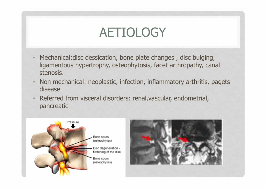
AETIOLOGY
• Mechanical:disc dessication, bone plate changes , disc bulging, ligamentous hypertrophy, osteophytosis, facet arthropathy, canal stenosis.
• Non mechanical: neoplastic, infection, inflammatory arthritis, pagets disease
• Referred from visceral disorders: renal,vascular, endometrial, pancreatic
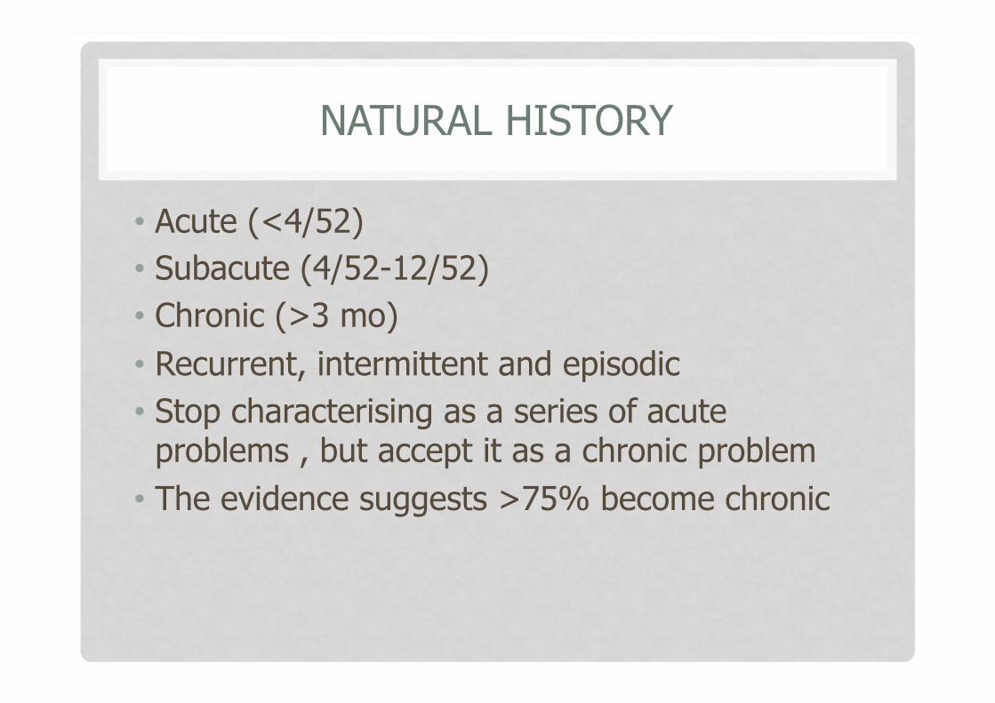
NATURAL HISTORY
• Acute (<4/52) • Subacute (4/52-12/52) • Chronic (>3 mo) • Recurrent, intermittent and episodic • Stop characterising as a series of acute
problems , but accept it as a chronic problem • The evidence suggests >75% become chronic

Date of download: 10/25/2013
Copyright © The American College of Physicians. All rights reserved.
From: Diagnosis and Treatment of Low Back Pain: A Joint Clinical Practice Guideline from the American College of Physicians and the American Pain Society
Ann Intern Med. 2007;147(7):478-491. doi:10.7326/0003-4819-147-7-200710020-00006
Initial evaluation of low back pain (LBPDo not use this algorithm for back pain associated with major trauma, nonspinal back pain, or back pain due to systemic illness. CRP = C-reactive protein; EMG = electromyography; ESR = erythrocyte sedimentation rate; MRI = magnetic resonance imaging; NCV = nerve conduction velocity.
Figure Legend:
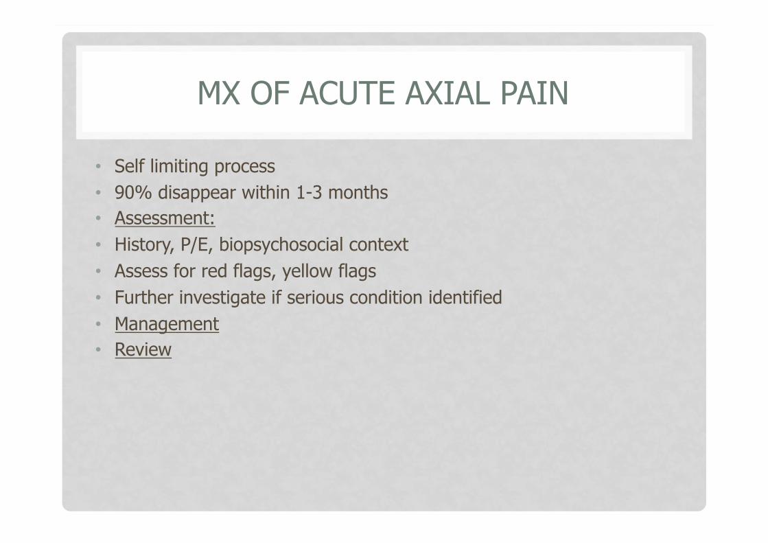
MX OF ACUTE AXIAL PAIN
• Self limiting process • 90% disappear within 1-3 months • Assessment: • History, P/E, biopsychosocial context • Assess for red flags, yellow flags • Further investigate if serious condition identified • Management • Review
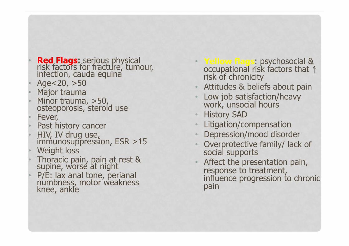
• Red Flags: serious physical risk factors for fracture, tumour, infection, cauda equina
• Age<20, >50 • Major trauma • Minor trauma, >50,
osteoporosis, steroid use • Fever, • Past history cancer • HIV, IV drug use,
immunosuppression, ESR >15 • Weight loss • Thoracic pain, pain at rest &
supine, worse at night • P/E: lax anal tone, perianal
numbness, motor weakness knee, ankle
• Yellow flags: psychosocial & occupational risk factors that ↑ risk of chronicity
• Attitudes & beliefs about pain • Low job satisfaction/heavy
work, unsocial hours • History SAD • Litigation/compensation • Depression/mood disorder • Overprotective family/ lack of
social supports • Affect the presentation pain,
response to treatment, influence progression to chronic pain

MANAGEMENT
• Provide information on nature of pain • Reassure natural history optimistic, address fears • Provide advice to remain active, resume normal activities as soon as
possible • Encourage activities to restore function, & avoid disability • General exercise program helps pt with chronic, subacute ,
postsurgical pain. • Non pharmacological Tx: passive: heat/massage/TENS, active:
strengthening, stretching • Pharmacological: paracetamol/NSAIDS/ opioids not indicated but if
used for severe pain , should be S.A, regular, not on pain contingent basis. Adjuvants : TCA, benzo,AED not indicated for acute pain.
• Identify concerns that may affect Mx

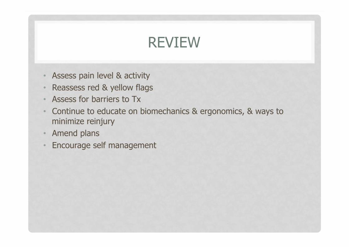
REVIEW
• Assess pain level & activity • Reassess red & yellow flags • Assess for barriers to Tx • Continue to educate on biomechanics & ergonomics, & ways to
minimize reinjury • Amend plans • Encourage self management

WADDELLS SIGNS OF NON ORGANIC BEHAVIOUR
• Presence >3 suggest non organic pain & more thorough assessment be made with psychological intervention
• tenderness: superficial, skin rolling or pinching, non anatomical • simulation: pain in back on axial loading head, rotation hip /
shoulders in line • distraction: SLR v sitting pt up in bed with legs at 90° • regional disturbances: sensory & motor loss non dermatomal/
anatomic • over reaction to pain stimulus, Sx magnified
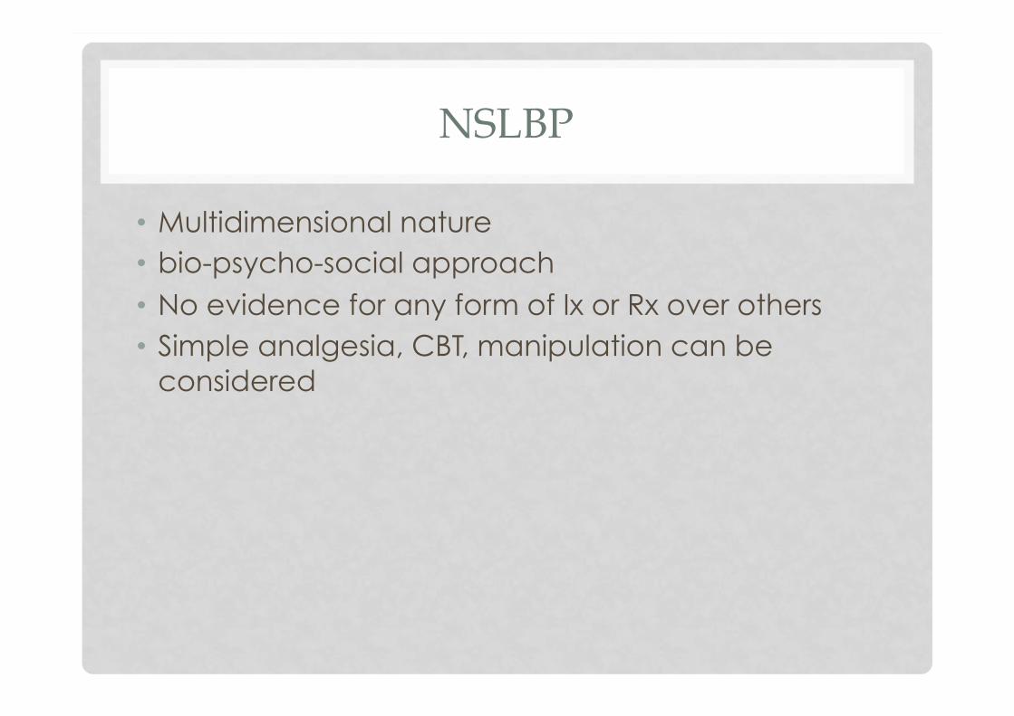
NSLBP
• Multidimensional nature • bio-psycho-social approach • No evidence for any form of Ix or Rx over others • Simple analgesia, CBT, manipulation can be
considered

INVESTIGATION
• CT/MRI to exclude red flag conditions / radiculopathy / LCS
• Local anaesthetic blocks to diagnose ligament sprain eg interspinous block
• Bone scan: diagnose painful phase of spondylolysis
• SI joint block
• Facet block
• Discogram

INTERVENTIONS

INTERVENTIONS

FACET SYNDROME
• Local pain, worse with movement, axial loading • Diagnostic blocks • RF of MB
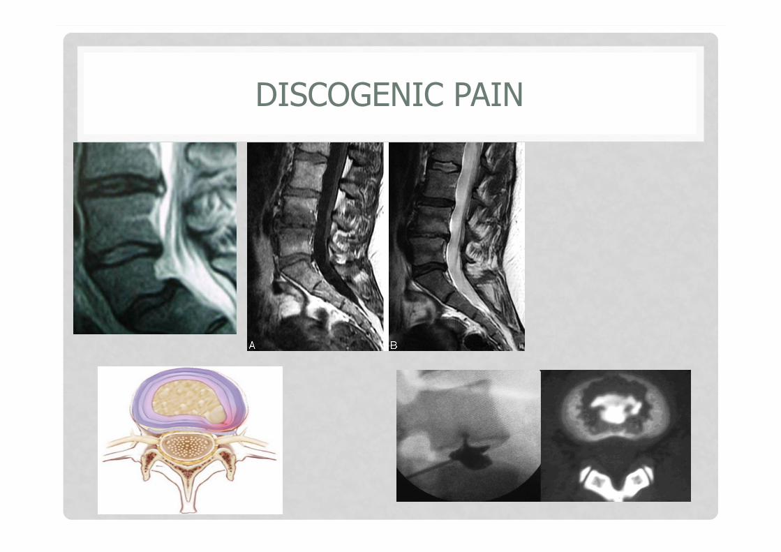
DISCOGENIC PAIN
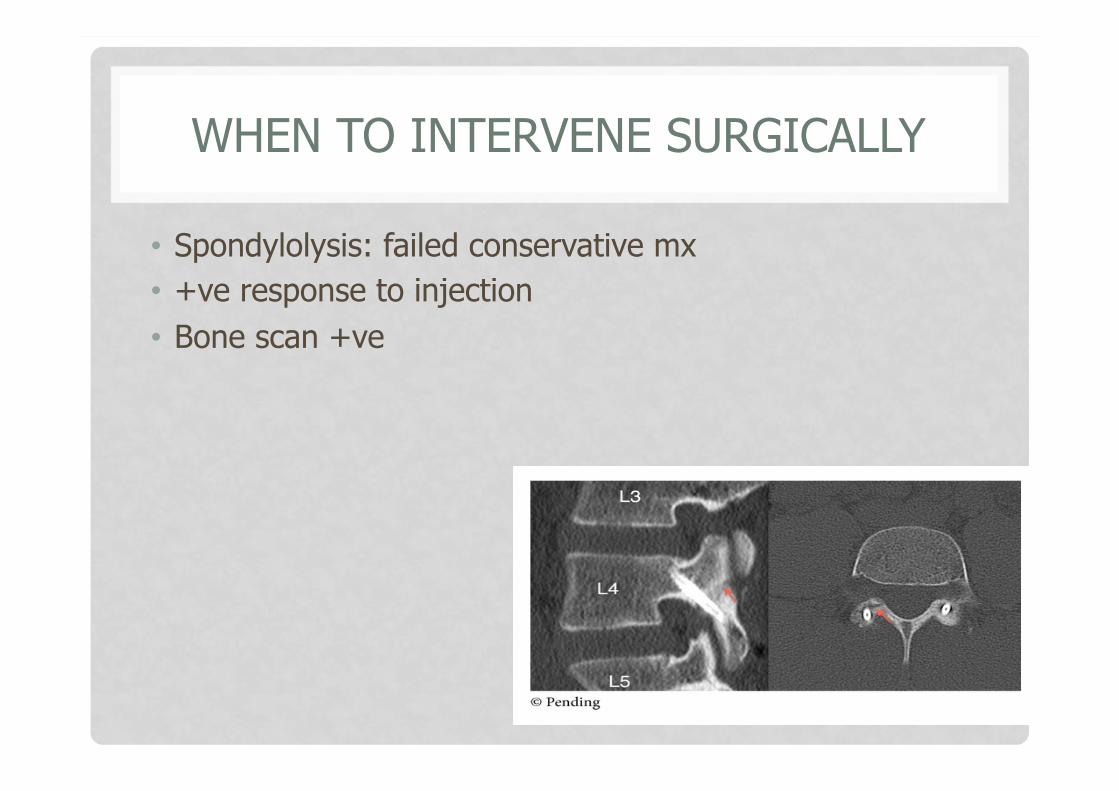
WHEN TO INTERVENE SURGICALLY
• Spondylolysis: failed conservative mx • +ve response to injection • Bone scan +ve

WHEN TO INTERVENE SURGICALLY
• Discogenic back pain
• IDET • TDR • Dynamic
stabilization • Fusion- ALIF/PLIF/
TLIF/PLF/360/DLF


