36
Spirochete infections • Boreliosis (Lyme disease) Borrelia burgdorferi • Syphylis (Lues) Treponema pallidum
| Date post: | 26-Dec-2015 |
| Category: |
Documents |
| Upload: | everett-kenneth-phelps |
| View: | 220 times |
| Download: | 3 times |

Spirochete infections
• Boreliosis (Lyme disease)Borrelia burgdorferi
• Syphylis (Lues)Treponema pallidum

Borrelia burgdorferi

Summary of reported cases of Lyme disease in the United States.
Hildenbrand P et al. AJNR Am J Neuroradiol 2009;30:1079-1087
©2009 by American Society of Neuroradiology

Lyme disease

Lyme disease
Skin lesionAfter tick bite

Erythema migrans rash with the typical target appearance that is virtually diagnostic of Lyme disease.
Hildenbrand P et al. AJNR Am J Neuroradiol 2009;30:1079-1087
©2009 by American Society of Neuroradiology

Lyme disease

Clinical feature
• 10-15% patients with untreated borreliosis – neuroborelliosis
• Primary location – ganglia od posterior roots, nerv roots, leptomeninges (macacus rhesus)
• Europe –– B. garinii

Clinical feature Periferal NS
• Sensory symptoms
• Painful radiculitis
• Painful lymphocytic meningoradiculitis – with/without paresis (Garin-Bujadoux-Bannwart syndróm)
• Facial palsy
• Pain – sharp, during night, weeks – months

Clinical feature Central NS
• Subsequent to the tick bite inoculation – B. reach reach the CNS
• Hematogenously or retrogradely via periferal nerves
• Encefalitis• Cranial neuritis • Motor or sensitive radikuloneuroitis • Encefalomyelitis - rare

Clinical feature late presentation
• Dementia – often in patients with artritis Desorientation, confusion, memory problems, cognitive dysfunction
• Chronic radikuloneuropathy – parestesia of acral parts, pain,
• EMG – axonal lesion

Diagnosis
• EMG – axonal lesion
• CSF:
• pleocytosis – Ly, proteins intratecal antibodies IgM, IgG against BB
• PCR

Facial neuritis.
Hildenbrand P et al. AJNR Am J Neuroradiol 2009;30:1079-1087
©2009 by American Society of Neuroradiology

Evolving cranial neuritis. Enhancement n. III, V l.dx., VII l. sin.
Hildenbrand P et al. AJNR Am J Neuroradiol 2009;30:1079-1087
©2009 by American Society of Neuroradiology

A 50-year-old woman with a history of tick bite and erythema migrans rash treated with doxycycline, who had recurrent erythema migrans rash with
headache, fever, nausea, and nuchal rigidity.
Hildenbrand P et al. AJNR Am J Neuroradiol 2009;30:1079-1087
©2009 by American Society of Neuroradiology

A 74-year-old man with 2-year cognitive decline and memory loss.
Hildenbrand P et al. AJNR Am J Neuroradiol 2009;30:1079-1087
©2009 by American Society of Neuroradiology

A 56-year-old woman with neck, bilateral shoulder, and bilateral arm pain.
Hildenbrand P et al. AJNR Am J Neuroradiol 2009;30:1079-1087
©2009 by American Society of Neuroradiology

A 17-year-old boy with right papilledema and orbital pain and rule out pseudotumor.
Hildenbrand P et al. AJNR Am J Neuroradiol 2009;30:1079-1087
©2009 by American Society of Neuroradiology

Boreliosis
• Th: Doxycycline (2x100 mg/D, 2T) CSF negat.
• i.v. ceftriaxone – likvor pozit.

Syphilis (Lues)
•1/3 nontreated patients – neurovascular complications of syphylis

Neurosyphilis
• Patogenesis• Perivascular infiltration of the meninges, focal
meningeal inflammation – formation of hypertrophic meninges, or gumma,
• Inflammatory cells invide blood vessel wal – arteritis (luminal occlusion)
• Parenchymal involvement – gliosis in late stages• Ly infiltration of preganglionic portion of dorsal
roots and posterior columns atrophy of posterior columns

Neurosyfilis – meningitis
• CSF Ly, ↓ Glu, proteins
• Pozit. VDRL test

Neurosyfilis – meningovascular
• Endarteritis – small and medium vessels (MCA) – can be stroke etiology in young people !!!
• Focal signs
• AG: nerrowing of arteries
• MRI: multiple infarcts • Spinal artery – transversal myelitis


Demencia paralyticaProgressive paralysis
• Decreased cognitive functions, memory problems, pupils abnormality

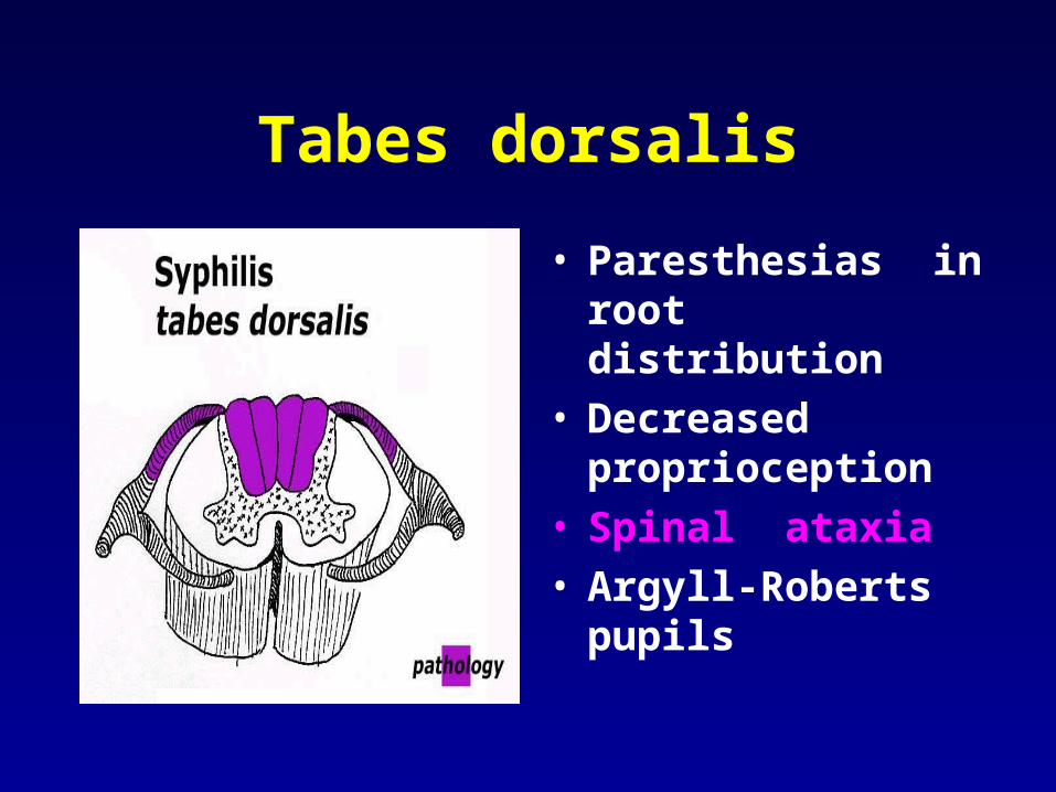
Tabes dorsalis
• Paresthesias in root distribution
• Decreased proprioception
• Spinal ataxia• Argyll-Roberts pupils


Acquired immunodeficiency syndrom (AIDS) Human immunodeficiency virus (HIV)
• I. stage
• Acute infection - 2. - 3. weeks after infection by HIV
• Symptoms like flu, or mononukleosis
• Acute retroviral syndrom

Stage II
• Period without symptoms – 2-10 years or more
• Decreased imunity in this period

Stage III, IV
• III – generalized lymphadenopathy, enlargement of LN
• IV- stage of AIDS • Weakened immune system fails • Fewer, lost of weight, weakness, fatigue,
muscle atrophy

Acquired immunodeficiency syndrom (AIDS)
• IV stage - symptoms of lesion of PNS and CNS
• Aseptic meningitis
• Cognitive decline
• Myelopathy • Neuropathy (inflammatory demyelinating
polyneuropathy, mononeuropathy, plexopathy)
• Myopathy – myositis

AIDS dementia complex (ADC)
• T2- MRI:
• Enlargement of ventricles
• Hyperintensity in subcortical white matter of frontal lobes
• Brain atrophy

HIV Opportunistic Infections
• People with advanced HIV infection are vulnerable to infections and malignancies that are called 'opportunistic infections' because they take advantage of the opportunity offered by a weakened immune system.
• Bacterial diseases such as tuberculosis, Mycobacterium avium complex, bacterial pneumonia and septicaemia (blood poisoning)
• Protozoal diseases such as toxoplasmosis, microsporidiosis, cryptosporidiosis, isopsoriasis and leishmaniasis

HIV Opportunistic Infections
• Fungal diseases such as Pneumocystis pneumonia, candidiasis, cryptococcosis and penicilliosis
• Viral diseases such as those caused by cytomegalovirus, herpes simplex and herpes zoster virus
• HIV-associated malignancies such as Kaposi's sarcoma, lymphoma and squamous cell carcinoma.


