Page 1
Spirometry practical guide and test
interpretation
Use of spirometry pre- lung surgery
Dr Paula Agostini PhD, Specialist Physiotherapist
Heart of England NHS Foundation Trust
Feb 2018
Acknowledgements: Sarah Cameron, HEFT CF Dept
Page 2
What is spirometry? ‘Method of assessing lung function by
measuring the volume of air that the patient is able to expel from the lungs after a maximal inspiration’ (NICE 2004)
Differentiates - obstructive/restrictive disorders
Most effective way of determining severity (not signs/symptoms alone)
MRC dyspnoea scale/QOL
Page 3
Training
‘All healthcare professionals managing patients with COPD should be competent in the interpretation of the results of spirometry and all healthcare professionals performing spirometry should have undergone appropriate training and keep their skills up to date’ (NICE 2004)
Association of Respiratory Technology and Physiology (ARTP)/BTS
Certificate of Competence
Page 5
Types of spirometer
• Many different types £300 - £3000
• Hand held - FEV1 & FVC readings
• Advanced - visual/printable traces
• Electronic - flow-volume curve
• Calculate %age predicted normal
values
• Reports - defects, severity
Page 6
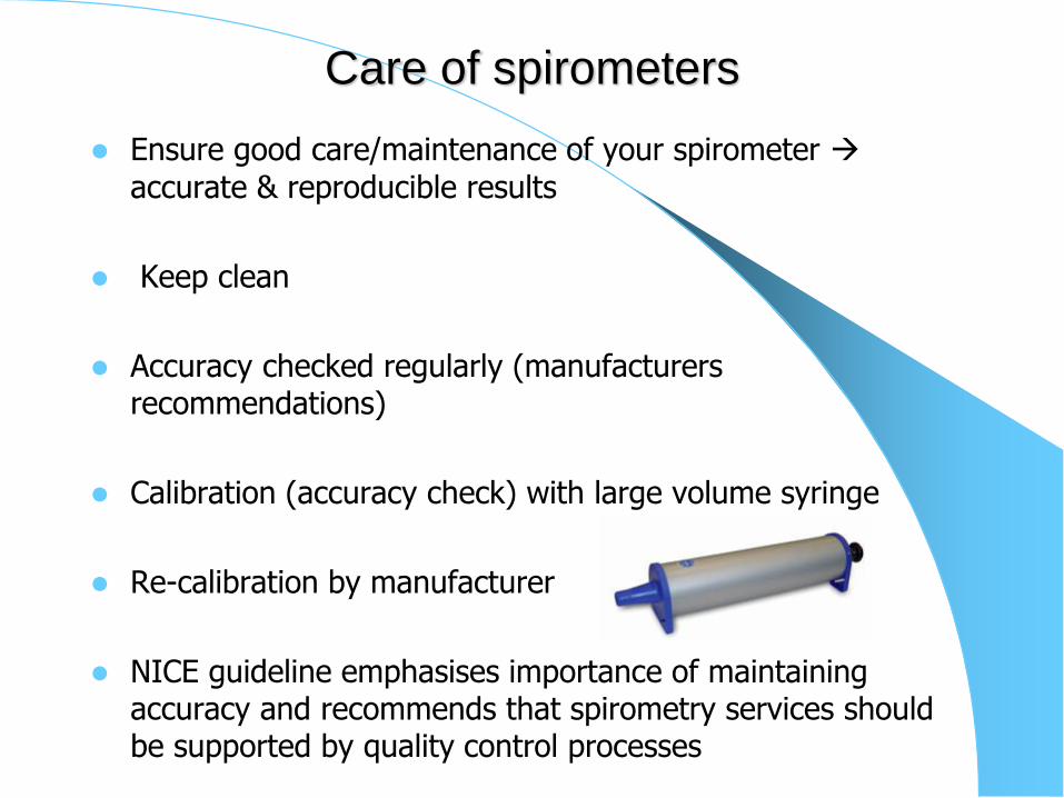
Care of spirometers
Ensure good care/maintenance of your spirometer
accurate & reproducible results
Keep clean
Accuracy checked regularly (manufacturers recommendations)
Calibration (accuracy check) with large volume syringe
Re-calibration by manufacturer
NICE guideline emphasises importance of maintaining accuracy and recommends that spirometry services should be supported by quality control processes
Page 7
Maintaining accuracy
Patient technique - most common reason for inconsistent readings
To detect errors observe patient and trace:
Inadequate/incomplete inhalation
Lack of ‘blast’ effort during exhalation
Additional breath taken during manoeuvre
Poor seal with mouthpiece
Slow start to forced exhalation
Exhalation stops before complete expiration
Some exhalation through the nose
Coughing
Page 8
Preparing the patient Comfortable/ seated
Explain purpose/demonstrate technique
Allow practice attempts
Encourage full exhalation
Limit total attempts to 8 or less/session
30 seconds rest between blows
Information:
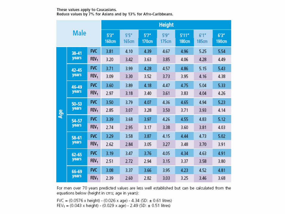
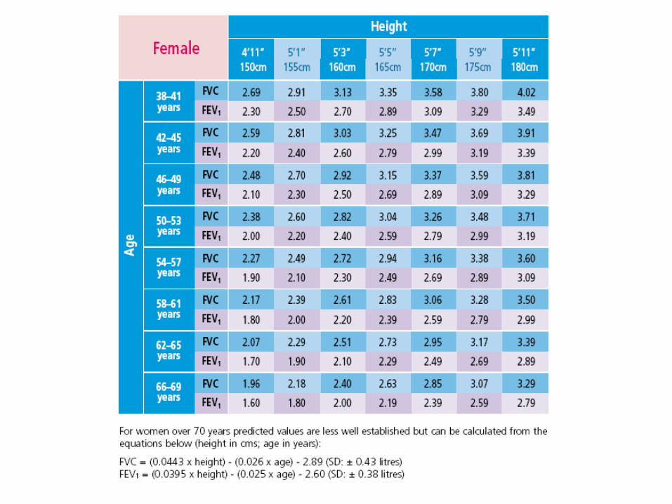
Age, gender, height
Adjust normal values (Asian/Afro-Carribean)
Note ??recent bronchodilator/exacerbation/pain
Page 9
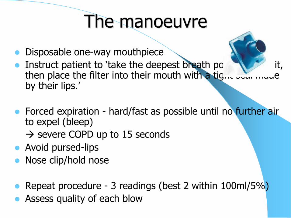
The manoeuvre
Disposable one-way mouthpiece
Instruct patient to ‘take the deepest breath possible, hold it, then place the filter into their mouth with a tight seal made by their lips.’
Forced expiration - hard/fast as possible until no further air to expel (bleep)
severe COPD up to 15 seconds
Avoid pursed-lips
Nose clip/hold nose
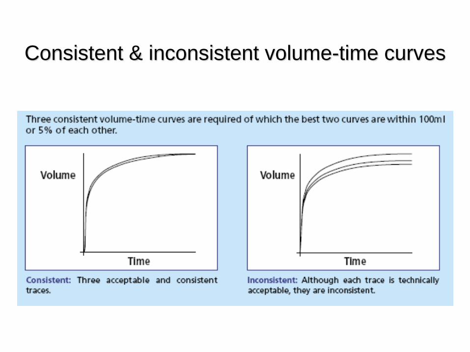
Repeat procedure - 3 readings (best 2 within 100ml/5%)
Assess quality of each blow
Page 10
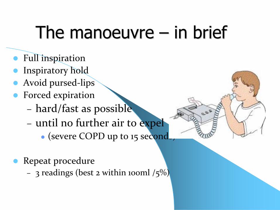
The manoeuvre – in brief
Full inspiration
Inspiratory hold
Avoid pursed-lips
Forced expiration
– hard/fast as possible
– until no further air to expel (severe COPD up to 15 seconds)
Repeat procedure– 3 readings (best 2 within 100ml /5%)
Page 11
Interpreting results
Best of 3 consistent readings (FEV1 & FVC)
Borderline normal results - repeat in few months to confirm diagnosis (especially > 75 years)
- Mild airflow obstruction FEV1 is between 50 and 80% of predicted normal &
FEV1/FVC is <0.7
Abnormality detected if any of following recorded:• FEV1 <80% predicted normal • FVC <80% predicted normal • FEV1/FVC ratio <0.7
Page 12
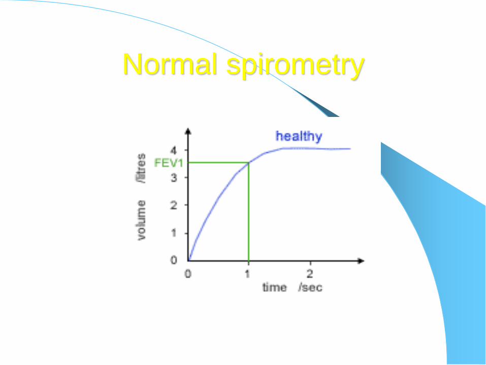
Normal spirometry
Page 13
- FEV1
- FVC- FEV1/FVC
- VC- FEV1/VC
- FEV1/FVC values based on age/gender/height- Predicted values lower in non-caucasions
Page 16
Consistent & inconsistent volume-time curves
Page 17
Identifying abnormalities
Obstructive disorder• FEV1 reduced (<80% predicted normal)• FVC usually reduced but to lesser extent than FEV1
• FEV1/FVC ratio reduced (<0.7)
Restrictive disorder• FEV1 reduced (<80% predicted normal)• FVC reduced (<80% predicted normal)• FEV1/FVC ratio normal (>0.7)
Page 18
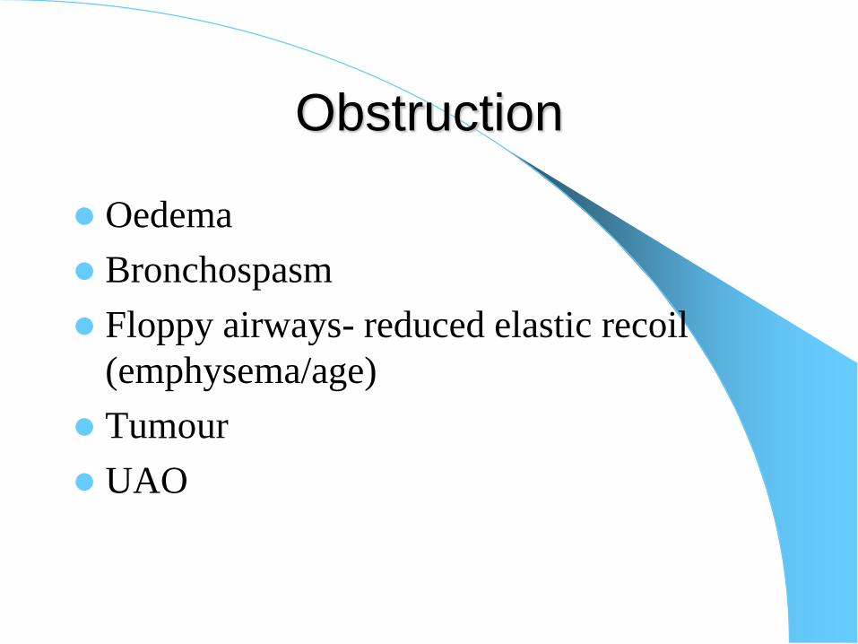
Obstruction
Oedema
Bronchospasm
Floppy airways- reduced elastic recoil
(emphysema/age)
Tumour
UAO
Page 19
Restriction
Lung
Pleural
Skeletal
Soft tissue
Abdominal
neurological
Page 20
Trouble shooting Slow start
Cough
Poor understanding/ submaximal effort- pain
Fatigue/bronchospasm
Early end of blow
Glottic closure
Leak- mouthpiece
Page 21
Identifying abnormalities
Coughing during exhalation• Abrupt stop in exhalation• Short intake of air (start of cough)• Irregular patter of exhalation
Slow start to forced exhalation• Marked increase in force of exhalation short time after start of manoeuvre (steep change in gradient on trace)
Page 22
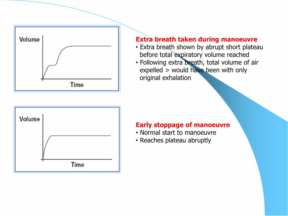
Early stoppage of manoeuvre• Normal start to manoeuvre• Reaches plateau abruptly
Extra breath taken during manoeuvre• Extra breath shown by abrupt short plateau before total expiratory volume reached
• Following extra breath, total volume of air expelled > would have been with only original exhalation
Page 23
Confirming COPD diagnosis via spirometry
FEV1 <80% predicted AND
FEV1/FVC <0.7 (70%)
Asthma can show same abnormalities reversibility testing
Spirometry - poor predictor of disability/QOL
Page 24
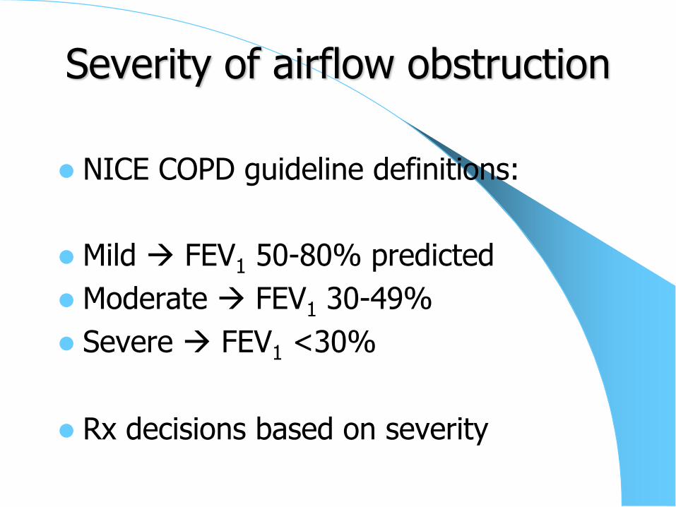
Severity of airflow obstruction
NICE COPD guideline definitions:
Mild FEV1 50-80% predicted
Moderate FEV1 30-49%
Severe FEV1 <30%
Rx decisions based on severity
Page 25
Consider COPD diagnosis in….
Smokers/ex-smokers > 35yrs
Exposure to respiratory irritants
Chronic SOB, cough, sputum, recurrent
chest infections, wheeze
Page 26
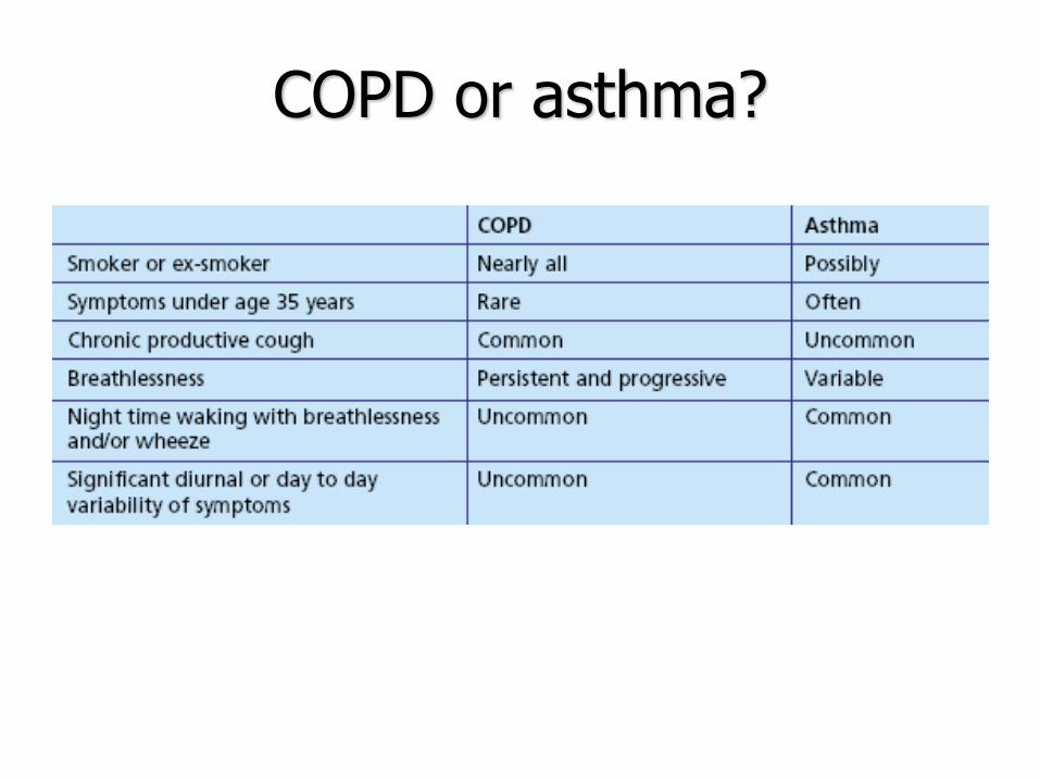
COPD or asthma?
Slow, progressive symptoms COPD
Symptoms pre-35 years asthma
Serial peak flow monitoring
NICE - bronchodilator reversibility testing not routinely used where clinical features/spirometry indicate COPD
Page 28
Reversibility testing
Asthma indicated large response to bronchodilator or
2/52 trial of 30mg Prednisilone daily (> 400ml)
or
Spirometry/clinical response 1/12 bronchodilator therapy
RT - not ‘gold standard’ interpret results with clinical Hx
Page 29
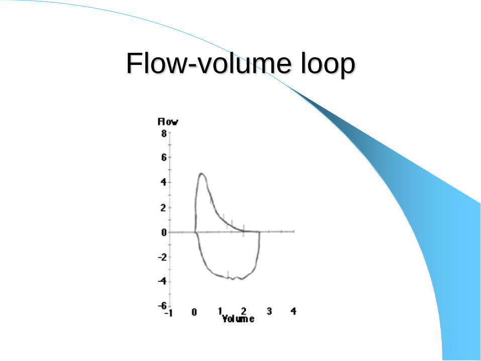
Flow-volume measurement
Basic spirometry volume-time curve
Flow-volume curve expiratory flow rate
plotted against the volume of air exhaled
Overall shape of flow-volume curve
detects airflow obstruction at an early stage/provides additional information
Page 32
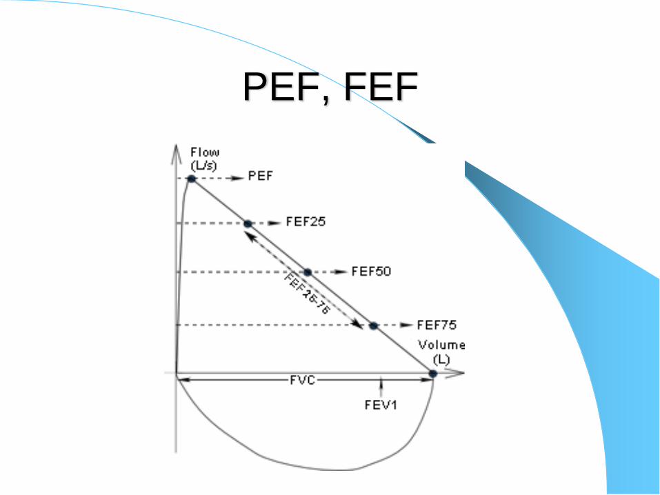
Identifying abnormalities
Obstructive disorder• Peak expiratory flow (PEF) is reduced • decline in airflow to complete exhalation follows a distinctive concave curve
Severe obstructive disorder• In severe airflow obstruction characteristic
‘steeple pattern’ in expiratory flow trace
Restrictive disorder• Pattern in expiratory trace normal in shapebut absolute reduction in volume
Page 34
Spirometry in practice
Case Studies
Page 35
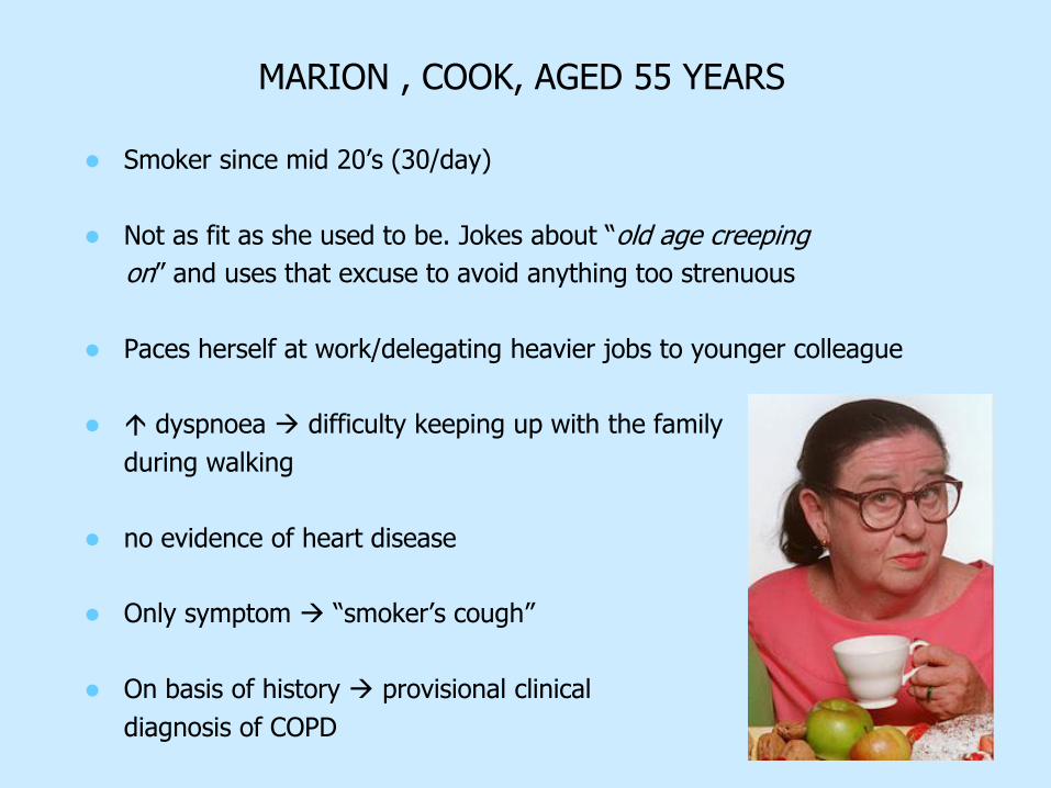
Smoker since mid 20’s (30/day)
Not as fit as she used to be. Jokes about “old age creeping
on” and uses that excuse to avoid anything too strenuous
Paces herself at work/delegating heavier jobs to younger colleague
dyspnoea difficulty keeping up with the family
during walking
no evidence of heart disease
Only symptom “smoker’s cough”
On basis of history provisional clinical
diagnosis of COPD
MARION , COOK, AGED 55 YEARS
Page 36
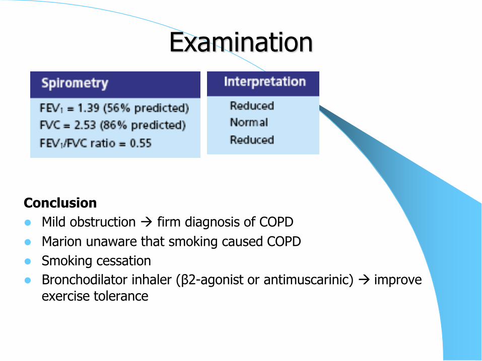
Examination
Conclusion
Mild obstruction firm diagnosis of COPD
Marion unaware that smoking caused COPD
Smoking cessation
Bronchodilator inhaler (β2-agonist or antimuscarinic) improve
exercise tolerance
Page 37
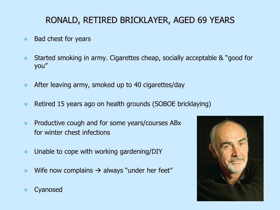
RONALD, RETIRED BRICKLAYER, AGED 69 YEARS
Bad chest for years
Started smoking in army. Cigarettes cheap, socially acceptable & “good for you”
After leaving army, smoked up to 40 cigarettes/day
Retired 15 years ago on health grounds (SOBOE bricklaying)
Productive cough and for some years/courses ABx
for winter chest infections
Unable to cope with working gardening/DIY
Wife now complains always “under her feet”
Cyanosed
Page 38
Examination
Conclusion
Severe COPD (FEV1 <30%)
Bronchodilator therapy stepped-up
Symptomatic benefit combination of beta-agonists & antimuscarinics
SPO2 89% on air
ABG chronic hypoxia (LTOT)
Started on long acting bronchodilator (beta agonist or antimuscarinic)
Due to FEV1 <50% predicted/frequent exacerbations started on
inhaled steroid
Page 39
JOHN, AN AREA SALES MANAGER, AGED 42 YEARS
Always been “chesty”
As a child considered “wheezy”/avoided PE
Started smoking early 20’s (10 cigarettes/day since)
Generally enjoyed good health/occasional URTI
coughing/wheeze
Prescribed ABX to treat “bronchitis” slow recovery (blamed
smoking)
Consulted his GP another cold had “gone to his chest”
Sleep disturbed by cough/wheeze
Unclear on basis of history asthma or COPD or both
Page 40
Examination
CONCLUSION
Mild degree of obstruction highly responsive (significant reversibility)
to bronchodilator
Significant reversibility/clinical history are highly indicative asthma
Advised on long-term impact of smoking/risk of developing COPD
Smoking cessation
Bronchodilator response tested (4 puffs salbutamol)
FEV1 re-measured after 30 minutes
Page 41
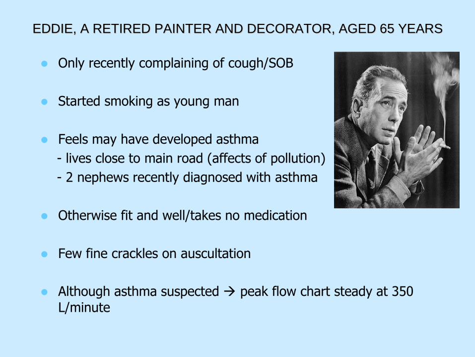
EDDIE, A RETIRED PAINTER AND DECORATOR, AGED 65 YEARS
Only recently complaining of cough/SOB
Started smoking as young man
Feels may have developed asthma
- lives close to main road (affects of pollution)
- 2 nephews recently diagnosed with asthma
Otherwise fit and well/takes no medication
Few fine crackles on auscultation
Although asthma suspected peak flow chart steady at 350
L/minute
Page 42
Examination
Conclusion
Abnormal FEV1 and FVC readings (both well below 80% of the predicted normal values)
However the FEV1/FVC ratio >70% restrictive disease
Fibrosing alveolitis diagnosed
Condition unrelated to environmental air pollution
Page 43
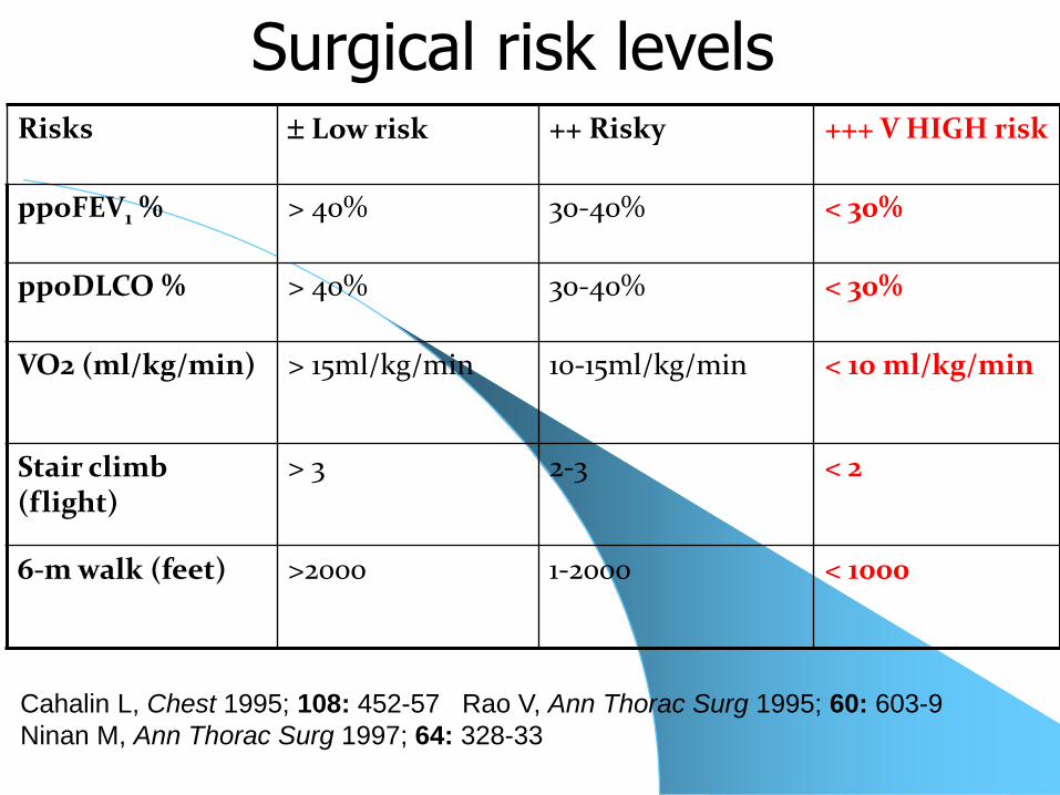
Surgical risk levelsRisks Low risk ++ Risky +++ V HIGH risk
ppoFEV1 % > 40% 30-40% < 30%
ppoDLCO % > 40% 30-40% < 30%
VO2 (ml/kg/min) > 15ml/kg/min 10-15ml/kg/min < 10 ml/kg/min
Stair climb (flight)
> 3 2-3 < 2
6-m walk (feet) >2000 1-2000 < 1000
Cahalin L, Chest 1995; 108: 452-57 Rao V, Ann Thorac Surg 1995; 60: 603-9
Ninan M, Ann Thorac Surg 1997; 64: 328-33
Page 46
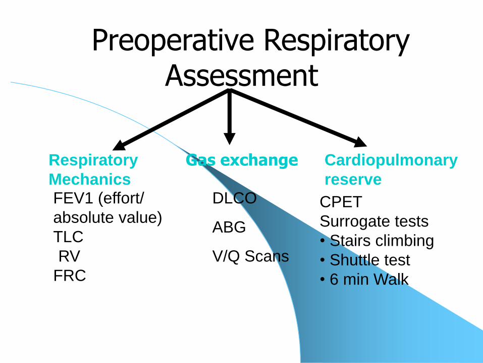
Preoperative Respiratory Assessment
Respiratory
MechanicsDLCO
ABG
V/Q Scans
CPET
Surrogate tests
• Stairs climbing
• Shuttle test
• 6 min Walk
FEV1 (effort/
absolute value)
TLC
RV
FRC
Cardiopulmonary
reserve
Gas exchange
Page 47
Predicted postoperative FEV1(ppoFEV1)
ppoFEV1 = pre FEV1 x (19 – segments to be removed)
19
obstructed segments
ppoFEV1 = pre FEV1 x (19 – a) - b)
(19-a)
a = obstructed segments
b = unobstructed segments to be resected
SEGMENTS UPPER 3 UPPER 5
RIGHT MIDDLE 2 LEFT
LOWER 5 LOWER 4
Page 48
V/Q Scans
Useful in prediction of postoperative function.
Postoperative FEV1= Preop FEV1 x % radioactivity contributed by the non operated lung
Better prediction is given by
– PPO FEV1 = Preoperative FEV1 x % perfusion of the non operated side.
Wernly JA et.al. J.Thorac.cardiovasc.surg.80:535-543,1980