Spiroplasma citri UGG and UGA Tryptophan Codons:Sequence of the Two Tryptophanyl-tRNAs and
Organization of the Corresponding GenesC. CIYII,l L. MARECHAL-DROUARD,2 C. SAILLARD,l* J. H. WEIL,2 AN J. M. BOVE1
Laboratoire de Biologie Cellulaire et Moleculaire, Institut National de la Recherche Agronomique,et Universite6 de Bordeaux II, Domaine de la Grande Ferrade, BP81 33 883 Villenave d'Ornon Cedex,and Institut de Biologie MoMculaire des Plantes, Universite Louis Pasteur, 67084 Strasbourg France
Received 6 May 1992/Accepted 7 August 1992
From the total tRNAs of Spiroplasma citri, we isolated and purified two tRNATrP species by usingchromatography on an RPC-5 column followed by denaturing polyacrylamide gel electrophoresis. Thesequence of the two tRNAs, as well as the sequences of the corresponding genes, were determined. One of thetwo tRNATrP species has a CCA anticodon and is able to pair with the universal UGG tryptophan codon, whilethe second has a U*CA (U* is a modified uridine) anticodon and is able to pair with UGA but also with UGGin accordance with the "U:N wobble" rule. Thus, in S. citri, UGA is not a stop codon but codes for tryptophan.The two tRNATrP genes, together with a third tRNA gene, ttRNAVr(CGA), belong to a single transcription unit.The nucleotide sequences of the two tRNATrP species show 82.9%o similarity. The two spiroplasmal tRNATrPspecies can be aminoacylated by using an aminoacyl-tRNA synthetase fraction from S. citri. In contrast, theenzyme fraction from Escherichia coli aminoacylates tRNATm"(CCA) but not tRNATrP(U*CA).
Members of the class Mollicutes are wall-less eubacteria.They have the smallest genomes of self-replicating, free-living organisms. Phylogenetic studies based on 16S rRNAsequence comparisons have shown that the class Mollicutesarose by regressive evolution, i.e., genome reduction, fromcommon ancestors of gram-positive eubacteria with lowguanine-plus-cytosine content, such as Clostridium ramo-sum and C. innocuum (39, 40). The class Mollicutes isdivided into six genera: Acholeplasma, Anaeroplasma, As-teroplasma, Mycoplasma, Spiroplasma, and Ureaplasma.Members of the genus Spiroplasma are motile and havehelical morphology (6).
In spiroplasmas, UGA is not used as a stop codon butcodes for tryptophan (27, 33). This peculiar codon usage wasdiscovered while studying the capsid protein gene of Spiro-plasma melliferum virus SPV4. This protein has a molecularmass of 63 kDa. No open reading frame large enough to codefor a 63-kDa protein could be identified on the sequencedviral genome when UAG, UAA, and UGA were all consid-ered as stop (nonsense) codons. Only when UGA was takenas a sense codon could an open reading frame with the rightsize be identified (28). This open reading frame was shown tobe the capsid protein gene by comparison of its deducedamino acid sequence with the N-terminal sequence of thepurified capsid protein. The capsid protein gene containsnine UGA codons. It could be cloned in a plasmid vector andamplified in Escherichia coli, but no capsid protein wasfound in the bacterial clones (25), whatever the cloningstrategy. In E. coli, UGA is recognized as a stop codon, andtranslation of the viral protein is expected to stop at the firstUGA codon encountered by the ribosome. In contrast, thegene for spiralin, the major membrane protein of S. citri, wasefficiently translated when introduced and amplified in E.coli (19). No UGA codon occurs in the spiralin gene (7), andhence, translation of this gene is not affected by premature
* Corresponding author.
termination at a UGA codon. The interpretation of thesedata benefited from the discovery of Yamao et al. (42) that inMycoplasma capricolum UGA is read as tryptophan. Thissuggested that in spiroplasmas UGA also codes for tryp-tophan. In agreement with this hypothesis, the absence ofUGA (and UGG) codons in the spiralin gene explained theabsence of tryptophan in the protein. M. capricolum has twotRNATrP species (41, 42). One tRNATrP has a CCA anti-codon and is able to decode the normal UGG tryptophancodon in accordance with the universal genetic code; theother tRNATrP has a U*CA (U* is a modified uridine)anticodon and is able to recognize both UGA and UGG inaccordance with the "U:N wobble" mechanism (9). M.gallisepticum also has the two tRNATrP species. However,other mycoplasmas, such as M. pneumoniae and M. geni-talium, possess only tRNATrP(UCA) (12).
In contrast, in Acholeplasma laidlawii, UGA functions asa stop codon and no tRNATrP(UCA) has been found (8, 35).The situation in ureaplasmas is not known.Sequence determination of several spiroplasmal genes has
shown that both UGG and UGA are used as tryptophancodons (5, 7, 28, 38). In the present study, we analyzed thetRNATrP situation in S. citri. Two different tRNATrP specieswere found. The sequences of these two tRNATrP speciesand the sequences of the corresponding genes were deter-mined. The comparative organization of tRNATrP genes in S.citri and other members of the class Mollicutes is discussed.
MATERIALS AND METHODS
Bacterial and spiroplasmal strains. E. coli DH5a(F') wasused as the host for propagation of plasmid pUC18 andbacteriophage M13.
S. citri Morocco strain R8A2 HP (high passage) (ATCC27556) was used to prepare total tRNAs, genomic DNA, andthe enzymatic fraction containing the aminoacyl-tRNA syn-thetase activities (fraction S100).Enzymes and chemicals. Voltalef300 LDPLmicro (Atochem)
was used as RPC-5 support. Trioctylmethylammonium chlo-ride (Serva, Heidelberg, Germany) was used for RPC-5preparation.Nuclease P1 from Penicihium citnim, T4 DNA ligase, iso-
propyl-p-D-thiogalactopyranoside, and 5-bromo-4-chloro-3-in-dolyl-3-D-galactopyranoside were purchased from Boehringer(Mannheim, Germany). N,N'-Methylenebisacrylamide, acryl-amide, and agarose were from Appligene (Strasbourg,France), and nylon N+ membrane, vector pUC18, T4 poly-nucleotide kinase, T4 RNA ligase, and radiolabelled com-pounds [32P]pCp (110 TBq/mol) ['32P]ATP (110 TBq/mol),[a-35S]dATP (22 TBq/mol), and [HJtryptophan (110 TBq/mol) were from Amersham Corp. (Arlington Heights, Ill.).Restriction endonucleases were obtained from BethesdaResearch Laboratories, Inc. (Gaithersburg, Md.).M13 DNA sequencing was performed by using the Seque-
nase version 2.0 sequencing kit from United States Biochem-ical Corporation (Cleveland, Ohio). Polygram cel 300DEAE/AR-2/15 for thin-layer chromatography was fromMacherey-Nagel (Duren, Germany).
Isolation of total tRNA. S. citri cells were grown in 5 litersof BSR medium (37) at 32°C and harvested by centrifugation(20,000 x g for 30 min). The following procedure for totaltRNA extraction was based on the method of Zubay (44).After direct phenol extraction, total tRNA was deacylatedby incubation in 1 M Tris-HCl (pH 9.0) at 37°C for 1 h. ThepH was adjusted to 7 by addition of 2 M HCl, and the tRNAswere precipitated by addition of a 0.01% solution of 20%(wt/vol) potassium acetate and 2.5 volumes of absoluteethanol. Nucleic acids were sedimented by centrifugation.The pellet was suspended in 1 ml of 10-4 M potassiumacetate and dialyzed overnight against 2 liters of the samebuffer.
Fractionation of total tRNA. Total tRNA of S. citri wasfractionated on an RPC-5 column (13) by using an NaClgradient from 0.4 to 0.8 M in 0.01 M sodium acetate buffer(pH 7.4) containing 0.01 M MgCl2 at a final volume of 100ml and a flow rate of 1 ml/min. Two peaks showing tryp-tophan-accepting activity were identified by aminoacylationin the presence of [3H]tryptophan using aminoacyl-tRNAsynthetase S100 fractions from S. citri or E. coli (seebelow). Fractions containing the tRNATIP present in peak 1(tRNATrPl) were pooled, concentrated, and further sub-jected to 10% polyacrylamide gel electrophoresis followedby denaturing 15% polyacrylamide gel electrophoresis asdescribed by Green et al. (10). Purification of the secondtRNATrP (tRNATrP2), contained in fractions of peak 2, wasachieved directly by denaturing 15% polyacrylamide gelelectrophoresis. The presence of the two tRNATrP specieswas monitored at each purification step by aminoacylationassays (see below).
Preparation of aminoacyl-tRNA synthetase fraction S100from S. citri and E. coli and conditions of aminoacylation. Theaminoacyl-tRNA synthetase fractions (S100) of S. citri andE. coli were prepared as described by Andachi et al. (1). Foraminoacylation assays, 0.05- to 20-pLg samples of tRNAswere incubated at 37°C for 20 min in 100 ,ul of a mixturecontaining 10mM ATP, 20 mM MgCl2, 50 mM Tris-HCl (pH7.5), 0.4 mM glutathione, 0.1 mg of bovine serum albuminper ml, 2 ,Ci of [3H]tryptophan, and 10 to 50 p,l of enzymaticextract (depending of the enzymatic activity).
Sequencing of tRNAP. The nucleotide sequences of thetwo S. citri tRNAT1P species were determined by using invitro postlabelling methods (30) and approaches describedpreviously (26). Most data were obtained by using thetechnique of Stanley and Vassilenko (34). Some RNA frag-
ments were also sequenced by using mobility shift analysis(30). All suspected modified residues were identified bytwo-dimensional thin-layer chromatography in different sol-vent systems (22).
Oligonucleotide synthesis and labelling. Oligonucleotideswere synthesized by J. C. Gandar, Institut de BiochimieCellulaire et de Neurochimie, Bordeaux, France. Oligonu-cleotide TRP1 (5'GGGGTAGCAGGACTTGAAC3') was 5'end labelled by using [_y-32P]ATP (110 TBq/mol) and T4polynucleotide kinase as described by Maxam and Gilbert(16). The labelled oligonucleotide was purified by using aNensorb 20 Nucleic Acid Purification Cartridge (DupontCo., Boston, Mass.) as recommended by the manufacturer.DNA isolation and Southern hybridization. Isolation of
genomic DNA from S. citri was performed by the method ofMarmur (15). Recombinant plasmid DNAs were analyzed byrapid alkaline extraction (2). Extraction of large amounts ofplasmid DNA was performed by using the same method;purification was achieved by CsCl gradient as the standardprocedure (14).
Southern blot (31) hybridization with an oligonucleotide asthe probe was performed as described by Zeff and Geliebter(43).
Cloning and sequencing of the tRNATrP genes. S. citrigenomic DNA was digested to completion with endonucle-ase EcoRI. The resulting DNA fragments were ligated withdephosphorylated, EcoRI-digested pUC18 plasmid DNA.Bacterial transformants containing a recombinant plasmidcarrying tRNATrP genes were screened for by colony hybrid-ization with the oligonucleotide TRP1 probe by standardprocedures (14).A partial restriction map of cloned inserts was con-
structed, and restriction fragments were subcloned intovector M13mpl8. Procedures used for restriction enzymedigestions, electrophoresis, and cloning of DNA were thosedescribed by Maniatis et al. (14).
Preparation of single-stranded DNA templates and anneal-ing and sequencing reactions were performed in accordancewith the Sequenase version 2.0 kit handbook (United StatesBiochemical Corp.). Double-stranded plasmid DNAs wereprepared as described by to Mierendorf et al. (18) andpartially sequenced by using synthesized oligonucleotides asprimers. Sequencing gel electrophoresis and computer pro-grams used for analysis of nucleotide sequences have beendescribed by Renaudin et al. (28).Sequences which are not of S. citi origin were imported
through the GenBank server (Los Alamos, N.Mex.) on theInternet network.
Nucleotide sequence accession number. The accession num-ber of the nucleotide sequence reported here is M91385.
RESULTS
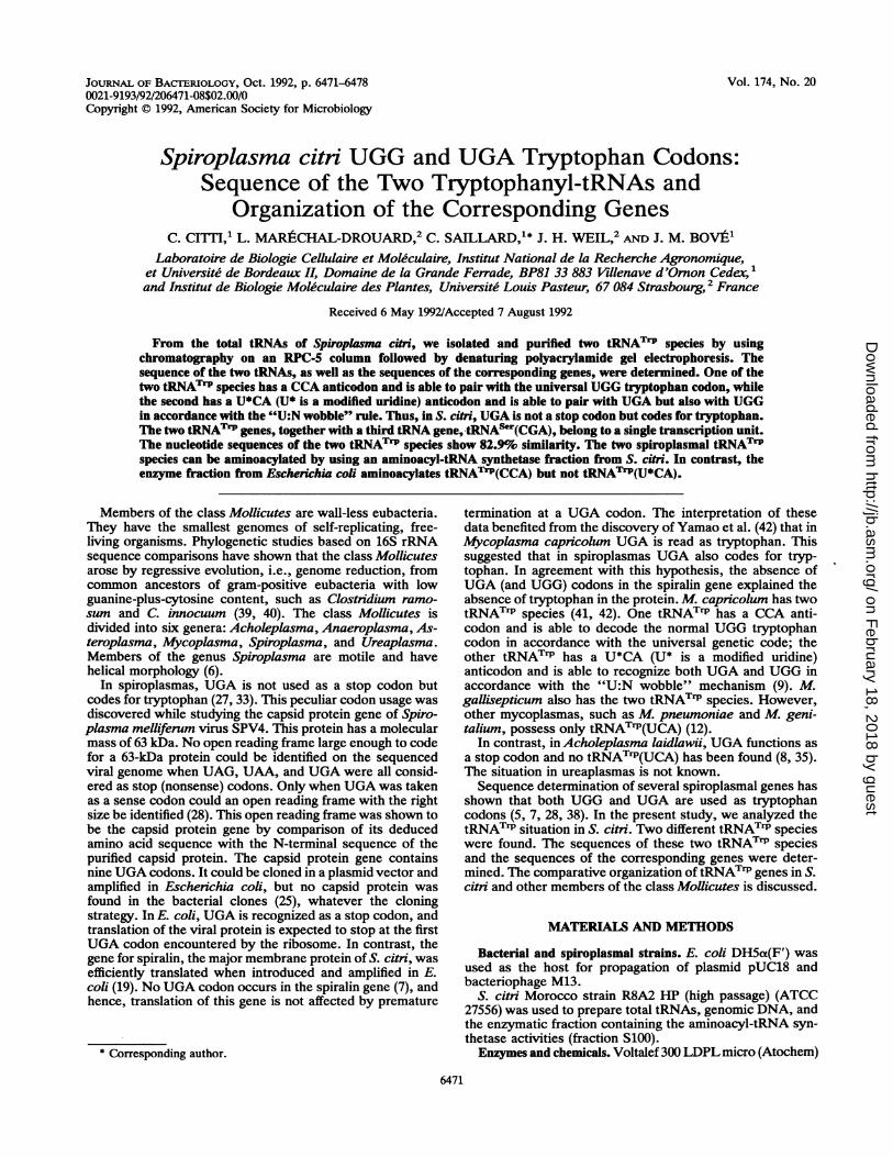
Purification and sequencing of S. citi tRNATrP species.Fractionation of the total tRNA of S. citri was achieved byRPC-5 column chromatography. The tRNAs in each elutedfraction were tested for tryptophan-accepting activity withS. citri and E. coli aminoacyl-tRNA synthetase fractions(Fig. 1). When the spiroplasmal enzyme fraction was used,two peaks of tryptophan-accepting activity, numbered 1 and2 on the basis of their elution order, were detected, while asingle peak (corresponding to peak 2) was found when the E.coli enzymatic extract was used.Each of these two peaks yielded several tRNA bands after
fractionation on denaturing polyacrylamide gel. For eachpeak, one of the tRNA bands was found by aminoacylation
FIG. 1. RPC-5 fractionation of S. citri total tRNA. One milligramof total tRNA in 0.01 M sodium acetate buffer (pH 4.7), containing0.01 M MgCl2 and 0.3 M NaCl was loaded on the column (70 by 0.45cm). Elution was performed in the same buffer with a linear gradient(2 x 50 ml) from 0.4 to 0.8M NaCL. Fractions of 1 ml were collected.Symbols: *, optical density (OD) at 260 nm; 0, [3H]tryptophan-accepting activity determined by using the S. citni enzyme fraction;*, [ H]tryptophan-accepting activity detected with the E. colienzyme fraction.
to contain a tRNATrP. Two tRNATr1P species were thusdetected.The nucleotide sequences of these two S. citri tRNATrP
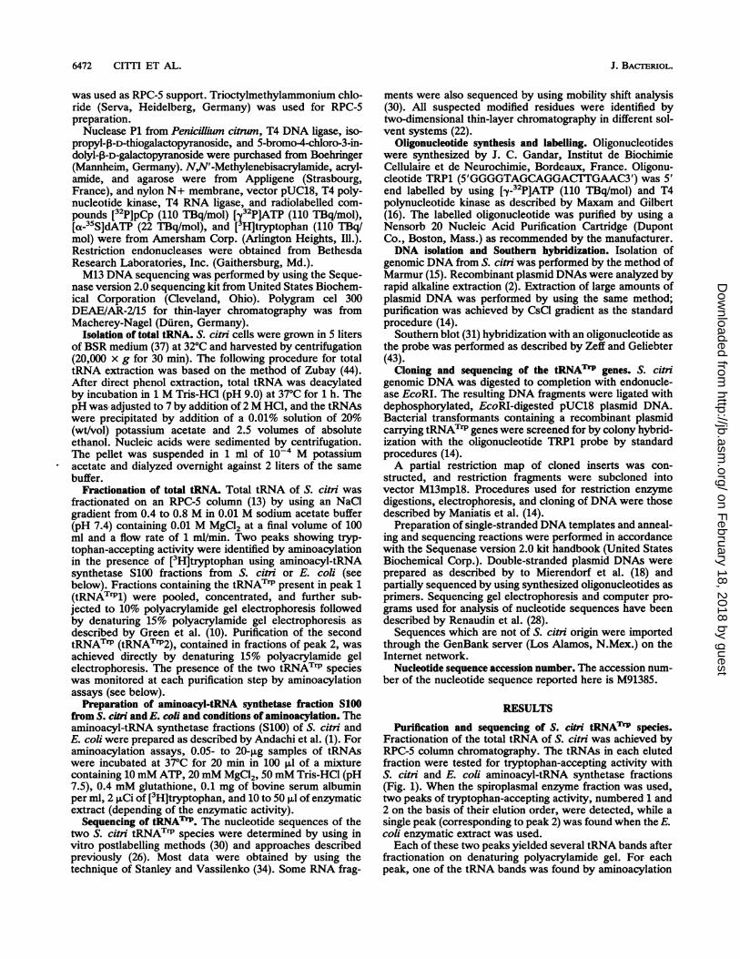
isoacceptors are shown in Fig. 2. Although the sequences ofthe two tRNATrP species show high similarity 182.9%), oneof the main differences between the two tRNA"rP species isat the anticodon level: tRNATrP, has an NCA anticodon,whereas tRNATrP2 has a CCA anticodon. The NCA anti-codon found in tRNATrPl contains a modified nucleotide inthe wobble position (position 34) of the anticodon. Thenucleotide 34-nucleotide 35 phosphodiester bond is resistantto water, partially resistant to nuclease P1, and digested bysnake venom phosphodiesterase. These data suggest thatnucleotide 34 contains an 0-methylated ribose. In addition,the sequence of the tRNATrPI gene revealed that nucleotide34 is a derivative of U (U*) (see below). tRNATrP(U*CA)(Fig. 2A) is 76 nucleotides long and contains one more
nucleotide, a C at position 17, than tRNATrP(CCA) (Fig. 2B).
AA 3'
C
G-c0-Co0-C70
0-C
G-C
U-At 00
IXH u° au cc A
A A 1oA ?I A
U rfU0 GUGG0 C
RI LTi U° U4GAAC ,Is.
u A A 1J76J20 [C-Gw1GU
0-C
30G C40
UCU A
C
B
3'
C
C
5'A-U
0-C
G-C70C-Ca-c¢-C
soU C
UUGUCC
AAa oA Hllllu UUUG GUGGO c¢ li1 us50 u4
GAAC U
20 a-U A C
C-C
a;-c30a-C40
C C
UA*
C
C
FIG. 2. Nucleotide sequences of the two S. citri tRNATrP spe-cies. (A) S. citri tRNATrP(U*CA). (B) S. citri tRNATrP(CCA). Boxesin panel A indicate the nucleotides of tRNATrP(U*CA) which differfrom those of tRNATrP(CCA). U* at position 34 is a hypermodifiednucleotide which has been identified as a derivative of U by genesequencing, and A* at position 37 is a hypermodified adenosine.
The two tRNATrP species also differ in their modified nu-cleotide contents. While tRNATrP(CCA) contains onlytwo modified nucleotides, a hypermodified adenosine (A*)at position 37 and a pseudouridine (4) at position 55,tRNATrP(U*CA) has five modified nucleotides, namely, two%'s at positions 39 and 55, a modified U (U*) at position 34,A* at position 37, and a 7-methylguanosine (m7G) at position46. The hypermodified A (A*) found at position 37 in bothtRNATIP species shows similar chromatographic behaviorand probably corresponds to the same posttranscriptionalmodification in the two tRNATrP species. Furthermore, thechromatographic mobilities (in the two systems used) of thismodified A are very close to the mobilities found for N6_A2_isopentenyladenosine or 2-methylthio-N6-A2-isopentylade-nosine. This suggests that this modified A is a derivative ofthese two hypermodified adenosines.
Localization of the tRNATOP genes on the spiroplasmalDNA. Each purified tRNATrP species was labelled at its 5'end with 32P and used as a probe in Southern blot hybrid-ization with EcoRI- or HindIlI-restricted S. citri genomicDNA. A single EcoRI fragment of 3.8 kbp and a singleHindIII fragment of 6.2 kbp hybridized with both tRNA:rIPprobes (data not shown), suggesting that the two tRNATrPgenes were located within the same EcoRI or HindIIIrestriction fragment. To determine the position of onetRNATrP gene with regard to the other, it was necessary toclone and sequence them. For this purpose, a 19-mer oli-godeoxynucleotide (TRP1) complementary to the 3' end oftRNATrP(U*CA) and 89.5% homologous to the 3' end oftRNATrP(CCA) was used as a probe to screen an S. citigenomic library. To check the specificity of this oligonucle-otide, it was first used to probe Southern blots of restrictedS. citri genomic DNA. This TRP1 oligonucleotide hybridizedto only one fragment, regardless of the restriction enzymeused; the EcoRI and HindIII hybridizing fragments had sizesof 3.8 and 6.2 kbp, respectively, in agreement with thehybridization pattern obtained when pure tRNATrP(U*CA)was used as the probe. Thus, TRP1 was suitable for screen-ing of a genomic DNA library of S. citri.
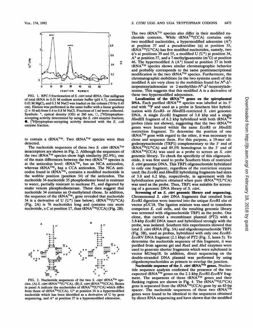
Screening of an S. citri genomic library and sequencing.Chromosomal S. citri DNA fragments that resulted fromEcoRI digestion were inserted into the unique EcoRI site ofvector pUC18. The ligation mixture was used to transformcompetent E. coli cells, and the resulting genomic librarywas screened with oligonucleotide TRP1 as the probe. Oneclone, that carried a recombinant plasmid (PT2) with a3.8-kbp EcoRI DNA insert and hybridized strongly with theprobe, was isolated. Southern blot experiments showed thattotal S. citri tRNA (Fig. 3A) and oligodeoxynucleotide TRP1(Fig. 3B), used as probes, hybridized with only one EcoRI-EcoRV DNA fragment (2.1 kbp) of PT2 (Fig. 3, lanes 5). Todetermine the nucleotide sequence of this fragment, it waspurified from agarose gel and RsaI and AluI enzymes wereused to generate shorter fragments which were subcloned invector M13mpl8. In addition, direct sequencing on thedouble-stranded DNA plasmid was performed by usingoligodeoxynucleotides as primers to overlap the junction.
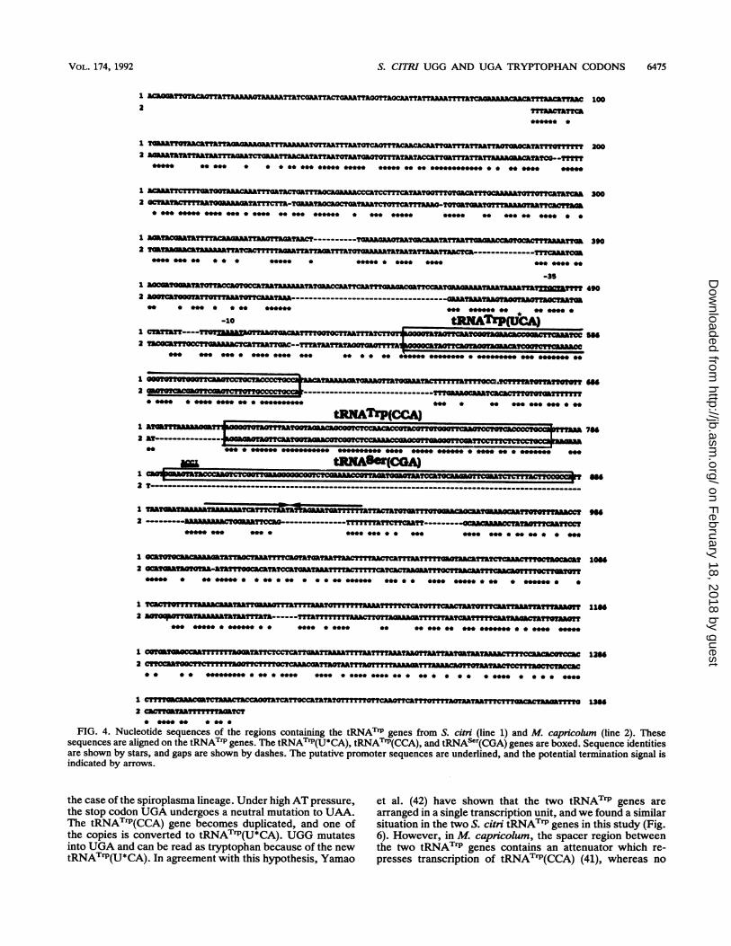
Nucleotide sequence of the S. citi tRNATrP genes. Nucleo-tide sequence analysis confirmed the presence of the twoexpected tRNATrP genes on the 2.1-kbp EcoRI-EcoRV frag-ment. The sequences of these tRNATrP genes and theirflanking regions are shown in Fig. 4. The tRNATrP(U*CA)gene is separated from the tRNATrP(CCA) gene by an 83-bpspacer. The nucleotide sequences of these two tRNATrPgenes were found to be identical to the sequences obtainedby direct RNA sequencing and have shown that the modified
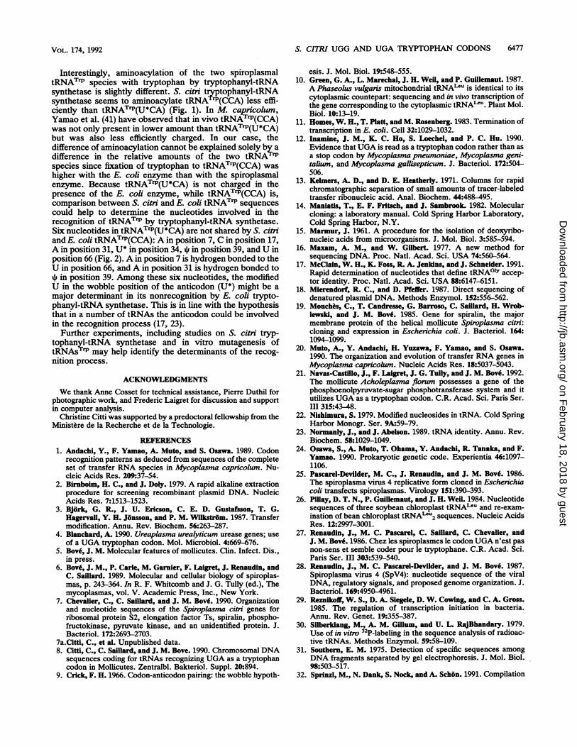
FIG. 3. Southern blot hybridizatioiDNA with total spiroplasmal tRNAprobes. Lanes: 1, plasmid PT2 DNA ri
3, AccI; 4, HindIII; 5, EcoRI and Ecc
nucleotide at position 34 of tRNacid residue.Another tRNA gene was found
tRNATrP(CCA) gene. This tRNAis a serine tRNA, in accordancecode. A potential promoterlike re-10 region and a 5'TTGCTA3upstream of the tRNATrP(U*CAthese -10 and -35 regions are siconsensus sequences, TATPuA1tively, of bacterial promoters reczase functioning with the general sAn inverted repeat sequence oc
the tRNASer gene and is a potentiasite (11). These data suggest thbelong to a single transcription ur(CGA) does not exist in M. capnictthis tRNA is expressed in S. citritRNA as a probe in Southern blotAccI-restricted plasmid DNA (lasequencing results, the AccI erspiroplasmal DNA insert at the 5'(Fig. 4). Total tRNA hybridized 1
fragments (Fig. 3A, lane 3), w]TRP1 hybridized with a single frshowing that tRNAser is expresse
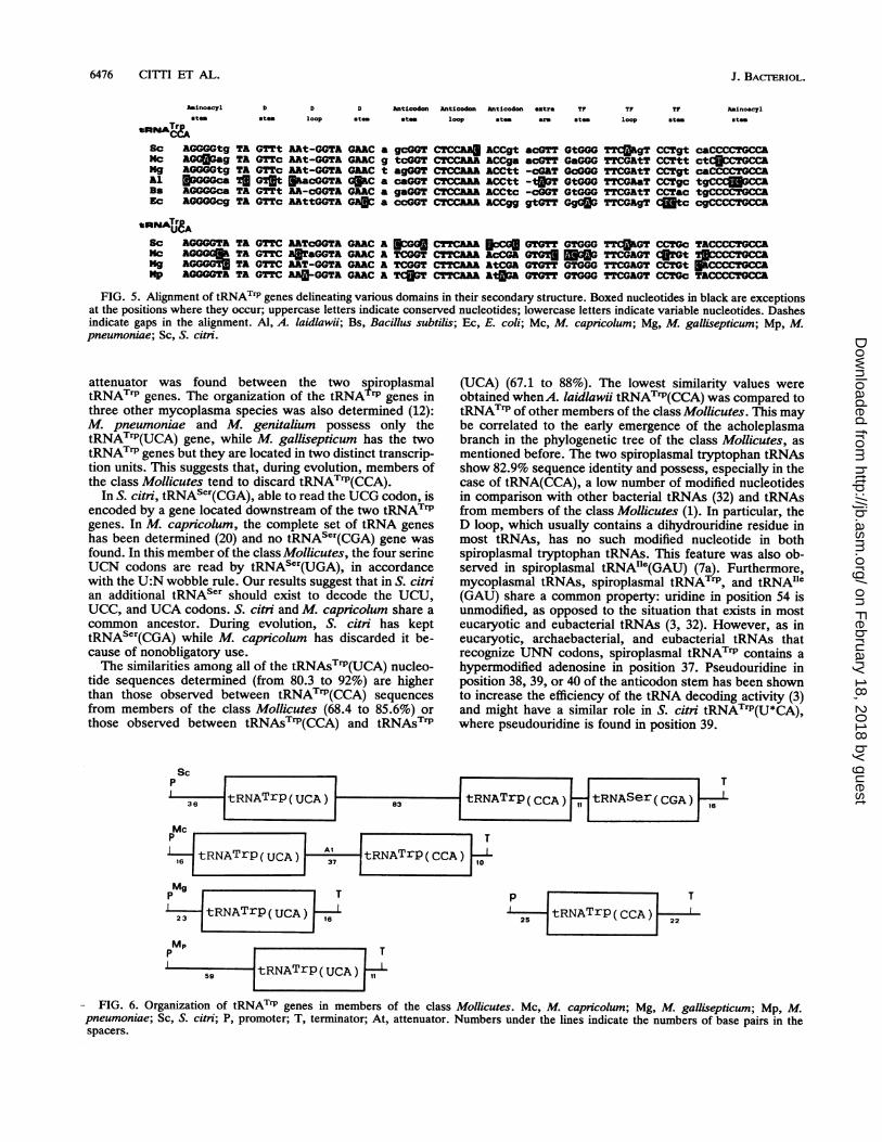
Sequence comparison with otheiAlignment of the 994-bp DNA fr(42) which carries the two tRN/sponding S. citri fragment is showthe regions flankingM. capricolumof S. citri showed 44.4% similarit'were obtained, as expected, betthemselves.Comparison of the tRNATrP ge
bers of the class Mollicutes (1delineating various domains in(Fig. 5), was also performed. I
conserved and variable regions ca5 shows that the length of the D lIcThe TF loop is highly conserved,
B mal tRNAT"P(CCA) and tRNATrP(U*CA), where the con-served G residue is replaced by an A. Similarly, in the
1 2 3 4 5 anticodon stem of S. citri tRNATrIP(U*CA), two bases in-volved in base pairing are different from those found in theother nine tRNAs. The extra arm is relatively less conservedin tRNA(CCA) than in tRNA(UCA). In general, the se-quences of tRNATrP(CCA) are more variable than those of
* a tRNATrP(UCA).
mDISCUSSION
We isolated and sequenced two S. citri tRNATFP species.1.3kbp......*. One has a CCA anticodon and is able to read the universalUGG tryptophan codon. The second has a U*CA anticodonthat is able to pair with UGA but also with UGG inaccordance with the U:N wobble rule (9). Furthermore,tRNATIP(U*CA) was shown to be correctly aminoacylatedin vitro by using an S. citri crude extract as a source oftryptophanyl-tRNA synthetase. These results show that in
n of recombinant plasmid PT2 S. citni, UGA is not a stop codon but in fact codes for(A) and TRP1 (B) used as tryptophan.
estricted by EcoRI; 2, EcoRV The occurrence of a tRNATIP that reads UGA should beoRV. considered from the point of view of evolution. Acholeplas-
mas (an anaerobic members of the class Mollicutes) are seenphylogenetically as the first members of the class Mollicutes
TATrP(U*CA) is a uridylic to have appeared by regressive evolution from their bacterial(clostridial) ancestors. The spiroplasmas have evolved from
10 bp downstream of the an early splitting of the acholeplasma branch, and thehas a CGA anticodon and mycoplasmas and ureaplasmas are thought to have spiro-with the universal genetic plasmal precursors (36). A. laidlawii, like its eubacterialgion, with a 5'TAAAAT3' ancestors, uses UGG as its sole tryptophan codon; to do-35 region, is present that, it has a tRNATrP with a CCA anticodon (8, 35). S. citrigene. The sequences of and M. capncolum belong to the same phylogenetic group
imilar to the -10 and -35 (spiroplasma group) (36). They use both UGG and UGA asF and TTGACA, respec- tryptophan codons (7, 28, 38) and have two tRNATrP spe-ognized by RNA polymer- cies. M. gallisepticum, M. pneumoniae, M. genitalium, andigma factor (29). Ureaplasma urealyticum are members of the pneumoniae:curs 18 bp downstream of phylogenetic group (36). M. gallisepticum resembles S. citriLl transcription termination and M. capricolum in that it has the two tRNATrP speciesat the three tRNA genes (12). However, M. pneumoniae and M. genitalium have lostait. Interestingly, tRNAser the usual tryptophan tRNA, tRNATrP(CCA), and have onlyolum (1, 20). To verify that one left: tRNA rP(UCA) (12). U. urealyticum reads UGA as, we used the total S. citri tryptophan (4) and very probably has tRNATrP(UCA), but itexperiments (Fig. 3A) with remains to be seen whether it still possesses the ancestralnes 3). According to the tryptophan tRNA, tRNATrP(CCA). Anaeroplasmas and as-nzyme should cleave the teroleplasmas have not been examined for the use ofUGA as' end of the tRNAser gene a tryptophan codon, but these anaerobic members of thewith two AccI-AccI DNA class Mollicutes are members of the same phylogenetichile oligodeoxynucleotide group as the acholeplasmas (anaeroplasma group). It is.agment (Fig. 3B, lane 3), therefore predictable that they have not evolved UGA as ad. tryptophan codon and possess only one tryptophan tRNA:r tRNAT"P gene sequences. tRNA rP(CCA), which reads UGG.agment of M. capricolum A very interesting case is that of Acholeplasma florum.K rP genes and the corre- This member of the class Mollicutes, as well as severaln in Fig. 4. Comparison of others, of plant or insect origin, does not need sterol forX tRNATrPgenes and those growth and was therefore classified as an acholeplasma,y. Higher similarity values even though it does require 0.04% Tween in its medium.ween the tRNATrI genes Phylogenetic analysis (36) has shown that this organism is a
member of the spiroplasma phylogenetic group! It has been-ne sequences from mem- found that this so-called acholeplasma, unlike A. laidlawii,2, 42) and bacteria (32) uses UGA as a tryptophan codon (21). Therefore, determi-their secondary structure nation of UGA as a tryptophan codon confirms its phylo-With this representation, genic position.In be distinguished. Figure A scenario for the reassignment of UGA from a stop)op can vary by 1 residue. codon to a tryptophan codon in the M. capncolum lineageexcept in both spiroplas- has been proposed by Osawa et al. (24) and might be valid in
FIG. 4. Nucleotide sequences of the regions containing the tRNATrP genes from S. citri (line 1) and M. capncolum (line 2). Thesesequences are aligned on the tRNATP genes. The tRNATrP(U*CA), tRNATrP(CCA), and tRNAS"r(CGA) genes are boxed. Sequence identitiesare shown by stars, and gaps are shown by dashes. The putative promoter sequences are underlined, and the potential termination signal isindicated by arrows.
the case of the spiroplasma lineage. Under high AT pressure,the stop codon UGA undergoes a neutral mutation to UAA.The tRNATrP(CCA) gene becomes duplicated, and one ofthe copies is converted to tRNATrP(U*CA). UGG mutatesinto UGA and can be read as tryptophan because of the newtRNATrP(U*CA). In agreement with this hypothesis, Yamao
et al. (42) have shown that the two tRNATrP genes arearranged in a single transcription unit, and we found a similarsituation in the two S. citri tRNATrP genes in this study (Fig.6). However, in M. capricolum, the spacer region betweenthe two tRNATrP genes contains an attenuator which re-presses transcription of tRNAT"P(CCA) (41), whereas no
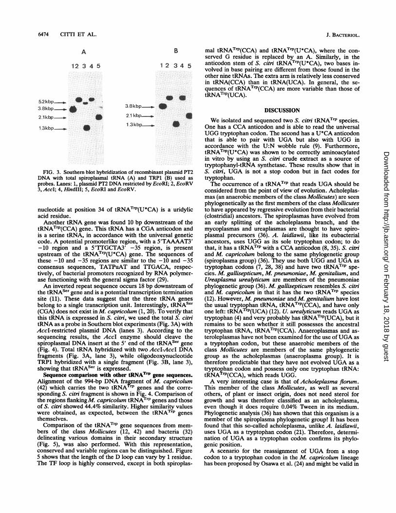
A lnoacyl D D D Antloodon Anticodon Anticodon extraatom atom loop atm atem loop atm are
TY
*too
EAAGGGGtg TA GTTt AAt-GGTA GAAC a gcGGT CTCCAU ACCgt acGTT GtGGGAGGWag TA GTTc AAt-GGTA GAAC g tcGGT CTCCAAA ACCga acGTT GaGGGAOGGGtg TA GTTc AAt-GGTA GAAC t agoGT CTCCAAA ACCtt -CGAT GcOGOOcak TM GT1t >acGGTA G C a caGGT CTCCAAA ACCtt -tIOT GtGGG
AGGGGca TA GTTt AA-cGGTA GAAC a gaGGT CTCCAAA ACCtc -cGGT GtGGGAGGGOcg TA GTTc AAttGGTA GOU a ccGGT CTCCAAA ACCgg gtGTT Gg00
Sc AGGOOTA TA GTTC AATcOGTA GAAC A C0G CTTCAAA ECGS GTOTT GTOGG TTCUIGT CCTOc TACCCCTOCCA
Nc aGGGGER TA GTTC AITaGGTA GAAC A TCGGT CTTCAAA AcCGA OTOTIEG TTCGAGT CIGtMg AGGGGT TA GTTC AAT-GGTA GAAC A TCGGT CTTCAAA AtCoA GTGTT GTGGG TTCGAGT CCTGt CCCCTOCCA
Mp AGGGGTA TA GTTC AAU-GGTA GAAC A TCOT CTTCAAA AtIGA GTGTT OTOGG TTCGAGT CCTOc
FIG. 5. Alignment of tRNATrP genes delineating various domains in their secondary structure. Boxed nucleotides in black are exceptionsat the positions where they occur; uppercase letters indicate conserved nucleotides; lowercase letters indicate variable nucleotides. Dashesindicate gaps in the alignment. Al, A. laidlawii; Bs, Bacillus subtilis; Ec, E. coli; Mc, M. capricolum; Mg, M. gallisepticum; Mp, M.pneumoniae; Sc, S. citri.
attenuator was found between the two spirctRNATrP genes. The organization of the tRNATrP Ithree other mycoplasma species was also determinM. pneumoniae and M. genitalium possess otRNATrP(UCA) gene, while M. gallisepticum hastRNATrP genes but they are located in two distinct trtion units. This suggests that, during evolution, merthe class Mollicutes tend to discard tRNATrP(CCA)
In S. citni, tRNASer(CGA), able to read the UCG cencoded by a gene located downstream of the two tgenes. In M. capricolum, the complete set of tRN.has been determined (20) and no tRNASer(CGA) gfound. In this member of the class Mollicutes, the foiUCN codons are read by tRNASCr(UGA), in accwith the U:N wobble rule. Our results suggest that itan additional tRNAser should exist to decode th4UCC, and UCA codons. S. citri and M. capricolumcommon ancestor. During evolution, S. citri htRNASer(CGA) while M. capricolum has discardecause of nonobligatory use.The similarities among all of the tRNAsTrP(UCA)
tide sequences determined (from 80.3 to 92%) artthan those observed between tRNATrP(CCA) seifrom members of the class Mollicutes (68.4 to 85those observed between tRNAsTrP(CCA) and tF
Sc
36tRNATrp(UCA)
(UCA) (67.1 to 88%). The lowest similarity values wereobtained whenA. laidlawii tRNATrP(CCA) was compared totRNATrP of other members of the class Mollicutes. This maybe correlated to the early emergence of the acholeplasmabranch in the phylogenetic tree of the class Mollicutes, asmentioned before. The two spiroplasmal tryptophan tRNAsshow 82.9% sequence identity and possess, especially in thecase of tRNA(CCA), a low number of modified nucleotidesin comparison with other bacterial tRNAs (32) and tRNAsfrom members of the class Mollicutes (1). In particular, theD loop, which usually contains a dihydrouridine residue inmost tRNAs, has no such modified nucleotide in bothspiroplasmal tryptophan tRNAs. This feature was also ob-served in spiroplasmal tRNAIle(GAU) (7a). Furthermore,mycoplasmal tRNAs, spiroplasmal tRNATrP, and tRNAI"e(GAU) share a common property: uridine in position 54 isunmodified, as opposed to the situation that exists in mosteucaryotic and eubacterial tRNAs (3, 32). However, as ineucaryotic, archaebacterial, and eubacterial tRNAs thatrecognize UNN codons, spiroplasmal tRNATrP contains ahypermodified adenosine in position 37. Pseudouridine inposition 38, 39, or 40 of the anticodon stem has been shownto increase the efficiency of the tRNA decoding activity (3)and might have a similar role in S. citri tRNATrP(U*CA),where pseudouridine is found in position 39.
T
~tRNATrp(CCA) tRNASer(CGA) 16
Mc
16 tRNATrp(UCA) 37 tRNATrp(CCA) 10
MgT
23 tRNATrp(UCA) 16
p T
25 tRNATrp(CCA) 2
pMp T
-mtRNATrP(UCA)1
- FIG. 6. Organization of tRNATrP genes in members of the class Mollicutes. Mc, M. capricolum; Mg, M. gallisepticum; Mp, M.pneumoniae; Sc, S. citri; P, promoter; T, terminator; At, attenuator. Numbers under the lines indicate the numbers of base pairs in thespacers.
Interestingly, aminoacylation of the two spiroplasmaltRNATTP species with tryptophan by tryptophanyl-tRNAsynthetase is slightly different. S. citn tryptophanyl-tRNAsynthetase seems to aminoacylate tRNA TI(CCA) less effi-ciently than tRNATrP(U*CA) (Fig. 1). In M. capricolum,Yamao et al. (41) have observed that in vivo tRNATP(CCA)was not only present in lower amount than tRNATrP(U*CA)but was also less efficiently charged. In our case, thedifference of aminoacylation cannot be explained solely by a
difference in the relative amounts of the two tRNA rPspecies since fixation of tryptophan to tRNATrP(CCA) was
higher with the E. coi enzyme than with the spiroplasmalenzyme. Because tRNATrP(U*CA) is not charged in thepresence of the E. coli enzyme, while tRNAT'P(CCA) is,comparison between S. citni and E. coli tRNATTP sequences
could help to determine the nucleotides involved in therecognition of tRNAT by tryptophanyl-tRNA synthetase.Six nucleotides in tRNAT P(U*CA) are not shared by S. citniand E. coli tRNATrP(CCA): A in position 7, C in position 17,A in position 31, U* in position 34,+ in position 39, and U inposition 66 (Fig. 2). A in position 7 is hydrogen bonded to theU in position 66, and A in position 31 is hydrogen bonded to
* in position 39. Among these six nucleotides, the modifiedU in the wobble position of the anticodon (U*) might be a
major determinant in its nonrecognition by E. coli trypto-phanyl-tRNA synthetase. This is in line with the hypothesisthat in a number of tRNAs the anticodon could be involvedin the recognition process (17, 23).
Further experiments, including studies on S. citi tryp-
tophanyl-tRNA synthetase and in vitro mutagenesis oftRNAs-' may help identify the determinants of the recog-
nition process.
ACKNOWLEDGMENTS
We thank Anne Cosset for technical assistance, Pierre Duthil forphotographic work, and Frederic Laigret for discussion and supportin computer analysis.
Christine Citti was supported by a predoctoral fellowship from theMinistere de la Recherche et de la Technologie.
REFERENCES1. Andachi, Y., F. Yamao, A. Muto, and S. Osawa. 1989. Codon
recognition patterns as deduced from sequences of the completeset of transfer RNA species in Mycoplasma capnicolum. Nu-cleic Acids Res. 209:37-54.
2. Birnboim, H. C., and J. Doly. 1979. A rapid alkaline extractionprocedure for screening recombinant plasmid DNA. NucleicAcids Res. 7:1513-1523.
3. Bjork, G. R., J. U. Ericson, C. E. D. Gustafsson, T. G.Hagervall, Y. H. Jfnsson, and P. M. Wdkstrfm. 1987. Transfermodification. Annu. Rev. Biochem. 56:263-287.
4. Blanchard, A. 1990. Ureaplasma urealyticum urease genes; use
of a UGA tryptophan codon. Mol. Microbiol. 4:669-676.5. Bove, J. M. Molecular features of mollicutes. Clin. Infect. Dis.,
in press.6. Bov6, J. M., P. Carle, M. Garnier, F. LaIgret, J. Renaudin, and
C. Saillard. 1989. Molecular and cellular biology of spiroplas-mas, p. 243-364. In R. F. Whitcomb and J. G. Tully (ed.), Themycoplasmas, vol. V. Academic Press, Inc., New York.
7. Chevalier, C., C. Saillard, and J. M. Bove. 1990. Organizationand nucleotide sequences of the Spiroplasma citri genes forribosomal protein S2, elongation factor Ts, spiralin, phospho-fructokinase, pyruvate kinase, and an unidentified protein. J.Bacteriol. 172:2693-2703.
7a.Citti, C., et al. Unpublished data.8. Citti, C., C. Saillard, and J. M. Bove. 1990. Chromosomal DNA
sequences coding for tRNAs recognizing UGA as a tryptophancodon in Mollicutes. Zentralbl. Bakteriol. Suppl. 20:894.
9. Crick, F. H. 1966. Codon-anticodon pairing: the wobble hypoth-
esis. J. Mol. Biol. 19:548-555.10. Green, G. A., L. Marechal, J. H. Weil, and P. Guillemaut. 1987.
A Phaseolus vulganis mitochondrial tRNAIzU is identical to itscytoplasmic countepart: sequencing and in vivo transcription ofthe gene corresponding to the cytoplasmic tRNAL*u. Plant Mol.Biol. 10:13-19.
11. Homes, W. H., T. Platt, and M. Rosenberg. 1983. Termination oftranscription in E. coli. Cell 32:1029-1032.
12. Inamine, J. M., K C. Ho, S. Loechel, and P. C. Hu. 1990.Evidence that UGA is read as a tryptophan codon rather than asa stop codon by Mycoplasma pneumoniae, Mycoplasma geni-talium, and Mycoplasma gailisepticum. J. Bacteriol. 172:504-506.
13. Kelmers, A. D., and D. E. Heatherly. 1971. Columns for rapidchromatographic separation of small amounts of tracer-labeledtransfer ribonucleic acid. Anal. Biochem. 44:488-495.
14. Maniatis, T., E. F. Fritsch, and J. Sambrook 1982. Molecularcloning: a laboratory manual. Cold Spring Harbor Laboratory,Cold Spring Harbor, N.Y.
15. Marmur, J. 1961. A procedure for the isolation of deoxyribo-nucleic acids from microorganisms. J. Mol. Biol. 3:585-594.
16. Maxam, A. M., and W. Gilbert. 1977. A new method forsequencing DNA. Proc. Natl. Acad. Sci. USA 74:560-564.
17. McClain, W. H., K Foss, R. A. Jenkins, and J. Schneider. 1991.Rapid determination of nucleotides that define tRNA0ly accep-tor identity. Proc. Natl. Acad. Sci. USA 88:6147-6151.
18. Mlerendorf, R. C., and D. Pfeffer. 1987. Direct sequencing ofdenatured plasmid DNA. Methods Enzymol. 152:556-562.
19. Mouchis, C., T. Candresse, G. Barroso, C. Saillard, H. Wrob-lewski, and J. M. Bov6. 1985. Gene for spiralin, the majormembrane protein of the helical mollicute Spiroplasma citni:cloning and expression in Escherichia coli. J. Bacteriol. 164:1094-1099.
20. Muto, A., Y. Andachi, H. Yuzawa, F. Yamao, and S. Osawa.1990. The organization and evolution of transfer RNA genes inMycoplasma capricohwn. Nucleic Acids Res. 18:5037-5043.
21. Navas-Castillo, J., F. Laigret, J. G. Tully, and J. M. Bov. 1992.The mollicute Acholeplasma flomum possesses a gene of thephosphoenolpyruvate-sugar phosphotransferase system and itutilizes UGA as a tryptophan codon. C.R. Acad. Sci. Paris Ser.III 315:43-48.
22. Nishimura, S. 1979. Modified nucleosides in tRNA. Cold SpringHarbor Monogr. Ser. 9AM59-79.
23. Normanly, J., and J. Abelson. 1989. tRNA identity. Annu. Rev.Biochem. 58:1029-1049.
24. Osawa, S., A. Muto, T. Ohama, Y. Andachi, R. Tanaka, and F.Yanao. 1990. Prokaryotic genetic code. Experientia 46:1097-1106.
25. Pascarel-Devilder, M. C., J. Renaudin, and J. M. Bove. 1986.The spiroplasma virus 4 replicative form cloned in Eschenchiacoli transfects spiroplasmas. Virology 151:390-393.
26. Pillay, D. T. N., P. GuiUemaut, and J. H. Weil. 1984. Nucleotidesequences of three soybean chloroplast tRNAI4U and re-exam-ination of bean chloroplast tRNAlu2 sequences. Nucleic AcidsRes. 12:2997-3001.
27. Renaudin, J., M. C. Pascarel, C. Saillard, C. Chevalier, andJ. M. Bov6. 1986. Chez les spiroplasmes le codon UGA n'est pasnon-sens et semble coder pour le tryptophane. C.R. Acad. Sci.Paris Ser. III 303:539-540.
28. Renaudin, J., M. C. Pascarel-Devilder, and J. M. Bov6. 1987.Spiroplasma virus 4 (SpV4): nucleotide sequence of the viralDNA, regulatory signals, and proposed genome organization. J.Bacteriol. 169:4950-4961.
29. Rezniloff, W. S., D. A. Siegele, D. W. Cowing, and C. A. Gross.1985. The regulation of transcription initiation in bacteria.Annu. Rev. Genet. 19:355-387.
30. Silberklang, M., A. M. Gillum, and U. L. RajBhandary. 1979.Use of in vitr 3P-labeling in the sequence analysis of radioac-tive tRNAs. Methods Enzymol. 59:58-109.
31. Southern, E. M. 1975. Detection of specific sequences amongDNA fragments separated by gel electrophoresis. J. Mol. Biol.98:503-517.
32. Sprinzl, M., N. Dank, S. Nock, and A. Schon. 1991. Compilation
of tRNA sequences and sequences of tRNA genes. NucleicAcids Res. 19:2127-2171.
33. Stamburski, C., J. Renaudin, and J. M. Bov6. 1992. Mutagenesisof a tryptophan codon from TGG to TGA in the CAT gene doesnot affect the expression of the CAT gene in Spiroplasma citn.Gene 110:133-134.
34. Stanley, J., and S. Vassilenko. 1978. A different approach totRNA sequencing. Nature (London) 274:87-89.
35. Tanaka, R., A. Muto, and S. Osawa. 1989. Nucleotide sequenceof tryptophan tRNA gene in Acholepk.sma laidlawii. NucleicAcids Res. 17:5842.
36. Weisburg, W. G., J. G. Tully, D. L. Rose, J. P. Petzel, H.Oyaaizu, D. Yang, L. Mandelco, J. Sechrest, T. G. Lawrence, J.Van Etten, J. Maniloff, and C. R. Woese. 1989. A phylogeneticanalysis of the mycoplasmas: basis for their classification. J.Bacteriol. 171:6455-6467.
37. Whitcomb, R. F. 1983. Culture media for spiroplasmas. Meth-ods Mycoplasmol. 1:147-158.
38. Williamson, D. L., J. Renaudin, and J. M. Bov6. 1991. Nucleo-
tide sequence of the Spiroplasma citni fibril protein gene. J.Bacteriol. 173:4353-4362.
39. Woese, C. R. 1987. Bacterial evolution. Microbiol. Rev. 51:221-271.
40. Woese, C. R., E. Stackebrandt, and W. Ludwig. 1985. What aremycoplasmas: the relationship of tempo in bacterial evolution.J. Mol. Biol. 21:305-316.
41. Yamao, F., S. Iwagami, Y. Azumi, A. Muto, S. Osawa, N. Fujita,and A. Ishihama. 1988. Evolutionary dynamics of tryptophantRNAs in Mycoplasma capricolum. Mol. Gen. Genet. 212:364-369.
42. Yamao, F., A. Muto, Y. Kawauchi, M. Iwami, S. Iwagami, Y.Azumi, and S. Osawa. 1985. UGA is read as tryptophan inMycoplasma capricolum. Proc. Natl. Acad. Sci. USA 82:2306-2309.
43. Zeff, R. A., and J. Geliebter. 1987. Oligonucleotide probes forgenomic DNA blots. Focus (Gibco BRL) 9:1-2.
44. Zubay, G. 1962. The isolation and fractionation of solubleribonucleic acid. J. Mol. Biol. 4:347-356.