16
Splash M-Knife Case Studies Case studies from clinical practice with Splash M-Knife Esophagus Stomach Colon

Splash M-Knife Case StudiesCase studies from clinical practice with Splash M-Knife
Esophagus
Stomach
Colon
1 2
Index Introduction
Splash M-Knife Features and Functions
Case Reports
Prof. Bo-in, Lee
Laterally spreading tumor at the Upper Rectum
Laterally spreading tumor involving the entire rectum
Visible Dysplasia in a patient with Ulcerative Pancolitis
Prof. Woo-Chul, Chung
ESD for Early Esophageal Cancer
ESD for EGCa with Severe Fibrosis
ESD of the lesion involving pyloric channel
ESD of the Stomach Ulceration
Asso. Prof. Jae-Myeong, Cha
LST of Rectum Case
SET of Stomach
Prof. Hyun-Gun, Kim
LST of Rectum Case
NET of Rectum Case
Product Specifications
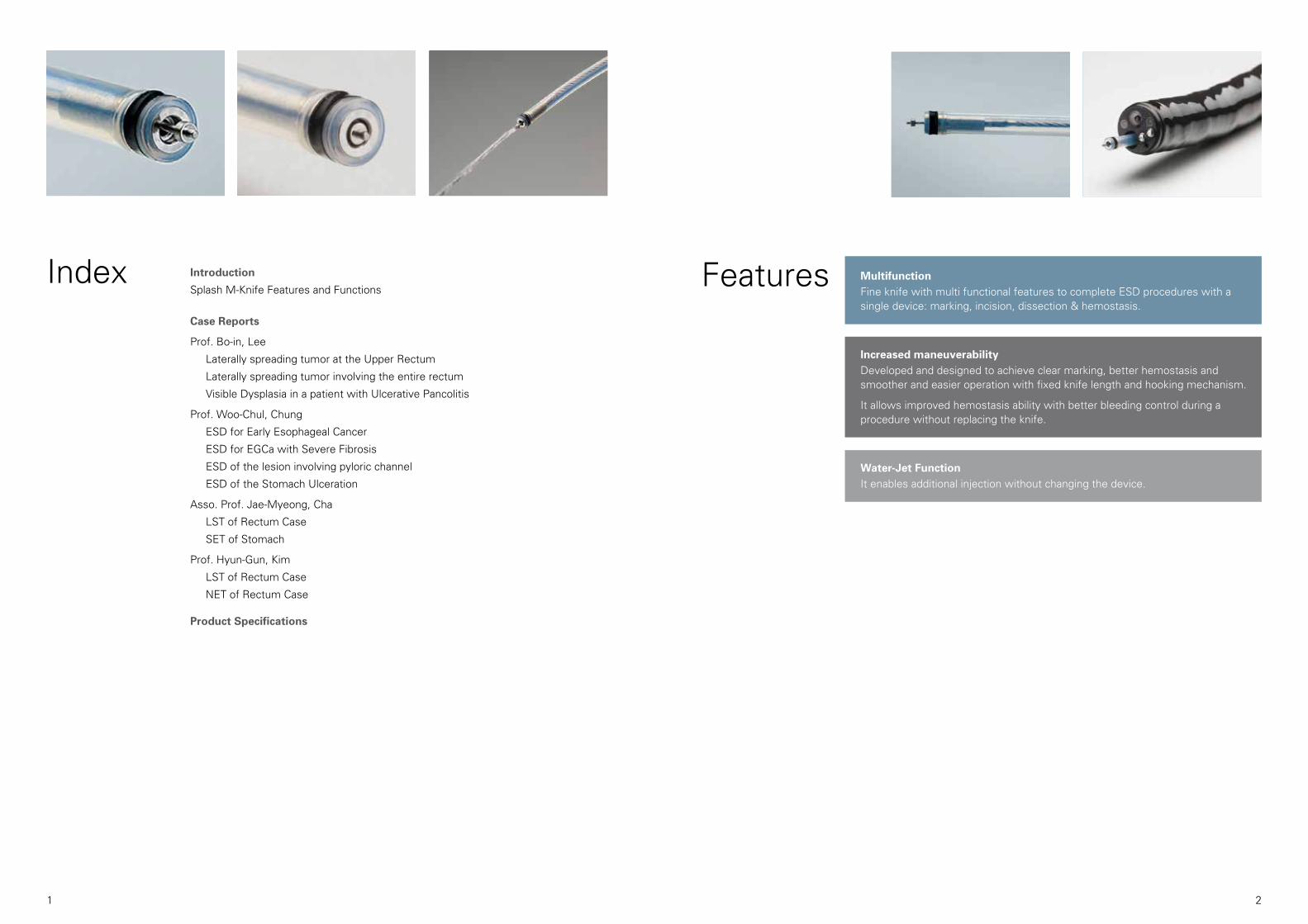
Features MultifunctionFine knife with multi functional features to complete ESD procedures with a single device: marking, incision, dissection & hemostasis.
Increased maneuverabilityDeveloped and designed to achieve clear marking, better hemostasis and smoother and easier operation with fixed knife length and hooking mechanism.
It allows improved hemostasis ability with better bleeding control during a procedure without replacing the knife.
Water-Jet FunctionIt enables additional injection without changing the device.
3 4
PENTAX Medical has been working closely with this group of doctors so that their best practices and scientific evidence using its ESD knife, the Splash M-Knife, can be shared in a peer environment. It will be highly beneficial for those who are starting off in ESD procedures.
Prof. Bo-in, LeeThe Catholic University of Korea, Seoul St. Mary’s Hospital, South Korea
Prof. Woo-Chul, ChungThe Catholic University of Korea, St. Vincent’s Hospital, South Korea
Asso. Prof. Jae-Myeong, ChaKyung-Hee University Hospital at Gang-dong, South Korea
Prof. Hyun-Gun, KimSoon Chun Hyang University Hospital Seoul,South Korea
The Key Opinion Leaders
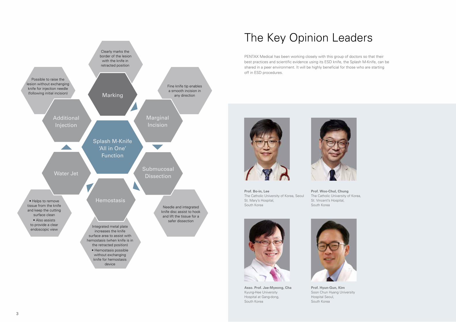
Splash M-Knife‘All in One’Function
MarginalIncision
Submucosal DissectionWater Jet
AdditionalInjection
Hemostasis
Marking
• Helps to removetissue from the knife and keep the cutting
surface clean
• Also assiststo provide a clear endoscopic view
Possible to raise the lesion without exchanging knife for injection needle (following initial incision)
Needle and integrated knife disc assist to hook and lift the tissue for a
safer dissection
Fine knife tip enables a smooth incision in
any direction
Clearly marks the border of the lesion
with the knife in retracted position
Integrated metal plate increases the knife
surface area to assist with hemostasis (when knife is in
the retracted position)
• Hemostasis possiblewithout exchangingknife for hemostasis
device

5 6
Figure 1
5. Clinical Images
Figure 2 Figure 3
Laterally spreading tumor at the Upper Rectum 1. Patient history and information
A 64-year-old male visited the hospital due to a 42-mm laterally spreading tumor [granular type, homogeneous (LST-G, H)] at the upper rectum which was found during a screening colonoscopy (Figure 1).
2. ESD procedure/Treatment
The patient underwent endoscopic submucosal dissection (ESD). A 10% glycerol, 5% fructose, and 0.9% saline mixture solution was used for submucosal injection. A Splash M-Knife was mainly used for mucosal incision and submucosal dissection. Submucosal injection through Splash M-Knife without changing to an injection needle
reduced procedure time significantly (Figure 2). Oozing during mucosal incision and submucosal dissection was easily controlled by the retracted Splash M-Knife. Hemostatic function can be maximized when the knife is retracted since the contact area is increased by the metal ring of the sheath tip.
Submucosal dissection was continued mainly using pocket-creation method (Figure 3).
3. Final Pathology
Colon, ESD, laterally spreading tumor at the upper rectum
• Size: 42 mm
• Granular type, homogeneous (LST-G, H)
• Final histology was consistent with low grade dysplasia. Tumor involvement of vertical and horizontal margin was negative.
4. Outcome/Follow-up
Procedure time: 40 minutes (Figure 4 & 5)
Complications: None
Outcome: Endoscopic and histologic complete resection, en bloc.
Figure 4 Figure 5
“The Splash M-Knife has a lot of benefits. I used it mainly for mucosal incision
and submucosal dissection during ESD procedure due to safer, faster and more
effective dissection outcome over others. Especially for fibrotic lesion in the
submucosal layer, the Splash M-Knife showed the true function of the knife disk:
“Hooking the tissue”. It is effective and safe for fibrotic portions.
In addition, without changing therapeutic devices, it reduces ESD procedure time
effectively. One of my favorite features is the maximized hemostatic function.
When the knife is retracted, the contact area with bleeding point is increased
through the metal ring of the sheath tip.
However, it does not mean I am only using Splash M-Knife. Depending on
the depth, type, size and location of lesion, I will select the most suitable and
safest knife for my patient. Generally, I am satisfied with the functionality of the
Splash M-Knife.”
Prof. Bo-in, LeeThe Catholic University of Korea, Seoul St. Mary’s Hospital, South Korea
Procedure Mode Effect Watt Duration Interval Max. Rated Voltage
Marginal incision
Endocut I 3 - 3 1
Submucosal dissection
Endocut I 3 - 3 1
Submucosal dissection(Hypervascular tissue)
Forced Coagulation
2 30
Hemostasis Soft Coagulation
4 50 - -
Electrosurgical Unit (Erbe 300D)
7 8
Figure 4 Figure 5
Figure 6
1. Patient history and information
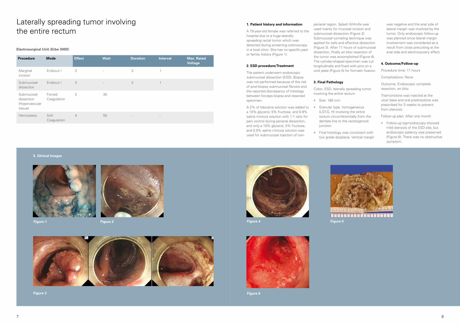
A 70-year-old female was referred to the hospital due to a huge laterally spreading rectal tumor which was detected during screening colonoscopy in a local clinic. She has no specific past or family history (Figure 1).
2. ESD procedure/Treatment
The patient underwent endoscopic submucosal dissection (ESD). Biopsy was not performed because of the risk of post-biopsy submucosal fibrosis and the reported discrepancy of histology between forceps biopsy and resected specimen.
A 2% of lidocaine solution was added to a 10% glycerol, 5% fructose, and 0.9% saline mixture solution with 1:1 ratio for pain control during perianal dissection, and only a 10% glycerol, 5% fructose, and 0.9% saline mixture solution was used for submucosal injection of non-
perianal region. Splash M-Knife was used mainly for mucosal incision and submucosal dissection (Figure 2). Submucosal tunneling technique was applied for safe and effective dissection (Figure 3). After 11 hours of submucosal dissection, finally en bloc resection of the tumor was accomplished (Figure 4). The cylinder-shaped specimen was cut longitudinally and fixed with pins on a cork plate (Figure 5) for formalin fixation.
3. Final Pathology
Colon, ESD, laterally spreading tumor involving the entire rectum
• Size: 180 mm
• Granular type, homogeneous (LST-G, H) involving the entire rectum circumferentially from the dentate line to the rectosigmoid junction
• Final histology was consistent with low grade dysplasia. Vertical margin
was negative and the anal side of lateral margin was involved by the tumor. Only endoscopic follow-up was planned since lateral margin involvement was considered as a result from close precutting at the anal side and electrocautery effect.
4. Outcome/Follow-up
Procedure time: 11 hours
Complications: None
Outcome: Endoscopic complete resection, en bloc
Triamcinolone was injected at the ulcer base and oral prednisolone was prescribed for 3 weeks to prevent from stenosis.
Follow-up plan: After one month
• Follow-up sigmoidoscopy showed mild stenosis of the ESD site, but endoscopic patency was preserved (Figure 6). There was no obstructive symptom.
Figure 1
5. Clinical Images
Figure 2
Figure 3
Laterally spreading tumor involving the entire rectum
Procedure Mode Effect Watt Duration Interval Max. Rated Voltage
Marginal incision
Endocut I 3 - 3 1
Submucosal dissection
Endocut I 3 - 3 1
Submucosal dissection (Hypervascular tissue)
Forced Coagulation
2 30
Hemostasis Soft Coagulation
4 50 - -
Electrosurgical Unit (Erbe 300D)
9 10
1. Patient history and information
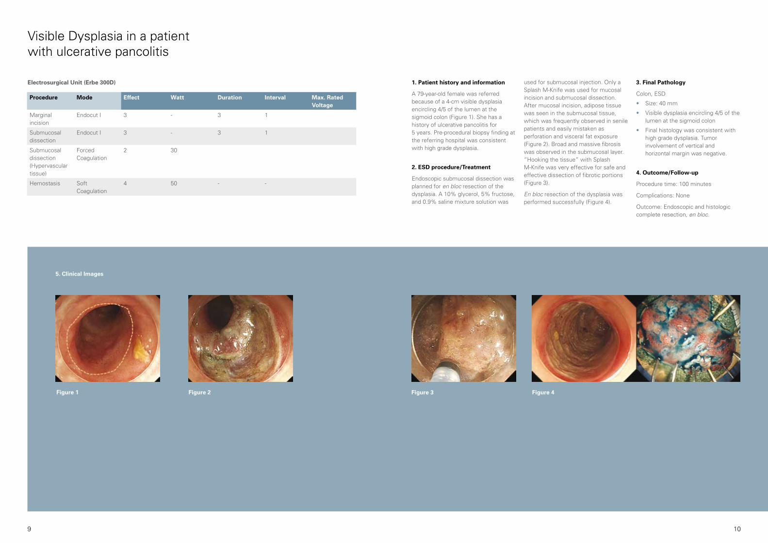
A 79-year-old female was referred because of a 4-cm visible dysplasia encircling 4/5 of the lumen at the sigmoid colon (Figure 1). She has a history of ulcerative pancolitis for 5 years. Pre-procedural biopsy finding at the referring hospital was consistent with high grade dysplasia.
2. ESD procedure/Treatment
Endoscopic submucosal dissection was planned for en bloc resection of the dysplasia. A 10% glycerol, 5% fructose, and 0.9% saline mixture solution was
used for submucosal injection. Only a Splash M-Knife was used for mucosal incision and submucosal dissection. After mucosal incision, adipose tissue was seen in the submucosal tissue, which was frequently observed in senile patients and easily mistaken as perforation and visceral fat exposure (Figure 2). Broad and massive fibrosis was observed in the submucosal layer. “Hooking the tissue” with Splash M-Knife was very effective for safe and effective dissection of fibrotic portions (Figure 3).
En bloc resection of the dysplasia was performed successfully (Figure 4).
Figure 3 Figure 4
3. Final Pathology
Colon, ESD
• Size: 40 mm
• Visible dysplasia encircling 4/5 of the lumen at the sigmoid colon
• Final histology was consistent with high grade dysplasia. Tumor involvement of vertical and horizontal margin was negative.
4. Outcome/Follow-up
Procedure time: 100 minutes
Complications: None
Outcome: Endoscopic and histologic complete resection, en bloc.
Visible Dysplasia in a patient with ulcerative pancolitis
Electrosurgical Unit (Erbe 300D)
Figure 1 Figure 2
5. Clinical Images
Procedure Mode Effect Watt Duration Interval Max. Rated Voltage
Marginal incision
Endocut I 3 - 3 1
Submucosal dissection
Endocut I 3 - 3 1
Submucosal dissection(Hypervascular tissue)
Forced Coagulation
2 30
Hemostasis Soft Coagulation
4 50 - -
11 12
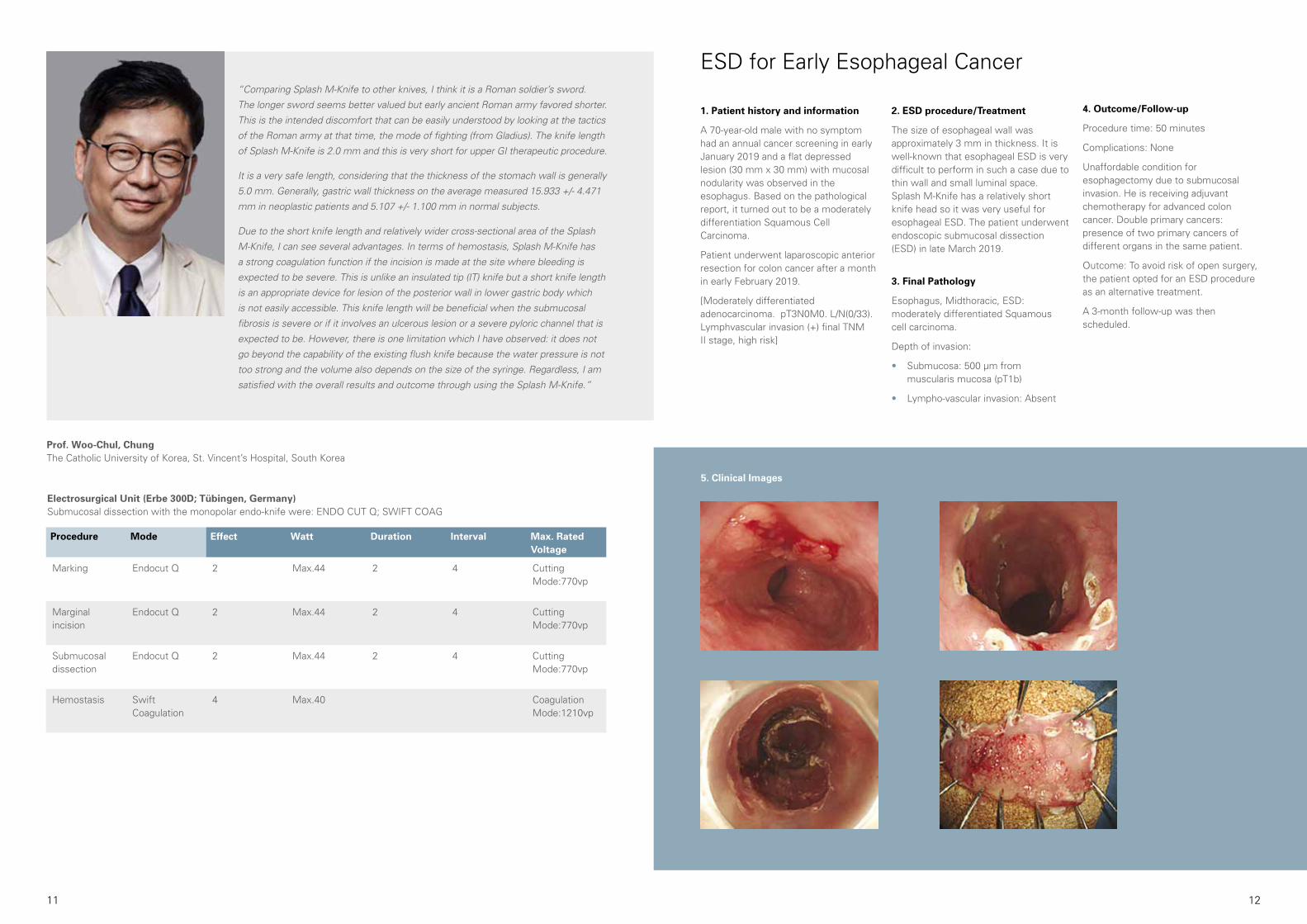
ESD for Early Esophageal Cancer
1. Patient history and information
A 70-year-old male with no symptom had an annual cancer screening in early January 2019 and a flat depressed lesion (30 mm x 30 mm) with mucosal nodularity was observed in the esophagus. Based on the pathological report, it turned out to be a moderately differentiation Squamous Cell Carcinoma.
Patient underwent laparoscopic anterior resection for colon cancer after a month in early February 2019.
[Moderately differentiated adenocarcinoma. pT3N0M0. L/N(0/33). Lymphvascular invasion (+) final TNM II stage, high risk]
2. ESD procedure/Treatment
The size of esophageal wall was approximately 3 mm in thickness. It is well-known that esophageal ESD is very difficult to perform in such a case due to thin wall and small luminal space. Splash M-Knife has a relatively short knife head so it was very useful for esophageal ESD. The patient underwent endoscopic submucosal dissection (ESD) in late March 2019.
3. Final Pathology
Esophagus, Midthoracic, ESD: moderately differentiated Squamous cell carcinoma.
Depth of invasion:
• Submucosa: 500 µm from muscularis mucosa (pT1b)
• Lympho-vascular invasion: Absent
4. Outcome/Follow-up
Procedure time: 50 minutes
Complications: None
Unaffordable condition for esophagectomy due to submucosal invasion. He is receiving adjuvant chemotherapy for advanced colon cancer. Double primary cancers: presence of two primary cancers of different organs in the same patient.
Outcome: To avoid risk of open surgery, the patient opted for an ESD procedure as an alternative treatment.
A 3-month follow-up was then scheduled.
5. Clinical Images
“Comparing Splash M-Knife to other knives, I think it is a Roman soldier’s sword.
The longer sword seems better valued but early ancient Roman army favored shorter.
This is the intended discomfort that can be easily understood by looking at the tactics
of the Roman army at that time, the mode of fighting (from Gladius). The knife length
of Splash M-Knife is 2.0 mm and this is very short for upper GI therapeutic procedure.
It is a very safe length, considering that the thickness of the stomach wall is generally
5.0 mm. Generally, gastric wall thickness on the average measured 15.933 +/- 4.471
mm in neoplastic patients and 5.107 +/- 1.100 mm in normal subjects.
Due to the short knife length and relatively wider cross-sectional area of the Splash
M-Knife, I can see several advantages. In terms of hemostasis, Splash M-Knife has
a strong coagulation function if the incision is made at the site where bleeding is
expected to be severe. This is unlike an insulated tip (IT) knife but a short knife length
is an appropriate device for lesion of the posterior wall in lower gastric body which
is not easily accessible. This knife length will be beneficial when the submucosal
fibrosis is severe or if it involves an ulcerous lesion or a severe pyloric channel that is
expected to be. However, there is one limitation which I have observed: it does not
go beyond the capability of the existing flush knife because the water pressure is not
too strong and the volume also depends on the size of the syringe. Regardless, I am
satisfied with the overall results and outcome through using the Splash M-Knife.”
Prof. Woo-Chul, ChungThe Catholic University of Korea, St. Vincent’s Hospital, South Korea
Procedure Mode Effect Watt Duration Interval Max. Rated Voltage
Marking Endocut Q 2 Max.44 2 4 Cutting Mode:770vp
Marginal incision
Endocut Q 2 Max.44 2 4 Cutting Mode:770vp
Submucosal dissection
Endocut Q 2 Max.44 2 4 Cutting Mode:770vp
Hemostasis Swift Coagulation
4 Max.40 Coagulation Mode:1210vp
Electrosurgical Unit (Erbe 300D; Tübingen, Germany)Submucosal dissection with the monopolar endo-knife were: ENDO CUT Q; SWIFT COAG
13 14
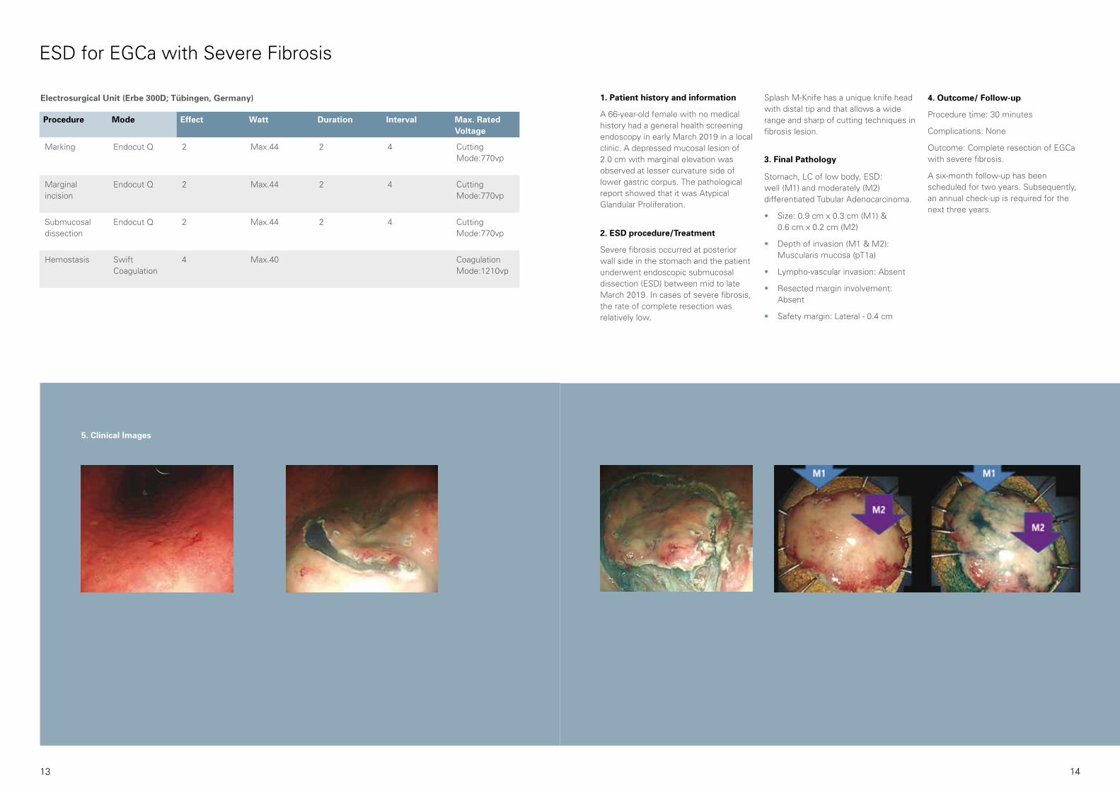
Splash M-Knife has a unique knife head with distal tip and that allows a wide range and sharp of cutting techniques in fibrosis lesion.
3. Final Pathology
Stomach, LC of low body, ESD: well (M1) and moderately (M2) differentiated Tubular Adenocarcinoma.
• Size: 0.9 cm x 0.3 cm (M1) & 0.6 cm x 0.2 cm (M2)
• Depth of invasion (M1 & M2): Muscularis mucosa (pT1a)
• Lympho-vascular invasion: Absent
• Resected margin involvement: Absent
• Safety margin: Lateral - 0.4 cm
4. Outcome/ Follow-up
Procedure time: 30 minutes
Complications: None
Outcome: Complete resection of EGCa with severe fibrosis.
A six-month follow-up has been scheduled for two years. Subsequently, an annual check-up is required for the next three years.
1. Patient history and information
A 66-year-old female with no medical history had a general health screening endoscopy in early March 2019 in a local clinic. A depressed mucosal lesion of 2.0 cm with marginal elevation was observed at lesser curvature side of lower gastric corpus. The pathological report showed that it was Atypical Glandular Proliferation.
2. ESD procedure/Treatment
Severe fibrosis occurred at posterior wall side in the stomach and the patient underwent endoscopic submucosal dissection (ESD) between mid to late March 2019. In cases of severe fibrosis, the rate of complete resection was relatively low.
ESD for EGCa with Severe Fibrosis
Procedure Mode Effect Watt Duration Interval Max. Rated Voltage
Marking Endocut Q 2 Max.44 2 4 Cutting Mode:770vp
Marginal incision
Endocut Q 2 Max.44 2 4 Cutting Mode:770vp
Submucosal dissection
Endocut Q 2 Max.44 2 4 Cutting Mode:770vp
Hemostasis Swift Coagulation
4 Max.40 Coagulation Mode:1210vp
Electrosurgical Unit (Erbe 300D; Tübingen, Germany)
5. Clinical Images
15 16
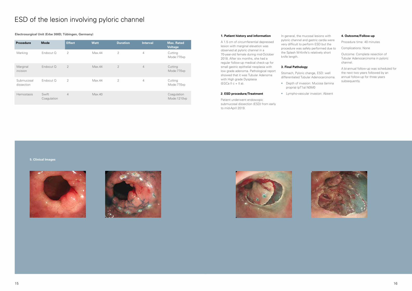
In general, the mucosal lesions with pyloric channel and gastric cardia were very difficult to perform ESD but the procedure was safely performed due to the Splash M-Knife’s relatively short knife length.
3. Final Pathology
Stomach, Pyloric change, ESD: well differentiated Tubular Adenocarcinoma.
• Depth of invasion: Mucosa (lamina propria) (pT1a) N0M0
• Lympho-vascular invasion: Absent
1. Patient history and information
A 1.5 cm of circumferential depressed lesion with marginal elevation was observed at pyloric channel in a 70-year-old female during mid-October 2018. After six months, she had a regular follow-up medical check-up for small gastric epithelial neoplasia with low grade adenoma. Pathological report showed that it was Tubular Adenoma with High grade Dysplasia (EGCa II c + II a).
2. ESD procedure/Treatment
Patient underwent endoscopic submucosal dissection (ESD) from early to mid-April 2019.
4. Outcome/Follow-up
Procedure time: 40 minutes
Complications: None
Outcome: Complete resection of Tubular Adenocarcinoma in pyloric channel.
A bi-annual follow-up was scheduled for the next two years followed by an annual follow-up for three years subsequently.
ESD of the lesion involving pyloric channel
Procedure Mode Effect Watt Duration Interval Max. Rated Voltage
Marking Endocut Q 2 Max.44 2 4 Cutting Mode:770vp
Marginal incision
Endocut Q 2 Max.44 2 4 Cutting Mode:770vp
Submucosal dissection
Endocut Q 2 Max.44 2 4 Cutting Mode:770vp
Hemostasis Swift Coagulation
4 Max.40 Coagulation Mode:1210vp
Electrosurgical Unit (Erbe 300D; Tübingen, Germany)
5. Clinical Images
17 18
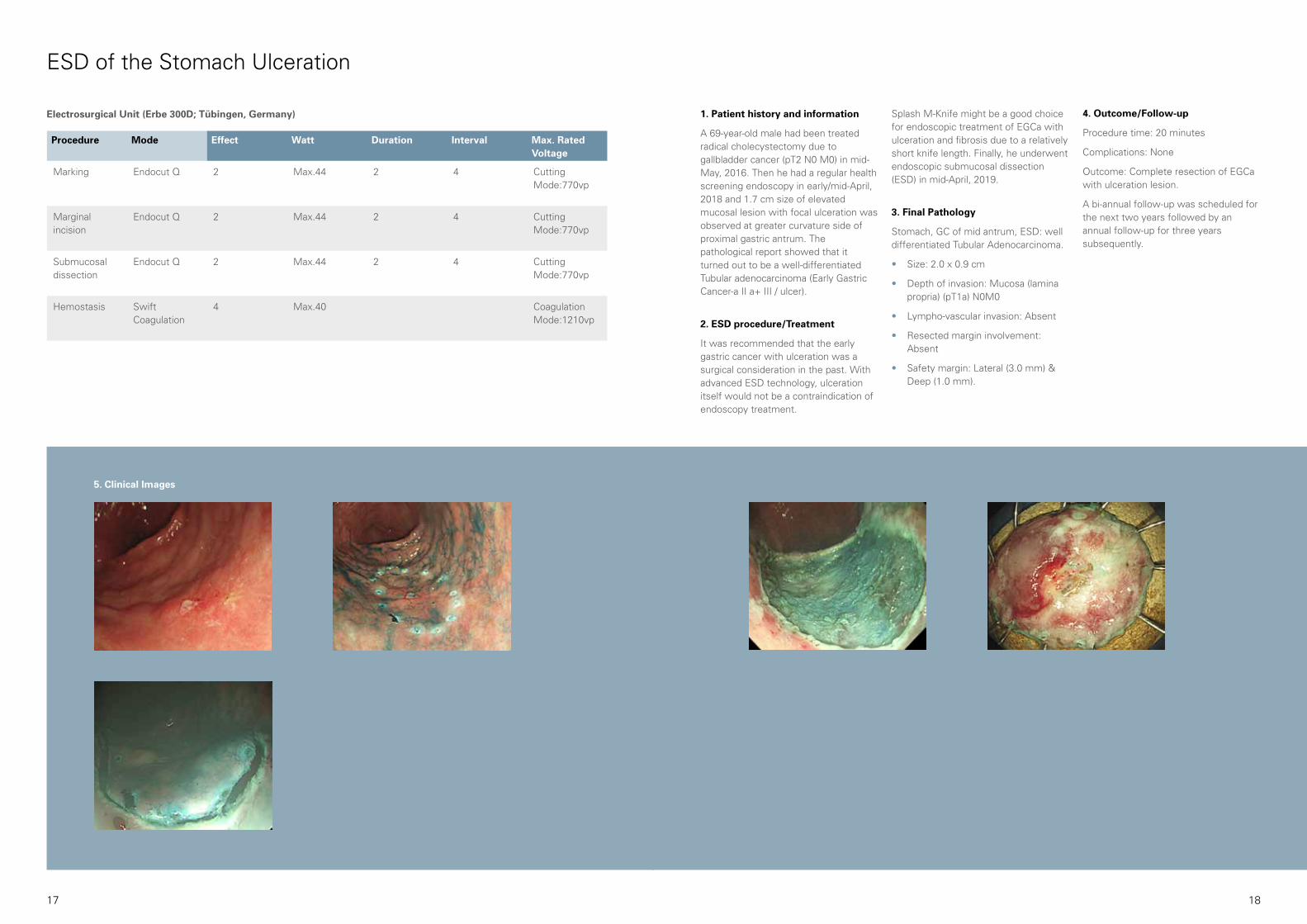
1. Patient history and information
A 69-year-old male had been treated radical cholecystectomy due to gallbladder cancer (pT2 N0 M0) in mid-May, 2016. Then he had a regular health screening endoscopy in early/mid-April, 2018 and 1.7 cm size of elevated mucosal lesion with focal ulceration was observed at greater curvature side of proximal gastric antrum. The pathological report showed that it turned out to be a well-differentiated Tubular adenocarcinoma (Early Gastric Cancer-a II a+ III / ulcer).
2. ESD procedure/Treatment
It was recommended that the early gastric cancer with ulceration was a surgical consideration in the past. With advanced ESD technology, ulceration itself would not be a contraindication of endoscopy treatment.
Splash M-Knife might be a good choice for endoscopic treatment of EGCa with ulceration and fibrosis due to a relatively short knife length. Finally, he underwent endoscopic submucosal dissection (ESD) in mid-April, 2019.
3. Final Pathology
Stomach, GC of mid antrum, ESD: well differentiated Tubular Adenocarcinoma.
• Size: 2.0 x 0.9 cm
• Depth of invasion: Mucosa (lamina propria) (pT1a) N0M0
• Lympho-vascular invasion: Absent
• Resected margin involvement: Absent
• Safety margin: Lateral (3.0 mm) & Deep (1.0 mm).
4. Outcome/Follow-up
Procedure time: 20 minutes
Complications: None
Outcome: Complete resection of EGCa with ulceration lesion.
A bi-annual follow-up was scheduled for the next two years followed by an annual follow-up for three years subsequently.
Procedure Mode Effect Watt Duration Interval Max. Rated Voltage
Marking Endocut Q 2 Max.44 2 4 Cutting Mode:770vp
Marginal incision
Endocut Q 2 Max.44 2 4 Cutting Mode:770vp
Submucosal dissection
Endocut Q 2 Max.44 2 4 Cutting Mode:770vp
Hemostasis Swift Coagulation
4 Max.40 Coagulation Mode:1210vp
ESD of the Stomach Ulceration
Electrosurgical Unit (Erbe 300D; Tübingen, Germany)
5. Clinical Images

19 20
LST of Rectum Case
1. Patient history and information
A 71-year-old female with postoperative hysterectomy in 1993 was in Diabetes Mellitus/Hypertension (-/-) condition. 4.5 cm x 5.0 cm size of laterally spread-ing tumor (LST) was observed at lower rectum. Pathological report showed that it is Tubulovillous Adenoma with Low Grade Dysplasia (TA-LGD).
2. ESD procedure/Treatment
The patient underwent endoscopic submucosal dissection (ESD). The Splash M-Knife enabled smooth dissection and was easy to control
which not only for decreased the procedure time but also generated strong hemostasis effect. Therefore, it is a considerably good multi-functional accessory for completing ESD procedures with a single device.
3. Final Pathology
Colon, LST of Rectum, ESD: TA-LGD
• Size: 4.5 cm x 5.0 cm
• Tubulovillous Adenoma with low grade dysplasia
• Clear resection margin
5. Clinical Images
4. Outcome/Follow-up
Procedure time: 62 minutes
Complications: None
Outcome: Clear resection margin. The patient was discharged from the hospital after 36 hours.
A colonoscopy was scheduled after 12 months and 36 months.
Asso. Prof. Jae-Myeong, ChaKyung-Hee University Hospital at Gang-dong, South Korea
“The ideal ESD knife should avoid making complications such as perforations
and heavy bleeding and yet can still allow a clean cut of lesion at a time. I don’t
think there is a perfect ESD knife due to many reasons. However, I would say the
Splash M-Knife is one of preferred options for good ESD knives because it is able
to perform not only dissection but also lifting of the mucosa by injecting solution
into submucosal layer. This leads to shorter procedure time and increase efficiency
because I do not need to change device. Another strength of the Splash M-Knife is
its hemostasis function. With the large disk of the knife distal end, it is much easier
and faster to make bleeding stop during the procedure.
However, it does not mean Splash M-Knife is perfect. It also has its limitation but
I am fairly satisfied with Splash M-Knife. In certain occasions, completion of ESD
procedure may not be required but some cases are necessary to diagnose the
lesion accurately and perform ESD.”
Procedure Mode Effect Watt Duration Interval Max. Rated Voltage
Marking Endocut Q 2 - 4 3 Cutting Mode: 155 vp
Marginal incision
Endocut Q 2 - 4 3 Cutting Mode: 155vp
Submucosal dissection
Endocut Q 2 - 4 3 Cutting Mode: 155vp
Hemostasis Forced Coagulation
2 80 - - Coagulation Mode: 1100vp
Electrosurgical Unit (Erbe 300D)
21 22
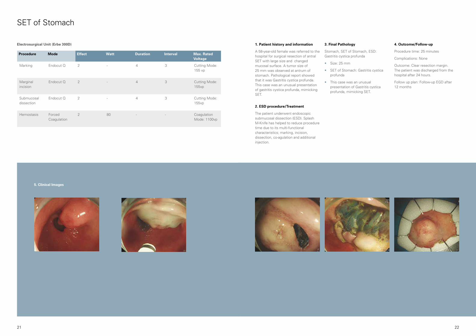
1. Patient history and information
A 58-year-old female was referred to the hospital for surgical resection of antral SET with large size and changed mucosal surface. A tumor size of 25 mm was observed at antrum of stomach. Pathological report showed that it was Gastritis cystica profunda. This case was an unusual presentation of gastritis cystica profunda, mimicking SET.
2. ESD procedure/Treatment
The patient underwent endoscopic submucosal dissection (ESD). Splash M-Knife has helped to reduce procedure time due to its multi-functional characteristics; marking, incision, dissection, co-agulation and additional injection.
3. Final Pathology
Stomach, SET of Stomach, ESD: Gastritis cystica profunda
• Size: 25 mm
• SET of Stomach: Gastritis cystica profunda
• This case was an unusual presentation of Gastritis cystica profunda, mimicking SET.
4. Outcome/Follow-up
Procedure time: 25 minutes
Complications: None
Outcome: Clear resection margin. The patient was discharged from the hospital after 24 hours.
Follow up plan: Follow-up EGD after 12 months
SET of Stomach
Procedure Mode Effect Watt Duration Interval Max. Rated Voltage
Marking Endocut Q 2 - 4 3 Cutting Mode: 155 vp
Marginal incision
Endocut Q 2 - 4 3 Cutting Mode: 155vp
Submucosal dissection
Endocut Q 2 - 4 3 Cutting Mode: 155vp
Hemostasis Forced Coagulation
2 80 - - Coagulation Mode: 1100vp
Electrosurgical Unit (Erbe 300D)
5. Clinical Images

23 24
1. Patient history and information
A 73-year-old male with no medical and family history had a 2.5 cm x 2.0 cm LST nodular mixed type with central dimpling. Previous biopsy result showed that it was Tubulovillous Adenoma under Type 2 and 2A in NICE and JNET classification. Pathological report showed that it was Tubulovillous Adenoma with Low Grade Dysplasia (TA-LGD).
2. ESD procedure/Treatment
He underwent endoscopic submucosal dissection (ESD). Saline with Indigocarmine mixture solution was used for submucosal injection.
According to the previous multiple biopsies performed at a local clinic, the lesion had a moderate to severe fibrosis in the center as well as a large central nodule. The knife was handy to dissect fibrotic tissue on the central lesion, and procedure time could be saved by direct injection of water during the procedure. And, bleeding during the procedure was easily controlled by smooth and effective hemostasis using the knife.
3. Final Pathology
Colon, LST of Rectum, ESD: TA-LGD
• Size: 2.5 cm x 2.0 cm
• Tubulovillous Adenoma with low grade dysplasia
• Clear resection margin
4. Outcome/Follow-up
Procedure time: 19 minutes
Complications: None
Outcome: Clear resection margin.
LST of Rectum Case
5. Clinical Images
Prof. Hyun-Gun, KimSoon Chun Hyang University Hospital Seoul, South Korea
“Splash M-Knife is convenient for performing ESD procedure because this knife
saves procedure time due to its multi-functions such as injection, pre-cutting and
dissection even co-agulation. A complete ESD is not always necessary but is
necessary in cases such as an exact diagnosis of lesion.
Particularly for fibrosis, this knife shows a strong advantage. Direct water injection
into the submucosal layer is possible and this makes it easy to dissect the fibrotic
tissue. In addition, the contact area of the tissue is wider than other knives, so it is
able to catch the bleeding quickly.”
Procedure Mode Effect Watt Duration Interval Max. Rated Voltage
Marking Endocut I 1 - 3 3
Marginal incision
Endocut I 1 - 3 3
Submucosal dissection
Endocut I 1 - 3 3
Hemostasis Swift Coagulation
4 40 - -
Electrosurgical Unit (Erbe 300D)

25 26
1. Patient history and information
A 52-year-old male with no medical and family history; had 30 mm LST NG type at hepatic flexure on TC.
Previous biopsy result was Tubular adenoma and final pathology result also showed Tubular adenoma with focal high-grade dysplasia.
2. ESD procedure/Treatment
The patient underwent endoscopic submucosal dissection (ESD). Saline with Indigocarmine mixture solution was used for submucosal injection.
The lesion was NG type LST, en-bloc resection was planned. However, as the lesion was located at the hepatic flexure medial side on T-colon, dependent portion, the procedure needed very high techniques. Usually, I set Endo-cut I during the ESD, but Endo-cut I mode made some burning effect during incision with Splash M-Knife. I had to switch to Endo-cut Q mode during the next procedure. Moderate to severe fibrosis was diffusely observed on the entire lesion. Direct water injection into the submucosal layer using Splash M-Knife was very useful in dissecting the fibrotic tissues and shortened the procedure time.
3. Final Pathology
Colon, LST of Rectum, ESD: TA-LGD
• Size: 30 mm
• Tubular adenoma with focal high-grade dysplasia
• Clear resection margin
4. Outcome/Follow-up
Procedure time: 83 minutes
Complications: None
Outcome: Clear resection margin.
LST of Hepatic flexure case
5. Clinical Images
Procedure Mode Effect Watt Duration Interval Max. Rated Voltage
Marking Endocut Q 2 - 2 3
Marginalincision
Endocut Q 2 - 2 3
Submucosal dissection
Endocut Q 2 - 2 3
Hemostasis Swift Coagulation
4 40 - -
Electrosurgical Unit (Erbe 300D)

27 28
Product Name
Working Length
Maximum Insertion Portion Width 2.7 mm 2.7 mm
2.0 mm 2.0 mm
0.5 mm
1500 mm or less
2.8 mm or more
Yes
0.5 mm
1900 mm or less
2.8 mm or more
Yes
Extended
Minimum Instrument Channel Width
Retracted
Working Length
1800 mm 2200 mm
DN-D2718B DN-D2722B
Active Portion Length
Compatible Endoscopes
Water Jet Function
Metal Plate: 1.8 mmNozzle
Knife Disk:0.8 mm
Knife: 0.3 mm
Marker
Inlet
(optionally available)
Active Cord Jack
connect to plug
plug
ACTIVE CORD
Insertion Portion
connect to an electro-surgical generator
Active Cord Plug
Finger Grip
2.0 mm
0.5 mm
Active portion length
Active portion length
Product Specifications
PENTAX Medical’s Triple Aim
Our Triple Aim program is designed to deliver on our commitment to support you and your healthcare organization’s wider objectives by providing programs, products and solutions to help you reach your goals.

LCM
/01/
09/2
0/32
0526
/02
TÜV Süd CE0123 • Medical device class: IIb • This product must be used only by healthcare professionals. Before usage and for detailed product specifications, please refer to the instructions for use. In the interest of technical process, specifications may change without notice.
Japan HOYA Corporation6-10-1 Nishi-shinjuku
Shinjuku-kuTokyo 160-0023
EMEA HeadquarterGermany
PENTAX Europe GmbHJulius-Vosseler-Straße 10422527 HamburgTel.: +49 40 / 5 61 92 - 0Fax: +49 40 / 5 60 42 13E-mail: [email protected]
0123


