Immunology, 1964, 7, 363. Antigenic Properties of Microsomes from Guinea-Pig Spleen, Liver and Lymph Nodes* ELAINE G. WHITBECKt AND L. T. ROSENBERG Department of Medical Microbiology, Stanford University, Stanford, California (Received 13th August 1963) Summary. The antigenic properties of guinea-pig spleen, liver and lymph node microsomes have been studied. These subcellular particles were isolated by differential centrifugation. Ribosomes were prepared by treating the microsomes with sodium desoxycholate. Double-diffusion in agar and immunoelectrophoresis were the principal serological techniques employed. The use of appropriately absorbed as well as unabsorbed antisera has permitted the recognition of spleen- specific antigens associated with membranous elements of the cell. A liver-specific antigen has also been found. Serum protein antigens associated with the micro- somes and a particle-specific antigen associated with ribosomes have been re- cognized. The microsomes from the three tissues contain the Forssman antigen. No evidence was obtained to indicate that the RNA present functioned as an antigenic determinant. INTRODUCTION The development of differential centrifugation techniques by Claude (1946a, b) and others has made it possible to isolate reproducibly various subcellular fractions from tissue homogenates. It has been possible to relate certain of these fractions to recognizable structures in the intact cell, and in turn biochemical studies have enabled the function of some of these subcellular components to be identified. Immunological studies of sub- cellular tissue components offer an approach to the study of the relationships between different structures in the same cell, or between similar structures isolated from different cells. One subcellular fraction of particular interest is that to which Claude gave the name microsomes (Claude, 1943). This fraction, with its associated ribonucleoprotein (RNP) particles, is derived from the endoplasmic reticulum (Palade and Siekevitz, 1956), a principal site of protein synthesis (Simkin, 1959). Indeed, it has been demonstrated that part of the protein associated with these particles may be the product of protein synthesis (Peters, 1957; Kern, Helmreich and Eisen, 1959). Several studies dealing with the antigenic properties of mammalian microsomes and ribosomes as well as bacterial ribosomes have been reported in recent years (Barbu, Panijel, Cayeux and Wahl, 1959; Barbu and Panijel, 1960; Barbu, Panijel and Quash, 1961; Panijel and Barbu, 1960; Quash, Dandeu, Barbu and Panijel, 1962; Lacour, Harel, * This investigation was supported in part by Public Health Service Research Grant AI-04442 from the National Institute of Allergy and Infectious Diseases. t Alice Freeman Palmer Fellow of the AAUW Educational Foundation. This study was taken in part from a dissertation submitted in partial fulfilment of the requirements for the Ph.D. 363
Transcript
Immunology, 1964, 7, 363.
Antigenic Properties of Microsomes from Guinea-PigSpleen, Liver and Lymph Nodes*
ELAINE G. WHITBECKt AND L. T. ROSENBERG
Department of Medical Microbiology, Stanford University, Stanford, California
(Received 13th August 1963)
Summary. The antigenic properties of guinea-pig spleen, liver and lymph nodemicrosomes have been studied. These subcellular particles were isolated bydifferential centrifugation. Ribosomes were prepared by treating the microsomeswith sodium desoxycholate. Double-diffusion in agar and immunoelectrophoresiswere the principal serological techniques employed. The use of appropriatelyabsorbed as well as unabsorbed antisera has permitted the recognition of spleen-specific antigens associated with membranous elements of the cell. A liver-specificantigen has also been found. Serum protein antigens associated with the micro-somes and a particle-specific antigen associated with ribosomes have been re-cognized. The microsomes from the three tissues contain the Forssman antigen.No evidence was obtained to indicate that the RNA present functioned as anantigenic determinant.
INTRODUCTION
The development of differential centrifugation techniques by Claude (1946a, b) andothers has made it possible to isolate reproducibly various subcellular fractions from tissuehomogenates. It has been possible to relate certain of these fractions to recognizablestructures in the intact cell, and in turn biochemical studies have enabled the function ofsome of these subcellular components to be identified. Immunological studies of sub-cellular tissue components offer an approach to the study of the relationships betweendifferent structures in the same cell, or between similar structures isolated from differentcells.One subcellular fraction of particular interest is that to which Claude gave the name
microsomes (Claude, 1943). This fraction, with its associated ribonucleoprotein (RNP)particles, is derived from the endoplasmic reticulum (Palade and Siekevitz, 1956), aprincipal site of protein synthesis (Simkin, 1959). Indeed, it has been demonstrated thatpart of the protein associated with these particles may be the product of protein synthesis(Peters, 1957; Kern, Helmreich and Eisen, 1959).
Several studies dealing with the antigenic properties of mammalian microsomes andribosomes as well as bacterial ribosomes have been reported in recent years (Barbu,Panijel, Cayeux and Wahl, 1959; Barbu and Panijel, 1960; Barbu, Panijel and Quash,1961; Panijel and Barbu, 1960; Quash, Dandeu, Barbu and Panijel, 1962; Lacour, Harel,
* This investigation was supported in part by Public Health Service Research Grant AI-04442 from the NationalInstitute of Allergy and Infectious Diseases.
t Alice Freeman Palmer Fellow of the AAUW Educational Foundation. This study was taken in part from adissertation submitted in partial fulfilment of the requirements for the Ph.D.
363
Elaine G. Whitbeck and L. T. RosenbergHarel and Hermet, 1962a; Lacour, Harel, Harel and Nahon, 1962b). An extensive investi-gation of the soluble antigens of rat liver microsomes, mitochondria and other cell fractionshas been carried out by Perlmann and co-workers and D'Amelio et al. (Perlmann andD'Amelio, 1958; Perlmann, Hultin, D'Amelio and Morgan, 1959; D'Amelio and Perl-mann, 1960; D'Amelio, Mutolo and Barbarino, 1963). They found a high degree of cellfraction specificity, as well as some antigens extractable by desoxycholate which wereshared by the various cell fractions. The anti-microsome sera reacted with normal ratserum (Perlmann et al., 1959). Okada (1962) has found tissue specific antigens associatedwith chicken kidney microsomes. Not only have protein components of these particlesbeen implicated as determinants of antigenic specificity, but in some instances the RNAhas also been found to function as antigen or hapten (Barbu and Panijel, 1960, 1961;Lacour et al., 1962a, b).
In the present investigation microsomes and ribosomes were isolated from guinea-pigspleen, liver and lymph nodes. The reactivity of these preparations with anti-spleenmicrosome serum has been studied to determine whether the spleen preparations havetissue-specific, particle-specific or product-specific antigens associated with them. Theantisera have also been examined for the presence of antibodies to RNA.
MATERIALS AND METHODS
Guinea-pigs, obtained from a local supplier, were sacrificed and the spleen, liver andlymph nodes removed. The tissues were stored frozen until used. Microsomes were isolatedby thawing and mincing the tissues in Littlefield and Keller's medium A (Littlefield andKeller, 1957). The entire isolation procedure was carried out in the cold. The mincedtissues were washed three times in this medium, then homogenized. The homogenates werecentrifuged at 10,000-12,000 rev./min. for 1 hour. The upper two-thirds to three-quartersof the clear supernates were decanted and centrifuged at 105,000 g for 1 hour in aSpinco Model L ultracentrifuge. The microsomal pellets were rinsed three times inmedium A and stored, either in a desiccator following lyophilization, or in a freezer in theplastic centrifuge tubes.
Ribosomes were prepared by homogenizing freshly-prepared microsomal pellets in 0 75per cent sodium desoxycholate (DOC) using 1 ml./pellet. After standing 10-15 minutes,these homogenates were centrifuged at 105,000 g for 2 hours. The pellets were rinsedthree times and stored at -20°. The microsome and ribosome preparations werecharacterized chemically (see below) and by their U.V. absorption spectra.
Rabbits were immunized with microsomes from the three tissues. A saline suspension ofthe lyophilized material was incorporated in complete Freund's adjuvant, at a final con-centration of 2 mg./ml. Subcutaneous injections of 1 ml. each were given at weekly inter-vals for 3 weeks. The rabbits were bled 1 week later. A second series of injections was givenafter which the animals were bled and sacrificed.The antigen-antibody reactions were studied primarily by double diffusion in agar and
immunoelectrophoresis. The agar diffusion studies were done on microscope slides, ontowhich 4 ml. of 1 per cent Difco agar in veronal buffered saline was pipetted. Immuno-electrophoresis was carried out essentially as described by Grabar and Williams (1955).
Antisera absorbed with heterologous microsomes, guinea-pig serum, spleen cells orsheep erythrocytes, as well as unabsorbed antisera were used. The lyophilized microsomeswere used for absorption. The dry powder was added to the serum which was then in-
364
Antigenic Properties of 'Microsomes
cubated in the refrigerator overnight. The mixture was centrifuged to remove the precipi-tate that had formed and a second aliquot of microsomes added. The mixture was againincubated overnight at 40, and centrifuged. This process was repeated until no furtherprecipitation occurred. The antisera were then reacted against microsomes in an agardiffusion plate to make certain that the absorption was complete. The same procedure wasfollowed for the absorption with guinea-pig serum. At the end of this absorption, the anti-sera had been diluted about one-half. The spleen cell and sheep erythrocyte absorptionswere carried out on antiserum previously absorbed with guinea-pig serum. Washedpacked cells were used. Two absorptions were carried out in each instance, the first at370 for 2 hours, the second at 40 for 24 hours.The ultraviolet absorption spectra of the microsomes and ribosomes were carried out in
the Beckman DK-2A spectrophotometer. The chemical analysis of the preparations in-cluded the following procedures:
1. Protein determinations by the Folin-Ciocalteau method as modified by Lowry,Rosebrough, Farr and Randall (1951).
2. Phosphate determinations by the method of Fiske and Subbarow (1925). Both totalphosphate and nucleic acid phosphate were determined. The nucleic acid phosphate wasextracted with trichloracetic acid (TCA).
3. The diphenylamine test for DNA was used (Colowick and Kaplan, 1957).The antisera were examined for the presence of antibody directed against RNA by the
precipitin reaction, by quantitative complement fixation (Rabat and Mayer, 1961), andby haemagglutination (Stavitsky, 1954; Miescher and Strassle, 1957). Anti-Forssmanantibody was measured by the agglutination of sheep erythrocytes.
RESULTS
The results of the chemical analyses of the microsomes and ribosomes are in Table 1.The efficiency of the isolation procedure as indicated by the protein determinations wassubject to some variation. The data also show that the composition of the isolates variedfrom one preparation to another. The liver microsomes are quite unlike the other two inthat only a small percentage of the phosphate present is nucleic acid phosphate. Even inthe liver ribosomes the proportion of nucleic acid phosphate is low, suggesting that theseparticles may still contain fairly large amounts ofmembrane material. The diphenylamine-test for DNA was negative throughout. The absorption spectra of the preparations areshown in Fig. 1.The results obtained with unabsorbed anti-spleen microsome serum and microsomes
and ribosomes from spleen, liver and lymph nodes are shown in Fig. 2(a). At least sixprecipitin lines are given by spleen and liver microsomes, fewer with lymph node micro-somes. The ribosomes, or DOC insoluble material, gave somewhat less complex reactionpatterns.Owing to the complexities of the reaction patterns, it is difficult to ascertain whether the
several precipitin lines cross react or give reactions of identity. The relationships of theantigen-antibody systems present can be more easily and clearly determined by absorbingthe antiserum with appropriate antigenic preparations. The removal by a given antigenof antibody reacting with the homologous antigen is proof of the presence of commonantigenic determinants in the preparations. This process also enables the presence ofantigens characteristic of the immunizing antigen to be more easily recognized.
365
Elaine G. Whitbeck and L. T. Rosenberg
TABLE 1
CHEMICAL ANALYSES OF MICROSOMES AND RIBOSOMES
Protein Phosphate Phosphate! Nucleic acid Nucleic acid(pg./g. wet (pg./g. wet Protein phosphate phosphate!
Absorption of the antiserum with guinea-pig serum resulted in the loss of several of theprecipitin lines (Fig. 2b). One line, giving a reaction of identity is seen with all of themicrosome and ribosome preparations. An additional line is present with the homologousantigen, spleen microsomes. These results indicate that the microsomes share antigenswith the serum proteins. They also suggest that the microsomes and ribosomes have acommon antigen, and that spleen microsomes have antigens not shared by the serum or bymicrosomes from the other two tissues.When the antiserum is absorbed with liver microsomes, almost all of the antibody
activity is removed from the serum (Fig. 2c). The only antigen preparation reacting with
FIG. 2. Reaction of microsomes and ribosomes with anti-spleen microsome serum. (a) Anti-spleenmicrosome serum; (b) antiserum absorbed with guinea-pig serum; (c) antiserum absorbed with livermicrosomes; (d) antiserum absorbed with guinea-pig serum and spleen cells. 1 = spleen microsomes;2 = spleen ribosomes; 3 = liver microsomes; 4 = liver ribosomes; 5 = lymph node microsomes;6 = lymph node ribosomes. On the original photograph of (c) two faint lines are visible between thecentre well and well No. 1, but the block-making process has not been able to reproduce these.
this serum is the homologous one. Two precipitin lines are given by spleen microsomes andthis absorbed serum. The liver microsomes have removed antibody directed against serumproteins as well as that against the common antigen.The fact that spleen ribosomes do not appear to share the antigens present in the spleen
microsomes which reacted with the above serum, suggested that the antigens involvedmight be membrane antigens. To investigate this possibility, spleen antiserum previouslyabsorbed with guinea-pig serum was absorbed with spleen cells. As is shown in Fig. 2(d),all the microsomes and ribosomes reacted with this doubly absorbed serum. This line gavea reaction of identity throughout. Spleen microsomes failed to reveal the presence of anyadditional antigen. The spleen cells removed antibody directed against any spleen specificantigens. This result supports the idea that the spleen antigens reacting with serum
367
Elaine G. Whitbeck and L. T. Rosenbergabsorbed with liver microsomes are membrane antigens and further suggests that the cellmembrane and endoplasmic reticulum are antigenically related.The spleen specific antigens can be shown to be distinct from the antigen shared by the
microsomes and ribosomes from the three tissues. The doubly absorbed serum gave onlyone identical precipitin line with all of the antigen preparations. In Fig. 3 spleen micro-somes and ribosomes have been reacted with this serum as well as with the liver microsome
FIG. 3. Demonstration of the non-identity of the spleen specific antigens and the ribosomal antigen.I = anti-spleen microsome serum absorbed with liver microsomes; 2 = spleen ribosomes; 3 = anti-serum absorbed with guinea-pig serum and spleen cells; 4 = spleen microsomes.
absorbed serum. The spleen specific antigens react only with the latter serum and arefound only in the spleen microsomes. One of these lines clearly crosses the line formed bythe common antigen present in both the microsomes and ribosomes indicating the non-identity of the antigens involved. The precipitin line caused by the other spleen specific
FIG. 4. Antigenic identity of spleen, liver and lymph node ribosomes. Centre row contains spleen, liverand lymph node ribosomes; upper and lower rows contain various anti-microsomal sera absorbed withguinea-pig serum.
antigen is weak and does not cross the common antigen line. The fact that no comparableline is given by the ribosomes indicates its non-identity with the common antigen.The common ribosomal antigen in the microsomes and ribosomes of the three tissues
demonstrable with the anti-spleen microsome serum can also be detected with antiseraagainst liver and lymph node microsomes. In Fig. 4, ribosomes from the three tissues havebeen reacted with the three anti-microsome sera. A single precipitin line developed, givinga reaction of identity throughout.
368
Antigenic Properties of Microsomes
Antibodies directed against serum proteins are present in the spleen antiserum. This isindicated by the loss of precipitin lines following absorption of the antiserum with guinea-pig serum. When guinea-pig serum is used as antigen in an agar double diffusion plate,three precipitin lines appear with whole anti-spleen serum. Antiserum which has beenabsorbed with liver microsomes fails to react with guinea-pig serum indicating that thisabsorption also removes antibody directed against serum proteins. A summary of thespleen antigens detected by double diffusion in agar is given in Table 2.
Electrophoresis of guinea-pig serum prior to reaction with anti-spleen microsomeserum results in the appearance of at least five precipitin lines. These are shown diagram-matically in Fig. 5. It can be seen that antibody reacting with y-globulin and serum albuminas well as serum components of intermediate mobility are present in this antiserum.
A I
81BI
B|
2
3B
c I4
FIG. 5. Immunoelectrophoresis reactions of guinea-pig serum and spleen microsomes and ribosomeswith anti-spleen microsome serum, unabsorbed and absorbed in various ways. A= guinea-pigserum; B = spleen microsomes; C = spleen ribosomes; 1 = anti-spleen microsome serum; 2 =antiserum absorbed with guinea-pig serum; 3 = antiserum absorbed with liver microsomes; 4 =antiserum absorbed with guinea-pig serum and spleen cells.
Electrophoresis of microsomes and ribosomes was also carried out. After reaction withantiserum, five or six lines developed with the microsomes. Although the microsomalantigens may or may not correspond to the albumin, a-, P- and y-globulin components ofserum, these designations are used throughout in identifying the lines obtained.
Precipitin lines are found in the pre-albumin, albumin, a-, P- and y-globulin regionswhen unabsorbed anti-spleen serum is used with electrophoresed microsomes. Absorptionsofthe antiserum with guinea-pig serum caused the loss of lines in the a-, y- and p-globulinand pre-albumin regions. The liver microsome absorption resulted in the loss of y and Plines while the doubly absorbed antiserum revealed the presence of only one antigeniccomponent, with the mobility of an albumin.
Immunoelectrophoretic studies with spleen ribosomes and the anti-spleen microsomeserum gave identical results whether whole or absorbed antiserum was used. The only line
369
3Elaine G. Whitbeck and L. T. Rosenberg
which developed was in the albumin region. In some experiments this was a single line, inothers it seemed to have split into a double line.
Both the microsomal and ribosomal supernates were examined immunologically. Themicrosomal supernate is the cell sap, the material which is not sedimentable at 105,000 gin 1 hour. The ribosomal supernate contains the microsomal material that was solubilizedby the DOC treatment.
TABLE 2
SUMMARY OF THE REACTIONS OF ANTI-SPLEEN MICROSOME SERUM INAGAR DOUBLE DIFFUSION SLIDES
Anti-spleen microsome serum absorbedwith:
Antigen UnabsorbedGPS GPS+SC LM
SM S1, S2 SI, S2 S1, S2C C CPlP2,P3 _
SR C C C
LM C C CPl, P2
LR C C C
LyM C C CPl, P2, P3 _
LyR C C C -
Key: SM = spleen microsomes; SR = spleen ribosomes; LM =liver microsomes; LR = liver ribosomes; LyM = lymph nodemicrosomes; LyR = lymph node ribosomes; S1, 2S = spleenspecific antigens; C = common ribosomal antigen; P1, P2, P3 =serum protein antigens; GPS = guinea-pig serum; SC = spleencells.
The microsomal supernate is very complex antigenically. At least six precipitin linesdeveloped in agar diffusion plates with unabsorbed anti-spleen microsome serum. Theribosomal supernate gave at most three lines with this antiserum. The three antigenspresent in the latter supernate proved to be identical with three in the microsomal super-nate, and with two present in microsomes and in guinea-pig serum. None of these wereshared by ribosomes (Fig. 6).
In addition to the three antigens shared with the ribosomal supernate, the cell sapshared two additional antigens with ribosomes which were also present in the microsomes(Fig. 6). Thus six antigens present in the microsomal supernate can be accounted for:three are shared by the ribosomal supernate and are the only antigens present in thisfraction; two of these antigens are also present in the microsomes; antigens 4 and 5 areshared by the cell sap, ribosomes and microsomes. There is at least one additional antigenin the microsomal supernate which cannot be detected in the other subcellular fractions.
Although only two of the three antigens in the ribosomal supernate gave reactions ofidentity with guinea-pig serum antigens, antiserum which had been absorbed with guinea-pig serum failed to react with this supernatant fraction. However, as was pointed outpreviously, this absorption resulted in considerable dilution of the antiserum and it may
370
Antigenic Properties of Microsomes
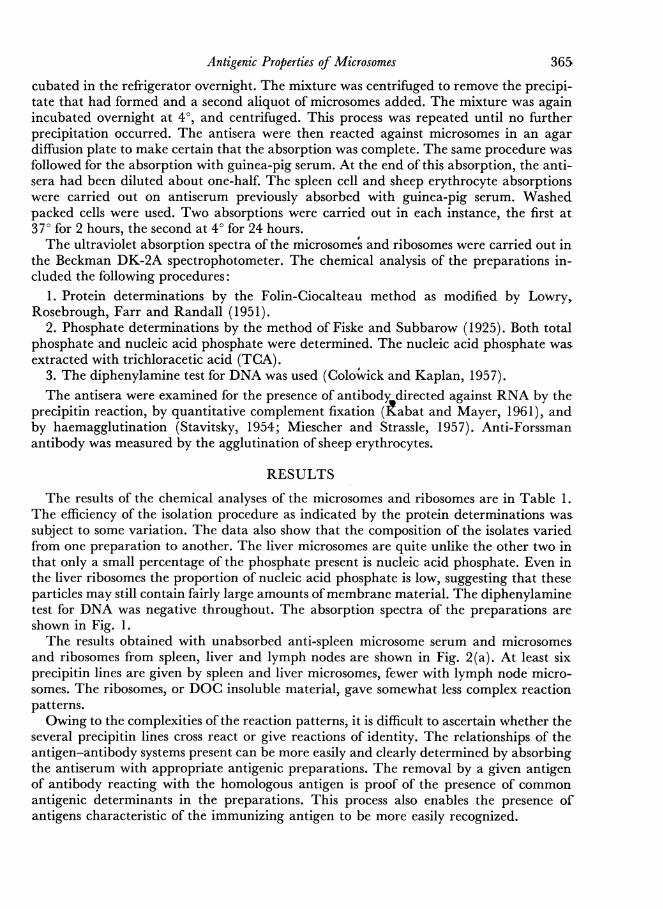
be that any remaining antibody activity for this fraction was too dilute to give an observ-able precipitin line. This same absorbed serum still reacted with the microsomal supernate,with three precipitin bands developing, two of which gave reactions of identity with bothmicrosomes and ribosomes. The third was not shared by either ofthese subcellular particles.Liver microsomes removed all activity for the ribosomal supernate from the serum. The
5 QMs SRO QGPS 0 0 0
0 ~~~~00
0~~~~~~~~~~~~~0( 0SM RS MS SR RS SR
(a) (b) (c) (d)
FIG. 6. Reactions ofspleen supernates in agar double-diffusion slides. (a) and (b) Anti-spleen microsomeserum; (c) antiserum absorbed with guinea-pig serum; (d) antiserum absorbed with liver microsomes.SR = spleen ribosomes; SM = spleen microsomes; MS = microsomal supernate; RS = ribosomalsupernate; GPS = guinea-pig serum.
microsomal supernate does contain one of the spleen specific antigens. One precipitin linedeveloped between this material and the serum absorbed with liver microsomes.In immunoelectrophoretic studies, the ribosomal supernate showed one component only,
which did not migrate from the well (Fig. 7). This component did not react with either ofthe absorbed antisera. The reaction pattern displayed by the cell sap was quite complex.A heavy line was present in the y-globulin region as well as two or three lines in the P-
globulin zone, an a-globulin, an albumin and pre-albumin. As with the microsomes,A I
Al
2Al
3
BI
FIG. 7. Electrophoresis of spleen microsome and ribosome supernates tested against anti-spleenmicrosomal serum. A = microsomal supernate; B = ribosomal supernate; 1 = anti-spleen microsomeserum; 2 = antiserum absorbed with guinea-pig serum; 3 = antiserum absorbed with liver microsomes.
absorption of the antiserum with guinea-pig serum left detectable antibodies for two com-ponents which migrated in the albumin and pre-albumin zones. The antiserum which hadbeen absorbed with liver microsomes reacted only with the pre-albumin component.
Similar studies have been carried out with anti-liver and anti-lymph node microsomesera. An antigen specific for liver microsomes has been found. The membrane location ofthis antigen has not been clearly established. An antigen specific for lymph node micro-somes has not as yet been demonstrated.
371
32Elaine G. Whitbeck and L. T. RosenbergA total of twenty antisera have been examined by the methods described above for the
presence of anti-RNA antibody. Anti-RNA antibody was not found in any of the antisera.
DISCUSSION
The immunological techniques used in this study have permitted the identification oftissue-specific antigens associated with the spleen microsomes. The data are quite clear inindicating that these spleen specific antigens are associated with the membranous elementof the microsomes. The DOC-insoluble ribosomes fail to show the presence of this antigen,indicating that it was dissolved with the membrane. It is not possible to demonstrate thepresence of these tissue-specific antigens in the ribosomal supernate, which contains theDOC-soluble material. When this supernatant fraction is reacted against anti-spleenmicrosome serum absorbed with liver microsomes, no precipitin lines appear. While it ispossible that the explanation for this is quantitative, the material rendered soluble beingdiluted in the procedure, it may equally well be that the DOC treatment has resulted inan alteration ofthe molecular configuration of the antigenic material. The loss ofstructuralintegrity may well be accompanied by a loss of antigenic specificity.
Further indication of the membranous nature of the spleen specific antigens wasobtained by absorbing the anti-spleen microsome serum with washed intact spleen cells.This absorption removed antibody directed against both spleen-specific antigens, indicat-ing that the microsome antigens are shared by the cell membrane. The spleen-specificantigens which have been demonstrated are in effect spleen membrane antigens. Electronmicroscopic studies have shown that the endoplasmic reticulum is continuous with theplasma membranes (Watson, 1955). The antigenic identity of the cell membranes is ingood agreement with their demonstrated physical continuity.The microsomes of the three tissues used contain the Forssman antigen. The spleen-
specific antigens are distinct from the Forssman antigen. Liver microsomes which could beused to absorb the anti-Forssman antibody left intact the spleen-specific antibody.Absorption of the antiserum with sheep erythrocytes also did not remove antibodydirected against the spleen-specific antigens.The anti-spleen microsome serum contained antibodies directed against several of the
serum proteins. Since the spleen is active in the synthesis of antibodies, it is not surprisingto find antibody to serum globulins in the anti-serum. However, immunoelectrophoreticanalysis revealed anti-serum albumin activity to be present also. The question of whetherthis antibody is the result of non-specific adsorption of albumin to the spleen microsomesor a reflection of proteins normally associated with this subcellular fraction cannot beresolved at present. A variety of esterases found in the serum on electrophoresis migrate inthe albumin zone. One is immunologically identical with serum albumin (Omachi,Barnum and Glick, 1948; Augustinsson, 1961; Uriel, 1961; Talal, Hormon, de Vaux St.Cyr and Grabar, 1963). Perhaps this one enzyme in spleen microsomes explains theresults obtained.The particle-specific antigen which has been demonstrated is associated with the DOC-
insoluble ribosomes. The chemical nature of this antigen has not been determined but itseems likely that it is protein. The ribosomes have associated with them, in addition to theproducts of protein synthesis, 'structural' proteins, that is, proteins for which no specificfunction is known. It is tempting to assume that this so-called 'structural' protein is beingrecognized here. Ribosomes from each of the three tissues gave only one precipitin line
372
Antigenic Properties of Microsomes 373
with the anti-microsome sera. On immunoelectrophoresis the antigen was found tomigrate to the same position in every instance. Although these results do not rule out thepossibility that a protein synthesized by these tissues is the antigen involved, it seems morelikely that a common protein, perhaps an enzyme, is responsible for the antigenicity of thepreparations.The possibility that RNA might be the common antigen in these particles was also
considered. Some investigators have presented evidence that RNA in ribosomes canfunction as a determinant of antigenic specificity. All attempts to show such a role forRNA in the present investigation have been singularly unsuccessful. It was possible toextract RNA from the precipitate which formed when a suspension of microsomes wasadded to anti-microsome serum in a test tube. This experiment showed only that RNAwas present in the precipitate and in no way indicated that it was involved as a deter-minant of specificity of the antigen-antibody reaction.
This investigation has shown that microsomes are antigenically complex particles.Immunological techniques such as cross absorption of the antisera, the use of doublediffusion in agar and of immunoelectrophoresis have made it possible to identify, in asense, some of these antigens. ,
These data are of interest from the cytological and biochemical points of view. Themembrane location of the spleen specific antigens is in accord with the demonstratedphysical continuity of cell membrane structures. The demonstration of serum proteinantigens associated with the microsomes from these tissues is in agreement with otherstudies indicating that the products of protein synthesis are associated with these particles.The presence of a common antigen in the ribosomes may have significance in regard tothe role of these particles in protein synthesis. Further studies aimed at elucidating thechemical nature and possible enzymatic activity of these antigens is warranted.The sensitivity and specificity of antigen-antibody reactions makes the immunological
approach to the study of cell components an extremely useful one. The informationobtained helps to establish the location of various components within the cell and todetermine the degree of relationship between cell components and between cells fromdifferent tissues. In conjunction with histochemical and biochemical techniques it may aidin the study of various biochemical problems.
REFERENCES
AUGUSTINSSON, K.-B. (1961). 'Multiple forms of esterasein vertebrate blood-plasma.' Ann. N. r. Acad. Sci.,94, 844.
BARBU, E., PANIJEL, J., CAYEUX, P. and WAHL, R.(1959). 'Characterisation immuno-chemique desribonucleoproteines bacteriennes.' C.R. Acad. Sci.(Paris), 249, 338.
BARBU, E. and PANIJEL,J. (1960). 'Presence d'anticorpsanti-acide ribonucleic dans les immunserums anti-ribosomes.' C.R. Acad. Sci. (Paris), 250, 1382.
BARBU, E. and PANIJEL, J. (1961). 'Concentration etproprietes des anticorps anti-acide ribonucleique.'C.R. Acad. Sci. (Paris), 252, 3157.
BARBU, E., PANIJEL,J. and QUASH, G. (1961). 'Charac-terisation immunochemique des ribosomes.' Ann.Inst. Pasteur, 100, 725.
CLAUDE, A. (1943). 'The constitution of protoplasm.'Science, 97, 451.
CLAUDE, A. (1946a). 'Fractionation of mammalianliver cells by differential centrifugation. I. Problems,methods and preparation of extract.' J. exp. Med.,84, 51.
CLAUDE, A. (1946b). 'Fractionation of mammalianliver cells by differential centrifugation. II. Experi-mental procedures and results.' J. exp. Med., 84,61.
COLOWICK, S. P. and KAPLAN, N. D. (1957). Methods inEnzymology, vol. III, pp. 99-100. Academic Press,New York.
D'AMELIO, V. and PERLMANN, P. (1960). 'The distribu-tion of soluble antigens in cellular structures of ratliver.' Exp. Cell Res., 19, 383.
D'AMELIO, V., MUTOLO, V. and BARBARINO, A.(1963). 'Immunological and electrophoretic analysisofrat liver mitochondria and other cellular fractions.'Exp. Cell Res., 29, 1.
374 Elaine G. Whitbeck and L. T. RosenbergFISKE, C. H. and SUBBAROW, Y. (1925). 'The colori-
metric determination of phosphorus.' J. biol. Chem.,66, 375.
GRABAR, P. and WILLIAMS, C. A. (1955). 'Methodeimmunodlectrophoreitique d'analyse de me1anges desubstances antig6nique.' Biochem. biophys. Acta, 17, 67.
KABAT, E. and MAYER, M. M. (1961). ExperimentalImmunochemistry, 2nd edn., pp. 133-240. Thomas,Springfield.
KERN, M., HELMREICH, E. and EISEN, H. N. (1959).'A demonstration of antibody activity on micro-somes.' Proc. nat. Acad. Sci. (Wash.), 45, 862.
LACOUR, F., HAREL, J., HAREL, L. and HERMET, J.(1962a). etudes antigenique des ribosomes decellules mammiferes.' C.R. Acad. Sci. (Paris), 255,1161.
LACOUR, F., HAREL, J., HAREL, L. and NAHON, E.(1962b). 'Immunological study of RNA by themicromethod of double diffusion in agar.' C.R.Acad. Sci. (Paris), 255, 2322.
LITTLEFIELD, J. W. and KELLER, E. B. (1957). 'In-corporation of C14-amino acids into ribonucleo-protein particles from the Ehrlich mouse ascitestumor.'_7. biol. Chem., 224, 13.
LOWRY, 0. H., ROSEBROUGH, A. L., FARR, A. L. andRANDALL, R. J. (1951). 'Protein measurement withthe Folin-Ciocalteau reagent.'J. biol. Chem., 193, 265.
MIESCHER, P. and STRASSLE, R. (1957). 'New serologicalmethods for the detection of the L.E. factor.' VoxSang., 2, 283.
OKADA, T. S. (1962). 'Tissue specificity in the solubleantigens in kidney microsomes.' Nature (Lond.), 194,306.
OMACHI, A., BARNUM, C. P. and GLICK, D. (1948).'Quantitative distribution ofan esterase among cyto-plasmic components of mouse liver cells.' Proc. Soc.exp. Biol. (N.Y.), 67, 133.
PALADE, G. E. and SIEKEVITZ, P. (1956). 'Livermicrosomes. An integrated morphological and bio-chemical study.' 7. biophys. biochem. Cytol., 2, 17 1.
PANIJEL, J. and BARBU, E. (1960). 'Communaute anti-genique entre ribonuclkoproteines d'originesdiverses.' C.R. Acad. Sci. (Paris), 250, 232.
PERLMANN, P. and D'AMELIO, V. (1958). 'Solubleantigens in microsomes and other cell fractions ofrat liver.' Nature (Lond.), 181, 491.
PERLMANN, P., HULTIN, T., D'AMELIO, V. and MORGAN,W. S. (1959). 'Distribution and metabolism ofprotein antigens in rat liver.' Exp. Cell Res. 7 (Suppl.),279.
PETERS, T. (1957). 'A serum albumin precursor incytoplasmic particles.'J. biol. Chem., 229, 659.
QUASH, G., DANDEU, J.-P., BARBU, E. and PANIJEL, J.(1962). 'Recherches preliminaire sur les antigenes desribosomes.' Ann. Inst. Pasteur, 103, 1.
SIMKIN, J. L. (1959). 'Protein biosynthesis.' Annu. Rev.Biochem., 28, 145.
STAVITSKY, A. (1954). 'Micromethods for the study ofproteins and antibodies. I. Procedure and generalapplications of hemagglutination and hemagglutina-tion inhibition reactions with tannic acid and proteintreated erythrocytes.'_7. Immunol., 72, 360.
TALAL, N., HORMANN, G., DE VAUX ST. CYR, C. andGRABAR, P. (1963). 'Immunoelectrophoretic studyof rat serum and urinary esterases.'_7. Immunol., 90,246.
URIEL, J. (1961). 'Characterisation des cholinesteraseset d'autres esterases carboxylique apres electro-phorese et immunoelectrophorese en gelose. I.Application 'a l'etude des esterases du serum humainnormal.' Ann. Inst. Pasteur, 101, 104.
WATSON, M. L. (1955). 'The nuclear envelope. Itsstructure and relation to cytoplasmic membranes.'7. biophys. biochem. Cytol., 1, 257.


![Leishmania and Leishmaniasis [Kompatibilitätsmodus] of Specific Prophylaxis and Tropical ... • migrates to liver, spleen, lymph nodes, & bone marrow • fever, weight loss, ...](https://static.documents.pub/doc/80x56/5b0c62677f8b9a2f788c1de4/leishmania-and-leishmaniasis-kompatibilittsmodus-of-specific-prophylaxis-and-tropical.jpg)


