Rev Colomb Radiol. 2012; 23(3): 3535-40 3535 case series SPONTANEOUS DISSECTION OF THE CAROTID AND VERTEBRAL ARTERIES. DOPPLER ULTRASOUND DIAGNOSIS. CASE SERIES DISECCIÓN ESPONTÁNEA DE LAS ARTERIAS CARÓTIDA Y VERTEBRAL. DIAGNÓSTICO POR ULTRASONIDO DOPPLER. SERIE DE CASOS Álvaro Ariza Fonseca 1 SUMMARY The carotid color Doppler ultrasound is usually the first imaging modality employed for the assessment of patients with cerebrovascular symptoms. The recognition of the distal steno- occlusive flow pattern is key for the detection of spontaneous dissection of the internal carotid and vertebral arteries in young patients who present cerebral ischaemic signs. We present six cases of patients whose diagnosis was initially suggested by carotid color Doppler ultrasound. RESUMEN Usualmente, el ultrasonido Doppler carotídeo es la primera modalidad de estudio para evaluar a los pacientes con sintomatología de origen cerebrovascular. El reconocimiento del patrón de flujo esteno-oclusivo distal es clave en detectar la disección espontánea de las arterias carótida interna y vertebral en pacientes jóvenes que se presentan con sintomatología de accidente isquémico cerebral. En el artículo se presentan seis casos de pacientes cuyo diagnóstico fue sugerido inicialmente por la realización de un estudio de Doppler carótido- vertebral. Introduction The spontaneous dissection of the carotid artery or the vertebral artery is an unusual event which causes approximately 2%of all cerebral ischaemic events. However, in young populations, this event is an important cause of this pathol- ogy, which causes between 10% and 25% of all cases(1). The distal steno-occlusive flow pattern in the carotid vertebral Doppler ultrasound is a sign which suggests the diagnosis and must the recognized, given that the ultrasound is usually available in a hospital environment. Moreover, the ultrasound is usually one of the first stud- ies that are carried out in patients with cerebral ischaemic signs. Following are six carotid and vertebral dis- section cases, which are evaluated by this diag- nostic method. In all these cases, an important hemodynamic alteration which is compatible with the steno-occlusive flow pattern is ob- served, which is consistent at slow speed, with an absence of flow during diastole, increased resistance and spectral widening due to turbu- lence (figure 1). First Case This case corresponds to a 23 year old man, who entered emergency care while in a drunken state. After vomiting several times and “falling asleep on a table while his neck was twisted”. When he entered, the patient showed signs of cerebral lesions in the mid right cerebral artery. The patient died after 24 hours had passed (figures 2,3,4, and 5). Second Case 46 year old woman, who entered emergency care with signs of a stroke in the left part of her brain. As background information, the patient vomited the day before, probably in relation to food poisoning (figure 6 and 7). 1 Radiological Doctor, Department of Radiology and Ultrasound of Country Clinic, Department of Diagnostic Images of the New Clinic, Bogotá, Colombia KEY WORDS (MeSH) Spontaneous vertebral artery dissection Internal carotid artery dissection Color Doppler ultrasonography PALABRAS CLAVE (DeCS) Disección de la arteria carótida interna Disección de la arteria vertebral Ultrasonografía Doppler
Transcript
Rev Colomb Radiol. 2012; 23(3): 3535-40 3535
case series
SPONTANEOUS DISSECTION OF THE CAROTID AND VERTEBRAL ARTERIES. DOPPLER ULTRASOUND DIAGNOSIS. CASE SERIESDISECCIóN ESPONTáNEA DE LAS ARTERIAS CARóTIDA y VERTEBRAL. DIAGNóSTICO POR ULTRASONIDO DOPPLER. SERIE DE CASOS
Álvaro Ariza Fonseca1
SUMMARYThe carotid color Doppler ultrasound is usually the first imaging modality employed for the
assessment of patients with cerebrovascular symptoms. The recognition of the distal steno-occlusive flow pattern is key for the detection of spontaneous dissection of the internal carotid and vertebral arteries in young patients who present cerebral ischaemic signs. We present six cases of patients whose diagnosis was initially suggested by carotid color Doppler ultrasound.
ReSUMenUsualmente, el ultrasonido Doppler carotídeo es la primera modalidad de estudio para
evaluar a los pacientes con sintomatología de origen cerebrovascular. El reconocimiento del patrón de flujo esteno-oclusivo distal es clave en detectar la disección espontánea de las arterias carótida interna y vertebral en pacientes jóvenes que se presentan con sintomatología de accidente isquémico cerebral. En el artículo se presentan seis casos de pacientes cuyo diagnóstico fue sugerido inicialmente por la realización de un estudio de Doppler carótido-vertebral.
IntroductionThe spontaneous dissection of the carotid
artery or the vertebral artery is an unusual event which causes approximately 2%of all cerebral ischaemic events. However, in young populations, this event is an important cause of this pathol-ogy, which causes between 10% and 25% of all cases(1). The distal steno-occlusive flow pattern in the carotid vertebral Doppler ultrasound is a sign which suggests the diagnosis and must the recognized, given that the ultrasound is usually available in a hospital environment. Moreover, the ultrasound is usually one of the first stud-ies that are carried out in patients with cerebral ischaemic signs.
Following are six carotid and vertebral dis-section cases, which are evaluated by this diag-nostic method. In all these cases, an important hemodynamic alteration which is compatible with the steno-occlusive flow pattern is ob-served, which is consistent at slow speed, with
an absence of flow during diastole, increased resistance and spectral widening due to turbu-lence (figure 1).
First Case This case corresponds to a 23 year old man,
who entered emergency care while in a drunken state. After vomiting several times and “falling asleep on a table while his neck was twisted”. When he entered, the patient showed signs of cerebral lesions in the mid right cerebral artery. The patient died after 24 hours had passed (figures 2,3,4, and 5).
Second Case46 year old woman, who entered emergency
care with signs of a stroke in the left part of her brain. As background information, the patient vomited the day before, probably in relation to food poisoning (figure 6 and 7).
1Radiological Doctor, Department of Radiology and Ultrasound of Country Clinic,
Department of Diagnostic Images of the New Clinic,
Spontaneous Dissection of the Carotid and Vertebral Arteries. Diagnosis by Doppler Ultrasound. Case Series. Ariza Á.3536
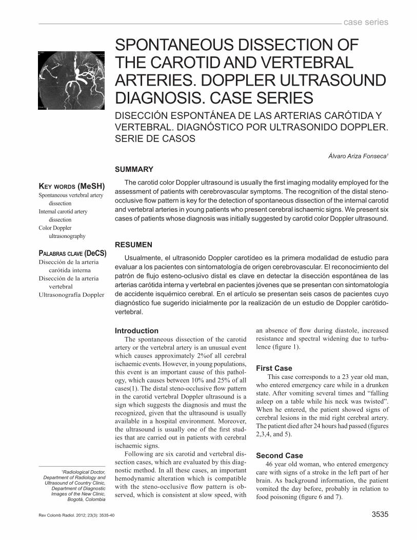
Figure 1. Normal flow pattern in the internal carotid and vertebral compared to the steno-occlusive flow pattern. The diagram shows abundant flow in a normal artery (left) during the cardiac cycle. Visual findings are confirmed by a measurement of velocities: Peak systolic velocity and end diastolic velocity (V1 and V2, respectively). The resistance index (IR) of the carotid and normal vertebral arteries is lesser than 0.7. The absence of flow at the end of the diastole (relaxation) (V2) in case of stenosis or distal occlusion conditions the increase of IR until 1.
Figure 3. Tomography, sequence with information in T2 with an increase in the right frontal-parietal cortical signal.
Figure 4: ADC Map which shows the disorder affecting the right hemisphere, which respects the territory of the frontal cerebral artery.
Figure 5: Enhanced contrast tomography with an absence of signs in the right internal carotid, as well as in its anterior and middle cerebral branches (arrow points).
Figure 2. The distal steno-occlusive flow pattern in the internal carotid leads to an important alteration of the internal carotid flow spectrum, due to a high-grade obstruction (or total obstruction) of the distal vase light with low velocity (peak systolic velocity or Vmax under 40 cm/s}, absence of end of diastole flow (Ved equal to 0), increase of resistance (RI equal to 1), and turbulence with spectral widening which implies turbulent flow (arrows).
case series
Rev Colomb Radiol. 2012; 23(3): 3535-40 3537
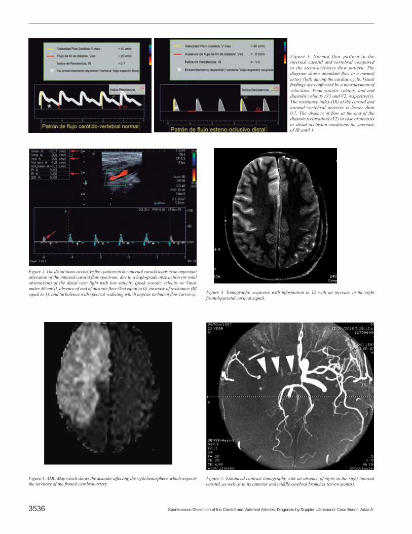
Figure 6: Distal steno-occlusive pattern with slow velocity, spectral widening and an absence of flow to the end of the diastole. In this case, one can see a thrombus in the vascular light (arrow points).
Figure 7. Enhanced contrast tomography by digital subtraction of the common carotid, which confirms the dissection of the left internal carotid (arrow points). The flow of the external carotid is normal.
Figure 8. Right vertebral artery. Distal steno-occlusive pattern with a reduction in flow velocity, high resistance and lack of flow at the end of the diastole in the right vertebral artery. The second image shows the contra-lateral vertebral artery with increased flow, with low resistance and of a compensatory nature. (Peak systolic velocity: 51.7; end diastole velocity: 23.9; resistance index: 0.54).
Third case35-year old woman who entered emergency care due to ischemia
in the area of the right lower back cerebellar artery (PICA), suffering from Wallenberg syndrome. No background information was given when she entered, except for the fact that she has vomited the previous day, probably related to food poisoning. The carotid-vertebral Doppler ultrasound showed a distal steno-occlusive pattern in the right vertebral artery. A normal study was carried out in the remaining aspects (figure 8).
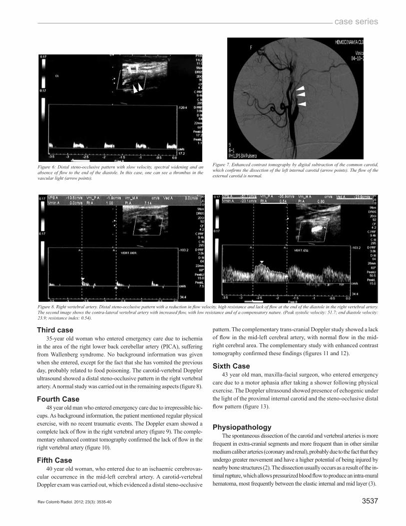
Fourth Case48 year old man who entered emergency care due to irrepressible hic-
cups. As background information, the patient mentioned regular physical exercise, with no recent traumatic events. The Doppler exam showed a complete lack of flow in the right vertebral artery (figure 9). The comple-mentary enhanced contrast tomography confirmed the lack of flow in the right vertebral artery (figure 10).
Fifth Case40 year old woman, who entered due to an ischaemic cerebrovas-
cular occurrence in the mid-left cerebral artery. A carotid-vertebral Doppler exam was carried out, which evidenced a distal steno-occlusive
pattern. The complementary trans-cranial Doppler study showed a lack of flow in the mid-left cerebral artery, with normal flow in the mid-right cerebral area. The complementary study with enhanced contrast tomography confirmed these findings (figures 11 and 12).
Sixth Case43 year old man, maxilla-facial surgeon, who entered emergency
care due to a motor aphasia after taking a shower following physical exercise. The Doppler ultrasound showed presence of echogenic under the light of the proximal internal carotid and the steno-occlusive distal flow pattern (figure 13).
PhysiopathologyThe spontaneous dissection of the carotid and vertebral arteries is more
frequent in extra-cranial segments and more frequent than in other similar medium caliber arteries (coronary and renal), probably due to the fact that they undergo greater movement and have a higher potential of being injured by nearby bone structures (2). The dissection usually occurs as a result of the in-timal rupture, which allows pressurized blood flow to produce an intra-mural hematoma, most frequently between the elastic internal and mid layer (3).
Spontaneous Dissection of the Carotid and Vertebral Arteries. Diagnosis by Doppler Ultrasound. Case Series. Ariza Á.3538
Figure 11.Distal steno-occlusive flow pattern in the left internal carotid, compatible with the distal spontaneous dissection at the moment of interrogation. Enhanced contrast tomography which shows a lack of signs in the internal carotid (arrow), and in the territory of the left medial cerebral artery (arrow points).
Figure 12. Trans-cranial Doppler of the right medial cerebral artery with normal flow (left). The flow is absent in the left medial cerebral artery (right).
Figure 9. A complete lack of flow in the right vertebral artery is observed, with a suggestive image of the thrombus inside, without an intimal flap or mural hematoma. This finding is uncommon, given the fact that the thrombus is usually distal to the dissection, which occurs more frequently in segment V4 (level C1), where vessel insonation is not easy. (2). The differential diagnosis includes the obstruction of arteriosclerotic origin, which can be suspected by observing the existing layer load in the rest of the artery tree (in this case absent).
Figure 10. Arteriography of the brachiocephalic artery, in which an absence of flow can be seen in the right vertebral artery. Only the ascending cervical artery is evident, which is a branch of the thyrocervical scapular artery (arrow). The flow of the right carotid and in the subclavian flap is normal.
case series
Rev Colomb Radiol. 2012; 23(3): 3535-40 3539
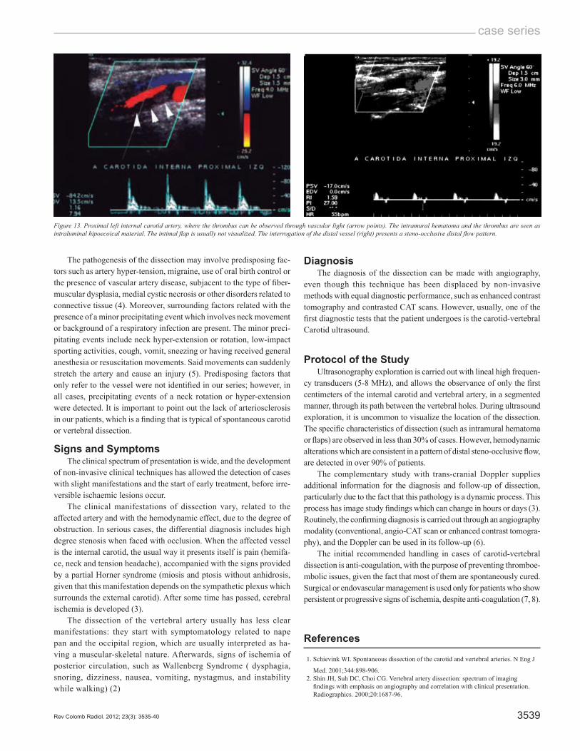
Figure 13. Proximal left internal carotid artery, where the thrombus can be observed through vascular light (arrow points). The intramural hematoma and the thrombus are seen as intraluminal hipoecoical material. The intimal flap is usually not visualized. The interrogation of the distal vessel (right) presents a steno-occlusive distal flow pattern.
The pathogenesis of the dissection may involve predisposing fac-tors such as artery hyper-tension, migraine, use of oral birth control or the presence of vascular artery disease, subjacent to the type of fiber-muscular dysplasia, medial cystic necrosis or other disorders related to connective tissue (4). Moreover, surrounding factors related with the presence of a minor precipitating event which involves neck movement or background of a respiratory infection are present. The minor preci-pitating events include neck hyper-extension or rotation, low-impact sporting activities, cough, vomit, sneezing or having received general anesthesia or resuscitation movements. Said movements can suddenly stretch the artery and cause an injury (5). Predisposing factors that only refer to the vessel were not identified in our series; however, in all cases, precipitating events of a neck rotation or hyper-extension were detected. It is important to point out the lack of arteriosclerosis in our patients, which is a finding that is typical of spontaneous carotid or vertebral dissection.
Signs and SymptomsThe clinical spectrum of presentation is wide, and the development
of non-invasive clinical techniques has allowed the detection of cases with slight manifestations and the start of early treatment, before irre-versible ischaemic lesions occur.
The clinical manifestations of dissection vary, related to the affected artery and with the hemodynamic effect, due to the degree of obstruction. In serious cases, the differential diagnosis includes high degree stenosis when faced with occlusion. When the affected vessel is the internal carotid, the usual way it presents itself is pain (hemifa-ce, neck and tension headache), accompanied with the signs provided by a partial Horner syndrome (miosis and ptosis without anhidrosis, given that this manifestation depends on the sympathetic plexus which surrounds the external carotid). After some time has passed, cerebral ischemia is developed (3).
The dissection of the vertebral artery usually has less clear manifestations: they start with symptomatology related to nape pan and the occipital region, which are usually interpreted as ha-ving a muscular-skeletal nature. Afterwards, signs of ischemia of posterior circulation, such as Wallenberg Syndrome ( dysphagia, snoring, dizziness, nausea, vomiting, nystagmus, and instability while walking) (2)
DiagnosisThe diagnosis of the dissection can be made with angiography,
even though this technique has been displaced by non-invasive methods with equal diagnostic performance, such as enhanced contrast tomography and contrasted CAT scans. However, usually, one of the first diagnostic tests that the patient undergoes is the carotid-vertebral Carotid ultrasound.
Protocol of the StudyUltrasonography exploration is carried out with lineal high frequen-
cy transducers (5-8 MHz), and allows the observance of only the first centimeters of the internal carotid and vertebral artery, in a segmented manner, through its path between the vertebral holes. During ultrasound exploration, it is uncommon to visualize the location of the dissection. The specific characteristics of dissection (such as intramural hematoma or flaps) are observed in less than 30% of cases. However, hemodynamic alterations which are consistent in a pattern of distal steno-occlusive flow, are detected in over 90% of patients.
The complementary study with trans-cranial Doppler supplies additional information for the diagnosis and follow-up of dissection, particularly due to the fact that this pathology is a dynamic process. This process has image study findings which can change in hours or days (3). Routinely, the confirming diagnosis is carried out through an angiography modality (conventional, angio-CAT scan or enhanced contrast tomogra-phy), and the Doppler can be used in its follow-up (6).
The initial recommended handling in cases of carotid-vertebral dissection is anti-coagulation, with the purpose of preventing thromboe-mbolic issues, given the fact that most of them are spontaneously cured. Surgical or endovascular management is used only for patients who show persistent or progressive signs of ischemia, despite anti-coagulation (7, 8).
References
1. Schievink WI. Spontaneous dissection of the carotid and vertebral arteries. N Eng J
Med. 2001;344:898-906.2. Shin JH, Suh DC, Choi CG. Vertebral artery dissection: spectrum of imaging findings with emphasis on angiography and correlation with clinical presentation. Radiographics. 2000;20:1687-96.
Spontaneous Dissection of the Carotid and Vertebral Arteries. Diagnosis by Doppler Ultrasound. Case Series. Ariza Á.3540
3. Rodallec MH, Marteau V, Gerber S, et al. Craniocervical arterial dissection: spectrum of imaging findings and differential diagnosis. Radiographics. 2008;28:1711-28.
4. Lam CS, YeeYK, Tsui YK, et al. Vertebral artery dissection: a treatable cause os ischaemic stroke. HKMJ. 1999;5:398-401.5. Bin Saeed A, Shuaib A, Al-Sulaiti G. Vertebral artery dissection: warning symptoms, clinical features and prognosis in 26 patients. Can J Neurol Sci. 2000;27:292-6.6. Provenzale JM. Dissection of the internal carotid and vertebral arteries: imaging features. AJR Am J Roentgenol. 1995;165:1099-104.7. O’Dwyer JA, Moscow N, Trevor R. Spontaneous dissection of the carotid artery. Radiology. 1980;137:379-85.8. Pugliese F, Crusco F, Cardaioli G. CT Angiography versus colour-Doppler US in acute dissection of the vertebral artery. Radiol Med. 2007;112:435-43.