BACTERIOLOGICAL REVIEWS, Mar. 1969, p. 48-71 Vol. 33, No. 1 Copyright @ 1969 American Society for Microbiology Printed in U.S.A. Sporulation and the Production of Antibiotics, Exoenzymes, and Exotoxins P. SCHAEFFER' Physiologie Cellulaire, Institut Pasteur, Paris 15eme, France INTRODUCTION ............................................................. 48 SPORULATION IN EUBACTERIA ............................................ 49 Cytological Aspect ........................................................... 49 Physiological Aspect: Catabolic Repression of Sporulation ........................... 49 Biochemical Aspect........................................................... so Turnover of RNA .......................................................... 50 Turnover of proteins ........................................................ 50 Intracellular metabolites ..................................................... 51 Extracellular products . ...................................................... 51 Antibiotics ................................................................ 51 Proteases ................................................................. 51 RNA-degrading enzymes .................................................... 52 Wall-lytic enzymes ......................................................... 52 Amylase .................................................................. 52 Lethal toxins . ............................................................. 52 Miscellaneous ............................................................. 53 GENETICAL ASPECT: SPORULATION MUTANTS IN EUBACTERIA ............. 53 Isolation of the Mutants ........ .............................................. 53 Random picking of mutants; sp- vs. osp strains ................................. 53 Selection of mutants, and the catabolic repression hypothesis ...................... 54 Induction of mutants and the sporulation-episome hypothesis ...................... 54 Indirect isolation of mutants ................................................. 55 Cytological Classification of the Mutants ........................................ 55 Biochemical Classification of the Mutants ........................................ 57 General absence of cross-feeding with the sp- mutants ........................... 57 Respiratory deficiencies in sporulation mutants .................................. 57 Ability to produce antibiotic(s) and exoenzymes in sp- mutant strains of B. subtilis: the various spo- phenotypes ............. ................................... 57 Ability to sporulate of antibiotic-deficient and exoenzyme-deficient mutants of B. subtilis 58 Production of lethal exotoxin in sp- mutant strains of C. histolyticumn and C. per- fringens ................................................................. 58 Genetics of Sporulation (in B. subtilis) ......... ................................. 59 Genetical classification of the mutants ......................................... 59 Chromosomal mapping of the mutants: the episome hypothesis ....... ............. 59 Nature of the pleiotropic spo- mutants ........................................ 60 Discussion of Sporulation in Eubacteria .......................................... 60 Exoenzymes, antibiotics, and exotoxins: roles in the sporulation process ..... ......... 61 Problem of the regulation mechanisms controlling the triggering and the unfolding of sporulation ........................................................... 63 SPORULATION AND THE PRODUCTION OF ANTIOBIOTICS, EXOENZYMES, AND EXOTOXINS IN ACTINOMYCETES AND THE LOWER FUNGI ........ 64 SPORULATION MUTANTS IN GENERAL AND APPLIED MICROBIOLOGY ... 65 LIrERATURE CT1ED ......................................................... 66 INTRODUCTION the production of antibiotics, exoenzymes, and exotoxins. It consists of three parts. In the first, of This article has two purposes: to illustrate the an inrdcoyntrreeatifraino usefulness of asporogenous mutants in studying spoa lation in bacteria is mentioned briefly A the mechanism of the sporulation process, and to sprlto inbceaism toed refyA discuss the relationship between this process and number of important points are left out, which are very adequately covered in recent reviews Present address: Institut de Microbiologie, Facultd des Sci- (76, 77, 133, 201). The main part of this review is ences, 91, Orsay, France. the second one, which reports and discusses the 48 on May 28, 2018 by guest http://mmbr.asm.org/ Downloaded from
Transcript
BACTERIOLOGICAL REVIEWS, Mar. 1969, p. 48-71 Vol. 33, No. 1Copyright @ 1969 American Society for Microbiology Printed in U.S.A.
Sporulation and the Production of Antibiotics,Exoenzymes, and Exotoxins
P. SCHAEFFER'Physiologie Cellulaire, Institut Pasteur, Paris 15eme, France
INTRODUCTION............................................................. 48SPORULATION IN EUBACTERIA............................................ 49
Cytological Aspect........................................................... 49Physiological Aspect: Catabolic Repression of Sporulation........................... 49Biochemical Aspect........................................................... so
GENETICAL ASPECT: SPORULATION MUTANTS IN EUBACTERIA............. 53Isolation of the Mutants...................................................... 53Random picking of mutants; sp- vs. osp strains................................. 53Selection of mutants, and the catabolic repression hypothesis...................... 54Induction of mutants and the sporulation-episome hypothesis...................... 54Indirect isolation of mutants................................................. 55
Cytological Classification of the Mutants........................................ 55Biochemical Classification of the Mutants........................................ 57
General absence of cross-feeding with the sp- mutants........................... 57Respiratory deficiencies in sporulation mutants.................................. 57Ability to produce antibiotic(s) and exoenzymes in sp- mutant strains of B. subtilis:
the various spo- phenotypes ............. ................................... 57Ability to sporulate of antibiotic-deficient and exoenzyme-deficient mutants of B. subtilis 58Production of lethal exotoxin in sp- mutant strains of C. histolyticumn and C. per-
fringens................................................................. 58Genetics of Sporulation (in B. subtilis) ......... ................................. 59
Genetical classification of the mutants......................................... 59Chromosomal mapping of the mutants: the episome hypothesis ....... ............. 59Nature of the pleiotropic spo- mutants........................................ 60
Discussion of Sporulation in Eubacteria.......................................... 60Exoenzymes, antibiotics, and exotoxins: roles in the sporulation process ..... ......... 61Problem of the regulation mechanisms controlling the triggering and the unfolding
of sporulation ........................................................... 63SPORULATION AND THE PRODUCTION OF ANTIOBIOTICS, EXOENZYMES,AND EXOTOXINS IN ACTINOMYCETES AND THE LOWER FUNGI........ 64
SPORULATION MUTANTS IN GENERAL AND APPLIED MICROBIOLOGY... 65LIrERATURE CT1ED......................................................... 66
INTRODUCTION the production of antibiotics, exoenzymes, andexotoxins. It consists of three parts. In the first, ofThis article has two purposes: to illustrate the an inrdcoyntrreeatifraino
usefulness of asporogenous mutants in studying spoa lation in bacteria is mentioned briefly Athe mechanism of the sporulation process, and to sprlto inbceaism toed refyAdiscuss the relationship between this process and number of important points are left out, which
are very adequately covered in recent reviewsPresent address: Institut de Microbiologie, Facultd des Sci- (76, 77, 133, 201). The main part of this review is
ences, 91, Orsay, France. the second one, which reports and discusses the48
work on asporogenous bacterial mutants up tothe summer of 1968. A mere addendum to thesecond part, the third is an attempt to evaluate,from a survey of the literature, the possible exten-sion to actinomycetes and lower fungi of theconclusions reached from work on bacteria. Suc-cessive approaches to this presentation havealready been published (172-175, 179).
SPORULATION IN EUBACUrERIASporulation of bacilli and clostridia is
considered here in its cytological, physiological,and biochemical aspects; the genetic approach isalso discussed.
Cytological AspectSuccessive changes in cell structure which
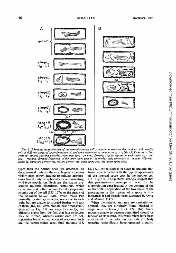
occur during sporulation were first described inBacillus cereus by Young and Fitz-James (240,241); their description is generally valid for otherspecies (18, 82, 159, 165). These changes arecontinuous, but it is convenient to select a fewtypical stages (Fig. 1A) and ascribe a number toeach (61, 179). Cells are said to be at stage I whenthe chromatin is visible as a single axial rod,rather than the one or two nuclear bodies ob-served in the growing cell (168). Stage II beginswith an invagination of the cytoplasmic mem-brane and ends with the completion of a sporeseptum, dividing the nuclear material in two, andcreating a double-celled organism, the spor-angium. This septum is easily distinguished froma division septum by its location close to a poleof the cell and by the absence of wall material be-tween the two membrane layers. Further elonga-tion of the double membrane, which bulges intothe bigger part of the cell, is accompanied by apolewards displacement of the insertion point ofthe septum, until an entirely intracytoplasmicforespore is "carved out" of the mother cell(state III). The next two stages are characterizedby the formation of new envelopes around theforespore: the cortex, formed between the twomembranes at stage IV, and the two layers ofthe spore coat, exterior to the cortex, formed atstage V. A change in the structure of the cortexthen occurs, as the thermoresistance of theenclosed spore is being built up (stage VI). Lysisof the mother cell eventually sets the maturespore free. Schematic representations of thevarious stages, as observed in B. subtilis, arepresented in Fig. 1A.
Unless mentioned otherwise, the following ob-servations apply to the Marburg strain of B. sub-tilis. Nutrient broth cultures, continuously aerated
at 37 C, yield a full crop of spores (at least 60% ofthe population present at to) 6.5 to 7 hr after theend of exponential growth, arbitrarily taken asthe to of sporulation (179). The times at whichcells at a given stage of their development predom-inate in the culture are also indicated in Figure1A. If enough glucose to support growth for 2 hris added to the culture at to, growth continues atan undisturbed rate; thus, nitrogen is in excess inthe medium. Under such conditions, less than 1%of the cells are found as thermoresistant spores att6.5. Addition of glucose at to.5, however, doesnot reduce the yield of spores at t6.5; by thattime, apparently, all the cells that were to sporu-late were committed to do so (64, 69, 70). Tworemarks are in order here, the first on Foster'sdefinition of commitment. A cell is considered tobe committed to sporulate at time t, if glucoseaddition at that time no longer prevents it fromforming a mature spore in due time (64). If themedium contains a nitrogen source, vegetativecells increase in number after addition of glucose.This is because a few per cent of the cells fail tosporulate, escape commitment, and continue togrow in the enriched medium. Also, up to stage IIinclusively, the sporangium of a committed cell isstill able to resume its multiplication (61, 239). Ifenough glucose is added, however, these newlyformed cells will not contribute to the yield ofspores at t7. Incidently, resumption of growthfrom the nonsporal part of a sporangium showsthat its nucleus, too, contains at least one com-plete genome (239).The other remark concerns the notion of to . It
is nothing more than an admittedly arbitrary butconvenient time mark, which cannot be devoid ofphysiological meaning, since it is the time of ex-haustion of the growth substrate. Its use does notnecessarily imply, however, that no sporulation-specific event took place previously, even in anideally synchronized culture. The time of com-mitment might conceivably be taken also as anarbitrary to, but cannot be determined in thecourse of an experiment. Whereas the adoption ofa to of sporulation has been found useful, Wright'swarning (230) that a differentiation process mayhave no to at all is to be kept in mind.
In contrast to B. subtilis, starvation of B.megaterium does not promote cell lysis. As is wellknown, sporulation occurs when either carbon ornitrogen source, or both, are lacking (69-71).Transfer experiments (64) were made with B.subtilis cells grown in broth, filtered, and resus-pended in mineral medium lacking either a carbonor a nitrogen source. Lysis by starvation (128)occurred in the (>N+ medium, but good sporula-tion was often obtained in the C+N- medium(unpublished data). Thus, it appearsthat, at least in
B. megaterium and in B. subtilis, sporulation isrepressed by glucose only in the presence of autilizable nitrogen source (i.e., under conditionssupporting growth). Perhaps some Bacillus speciesdraw more easily on their intracellular nitrogensources than others, since glucose alone in somecases seems sufficient to repress sporulation (64).
In B. megaterium, spores are formed duringexponential growth on glucose-mineral medium;their number increases in parallel with that of theviable cells (9), the spore-to-cell ratio at equilib-rium depending on the nature of the carbonsource (6). Experiments with B. subtilis confirmedthis finding and showed this ratio to be dependentalso on the nature of the nitrogen source (180).Many amino acids, added singly to the minimalmedium, lower the ratio considerably (down to0.1% of the minimal-medium value in the caseof glutamic acid); their sporulation-depressingactivity is additive, since in cultures growing in thepresence of them all, no spores could be detectedin 108 cells (102, 180).The facts are thus consistent with the following
working hypothesis. Sporulation is repressed bynitrogen-containing metabolites, just as the syn-thesis of histidase is in Salmonella (112); the levelof this repression would then reflect the intra-cellular concentration of these repressing metabo-lites, of which not all carbon and nitrogen sourcesare equally fast suppliers. The intracellular con-centration of repressors may vary in individualcells, particularly when grown in a not very richmedium. Therefore, not all cells will have thecritical concentration.
It could be argued that the nature of thenutrilites cannot be varied without also changingthe growth rate, and that repression really is afunction of that rate. That it is not so has beenshown by varying, within physiological limits, thetemperature at which cultures growing in variousmedia are incubated. In a given medium, a B.subtilis cell is equally likely to sporulate at highand low temperature (and growth rate). In spiteof the higher growth rate, it is more likely tosporulate in minimal medium at the higher tem-perature than in amino acid-containing mediumat the lower temperature (180). Experimentaltests further supporting the proposed interpreta-tion will be encountered as we go along.
First thought of as a nonspecific "glucose inhi-bition," catabolic repression (112) now appears toreflect the activity of a regulatory system on thesynthesis of catabolic enzymes, functioning inmuch the same way as the operon-specific end-product repression of anabolic enzymes (105; see45, however). In operons regulated by both cata-bolic repression and an operon-specific system,like the lac operon of Escherichia coli, the two
systems act in an entirely independent manner(104, 115, 116). Catabolic repression has its ownregulatory gene, CR, which mutates independ-ently from the operon-specific lacd gene, and isbelieved to produce a cytoplasmic repressor,activated by a catabolite (105). No messengerribonucleic acid (RNA) is made from a cataboli-cally repressed operon (134). This blocked tran-scription might only be a secondary effect of atranslation blocked directly by the catabolite (seereference 45).The lack of specificity of catabolite repression
has been challenged; in the repression of distinctenzymes, the participation of distinct cataboliteshas been demonstrated (112). A catabolite re-presses only the enzymes directly or indirectlyproducing it (117). This new picture of cataboliterepression will help us interpret the nature of someof the mutants described later.
Biochemical Aspect
The reader is referred to the reviews cited in theIntroduction for information on the antigenic andintracellular enzymatic changes taking placeduring sporulation. Special attention will be paidhere to the extracellular products of the sporulat-ing bacteria. Proteases and nucleases being amongthe excreted products, turnover of RNA andproteins will be considered first.
Turnover of RNA. At to, net RNA synthesisstops abruptly, and total RNA per cell slowlydecreases afterwards (61, 194, 203, 240, 241).Some synthesis persists, however (14, 65, 194), inboth nuclei (166); it depends on amino acids (15),acting as unspecific carbon sources (65), and itbarely compensates for degradation. Even theRNA species which are stable during growthparticipate in this turnover (14, 16, 194). Newkinds of mRNA (messenger RNA) molecules,not produced during growth, appear early insporulation (4, 56).Among the mRNA molecules produced at the
onset of sporulation, some, detected by Aronson(4) as being relatively stable, might play an im-portant role in the commitment phenomenon;this stability could not be detected in B. subtilishowever (14, 194). It has been demonstrated, withdifferentiating slime molds, that commitment doesnot necessarily mean the end of transcription(106).Turnover of proteins. "Under conditions of star-
vation, when no net synthesis is possible, turnover(of protein and RNA) is probably the only mech-anism available for achieving adaptation." Thisremark (114) applies in the case of sporulation,during which protein turnover has indeed beenobserved (7, 64, 129), and is reflected in the
changes occuring in the antigen makeup of thecell (13).Not all protein species are degraded at the same
rate during sporulation (64). The same is true ofE. coil during starvation (150, 151, 222); someproteolysis was found to occur even duringgrowth, and Pine proposed that additional classesof proteins are submitted to degradation by "theproteolytic system" of the cell when the mediumbecomes exhausted. Equally plausible would bethe hypothesis that under starvation a derepressedsynthesis occurs of new kinds of proteases, whichattack new classes of preexisting proteins. Themultiplicity of proteases synthesized by spore-formers is now well documented and will be dis-cussed later. The various deprivations (of phos-phorus, carbon, or nitrogen) which in spore-formers trigger sporulation (70), also increaseprotein degradation in E. coli (222).Where in the sporangium are the new proteins
synthesized? The location may be in the mothercell or in the prespore, depending on the enzymebeing considered (7); this is in keeping with thefact that RNA synthesis in the sporangium occursat similar rates in association with both nuclearbodies (166). Enzymes nonessential to spore for-mation can still be produced during sporulation(7); suggestive evidence that some spore proteins,the coats, are synthesized in the mother cell (18,147) is discussed in the succeeding pages.
Intracellular metabolites. Besides dipicolinicacid and N-succinyl glutamic acid, a few new
low-molecular-weight compounds, found only insporulating cells, have recently been described.The activity of one of them, sporogen (chemicallyunidentified), seems particularly interesting; in amedium not supporting growth, it promotessporulation in nongranular cells which in itsabsence would lyse (195, 196).
Also to be mentioned here is the case of somebranched (ante-iso) fatty acids, which are presentonly in sporulating cells or spores (41). This drawsattention to important changes occuring in thecomposition of phospholipids. In bacteria, thesecompounds are exclusively located in the cyto-plasmic membrane, the role of which is conspic-uous in spore formation (62).
Extraceliular products. The multiplicity of thebiologically active substances excreted by spore-
formers into their culture medium is well known,but remains unexplained. In bacilli, which will beconsidered first, these are mainly antibiotics andexoenzymes; in clostridia, they include toxins.
Antibiotics. It was already known in 1949 thatbacilli produce many antibiotics (1). Too nephro-toxic to be widely used in therapeutics, theirphysiological study was first neglected, but theirchemical study revealed many striking facts.
Many of them are cyclic peptides, containing un-usual amino acids, often with the D configuration;some peptidic antibiotics behave like epoxides(160). As a consequence of their odd composition,the peptidic antibiotics are resistant to proteases-at least to those of animal origin usually tested.Like the cell wall peptides, but unlike cytoplasmicproteins, they are not synthesized on ribosomesfrom aminoacyl-transfer RNA (33, 66).At least some of these antibiotics are species-
specific, and one species may produce several. B.subtilis is a good example: in addition to anti-bacterial antibiotics (1), this species also producesseveral antifungal ones (113, 185). Among theantibiotics described, it is impossible to knowfrom the literature just how many are producedby one and the same strain, but one strain wasshown to produce at least three (139).No attempt will be made here to review the
problem of the mode of action of these antibioticson sensitive bacteria, beyond saying that manycell functions are affected by one antibiotic oranother: cell wall synthesis, oxidative phosphoryl-ations, membrane permeability, etc. More to thepoint is the question of the physiological meaningof antibiotic production in the life cycle of theproducer organism. An important remark in thisrespect is that antibiotics are never produced byrapidly growing bacilli (25, 26, 227, 229). In thecase of bacitracin production by B. licheniformis[studied by Bernlohr and Novelli (27-29)1, theauthors concluded that the antibiotic molecule isa subunit of the spore-coat protein and is incor-porated as such in the late stages of spore forma-tion. Mass incorporation of ornithine, a compo-nent of bacitracin, into spore coats was not con-firmed by Snoke (189), however. Antibioticincorporation into spore coats has been claimed inthe case of circulin (G. J. Jann and H. H. Eichorn,Bacteriol Proc., p. 13, 1964), but could not bedetected with either polymyxin (39) or myco-bacillin (32). Whether or not mass incorporationoccurs, the notion has been introduced that anti-biotic production may be an essential step in theprocess of spore formation.
Proteases. Culture filtrates of sporeformingbacteria, usually studied after the end of growth,have strong proteolytic activity (50, 152, 223). Acorrelation between protease production andspore formation was suggested when kineticstudies carried out with B. larvae (84), B. licheni-formis (23), B. subtilis (48, 120), and B. cereus(102) showed this activity to increase rapidly atthe end of growth as a result of de novo syn-thesis, rather than as a result of delayed excre-tion or activation of preformed enzyme (23, 48,102, 119). A single enzyme was first assumedresponsible for the observed activity and, in con-
trast to intracellular proteolytic enzymes (8, 42),it seemed controlled by amino acid repression(42, 43, 138).
It was later realized that several proteases maybe excreted by sporeforming bacteria. This wasan already familiar notion with clostridia, becausecollagenase and elastase activity is easily detectedin the presence of other proteases. With bacilli,endopeptidases of the two types described byHagihara (74) and Morihara (130) may be pro-duced: a metal-enzyme active at neutral pH andan alkaline serine-enzyme endowed with esteraseactivity. This is true at least of B. subtilis (73, 111,121, 153; J. Millet, unpublished data) and of B.licheniformis (75). Only the neutral enzyme seemsto be excreted by B. megaterium (126, 127) andB. cereus (102), however.
Since in complex media the overall proteaseactivity first appears after to, one is led to suspectthat with B. subtilis the synthesis of both enzymesis controlled by the same mechanism. This wasrecently tested by Millet, who found the ratio ofthe two activities to be nearly constant betweento and t4 , when the total activity sharply increases(unpublished data). Amylase, ribonuclease, andoverall protease activities had already been shownto increase parallel with each other (48).RNA-degrading enzymes. Two RNA-hydrolyz-
ing enzymes have been isolated and characterizedin culture filtrates of B. subtilis: a neutral ribo-nuclease, appearing at the end of growth (48, 117,144), and a phosphodiesterase, degrading alsodeoxyribonucleic acid (DNA), both native anddenatured (93, 135, 148). The specificity of thelatter enzyme in vitro has several interestingfeatures which might make it a versatile tool in theliving cell.The isolation of guanosine-5'-diphosphate in
culture filtrates suggests that polynucleotide phos-phorylase also may be excreted (52). At least theneutral ribonuclease is inhibited by a proteinfound in cell extracts (188).
Mutants deficient in extracellular ribonucleaseactivity have been described in B. subtilis (97); theresidual activity that they still possess seems easilyexplained by the plurality of the enzymes attack-ing RNA. More will be said about such strainswhen sporulation mutants are described.
Wali-lytic enzymes. The presence of an autol-ysin in the medium of sporulated cultures, asopposed to growing cultures, of bacilli has longbeen known (68). The autolysin of B. subtilis is atrue exoenzyme (145), produced shortly afterthe end of growth, before lysis of the sporangiasets in. Norris detected its production duringgrowth in B. cereus (146), but this was on solidmedium where the physiological age of thepopulation is heterogenous. These early observa-
tions are difficult to interpret today. Severallytic enzymes have now been shown to be asso-ciated with the cell wall of sporulating bacteriaand to be liberated by their autolysis (199;S. L. Kingan and J. C. Ensign, Bacteriol. Proc.,p. 30, 1967). An N-acetylmuramyl-L-alanineamidase is among them (236). The only lyticenzyme shown in B. subtilis to be truly extra-cellular is lysozyme, an a-N-acetylglucosamini-dase (156, 157); it is produced during growth,but at a constantly increasing differential rate.This seems to be precisely the kinetics observedwith three other exoenzymes (48). Similar datawith the other lytic enzymes are lacking. Whetherlysis of the sporangia is carried out by thesame enzyme(s) as that induced in vegetativecells by starvation (128) or by an anaerobicshock (145) is also unknown.
Amylase. A single exoenzyme, a-amylase, isresponsible for the amylolytic activity of theculture filtrates of B. subtilis (152). Once dis-puted, the time course of its appearance is thesame as that of the other exoenzymes (48, 96,145, 226).
Lethal toxins. A variety of substances injuringanimal cells (hemolysins, phospholipases) ortissues (collagenase, elastase) are excreted bysporeforming bacteria. Only those with anestablished lethal activity on whole animals willbe considered, this arbitrary limitation beingjustified partly by the scarcity of information andpartly by the incompetence of the writer as apathologist. Why do so many clostridia elaborateexotoxins, and what is the role of these proteinsin the physiology of the cells producing them?These questions remain unanswered (216). Thetime at which toxins first appear, in cell extractsand in culture filtrates, is an important pre-liminary question to which there is no uniformanswer. Tetanospasmin does not appear untilthe phase of active growth is over (223). Accord-ing to Raynaud et al. (154), the same is true ofthe toxins produced by Clostridium botulinum,C. histolyticum, C. sordelli, and C. hemolyticum,but not of that of C. oedematiens; also, the timeelapsed between synthesis and excretion may notbe negligible (C. perfringens). The picture isfurther complicated, in some cases, by thepossibility that an increase in toxicity is due notto de novo toxin synthesis, but to activation of atoxin precursor by proteases (35, 100, 101).Toxic activity in filtrates is sometimes said toincrease when the cells autolyse (34, 36). It isnot clear whether the autolysis referred to issporangial lysis after sporulation. In conclusion,toxin production in clostridia may occur, in somespecies, only after the end of exponential growth,
but information obtained from kinetic studiesalone is insufficient to settle the point.
Nishida and his collaborators compared toxinproduction in many strains of the same Clos-tridium species, isolated from heat-treated soilsamples. They concluded that the more severethe heat treatment, the lower the production oftoxin by strains isolated from the sample. Thisconclusion applies to C. perfringens (233), C.novyi (141, 142), C. sordeliii (143, 211), andC. tetani (170), but apparently not to C. histo-lyticum (140).
Stimulating as it is, this conclusion raises manyquestions, the most obvious being: what is theheat treatment doing? If it is assumed that allstrains present in soil had sporulated equally well,then heat must have selected the strains formingthe most heat-resistant spores (or forming sporesactivated by heat), and the relationship betweenheat resistance of spores and toxin production isan inverse one. This seems to be the case at leastwith C. perfringens (137, 219). But it may also beassumed that oligosporogenous strains are presentin soil, along with sporogenous ones; being asmall minority, their spores, although heat-resistant, might escape detection after heat treat-ment. Selection by the latter would then favorstrains with a high frequency of sporulation, andagain we would have an inverse relation, but nowbetween toxin production and ability to formspores. This seems to be the case with C. novyi(142). The case of C. histolyticum, in which adirect relationship between toxin production andability to form spores is observed (140, 184),remains a different one. Confronted with anapparently complex situation, Nishida et al.made no comment on the possible nature of thevarious correlations observed.A word may be added on toxin production in
bacilli. In B. cereus (92), a soluble, lethal toxin isproduced at the end of growth, at least when pHchanges, announcing a pending sporulation,occur. (Sporulation itself was not studied.) InB. cereus var. alesti and in B. thuringiensis, anintracellular protein crystal forms, which requirespartial digestion in the gut of the insect host toreveal its lethal property. Crystal formation onlyoccurs during the early stages of sporulation(129, 241). The case of B. anthracis will not beconsidered; the recent finding that as many asthree distinct factors cooperate in the toxicity ofculture filtrates (19, 197) complicates the pictureso much that it cannot be profitably discussed inthe present context.
Miscellaneous. A nucleotidase (53), a y-glutamyl transferase (Fukumoto, cited in 226),and a hemolysin (22) have also been noted inculture filtrates of bacilli. RNA (52) and DNA
(12, 59) are excreted by B. subtilis in the apparentabsence of cell lysis; the physiological meaning ofthese facts is unknown.
GENETICAL ASPECT: SPORULATIONMUTANTS IN EUBACTERIA
"All this to illustrate that when you havemutants you are better off than
when you don't."(The foregoing was a remark by S. E. Luria,
which he considers too trivial to go into print.)As it well appears from the exhaustive reviewwritten by Murrell in 1961 (132), and in spite ofone notable exception (214), the impact of bio-chemical genetics was not felt in sporulationresearch for a long while. Truly, genetic recombi-nation was not known to occur in sporeformingbacteria, but a good deal might have beenlearned from a systematic study of asporogenousmutants, discovered in B. anthracis as early as1890 (162). In 1958, when attention was drawn bySpizizen to the ability of the Marburg strain ofB. subtilis to be transformed (176, 190, 191),interest in these mutants increased and a searchfor them was started independently in Lundgren's(31, 107, 109) and in my laboratory. The laterdiscovery of transducing phages added a newtool for genetic analysis (85, 206, 207, 213).
Isolation of the MutantsWere it not for their relatively high frequency,
sporulation mutants would be hard to find;methods for selecting them are not a prioriobvious.Random picking of mutants; sp- vs. osp strains.
Melanins accumulate in the spores of the Mar-burg strain. This facilitates the isolation ofmutant colonies, which on nutrient agar remainunpigmented, whereas the wild type turn brown(89, 177). Two types of white colonies are ob-served, opaque and translucent; sporulation isblocked at an early stage (stage zero) in mutantsforming translucent colonies. [Even in speciesforming nonpigmented spores, asporogenousmutant colonies may be recognized by theirgreater transparency (10, 18, 184).]Some of the white-colony mutants never form
a heat-resistant spore, except by reversion, andare properly called asporogenous (sp-); others,called oligosporogenous (osp), behave as leakymutants and sporulate at low frequencies (37,65, 177). All degrees of leakiness and colonyshade are observed. At the extremes, osp strainsmay be difficult to distinguish from either sp+ orsp- strains.Melanin synthesis itself may be lost by muta--
also be maintained in some spa mutants, blockedat one of the latest stages of sporulation (J.Millet, unpublished data). Clearly a dispensablestep in spore formation (177, 179), melaninformation is nonetheless a useful guide in theisolation of sporulation mutants from B. subtilisMarburg.
Selection of mutants, and the catabolic repressionhypothesis. If the hypothesis of catabolic repres-sion, as discussed earlier, is sound, adaptation toa new carbon or nitrogen source ought to beginwith a reduction of the intracellular concentrationof the corepressor(s), which might cause sp+cells to become committed to sporulate. Anynoncommittable mutant present in the populationshould then grow freely after adaptation and beselected out. The outcome of the adaptationexperiments is obscured somewhat by the factthat not all the cells ever sporulate in the parentalsp+ population; however, the expected selectiontakes place and, in the case of adaptation tonitrate utilization as a nitrogen source, is ex-tremely effective (123, 124). Its occurrence is inter-preted as further support of the catabolite repres-sion hypothesis.
It is generally known, as an empirical method,that sporulation mutants accumulate in oldcultures of bacilli that are kept aerated aftersporulation is completed. When this selection isfollowed against time, a 100-fold increase in themutant population occurs after autolysis of thesporangia, which must supply new nutrients forgrowth of noncommitted cells. Thus, the selectivemechanism at work in aging cultures is apparentlythe same as that in the adaptation experiments(124). In a third variation on the same theme,also resulting in mutant selection, glucose isadded back to a sp+ culture after commitment(10).To examine the validity of the catabolic repres-
sion hypothesis further, two more tests have beenused. In the first test, suggested by the fact thatin E. coli catabolic repression of f3-galactosidasesynthesis is relieved by a shift to anaerobic con-ditions (47, 55), the assumption was made that ashort anaerobic shock might derepress sporula-tion, even in a nonexhausted medium. No dere-pression was observed, however, when the experi-ment was tried with B. subtilis Marburg, a strainwhich in nutrient broth is relatively insensitive toanaerobic lysis (unpublished data); a facultativeanaerobe, like B. cereus, might be better suited tothe purpose of the experiment, however.
In the second test, the behavior in the chemo-stat of a sp+ ade- (adenine-requiring) strain ofB. megaterium was investigated. In the test tube,this strain sporulates in the absence of bothadenine and glucose, but not when adenine aloneis missing. In the chemostat, growth at low rates
could easily be maintained for several days whenadenine was the rate-limiting factor, but wheneither glucose or ammonium (the nitrogen source)was limiting, sporulation set in and the chemostatwas washed out. No washing out was observedunder the latter conditions, however, when anspa derivative, isolated after selection, was sub-stituted for its sp+ parent strain. These are theexpected results if sporulation is repressed bynitrogen-containing metabolites (11, 180).
Catabolic repression of sporulation, suggestedby these physiological observations, receivedstrong support from enzymological work. Inbacilli a functional Krebs cycle is dispensable forgrowth, but not for sporulation (63, 81); enzymesof this cycle can thus be thought of as sporulation-specific. Repression of their synthesis shows "adual requirement for a glucose (or glycerol)catabolite and an organic nitrogen source" (79,204). Similarly, the three enzymes of the argininedegradative pathway, detectable only in sporulat-ing cells, are catabolically repressed duringgrowth; they have not been shown to be sporula-tion-specific, however (95). The regulatory func-tion of metabolites, in sporeforming organisms,is not restricted to enzyme repression and inhibi-tion. In addition, at least in vivo, they increasethe activity of some allosteric "growth enzymes",a phenomenon termed "metabolite control" byLeitzman and Bernlohr (99). Thus, exhaustion ofthe growth substrate may simultaneously dere-press the synthesis of sporulation enzymes anddecrease the activity of some growth enzymes.
Induction of mutants and the sporulation-episome hypothesis. Some sp mutants grownormally and do not revert. The sporulation func-tion is thus dispensable. It has been speculatedthat the entire genetic information specificallyrequired for sporulation might be carried by anepisome, which becomes autonomous and ex-presses itself under conditions of starvation andreturns to the inactive integrated state duringspore germination (91). Difficulties were en-countered in my laboratory, however, whenattempts were made to demonstrate the validityof the hypothesis. The episome had to be non-infectious, and all attempts to cure the sp+strain of its hypothetical episome failed, includ-ing acriflavine treatment (see discussion in refer-ence 172). Pale brown colonies of osp mutantswere often obtained, however, the appearance ofwhich could not be explained. The sporulation-specific genetic information obviously was stillpresent in these osp mutants, even if it was onlyrarely expressed. Since the hypothesis was notinvalidated by these negative results, anotherapproach was resorted to: the chromosomalmapping of various sporulation markers.
Successful curing by acridine orange treatmentwas later claimed by Rogolsky and Slepecky(161). Under the conditions described, it couldnot be repeated in my laboratory; killing was highand no mass curing occurred. A thorough rein-vestigation by Bott and Davidoff-Abelson (37)may explain these discrepancies. With a purifieddye of reduced killing activity, used in sublethalconcentration, cultures were obtained containingover 20% of mutant cells. The clones isolatedagain were exclusively of the osp type, but someof them had very low frequencies of sporulation.Up to seven distinct classes could be distinguishedamong them. The impaired respiratory activity ofthe mutants obtained, the necessity for theirappearance of several divisions in the presence ofthe dye, and the poor reproducibility of the curingexperiment are emphasized in the paper. Bott'ssuspicion is that the dye exerts its action by pre-venting the multiplication of a membrane-boundrespiratory component (37). Acridine yellow hasrecently been shown by Stewart to induce genemutations in B. subtilis (198). The frequency ofinduced mutations observed by Stewart is muchlower than the frequency of appearance ofacridine-induced osp strains, however. Acridinedyes may thus still have, on some sporulationgenes, an additional and specific effect.
J. Northrop and R. A. Slepecky exposed spores
to dry heat, under conditions leading to lowkilling (Bacteriol. Proc., p. 16, 1966), as anothermeans of curing sp+ cells of a cytoplasmic ele-ment. Few auxotrophs, but many (10%) white-colony mutants, were found among the survivors;the mutants were not characterized further.These results were easily confirmed in my lab-oratory, but, again, most of the mutants were
osp strains. Thus, in spite of many efforts, noconclusive evidence has been produced concern-ing the validity of the initial hypothesis, but themode of action of the "curing" treatments hasraised new and unsolved problems.
Indirect isolation of mutants. Mutants of var-ious kinds have been found to be affected also intheir ability to form spores; among those are anaconitase mutant (78, 204), a mutant requiringorganic sulfur for growth (108), the mutantsresistant to high concentrations of actinomycin D(187), the cytochrome a-deficient mutants (H.Taber and F. Sherman, Bacteriol. Proc., p. 31,1966) and the hemin auxotrophs (3). The func-tions altered in all these strains would not seemto be sporulation-specific. Other cases of indirectselection will be discussed.
Cytological Classification of the MutantsExamination of the sporulation mutants in the
electron microscope at t8 or later makes it pos-
sible to classify them cytologically (172). This isalso true of the osp mutants, since in most osppopulations the sporulating cells are too few tobe detected by direct examination. In the de-scription to follow, the selected mutants will beopposed to the unselected; it may thus be usefulto recall that both are isolated as white colonieson nutrient agar, the latter from cultures grow-ing in broth and the former from cultures sub-mitted to one of the enrichment proceduresdescribed in a previous section. Results withunselected mutants will be considered first.
Classification was found to be possible (167)and applicable even to osp mutants (since in mostosp populations the sporulating cells are too fewto be detected by direct examination). Mutationalblocks at all stages of the normal process can befound in B. subtilis (167, 169, 179), B. cereus(58, 61), and C. histolyticum (18) when a largeenough collection of mutants is examined. Theclassification so obtained greatly facilitates sub-sequent biochemical and genetical studies of themutant strains (61, 169, 175, 179). The stage atwhich sporulation is blocked (Fig. 1A) is con-veniently indicated as a Roman number in sub-script to the spore character of the strain (e.g.,sPif' ospiIIl' etc; 169).
In our collection of white-colony mutants of theMarburg strain, blocks at stages I, IV, and V arefew. In C. histolyticum, however, stage V mutantsare frequently encountered among transparentcolonies (18, 182). The relative frequency ofmutants blocked at a given stage might be areflection either of a bias introduced by relying oncolonial aspects for the isolation of mutants, orof the number ofenzymatic steps required to reachthe next stage. Recently, J. Millet (unpublisheddata) found that some "late" sp- mutants of theMarburg strain are producers of melanins. Thus,the picking of white colony mutants introduced abias in the isolation of "late" mutants in thisstrain.The situation seems to be different with spif
mutants which, although repeatedly encountered(61, 110, 167), are rare in B. subtilis, and couldnot be found at all in C. histolyticum (18). Be-cause the cytological aspect of cells at stage I ofsporulation is also seen in growing cells, andbecause the nuclear rod in some stage I cells hasonly one mesosome, the justification for main-taining stage I as a sporulation-specific stage dueto the fusion of two nuclei has been questioned(168, 169). The recently recognized fact that mostcells at to are uninucleated, and thus must gothrough one last nuclear division (in the absenceof cell division) in order to reach stage II, rulesout a nuclear fusion occurring at stage I (12).Among the unselected mutants, there are other
FIG. 1. Schematic representation of the ultramicroscopic cell structure observed on thin sections of B. subtiliscellsat different stages of spore formation (A) andsome monstrous sp- mutants at t8 to t20 (B). (B) From top to bot-tom: sp- mutant showing anarchic septations: spir Bmutant, forming a spore septum at each pole; spir- andspjiz- mutants, forming fragments of the inner spore coat in the mother cell; cortexless sp- mutant. Abbrevia-tions: ic, immature cortex; mc, mature cortex; osc, outer spore coat; isc, inner spore coat.
types than the normal ones just described. Inthe abnormal mutants, the morphogenetic processvisibly goes astray, leading to cellular architec-tures found only exceptionally in a sporulating,wild-type population. Such are the strains pre-senting multiple disordered septations, whichcarve unequal, often nonnucleated cytoplasmicchunks out of the cell (155, 167); or the strains ofthe so-called SPIIB- type, which make twonormally located spore septa, one close to eachpole, but are unable to proceed further with anyof them (167, 169, 239). Not all these "monsters",depicted in Fig. 1B, are easy to classify; thedifficulty stems from the fact that late structuresmay be formed, whereas earlier ones are not,suggesting branched sequences of reactions. Suchare the cortex-minus (coat-plus) mutants (18,
61, 182), or the stage II or stage III mutants thatform dense lamellae with the typical appearanceof the internal spore coat in the mother cell(18; Fig. 1B). The pictures strongly suggest thatthis proteinaceous envelope is coded for bya sporulation gene located in the genome of themother cell. Cooperation of the two nuclei of thesporangium in the making of a spore is thusindicated; it had already been suspected by Ohyeand Murrell (147).When the selected mutants are similarly ex-
amined, they are strikingly found blocked atstage zero exclusively (123, 124, 169). Sincemutants unable to become committed should beblocked at stage zero, this result might have beenanticipated if the selection methods are trulyselecting catabolically hyperrepressed mutants.
The agreement between observation and expecta-tion (123, 124) lends further support to the theoryof a catabolic repression of sporulation (180).The selected strains, which are similar in cyto-logical appearance, can be subdivided by phys-iological tests, however.No cytological typing has yet been published
of the induced or indirectly isolated mutants;unpublished work by Ryter shows, however, thatblocks at various stages occur among both theacridine orange mutants of Bott and Davidoff-Abelson (37) and the cytochrome a mutants ofH. Taber and F. Sherman (Bacteriol. Proc.,p. 31, 1966).
Biochemical Classification of the MutantsGeneral absence of cross-feeding with the sp-
mutants. When a collection of asporogenousmutants of the Marburg strain became availablein my laboratory, the possibility was investigatedthat some might feed others and help them toform spores. Although several methods weretried in this investigation, no feeding was ob-served with any of the mutants studied (175, 179).Identification of the biochemical steps of thesporulation process is hampered by this absenceof cross-feeding. Some of the blocked steps canbe bypassed by supplying the missing product,however, as the recent isolation, in C. histolyticum(182) and B. cereus (228), of mutants deficientin the synthesis of dipicolinate illustrates. Thelatter substance might account for the activitydetected by S. S. Witkin and J. M. Kornfeld ingermination exudates (Bacteriol. Proc., p. 23,1967).
Respiratory deficiencies in sporulation mutants.In bacilli, some tricarboxylic acid cycle enzymes(80, 81) and NADH2 oxidase (205) first appear,or greatly increase in specific activity, early duringsporulation. This work has already been reviewed(77, 133, 201).Krebs cycle (204) and cytochrome-deficient
mutants (H. Taber and F. Sherman, Bacteriol.Proc., p. 31, 1966) isolated as such, were foundsp- as well. Inversely, many sp- mutants isolatedfrom white colonies are deficient in Krebs cycleenzymes (63, 65), NADH2 oxidase (37, 205), orglucose dehydrogenase (37) activity. Deficienciesof this kind would account for the absence ofcross-feeding reported in the previous section. Itwill be interesting to confront this biochemicalclassification of the sp- strains with the cytologicalone already described.
Ability to produce antibiotic(s) and exoenzymesin sp mutant strains of B. subtilis: the variousspo- phenotypes. To characterize the ability ofsporulation mutants to synthesize the post-logarithmic phase products previously listed, thefollowing phenotypic symbols are adopted. The
abilities to produce antibiotic (on Staphylococcusaureus), extracellular amylase, protease, wall-lytic, and ribonuclease activities are designatedab+, amy+, prot+, lyt+, and mg+, respectively.Genetic transformability is termed tfm+. Allthese abilities are possessed by the Marburgstrain.There have been isolated sp- strains which are
completely or incompletely deficient in one ormore of these traits. References are as follows:prot: 37, 65, 102, 175, 192, 193; ab: 17, 37, 175,179 [Tested on B. subtilis, the antibiotic activityreported by Spizizen (192) is distinct from theone detected on staphylococcus (G. Balassa, un-published data).]; tfm: 175, 193, 206, 238; lyt:175, 193; ins: J. P. Aubert and P. Schaeffer,unpublished data. Not a single case of amylasedeficiency was observed in the sp- strains studied(175). Recognition of these traits in many mutantclones had to rely on bacteriological plate-testsof admittedly questionable sensitivity and specific-ity. When all these traits, together with the stageat which sporulation is blocked cytologically ineach strain, are taken into consideration, thefollowing conclusions can be drawn (175). (i) Allthe sp- mutants capable of reaching at leaststage II have retained intact all the abilities of thewild type. [Strains with an ab+ prott sp- pheno-type have repeatedly been described; the sp-strain producing subtilisin (an alkaline protease;72) and the sp- ab+ strain of Shaw et al. (186)may belong in this class of mutants blocked at alate stage of sporulation.] (ii) All the mutantsaffected in the prot character (more than 50) areblocked at stage zero; the reciprocal is not true,however, a few spo- mutants, said to be of typeC (spod-) being ab+ proti. (iii) All sp- ab- strains(more than 50) are also affected in their protcharacter and blocked at stage zero. Dependingon whether the prot character is lost (by theplate test), or merely attenuated, the spo- ab-strains are said to be of type A (sPoA-) or B(sPoB-). (iv) The tfm trait is affected regularly inspoA- mutants (over 20 strains tested repeatedly),but only exceptionally in all other mutant types,including SPOB- and spoc-. [Occasional tfmistrains are similarly found among various auxo-trophic mutants; this may reflect the fact thatloss of transformability may have more than onecause. The claim that sp- mutants cannot betransformed (206, 238) must have been based onthe examination of only a few strains.] (v)Several SPOA- strains are affected in the lytcharacter [not all of them, however, as was firstthought (175)], and some in the ms trait. [Thisis consistent with Balassa's finding, that mutantswith an impaired turnover of RNA are sp -
mutants (16).] (vi) As these conclusions alreadysuggest, the abilities to excrete the various late
products are not independently lost by mutation.This apparent pleiotropy will be discussed, to-gether with the results reported in the next section.Some of the conclusions are presented in Table 1(adapted from reference 175).The list of mutant types appearing in Table 1
is not exhaustive; ab+ prot- tfm+ strains havealso been observed (65). Osp mutants are moredifficult to characterize than sp- ones, particu-larly when they sporulate at a relatively highfrequency, because of the heteregenous behaviorof their populations; their phenotypes are verysimilar to those of the sp- strains described,however (37, 65).
Ability to sporulate of antibiotic-deficient andexoenzyme-deficient mutants of B. subtilis. Mostof the sp&- mutants, as we have just seen, seemto be deficient in the synthesis of several extra-cellular products. The finding suggests that thesesyntheses might be essential early steps of thesporulation process. If this is so, it should bepossible to isolate spo- mutants indirectly fromthe wild-type strain by picking colonies shown onplates to be deficient in the synthesis of one ofthese products. Moreover, if the latter are notformed independently, as suggested in the preced-ing section, a mutant isolated as deficient for oneextracellular product should also be deficient forothers. A study conducted in this fashion (175)confirmed these predictions except in the case ofthe amy- mutants isolated, which still had allother traits unchanged. But the mutant strainsfully deficient for the ab, prot, lyt, or rns char-acters (admittedly few in each case) were allfound to be pleiotropic spo- mutants. (This,incidentally, may be why ab- strains do notrespond to the addition of the crude antibiotic.)Whether isolated as a ab-, an prot-, a lyt-, arns-, or an unpigmented translucent colony, thesporulation mutants obtained have similar, if notidentical, phenotypes. The various activites maynot all be completely abolished in every mutant,but as a rule they do not have the wild-type level.The method used for the isolation of a mutantbears no apparent relation to the nature of thecharacters, which happen to be attenuated ratherthan suppressed. As an example of the inter-dependance of the characters studied, the case of
a strain producing no detected trace of ribonu-clease activity may be mentioned. Isolated as aprotC strain, in is also abE lyt± tfm- and ospo(sporulating in broth at a frequency of 1 X 10⁻⁶).The only constant associations so far noted arebetween the tfm- and the prot- (as against protI)traits, and between the ab- and either the proCror the prot± traits (one exception in reference 65,however).Some mutants isolated as not fully deficient for
one trait are also instructive; prot+, rns±, andlyt4 strains were found to be ospo; five abdstrains isolated as such were sp+ and normal inevery other respect, however. A possible explana-tion for the latter finding is proposed in a sub-sequent section.
Production of lethal exotoxin in sjr mutantstrains of C. histolyticum and C. perfringens.The synthesis and excretion of a biologicallyactive protein by a sporeforming bacteriummay, as we have seen, be restricted to thesporulation period. This, which seems to applyto many clostridial toxins, prompted an inves-tigation of toxin production by sp- mutantsof clostridia (183, 184). C. histolyticum was firstchosen as the material (184), since it yields sp-mutants easily and produces substantial amountsof toxin (600 to 640 minimum lethal dose (MLD)per ml at t4). In a collection of randomly picked,transparent colony mutants, blockages at allstages of sporulation except stage I were ob-tained. Most of the mutants were still normalproducers of the lethal a-toxin, but three of themproduced a maximum of 10 to 20 MLD per ml,and were provisionally called tox-. When thecrude a-toxin from the wild-type strain is studiedby immunoelectrophoresis, the presence of twotoxic components, aA and aB', was revealed. Onlycomponent B was present, however, in the super-nates of the three tox- strains, which thus deservethe designation sp- aA- a1B+.
Determination of the stage at which sporula-tion was blocked in the various sp- mutants (18)revealed a remarkable fact: the three tox- strainsare spo- strains whereas the sp- aA+ aB+ mutantsare blocked at, or later than stage II (184).
Genetic recombination is not known to occurin clostridia. Transducing phages were looked forunsuccessfully, but a search for sp+ revertantsyielded, from one of the tox- strains, a pseudo-revertant osp strain sporulating, under thestandard conditions, at a frequency of 1 to 5 X10⁻² (as against < 4 X 10⁻⁷ in its tox7 parentstrain and 2 to 6 X 10⁻¹ in the wild type). Themaximal titer of toxin in the supernate of thisrevertant was 100 to 200 MLD per ml, andimmunoelectrophoresis showed the presenceagain of component aA.A different situation was encountered when
work of this kind was repeated with C. perfringensATCC 3624 (183). Although forming opaquecolonies, this organism sporulates at a frequency10-2 in the best sporulation media. In anotherbroth, in which no thermoresistant spores wereformed, a large proportion of the cells reachedstage II. The designation osp12 for the wild-typestrain, which describes this behavior best, mightlead to confusion, however, because ospo mu-tants, sporulating at a frequency of about 10-i,were isolated from it. The designation spw wasthus adopted instead. Other characteristics ofATCC 3624 were as follows. Titers of a-toxin(phospholipase C) of 20 pig/ml at to and 30 to40 jug/ml at t4 were obtained in the best toxigenicmedium, the latter figure being the maximaltiter; this behavior is called at. The hemolyticactivity of 6 toxin and the overall proteolyticactivity, estimated from the size of the halos sur-rounding isolated colonies on suitable solid media,were recorded as 0+ and prot+, respectively.Up to 50% of the colonies obtained by plating
wild-type cultures after various acridine treat-ments showed transparent sectors, seldom seenin untreated controls. Of the strains reisolatedfrom these sectors, about 50% were sp- (usuallyspo-, but a few SPIF were noted), 40% werespW and 10% were ospo. Both hypoproducers(no enzyme at to and, at most, 5 to 8 jog/ml att4 to t6) and hyperproducers (maximal titers of100 to 1,000 ,ug/ml) of a-toxin were encounteredin each of these three classes (183). These resultswill be discussed in a later section.
Genetics of Sporulation (in B. subtilis)Genetical classification of the mutants. The
genetic analysis of the sporulation process beganwith the observation that, in a series of (randomlypicked) independent sporulation mutants, theability to form spores can be regained by trans-formation (177, 178). Sporulation being poor inthe usual transformation medium (2), and trans-formation being poor in sporulation media, theadopted procedure was as follows: After trans-formation in Anagnostopoulos and Spizizen'smedium, the DNA-treated bacteria were trans-ferred into an exhausted sporulation broth con-taining deoxyribonculease, and the sp+ trans-formants were allowed to form spores before theselective heat treatment was applied (87). Byusing this quantitative method on several samplesof the same competent culture, each treated withDNA from a different strain, the same numberof sp+ transformants is obtained with wild-typeDNA and DNA from other sp- mutants. Thisbeing true with many sp- strains used as recipient,it is concluded that the genes determining the sp-phenotype are many, and generally unlinked bytransformation (177, 179).
When pairs of mutants blocked at the samestage are used in the cross, however, some pairsshow genetic linkage; this has been observedwith spI, SPII (163), and spo mutants (122).Not all the mutants blocked at the same stageare linked (by transformation), however, and asmany as 3, 4, and 5 unlinked loci have beendetected in spo-, sPII and SPIII mutants,respectively (122, 163). This is interpreted tomean that several biochemical steps are requiredfor a cell to pass from one cytological stage tothe next. Although these results raise many unan-sweredquestions, some ofwhich are considered be-low, they show at least (i) that the mutants can beclassified also genetically and that mutations oc-curring at different geneticlocations may block thesporulation process at different stages. The grandtotal of the sites unlinked by transformation, atwhich a mutation may lead to a (prototrophic) sp-phenotype, presently is 15, a figure certainlygreatly underestimated; the corresponding num-ber of cistrons is unknown, but cannot be smaller.Chromosomal mapping of the mutants: the
episome hypothesis. The situation created by thesporulation-episome hypothesis and its subse-quent developments has already been described.To investigate whether a chromosomal locationcan be demonstrated for several sporulationmarkers would seem to be a more direct approachto the problem. (The results just reported bringno information concerning the nature of thegenophore carrying the markers employed.) Inwhat follows, the four genetic segments of the B.subtilis genome that have already been mapped(57) will be assumed to be parts of just onechromosome, although some doubt on this maystill persist. In trying to establish the chromosomallocation of the spore markers, two different ex-perimental approaches have been followed.Takahashi (208-210) started with auxotrophic
mutants, of which he isolated sp- derivatives in asecond step. Using preparations of phage PBS1grown on a prototrophic sp+ streptomycin-resistant host, he then transduced (206) thedouble mutants to prototrophy or to resistance.With four of the sp- strains (smi, ser-, trp-, andphe-), he observed a high frequency of cotransferof the sp+ trait in the transductants. Four casesof linkage between spore and nonspore markersbeing thus detected, he studied the frequency ofthe nonspore markers relative to methionine,following the method of Yoshikawa and Sueoka(235). Assuming a (likely) chromosomal locationfor the nonspore markers, he thus measured theirdistance from the origin of duplication and foundthem to vary from 0.23 (sm) to 0.88 (ser).
Ionesco and Schaeffer (88) started with alreadymapped auxotrophs, sp+, and transformed them
to prototrophy with 90 to 95% nitrous acid-inactivated wild-type DNA (103); with two ofthe strains, phe- and lys-, they obtained spprototrophs, the sp- mutations of which (createdby the nitrous acid treatment) were linked withthe mutant sites of the recipient auxotrophs. OnesPoB- and one SPI, gene were thus located closeto phe and lys, respectively (88). Lastly, anabstract of recent work by M. Rogolsky and J.Spizizen mentions that the mutation in an acridineorange-induced sp1i- mutant is linked to his,(Bacteriol. Proc., p. 22, 1967). Whether allacridine orange-induced mutations occur at thesame site is disagreed upon (compare K. F.Bott, Bacteriol. Proc., p. 52, 1967, and M.Rogolsky and J. Spizizen, Bacteriol. Proc., p. 22,1967), but a single site seems hardly compatiblewith the multiplicity of the phenotypes observed.To conclude this section, several reactions,
unidentified chemically but required at differentstages of the sporulation process, are controlledby chromosomal genes that are unlinked bytransformation. The stage at which a mutationwill interrupt the sporulation process is thusdetermined, in some cases at least, by the chromo-somal location at which this mutation occurs. Theunexplained effects of the acridine or heat treat-ment cannot be invoked against these facts, whichclearly rule out the episome hypothesis in itsoriginal form [i.e., the possibility that the entireinformation required for sporulation might becarried by an episome (91)]. Revised forms ofthe hypothesis are still conceivable in which onlya few sporulation genes would be episomal; theywill become untenable, however, if it is confirmedthat only incomplete blocks are observed in thepresumably cured strains (37).
Nature of the pleiotropic sp - mutants. At leasttwo types of spo- mutants have been found to bepleiotropic; those with the ab- prot- tfm- pheno-type (122-124, 169, 175, 192), now called SPoA-mutants, and the SPOB- mutants, defined as ab-prot' tfm+ (123, 124, 175). Some, but not all, ofthe SPOA- mutants were later found to be affectedin the rns or lyt trait as well.A remark on the use being made of the term
pleiotropic may be appropriate here. The spo-mutants are not called pleiotropic because oftheir deficiency in one extracellular product(which might provoke the sp- character) whilebeing asporogenous at the same time, butbecause they are deficient in at least twoapparently unrelated extracellular activities. Likeother early blocked mutants already described(109, 182), they produce no dipicolinic acid(DPA) but they do not respond to it either.Rather than specifically suppressed by the muta-tion, DPA synthesis in these strains would seem
to stay repressed as a result of some regulatorydisturbance affecting, inter alia, an early step ofsporulation as well.Many questions are raised by these intriguing
pleiotropic mutants, but the answers as yet arefew. The first question, i.e., whether they aresingle step, revertible mutants, has been answeredaffirmatively (122). The mutants are detectedearly, with their full complement of characters,during their selection in nitrate medium; atleast some of them revert spontaneously to thesp+ condition, and the revertants have recoveredall their lost functions. No segregation of theircharacters is observed in the transformants ob-tained when a wild-type culture is treated withtheir DNA. (122). [This last observation, forunknown reasons, contradicts that of Spizizen,(192).]The other questions raised by the pleiotropic
mutants have no answer yet; they will be dis-cussed in the next section.
Discussion of Sporulation in EubacteriaThe evidence collected in this review illustrates
the existence of a direct correlation betweensporulation and the production of some exo-enzymes, antibiotics, and exotoxins. The crucialpoint is whether the relation is causal or "fortui-tous." Two phenomena need not be functionallyrelated just because they have a simultaneousoccurrence. Causality is an appealing workinghypothesis, since the finality of the productionof these biologically active substances is unknown,and some finality is teleonomically needed (51).
Fortuity on the other hand would not be afruitless notion if it meant a common origin forboth phenomena. Relief from metabolic repres-sion (180) might be this common origin. Froma rapidly metabolized growth substrate, such asglucose or glycerol, and in the presence of anutilizable nitrogen source, metabolites would beformed that would repress the synthesis of extra-cellular enzymes and of at least one early sporu-lation-specific enzyme, possibly related to theKrebs cycle. The repressing metabolites, whichneed not to be the same for all these enzymes,should not be produced from the substratesoxidized during sporulation, i.e., organic acids(80, 137) and amino acids (24).Another suggestion was made by Coleman (48),
who thinks the reason for exoenzyme synthesislagging behind growth is a shortage of RNAprecursors while ribosomes are being activelysynthesized. The reasoning could be applied tosporulation-specific enzymes as well. The weakpoint in this argument seems to be that manygenes unconcerned with ribosome synthesis be-come expressed during growth. The dissimilarity
in expression of growth genes and sporulationgenes, or exoenzyme structural genes, remainsunexplained in this theory, unless a differential(catabolic?) repression is reintroduced, makingthe theory unnecessary. In addition, determina-tions of the pool of acid-soluble nucleotides dur-ing growth and sporulation (98) show a steady,slow rise, rather than the abrupt increase at torequired by the theory. Imprecise as it is (orperhaps because of its impreciseness?), themetabolic repression hypothesis still appears tofit the facts best. It may also be considered apriori that no uniform answer is to be expectedin this debate: causality and fortuity might bothbe encountered, depending on the particularproduct under consideration.Exoenzymes, antibiotics, and exotoxins: roles
in the sporulation process. Let us symbolicallycall x, y, and z three postlogarithmic phaseextracellular products. In discussing whethertheir synthesis is an integral part of the sporula-tion process, the following two rules will beapplied. (i) A single x- sp+ mutant isolated isenough to rule out causality. (ii) A causal relation-ship implies not only that all x- mutants be sp-as well, but also that they still be y+ z+. Other-wise, the mutants would be pleiotropic andprobably derived by mutation in a regulatoryCR gene (105). Their asporogeny might then beaccounted for by the disturbed regulation, andshould not be construed to mean that synthesisof x, y, or z is an essential step in sporulation.Applying these rules, an attempt will now bemade to discuss in each case the possible causalityof the observed relationship.
Sporulation is unaffected when a-amylase issuppressed by mutation. The enzyme clearlyplays no role in sporulation (rule i). Its synthesisis catabolically repressed, as is further illustratedby the diauxic growth curve obtained whenstarch is added to a complex growth medium(unpublished data). Its function should be that ofa food supplier scouting for a new substrate whenthe medium is approaching exhaustion. Itsteleonomic significance is clear and the observedcorrelation must originate in the common type ofrepression controlling sporulation and enzymesynthesis.One difficulty in discussing the role possibly
played by proteases in sporulation stems fromthe fact that cell physiologists (who used mutantsand paid attention to the state of sporulation orcell lysis in the cultures studied) assumed asingle enzyme responsible for the observed activ-ity, whereas biochemists demonstrated the pres-ence of at least two well-characterized enzymesbut did so in filtrates of cultures (of wild-typestrains exclusively) merely described as being
"24 hours old" or "in the stationary phase",with no reference to sporulation or lysis.Two types of situation have apparently been
encountered, of which B. megaterium and B.subtilis are good examples. In the former type oforganism (126, 127), and apparently in B. cereusalso (102; N. Angelo and A. Aronson, Bacteriol.Proc., p. 29, 1967), only one enzyme, a neutralendoprotease, is excreted. (Organisms of thistype are thus pm+.) Acting on proteins, thisenzyme liberates peptides rather than aminoacids (60, 125, 126). Its synthesis, unlike that ofintracellular proteases (8, 42), is repressed byamino acids, of which one pair at least is requiredfor maximal repression (42, 43, 102, 119, 126,127, 138). According to Levisohn and Aronson,amino acids repress indirectly by sparing aprotease-repressing metabolite which is, directlyor indirectly, an intermediate in many biosyn-thetic pathways (102). Prm- mutants have beenisolated in which protein turnover and sporulationstill occur normally (126, 127; N. Angelo and A.Aronson, Bacteriol. Proc., p. 29, 1967). The firstrule applies; the neutral protease is not essential.An intracellular enzyme must be responsible forprotein turnover.
In the second type of organism, B. subtilis,both neutral and alkaline proteases are syn-thesized in a coordinate fashion (J. Millet,unpublished data); the organism is prn+ pra+. Anapparently one-step mutation leads to SPOA-mutants (122), phenotypically prn- pra- ab-tfm-, and sometimes lyt- or rns-, as well. Shortof a large deletion (231), which should not berevertible [whereas some SPoA- mutants are(122)], only a regulatory gene could by mutationhave such a pleiotropic effect. Nothing useful tothis discussion can be learned from such mutants(rule ii). Analogy with B. megaterium, however,would suggest that none of the proteases isessential. Mutant strains of B. subtilis deficientin the synthesis of just one protease would beuseful; perhaps this is what SPOB- mutants are.B. licheniformis, which excretes three proteases,might belong in the same group with B. subtilis.
Extracellular RNA-degrading enzymes havebeen studied only with B. subtilis. At least twoenzymes endowed with this activity are knownto be produced by this organism. Since overallribonuclease activity is found to first appear whengrowth is over (48, 144), this must be true of bothenzymes. Only overall activity has been studiedwith rns mutants. Nothing was said of the abilityto sporulate of those obtained by Lanyi andLederberg (97), but a picture in the article showstheir growth on plates to have a translucentappearance, familiar with spo- mutants. Thedecreased ribonuclease activity of these strains
may have been due entirely to one or the otherenzyme. Out of seven strains isolated by J. P.Aubert and myself (unpublished data) as rns-deficient on RNA plates, nuclease productionwas merely belated in one (as was spore forma-tion), decreased in four (osp strains blocked atundetermined stages), and absent in two, whichhad the SPOA- phenotype. To conclude, norns sp+ and no nonpleiotropic rns- sp- strainhas yet been described and progress will be madeonly when the various RNA-degrading exo-enzymes are tested individually.
Several enzymes are now known to disrupt oneor the other bond of the peptidoglycan sacculus.In bacilli, some have been found associated withcell wall preparations and liberated by artificialmeans; others are known to be released normally.When are these enzymes synthesized, and arethey excreted during sporulation? Except forlysozyme, this has apparently not been investi-gated. Neither do we know precisely which ofthese enzymes is responsible for lysis of sporangia.According to Chaloupka, at least part of the wallmaterial turns over, even in nongrowing bacteria(44). The same questions can be asked with wallturnover in mind, rather than sporangial lysis.Only the overall wall-lytic activity of culture
filtrates has so far been investigated, compara-tively in wild-type and mutant strains of B.subtilis (175, 193). Some sPoA- and sPoB- mu-tants have been found to be deficient; inversely,five lyt- mutants, isolated as such, proved to besPo - or SPoB- strains as well. Of 14 lyt mutantsisolated, none was sp+ (175). This conclusion issimilar to that reached with the nucleases.
In spite of extensive screenings, not all spore-formers are known to produce antibiotics. Itmay seem unlikely, for this reason alone, thatsporulation requires their production. A morecareful attitude is needed, however, since not allantibiotics are easily detected. Some are formedbut never excreted (227); others, for unknownreasons, are not found in liquid cultures.One approach to the problem that has been
used extensively involves looking for a massincorporation of the antibiotic into the maturespores. The situation arrived at, reported pre-viously, is rather confusing. This may haveseveral reasons. (i) Antibiotics are extremelydiverse chemically and in their mode of action;only a few of them might play a role in sporeformation. In a strain producing several, sporu-lation may not require them all. (ii) Antibioticsmight be essential in sporulation by exerting theirspecific action at some critical time without beingeventually incorporated into spores. For thisreason, a search for sporulation antibiotics,based on antibiotic-deficient mutants, may be
more sensitive than one based on incorporationexperiments. This approach, followed with B.subtilis in my laboratory, suffers from a greathandicap: the nature and the very number of theantibiotics produced are still unknown.
In B. subtilis, the one ab- strain isolated assuch was a pleiotropic spo- mutant. No conclu-sion can be drawn from this single case. Fivepartially deficient (abI) strains were sp+ andnormal in every other respect. The workinghypothesis here is that one nonessential antibioticout of several produced has been lost in theseabA sp+ strains. Many more ab mutants shouldbe tested and characterized for each of the anti-biotics produced. In conclusion, with the pos-sible exception of circulin (G. J. Jann and H. H.Eichorn, Bacteriol. Proc., p. 13, 1964), no singleantibiotic has yet been unambiguously shown tobe essential to bacterial sporulation.That toxin production may be required early in
spore formation was suggested by the isolationin C. histolyticum of spo- tox- mutants (184).The hypothesis is consistent with the later findingsof Nishida and Imaizumi (140). The demonstra-tion that two electrophoretically distinct com-ponents, A and B, participate in the neurotoxicityof the crude toxin, and that component A aloneis missing in the supernates of the "tox-" mu-tants (184), introduces a complication. Thetoxins of C. botulinum type A (49) and C. per-fringens (22) have also been found to contain twocomponents. If it is assumed that the toxin is anhomopolymeric protein, one component mightbe another form of the same polypeptide chain,and the mutations observed might have affectedits ability to polymerize, rather than its synthesis.When larger numbers of sp - tox mutants areisolated, some true tox- strains might be found.Activation, for instance by a protease, of aprotoxin already endowed with some toxicity, isanother possibility; tox- strains might turn out tobe prot- mutants in this case. Whatever themechanism, the suggestion remains that toxinproduction is required for sporulation in C.histolyticum; this suggestion must now be ap-praised critically.
Three colonial mutants have been found to bespc- and tox-, as well. No tox mutants have beenisolated directly as such, however, and only thosecould tell us whether sp+ strains are found amongthem. The available information is thus inade-quate for this discussion. Since reversion affect-ing both traits has once been observed in a sp&-tox- strain, it may be admitted provisionally thatthese strains arose by single gene mutation.Whether the latter affected the structural gene ofthe toxin, rather than a CR gene (105) controllingboth sporulation and toxin production, remains
undecided. The previously expressed opinionthat toxin is required for sporulation in C.histolyticum (184) resulted from a misunderstand-ing of the basic relations now clearly expressed inthe two rules. The situation will be clarified onlywhen tox mutants are isolated directly as suchin greater number, and this in turn requires thatpure antitoxin antibodies be available in largeamounts.Turning now to C. perfringens, mention must
first be made of a paper by Paquette and Fredettedescribing the induction of tox- strains by acri-dine treatment (149). Here again, toxin produc-tion was much reduced in the mutant strainsrather than abolished. A hypothetical episome,from which the mutants were supposedly cured,was held responsible for a-toxin production. Noattention was paid to the ability of the strains toform spores.The ability of acridine treatments to induce
sporulation mutants suggested an alternativeinterpretation. If one assumes sporulation in C.perfringens to require the production of phos-pholipase C, then the acridine-induced tox-strains should be spa as well. The work of Sebaldand Cassier was started to test this possibility.The outcome was that strains with an unalteredcapacity to form spores (i.e., spw still) and witha reduced toxic activity were isolated. It is notquite clear whether the first rule applies here, forseveral reasons. (i) The mutants were isolated onthe basis of their colonial appearance, which isnot regularly associated with either the tox orthe spa trait. An element of uncertainty may beintroduced with this indirect method of isolation.(ii) The toxin should have been estimated sero-logically as well. (iii) spw strains sporulate at lowfrequency (10-2), whereas some toxin is stillproduced by the tox- strains. For these reasons,no conclusion can yet be drawn.
Incidentally, a- strains, producing filtrates ofhigher than normal activity, have also beenencountered after acridine treatment. (Both spwand spg strains are found among them.) Thisdoes not support the curing hypothesis ofPaquette and Fredette. Some a- strains, whichmay be spw or sp-, are OP as well, as if a commonregulation mechanism were controlling the pro-duction of both toxins. prot- strains also havebeen noted. To conclude, an approach has beenfollowed which has not yet led to an understand-ing of the observed correlations, but which may
already have useful practical applications.No attempt will be made to extend this discus-
sion to other clostridia. The literature concerningC. botulinum has been reviewed in contributions54 and 55 to reference 86. Toxin location on thinsections of protoplasts, determined with ferritin-
labeled type A antitoxin, led to the suggestionthat "the spore may play a role in toxin produc-tion" (J. J. Duda and J. M. Slack, Bacteriol.Proc., p. 41, 1968).
Finally, in B. cereus var. alesti, crystallessmutants remain sp+ (239). If the crystal-formingprotein is not synthesized in these strains, it isclearly not required for the sporulation of thisorganism.To summari this section, the same, or similar,
metabolic repression mechanisms seem to controlsporulation and the synthesis of the postlogarith-mic products. Some of those have been shown tobe functionally unrelated to the sporulationprocess; a functional relation is still possible withthe others. The nature and function of these prod-ucts are so diverse that no generalization ispossible; each must be studied individually.Problem of the regulation mechanisms con-
trolling the triggering and the unfolding of sporu-lation. At least 15 genes controlling sporulationhave been detected in B. subtilis, and about halfof them have been mapped. It seems reasonableto expect that both structural and regulatory geneswill be found among them. Many of the reactionsinvolved in sporulation still await identification,and we have no way of knowing the dominancerelationship between the wild and mutant allelesof the regulatory genes, since no stable heteroge-notes have yet been made in sporeforming bac-teria. A discussion on regulation in this contextcan hardly go beyond a priori considerations.
Sporulation soon becomes an irreversibleprocess. It should be profitable to distinguish itstriggering from its subsequent unfolding, sinceonly the former is catabolically repressed (64,69, 70). In E. coli, catabolic (105) and operon-specific (90) repressions alike depend on a two-gene regulation system, one of which, the opera-tor, responds to a repressor produced by theregulator gene (see 158 and 215, however). [Inthe summer of 1968, when this review was readyfor publication, the two-gene regulatory systemproposed by Loomis and Magasanik to accountfor catabolic repression (105) stood as the sim-plest model into which the data gathered in thisreview had to be tentatively fitted. The validity ofthis model has recently been challenged byRickenberg et al., however. Their data stronglysuggest that the CR gene codes for an enzyme ofglucose metabolism, rather than for a catabolicaporepressor (215). Thus, ways of thinking havebeen opened which must be explored and testedbefore another interpretation of sporulation-controlling mechanism is proposed.] Both dere-pressed and hyper-repressed (224) mutants arefound in operon-specific systems. One maywonder whether mutants with a derepressed or
an hyper-repressed sporulation have similarlybeen found.
Catabolically derepressed mutants, massivelysporulating in the presence of both a growth sub-strate and a usable nitrogen source, would belethal unless conditional. Temperature-dependentstrains have been looked for in my laboratory,but so far none has been found. Thermosensitivesporulation mutants have been described, but atthe 'permissive temperature sporulation stilloccurred only in an exhausted medium (107). Amutant strain, isolated after repeated exposuresto ultraviolet irradiation, has been observed toproduce spores during exponential growth innutrient broth (202), but the spore-to-cell ratioin the growing culture at equilibrium was about10-2 to 10-s; thus, the strain is far from escapingcatabolite repression. Mutants sporulating inhigh amino acid concentrations, inhibitory tosporulation of the wild-type strain, have beenisolated (102), but they grow normally as long asglucose is present and thus cannot be consideredcatabolically derepressed in the sense given tothis expression. In the amino acid-rich medium,in contrast to wild type, the mutants also syn-thesize protease in large amounts. According toLevisohn and Aronson, amino acids spare apostulated protease-repressing metabolite (PR),derived from glucose but not produced bytheir own degradation. Protease production andsporulation would be derepressed in the mutants,but only when glucose degradation no longersupplies PR in large amounts. Another interest-ing question raised by the authors is that of theidentity ofPR and SR, the postulated sporulation-repressing metabolite. The arguments presentedfor their being distinct substances seem question-able to the reviewer, who would rather leave thequestion open.
Let us now examine whether mutants with ahyper-repressed sporulation have been found. Inlight of recent findings by Fortnagel and Freese(63), it now appears that prerequisites for sporu-lation (and for postlogarithmic-phase synthesis ofexoenzymes) necessarily include (i) buildingblocks for RNA and protein synthesis. Sincethey need not be supplied, they must be liberatedby enzymes degrading preexisting biopolymers.(ii) They also include ATP, supplying energy forthe synthesis of new polymeric molecules. Inbacilli, only a functional Krebs cycle, with anintact respiratory chain, will provide enoughenergy for sporulation (63). (iii) Finally, they in-clude expression of sporulation-specific genes,unexpressed during growth and encoding newspecies of proteins. Mutations affecting any one
of these prerequisites should lead to enzymeless,sporeless, pleiotropic mutants.Krebs cycle (63, 204) and respiratory (H.
Taber and F. Sherman, Bacteriol. Proc., p. 31,1966) mutants have both been found sp- aswell. Those among them which are affected inthe ab or in the prot character are likely to beblocked at stage zero, although this has not beenchecked cytologically. Data on their pleiotropicnature are scant at best.
Strains isolated as prot, lyt-, rns- could notbe distinguished phenotypically from the pleio-tropic SPA07 mutants selected by growth onnitrate. A block in glucose oxidation seems un-likely with these mutants for the followingreasons. They grow well on glucose mineralmedium. and thus synthesize glutamate fromglucose; they also grow (slowly, but no slowerthan their sp+ parent strain) on glutamate mineralmedium, and thus do not seem to be blockedbetween a-ketoglutarate and oxaloacetate either(unpublished data). Thus, by these criteria also,sporulation and the synthesis of several exo-enzymes seem hyper-repressed in SPA0- mutants,as selection of the latter in the nitrate mediumhad already suggested. The case of the SPOB-strains is not as clear yet.Very little can be guessed of the regulation of
sporulation-specific reactions once the process istriggered. An ingenious method for orderingthese biochemical events sequentially has re-cently been devised (G. Balassa, Bacteriol. Proc.,p. 25, 1968), and should be useful when cleanbiochemical tests are substituted for the bacter-iological plate-tests. The polypeptide chain(s), ofwhich spore coats are made, are synthesizedearly during sporulation, but they only appear asmorphologically distinct structures on electronmicrographs at stage V, when exogenous cystineis incorporated (5). The coat structure stillappears in some coretexless mutants (18, 61) andeven in mutants blocked as early as stage 11 (18;Fig. 1B). Some late reactions, like cystine incor-poration, can thus still occur when earlier onesfail. This is not always the case, as was previouslynoted 'a propos dipicolinate synthesis.SPORULATION AND PRODUCTION OFANTIBIOTICS, EXOENZYMES, AND EX-OTOXINS IN ACTINOMYCETES AND
THE LOWER FUNGIEndospore formation in eubacteria, conidiation
in actinomycetes and in lower fungi, meiosis inyeast, etc., are often designated under the commonname of sporulation. Much more than the nameis common to these phenomena in spite of theconspicuous differences existing among them; they
all are intracellular differentiation processes, sub-dividing the cell by neomembrane formation, andremaining unexpressed as long as rapid growth ispossible. The basically similar physiological situa-tion is reflected in the striking similarity of theusual sporulation media, empirically and inde-pendently worked out for yeast and bacilli. Withspecies ofPenicilliwm, therecommended procedureto obtaingoodsporulation in submerged cultures isto transfer the organism into a medium containingsugars in high concentration, but no nitrogensource; the absence of sugar is conducive to autol-ysis of the mycelium (131). These considerationsprompted a search in the literature, reported inthis section, to determine whether the productionof antibiotics, exoenzymes, and exotoxins wasalso correlated with sporulation in actinomycetesand the lower fungi.
Together with the ability to form aerial hyphae(conidia?), colonial morphology variants ofActinomyces griseus were found, as early as 1945,to have lost the capacity to produce streptomycin(smr); both properties were regained together inthe revertant strains isolated (181). Essentiallythe same situation was encountered when strepto-thrycin production by A. lavendulae was studied(218). These observations should perhaps not beascribed to the ability of the strains to conidiate,however, but rather to a change in the branchinghabits of the growing mycelium, since the sm-mutants described by Szabo et al. (200), althoughpleiotropic, were sp+ still.The incompatibility of cell growth and anti-
biotic biosynthesis, "a commonplace of fermenta-tion practice" (40), is generally agreed upon (83,217, 229). The function of "secondary metabo-lites" (antibiotics and their precursors) may wellbe "the formation of specialized structures per-mitting survival under adverse conditions, i.e.,the formation of spores, conidia, sclerotia, etc"(40). To quote Woodruff (229), antibiotic pro-duction might be "a fundamental property of thedifferentiating cell," and its raison d'etre "thesurvival advantage conferred to the organism byits differentiation, under conditions adverse togrowth." The general impression so expressed,short of a rigorous demonstration, is certainly inkeeping with the situation reported with bacilli.The view sometimes expressed that antibioticsmight result from inborn errors of metabolism iseither ignored or denounced as heretical.Worth mentioning in the present context, al-
though not endowed with antibiotic activity, arethe peptolides (sporidesmin and sporidesmolides)produced at sporulation by certain fungi (54,164); they are located at the surface layers of thefungal spores (30).
Little information could be found, beyond themere mention of their existence (46, 217), onprotease and nuclease production by actinomy-cetes, but the case of extracellular wall-lyticenzymes seems better documented (221). Em-phasis was put on the fact that these enzymes areproduced exclusively by the aerial myceliumwhen sporulation sets in (220). N-acylmuramyl-L-alanine amidase was first identified in the cul-ture filtrates of a Streptomyces (67).
Extracellular wall-lytic enzymes are also pro-duced by fungi, together with proteases andnucleases (50, 74). The correlation between pro-tease excretion and sporulation was probablyfirst noted with a fungal enzyme (94). Threedistinct proteolytic enzymes are excreted at sporu-lation by a species of Aspergillus (20, 74). Theregion of protease formation is at a constantdistance from the hyphal tip, and thus bears aconstant spatial relationship with the membranesite where the first conidiation septum is formed(234). In a Penicilliwn species the causal relation-ship has been objected to: complete suppression ofprotease formation could be obtained with littleaction on sporulation (212). In Takadiastase, acommercial crude enzyme preparation from anAspergillus species, two distinct (extracellular)ribonculeases have been found (171). Lastly,lower fungi produce many toxins, including thewell-known aflatoxin (118, 225); their possiblerelation to sporulation apparently has not beenlooked into.
SPORULATION MUTANTS IN GENERALAND APPLIED MICROBIOLOGY
The complex process of sporulation, as thisreview illustrates, can be chopped down to moreeasily analyzed pieces by studying asporogenousmutants in large numbers. This is a classical wayto conduct the analysis of a physiological process;the only surprise here is that it was not followedearlier. An additional virtue of this approach isthat providence often adds some traits of its ownto those expected of the mutants. This is whatmakes a sensible statement of Luria's joke, citedat the beginning of the second part of this review,and justifies (so it is hoped) the otherwise"antiethical" quotation of a reluctant author.The providential traits, in the present case, arein some mutants the inability to produce anti-biotics, exoenzymes, and exotoxins and, inothers, the ability to produce specific types ofmonstrous cell structures (Fig. 1B).What kinds of work could sporulation mutants
be put to, besides the analysis of sporulation itselfand of its regulation? The search for better pro-
ducers of antibiotics has already been conductedempirically on such a large scale that the presentsituation is unlikely to be further improved by theuncertain isolation of mutant strains defective orderepressed in their sporulation; a posteriorirecognition that already exploited mutants areof such a nature seems possible if such profitablestrains were made available to scholars, a condi-tion unlikely to be soon fulfilled.
Antibiotics required for sporulation, if someare found, might conceivably be excreted only inloosely regulated strains; those tightly regulatedwould then appear to produce none. A search incell extracts would be rewarding in this case,particularly if carried out comparatively in sp+and sp- strains. Never excreted, gramicidin Smight offer an example of such a situation (227).The mode of action of an antibiotic, when it is
known, is generally established on sensitive bac-teria, unrelated to the producer organism, andtested in growth-supporting media. One wouldlike to know whether the same action is exerted inthe producer by the endogenous antibiotic, aquestion seldom raised, and one which couldperhaps be answered by comparing sp+ and sp-strains under conditions conducive to sporulation.
Extracellular enzymes of microbial origin havelong been commercialized and their market seemsto be expanding. It may be of some economicinterest that overproducers of protease (J. F.Michel, personal communication) are foundamong sporulation mutants. Exotoxins are usedfor toxoid and antitoxin production; mutantsproducing increased amounts of toxin have simi-larly been found among sporulation mutants(183).
If some toxins, the "sporulation toxins," areactually required for sporulation, a single bio-logical activity is suggested by Ockham's principleas responsible for both this requirement and thetoxicity for animal cells. Pursuing this reasoningfurther, a comparative biochemical study of tox+and tox- bacteria, incubated in a sporulationmedium, should be a simpler way to identify thisbiological activity than a study of the intoxicatedanimal.Asporogenous mutants derived from transform-
able strains might also be a good tool for study-ing the building of the competent state, sincetransformability is usually normal in SPOB- mu-tants and deficient in SPOA- strains; the loss oftransformability in type A mutants cannot beascribed to their prot trait, however, since prortfm+ strains have been observed (65). Auto-radiographs of sporulating bacteria, washedwith deoxyribonuclease after exposure to 3H-labeled DNA, frequently show the radioactivityin close association with the forespore membrane
(237); in the medium of Bott and Wilson, com-petence regularly appears at t3 (38). These obser-vations might suggest that ability to reach stageIII of sporulation is somehow essential for trans-formation to occur; this cannot be so, however,since mutants blocked at an earlier stage can benormally transformable.
Finally, biochemists often seem worried aboutthe instability of enzymes or ribosomes in ex-tracts of B. subtilis cells. Inasmuch as this in-stability may be due to extracellular enzymespresent in the extracts, some sp mutants, devoidof these enzymes, might be used advantageously.
In conclusion, sporulation mutants appear asversatile tools which have been neglectedtoo long.
ACKNOWLEDGMENTS
Access to and fruitful discussions of unpublished data ofJ. P. Aubert, H. Ionesco, J. Michel, J. Millet, M. Sebald, and A.Ryter are thankfully acknowledged. Private communicationsfrom C. Anagnostopoulos, G. Balassa, K. F. Bott, J. Chaloupka,E. Freese, R. A. Slepecki, and L. Takahashi are highly appreciated.Genetically marked strains were received from C. Anagnostopou-los, K. F. Bott, J. Marmur, H. Taber, and I. Takahashi. We areindebted to Brigitte Cami for her excellent technical assistanceand to G. D. Shockman for reading the manuscript.
This investigation was supported by grants from the followingagencies: Centre National de la Recherche Scientifique, DelegationGen6rale a la Recherche Scientifique et Technique, Commissariata 1'Energie Atomique (Departement de Biologie) and Fondationpour la Recherche Medicale.
LITERATURE CITED
1. Abraham, E. P., and H. W. Florey. 1949. Antibiotics frombacteria in the genus Bacillus, p. 471-492 and 1533-1541.In Antibiotics. Oxford University Press, Oxford, England.
2. Anagnostopoulos, C., and J. Spizizen. 1961. Requirementsfor transformation in Bacillus subtilis. J. Bacteriol. 81:741-746.
3. Anderson, T. J., and G. Ivanovics. 1967. Isolation and somecharacteristics of haemin-dependent mutants of Bacillussubtilis. J. Gen. Microbiol. 49:31-40.
4. Aronson, A. I. 1965. Characterization of messenger RNAin sporulating Bacillus cereus. J. Mol. Biol. 11:576-588.
5. Aronson, A. I., and P. C. Fitz-James. 1968. Biosynthesis ofbacterial spore coats. J. Mol. Biol. 33:199-212.
6. Aubert, J. P., and J. Millet. 1961. Influence de l'alimentationcarbonee sur le taux de sporulation de mutants de sporu-lation de Bacillus megaterium. Compt. Rend. 253:1880-1882.
7. Aubert, J. P., and J. Millet. 1963. Induction de la p-galac-tosidase ches Bacillus megaterium au cours de la sporula-tion. Colloq. Intern. Centre Nati. Rech. Sci. (Paris)124:545-551.
8. Aubert, J. P., and J. Millet. 1965. Etude d'une L-leucyl-jI-naphtyl-amide hydrolase en relation avec la sporulationchez Bacillus megaterium. Compt. Rend. 261:4274-4277.
9. Aubert, J. P., J. Millet, and C. Castoriadis-May. 1961.Relation entre le taux de croissance et la sporulation chezBacillus megaterium. Compt. Rend. 253:1731-1733.
10. Aubert, J. P., J. Millet, E. Pineau, and G. Milhaud. 1961.L'acide N-succinyl-L(+) glutamique chez Bacillus mega-terium en voie de sporulation. Biochim. Biophys. Acta51:529-537.
11. Aubert, J. P., J. Millet, and P. Schaeffer. 1965. Croissanceet sporulation de Bacillus megaterium en culture continue.Compt. Rend. 261:2407-2409.
12. Aubert, J. P., A. Ryter, and P. Schaeffer. 1968. Comporte-ment de I'ADN des bact&ries et des spores au cours d'uncycle sporal chez Bacillus subtilis. Ann. Inst. Pasteur115:989-1007.
13. Baillie, A. 1967. Antigens of Bacillus cereus: a comparisonof a parent strain, an asporogenic variant and cell frac-tions. J. Appl. Bacteriol. 30:230-238.
14. Balassa, G. 1963. Le m6tabolisme des acides ribonucldiquesau cours du cycle sporal de Bacillus subtills. Colloq. In-tern. Centre Nati. Rech. Sci. (Paris) 124:565-582.
15. Balassa, G. 1964. Quantitative regulation of RNA synthesisduring sporulation of Bacillus subtilis. Biochem. Biophys.Res. Commun. 15:236-239.
16. Balassa, G. 1964. Genetic control of RNA turnover in
sporulation mutants of Bacillus subtilis. Biochem. Bio-
phys. Res. Commun. 15:240-242.17. Balassa, G., H. Ionesco, and P. Schaeffer. 1963. Preuve
g6ndtique d'une relation entre la production d'un anti-
biotique par Bacillus subtilis et sa sporulation. Compt.Rend. 257:986-988.
18. Bayen, H., C. Frehel, A. Ryter, and M. Sebald. 1967. Etudecytologique de la sporulation chez Clostridium histolyti-cum: souche sporogene et mutants de sporulation. Ann.Inst. Pasteur 113:163-173.
19. Beall, F. A., M. J. Taylor, and C. B. Thorne. 1962. Rapidlethal effect in rats of a third component found upon frac-
tionating the toxin of Bacillus anthracis. J. Bacteriol.83:1274-1280.
20. Bergkvist, R. 1963. The proteolytic enzymes of Aspergillusorizae. Acta Chem. Scand. 17:1521-1551.
21. Bernheimer, A. W., and P. Grushoff. 1967. Extracellularhemolysins of aerobic sporogenic bacilli. J. Bacteriol.93:1541-1543.
22. Bernheimer, A. W., P. Grushoff, and L. S. Avigad. 1968.Isoelectric analysis of cytolytic bacterial proteins. J.
Bacteriol. 95:2439-2441.23. Bernlohr, R. W. 1964. Postlogarithmic phase metabolism of
sporulating micro-organisms. I. Protease of Bacillus
licheniformis. J. Biol. Chem. 239:538-543.24. Bernlohr, R. W. 1965. Role of amino acids in sporulation,
p. 75-87. In L. L. Campbell and H. 0. Halvorson (ed.),Spores III. American Society for Microbiology, Ann
Arbor, Mich.25. Bernlohr, R. W., and G. D. Novelli. 1959. Antibiotic pro-
duction as a function of spore formation in Bacillus
licheniformis. Nature 184:1256-1257.26. Bernlohr, R. W., and G. D. Novelli. 1960. Some character-
istics of bacitracin production by Bacillus licheniformis.Arch. Biochem. Biophys. 87:232-238.
27. Bernlohr, R. W., and G. D. Novelli. 1960. Uptake of baci-tracin by sporangia and its incorporation into the sporesof Bacillus licheniformis. Biochim. Biophys. Acta 41:541-543.
28. Bernlohr, R. W., and G. D. Novelli. 1963. Bacitracin and
spore formation: the physiological role of an antibiotic.Arch. Biochem. Biophys. 103:94-104.
29. Bernlohr, R. W., and C. Sievert. 1962. Preliminary chemicalcharacterization of the spore coats of Bacillus lichenifor-mis. Biochem. Biophys. Res. Commun. 9:32-37.
30. Bertaud, W. S., I. M. Morice, D. W. Russel, and A. Taylor.1963. Cristalline spicules of sporidesmolides from thesurface layers of spores. J. Gen. Microbiol. 32:385-395.
31. Beskid, G., and D. G. Lundgren. 1961. Biochemical changesoccurring in a minimal medium during growth and sporu-lation: comparison of sporogenic Bacillus cereus withasporogenic mutants. Can. J. Microbiol. 7:543-551.
32. Bhattacharyya, P., and S. K. Bose. 1967. Amino acid com-
position of cell wall and spore coat of Bacillus subtilis in
relation to mycobacillin production. J. Bacteriol. 94:2079-2080.
33. Bodanszky, M., and D. Perlman. 1964. Are peptide anti-
biotics small proteins? Nature 204:840-844.
34. Bonventre, P. F., and L. L. Kempe. 1960. Physiology oftoxin production by Clostridium botulinum types A andB. L. Growth, autolysis, and toxin production. J. Bac-teriol. 79:18-23.
35. Bonventre, P. F., and L. L. Kempe. 1960. Physiology of
toxin production by Clostridium botulinum types A and
B. IV. Activation of the toxin. J. Bacteriol. 79:24-32.36. Boroff, D. A. 1955. Study of toxins of Clostridium botulnumn.
Ill. Relation of autolysis to toxin production. J. Bacteriol.70:363-367.
37. Bott, K. F., and R. Davidoff-Abelson. 1966. Altered sporu-lation and respiratory patterns in mutants of Bacillus
subtills induced by acridine orange. J. Bacteriol. 92:229-
240.38. Bott, K. F., and G. A. Wilson. 1967. Development of com-
petence in the Bacillus subtilis transformation system. J.
Bacteriol. 94:562-570.
39. Brenner, M., E. Gram, and H. Paulus. 1964. The relation of
polymyxin B to the spore coat of Bacillus polymyxa. Bio-chim. Biophys. Acta 90:401-403.
40. Bu'lock, J. D. 1961. Intermediary metabolism and antibiotic
synthesis. Adv. Appl. Microbiol. 3:293-342.
41. Bureau, G., and P. Mazliak. 1967. Apparition d'acides grasa chaines ramifiaes au cours du d6veloppement en pdnuriede phosphore de Bacillus subtilis var. niger. Compt. Rend.
264:1124-1127.42. Chaloupka, J., and P. Kreckova. 1962. Protease repression
in Bacillus megaterium KM. Biochem. Biophys. Res. Com-mun. 8:120-124.
43. Chaloupka, J., P. Kreckova, and L. Ribova. 1963. Repres-sion of protease in Bacillus megateriwn by single amino
acids. Biochem. Biophys. Res. Commun. 12:380-382.
44. Chaloupka, J. P. 1967. Synthesis and degradation of sur-
face structures by growing and non-growing Bacillus
60. Feder, J. 1967. Studies on the specificity of Bacillus subtilisneutral protease with synthetic substrates. Biochemistry6:2088-2093.
61. Fitz-James, P. C. 1963. Spore formation in wild and mutant
strains of B. Cereus and some effects of inhibitors. In
Mecanismes de regulation chez les microorganismes.Colloq. Intern. Centre Natl. Rech. Sci. (Paris) 124:529-
544.
62. Fitz-James, P. C. 1965. A consideration of bacterial mem-
brane as the agent of differenciation. Symp. Soc. Gen.Microbiol. 15:369-378.
63. Fortnagel, P., and E. Freese. 1968. Analysis of sporulationmutants. H. Mutants blocked in the citric acid cycle. J.Bacteriol. 95:1431-1438.
64. Foster, J. W. 1956. Morphogenesis in bacteria: some aspects
of spore formation. Quart. Rev. Biol. 31:102-118.65. Freese, E., and P. Fortnagel. 1967. Analysis of sporalation
mutants. I. Response of uracil incorporation to carbonsources and other mutant properties. J. Bacteriol. 94:1957-1969.
66. Gevers, W., H. Kleinkauf, and F. Lipmann. 1968. Activa-tion of amino acids for biosynthesis of Gramicidin. Proc.
Natt. Acad. Sci. U.S. 60:269-276.67. Ghuysen, J. M., M. Leyh-Bouille, and L.Dierickx. 1962.
Isolement del'amidase et d'un enzyme nouveausecrrtespar Sireptomyces albus C et actifs sur les parois de Bacillusmegaterium KM et de Micrococcus lysodeikticus. Biochim.Biophys. Acta 63:286-296.
68. Greenberg, R. A., and H. 0. Halvorson. 1955. Studies on
an autolytic substance produced by an aerobic sporeform-ing bacterium. J. Bacteriol. 69:45-50.
69. Grelet, N. 1951. Le d6terminisme dela sporulation deBacillus megaterium.I.L'effet del'epuisement del'alimentcarbone en milieu synthetique. Ann. Inst. Pasteur 81:430-440.
70. Grelet, N. 1952. Le d6terminisme dela sporulation deBacillus megaterium.I. L'effet dela pednurie des constitu-ants mineraux du milieu synthetique. Ann. Inst. Pasteur82:62-77.
71. Grelet, N. 1957. Growth limitation and sporulation. J.Appl. Bacteriol. 20:315-324.
72. Guntelberg, A. V. 1954. A method for the production of the
plakalbumin-forming proteinase from Bacillus subtills.Compt. Rend. Trav. Lab. Carlsberg 29:27-35.
73. Guntelberg, A. V., and M. Ottesen. 1954. Purification of theproteolytic enzyme from Bacillus subtilis. Compt. Rend.
Trav. Lab. Carlsberg 29:36-48.74. Hagihara, B. 1960. Bacterial and mold proteases, p. 193-
213. In P. D. Boyer, H. Lardy, and K. Myrback (ed.),
The enzymes. Vol. 4. Academic Press, Inc., New York.
75. Hall, F. F., H. 0. Kunkel, and J. M. Prescott. 1966. Multiple
proteolytic enzymes of Bacillus licheniformls. Arch. Bio-chem. Biophys. 114:145-153.
76. Halvorson, H. 0. 1962. Physiology of sporulation. p. 223-
264. InL. C. Gunsalus and R. Y. Stanier (ed.), The bac-teria, Vol. 4 Academic Press, Inc., New York.
77. Halvorson, H. 0. 1965. Sequential expression of biochemi-
cal events during intracellular differenciation. Symp. Soc.Gen. Microbiol. 15:343-368.
78. Hanson, R. S., J. Blicharska, and J. Szulmajster. 1964.
Relationship between the tricarboxylic acid cycle enzymes
and sporulation of B. subtilis. Biochem. Biophys. Res.
Commun. 17:1-7.
79. Hanson, R. S., and D. P. Cox. 1967. Effect of different nu-
tritional conditions on the synthesis of tricarboxylic acid
cycle enzymes. J. Bacteriol. 93:1777-1787.80. Hanson, R. S., V. R. Srinivasan, and H. 0. Halvorson.
1963. Biochemistry of sporulation. I. Metabolism of ace-
tate by vegetative and sporulating cells. J. Bacteriol.85:451-460.
81. Hanson, R. S., V. R. Srinivasan, and H. 0. Halvorson.
1963. Biochemistry of sporulation.II. Enzymatic changes
during sporulation of Bacillus cereus. J. Bacteriol. 86:
45-50.
82. Hashimoto, T., S. H. Black, and P. Gerhardt. 1960. De-
velopment of fine structure, thermostability and dipicoli-
nate during sporogenesis in a Bacillus. Can. J. Microbiol.6:203-212.
83. Hockenhull, D. J. D. 1963. Antibiotics. p. 227-299. In C.
Rainbow and A. H. Rose (ed.), Biochemistry of industrial
micro-organisms. Academic Press, Inc. New York.
84. Hoist, E. C., and A. P. Sturtevant. 1940. Relation of proteo-
lytic enzymes to phase of life of Bacillus larvae. J. Bac.
teriol. 40:723-731.85. lijima, T., and Y. Ikeda. 1963. Transduction of amylase
productivity of Bacillus subillis by bacteriophage. J. Gen.
Appl. Microbiol. 9:97-100.86. Ingram, M., and T. G. Roberts. 1966. Botulism. Chapman
and Hall, Ltd., London, England.87. Ionesco, H. 1967. Etude quantitative de la transformation
chez des mutants de sporulation de Bacillus subtills. Ann.
Inst. Pasteur 113:157-162.88. lonesco, H., and P. Schaeffer. 1968. Localisation chromo-
somique de certains mutants asporgees, de Bacillus sub-tills. Ann. Inst. Pasteur 114:1-9.
89. Itchinska, E. 1960. Some physiological peculiarities of
asporogenous mutants of Bacilli. Microbiology (USSR)
English Transl. 29:195-200.90. Jacob, F., and J. Monod. 1961. Genetic regulatory mecha-
nisms in the synthesis of proteins. J. Mol. Biol. 3:318-356.91. Jacob, F., P. Schaeffer, and E. L. Wollman. 1960. Episomic
elements in bacteria. Symp. Soc. Gen. Microbiol. 10:67-
91.92. Johnson, C. E., and P. P. Bonventre. 1967. Lethal toxin of
Bacillus cereus.I. Relationship and nature of toxin,
hemolysin, and phospholipase. J. Bacteriol.94:306-316.93. Kerr,I. M., J. R. Chien, and I. R. Lehman. 1967. Exonucleo-
lytic degradation of high molecular weight DNA and
RNA to nucleoside3'-phosphates by a nuclease from
Bacillus subtils. J. Biol. Chem. 242:2700-2708.94. Kunitz, M. 1938. Formation of trypsin from trypsinogen
by an enzyme produced by a mold of the genus Penicil-
lium. J. Gen. Physiol. 21:601-620.95. Laishley, E. J., and R. W. Bernlohr. 1966. Catabolite repres-
sion of three "sporulation enzymes" during growth of
101. Lettl, A., K. Nekvasilova, K. Moravec, and A. Stejskal.1966. Etude de la toxinoginise chez Plectridium tetani.III. Synthase intracellulaire du pr6curseur et son activa-tion. Ann. Inst. Pasteur 111:622-629.
102. Levisohn, S., and A. I. Aronson. 1967. Regulation of extra-cellular protease in Bacillus cereus. J. Bacteriol. 93:1023-1030.
103. Litman, R. M., and H. Ephrussi-Taylor. 1959. Inactiva-tion et mutation des facteurs gin6tiques de l'ADN dupneumocoque par l'ultraviolet et par l'acide nitreux.Compt. Rend. 249:838-840.
104. Loomis, W. F., Jr., and B. Magasanik. 1964. The relationof catabolite repression to the induction system for0-galactosidase in Escherichia coli. J. Mol. Biol. 8:417-426.
105. Loomis, W. F., Jr., and B. Magasanik. 1965. Genetic con-
trol of catabolite repression of the Lac operon in Escher-ichia coli. Biochem. Biophys. Res. Commun. 20:230-234.
106. Loomis, W. F., Jr., and M. Sussman. 1967. Commitment tothe synthesis of a specific enzyme during cellular slimemold development. J. Mol. Biol. 23:401-404.
107. Lundgren, D. G., and G. Beskid. 1960. Isolation and inves-tigation of induced asporogenic mutants. Can. J. Micro-biol. 6:135-151.
108. Lundgren, D. G., and K. F. Bott. 1963. Growth and sporu-
lation characteristics of an organic sulfur-requiring auxo-
troph of Bacillus cereus. J. Bacteriol. 86:462-472.109. Lundgren, D. G., and J. J. Cooney. 1962. Chemical analysis
of asporogenic mutants of Bacillus cereus. J. Bacteriol.83:1287-1293.
110. Lundgren, D. G., and C. C. Remsen. 1966. Fine structureof an asporogenic mutant of Bacillus cereus. J. Bacteriol.91:2096-2098.
111. McConn, J. W., D. Tsuru, and K. T. Yasunobu. 1964.Bacillus subtilis neutral proteinase. . A zinc-enzyme ofhigh specific activity. J. Biol. Chem. 239:3706-3715.
113. Majumdar, S. K., and S. K. Bose. 1958. Mycobacillin, a
new antifungal antibiotic produced by B. subtilis. Nature181:134-135.
114. Mandelstam, J. 1960. The intracellular turnover of proteinand nucleic acids and its role in differenciation. Bacteriol.Rev. 24:289-308.
115. Mandelstam, J. 1962. The repression of constitutive ,3-galac-tosidase in Escherichia coli by glucose and other carbonsources. Biochem. J. 82:489-493.
116. Mandelstam, J. 1963. Protein turnover and its function in theeconomy of the cell. Ann. N.Y. Acad. Sci. 102:621-636.
117. Mandelstam, J. 1967. End product repression and the regu-
lation of degradative enzymes. p. 351-356. In V. V.Koningsberger and L. Bosch (ed.), Regulation of nucleicacid and protein biosynthesis. Elsevier Publishing Co.,Amsterdam.
118. Mateles, R. I., and C. N. Wogan. 1967. Biochemistry ofsome food-borne microbial toxins. The M.I.T. Press,Cambridge, Mass.
119. May, B. K., and W. H. Elliott. 1968. Characteristics ofextracellular protease formation by Bacillus subtilis andits control by amino acid repression. Biochim. Biophys.Acta 157:607-615.
120. Michel, J. F. 1966. L'activit6 prot6olytique du milieu deculture au cours de la croissance et de la sporulation deBacillus subtilis. Ann. Inst. Pasteur 111:14-24.
121. Michel, J. F. 1967. An exocellular protease involved in thesporulation process of Bacillus subtilis Marburg. FoliaMicrobiol. Acad. Sci. Bohemoslov. 12:297-300.
122. Michel, J. F., and B. Cami. 1969. Selection de mutants deBacillus subtilis bloques au debut de la sporulation. IHI.Nature des mutations selectionnees. Ann. Inst. Pasteur116:3-18.
123. Michel, J. F., B. Cami, and P. Schaeffer. 1968. Selection de
mutants de Bacillus subtilis bloquis au d6but de la sporu-lation. I. Mutants asporogenes pl6otropes s6lectionnispar croissance en milieu au nitrate. Ann. Inst. Pasteur114:11-20.
124. Michel, J. F., B. Cami, and P. Schaeffer. 1968. S6lection demutants de Bacillus subtilis bloques au d6but de la sporu-lation. II. Selection par adaptation A une source de car-
bone et par vieilissement de cultures sporulecs. Ann.Inst. Pasteur 114:21-27.
125. Millet, J., and R. Acher. 1968. Spdcificit6 hydrophobiqued'une endopeptidase isolde de Bacillus megaterium. Bio-chim. Biophys. Acta 151:302-305.
126. Millet, J., R. Acher, and J. P. Aubert. 1968. Biochemicaland physiological properties of an extracellular proteaseproduced by Bacillus megaterium. Third Intern. Fermen-tation Symp. Biotech. Bioeng., in press.
127. Millet, J., and J. P. Aubert. 1964. R6le d'une protdase exo-
cellulaire de Bacillus megaterium. Compt. Rend. 259:2555-2558.
128. Monod, J. 1941. Recherches sur la croissance des culturesbacteriennes. Hermann et Cie, Paris.
129. Monro, R. E. 1961. Protein turnover and the formation ofprotein inclusions during sporulation of Bacillus thuringi-ensis. Biochem. J. 81:225-232.
130. Morihara, K. 1967. The specificities of various neutral andalkaline proteinases from microorganisms. Biochem. Bio-phys. Res. Commun. 26:656-661.
131. Morton, A. C. 1961. The induction of sporulation in mouldfungi. Proc. Roy. Soc. (London) Ser. B 153:548-569.
132. Murrell, W. G. 1961. Sporulation and germination as a
microbial reaction to the environment. Symp. Soc. Gen.Microbiol. 11:100-150.
133. Murrell, W. G. 1967. The biochemistry of the bacterialendospore. Adv. Microbiol. Physiol. 1:133-251.
134. Nakada, D., and B. Magasanik. 1964. The roles of inducerand catabolite repressor in the synthesis of 0-galactosidaseby Escherichia coli. J. Mol. Biol. 8:105-127.
135. Nakai, M., Z. Minami, T. Yamazaki, and A. Tsugita. 1965.Studies on the nucleases of a strain of Bacillus subtilis. J.Biochem. (Tokyo) 57:96-99.
136. Nakamura, M., and W. R. Cross. 1968. The lecithinase(a-toxin) activity of strains of Clostridium perfringens.Proc. Soc. Exptl. Biol. Med. 127:719-722.
137. Nakata, H. M., and H. 0. Halvorson. 1960. Biochemicalchanges occurring during growth and sporulation ofBacillus cereus. J. Bacteriol. 80:801-810.
138. Neumark, R., and N. Citri. 1962. Repression of protease
formation in Bacillus cereus. Biochim. Biophys. Acta59:749-751.
139. Newton, C. G. P. 1949. Antibiotics from a strain of B. sub-tilis: bacilipin A and B and bacilysin. Brit. J. Exptl.Pathol. 30:306-319.
140. Nishida, S., and M. Imaizumi. 1966. Toxigenicity of Clos-tridium histolyticum. J. Bacteriol. 91:477-483.
141. Nishida, S., and C. Nakagawara. 1964. Isolation of toxigenicstrains of Clostridium novyi from soil. J. Bacteriol. 88:1636-1640.
142. Nishida, S., and G. Nakagawara. 1965. Relationship be-tween toxigenicity and sporulating potency of Clostridiumnovyi. J. Bacteriol. 89:993-995.
143. Nishida, S., K. Tamai, and T. Yamagishi. 1964. Taxonomyof Clostridium bifermentans and Clostridium sordellii. I.Their toxigenicity, urease activity, and sporulating po-
tency. J. Bacteriol. 88:1641-1646.144. Nishimura, S., and M. Nomura. 1959. Ribonuclease of
Bacillus subtilis. J. Biochem. (Tokyo) 46:161-167.145. Nomura, M., J. Hosoda, and H. Yoshikawa. 1958. Studies
on amylase formation by Bacillus subtilis. VI. The mecha-nism of amylase excretion and cellular structure of Bacillussubtilis. J. Biochem. (Tokyo) 45:734-744.
146. Norris, J. R. 1957. A bacteriolytic principle associated withcultures of Bacillus cereus. J. Gen. Microbiol. 16:1-8.
147. Ohye, D. F., and W. G. Murrell. 1962. Formation and struc-ture of the spore of Bacillus coagulans. J. Cell Biol. 14:111-123.
148. Okazaki, R., T. Okazaki, and K. Sakabe. 1966. An extra-cellular nuclease of Bacillus subtilis: some novel proper-ties as a DNA exonuclease. Biochem. Biophys. Res.Commun. 22:611-619.
149. Paquette, G., and V. Fredette. 1967. Avirulent Clostridiumperfringens strains obtained by euflavine treatment. J.Bacteriol. 94:1437-1442.
150. Pine, M. J. 1966. Metabolic control of intracellular pro-teolysis in growing and resting cells of Escherichla col.J. Bacteriol. 92:847-850.
151. Pine, M. J. 1967. Response of intracellular proteolysis toalteration of bacterial protein and the implications inmetabolic regulation. J. Bacteriol. 93:1527-1533.
152. Pollock, M. R. 1962. Exoenzymes, p. 121-178. In I. C.Gunsalus and R. Y. Stanier (ed.), The bacteria. Vol. 4.Academic Press, Inc., New York.
153. Rappaport, H. P., W. S. Riggsby, and D. A. Holden. 1965.A Bacillus subtilis proteinase. J. Biol. Chem. 240:78-86.
154. Raynaud, M., A. Turpin, R. Mangalo, and R. Bizini. 1955.Croissance et toxinogdn~se. Ann. Inst. Pasteur 88:24-43.
155. Remsen, C. C., and D. L. Lundgren. 1965. Multiple septa-ion in variants of Bacillus cereus. J. Bacteriol. 90:1426-1431.
156. Richmond, M. H. 1959. Formation of a lytic enzyme by a
strain of Bacillus subtills. Biochim. Biophys. Acta 33:78-91.
157. Richmond, M. H. 1959. Properties of a lytic enzyme pro-duced by a strain of Bacillus subtilis. Biochim. Biophys.Acta 33:92-101.
158. Rickenberg, H. V., A. W. Hsie, and J. Janecek. 1968. TheCR mutation and catabolite repression in Escherichia coli.Biochem. Biophys. Res. Commun. 31:603-608.
159. Robinow, C. F. 1960. Morphology of bacterial spores,their development and germination, p. 207-248. In L. C.Gunsalus and R. Y. Stanier (ed.), The bacteria. Vol. L.
Academic Press, Inc., New York.160. Rogers, H. J., C. G. F. Newton, and E. P. Abraham. 1965.
Production and purification of bacilysin. Biochem. J.97:573-578.
161. Rogolsky, M., and R. A. Slepecky. 1964. Elimination of agenetic determinant for sporulation of B. subtilis withacriflavin. Biochem. Biophys. Res. Commun. 16:204-208.
162. Roux, E. 1890. Bacteridie charbonneuse asporogbne. Ann.Inst. Pasteur 4:25-34.
163. Rouyard, J. F., H. lonesco, and P. Schaeffer. 1967. Classifi-cation g6ndtique de certains mutants de sporulation deBacillus subtilis Marburg. Ann. Inst. Pasteur 113:675-683.
164. Russel, D. W., and M. E. Brown. 1960. Sporidesmolic acidB, a hydroxyacyldipeptide from Sporidesmium baker. Bio-chim. Biophys. Acta 38:382-383.
165. Ryter, A. 1965. Etude morphologique de la sporulation deBacillus subtilis. Ann. Inst. Pasteur 108:40-60.
166. Ryter, A., B. Bloom, and J. P. Aubert. 1966. Localisationintracellulaire des acides ribonucltiques synthttisds pen-dent la sporulation chez Bacillus subtilils. Compt. Rend.262:1305-1307.
167. Ryter, A., H. lonesco, and P. Schaeffer. 1961. Etude au
microscope dlectronique de mutants asporogenes deBacillus subtilis. Compt. Rend. 252:3675-3677.
168. Ryter, A., and F. Jacob. 1964. Etude au microscope dlec-tronique de la liaison entre noyau et mdsosome chezBacillus subtilis. Ann. Inst. Pasteur 107:384-400.
169. Ryter, A., P. Schaeffer, and H. lonesco. 1966. Classificationcytologique, par leur stade de blocage, des mutants desporulation de Bacillus subtifis Marburg. Ann. Inst.Pasteur 110:305-315.
170. Sanada, I., and S. Nishida. 1965. Isolation of Clostridiumtetani from soil. J. Bacteriol. 89:626-629.
171. Sato, K., and F. Egami. 1957. Studies on tibonucleases inTakadiastase. J. Biochem. (Tokyo) 44:753-767.
172. Schaeffer, P. 1963. On the regulation meanisms controllingbacterial sporulation. Symp. Biol. Acad. Sci. (Hungary)6:147-152.
173. Schaeffer, P. 1964. Transformation, p. 87-153. In L C.
Gunsalus and R. Y. Stanier (ed.), The bacteria. Vol. 5.Academic Press, Inc., New York.
174. Schaeffer, P. 1965. Probleme der Sporenbildung in Bakterien.Zentr. Baktetiol. Parasitenk. Abt. I Otig. 198:72-75.
175. Schaeffer, P. 1967. Asporogenous mutants of Bacillus sub-tills Marburg. Folia Microbiol. Acad. Sci. Bohemoslov.12:291-296.
176. Schaeffer, P., and H. Ionesco. 1959. Sur la transformation deBacillus subtilis. Compt. Rend. 249.481-482.
177. Schaeifer, P., and H. lonesco. 1960. Contribution a I'dtudegdndtique de la sporogdnese. Compt. Rend. 251:3125-3127.
178. Schaeffer, P., H. Ionesco, and F. Jacob. 1959. Sur le diter-minisme g6ndtique de la sporulation bactdrienne. Compt.Rend. 249:577-578.
179. Schaeffer, P., H. Ionesco, A. Ryter, and G. Balassa. 1963.La sporulation de Bacillus subtilis: Etude gdnttique etphysiologique. Colloq. Intern. Centre NatL. Rech. Sci.(Paris) 124:553-563.
180. Schaeffer, P., J. Millet, and J. P. Aubert. 1965. Catabolicrepression of bacterial sporulation. Proc. Natd. Acad.Sci. U.S. 54:704-711.
181. Schatz, A., and S. A. Waksman. 1945. Strain specificity andproduction of antibiotic substances. IV. Variations amongactinomycetes, with special reference to A. griseus. Proc.Nail. Acad. Sci. U.S. 31:129-137.
182. Sebald, M. 1968. Sur un mutant asporogbne de Clostridlumhistolyticum incapable de synthdtiser l'acide dipicolinique.Ann. Inst. Pasteur 114:265-276.
183. Sebald, M., and M. Cassier. 1969. Sporulation and toxi-genicity in mutant strains of Clostridlum perfringens.In L. L. Campbell (ed.), Spores IV. American Societyfor Microbiology, Bethesda, Md. (in press).
184. Sebald, M., and P. Schaeffer. 1965. Toxinogdnase et sporu-lation chez Clostridium histolyticum. Compt. Rend.260:5398-5400.
185. Sharon, N., A. Pinsky, R. Turner-Graff, J. Babad, and A. P.Cercos. 1954. Classification of the antifungal antibioticsfrom Bacillus subtilis. Nature 174:1190-1191.
186. Shaw, M., R. Brown, and A. G. Martin. 1966. Polypeptideantibiotic 4205 from a soil Bacillus. Appl. Microbiol.14:79-85.
187. Slotnick, L. J. 1959. Asporogeny in Bacillus subtills associ-ated with development of resistance to actinomycin D.J. Bacteriol. 78:893-895.
188. Smeaton, J. R., W. H. Elliott, and G. Coleman. 1965. Aninhibitor in Bacillus subtilis of its extracellular ribonucle-ase. Biochem. Biophys. Res. Commun. 18:36-42.
189. Snoke, J. E. 1964. Aminoacid composition of Bacilluslichenlformis sporecoat. Biochem. Biophys. Res. Commun.14:571-574.
190. Spizizen, 3. 1958. Transformation of biochemically deficientstrains of Bacillus subtiilis by deoxyribonucleate. Proc.Nadl. Acad. Sci. U.S. 44:1072-1078.
191. Spizizen, J. 1959. Genetic activity of DNA in the recon-struction of biosynthetic pathways. Federation Proc. 18:
957-965.192. Spizizen, J. 1965. Analysis ofasporogenic mutants in Bacillus
subtilis by genetic transformation, p. 125-137. In L. L.Campbell and H. 0. Halvorson (ed.). Spores m. Ameri-can Society for Microbiology, Ann Arbor, Mich.
193. Spizizen, J., B. Reilly, and B. Dahl. 1963. Transformationof genetic traits associated with sporulation in Bacillussubtilis, p. 31. In S. G. Geerts (ed.), Genetics today. Vol.L. Pergamon Press, Oxford, England.
194. Spotts, C. R., and J. Szulmajster. 1962. Synthbse de l'acide
ribonucleique et des protdines chez Bacillus subtilissporogene et asporogene. Biochim. Biophys. Acta 61:635-638.
195. Srinivasan, V. R. 1965. Intracellular regulation of sporula-tion of bacteria, p. 64-74. In L L Campbell and H. 0.
Halvorson (ed.), Spores II. American Society for Micro-biology, Ann Arbor, Mich.
196. Srinivasan, V. R., and H. 0. Halvorson. 1963. Endogenousfactor in sporogenesis in bacteria. Nature 197:100-101.
197. Stanley, J. L, and H. Smith. 1961. Purification of factor I,
and recognition of a third factor of the anthrax toxin. J.Gen. Microbiol. 26:49-66.
198. Stewart, C. R. 1968. Mutagenesis by acridine yellow inBacillus subtilis. Genetics 59:23-31.
199. Strange, R. E., and F. A. Dark. 1957. Cell-wall lytic enzymesat sporulation and spore germination in Bacillus species.J. Gen. Microbiol. 17:525-537.
200. Szabo, G., G. Barabas, and T. Valyi-Nagy. 1961. Compari-son of Sir. Griseus strains which produce streptomycinand those which do not. Arch. Mikrobiol. 40:261-274.
201. Szulmajster, J. 1964. Biochimie de la sporogdnese chezBacillus subtilis. Bull. Soc. Chim. Biol. 46:443-481.
202. Szulmajster, J., J. Blicharska, and C. C. Spotts. 1962. Isole-ment d'un mutant de B. subtilis capable de sporulerpendant la croissance. Compt. Rend. 254:4533-4535.
203. Szulmajster, J., and R. E. Canfield. 1963. Changements bio-chimiques associds a la sporulation de B. subtllis. Colloq.Intern. Centre Natl. Rech. Sci. (Paris) 124:587-596.
204. Szulmajster, J., and R. S. Hanson. 1965. Physiological con-trol of sporulation in Bacillus subtilis, p. 162-173. InL. L. Campbell and H. 0. Halvorson (ed.), Spores IIIAmerican Society for Microbiology, Ann Arbor, Mich.
205. Szulmajster, J., and P. Schaeffer. 1961. Augmentation del'activit6 DPNH-oxydasique au cours de la sporulationde Bacillus subtilis. Compt. Rend. 252:220-222.
206. Takahashi, L. 1961. Genetic transduction in Bacillus subtilt.Biochem. Biophys. Res. Commun. 5:171-175.
207. Takahashi, L. 1963. Transducing phages for Bacillus subtilws.J. Gen. Microbiol. 31:211-217.
208. Takahashi, I. 1965. Transduction of sporogeneis in Bacillussubtilis. J. Bacteriol. 89:294-298.
209. Takahashi, L. 1965. Localization of spore markers on thechromosome of Bacillus subtilis. J. Bacteriol. 89:1065-1067.
210. Takahashi, I. 1965. Mapping of spore markers on the chro-mosome of Bacillus subtilis, p. 138-143. In L. L Campbelland H. 0. Halvorson (ed.), Spores IIL American Societyfor Microbiology, Ann Arbor, Mich.
211. Tamai, K., and S. Nishida. 1964. Taxonomy of Clostrflmbifermentans and Clostridium sordellit. IL Toxigenic andsporulating potencies in substrains of a Clostridium sor-dellil strain. J. Bacteriol. 88:1647-1651.
212. Thangamani, A., and T. Hofmann. 1966. The role of a pro-tease in sporulation of Penicillium janthinellum. Can. J.Biochem. 44:579-584.
213. Thorne, C. T. 1962. Transduction in Bacillus subtilis. J.Bacteriol. 83:106-111.
214. Tinelli, R. 1955. Etude du comportement d'une souche deB. megaterium asporogene mise dans les conditions desporulation. Ann. Inst. Pasteur 88:642-649.
215. Tyler, B., W. F. Loomis, J. R. Magasanik, and B. Magasanik.1967. Transient repression of the lac operon. J. Bacteriol.94:2001-2011.
216. Van Heyningen, W. E., and S. M. Arseculerante. 1964.Exotoxins. Ann. Rev. Microbiol. 18:195-216.
217. Waksman, S. A. 1959. The actinomycetes. The Williams &Wilkins Co., Baltimore.
218. Waksman, S. A., and A. Schatz. 1945. Strain specificity and
production of antibiotic substances. VI. Strain variationand production of streptothrycin by Actinomyces laven-dulae. Proc. Natl. Acad. Sci. U.S. 31:208-214.
219. Weiss, K. F., and D. H. Strong. 1967. Some properties ofheat-resistant and heat-sensitive strains of Clostridiumperfringens. I. Heat resistance and toxigenicity. J. Bac-teriol. 93:21-26.
220. Welsch, M. 1936. Proprintes bact6riolytiques du Streptothrixet sporulation. Compt. Rend. Soc. Biol. 123:1013-1017.
221. Welsch, M. 1957. Activites bact6riolytiques des microor-ganismes. Ergeb. Mikrobiol. 30-.217-279.
222. Willets, N. S. 1967. Intracellular protein breakdown innongrowing cells of Escherichia coli. Biochem. J. 103:453-461.
223. Willis, A. T. 1964. Anaerobic bacteriology in clinical medi-cine. Butterworths Publishing Co., London.
224. Willson, C., D. Perrin, M. Cohn, F. Jacob, and J. Monod.1964. Non-inducible mutants of the regulator gene in the"Lactose" system of Escheichia coli. J. Mol. Biol. 8:582-592.
225. Wilson, B. J. 1966. Toxins other than aflatoxins producedby Aspergillusflavus. Bacteriol. Rev. 30:478-484.
226. Windish, W. W., and N. S. Mhatre. 1965. Microbial amy-lases. Adv. Appl. Microbiol. 7:273-297.
227. Winnick, R. E., H. Lis, and T. Winnick. 1961. Biosynthesisof gramicidin S. L. General characteristics of the processin growing cultures of Bacillus brevis. Biochim. Biophys.Acta 49:451-462.
228. Wise, J., A. Swanson, and H. 0. Halvorson. 1967. Dipicolinicacid-less mutants of Bacillus cereus. J. Bacteriol. 94:2075-2076.
229. Woodruff, H. E. 1966. The physiology of antibiotic produc-tion: the role of the producing organism. Symp. Soc. Gen.Microbiol. 16:22-46.
230. Wright, B. E. 1966. Multiple causes and controls in dif-ferentiation. Science 153:830-837.
231. Yamagishi, H., and L Takahashi. 1968. Genetic transcriptionin asporogenous mutants of Bacillus subtilis. Biochim.Biophys. Acta 155:150-158.
233. Yamagishi, T., S. Ishida, and S. Nishida. 1964. Isolation oftoxigenic strains of Clostridium perfringens from soil. J.Bacteriol. 88.-646-652.
234. Yanagita, T., and Y. Nomachi. 1967. Kinetic analysis of theregion of protease formation in the hypha of Aspergillusniger. J. Gen. Appl. MicrobioL 13:227-235.
235. Yoshikawa, H., and N. Sueoka. 1963. Sequential replicationof Bacillus subtilis chromosome. L. Comparison of markerfrequencies in exponential and stationary growth phases.Proc. Natl. Acad. Sci. U.S. 49:559-566.
236. Young, F. E. 1966. Fractionation and partial characteriza-tion of the products of autolysis of cell walls of B. subtills.J. Bacteriol. 92:839-846.
237. Young, F. E. 1967. Competence in Bacillus subtilis trans-formation system. Nature 213:773-775.
238. Young, F. E., and J. Spizizen. 1961. Physiological andgenetic factors affecting transformation of Bacillus subtills.J. Bacteriol. 81:823-829.
239. Young, I. E. 1964. Characteristics of an abortively disporicvariant of Bacillus cereus. J. Bacteriol. 88:242-254.
240. Young, I. E., and P. C. Fitz-James. 1959. Chemical andmorphological studies of bacterial spore formation. L Theformation of spores in Bacillus cereus. J. Biophys. Bio-chem. Cytol. 6:467-481.
241. Young, L E., and P. C. Fitz-James. 1959. Chemical andmorphological studies of bacterial spore formation. II.
Spore and parasporal protein formation in Bacillus cereus
var. Alesti. J. Biophys. Biochem. Cytol. 6:483-498.