Stagodontid marsupials from the Late Cretaceous of Canada and their systematic and functional implications RICHARD C. FOX and BRUCE G. NAYLOR Fox, R.C. and Naylor, B.G. 2006. Stagodontid marsupials from the Late Cretaceous of Canada and their systematic and functional implications. Acta Palaeontologica Polonica 51 (1): 13–36. Previously undescribed specimens of stagodontid marsupials from Late Cretaceous deposits in Alberta, Canada, reveal new information concerning the upper dentition of Eodelphis spp. and the lower dentition of Didelphodon coyi. Addition− ally, an incomplete upper dentition of D. coyi from the Scollard Formation extends the range of this species into the Lancian, co−eval with D. vorax and D. padanicus. Stagodontids are in accord with other North American Late Cretaceous marsupials for which the appropriate parts are known in lacking diastemata between the canines and the molars while pos− sessing well−developed palatal vacuities, implying that these morphologies characterized ancestral marsupials. If so, the diastema between P1 and P2 in the Asian middle Early Cretaceous “metatherian” Sinodelphys szalayi is convergent on that in Cenozoic didelphids, and the absence of palatal vacuities in South American Paleogene and Neogene borhyaenids is derived, representing a paedomorphic truncation of development. Claims that the Asian Late Cretaceous “metatherian” Deltatheridium pretrituberculare had a marsupial−like dental replacement pattern are tautological, deduced from an a pri− ori acceptance of a marsupial model of replacement to the exclusion of other, no less realistic, alternatives. The new speci− mens of Didelphodon coyi demonstrate that upper and lower premolars occluded broadly, implying that the inflated lin− gual lobes characteristic of Didelphodon premolars evolved primarily as a crushing mechanism, not for passive protec− tion of the gums. Recent speculations that stagodontids were aquatic are not based on credible morphologic or taphonomic evidence and are dismissed, as is speculation that the Judithian species of Eodelphis are sexual morphs of a single species. Current knowledge of Didelphodon compels correction of numerous errors concerning its morphology as presented in recent analyses of marsupial relationships. Key words: Mammalia, Marsupialia, Stagodontidae, Cretaceous, Alberta, Canada. Richard C. Fox [[email protected]], Laboratory for Vertebrate Paleontology, Department of Biological Sciences, University of Alberta, Edmonton, Alberta, Canada T6G 2E9 (corresponding author); Bruce G. Naylor [[email protected]], Royal Tyrrell Museum of Palaeontology, Drumheller, Alberta, Canada T5J 0Y0. Introduction The Stagodontidae are a curious family of early marsupials known from only the Late Cretaceous of North America. The first stagodontid that was described, Didelphodon vorax Marsh, 1889a from the Lance Formation, Wyoming (Clem− ens 1966), is one of the largest North American Late Creta− ceous mammals so far discovered and was probably about the size of a small domestic cat (see e.g., Gordon 2003). Geologically earlier stagodontids, classified as species of Eodelphis Matthew, 1916 (Fox 1971, 1981), were smaller than D. vorax but nonetheless were among the largest mam− mals in the communities in which they lived. As suggested by dental features, stagodontids were probably predators and/or scavengers: for example, specialized aspects of the premolars of the best known stagodontid, Didelphodon vo− rax, resemble the premolars in the extant Tasmanian devil, Sarcophilus harrisii (Boitard, 1841) (see Clemens 1966, 1968), a marsupial that takes both carrion and living prey (Macdonald 1984). Stagodontid fossils that have been reliably identified tax− onomically are strongly biased anatomically, consisting only of isolated teeth and tooth fragments, incomplete dentulous and edentulous jaws, and rare fragmentary skull bones (e.g., Matthew 1916; Smith Woodward 1916; Simpson 1928, 1929; Clemens 1966, 1973; Sahni 1972; Fox 1981; Fox and Naylor 1986; Lofgren 1992; Montellano 1992). Isolated postcranial elements have been referred to stagodontids as well (e.g., Szalay 1994; Kielan−Jaworowska et al. 2004; Longrich 2004; see below), but we emphasize that in the absence of articula− tion with specimens having dentitions, the taxonomic identi− fication of these elements is impossible to determine, even at the family level. Of undoubted stagodontid fossils, the strati− graphically oldest are a few isolated lower molars from the continental Deadhorse Coulee Member of the Milk River Formation (Meijer Drees and Myhr 1981), southernmost Al− berta; Fox (1971) referred these teeth to Eodelphis sp. The Deadhorse Coulee Member is of Aquilan or late ?Santonian– earliest Campanian age and was deposited approximately 83.5 Myr before the present (Braman 2001; Payenberg et al. 2002). A somewhat younger and much richer record of Eodelphis comes from the Dinosaur Park Formation of sou− thern Alberta. This unit (which was included in the Belly River, Oldman, and Judith River formations of earlier au− http://app.pan.pl/acta51/app51−013.pdf Acta Palaeontol. Pol. 51 (1): 13–36, 2006
Transcript
Stagodontid marsupials from the Late Cretaceous ofCanada and their systematic and functional implications
RICHARD C. FOX and BRUCE G. NAYLOR
Fox, R.C. and Naylor, B.G. 2006. Stagodontid marsupials from the Late Cretaceous of Canada and their systematic andfunctional implications. Acta Palaeontologica Polonica 51 (1): 13–36.
Previously undescribed specimens of stagodontid marsupials from Late Cretaceous deposits in Alberta, Canada, revealnew information concerning the upper dentition of Eodelphis spp. and the lower dentition of Didelphodon coyi. Addition−ally, an incomplete upper dentition of D. coyi from the Scollard Formation extends the range of this species into theLancian, co−eval with D. vorax and D. padanicus. Stagodontids are in accord with other North American Late Cretaceousmarsupials for which the appropriate parts are known in lacking diastemata between the canines and the molars while pos−sessing well−developed palatal vacuities, implying that these morphologies characterized ancestral marsupials. If so, thediastema between P1 and P2 in the Asian middle Early Cretaceous “metatherian” Sinodelphys szalayi is convergent onthat in Cenozoic didelphids, and the absence of palatal vacuities in South American Paleogene and Neogene borhyaenidsis derived, representing a paedomorphic truncation of development. Claims that the Asian Late Cretaceous “metatherian”Deltatheridium pretrituberculare had a marsupial−like dental replacement pattern are tautological, deduced from an a pri−ori acceptance of a marsupial model of replacement to the exclusion of other, no less realistic, alternatives. The new speci−mens of Didelphodon coyi demonstrate that upper and lower premolars occluded broadly, implying that the inflated lin−gual lobes characteristic of Didelphodon premolars evolved primarily as a crushing mechanism, not for passive protec−tion of the gums. Recent speculations that stagodontids were aquatic are not based on credible morphologic ortaphonomic evidence and are dismissed, as is speculation that the Judithian species of Eodelphis are sexual morphs of asingle species. Current knowledge of Didelphodon compels correction of numerous errors concerning its morphology aspresented in recent analyses of marsupial relationships.
Key words: Mammalia, Marsupialia, Stagodontidae, Cretaceous, Alberta, Canada.
Richard C. Fox [[email protected]], Laboratory for Vertebrate Paleontology, Department of Biological Sciences,University of Alberta, Edmonton, Alberta, Canada T6G 2E9 (corresponding author);Bruce G. Naylor [[email protected]], Royal Tyrrell Museum of Palaeontology, Drumheller, Alberta, Canada T5J 0Y0.
Introduction
The Stagodontidae are a curious family of early marsupialsknown from only the Late Cretaceous of North America.The first stagodontid that was described, Didelphodon voraxMarsh, 1889a from the Lance Formation, Wyoming (Clem−ens 1966), is one of the largest North American Late Creta−ceous mammals so far discovered and was probably aboutthe size of a small domestic cat (see e.g., Gordon 2003).Geologically earlier stagodontids, classified as species ofEodelphis Matthew, 1916 (Fox 1971, 1981), were smallerthan D. vorax but nonetheless were among the largest mam−mals in the communities in which they lived. As suggestedby dental features, stagodontids were probably predatorsand/or scavengers: for example, specialized aspects of thepremolars of the best known stagodontid, Didelphodon vo−rax, resemble the premolars in the extant Tasmanian devil,Sarcophilus harrisii (Boitard, 1841) (see Clemens 1966,1968), a marsupial that takes both carrion and living prey(Macdonald 1984).
Stagodontid fossils that have been reliably identified tax−onomically are strongly biased anatomically, consisting only
of isolated teeth and tooth fragments, incomplete dentulousand edentulous jaws, and rare fragmentary skull bones (e.g.,Matthew 1916; Smith Woodward 1916; Simpson 1928, 1929;Clemens 1966, 1973; Sahni 1972; Fox 1981; Fox and Naylor1986; Lofgren 1992; Montellano 1992). Isolated postcranialelements have been referred to stagodontids as well (e.g.,Szalay 1994; Kielan−Jaworowska et al. 2004; Longrich 2004;see below), but we emphasize that in the absence of articula−tion with specimens having dentitions, the taxonomic identi−fication of these elements is impossible to determine, even atthe family level. Of undoubted stagodontid fossils, the strati−graphically oldest are a few isolated lower molars from thecontinental Deadhorse Coulee Member of the Milk RiverFormation (Meijer Drees and Myhr 1981), southernmost Al−berta; Fox (1971) referred these teeth to Eodelphis sp. TheDeadhorse Coulee Member is of Aquilan or late ?Santonian–earliest Campanian age and was deposited approximately83.5 Myr before the present (Braman 2001; Payenberg et al.2002). A somewhat younger and much richer record ofEodelphis comes from the Dinosaur Park Formation of sou−thern Alberta. This unit (which was included in the BellyRiver, Oldman, and Judith River formations of earlier au−
thors; see Eberth and Hamblin 1993) is Judithian or lateCampanian in age, between 76 and 74.5 Myr old (Eberth andDeino 1992; Eberth 1997b). Two species, E. browni Mat−thew, 1916 and E. cutleri (Smith Woodward, 1916), whichdiffer somewhat in proportions of the dentary and dentition(Clemens 1966; Fox 1981), occur in the formation. Eodel−phis is also documented by isolated teeth found at Judithianhorizons in northern Montana (e.g., Sahni 1972; Montellano1992; see Discussion below), and may range into the Edmon−tonian St. Mary River Formation at Scabby Butte, southwest−ern Alberta (Sloan and Russell 1974). Archibald (1982) re−ported that an edentulous mandible from the Lancian HellCreek Formation, Montana, that he tentatively identified aspertaining to ?Pediomys cf. P. florencae Clemens, 1966,could with near equal plausibility be referred to E. browni.
Didelphodon, the youngest and most derived stagodon−tid, is best known at Lancian (latest Maastrichtian) hori−zons. Based initially on fossils collected from the LanceFormation, Wyoming (Marsh 1889a; Clemens 1966, 1973),Didelphodon has since been found in the Hell Creek Forma−tion of the Dakotas (Cope 1892; Wilson 1965; Hunter andPearson 1996) and Montana (Simpson 1927a; Sloan andVan Valen 1965; Clemens 1968; Archibald 1982; Lofgren1995), the Scollard Formation, Alberta (Lillegraven 1969),and the Frenchman Formation, Saskatchewan (Fox 1989;Storer 1991), all Lancian in age. Two species, D. voraxMarsh, 1889a and D. padanicus (Cope, 1892), have tradi−tionally been recognized (Clemens 1966, 1973), but in1986, Fox and Naylor named a third, earlier species, Didel−phodon coyi, from the Edmontonian (late Campanian/earlyMaastrichtian) Horseshoe Canyon Formation near Drum−heller, Alberta, a unit deposited approximately 73–68 Myrago (Eberth 1997a). Fox and Naylor (1986) also describedisolated teeth from Scabby Butte that possibly belong to yetanother species of Didelphodon, which they did not name.In as far as is known, stagodontids themselves failed to sur−vive the end−of−Cretaceous extinction event approximately65 Myr ago and were not ancestral to other marsupials(Clemens 1966; Fox 1981).
Stagodontids possess a unique combination of dentalspecializations that set them apart from all of their mamma−lian contemporaries. The molars are of tribosphenic grade,are relatively large, and are massive in their construction. Inthe lowers, the trigonid is anteroposteriorly compressed, theparaconid high and blade−like and subequal in height withthe protoconid, a conspicuous carnassial notch is developedwithin the paracristid, the metaconid is reduced, and thecristid obliqua meets the posterior wall of the trigonid far la−bially. In the uppers, the metacone is robust and the post−metacrista long and high, the paracone is small, the pre−paracrista short, the conules are well developed and closelyappressed against the bases of the paracone and metacone,and there is no metacingulum. As a biomechanical conse−quence of many of these features, the capacity for pre−vallum/postvallid shear, the primitive pattern in therians(Patterson 1956; Crompton 1971; Fox 1975), was reduced
and that for postvallum/prevallid shear enhanced. This spe−cialized stagodontid shearing pattern was functional inyoung animals when the molars erupted and were firstbrought into use, but thereafter the molar cusps and crestswere gradually truncated and then erased by horizontalwear; in time, the crowns of the molars were reduced tobroad crushing or grinding platforms with no capacity forshear (Fox and Naylor 1995). This pattern proceeded fromanterior to posterior along the molar row; moreover, it iswidespread among other early marsupials (e.g., AlphadonSimpson, 1927b, “Pediomys”) that lack stagodontid coro−nal specializations (Fox 1979; Fox and Naylor 1995), sug−gesting it may be of taxonomic significance in diagnosingearly marsupials versus other tribosphenic therians contem−porary with them. A second, more dramatic feature of thestagodontid dentition is the evolution of large, crushing pre−molars within the history of the family. The premolars arenot known in the Aquilan species, but in the JudithianEodelphis cutleri, P3/p3 had increased in size relative totheir counterparts in E. browni and more generalized earlymarsupials (Clemens 1966; Fox 1981). With the adventof Didelphodon, all of the premolars displayed inflatedcrowns, having become highly specialized crushing teethsuitable for breaking up bones or molluscan shells (Clem−ens 1966, 1968, 1973; Lillegraven 1969; Lofgren 1992).
In addition to the undoubted stagodontids Eodelphis andDidelphodon, five other taxa have been allied with the Stago−dontidae on the basis of certain dental resemblances and arebriefly considered here. First among these is Pariadens kirk−landi Cifelli and Eaton, 1987, founded on a partial lowerdentition from the middle Cenomanian (earliest Late Creta−ceous) Dakota Formation of Utah. As indicated by the holo−type (UCM 54155, an incomplete dentary containing m2?–4?), however, P. kirklandi clearly lacks crucial stagodontidfeatures, including the high blade−like paracristid containinga large, keyhole−like carnassial notch, anteroposterior com−pression of the lower molar trigonids, and labial position ofthe cristid obliqua on all of the lower molars (Cifelli andEaton 1987; Eaton 1993). Cifelli (2004) described a secondspecies of Pariadens, P. mckennai, based on three isolatedlower molariform teeth from the Albian/Cenomanian CedarMountain Formation, Utah. Unaccountably, the holotype ofthis species, a presumed m4 (OMNH 33072), not only lacksdiagnostic stagodontid characters [as Cifelli (2004) noted,the trigonid is not compressed anteroposteriorly and thecristid obliqua is lingually positioned, for example, meetingthe postvallid wall beneath the protocristid notch], but doesnot even show closely approximated (“twinned”) hypoconu−lid−entoconid cusps, unlike m4 in undoubted stagodontids(Clemens 1966; Fox 1981) and indeed, m4 in all other earlymarsupials known to us. Based on these considerations, wedo not include Pariadens in the Stagodontidae and findP. kirklandi best classified as “Marsupialia, incertae sedis”[contra Cifelli et al. (2004) and Kielan−Jaworowska et al.(2004)]. From the available evidence, “P.” mckennai showsno special resemblances to stagodontids nor to Marsupialia
14 ACTA PALAEONTOLOGICA POLONICA 51 (1), 2006
more generally, and seems best classified as incertae sedisamong therians of tribosphenic grade.
The third purported stagodontid is Boreodon matutinusLambe, 1902, established on a large isolated premolar, NMC1887, from the Dinosaur Park Formation, Alberta. We con−sider B. matutinus to be a nomen dubium: NMC 1887 is ofuncertain premolar position and lacks taxonomically diag−nostic features (see Russell 1952; Clemens 1966; Sahni 1972;Fox 1981). We have similar doubts about the fourth pur−ported stagodontid record, that by Rigby and Wolberg (1987):they described several fragmentary teeth from the KirtlandShale [?late Campanian (Cifelli et al. 2004)], New Mexico,referring them to “cf. Eodelphis”, but in the absence ofwell−preserved specimens (and the original fossils were notillustrated), this record is considered no further in this paper.Finally, McKenna and Bell (1997: 52) included DelphodonSimpson, 1927a in the Stagodontidae; Clemens (1966: 107,109) had earlier concluded that this genus contained speciesfounded on teeth that probably pertain to the early marsupialsPediomys sp. or Alphadon sp. as then recognized, and thatview is followed here.
The present paper describes incomplete jaws with teethof Eodelphis and Didelphodon that preserve features notknown in stagodontids before, thereby increasing under−standing of stagodontid anatomy and inferences of relation−ship based on it. Moreover, the new information about den−tal and gnathic structure allows better insight into dentalfunction in stagodontids than was previously possible; fun−ctional inferences can be a valid primary source of evidenceas to relationships (e.g., Fox 1979; O’Keefe and Sander1999; Vermeij 1999, 2001; Shu et al. 2004) as well as en−hancing interpretation of the ways of life of long−extinct or−ganisms. Finally, the specimens on which this paper isbased compel correction of important errors made in stago−dontid character descriptions in recent analyses of marsu−pial evolution (e.g., Luo et al. 2003).
Institutional abbreviations.—AMNH, American Museum ofNatural History, New York, USA; LACM, Los AngelesCounty Museum, Los Angeles, USA; MAE, MongolianAcademy of Sciences–American Museum of Natural His−tory Expeditions; NMC, Canadian Museum of Nature (previ−ously the National Museum of Canada), Ottawa, Canada;OMNH, Oklahoma Museum of Natural History, Norman,USA; PSS, Paleontology and Stratigraphy Section (Geologi−cal Institute), Mongolian Academy of Sciences, Ulaanbaatar,Mongolia; TMP, Royal Tyrrell Museum of Palaeontology,Drumheller, Canada; UALVP, Laboratory for VertebratePaleontology, University of Alberta, Edmonton, Canada;UCM, University of Colorado Museum, Boulder, USA;UCMP, Museum of Paleontology, University of California,Berkeley, USA.
Other abbreviations.—L = length; W = width; WTri = widthof the trigonid; WTal = width of the talonid; AW = anteriorwidth; PW = posterior width. The terms “Lancian”, “Edmon−tonian”, “Judithian”, and “Aquilan” refer to North American
Land Mammal Ages, which are biochrons, i.e., intervals oftime defined by the mammalian species that lived duringthose intervals (Lillegraven and McKenna 1986; Woodburne2004). The traditional convention of designating the primi−tive dental complement of adult marsupials as I1–5/1–4,C1/1, P1–3/1–3, M1–4/1–4 is followed here, while acknowl−edging that teeth at the anteriormost two premolar loci areprobably retained deciduous teeth lacking successors (Clem−ens 1966; Luckett 1993; Cifelli et al. 1996). All measure−ments are in millimeters; measurements in square bracketsindicate that the tooth in question is damaged and the mea−surement compromised to a minor degree.
Systematic paleontology
Cohort Marsupialia Illiger, 1811Remarks.—At present, little consensus exists as how best toclassify marsupials (compare, e.g., Aplin and Archer 1987;Reig et al. 1987; Marshall et al. 1990; Szalay 1994; McKennaand Bell 1997; Kielan−Jaworowska et al. 2004; Case et al.2004). Moreover, some recent authors (Rougier et al. 1998;Luo et al. 2003; Horovitz and Sánchez−Villagra 2003; Asher etal. 2004) have preferred to limit the name “Marsupialia” to thecrown clade, i.e., living marsupials, their last common ances−tor, and all descendants of that last common ancestor. As aconsequence, many major fossil groups of what for over a cen−tury have been considered marsupials by paleontologists andmammalogists have been relegated to a non−marsupial cate−gory informally termed “stem−Metatheria” or “basal Meta−theria.” This revision is not owing to the anatomical charactersthat these “stem−” or “basal−metatherians” may or may notshare with “crown marsupials”, but merely as a consequenceof the crown clade definition being based on the occurrence ofspecies in an arbitrarily selected time horizon, the Recent (Ho−locene). In actual practice, however, the scope of crown Mar−supialia is sometimes even more limited than this, denotingonly living species: contrary to the implications of crown−clade definitions, none of the four papers cited above includein their analyses characters of extinct groups that descendedfrom the “last common ancestor” of the crown clade. As an ex−ample of the consequences of such omissions, Luo et al.(2003) included loss of conules as an unambiguous synapo−morphy of crown Marsupialia, while ignoring the well−docu−mented presence of conules in Herpetotheriinae (Korth 1994;Johanson 1996a, b). Herpetotheriines are Tertiary opossum−like marsupials that are near universally accepted as includedin Didelphidae (e.g., Simpson 1945; Fox 1983; Marshall 1987;Reig et al. 1987; Marshall et al. 1990; Korth 1994; Johanson1996b; McKenna and Bell 1997) and, hence, are crown clademarsupials even though extinct. The obvious fallacy here isthat “commonness” in the living species was typologically as−sumed to be primitive for the crown clade and therefore musthave characterized its last common ancestor and all descen−dants of that ancestor.
http://app.pan.pl/acta51/app51−013.pdf
FOX AND NAYLOR—STAGODONTIDS FROM CANADA 15
Regardless of the practices of individual paleontologists,however, the basic conceptual weakness of crown−grouptaxa is that they are defined by extinction events (Lucas1990; Miao 1991), an objection mostly ignored but validnonetheless. By definition, crown−group taxa are those thathave survived to the Recent, a criterion that muddles the dis−tinction between adaptation of the organisms concerned andtheir descent relationships, yet it is the latter that furnishesthe basis for classification in modern biology. Moreover,crown−group nomina are defined by reference to other no−mina (definition by extension, i.e., a listing of items to whichthe definition applies), which in themselves have no materialsubstance, leaving their reality impossible to demonstrate orrefute. We believe that in order to be useful and subject tocritical examination, membership in the units that are named,and hence the reality of those units and the utility of their def−initions, can only be by reference to the material charactersthat they possess (definition by intension, i.e., by a list ofproperties required of all individuals included in the defini−tion). In other words, for the working systematist, marsupialsare marsupials because of the material features that allowtheir recognition and testify to their evolutionary history, notbecause of the taxonomic nomina that the name “Marsu−pialia” subsumes (see e.g., Kielan−Jaworowska et al. 2004).That being the case, the recent claim that no marsupials areknown from the Cretaceous (Rougier et al. 1998: 462) isbased only on semantics, i.e., “language used to have a de−sired effect as in advertising or political propaganda” (Mish1983: 1068), not on the distribution of material charactersthat imply relationships among real organisms.
Holotype: AMNH 14169, left dentary, symphyseal region of right den−tary, and fragments of the skull. Judithian Land Mammal Age (lateCampanian), Sand Creek, Red Deer River Valley, Alberta.
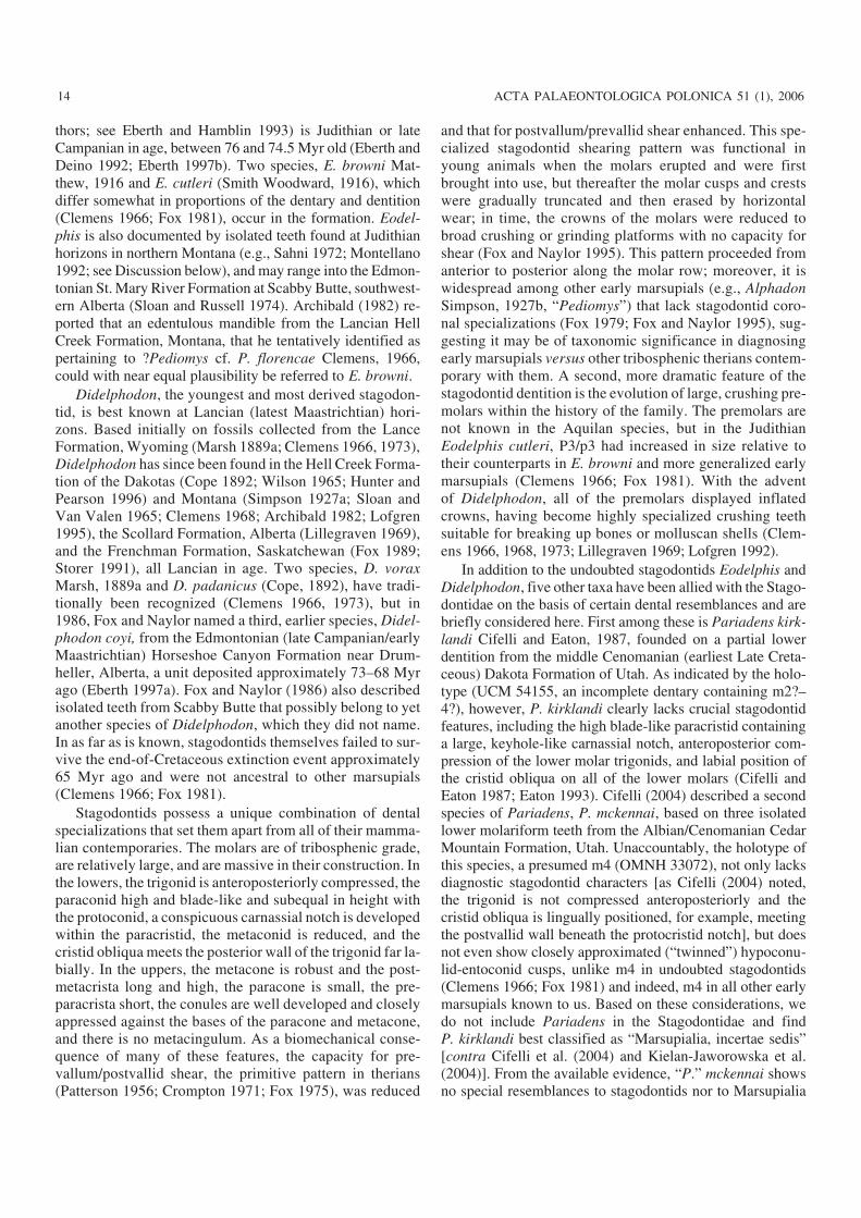
New material.—TMP 85.53.3, an incomplete left maxilla,containing P1–2 and M1, and alveoli for C (incomplete), P3and M2; from Dinosaur Park Formation, Dinosaur ProvincialPark, Alberta.
DescriptionMaxilla and upper dentition.—In TMP 85.53.3 (Fig. 1A),the preserved part of the maxilla extends from the caninealveolus to the posterolabial alveolus of M2. P1–2 and M1are in place, whereas the canine, P3, and M2 are representedonly by their alveoli. A rounded notch in the broken dorsalborder of the facial process of the maxilla is the remnant ofthe infraorbital foramen above P3; the foramen opens aboveM1 in Didelphodon (see below) and above P2 or between P2and P3 in uncatalogued specimens of the Virginia opossum,Didelphis virginiana Kerr, 1792, at hand and available for
comparison. A finished edge on the palatal process of themaxilla opposite M1 indicates the presence of a palatal vacu−ity in this specimen; the anterior extremity of the palatal va−cuity is commonly opposite M1 in D. virginiana.
The canine alveolus is incomplete but obviously waslarge originally, and it is substantially larger than the alveolimore posteriorly in the specimen, as is the case in D. virgi−niana. In TMP 85.53.3, the maxilla probably furnishedmuch of the walls of the canine alveolus. As preserved, thisbone extends further anteriorly on the medial side of thealveolus than laterally, but this may be only an artifact. Bycomparison, in the Virginia opossum the maxilla furnishesthe entire lateral wall of the canine alveolus but the bonethere is very thin; had the maxilla a similar configuration inTMP 85.53.3, the lateral wall of the canine alveolus doubt−less would not have been preserved. The canine alveolusdisplays no evidence of subdivision, leading to the conclu−sion that the upper canine of Eodelphis browni, like that ofDidelphodon vorax (Lillegraven 1969; Lofgren 1992), wassingle−rooted.
P1 [L = (1.8); W = (1.5)] is a small, two−rooted tooth inTMP 85.53.3, and is located directly behind the canine, with−out a diastema intervening. Indeed, the anterior root of P1 ispartly exposed in the posterolabial wall of the canine alveo−lus. The same tight spacing between the upper canine and P1,including the exposure of the anterior root in the postero−labial wall of the canine alveolus, is seen in Didelphis virgi−niana. Most of the crown of P1 in TMP 85.53.3 has beeneroded away, leaving little useful information about its mor−phology. From the outline of its base, however, the crownwas stout, somewhat wider posteriorly than anteriorly, butlonger than wide overall. Its long axis is oblique, fromanterolabial to posterolingual, and the anteriormost extrem−ity of the crown and the anterior root are labial to the midlineof the canine alveolus, as in D. virginiana, although P1 in theVirginia opossum extends still further anterolabially relativeto the canine. In TMP 85.53.3, the posterior root of P1, whichis substantially larger in cross section than the anterior root,is immediately adjacent to the anterior root of P2, with spacefor only a thin wall of bone between their respective alveoli;hence, it is clear that there was no diastema between P1 andP2 in this specimen, with the short gap between the crowns ofthe two teeth as preserved being due to breakage and erosionof P1 posteriorly and erosion of P2 anteriorly. The anteriorroot of P1 is vertical, not angled obliquely posteriorly, nordoes the remnant of the crown lean anteriorly. The posteriorroot slants posterolingually and probably passes lingual tothe anterior root of P2 within the maxilla, but in lateral viewthe posterior root is nearly vertical, not angled strongly pos−teriorly as in D. virginiana; its proportions do not suggestthat it supported an expanded lingual lobe of the crown. InDidelphodon vorax, P1 is single rooted and there are nodiastemata between the upper canine, P1, and P2 (Lofgren1992: fig. 1). A procumbent P1 and a diastema between P1and P2 have been claimed to be diagnostic for “metatheri−ans” (Rougier et al. 1998: fig. 5; see below).
16 ACTA PALAEONTOLOGICA POLONICA 51 (1), 2006
http://app.pan.pl/acta51/app51−013.pdf
FOX AND NAYLOR—STAGODONTIDS FROM CANADA 17
Fig. 1. A. Eodelphis browni Matthew, 1916, incomplete left maxilla, TMP 85.53.3, from the Dinosaur Park Formation, Judithian Land Mammal Age (lateCampanian), Dinosaur Provincial Park, Alberta, containing P1–2, M1 in occlusal (A1) and labial (A2) views. B. Eodelphis cutleri (Smith Woodward, 1916),incomplete right maxilla, UALVP 7031, from the Dinosaur Park Formation, Judithian Land Mammal Age (late Campanian), mouth of Sand Creek, Dino−saur Provincial Park, Alberta, containing M1 (broken), M2–3 in occlusal view; arrow shows posterior alveolus of P3. C. Didelphodon coyi Fox and Naylor,1986, incomplete right dentary, TMP 91.161.1, from the Horseshoe Canyon Formation, Edmontonian Land Mammal Age (early Maastrichtian), PaintearthCreek, Alberta, containing i2–3 (broken), c (broken), p1–3, m1–2, m3–4 (broken) in labial (C1), lingual (C2), and lingually oblique (C3) views. A1 and Bstereophoto pairs. Scale bar 5 mm.
In TMP 85.53.3, P2 (L = 3.3; AW = 1.4; PW = 1.8) is sub−stantially larger than P1. P2 is two−rooted, premolariform, bi−laterally compressed, and has a single main cusp, the para−cone. The outermost surface of enamel has been eroded fromover most of the crown, but little loss of morphological detailhas resulted. In lateral profile, the crown nearly forms anisosceles triangle but the posterior side is slightly longer andslightly less steep than the anterior side. A faint anterior ridgeextends from the apex of the paracone to the base of thecrown; this ridge curves gently labially along its length. Astronger ridge is present posteriorly and curves linguallyfrom the apex of the paracone to the base of the crown. Abasal cingulum is developed anteriorly; a second, widercingulum is developed posteriorly and terminates posteriorlyin a small cusp; the two cingula fail to meet either labially orlingually, but curve apically as they approach one another.The sides of the crown above the cingula are slightly hol−lowed out, more so above the posterior cingulum than anteri−orly, and the crown is moderately expanded posteriorly, es−pecially on its lingual side. In all of these features, P2 of TMP85.53.3 closely resembles P2 in Didelphis virginiana, exceptthat the anterior cingulum is better developed in the fossilspecimen. The tooth displays no trace of a swollen linguallobe as is present on P2 of Didelphodon vorax (Clemens1966: fig. 49; Lofgren 1992: fig. 1) and probably P2 ofDidelphodon coyi, as discussed below.
P3 in TMP 85.53.3 is represented by its two alveoli; theanterior alveolus contains a broken root. The alveoli arelarger than those for P2 and more widely spaced from one an−other anteroposteriorly, implying that P3 was larger than P2,undoubtedly the primitive proportions of these teeth in opos−sum−like marsupials generally (contra Luo et al. 2003). TheP3 alveoli are subcircular; the posterior alveolus is slightlywider than the anterior alveolus (anterior alveolus: W = 2.1;posterior alveolus: W = 2.4), but it is narrower than the coro−nal width of M1, in contrast to E. cutleri, in which the poste−rior alveolus alone of P3 is wider than the crown of M1 (seeUALVP 7031: Fig. 1B, arrow).
In TMP 85.53.3, the surface features of M1 (L = 3.4;AW = 3.4; PW = 3.9) are somewhat eroded diagenetically,but nonetheless the tooth displays the characteristic special−izations of M1 in stagodontids: the stylar shelf is wide, es−pecially posterior to the ectoflexus, the stylocone andprotocone are robust, the paracone is reduced and smallerthan the metacone, the conules are closely appressed to thelingual bases of the paracone and metacone, and there is nometacingulum.
Discussion
TMP 85.53.3 is the first known specimen of Eodelphis inwhich the maxilla extends anteriorly as far as the caninealveolus, thereby preserving evidence of the configuration ofthe upper premolars and of the size, at least, of the upper ca−nine. This specimen is relevant to several related issues asfollows:
Spacing of the upper premolars.—Luo et al. (2003: 1934–1935) claimed that in Late Cretaceous “metatherians” andCenozoic “didelphid−like” marsupials, P1 “is procumbentand close to the upper canine, followed by a large diastemabehind”, derived features that purportedly unite these groupswith the then−new middle Early Cretaceous “basal metathe−rian” Sinodelphys Luo et al. 2003 from China [as notedabove, Rougier et al. (1998: 462) had earlier cited these samecharacters as diagnostic of “Metatheria”, including “marsupi−als”]. In fact, these features are hardly known at all in NorthAmerican Late Cretaceous marsupials, the richest source ofinformation about early marsupial diversity and dental evolu−tion. Nonetheless, the evidence that the available specimensprovide plainly conflicts with the aspects of the anterior upperpremolars that Rougier et al. (1998) and Luo et al. (2003)have cited as diagnostic for metatherians. For example, Clem−ens (1966: fig. 27) illustrated an incomplete maxilla (UCMP52094) of Pediomys hatcheri (Osborn, 1898) in which P1 isindeed separated from P2 by a “large” diastema, but P1 is alsoseparated from the upper canine by a diastema that from spac−ing of the alveoli, appears nearly as long as that between P1and P2 (because the crown of P1 is missing from this speci−men whether or not it was procumbent cannot be determined).In no other specimens of the Lance marsupials that Clemens(1966, 1973) described are these parts preserved. Lillegraven(1969: fig. 14.3b) illustrated UALVP 2389, an incompletemaxilla of Alphadon marshi Simpson, 1927b (or A. jasoniStorer, 1991; see Johanson 1996a) preserving P2–3, M1 fromthe Lancian Scollard Formation, Alberta, but in this specimenthe posterior root of P1 is present as well and shows that thistooth was not separated from P2 by a diastema. None of theother marsupial specimens in Lillegraven’s (1969) descrip−tion of the Trochu local fauna are preserved this far anteriorly.Lofgren (1992) demonstrated that in Didelphodon vorax thealveolus for P1 is close behind the canine, but from the spac−ing of their alveoli, P1 and P2 were not separated by adiastema. As described above in the earlier and more primi−tive stagodontid Eodelphis browni, P1 is close behind the ca−nine, probably was not procumbent, and there is no diastemabetween P1 and P2. Given this pattern in Eodelphis, the lackof diastemata in the anterior upper postcanine dentition inDidelphodon cannot be explained away as a peculiarity re−stricted to that genus, i.e., a feature that is a correlate to theshortening of the jaws and specialized crowding of the ante−rior postcanine dentition that Didelphodon exhibits. Another,previously unpublished, example preserving the anterior up−per dentition in early marsupials agrees with those citedabove: reconstruction of the maxillary dentition of a Late Cre−taceous Alphadon−like didelphoid, TMP 95.178.26 (Fig. 3D)from the Judithian Devil’s Coulee locality, Alberta, to be de−scribed elsewhere, indicates that in this species P1 was erect(not procumbent) and there was no diastema between P1 andP2. Additional direct evidence as to the spacing of the upperpremolars in early marsupials is provided by a well−preservedmaxilla (UALVP 43007) of a new Paleocene species ofPeradectes Matthew and Granger, 1921 from locality DW−2
18 ACTA PALAEONTOLOGICA POLONICA 51 (1), 2006
(Fox 1990) also to be described elsewhere: this specimen con−tains the alveolus for the canine and P1, and P2–3, M1–4 arein place: P1 was single−rooted and (from the slope of itsalveolus), not procumbent and there are no diastemata any−where along the tooth row.
In sum, the spacing between the upper premolars consid−ered diagnostic by Rougier et al. (1998) and Luo et al. (2003:1934–1935) in “Late Cretaceous metatherians” (= Late Cre−taceous marsupials of this paper) is known to occur in only asingle specimen of P. hatcheri, but because of the diastemabetween the upper canine and P1, this resemblance is incom−plete. More importantly, the lack of a diastema between P1and P2 in Didelphodon is not a peculiar specialization lim−ited to this genus but has a wider distribution, including oc−currences in at least four genera of dentally less specializedearly marsupials. From these facts, we suggest that contraryto Rougier at al. (1998) and Luo et al. (2003), the primitivecondition in marsupials is one in which P1 is vertical (notprocumbent) and there are no diastemata along the tooth rowbetween the upper canine and the molars. If so, the resem−blances between Sinodelphys and Cenozoic “didelphid−like”marsupials involving a procumbent P1 and a diastema be−tween P1 and P2 are convergent, in keeping with the virtualcertainty that living didelphids share a more recent commonancestry with known North American Late Cretaceous opos−sum−like marsupials than with the Asian middle Early Creta−ceous Sinodelphys (see, e.g., Case et al. 2004 for discussionof paleobiogeography of early marsupials).
Palatal vacuities.—In 1995, Fox and Naylor described thefirst evidence of vacuities (fenestrae) in the secondary palateof North American Late Cretaceous marsupials, includingthe stagodontids Eodelphis cutleri and Didelphodon vorax.As noted above, TMP 85.53.3 displays a rounded, finishededge along the medial side of the maxilla opposite M1, whichextends the evidence of palatal vacuities in Late Cretaceousmarsupials to E. browni. Palatal vacuities were cited as asynapomorphy of “crown group Marsupialia” by Horovitzand Sánchez−Villagra (2003: fig. 1, appendix B), who evi−dently were unaware of the by−then well−documented occur−rence of these structures in diverse Late Cretaceous marsupi−als—perhaps reflecting a widespread view among neontolo−gists that incomplete fossils and the literature describingthem contain no useful anatomic and, hence, phylogeneticinformation. Palatal vacuities in generalized early marsupialsalso occur in Andinodelphys cochabambensis Marshall andMuizon, 1988 from the early Paleocene Tiupampa depositsof Bolivia (Muizon et al. 1997); this species was based on awell−preserved skull but was also omitted from Horovitz andSánchez−Villagra’s (2003) analysis. Evidence of palatal va−cuities occurs additionally in the new species of Peradectesfrom the Paleocene Paskapoo Formation of Alberta.
Marshall et al. (1995) and Muizon (1998) contended thatbecause palatal openings occur sporadically throughoutMammalia (e.g., in some multituberculates, in carpolestidplesia Erinaceus Linnaeus, 1758, etc.), they contain littlephylogenetic information in regards marsupials. We dis−
agree: we do not accept that the independent acquisition of acharacter among unrelated clades furnishes valid informa−tion as to the homology (or not) of that character as it occurswithin a clade (see Vermeij 1999, 2001; Van Valen 2004).Marshall et al. (1995) and Muizon (1998) further argued thatbecause palatal openings develop by the resorption of bonein marsupial pouch−young already having a complete sec−ondary palate (i.e., one lacking these openings), these open−ings cannot constitute a marsupial synapomorphy, a derivedcharacter arising at the origin of marsupials. We acknowl−edge that the late appearance of palatal openings in marsupialontogeny is at least consistent with a complete palate beingprimitive for marsupials (as for mammals generally), but wenote that the early establishment of a complete palate onto−genetically may instead be related to precocious suckling bythe developing pouch young (de Beer 1937) and hence con−vey no phylogenetic signal. In any case, the timing of devel−opment of palatal openings in extant marsupials can tell usnothing about when during marsupial geological historythese openings originated or the pattern of their distributionin extinct species thereafter. That information can be sup−plied only by the fossil record.
The fossil record reveals that palatal vacuities occur in allNorth American Late Cretaceous marsupials for which theappropriate parts have been discovered, from Judithian toLancian horizons, in both dentally generalized (e.g., Alpha−don) and specialized (e.g., Stagodontidae) species, includingbasal marsupials (Stagodontidae, “Pediomyidae”) (Fox andNaylor 1995). The taxonomic and temporal distributions ofthese specimens agrees with the long−standing hypothesisthat palatal vacuities are a marsupial synapomorphy (Simp−son 1947; Tyndale−Biscoe 1973; Reig et al. 1987; Fox andNaylor 1995), with absence of vacuities, as in Paleogene andNeogene South American borhyaenids, being a derived con−dition, reflecting truncation of palatal development in geo−logically younger species (Reig et al. 1987: 30). While theprecise source of Cenozoic marsupials among their Creta−ceous predecessors is not known, it probably is still mostclosely approximated by taxa having an Alphadon−like den−tition (Clemens 1966, 1979; Case et al. 2004) and hencelikely having palatal vacuities, as documented by specimensalready collected from the North American Western Interior.
Sexual dimorphism in Eodelphis.—Montellano (1992: 84)suggested that the two nominal Judithian species of Eodel−phis could well be sexual dimorphs of a single species. Wereject this interpretation: the qualitative differences in thedentition between E. browni and E. cutleri (see Fox 1981),with the enlarged posterior premolars in the latter antecedentat least phenetically to those of Didelphodon, imply very dif−ferent food niches, to a degree that would be unexpected be−tween the sexes of a single mammalian species. Moreover,dental differences of this magnitude are without parallelknown to us within single species of extant opossum−likemarsupials, the nearest living analogue to Late Cretaceousstagodontids.
http://app.pan.pl/acta51/app51−013.pdf
FOX AND NAYLOR—STAGODONTIDS FROM CANADA 19
Genus Didelphodon Marsh, 1889aDidelphodon coyi Fox and Naylor, 1986Figs. 1C, 2, 3A, B.
Holotype: TMP 84.64.1, incomplete right dentary containing p3, m3–4(Fox and Naylor 1986: figs. 3, 4). Edmontonian Land Mammal Age(early Maastrichtian), Michichi Creek, Alberta.
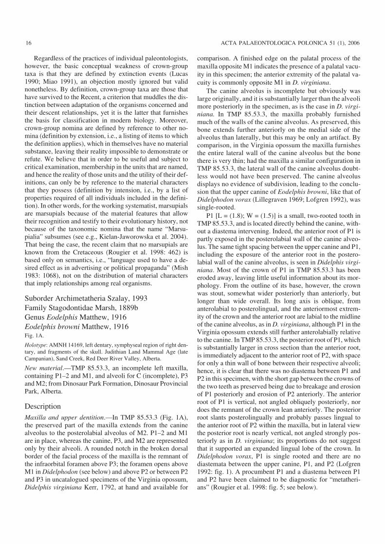
New material.—TMP 91.166.1, an incomplete right dentarycontaining p1–3, m1–2, m4 (broken), and roots of the canine,two incisors, and m3 contained in their alveoli, from Painte−arth Creek, Alberta; TMP 90.12.29, an incomplete left denta−ry with heavily worn and broken p3, alveoli for m1–4, the ca−nine, p1? and p2, from the type locality, Michichi Creek, Al−berta (Fox and Naylor 1986) (both of these localities are inthe Horseshoe Canyon Formation, with exact coordinates onfile at the Royal Tyrrell Museum of Palaeontology); TMP94.125.125, incomplete right maxilla with P3, M1 and dam−aged M2, from the latest Cretaceous (latest Maastrichtian;Lancian) Scollard Formation at KUA−1 [see Lillegraven(1969) for description of the locality and Archibald (1982)for account of the Trochu local fauna; see Fox (1974) andFox and Naylor (2003) for important additions to this localfauna].
Although the dentary TMP 90.12.29 was found at thetype locality and is from the opposite side of the jaw than theholotype, the two specimens are clearly from different indi−viduals as evidenced, for example, by the deep wear exhib−ited on p3 of TMP 90.12.29 and the virtually unworn p3 ofthe holotype.
Description
Dentary.—Of the two new dentaries of Didelphodon coyi,TMP 91.166.1 (Figs. 1C, 2A) is the better preserved, but it isbroken and missing from the base of the canine anteriorly,and posteriorly from just beyond the level of the mandibularforamen; nonetheless, it displays several features more ex−tensively than does the holotype or other available specimensof this species.
The dentary in TMP 91.166.1 is robustly constructed but isslightly smaller than the holotype dentary, a difference of nosignificance taxonomically. The alveolar and ventral marginsof the horizontal ramus are nearly parallel with each other, ex−cept anteriorly from beneath p3, where the ventral marginrises steeply at the level of the symphysis. In TMP 91.166.1,two mental foramina penetrate the outer side of the ramus, thelarger and more ventral one opening beneath p3, the other be−neath m1, matching their relative size and position in theholotype; a faint sulcus extends anteriorly from the anterior fo−ramen. However, as in the holotype, there is no evidencewithin the masseteric fossa of the “labial mandibular foramen”that has been reported in the early marsupials Kokopellia juddiCifelli, 1993a and Alphadon eatoni Cifelli and Muizon, 1998a(see also Cifelli and Muizon 1997, 1998b).
On the medial side of TMP 91.166.1, the symphysealboss is the most prominent feature of the horizontal ramus; itis better preserved and dorsoventrally shallower than in the
holotype. It forms a raised, elongate oval of bone that slopesposteroventrally to beneath p3, which in TMP 91.166.1 is atthe deepest part of the ramus (the holotype is deepest beneathm4). The articulating surface of the boss is covered by broad,shallow pits that give it an irregularly pock−marked texture.If this surface is held in the vertical plane, its presumed orien−tation in life when in full articulation with its counterpart onthe left side, the horizontal ramus at the level of the sym−physis leans laterally, causing the premolars to lean laterallyas well (Fig. 2A2); more posteriorly, the dentary graduallybecomes more vertical and the molars are vertically em−placed. This peculiar orientation of the lower premolars hasunusual functional implications that have not been recog−nized before and these are discussed below.
In TMP 91.166.1, a medial shelf on the horizontal ramusextends posteriorly from the upper margin of the symphysealboss, beginning below p1. This shelf slopes ventromedially,becoming steeper posteriorly; it is more prominent in thisspecimen than in the holotype. The ventral limits of the shelfare provided by a faint, narrow ridge that may be the mylo−hyoid line, which marks the origin of the mylohyoid muscles,although in the Virginia opossum, Didelphis virginiana, thesemuscles are reported to originate much more ventrally on themedial face of the dentary (Hiiemae and Jenkins 1969); be−neath m4 in TMP 91.166.1, this ridge curves dorsally towardsthe raised anterior margin of the pterygoid fossa. Although lit−tle of the pterygoid fossa is preserved in TMP 91.166.1, themandibular foramen has not been damaged and opens into thefossa just anterior to the broken posterior edge of the speci−men, well anterior to its position in D. virginiana and directlyabove the anteriormost extremity of the inflected angular pro−cess; in living didelphids, the foramen carries the inferior alve−olar branch of the mandibular nerve and blood vessels that ac−company it (Wible 2003: 177). Anterior to the foramen inTMP 91.166.1 (as in the holotype: Fox and Naylor 1986: fig.1C), there is no evidence of an internal mandibular groove(holding postdentary elements; e.g., Meng et al. 2003) or evena mylohyoid groove as seen in, e.g., D. virginiana [this groovemarks the passage of the mylohyoid vessels and nerve (Ben−sley 1902)].
The second new specimen, TMP 90.12.29 (Figs. 2B, 3A),is in poorer condition overall than TMP 91.166.1, but it in−cludes a more extensively preserved coronoid process, acomplete dentary peduncle or condylar process, the condyle,and part of the angular process. In TMP 90.12.29, the ante−rior margin of the coronoid process slopes at about 104 de−grees relative to the alveolar border, approximately at thesame angle as in the holotype (Fox and Naylor 1986: fig. 3).What remains of the angular process in TMP 90.12.29 is in−flected and forms a broad, medially directed shelf that is flaton its ventral side; the shelf is broken both medially and ante−riorly, so its full extent cannot be determined. The postero−medial margin of the shelf, however, is complete and curvessmoothly anteriorly; at least in the parts that remain, it doesnot form a posteriorly directed notch and acute posterior pro−cess, as in D. virginiana and the short−tailed opossum, Mono−
20 ACTA PALAEONTOLOGICA POLONICA 51 (1), 2006
delphis brevicaudata (Erxleben, 1777) (Wible 2003). At theanteriormost extent of this margin, the shelf turns abruptlymedially a short distance before reaching a now−broken
edge: perhaps a shallow notch and posterior process were de−veloped more medially here, although if so, they would havebeen well medial to their position in D. virginiana. In the
http://app.pan.pl/acta51/app51−013.pdf
FOX AND NAYLOR—STAGODONTIDS FROM CANADA 21
Fig. 2. Didelphodon coyi Fox and Naylor, 1986 from the Horseshoe Canyon Formation, Edmontonian Land Mammal Age (early Maastrichtian). A. TMP91.161.1, Paintearth Creek, Alberta, incomplete right dentary, containing 12−3 (broken), c (broken), p1–3, m1–2, m3–4 (broken) in occlusal (A1) and an−terior (A2) views. B. TMP 90.12.29, Michichi Creek (type locality), Alberta, incomplete left dentary, containing p3 (broken) in occlusal (B1) and labial(B2) views. A1 and B1 stereophoto pairs. Scale bars 5 mm.
holotype and TMP 91.166.1, the shelf reaches slightly ante−rior to the level of the mandibular foramen, almost as far an−teriorly as the base of the coronoid process above and wellanterior to the anteriormost extent of the shelf in the Virginiaopossum. Crompton and Lieberman (2004) associated the in−flected angle in living marsupials with insertion of a neo−morphic superficial division of the medial pterygoid muscu−lature not seen in extant eutherians; we conclude that thismuscle was present in Didelphodon, as well.
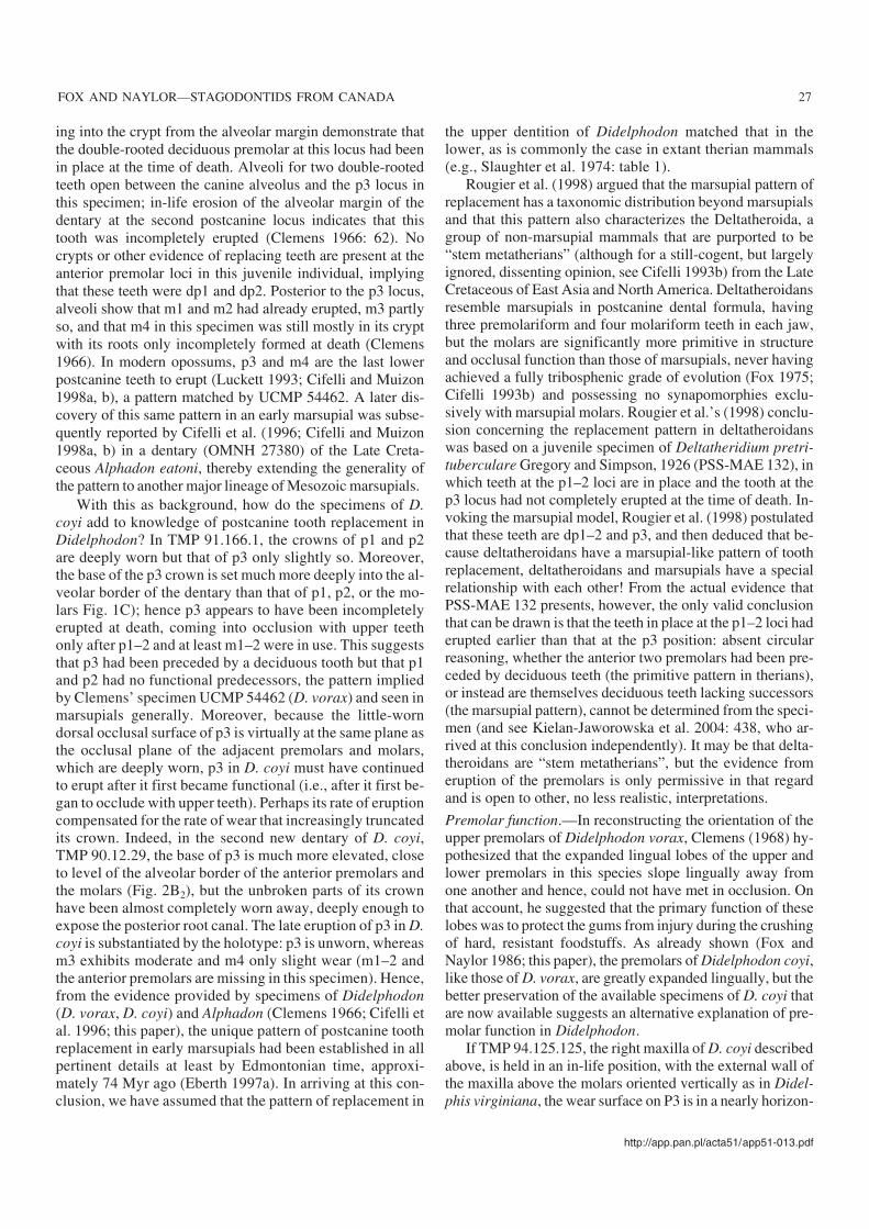
In TMP 90.12.29, a low ridge extends from the base ofthe angular process to the ventromedial margin of the con−dyle. In the holotype, the condyle is damaged, with its me−dial end missing (Fox and Naylor 1986: fig. 3B). In TMP90.12.29, the complete condyle is a transversely expandedsubcylindrical body 11.6 mm long that in posterior view isslightly deeper at its medial end than laterally (Fig. 3A). Re−
construction of the posterior part of the dentary based onTMP 90.12.29 and the holotype together demonstrates thatmost of the condyle in D. coyi is lateral to the vertical planeof the coronoid process. By contrast, in Didelphis virgi−niana, the condyle projects further laterally than mediallyrelative to this same plane but projects further medially thanin D. coyi.
On the lateral side of TMP 90.12.29, the posterior part ofthe masseteric shelf that ventrally borders the massetericfossa is still in place. As in the holotype (Fox and Naylor1986: fig. 3), the shelf narrows posteriorly to a low crest thatextends along the ventrolateral edge of the dentary peduncleto the lateral extremity of the condyle; in Didelphis virgi−niana, this shelf remains wide posteriorly to its junction withthe condyle. The articulating surface of the condyle is welldefined, especially dorsally, and presumably was covered by
22 ACTA PALAEONTOLOGICA POLONICA 51 (1), 2006
Fig. 3. A. Didelphodon coyi Fox and Naylor, 1986, condyle and inflected angle of TMP 90.12.29, from the Horseshoe Canyon Formation, EdmontonianLand Mammal Age (early Maastrichtian), Michichi Creek (type locality), Alberta, in posterior view with ventral surface of inflected angle horizontal (A1)and dorsal surface of condyle horizontal (A2). B. Didelphodon coyi Fox and Naylor, 1986, incomplete right maxilla, TMP 94.125.125, from the ScollardFormation, Lancian Land Mammal Age (late Maastrichtian), KUA−1 locality, Red Deer River Valley, Alberta, containing P3, M1, M2 (broken) in occlusalview. C. Eodelphis cutleri (Smith Woodward, 1916), incomplete right maxilla, UALVP 43005, from the Dinosaur Park Formation, Judithian Land Mam−mal Age (late Campanian), Onetree Creek, Dinosaur Provincial Park, Alberta, containing M2–3 in occlusal view. D. Undescribed Alphadon−like marsupial,incomplete left maxilla, TMP 95.178.26, from the Oldman Formation, Judithian Land Mammal Age (late Campanian), Devil’s Coulee, Alberta, containingP1–3, M1–4 in occlusal (D1) and labial (D2) views. B, C, and D1 stereophoto pairs. Scale bars 5 mm.
a thin layer of cartilage in life: in dorsal aspect, the articulat−ing surface extends slightly more anteriorly on the lateralside of the condyle than medially; in its curvature around theposteroventral aspect of the condyle it narrows from eitherside as in D. virginiana. If the dentary is held so that the ven−tral surface of the inflected angular process is horizontal, thecondyle slants obliquely (Fig. 3A1), from dorsomedial toventrolateral; alternatively, if the condyle is held horizon−tally (Fig. 3A2), the angular process slants ventromedially.These same relative orientations between condyle and angu−lar process are seen in some adults of D. virginiana. Untilbetter preserved specimens of D. coyi are collected, however,the actual orientation of these structures in life cannot be de−termined in this species.
Lower dentition.—TMP 91.166.1 is incomplete anteriorlybut its cleanly broken anterior surface reveals the roots of thecanine and incisors still within their alveoli (Fig. 2A2). Thelower canine, not known previously in Didelphodon coyi,was well developed as indicated by the dimensions of its bro−ken cross section (depth = 4.6; W = 3.8), which show thetooth to have been slightly compressed bilaterally. In TMP90.12.29, the canine is missing but its alveolus extends pos−teriorly to beneath p3. In addition to these aspects of the ca−nine, something of its orientation can be determined as well:if TMP 91.166.1 is held so that its symphyseal surface is ver−tical, a line through the greatest depth of the canine leans lat−erally at approximately 30 degrees from the vertical. InDidelphis virginiana, the snout at the level of the canines iswider than the mandible and the lower canines are splayed,allowing their tips to slide dorsally past the ventral margin ofthe maxillae into the maxillary fossae when the mandible iselevated and the postcanine teeth brought into occlusion.Perhaps the orientation of the lower canines in D. coyi is re−lated to similar differences in the relative widths of the upperand lower jaws.
In TMP 91.166.1, two small incisor alveoli are exposed incross section near the canine (Fig. 2A2). The larger of the twois ventromedial in position, between the canine and the sym−physeal surface; the root that it contains is ovate in cross sec−tion, being somewhat compressed bilaterally (depth = 1.7;W = 1.2), and is substantially less than the diameter of the ca−nine root. This alveolus is clearly the homologue of the ventro−medial incisor alveolus preserved in the holotype (Fox andNaylor 1986: fig. 4). The second incisor alveolus, not evidentin the holotype, is ventral to the canine and ventrolateral to themedial incisor; it is nearly circular in cross section and the rootis less than half the diameter of that of the medial incisor, ap−pearing to have been broken very near its end (depth = 0.5;W = 0.5). There is no indication of a third or fourth incisoralveolus in TMP 91.166.1 (for our assessment of the homo−logies of the lower incisors in D. coyi, see Discussion below).
In TMP 91.166.1, p1 [L = (2.4); W = 2.8] is located di−rectly behind the canine and is closely appressed to it. Clem−ens (1966) identified isolated p1s of Didelphodon vorax, andtwo alveoli for this tooth are present in dentaries of this spe−cies, UCMP 54462 and LACM 15433 (Clemens 1966, 1968),
but p1 of Didelphodon has not previously been known from atooth articulated in the lower jaw. The crown of p1 in TMP91.166.1 is anteroposteriorly compressed and orientednearly transversely across the summit of the dentary, from alabial and slightly anterior position to a lingual and slightlyposterior position. Accordingly, the crown is wider thananteroposteriorly long; in D. vorax, the position of its alveoliindicates that p1 was probably oriented more anteropos−teriorly (Clemens 1966: fig. 37, 1968: fig. 1), although inUSNM 2136, a dentary that Clemens (1966: fig. 36) referredto D. vorax but that may pertain to D. padanicus (Archibald1982: 158), the alveoli of p1 are nearly opposite one anotheracross the jaw. In occlusal view of p1 in TMP 91.166.1, themiddle parts of the crown are weakly constricted, with theanterior surface of the resulting “waist” fitting tightly overthe convex posterior side of the canine and the posterior sur−face of the “waist” receiving a swollen anterobasal cusp onp2. The labial part of the p1 crown has been lost but the lin−gual side is unbroken: it is expanded into a small lobe andsupports a steeply sloping wear facet in which the enamel hasbeen abraded away and the dentine exposed; deep irregularscratches are incised into the dentine. Labial to the wearfacet, a few irregular enamel ridges border the broken sur−face. A short cingulid is posteriorly adjacent to the wearfacet; this cingulid may have extended further lingually but ifso, it was worn away before death.
In TMP 91.166.1, p1 is two−rooted; both roots are stoutand descend on either side of the root of the canine; they arenearly opposite to one another across the jaw, with the labialroot only slightly more anterior in position. The lingual rootis angled steeply posteriorly, parallel to the side of the canineroot; the labial root is more nearly vertical and passes labialto the canine root; no accessory roots of p1 are visible in thespecimen. In the holotype of Didelphodon coyi, there ap−pears to be a single alveolus for p1, implying that the toothwas single−rooted in that specimen (Fox and Naylor 1986); ifso, p1 in D. coyi is polymorphic for the number of roots. InTMP 90.12.29, there is a single small circular alveolus thatopens between the posterior root of p2 and the canine alve−olus, but whether this was for one of two roots or if p1 wassingle rooted cannot be determined owing to the poor condi−tion of the specimen.
In TMP 91.166.1, p2 is well preserved and crowdedagainst p1. In occlusal outline, its crown (L = 5.5; W = 5.0) isover four times as large in areal dimensions as that of p1. It isnarrow anteriorly, expands into a greatly widened postero−lingual lobe, and is oriented obliquely across the alveolarborder of the dentary, so that its anteriormost parts projectanterolabially beyond the posterior margin of p1; unlike p1,the crown of p2 is slightly longer than wide. In LACM15433, the dentary of D. vorax described by Clemens (1968),p2 is in place but this tooth has not previously been known inD. coyi, being represented only by alveoli in the holotype.Like p1, p2 in TMP 91.166.1 is deeply worn: all of its origi−nal cuspation has been worn away, exposing dentine acrossmost of the remaining surface. Like that on p1, the wear sur−
http://app.pan.pl/acta51/app51−013.pdf
FOX AND NAYLOR—STAGODONTIDS FROM CANADA 23
face slopes steeply lingually, faces dorsolingually, and isfaintly convex from its labial to lingual side. From compari−son to p2 on LACM 15433, this wear surface encompassesthe main cusp (protoconid) and the swollen posterolinguallobe. In TMP 91.166.1, the lobe is worn down to nearly itsbase, leaving little of the lingual wall of the crown remaining.On the anterior side of p2, two small basal cusps are devel−oped, the more lingual of which fits into the hollow on theposterior side of p1; the labial basal cusp is free, not contact−ing p1. The enamel on the labial side of p2 forms exodaeno−dont lobes above the anterior and posterior roots, respec−tively. A short, deep, nearly vertical furrow is formed by theenamel on the side of the posterior lobe; by comparison withp3, in which a longer furrow is developed in this position,this furrow marks the junction between the talonid or “heel”and the main body of the crown (protoconid) and corre−sponds to the molar hypoflexid. In its turn, the talonid ispartly divided into two lobes on its posterior side. The morelingual of these projects posteriorly from the worn occlusalsurface and probably represents the hypoconulid. Two rootsare visible on p2; because the crown fits closely to the denta−ry, the complete dimensions of the roots cannot be deter−mined, but alveoli on the holotype indicate that the anteriorroot of p2 is small, circular in cross section, and in a labial po−sition; the posterior root is greatly widened transversely, ex−tending across much of the alveolar border of the dentary asin the holotype (Fox and Naylor 1986) and confirmed by thebroken roots of p2 in TMP 90.12.29. There is no evidencefrom these specimens that p2 possessed an accessory root asClemens (1966) reported on a p2 of D. vorax.
The p3 in TMP 91.166.1 is greatly enlarged (L = 7.3; W =4.7), with swollen coronal walls as is characteristic of p3 ofDidelphodon (Clemens 1966, 1968; Fox and Naylor 1986); itclosely resembles p3 on the holotype of D. coyi but is slightlysmaller. The crown is divided into a tall, massive protoconidand a lower, bulbous, unicuspid, unbasined talonid; there isno evidence of anterobasal cusps. A strong ridge is devel−oped posteriorly on the protoconid and terminates ventrallyat the heel, separated from it by a shallow notch; the heel isalso demarcated from the protoconid by deep furrows, oneeach on the labial and lingual sides of the crown. A very shortbasal cingulid is present anterolingually, and immediately la−bial to this, the anterior face of the protoconid is shallowlyconcave, fitting against the posterior side of p2. Towards itsbase, the crown is subdivided labially and lingually into twolobes at the anterior and posterior roots; the lingual lobes ex−tend further ventrally and are more swollen than those on thelabial side. On the labial side of the talonid a vertical ridge isdeveloped that approximates the position of the posteriorcingulid on the molars.
In TMP 91.166.1, the apex of the protoconid of p3 hasbeen truncated by wear, although much less deeply than onp1 or p2. The protoconid wear facet, in which dentine is al−ready exposed, slopes lingually but less steeply than the wearfacets on the more anterior premolars. The apex of the talo−nid displays the first stages of wear: two subcircular facets
are developed there, one anterior, which is larger, and a sub−stantially smaller facet posterior to it, immediately above thedorsal end of the posterior cingulid; this facet may mark theposition of the hypoconulid. Much of the enamel coveringthe unworn parts of the crown is weakly wrinkled, especiallyon the lower parts of the labial wall. The entire crown is setmore deeply into the dentary than are the crowns of the adja−cent teeth, as if its eruption had not been fully completed atthe time of death (see Discussion below).
In TMP 91.166.1, m1 and m2 are in place (they are miss−ing from the holotype); the crown of m3 has been lost, al−though its roots are contained in their alveoli, whereas m4 isrepresented by only the base of the crown and the roots, itsocclusal surface having been broken off before the specimenwas collected. As in the holotype of D. coyi (Fox and Naylor1986) and in D. vorax (Clemens 1966, 1968), the lower mo−lar row in TMP 91.166.1 crosses the summit of the dentaryobliquely, from anterolabial to posterolingual; hence, thelong axis of m1 (L = 4.8; WTri = 3.2; WTal = 3.5) is obliquerelative to the long axis of the dentary. The crown of m1 isdeeply worn; what remains of the trigonid has been abradeddown to the same level as the talonid, which is also worn. To−gether, these two parts of the crown form a nearly flatocclusal surface; dentine is broadly exposed over all of thissurface, except at the center of the talonid basin, where asmall, circular patch of enamel remains. The trigonid of m1is narrower but longer than the talonid. A strongly curvedcingulid is present on the anterolabial wall of the trigonid andmay have terminated anterodorsally in a distinct cuspule, al−though the uppermost parts of the cingulid have been wornaway. The anterolingual side of what remains of the para−conid projects anterolabially beyond the swollen postero−lingualmost parts of p3 to meet the talonid of p3 along a veryfaint vertical groove in its posterior wall beneath the hypo−conulid. This overlap was already suggested by the positionof the empty alveoli of m1 relative to p3 in the holotype andin TMP 83.33.7 (Fox and Naylor 1986: figs. 3B, 6A). Poste−riorly on m1, the tip of the hypoconulid has been truncated bywear but its base juts posteriorly, inserted into a notch on m2formed by the base of the paraconid and the anterior end ofthe basal cingulid in a tight interlock between the two teeth.A short posterior cingulid descends labially from the hypo−conulid and blends into a series of rounded, irregular verticalridges on the labial face of the talonid. By contrast, theenamel covering the lingual side of the crown is smooth. Theposterior labial cingulid and hypoconulid of m1 form a shal−low notch that receives the anterior cingulid of m2, enhanc−ing the interlock between the two molars.
In TMP 91.166.1, m2 (L = 5.1; WTri = 3.7; WTal = 3.8) isslightly longer and wider than m1 and is in line with it, so ittoo is oriented obliquely relative to the long axis of the denta−ry. The tooth is deeply worn, but less so than m1: the occlusalsurface of the trigonid has been worn flat but the trigonid re−mains slightly higher than the talonid. The talonid is slightlywider than the trigonid and the trigonid is more antero−posteriorly compressed than that of m1. The rim of the
24 ACTA PALAEONTOLOGICA POLONICA 51 (1), 2006
talonid is worn, but the cristid obliqua meets the posteriorwall of the trigonid in a labial position (a stagodontid fea−ture). The hypoconulid is still recognizable as a distinct cusp;a posterolingual furrow marks the division between thehypoconulid and entoconid, which were “twinned” postero−lingually on the rim of the talonid, with the hypoconulidposterolabial to the entoconid. On the lingual side of m2,swellings marking the base of the paraconid and metaconidindicate that the paraconid was substantially larger than themetaconid, as in stagodontids generally. The anterolabialcingulid is massively constructed, incompletely enclosing ashallow pocket on the anterolabial wall of the protoconid.The cingulid ends lingually at a cuspule that is labially adja−cent to the hypoconulid of m1; labially, the cingulid termi−nates in a cuspule on the side of the protoconid, near its base,and two smaller cuspules are developed along its length. Theposterior cingulid is short but prominent. The posterior wallof the talonid is marked by a broad interdental wear facet thatextends over much of its surface as evidence of the close in−terlock between m2 and m3. Nothing can be observed of thecrown morphology of m3 or m4, except that on m4, the ante−rior cingulid is strongly developed and subdivided into smallcuspules, not evident on m4 of the holotype owing to wear. Aline passing through the long axis of m3–4 would unambigu−ously extend medial to the coronoid process, as in the holo−type. The roots of the canine, premolars, and molars areheavily invested with cement. Further posteriorly, the man−dibular corpus twists increasingly into a vertical orientationcarrying the molar row with it. Interestingly, a similar trendin orientation of the lower tooth row is seen in Didelphisvirginiana, although it is not as strongly developed.
Maxilla and upper dentition.—TMP 94.125.125 (Fig. 3B) isan incomplete right maxilla containing P3, M1, and a brokenM2. The maxilla itself preserves little of anatomical interestexcept for a notch along the broken dorsal edge of the facialprocess. This notch marks the position of the infraorbital fo−ramen, which opened above M1, as in Didelphodon vorax(Clemens 1973).
Whereas TMP 94.125.125 clearly pertains to Didelpho−don, it is significantly smaller than specimens of D. vorax indental dimensions that can be compared (see Clemens 1966:table 13). Moreover, when held manually, M1–2 of TMP 94125.125 occlude readily with m1–2 on TMP 91.166.1, thespecimen of D. coyi that has the most extensively preservedlower dentition now known. Conversely, the teeth of TMP94.125.125 are too small to fit with the lower dentition of D.vorax as preserved in, e.g., a cast of LACM 15433, left denta−ry of D. vorax from the Hell Creek Formation cited above(Clemens 1968), or UALVP 1985, an incomplete right den−tary with c and m2 of D. vorax from the Scollard Formation(Lillegraven 1969). Hence, TMP 94.125.125 is referred to D.coyi and furnishes the first evidence of the upper dentition ofthis species.
In TMP 94.125.125, P3 is a large tooth (L = 5.0; W = 5.4)and the tallest on the specimen; it has an inflated crown con−sisting of a labial cusp (paracone) and a swollen lingual lobe.
The crown, which is bean−shaped in occlusal outline, iswider transversely than anteroposteriorly long. Its anteriorside is slightly concave, presumably meeting the convex pos−terior side of P2, an intuition corroborated by an interdentalwear facet incised into the enamel within the deep part of theconcavity. A narrow anterior cingulum extends along thebase of the main cusp and continues around the anterolabialcorner, where it breaks up into a labial patch of irregularcuspules. The anterolabial corner of P3, although rounded,projects somewhat anteriorly. A small cusp is present here, atthe junction of a crest from the paracone and the cingulum;by comparison with the molars, this cusp is the stylocone andthe crest, the preparacrista. The posterolabial corner of P3 issmoothly rounded and lacks a labial or posterior projection; apoorly defined vertical crest or postparacrista is developedon the posterior wall of the paracone. A short, rugose poste−rior cingulum meets the posterior crest at a raised cuspule,and lingually it divides into two ridges that curve towards theapex of the main cusp; the cingulum abuts the anterior side ofM1 at the level of the paracone. The lingual side of P3 is ex−panded into a low but inflated lobe. Wear has truncated theapex of the paracone, producing a flat, subovate facet inwhich dentine is widely exposed, but the facet had not ex−tended onto the lingual lobe by the time of death. Much of theenamel that remains is wrinkled, especially on the labial sideof the crown.
M1 is well preserved, although moderately deeply worn.It closely resembles M1 of Didelphodon vorax, but on asmaller scale (L = 4.0; AW = 4.6; PW = 5.5). The crown iswider transversely than long anteroposteriorly and has prom−inent, rounded parastylar and metastylar lobes separated by anarrow but deep ectoflexus; the metastylar lobe projects farlabially, as in M1 of D. vorax (Clemens 1966: fig. 52). Wearhas truncated stylar cusps A and B (stylocone), preparacrista,and paracone, but from the proportions that remain, stylarcusp B was robust, as in D. vorax; whether a paracingulumwas developed cannot be determined owing to wear. No cuspis present at the C position, and cusp D is massive and low,distinguishing features of M1–3 of Didelphodon. A low crestdescends posteriorly from the apex of the D cusp to theposterolabial corner of the crown, but whether a small E cuspmight have been present there is unclear. The postmetacristais a high crest that extends from the posterolabial corner ofthe crown to the base of the metacone, from which it is sepa−rated by a carnassial notch; a narrow, strap−like wear facetextends along the crest, exposing dentine through much of itslength. Most of the metacone has been worn away but fromthe parts that remain, it was distinctly larger than the para−cone, as in D. vorax and other stagodontids; there is nometacingulum. The conules are clearly developed structuresthat are appressed closely to the bases of the paracone andmetacone, respectively, as in other stagodontids. The proto−cone is anteroposteriorly short and transversely wide, as inD. vorax, but on this specimen its occlusal surface has beendeeply worn. There are no protoconal cingula.
M2 (AW = 5.5) is little worn but its posterolabial corner,
http://app.pan.pl/acta51/app51−013.pdf
FOX AND NAYLOR—STAGODONTIDS FROM CANADA 25
including the ectoflexus, stylar cusp D, the postmetacrista,and much of the metacone, are not preserved. The parastylarlobe extends anteriorly as a hook−like structure that meets theposterolabial side of M1. What remains of the labial borderof the parastylar lobe projects strongly labially, and thestylocone, although worn at its apex, is a massive cusp. Theparacone is reduced and is smaller than the stylocone; asnoted above, reduction of the paracone is a characteristic fea−ture of the upper molars of stagodontids (Clemens 1966).The size of the remnant of the metacone indicates that it waslarger than the paracone originally, and the conules are ap−pressed to the base of the paracone and metacone. Theparacingulum extends from the preparaconule crista to stylarcusp A. The protocone is unworn and is a prominent, tallcusp, as in D. vorax (it fits tightly into the talonid of m2 onTMP 91.166.1). There are no protoconal cingula.
Discussion
Lillegraven (1969) first documented the occurrence of Didel−phodon in the Lancian Trochu local fauna from the lower(i.e., uppermost Cretaceous) parts of the Scollard Formation,Red Deer River Valley, Alberta. He referred the stagodontidspecimens of his study to D. vorax; contra Fox and Naylor(2003), however, none of these specimens came from KUA−1,the locality that has yielded most of the Trochu local fauna.Hence, TMP 94.125.125 constitutes the first record of Didel−phodon at KUA−1 and is also the first record of D. coyi at aLancian horizon, all previous discovered occurrences beingin the Edmontonian Horseshoe Canyon Formation (Fox andNaylor 1986; this paper). From the dimensions of its knownparts, D. coyi appears to have been somewhat smaller in bodysize than D. vorax, although the p3 is more specialized thanin the younger species, being relatively larger and having amore complex coronal structure (Fox and Naylor 1986; thispaper). The new specimens of Didelphodon coyi describedabove contribute new information concerning several otherissues, as follows:
Lower incisors.—Matthew (1916) recorded three lower inci−sors, the second of which is enlarged, in Eodelphis browni(Matthew 1916: figs. 1, 2; pls. II–IV; Simpson 1929: fig. 48;Clemens 1966: 58). If there were three lower incisors in D.coyi and if they had the same relative sizes and positions as inE. browni, then the small medial incisor, i1 (of the three), isnot preserved in TMP 91.166.1. Alternatively, this tooth mayhave been lost evolutionarily, leaving only two lower inci−sors in at least TMP 91.166.1 (and perhaps in the speciesgenerally), with the more medial being the larger of the two.Clemens (1966: 58, 60) reported evidence of three lower in−cisor alveoli of subequal size in a dentary of Didelphodonvorax (UCMP 54462), and a second, undescribed, dentary ofD. vorax appears to have three incisor alveoli, as well(personal communication 2003, Shane Ziemmer, Childrens’Museum, Indianapolis, Indiana). The primitive number oflower incisors in marsupials is at least four [see discussion inCifelli and Muizon (1998)].
Hershkovitz (1982, 1995) cited Eodelphis browni as anearly marsupial in which i2 (his i3) is “staggered”, i.e., offsetlingually and supported labially by a high bony buttress, apurported synapomorphy of didelphimorphian marsupials;by contrast, Cifelli and Muizon (1998: 536; and see Muizonet al. 1997: 488) stated that although i2 is enlarged in E.browni it is not “staggered”, a view supported by Matthew’sillustration of the holotype of E. browni, AMNH 1416 (con−tra Hershkovitz 1982: 193, 1995: 165). The more dorsal (lin−gual?) position of the larger incisor in TMP 91.166.1 (Fig.2A2) resembles that of the “staggered” i2 in Didelphis virgi−niana, implying homology with that locus; if so, the smallerincisor in TMP 91.166.1 is presumably i3. If D. coyi had buttwo unequally−sized lower incisors, it could not have beenpart of the ancestry of D. vorax, barring a reversal in the num−ber and size of lower incisors for which there is no evidence.
Orientation of upper premolars.—At the time of Clemens’(1966) monograph on marsupials from the Lance Formation,Wyoming, Didelphodon vorax was known only from iso−lated upper and lower premolars and molars, several incom−plete dentaries with teeth, and fragments of the otic region ofthe skull. The crowns of the upper premolars consist of alarge main cusp (paracone) and a prominent basal lobe; lack−ing direct evidence from articulated dentitions, Clemens sug−gested that the lobes arise from the labial side of the crowns.In 1968, he re−addressed the orientation of the upper premol−ars in D. vorax based on premolar morphology and wear inLACM 15433, a previously undescribed left dentary of D.vorax containing p2–3, m3–4. He concluded that the basallobes on the upper premolars are lingual in position, corre−sponding to the expanded lobes on the lowers, which extendlingually in LACM 15433. By matching the roots of isolatedpremolars to the alveoli of incomplete maxillae of D. vorax,Clemens (1973) and Lofgren (1992) strengthened the evi−dence that the lobes on the upper premolars are lingual in po−sition. However, TMP 94.125.125, the maxilla of D. coyi de−scribed above, is the first discovered specimen of Didel−phodon having an upper premolar in place in the maxilla andthus demonstrates conclusively that P3s having the coronalmorphology of the isolated P3s referred to D. vorax do in−deed pertain to Didelphodon and that in these teeth, the basallobe is expanded lingually. In this light, and given the mor−phology and wear pattern of p1 and p2 in TMP 91.166.1, thecrowns of P1 and P2 in D. coyi must have been expanded lin−gually, as well.
Premolar replacement pattern.—Although largely over−looked since, Clemens (1966: 62, fig. 37) presented the firstdefinitive evidence showing that the replacement pattern ofthe postcanine dentition in Mesozoic marsupials is the sameas in living species [in postnatal marsupials, but not other ex−tant mammals, functional deciduous teeth are replaced atonly the third premolar locus (Luckett 1993)]. Clemens dis−covered an incomplete crown of a developing p3 deep withinits crypt in a dentary of a juvenile individual of Didelphodonvorax (UCMP 54462; Clemens 1966: 37); two alveoli open−
26 ACTA PALAEONTOLOGICA POLONICA 51 (1), 2006
ing into the crypt from the alveolar margin demonstrate thatthe double−rooted deciduous premolar at this locus had beenin place at the time of death. Alveoli for two double−rootedteeth open between the canine alveolus and the p3 locus inthis specimen; in−life erosion of the alveolar margin of thedentary at the second postcanine locus indicates that thistooth was incompletely erupted (Clemens 1966: 62). Nocrypts or other evidence of replacing teeth are present at theanterior premolar loci in this juvenile individual, implyingthat these teeth were dp1 and dp2. Posterior to the p3 locus,alveoli show that m1 and m2 had already erupted, m3 partlyso, and that m4 in this specimen was still mostly in its cryptwith its roots only incompletely formed at death (Clemens1966). In modern opossums, p3 and m4 are the last lowerpostcanine teeth to erupt (Luckett 1993; Cifelli and Muizon1998a, b), a pattern matched by UCMP 54462. A later dis−covery of this same pattern in an early marsupial was subse−quently reported by Cifelli et al. (1996; Cifelli and Muizon1998a, b) in a dentary (OMNH 27380) of the Late Creta−ceous Alphadon eatoni, thereby extending the generality ofthe pattern to another major lineage of Mesozoic marsupials.
With this as background, how do the specimens of D.coyi add to knowledge of postcanine tooth replacement inDidelphodon? In TMP 91.166.1, the crowns of p1 and p2are deeply worn but that of p3 only slightly so. Moreover,the base of the p3 crown is set much more deeply into the al−veolar border of the dentary than that of p1, p2, or the mo−lars Fig. 1C); hence p3 appears to have been incompletelyerupted at death, coming into occlusion with upper teethonly after p1–2 and at least m1–2 were in use. This suggeststhat p3 had been preceded by a deciduous tooth but that p1and p2 had no functional predecessors, the pattern impliedby Clemens’ specimen UCMP 54462 (D. vorax) and seen inmarsupials generally. Moreover, because the little−worndorsal occlusal surface of p3 is virtually at the same plane asthe occlusal plane of the adjacent premolars and molars,which are deeply worn, p3 in D. coyi must have continuedto erupt after it first became functional (i.e., after it first be−gan to occlude with upper teeth). Perhaps its rate of eruptioncompensated for the rate of wear that increasingly truncatedits crown. Indeed, in the second new dentary of D. coyi,TMP 90.12.29, the base of p3 is much more elevated, closeto level of the alveolar border of the anterior premolars andthe molars (Fig. 2B2), but the unbroken parts of its crownhave been almost completely worn away, deeply enough toexpose the posterior root canal. The late eruption of p3 in D.coyi is substantiated by the holotype: p3 is unworn, whereasm3 exhibits moderate and m4 only slight wear (m1–2 andthe anterior premolars are missing in this specimen). Hence,from the evidence provided by specimens of Didelphodon(D. vorax, D. coyi) and Alphadon (Clemens 1966; Cifelli etal. 1996; this paper), the unique pattern of postcanine toothreplacement in early marsupials had been established in allpertinent details at least by Edmontonian time, approxi−mately 74 Myr ago (Eberth 1997a). In arriving at this con−clusion, we have assumed that the pattern of replacement in
the upper dentition of Didelphodon matched that in thelower, as is commonly the case in extant therian mammals(e.g., Slaughter et al. 1974: table 1).
Rougier et al. (1998) argued that the marsupial pattern ofreplacement has a taxonomic distribution beyond marsupialsand that this pattern also characterizes the Deltatheroida, agroup of non−marsupial mammals that are purported to be“stem metatherians” (although for a still−cogent, but largelyignored, dissenting opinion, see Cifelli 1993b) from the LateCretaceous of East Asia and North America. Deltatheroidansresemble marsupials in postcanine dental formula, havingthree premolariform and four molariform teeth in each jaw,but the molars are significantly more primitive in structureand occlusal function than those of marsupials, never havingachieved a fully tribosphenic grade of evolution (Fox 1975;Cifelli 1993b) and possessing no synapomorphies exclu−sively with marsupial molars. Rougier et al.’s (1998) conclu−sion concerning the replacement pattern in deltatheroidanswas based on a juvenile specimen of Deltatheridium pretri−tuberculare Gregory and Simpson, 1926 (PSS−MAE 132), inwhich teeth at the p1–2 loci are in place and the tooth at thep3 locus had not completely erupted at the time of death. In−voking the marsupial model, Rougier et al. (1998) postulatedthat these teeth are dp1–2 and p3, and then deduced that be−cause deltatheroidans have a marsupial−like pattern of toothreplacement, deltatheroidans and marsupials have a specialrelationship with each other! From the actual evidence thatPSS−MAE 132 presents, however, the only valid conclusionthat can be drawn is that the teeth in place at the p1–2 loci haderupted earlier than that at the p3 position: absent circularreasoning, whether the anterior two premolars had been pre−ceded by deciduous teeth (the primitive pattern in therians),or instead are themselves deciduous teeth lacking successors(the marsupial pattern), cannot be determined from the speci−men (and see Kielan−Jaworowska et al. 2004: 438, who ar−rived at this conclusion independently). It may be that delta−theroidans are “stem metatherians”, but the evidence fromeruption of the premolars is only permissive in that regardand is open to other, no less realistic, interpretations.
Premolar function.—In reconstructing the orientation of theupper premolars of Didelphodon vorax, Clemens (1968) hy−pothesized that the expanded lingual lobes of the upper andlower premolars in this species slope lingually away fromone another and hence, could not have met in occlusion. Onthat account, he suggested that the primary function of theselobes was to protect the gums from injury during the crushingof hard, resistant foodstuffs. As already shown (Fox andNaylor 1986; this paper), the premolars of Didelphodon coyi,like those of D. vorax, are greatly expanded lingually, but thebetter preservation of the available specimens of D. coyi thatare now available suggests an alternative explanation of pre−molar function in Didelphodon.
If TMP 94.125.125, the right maxilla of D. coyi describedabove, is held in an in−life position, with the external wall ofthe maxilla above the molars oriented vertically as in Didel−phis virginiana, the wear surface on P3 is in a nearly horizon−
http://app.pan.pl/acta51/app51−013.pdf
FOX AND NAYLOR—STAGODONTIDS FROM CANADA 27
tal plane, having a slope that is no more than one or two de−grees more dorsal lingually than labially. If TMP 91.166.1,the right dentary of D. coyi described above, is held in a posi−tion so that the premolars and the dentary beneath are also ina vertical plane, the wear surfaces on the premolars facedorsomedially, i.e., the worn crowns of these teeth are higherlabially than lingually. Consequently, the occlusal surfaceson the upper and lower premolars meet only at their labialmargins, and slope away from one another more lingually,just as Clemens (1968) thought was the case in D. vorax. Butif that were so, how could the upper and lower premolarshave occluded to function as a crushing mechanism?
As described above, the intermandibular symphyseal sur−face is well preserved in TMP 91.166.1 and its orientationrelative to that of the main body of the dentary gives an im−portant clue to premolar function in Didelphodon. In life,when the two dentaries were in articulation at the symphysis,the symphyseal surface was oriented in the vertical plane.The relatively smooth topography of this surface suggeststhat the symphysis was a syndesmotic joint and mobile, as inDidelphis virginiana (Crompton and Hiiemae 1970). Asnoted above, if TMP 91.166.1 is held so that the symphysealsurface is vertically oriented, the anterior part of the dentaryand the premolars that it supports lean strongly labially (Fig.2A2). This orientation of the crowns brings the lingually ex−panded wear facets of the lower premolars into a nearly hori−zontal plane, subparallel with the plane of the uppers. Slightrotation around the long axis of the dentary coincident withelevation of the lower jaw would bring the biting surfaces ofthe lower premolars into full occlusion with those of the up−pers, allowing their entire areal extent of the upper and lowerpremolar biting surfaces to meet. Hence, the lingual lobesseem to have been directly engaged in the crushing func−tion—as already suggested by their remarkably massive de−velopment in Didelphodon—and not merely passive protec−tors of the gums.
Were stagodontids aquatic?—Szalay (1994) and Longrich(2004) have speculated that stagodontid marsupials wereaquatic, with Szalay (1994) citing the morphology of iso−lated tarsal bones and Longrich (2004) the morphology ofisolated caudal vertebral centra as well as the taphonomy ofstagodontid fossils to arrive at this conclusion. We findtheir arguments to be unconvincing, and we explain why asfollows:
(1) From 1884 (the year of Marsh’s description of the firstknown stagodontid, Didelphodon vorax) to the present, thetaxonomy of stagodontids has been based solely on charac−ters of the dentition. At no locality of Late Cretaceous age, inAlberta or elsewhere, have teeth of a stagodontid and post−cranial remains recognizable as belonging to the same indi−vidual ever been found. Instead, Szalay’s (1994) and Lon−grich’s (2004) identifications of isolated postcranial bones asstagodontid have depended primarily on the relatively largesize of the bones concerned, with these authors evidently as−suming that stagodontids are the only “large” mammals oc−curring in the North American Late Cretaceous. They are not:
among therians, large “pediomyids”, alphadontids, cimoles−tans, condylarths, and taeniodonts, each known from specieshaving molar dimensions that overlap with those of stago−dontids, occur in the same beds as stagodontids, as do cimolo−dontid and cimolomyid multituberculates, large multituber−culates of less certain affinities (such as Essonodon Simpson,1927a), and therians of presently unknown affinities (e.g.,Fox 1989). Whereas the astragali and calcanea of Late Creta−ceous therians can be readily distinguished from those ofmultituberculates, differences in these bones between LateCretaceous marsupials and placentals are less certain (cf.Szalay 1994; Luo et al. 2003), and we know of no basis what−ever for distinguishing the caudal vertebral centra of stago−dontids from those of other therians or even of multituber−culates, as Longrich’s (2004) conclusions require.
(2) Szalay (1994) considered that certain surfaces of ar−ticulation borne by the tarsals that he referred to “Eodelphisbrowni”, “Eodelphis sp.”, and “stagodontid, cf. Didelpho−dus” [sic] implied a flexible pes and, hence, aquatic habits.Primary among these surfaces is a nearly circular calcaneo−cuboid facet, indicating “the rotational ability of the foot ofan aquatic animal” (p. 144). We note, however, that thecalcaneocuboid facet in the arboreal tree−shrew Ptilocercuslowii Gray, 1848 is also circular (Szalay and Drawhorn 1980:fig. 9) as is that in the gliding dermopteran Cynocephalusvolans (Linnaeus, 1758) (Szalay and Drawhorn 1980: fig.11E), while the facet in the didelphid Chironectes minimus(Zimmermann, 1780), a specialized swimmer having web−bed hind feet (Macdonald 1984: 831), is clearly crescentic(Szalay 1994: fig. 7.12E) and, hence, not circular at all. In re−gards the purported astragalus of stagodontids, Szalay (1994:149) wrote that the “reduced [astragalonavicular] and thelarge, wedge−shaped and highly convex [astragalocuboid]facet [...] suggest extensive mobility between the cuboid andthe astragalus. Perhaps this also reflects habitual use of thefoot in water.” By contrast, in Chironectes minimus, theastragalonavicular facet is extensive and there is no well−de−veloped, separate astragalocuboid facet (Szalay 1994: fig.7.12). Indeed, in the absence of observations to the contrary,an expanded contact between the astragalus and the cuboid,as in the “stagodontid” astragalus, could be evidence of abracing between these bones and hence, increased rigidity ofthe foot (see, e.g., Hildebrand 1974: 535), not an increase inits flexibility: such a contact, for example, is seen in theechidna Tachyglossus aculeatus (Shaw and Nodder, 1792)(Szalay 1994: figs. 5.1, 5.3), having limbs and feet highlyspecialized for digging and hence resistant to mechanicalstress.
(3) Longrich (2004) cited an “unusual abundance in fluvialdeposits” of stagodontids in support of his conclusions thatthese marsupials were aquatic mammals. At no horizon in theNorth American Late Cretaceous, however, are stagodontids“unusually abundant”, being neither the most abundant mam−mals nor even the most abundant therians at any locality ofwhich we are aware, and at some localities that are especiallyrich in mammalian remains (e.g., KUA−1, Scollard Formation,
28 ACTA PALAEONTOLOGICA POLONICA 51 (1), 2006
Alberta), stagodontids are actually relatively rare (e.g., Lille−graven 1969). Moreover, all mammalian specimens fromNorth American Upper Cretaceous rocks known to us havebeen collected from fluvial deposits, not just the fossils ofstagodontids; hence, by Longrich’s reasoning, all of thesemammals must have been aquatic, along with dinosaurs andall other tetrapods having remains preserved in the samebeds—a conclusion that we find thoroughly implausible.
AcknowledgementsWe thank Wayne Marshall (East Coulee, Alberta), Kent Wallis (Olds,Alberta), and Gordon P. Youzwyshyn (Grant MacEwan College, Ed−monton) for collection of TMP 90.12.29, 91.166.1, and 94.125.125, re−spectively. We also thank James Gardner (Royal Tyrrell Museum ofPalaeontology, Drumheller) for permission to borrow specimens de−scribed herein, Gardner and David Eberth, also from the Tyrrell Mu−seum, for furnishing locality and stratigraphic information concerningthese specimens, Craig S. Scott (University of Alberta, Edmonton) forspecimen photography, and Michael W. Caldwell (University of Al−berta, Edmonton) for use of photographic equipment; Scott also washelpful in bringing aspects of the research literature to our attention andgenerously allowed us to cite Peradectes sp. nov., to be named and de−scribed by him as part of his own research program. The Department ofBiological Sciences has provided laboratory space for RCF to under−take this research; approximately 50 percent of funds supporting his re−search has been supplied by the Natural Sciences and Engineering Re−search Council of Canada.
ReferencesAplin, K.P. and Archer, M. 1987. Recent advances in marsupial systematics
with a new syncretic classification. In: M. Archer (ed.), Possums andOpossums: Studies in Evolution, Volume 1, xv–lxxii. Surrey Beatty &Sons Pty Limited, Chipping Norton, New South Wales.
Archibald, J.D. 1982. A study of Mammalia and geology across the Creta−ceous–Tertiary boundary in Garfield County, Montana. University ofCalifornia Publications in Geological Sciences 122: 1–286.
Asher, R.J., Horovitz, I., and Sánchez−Villagra, M.R. 2004. First combinedcladistic analysis of marsupial mammal interrelationships. MolecularPhylogenetics and Evolution 33: 240–250.
Beer, G., de. 1937. The Development of the Vertebrate Skull. 554 pp. OxfordUniversity Press, London.
Bensley, B.A. 1902. On the identification of the Meckelian and mylohyoidgrooves in the jaws of Mesozoic and recent mammals. University of To−ronto Studies, Biological Series 3: 75–81.
Boitard, P. 1841. Le Jardin des Plantes Descriptions et Moeurs des Mammi−fères de la Ménagerie et du Muséum d’Histoire naturelle. 472 pp. J.J.Dubochet, Paris.
Bown, T.M. and Kraus, M.J. 1979. Origin of the tribosphenic molar andmetatherian and eutherian dental formulae. In: J.A. Lillegraven, Z.Kielan−Jaworowska, and W.A. Clemens (eds.), Mesozoic Mammals:The First Two−thirds of Mammalian History, 172–181. University ofCalifornia Press, Berkeley.
Braman, D.R. 2001. Terrestrial palynomorphs of the upper Santonian–?lowest Campanian Milk River Formation, southern Alberta, Canada.Palynology 25: 57–101.
Butler, P.M. 1939. Studies of the mammalian dentition. Differentiation ofthe postcanine dentition. Proceedings of the Zoological Society of Lon−don 109: 1–36.
Case, J.A., Goin, F.J., and Woodburne, M.O. 2004. “South American” mar−supials from the Late Cretaceous of North America and the origin ofmarsupial cohorts. Journal of Mammalian Evolution 11: 223–255.
Cassiliano, M.L. and Clemens, W.A. 1979. Symmetrodonta. In: J.A. Lille−graven, Z. Kielan−Jaworowska, and W.A. Clemens (eds.), MesozoicMammals: The First Two−thirds of Mammalian History, 150–161. Uni−versity of California Press, Berkeley.
Cifelli, R.L. 1993a. Early Cretaceous mammal from North America and theevolution of marsupial dental characters. Proceedings of the NationalAcademy of Sciences USA 90: 9413–9416.
Cifelli, R.L. 1993b. Theria of metatherian−eutherian grade and the origin ofmarsupials. In: F.S. Szalay, M.J. Novacek, and M.C. McKenna (eds.),Mammal Phylogeny, Volume 1. Mesozoic Differentiation, Multitubercu−lates, Monotremes, Early Therians, and Marsupials, 205–215. Springer−Verlag, New York.
Cifelli, R.L. 2004. Marsupial mammals from the Albian–Cenomanian(Early–Late Cretaceous) boundary, Utah. Bulletin of the American Mu−seum of Natural History 285: 62–79.
Cifelli, R.L. and Eaton, J.G. 1987. Marsupial from the earliest Late Creta−ceous of Western US. Nature 325: 520–522.
Cifelli, R.L. and Muizon, C. de. 1997. Dentition and jaw of Kokopelliajuddi, a primitive marsupial or near−marsupial from the medial Creta−ceous of Utah. Journal of Mammalian Evolution 4: 241–258.
Cifelli, R.L. and Muizon, C. de. 1998a. Marsupial mammal from the UpperCretaceous North Horn Formation, central Utah. Journal of Paleontol−ogy 72: 532–537.
Cifelli, R.L. and Muizon, C. de. 1998b. Tooth eruption and replacement pat−tern in early marsupials. Comptes Rendus de l’Académie des Sciences,Sciences de la terre et des planètes, Paris 326: 215–220.
Cifelli, R.L., Eberle, J.J., Lofgren, D.L., Lillegraven, J.A., and Clemens,W.A. 2004. Mammalian biochronology of the latest Cretaceous. In:M.O. Woodburne (ed.), Late Cretaceous and Cenozoic Mammals fromNorth America: Biostratigraphy and Geochronology, 21–42. ColumbiaUniversity Press, New York.
Cifelli, R.L., Rowe, T.B., Luckett, W.P., Banta, J., Reyes, R., and Howes,R.I. 1996. Fossil evidence for the origin of the marsupial pattern of toothreplacement. Nature 379: 715–718.
Clemens, W.A., Jr. 1966. Fossil mammals of the type Lance Formation,Wyoming. Part II. Marsupialia. University of California Publications inGeological Sciences 62: 1–122.
Clemens, W.A., Jr. 1968. A mandible of Didelphodon vorax (Marsupialia,Mammalia). Contributions in Science, Los Angeles County Museum133: 1–11.
Clemens, W.A., Jr. 1973. Fossil mammals of the type Lance Formation,Wyoming. Part III. Eutheria and summary. University of CaliforniaPublications in Geological Sciences 94: 1–102.
Clemens, W.A. 1979. Marsupialia. In: J.A. Lillegraven, Z. Kielan−Jawo−rowska, and W.A. Clemens (eds.), Mesozoic Mammals: The FirstTwo−thirds of Mammalian History, 192–220. University of CaliforniaPress, Berkeley.
Cope, E.D. 1892. On a new genus of Mammalia from the Laramie forma−tion. American Naturalist 26: 758–762.
Crompton, A.W. 1971. The origin of the tribosphenic molar. In: D.M.Kermack and K.A. Kermack (eds.), Early Mammals. Zoological Jour−nal of the Linnean Society (Supplement 1) 50: 65–87.
Crompton, A. and Lieberman, D. 2004. Functional significance of the man−dibular angle. Journal of Vertebrate Paleontology (Supplement to Num−ber 3) 24: 49A.
Crompton, A.W. and Hiiemae, K. 1970. Molar occlusion and mandibularmovements during occlusion in the American opossum, Didelphismarsupialis L. Zoological Journal of the Linnean Society 49: 21–47.
Crompton, A.W. and Kielan−Jaworowska, Z. 1978. Molar structure and oc−clusion in Cretaceous therian mammals. In: P.M. Butler and K.A.Joysey (eds.), Development, Function and Evolution of Teeth, 249–287.Academic Press, New York.
Davis, B. 2003. Systematics and relationships of “pediomyid” marsupials
http://app.pan.pl/acta51/app51−013.pdf
FOX AND NAYLOR—STAGODONTIDS FROM CANADA 29
(Late Cretaceous, North America). Journal of Vertebrate Paleontology(Supplement to No. 3) 23: 44A.
Davis, B. 2004. The interrelationships of “pediomyid” marsupials (Late Creta−ceous, North America): a systematic revision of the family “Pedio−myidae”. Journal of Vertebrate Paleontology (Supplement to No. 3) 24:50A.
Eaton, J.G. 1993. Therian mammals from the Cenomanian (Upper Creta−ceous) Dakota Formation, southwestern Utah. Journal of VertebratePaleontology 13: 105–124.
Eberth, D.A. 1997a. Edmonton Group. In: P.J. Currie and K. Padian (eds.),Encyclopedia of Dinosaurs, 199–204. Academic Press, San Diego.
Eberth, D.A. 1997b. Judith River wedge. In: P.J. Currie and K. Padian(eds.), Encyclopedia of Dinosaurs, 379–385. Academic Press, SanDiego.
Eberth, D.A. and Deino, A. 1992. A geochronology of the non−marine Ju−dith River Formation of southern Alberta. Society for Sedimentary Ge−ology, Program with Abstracts, 24–25.
Eberth, D.A. and Hamblin, A.P. 1993. Tectonic, stratigraphic, and sedi−mentologic significance of a regional discontinuity in the upper JudithRiver Group (Belly River wedge) of southern Alberta, Saskatchewan, andnorthern Montana. Canadian Journal of Earth Sciences 30: 174–200.
Erxleben, J.C.P. 1777. Systema Regni Animalis per Classes, Ordines, Genera,Species, Varietates cum Synonomia et Historia Animalium. Classis I.Mammalia. xlviii + 636 pp. Weygand, Leipzig.
Fox, R.C. 1971. Marsupial mammals from the early Campanian Milk RiverFormation, Alberta, Canada. In: D.M. Kermack and K.A. Kermack(eds.), Early Mammals. Zoological Journal of the Linnean Society(Supplement 1) 50: 145–164.
Fox, R.C. 1974. Deltatheroides−like mammals from the Upper Cretaceousof North America. Nature 249: 392.
Fox, R.C. 1975. Molar structure and function in the Early Cretaceous mam−mal Pappotherium: evolutionary implications for Mesozoic Theria. Ca−nadian Journal of Earth Sciences 12: 412–442.
Fox, R.C. 1979. Ancestry of the ‘dog−like’ marsupials. Journal of Paleon−tology 53: 733–735.
Fox, R.C. 1981. Mammals from the Upper Cretaceous Oldman Formation,Alberta. V. Eodelphis Matthew, and the evolution of the Stagodontidae(Marsupialia). Canadian Journal of Earth Sciences 18: 350–365.
Fox, R.C. 1983. Notes on the North American Tertiary marsupials Herpeto−therium and Peradectes. Canadian Journal of Earth Sciences 20: 1565–1578.
Fox, R.C. 1987a. An ancestral marsupial and its implications for early mar−supial evolution. In: P.J. Currie and E.H. Koster (eds.), Fourth Sympo−sium on Mesozoic Terrestrial Ecosystems. Occasional Paper of the Tyr−rell Museum of Palaeontology 3: 101–105.
Fox, R.C. 1987b. Palaeontology and the early evolution of marsupials. In: M.Archer (ed.), Possums and Opossums: Studies in Evolution, 161–169.Surrey Beatty & Sons Pty Limited, Chipping Norton, New South Wales.
Fox, R.C. 1989. The Wounded Knee local fauna and mammalian evolutionnear the Cretaceous–Tertiary boundary, Saskatchewan, Canada. Palae−ontographica, Abteilung A 208: 11–59.
Fox, R.C. 1990. The succession of Paleocene mammals in western Canada.Geological Society of America Special Paper 243: 51–70.
Fox, R.C. and Naylor, B.G. 1986. A new species of Didelphodon Marsh(Marsupialia) from the Upper Cretaceous of Alberta, Canada: paleo−biology and phylogeny. Neues Jahrbuch für Geologie und Paläonto−logie Abhandlungen 172: 357–380.
Fox, R.C. and Naylor, B.G. 1995. The relationships of the Stagodontidae,primitive North American Late Cretaceous mammals. In: A. Sun and Y.Wang (eds.), Sixth Symposium on Mesozoic Terrestrial Ecosystems andBiota, 247–250. China Ocean Press, Beijing.
Fox, R.C. and Naylor, B.G. 2003. A Late Cretaceous taeniodont (Eutheria,Mammalia) from Alberta, Canada. Neues Jahrbuch für Geologie undPaläontologie Abhandlungen 229: 393–420.
Gordon, C.L. 2003. A first look at estimating body size in dentally conserva−tive marsupials. Journal of Mammalian Evolution 10: 1–21.
Gray, J.E. 1848. Description of a new genus of insectivorous Mammalia, orTalpidae, from Borneo. Proceedings of the Zoological Society of Lon−don 1848: 23–24
Gregory, W.K. and Simpson, G.G. 1926. Cretaceous mammal skulls fromMongolia. American Museum Novitates 225: 1–20.
Hershkovitz, P. 1982. The staggered marsupial lower third incisor (I3). In: E.Buffetaut, P. Janvier, J.−C. Rage, and P. Tassy (eds.), Phylogénie etPaléobiogéographie. Livre Jubillaire en l’honneur de Robert Hoffstetter,191–200. Geobios, Mémoire Spécial 6.
Hershkovitz, P. 1995. The staggered marsupial third lower incisor: hallmarkof cohort Didelphimorphia, and description of a new genus and specieswith staggered i3 from the Albian (Lower Cretaceous) of Texas. BonnerZoologische Beiträge 45: 153–169.
Hiiemae, K. and Jenkins, F.A., Jr. 1969. The anatomy and internal architec−ture of the muscles of mastication in the American opossum, Didelphismarsupialis. Postilla 140: 1–49.
Hildebrand, M. 1974. Analysis of Vertebrate Structure. 710 pp. John Wiley& Sons, New York.
Horovitz, I. and Sánchez−Villagra, M.R. 2003. A morphological analysis ofmarsupial mammal higher−level phylogenetic relationships. Cladistics19: 181–212.
Hunter, J.P. and Pearson, D.A. 1996. First record of Lancian (Late Creta−ceous) mammals from the Hell Creek Formation of southwestern NorthDakota, USA. Cretaceous Research 17: 633–643.
Illiger, C. 1811. Prodromus systematis mammalium et avium additis terminiszoographicus utrindque classis. 301 pp. C. Salfeld, Berlin.
Jenkins, F.A., Jr. and Crompton, A.W. 1979. Triconodonta. In: J.A. Lille−graven, Z. Kielan−Jaworowska, and W.A. Clemens (eds.), MesozoicMammals: The First Two−thirds of Mammalian History, 74–90. Uni−versity of California Press, Berkeley.
Johanson, Z. 1996a. Revision of the Late Cretaceous North American mar−supial genus Alphadon. Palaeontographica, Abteilung A 242: 127–184.
Johanson, Z. 1996b. New marsupial from the Fort Union Formation, SwainQuarry, Wyoming. Journal of Paleontology 70: 1023–1031.
Kerr, R. 1792. The Animal Kingdom, or Zoological System, of the Cele−brated Sir Charles Linnaeus. Class I. Mammalia: containing a com−plete systematic description, arrangement, and nomenclature, of all theknown species and varieties of the Mammalia, or animals which givesuck to their young, being a translation of that part of the SystemaeNaturae, as lately published, with great improvements, by ProfessorGmelin of Goettingen. Together with numerous additions from more re−cent zoological writers, and illustrated with copper plates. 651 pp. A.Strachan and T. Cadell, London.
Kielan−Jaworowska, Z. Bown, T.M., and Lillegraven, J.A. 1979. Eutheria.In: J.A. Lillegraven, Z. Kielan−Jaworowska, and W.A. Clemens (eds.),Mesozoic Mammals: The First Two−thirds of Mammalian History,221–258. University of California Press, Berkeley.
Kielan−Jaworowska, Z., Cifelli, R.L., and Luo, Z.−X. 2004. Mammals fromthe Age of Dinosaurs. 630 pp. Columbia University Press, New York.
Korth, W.W. 1994. Middle Tertiary marsupials (Mammalia) from NorthAmerica. Journal of Paleontology 68: 376–397.
Lambe, L.M. 1902. On vertebrates of the Mid−Cretaceous of the North WestTerritory—New genera and species from the Belly River Series (Mid−Cretaceous). Contributions to Canadian Palaeontology, GeologicalSurvey of Canada, Volume 3, Part 2: 25–81.
Lillegraven, J.A. 1969. Latest Cretaceous mammals of upper part of Ed−monton Formation of Alberta, Canada, and review of marsupial−placen−tal dichotomy in mammalian evolution. The University of KansasPaleontological Contributions 50 (Vertebrata 12): 1–122.
Lillegraven, J.A. and McKenna, M.C. 1986. Fossil mammals from the “Mesa−verde” Formation (Late Cretaceous, Judithian) of the Bighorn and WindRiver Basins, Wyoming, with definitions of Late Cretaceous North Amer−ican Land−Mammal “Ages”. American Museum Novitates 2840: 1–68.
Linnaeus, C. 1758. Systema naturae per regna tria naturae, secundumclasses, ordines, genera, species, cum characteribus, differentiis, syno−
30 ACTA PALAEONTOLOGICA POLONICA 51 (1), 2006
nymis, locis. Vol. 1: Regnum animale. Editio decima, reformata. 824pp. Laurentii Salvii, Stockholm.
Lofgren, D.L. 1992. Upper premolar configuration of Didelphodon vorax(Mammalia, Marsupialia, Stagodontidae). Journal of Paleontology 66:162–164.
Lofgren, D.L. 1995. The Bug Creek problem and the Cretaceous–Tertiarytransition at McGuire Creek, Montana. University of California Publi−cations in Geological Sciences 140: 1–185.
Longrich, N. 2004. Aquatic specialization in mammals from the Late Creta−ceous of North America. Journal of Vertebrate Paleontology (Supple−ment to Number 3) 24: 84A.
Lucas, S.G. 1990. The extinction criterion and the definition of the classMammalia. Journal of Vertebrate Paleontology (Supplement to Num−ber 3) 10: 33A.
Luckett, W.P. 1993. An ontogenetic assessment of dental homologies intherian mammals. In: F.S. Szalay, M.J. Novacek, and M.C. McKenna(eds.), Mammal Phylogeny, Volume 1. Mesozoic Differentiation, Multi−tuberculates, Monotremes, Early Therians, and Marsupials, 182–204.Springer−Verlag, New York.
Luo, Z.−X., Ji, Q., Wible, J.R., and Yuan, C.−X. 2003. An Early Cretaceoustribosphenic mammal and metatherian evolution. Science 302: 1934–1940.
Macdonald, D.W. (ed.). 1984. The Encyclopedia of Mammals. 895 pp. FactsOn File Publications, New York.
Marsh, O.C. 1889a. Discovery of Cretaceous Mammalia. American Journalof Science, Series 3 38: 81–92.
Marsh, O.C. 1889b. Discovery of Cretaceous Mammalia, part ii. AmericanJournal of Science, Series 3 38: 177–180.
Marshall, L.G. 1987. Systematics of Itaboraian (middle Paleocene) age“opossum−like” marsupials from the Limestone Quarry at São José deItaboraí, Brazil. In: M. Archer (ed.), Possums and Opossums: Studies inEvolution, Volume 1, 91–160. Surrey Beatty & Sons Pty Limited, Chip−ping Norton, New South Wales.
Marshall, L.G. and Muizon, C. de. 1988. The dawn of the Age of Mammalsin South America. National Geographic Research 4: 23–55.
Marshall, L.G., Case, J.A., and Woodburne, M.O. 1990. Phylogenetic rela−tionships of the families of marsupials. In: H.H. Genoways (ed.), Cur−rent Mammalogy, Volume 2, 433–505. Plenum Press, New York.
Marshall, L.G., Muizon, C. de, and Sigogneau−Russell, D. 1995. Pucadelphysandinus (Marsupialia, Mammalia) from the early Paleocene of Bolivia.Mémoires du Muséum national d’Histoire naturelle 165: 7–164.
Matthew, W.D. 1916. A marsupial from the Belly River Cretaceous. Withcritical observations upon the affinities of the Cretaceous mammals.Bulletin of the American Museum of Natural History 35: 477–500.
Matthew, W.D. and Granger, W. 1921. New genera of Paleocene mammals.American Museum Novitates 13: 1–7.
McKenna, M.C. and Bell, S.K. 1997. Classification of Mammals Above theSpecies Level. 631 pp. Columbia University Press, New York.
Meijer Drees, N.C., and Myhr, D.W. 1981. The Upper Cretaceous MilkRiver and Lea Park formations in southeastern Alberta. Bulletin of Ca−nadian Petroleum Geology 29: 42–74.
Meng, J., Hu, Y., Wang, Y., and Li, C. 2003. The ossified Meckel’s cartilageand internal groove in Mesozoic mammaliaforms: implications to ori−gin of the definitive mammalian middle ear. Zoological Journal of theLinnean Society 138: 431–448.
Miao, D. 1991. On the origins of mammals. In: H.−P. Schultze and L. Trueb(eds.), Origins of the Higher Groups of Tetrapods, 579–597. CornellUniversity Press, Ithaca.
Mish, F. C. (ed.). 1983. Webster’s Ninth New Collegiate Dictionary. 1563pp. Thomas Allen & Son Ltd., Markham, Ontario.
Montellano, M. 1992. Mammalian fauna of the Judith River Formation(Late Cretaceous, Judithian), northcentral Montana. University of Cali−fornia Publications in Geological Sciences 136: 1–115.
Muizon, C. de. 1998. Mayulestes ferox, a borhyaenoid (Metatheria, Mam−malia) from the early Palaeocene of Bolivia. Phylogenetic and palaeo−biologic implications. Geodiversitas 20: 19–142.
Muizon, C. de, Cifelli, R.L., and Céspedes Paz, R. 1997. The origin of thedog−like borhyaenoid marsupials of South America. Nature 389: 486–489.
O’Keefe, F.R. and Sander, P.M. 1999. Paleontological paradigms and infer−ences of phylogenetic pattern: a case study. Paleobiology 25: 518–533.
Osborn, H.F, 1898. Evolution of the Amblypoda. Part 1. Taligrada andPantodonta. Bulletin of the American Museum of Natural History 10:169–218.
Patterson, B. 1956. Early Cretaceous mammals and the evolution of mam−malian molar teeth. Fieldiana, Geology 13: 1–105.
Payenberg, T.H.D., Braman, D.R., Davis, D.W., and Miall, A.D. 2002. Litho−and chronostratigraphic relationships of the Santonian–Campanian MilkRiver Formation in southern Alberta and Eagle Formation in Montana uti−lising stratigraphy, U−Pb geochronology, and palynology. Canadian Jour−nal of Earth Sciences 39: 1553–1577.
Reig, O.A., Kirsch, J.A.W., and Marshall, L.G. 1987. Systematic relation−ships of the living and Neocenozoic American “opossum−like” marsu−pials (Suborder Didelphimorphia), with comments on the classificationof these and of the Cretaceous and Paleogene New World and Europeanmetatherians. In: M. Archer (ed.), Possums and Opossums: Studies inEvolution, Volume 1, 1–89. Surrey Beatty & Sons Pty Limited, Chip−ping Norton, New South Wales.
Rigby, J.K. Jr. and Wolberg, D.L. 1987. The therian mammal fauna (Cam−panian) of Quarry 1, Fossil Forest study area, San Juan Basin, NewMexico. Geological Society of America Special Paper 209: 51–79.
Rougier, G.W., Wible, J.R., and Novacek, M.J. 1998. Implications of Delta−theridium specimens for early marsupial history. Nature 396: 459–463.
Russell, L.S. 1952. Cretaceous mammals of Alberta. Bulletin of the NationalMuseum of Canada 126: 110–119.
Sahni, A. 1972. The vertebrate fauna of the Judith River Formation,Montana. Bulletin of the American Museum of Natural History 147:323–412.
Scott, C.S., Webb, M.W., and Fox, R.C. (in press). Horolodectes sunae, anenigmatc mammal from the late Paleocene of central Alberta, Canada.Journal of Paleontology.
Shaw, G. and Nodder, R.P. 1792. The Naturalists’ Miscellany; or ColouredFigures of Natural Objects Drawn and Described Immediately fromNature, vol. 3. 75–110. E. Nodder and Son, London.
Shu, D.−G., Conway Morris, S., Han, J., Zhang, Z.−F., and Liu, J.−N. 2004.Ancestral echinoderms from the Chengjiang deposits of China. Nature430: 422–428.
Simpson, G.G. 1927a. Mammalian fauna of the Hell Creek Formation ofMontana. American Museum Novitates 267: 1–7.
Simpson, G.G. 1927b. Mesozoic Mammalia. VIII: Genera of Lance mam−mals other than multituberculates. American Journal of Science, Series5 14: 121–130.
Simpson, G.G. 1928. A Catalogue of the Mesozoic Mammalia in the Geo−logical Department of the British Museum. 215 pp. British Museum(Natural History), London.
Simpson, G.G. 1929. American Mesozoic Mammalia. Memoirs of the Pea−body Museum 3: 1–171.
Simpson, G.G. 1945. Principles of classification and a classification of mam−mals. Bulletin of the American Museum of Natural History 85: 1–350.
Simpson, G.G. 1947. A new Eocene marsupial from Brazil. American Mu−seum Novitates 1357: 1–7.
Slaughter, B.H., Pine, R.H., and Pine, N.E. 1974. Eruption of cheek teeth inInsectivora and Carnivora. Journal of Mammalogy 55: 115–125.
Sloan, R.E. and Russell, L.S. 1974. Mammals from the St. Mary River For−mation (Cretaceous) of southwestern Alberta. Life Sciences Contribu−tions, Royal Ontario Museum 95: 1–21.
Sloan, R.E. and Van Valen, L. 1965. Cretaceous mammals from Montana.Science 148: 220–227.
Smith Woodward, A. 1916. On a mammalian mandible (Cimolestes cutleri)from an upper Cretaceous formation in Alberta, Canada. Proceedings ofthe Zoological Society of London 158: 525–528.
Storer, J.E. 1991. The mammals of the Gryde local fauna, Frenchman For−
http://app.pan.pl/acta51/app51−013.pdf
FOX AND NAYLOR—STAGODONTIDS FROM CANADA 31
mation (Maastrichtian: Lancian), Saskatchewan. Journal of VertebratePaleontology 11: 350–369.
Szalay, F.S. 1993. Metatherian taxon phylogeny: evidence and interpreta−tion from the cranioskeletal system. In: F.S. Szalay, M.J. Novacek, andM.C. McKenna (eds.), Mammal Phylogeny, Volume 1. Mesozoic Dif−ferentiation, Multituberculates, Monotremes, Early Therians, and Mar−supials, 216–242. Springer−Verlag, New York.
Szalay, F.S. 1994. Evolutionary History of the Marsupials and an Analysisof OsteologicalCharacters. 481 pp. Cambridge University Press, Cam−bridge.
Szalay, F.S. and Drawhorn, G. 1980. Evolution and diversification of theArchonta in an arboreal milieu. In: W.P. Luckett (ed.), Comparative Bi−ology and Evolutionary Relationships of Tree Shrews, 133–169. Ple−num Press, New York.
Thenius, E. 1989. Zähne und Gebiss der Säugetiere. Handbuch der Zoo−logie, Band VIII. Mammalia, 1–513. Walter de Gruyter, Berlin.
Tyndale−Biscoe, C.H. 1973. Life of Marsupials. 254 pp. American ElsevierPublishing Company, New York.
Van Valen, L. 2004. Adaptation and the origin of rodents. Bulletin of theAmerican Museum of Natural History 285: 110–119.
Vermeij, G.J. 1999. A serious matter with character−taxon matrices. Paleo−biology 25: 431–433.
Vermeij, G.J. 2001. Time and the comparative method. Paleobiology 27:179–180.
Walker, E.P. 1975. Mammals of the World, Third Edition. 644 pp. The JohnsHopkins University Press, Baltimore.
Wible, J.R. 1990. Petrosals of Late Cretaceous marsupials from NorthAmerica, and a cladistic analysis of the petrosal in therian mammals.Journal of Vertebrate Paleontology 10: 183–205.
Wible, J.R. 2003. On the cranial osteology of the short−tailed opossumMonodelphis brevicaudata (Didelphidae, Marsupialia). Annals of Car−negie Museum 72: 137–202.
Wilson, R.W. 1965. Type localities of Cope’s Cretaceous mammals. Pro−ceedings of the South Dakota Academy of Sciences 44: 88–90.
Woodburne, M.O. (ed.). 2004. Late Cretaceous and Cenozoic Mammals ofNorth America. 391 pp. University of California Press, Berkeley.
Zimmermann, E.A.W. von. 1780. Geographische Geschichte des Men−schen und der allgemein verbreiteten vierfüssigen Tiere, nebst einerhierher gehörigen zoologischen Weltkarte, vol. 2. 432 pp. Weygand−schen Buchhandlung, Leipzig.
32 ACTA PALAEONTOLOGICA POLONICA 51 (1), 2006
Appendix 1Here we present for purposes of completeness the correct de−scriptions of morphological characters of Didelphodon thatwere erroneously presented for this genus in the recent analy−sis of marsupial relationships by Luo et al. (2003); thesecorrections are documented directly from specimens in theUALVP and TMP collections or are taken from primary litera−ture sources, including those evidently overlooked by Luo etal. (2003) (e.g., Clemens 1968; Fox and Naylor 1986; Lofgren1992); we have added further remarks to our entries as neces−sary. Although we have not reviewed character states for alltaxa in Luo et al.’s analysis, we have found that other signifi−cant errors are included in it: for example, for none of the spe−cies of the North American Late Cretaceous marsupials tradi−tionally referred to the genus Pediomys are the ankle bones(calcaneum, astragalus) known—contrary to fig. 3 of Luo etal.—nor is Pediomys itself a monophyletic taxon (Fox 1987a,b; Davis 2003, 2004; Kielan−Jaworowska et al. 2004); othererrors are noted below, as appropriate. The original numbers{#} of the characters as given by Luo et al. (2003: Supplemen−tary Information) are retained here. Whereas the revised de−scription of the characters below undoubtedly would affectLuo et al.'s (2003) computer−assisted analysis of early marsu−pial relationships, that subject is beyond the scope of thispaper.12. Location of the mandibular foramen (posterior opening of the man−
dibular canal): Scored as “(1) In the pterygoid fossa and offset fromMeckel’s sulcus (the intersection of Meckel’s sulcus at the pterygoidmargin is ventral and posterior to the foramen).” However, there isno evidence of Meckel’s sulcus retained in the dentary of Didel−phodon (or that of Eodelphis), so the most nearly correct alternativeis “(3) In the pterygoid fossa but not associated with Meckel’ssulcus.”
22. Labial mandibular foramen inside masseteric fossa. Scored as: “(1)Present”, which is incorrect; it is absent in Didelphodon. See, e.g.,TMP 84.64.1, holotype of Didelphodon coyi (Fox and Naylor 1986:fig. 3).
23. Posterior vertical shelf of the masseteric fossa connected to the den−tary condyle: (0) Absent; (1) Present as a thin crest along the angu−lar margin of the mandible; (2) Present as a thick, vertical crest.Scored as “absent”, which is incorrect; the crest is present inDidelphodon coyi, and is best characterized by alternative (2) (seeFox and Naylor 1986: fig. 1; TMP 90.12.29, described above). Un−accountably, Luo et al. have also scored this crest as absent inDidelphis, but in reality, it is hyperdeveloped in that taxon relativeto its size in Didelphodon (see, e.g., Fox and Naylor 1986: fig. 5).
27. Orientation of the dentary peduncle (condylar process) and condyle.Scored as: “(1) Dentary condyle continuous with the semicircularposterior margin of the dentary; the condyle is facing up due to theupturning of the posterior−most part of the dentary”, which is incor−rect. The correct alternative is: “(0) Dentary peduncle more posteri−orly directed” (see TMP 84.64.1, holotype of D. coyi; TMP 90.12.29,D. coyi, this paper); in D. coyi, the condyle is even more posteriorlydirected than in Didelphis, which Luo et al. scored as “(0)”.
30. Tilting of the coronoid process of the dentary (measured as the an−gle between the anterior border of the coronoid process and the hor−
izontal alveolar line of all molars). Scored as unknown, which is in−correct. In TMP 84.64.1, the holotype of Didelphodon coyi, thecoronoid process is extensively preserved (see Fox and Naylor1986: fig. 3) and it “tilts” at about 104�, which is closest to alterna−tive (3): “Coronoid process near vertical (95�to 105�)”.
31. Alignment of ultimate molar (or posteriormost postcanine) to theanterior margin of the dentary coronoid process (and near thecoronoid scar if present). Scored as “(1) Ultimate molar alignedwith the coronoid process.” In respect to Didelphodon, that is incor−rect; the correct alternative is: “(0) Ultimate molar medial to thecoronoid process.” Both TMP 91.166.1 and 90.12.29, referredabove to D. coyi, show that an extension of the molar row posteri−orly would pass medial to the coronoid process, as was clear fromalready published illustrations of TMP 84.64.1, the holotype of thisspecies (Fox and Naylor 1986: fig. 3). Moreover, this pattern hadpreviously been documented in D. vorax: Clemens’ (1966: fig. 37)illustration of an edentulous dentary of D. vorax (UCMP 54462)shows that the alveoli for m3–4 are medial to the base of thecoronoid process, which extends further anteriorly than in D. coyi,and m2 and m1 are in successively more labial positions (and seeClemens 1968: figs. 1, 2). In another, more mature specimen of D.vorax, UALVP 1985, an incomplete right dentary from the ScollardFormation (Lillegraven 1969), m4 is well medial to the broken baseof the coronoid process. Additionally, Luo at al. (2003) stated thatin Didelphis m4 and coronoid process are in line. That is only partlycorrect, at least as seen in Didelphis virginiana: in young animals,in which dp3 is still in place, p3 just beginning to erupt, and m4 notcompletely erupted, the axis of the molar row passes medially to thecoronoid process; only in older individuals, in which p3 and m4 arefully erupted, is the axis of the lower molar row brought in line withthe long axis of the coronoid process (personal observation).Hence, relative to the condition in dentally mature D. virginiana,Didelphodon appears to be paedomorphic. The same relationshipbetween the lower molars and the coronoid process in Didelphodonoccurs in Eodelphis (Fox 1981: figs. 1, 2).
36. Ultimate upper premolar—protocone or protoconal swelling: “(0)Little or no lingual swelling; (1) Present.” Scored as present inDidelphodon and homologous with the protocone on the ultimateupper premolar in eutherians (P5), which is plainly incorrect. In D.vorax, Clemens (1968) identified this structure simply as a “linguallobe”, and Lillegraven (1969) identified it as an “accessory lobe”,as did Archibald (1982) and Lofgren (1992). Expressed differently,none of these major contributors to the modern, primary researchliterature concerning the anatomy and systematics of North Ameri−can Late Cretaceous marsupials concluded that the lingual lobe onP3 in D. vorax is a protocone. Moreover, in D. vorax similarlyswollen lobes are developed on the lingual side of P1 and P2 (Clem−ens 1966: figs. 48, 49; 1968; Lofgren 1992: fig. 1); if these lobes areserially homologous with the lingual lobe on P3 (as would appear tobe the case) and the latter is a protocone or protoconal swelling,then Didelphodon, unlike other marsupials (see, e.g., Clemens1979: 196), possesses semimolariform P1–3 (semimolariform up−per premolars of tribosphenic therians are defined by their posses−sion of a protocone and lack of a metacone: Scott et al. in press). Weemphasize that these lingual structures in Didelphodon clearly lackthe diagnostic features of protocones: they do not occlude within abasined talonid on the lower premolars nor are they modified fromancestral conditions displaying such a pattern (the lower premolars
http://app.pan.pl/acta51/app51−013.pdf
FOX AND NAYLOR—STAGODONTIDS FROM CANADA 33
of Didelphodon, the ancestral stagodontid Eodelphis, and all othermarsupials lack a basined talonid). Instead, the lobe on the upperpremolars met the lower premolar crowns in a mace−like fashion inwhich the occlusal surfaces were initially convex and became flatwith wear. By contrast, the protocone and basined talonid of tribo−sphenic dentitions “were initially developed to supply additionalshearing surfaces” (Crompton and Hiiemae 1970: 43 [emphasisadded]), not for crushing or grinding. The molars of Didelphodonstill retain these shearing surfaces, but there is no evidence of themon the premolars. Moreover, the lingual lobe on the upper premol−ars of Didelphodon is not supported by a separate lingual root(Lillegraven 1969; Clemens 1973; Lofgren 1992), as is the proto−cone in semimolariform premolars of eutherians. Plainly, the molardevelopmental field (Butler, 1939) in stagodontids or any othermarsupials had not extended anteriorly to produce a molar−likemorphology of the premolars. Consequently, to score the linguallobe of the upper premolars in Didelphodon as a “protocone orprotoconal swelling” as in eutherians is willfully to ignore an abun−dance of long−established evidence to the contrary and constitutesan hypothesis of homology between what are only superficiallysimilar structures containing no evidence of community of descent.
37. Penultimate upper premolar—protocone or protoconal swelling:Scored as “unknown” in Didelphodon. However, Clemens (1966:fig. 49; 1968) and Lofgren (1992: fig. 1) showed that the penulti−mate upper premolar of Didelphodon vorax is swollen lingually, al−though this lingual lobe is not homologous to the protocone (seeabove).
38. Position of the tallest posterior upper premolar within the premolarseries: scored as unknown. Clemens (1966) identified isolated teethof Didelphodon vorax at the P1–3 loci, with their positions corrob−orated by Lofgren (1992) and, for P3, by the present paper; of thethree upper premolars, P3 is clearly the tallest (Clemens 1966: figs.48–50). Whereas it is true that P1–2 of Didelphodon have yet to bediscovered articulated in a maxilla, there is far greater certainty asto the taxonomic identification and homologies of these as isolatedteeth than there is concerning any of the isolated tarsal bones thatLuo et al. have referred to Didelphodon and other North AmericanLate Cretaceous marsupials as a crucial part of their analysis (seeitems 313–338 below).
39. Diastema posterior to the first upper premolar (applicable to taxawith premolar−molar differentiation): (0) Absent; (1) Present. Luo etal. scored this character as unknown in Didelphodon, although Lof−gren (1992) had already shown that there is no diastema between P1and P2 in this genus. Luo et al. (2003: 1934–1935) considered adiastema between P1 and P2 a derived feature uniting Late Creta−ceous “metatherians” and Cenozoic “didelphid−like” marsupials (butsee Discussion above and {142} below).
42. Ultimate lower premolar—arrangement of principal cusp a, cusp b(if present), and cusp c (assuming the cusp to be c if there is onlyone cusp behind the main cusp a). Didelphodon is scored as alterna−tive (0): “Aligned in a single straight line or at a slight angle.” Thereare only two cusps on p3 of Didelphodon, the protoconid (cusp a)and a talonid cusp, located directly posterior to the protoconid(Clemens 1966, 1968; Fox and Naylor 1986; this paper), as in theless specialized p3 of Eodelphis (Fox 1981). We are puzzled as towhy, however, one would “assume”, as have Luo et al., that thissecond cusp on p3 of Didelphodon is a metaconid (cusp c of Luo etal.; see {59}). This “assumption” constitutes an a priori statementconcerning the homology of the cusp in question (i.e., it is by nomeans a neutral assumption) and as such contradicts long−estab−
lished evidence to the contrary: the metaconid—which would bepositioned posterolingual to the protoconid as a trigonid cusp, noton the talonid directly posterior to the protoconid—is not devel−oped on the lower premolars of any Late Cretaceous marsupial, in−cluding Didelphodon.
53. Development of postvallum shear on the second upper molar.Scored as “(2) Metacingulum/metaconule present, in addition topostprotocrista, but the metacingulum crest does not extend beyondthe base of the metacone.” This is incorrect: M1–3 of Didelphodonpossess a metaconule and postmetaconule crista but lack a meta−cingulum, i.e., no crest at all is developed that continues labiallyfrom the postmetaconule crista along the posterior side of themetacone at its base; hence, none of the alternatives by Luo et al.describe the actual disposition of these structures in Didelphodon(see, e.g., Clemens 1966: fig. 53b; Clemens 1973: fig. 32b; UALVP1600, Didelphodon vorax); the same pattern is seen in Eodelphis(UALVP 43005; Fig. 3C).
70. Paraconid position relative to the other cusps of the trigonid on thelower molars (based on the second lower molar). Scored as “(1)Paraconid lingually positioned (within lingual 1/4 of the trigonidwidth).” This is correct but incomplete, and (2) is the better alterna−tive: “Paraconid lingually positioned and appressed to the meta−conid.” Appression of the paraconid to the metaconid on m2–4 is acharacteristic feature of the stagodontid lower molar dentition (see,e.g., Fox and Naylor 1986: fig. 9; Cifelli and Eaton 1987).
72. Angle of the paracristid and the protocristid on the trigonid: (0)>90�; (1) 90~50�; (2) <35�. This is scored as (1), but the trigonidangle is about 35�on m2 and less than 30�on m3–4 of Didelphodoncoyi and D. vorax, with the paraconid increasingly appressed to themetaconid posteriorly along the molar row (see, e.g., Fox andNaylor 1986: fig. 9).
77. Morphology of the talonid (or the posterior heel [sic]) of the molar:Scored as (5), but no alternative “(5)” for this character is given. Thecorrect alternative is: “(4) Present as a functional basin, rimmed withthree functional cusps.”
82. Last lower molar—hypoconulid—orientation and relative size.Scored as “unknown” for Didelphodon. In UALVP 43006, a leftm4 of D. vorax from the Lance Formation, Wyoming, the hypo−conulid is erect but lower than the hypoconid, thereby matching thefirst alternative in this character description: “(0) Short and erect”(and matching the proportions of these cusps in m4 of D. vorax asalready illustrated by Clemens 1966: fig. 47).
86. The length vs. width ratio of the functional talonid basin of thelower molars (in occlusal view, measured at the cingulid level, andbased on the second molar): scored as “(1) Length equals width”.However, in Didelphodon, the talonid basin is wider than long onm2 (see Clemens 1966: fig. 45; TMP 91.161.1, D. coyi), as it is inDidelphis virginiana (contra Luo et al.).
101. Presence of the paraconule and metaconule on the upper molars:scored as “(0) Absent.” This is incorrect; the conules are presentand well developed in Didelphodon vorax (e.g., UALVP 1600, in−complete maxilla with M1–3, from the Lance Formation, Wyo−ming) and D. coyi (TMP 94.125.125, described above); they arealso well developed in Eodelphis (Fox 1981).
102. Relative position of the paraconule and metaconule on the upperfirst and second molars. Scored as: “ (1) Both positioned near themidpoint of the protocone−metacone.” This is incorrect; the correctalternative is: “(2) Paraconule and metaconule labial to the mid−
34 ACTA PALAEONTOLOGICA POLONICA 51 (1), 2006
point.” The conules are positioned at the base of the paracone andmetacone in stagodontids, including Didelphodon.
103. Internal conular cristae. Scored as: “(0) Cristae indistinctive”[sic]. This is incorrect; the correct alternative is “(1) Cristae distinctand winglike” (see, e.g., UALVP 1600, D. vorax; TMP 94.125.125,D. coyi).
107. Stylar cuspule “B” (opposite the paracone): “(0)Vestigial to ab−sent; (1) Small but distinctive [sic]; (2) Subequal to the parastyle;(3) Large (subequal to the parastyle), with an extra ‘B−1’ cuspule inaddition to ‘B’”; alternative “(3)” was scored for Didelphodon.Most students consider “stylar cuspule ‘B’” to be homologous tothe stylocone or primary cusp B of Crompton (1971) (see, e.g.,Patterson 1956; Clemens 1966; Fox 1975; Bown and Kraus 1979;Kielan−Jaworowska et al. 2004), as do we. In Didelphodon, thestylocone is substantially larger than the parastyle (stylar cuspule“A” of Luo et al.) and is not usually accompanied by a “B−1" cusp(e.g., Clemens 1966; Hunter and Pearson 1996: fig. 2)—conse−quently, none of the alternatives presented by Luo et al. describe thecondition in Didelphodon.
111. Position of the stylar cuspule “E” relative to “D” or “D−position”:Scored as “(0) ‘E’ more lingual to ‘D’ or ‘D−position’” in Didelpho−don, which is incorrect; the correct alternative is “(1) ‘E’ distal to orat same level as ‘D’ or ‘D−position’” (e.g., Clemens 1966: 71).
112. Upper molar interlock: scored as alternative (2) “Parastylar lobe ofa succeeding molar lumbricated with metastylar region of a preced−ing molar.” We do not know the intended relation characterizedhere because “lumbricated” is not a word in the English language.
118. Functional development of occlusal facets on individual molarcusps: (0) Absent; (1) Absent at eruption but developed later bycrown wear; (2) Wear facets match upon tooth eruption (inferredfrom the contact surface upon eruption). For this character, Luo etal. have scored the taxa in their data matrix only for alternative“(0)” and “(1)”; peculiarly, no taxa in the matrix have been scoredfor alternative “(2)”. Jenkins and Crompton (1979: 79) noted that inbasal non−therian mammals, such as morganucodontids, intercuspwear facets develop only as a consequence of tooth−on−tooth con−tact during occlusion. By contrast, in early mammals having a re−versed triangle dentition (Symmetrodonta and their derivatives),“the occluding crests....appear to have been under much more rigor−ous genetic control” (Cassiliano and Clemens 1979: 157), and op−posing wear facets match at eruption, before use. Although thesesame differences are expressed in Luo et al.’s alternatives above,their distribution and scoring in the data matrix itself is obviouslyerroneous. Didelphodon should have been scored as exhibiting al−ternative “(2)”, not “(1)”.
119. Topographic relationships of wear facets to the main cusps: (0)Lower cusps a, c support two different wear facets (facets 1 and 4)that contact the upper primary cusp A; (1) Lower cusps a, c supporta single wear facet (facet 4) that contacts the upper primary cusp B(this facet extends onto cusp A as wear continues, but 1 and 4 do notdevelop simultaneous [sic] in these taxa); (2) Multicuspate series,each cusp may support 2 wear facets. This character description isriddled with errors. According to homologies followed elsewherein Luo et al.’s character descriptions, lower molar primary cusps a,b, and c are the protoconid, paraconid, and metaconid, respectively(see {57} and {58}); these homologies follow those of Crompton[1971; also followed by Crompton and Kielan−Jaworowska (1978),Bown and Kraus (1979), and Kielan−Jaworowska et al. (2004) andare otherwise widely accepted in the literature]. As for the uppermolars, in Crompton’s (1971) system primary cusp A is the para−
cone and cusp B is the stylocone (and see Patterson 1956; Clemens1966; Jenkins and Crompton 1979), but in Luo et al.’s system, it isthe paracone that has been dubbed cusp B (see {96}), an unfortu−nate and unnecessary source of confusion. Luo et al. did not explic−itly state their opinion concerning the homology of their primaryupper molar cusp A, but from their description of molar wear pat−terns (see {56−1}), this cusp appears to be located immediately an−terior (mesial) to their cusp B; in Crompton’s (1971; Jenkins andCrompton 1979) system, the primary cusp immediately anterior tothe paracone in basal mammals is the stylocone (his cusp B).Hence, cusp A of Luo et al. is in the position of cusp B (stylocone)of Crompton (1971) but cusp B of Luo et al. is cusp A (paracone) ofCrompton (1971). Luo et al. equated upper molar primary cusp Cwith the metacone ({96}), an homology that is disputable but of norelevance here. In Crompton’s (1971) study of occlusal patterns inthe evolution of the tribosphenic molar, wear facet 1 in the tribo−sphenic dentition was reconstructed as extending along the anterioror prevallum side of the upper molar and the posterior or postvallidside of the lower molar trigonid, i.e., along the anterior side of theparacone [cusp A of Crompton (1971), cusp B of Luo et al., and thepreparacrista] and the posterior sides of the protoconid and meta−conid (cusps a and c and the protocristid) on the occluding lowermolar (see characters {121, 122}). In the tribosphenic dentition inCrompton’s (1971) system, wear facet 4, however, is developed onthe anterolingual side of the metacone and the posterolabial side ofthe lower molar talonid ({123}); hence, it is spatially separated(discontinuous) from wear facet 1 on both the upper and lower mo−lars and does not appear in mammals more basal than Peramus, i.e.,in mammals lacking a basined talonid (see Crompton, 1971: fig. 8).If primary upper molar cusp A is anterior to the paracone, as in Luoet al.’s terminology, alternative (0) in {119} “Lower cusps a, c sup−port two different wear facets (facets 1 and 4) that contact upper pri−mary cusp A” is physically impossible to achieve for occludingteeth at the same locus: by definition, the position of facet 4 on thelower molar is not on the metaconid but further posteriorly, on theposterolabial side of the talonid and well separated from the post−vallid wall and facet 1. Similarly, alternative (1) is incorrect: cusp c(metaconid) is the posterolingual cusp of the trigonid; it is not atalonid cusp and is not implicated in the development of facet 4,which is generated on the posterolabial side of the talonid. Luo et al.scored Didelphodon (and most other mammals in their data matrix)as exhibiting alternative (2), but this alternative describes the pat−tern of molar wear in tritylodontids and multituberculates (see{56−4}), not tribosphenic therians, whereas tritylodontids and thetwo groups of multituberculates included in this analysis, “plagiau−lacidans” and “cimolodontans”, were scored for alternative “(3)”,but there is no alternative “(3)” in this character description.
122. Upper molars—development of facet 1 and the preprotocrista (ap−plicable to molars with reversed triangulation): (0) Facet 1 (pre−vallum crest) short, not extending to the stylocone area; (1) Facet 1extending into the hook−like area near the stylocone; (2) Pre−protocrista long, extending labially beyond the paracone. Luo et al.scored Didelphodon as exhibiting alternative (1). The descriptionsand scoring for this character, however, include several errors.While the upper molars of Didelphodon do match alternative (1),they also match alternative (2), at least after the terminology of thatalternative has been corrected: In no therians having fully tribo−sphenic molars known to us does the preprotocrista extend labiallybeyond the paracone; instead, it terminates more lingually, at thebase of the paraconule (see, e.g., Bown and Kraus 1979: fig. 9−1).The preparaconule crista and paracingulum, not the preprotocrista,
http://app.pan.pl/acta51/app51−013.pdf
FOX AND NAYLOR—STAGODONTIDS FROM CANADA 35
are the second−rank prevallum crests that continue more labially tothe parastylar area, as part of the basic design of these teeth (Fox1975). Although not scored as such by Luo et al., this pattern isnonetheless matched by the upper molars of Didelphodon (e.g.,Clemens 1966: figs. 52, 53). Luo et al.’s alternative “(2)”, however,also includes reference to the “preprotocrista” as “long”, claimed tobe exhibited by, among others, the early insectivorans Gypso−nictops and Leptictis (see Lillegraven 1969: figs. 27, 28), the LateCretaceous/Paleocene archaic ungulate Protungulatum (see Crom−pton and Kielan−Jaworowska 1978: fig. 16; Kielan−Jaworowska etal. 1979: fig. 12−19A), and the extant lipotyphlan Erinaceus (The−nius 1989: fig. 126.). In fact, these eutherians exhibit a narrowstylar shelf and a correspondingly shorter “preprotocrista” than thatin Didelphodon and several other similarly scored Late Cretaceousmarsupials, such as Albertatherium (Fox 1971). Hence, not only isLuo et al.’s scoring of this character for Didelphodon mistaken butthat for several other taxa as well.
132. Upper canine—presence vs. absence, and size. Luo et al. scoredthe upper canine of Didelphodon as “unknown”, which is incorrectin terms of the alternatives cited here. Lofgren (1992) showed thatthis tooth in Didelphodon is large, as would be expected and as it isin Eodelphis (this paper).
142. Procumbency and diastema of first (functional) upper premolar orpostcanine in relation to upper canine: (0) Not procumbent andwithout a diastema; (1) Procumbent and with a diastema. This char−acter was scored as “?” for Didelphodon. Although it is still notknown whether P1 of Didelphodon is procumbent, Lofgren (1992)showed that P1 in this stagodontid follows closely behind the ca−nine, with no space intervening between the two teeth; this samepattern is now known in Eodelphis, as well (this paper). Luo et al.scored Didelphis as “1" for this character, but in fact there is nodiastema between C and P1 in the Virginia opossum (personal ob−servation). Moreover, neither of the character state alternatives in{142} express the purported synapomorphy that Luo et al. cited intheir text as uniting Late Cretaceous ”metatherians" and Cenozoic“didelphid−like” marsupials [“The first upper premolar (P1) isprocumbent and close to the upper canine, followed by a largediastema behind” (Luo et al.: 1934–1935)], nor is the distribution ofthis purported synapomorphy included in the data matrix (see {39}above).
156. Enamel microstructure. Scored as alternative “(2)”. Incorrect: Luoet al. present only alternatives “(0)” and “(1)” as states of this char−acter.
213–238. In Luo et al.’s analysis, twenty−six characters describing as−pects of the morphology of the proximal tarsals (astragalus, calca−neum) were scored for Didelphodon. The tarsals of stagodontids,
however, are unknown. Szalay (1994) referred an isolated calca−neum to “stagodontid, cf. Didelphodus”, the latter presumably alapsus for Didelphodon [Didelphodus is an Eocene cimolestideutherian (McKenna and Bell 1997)]; however, the specimen inquestion, TMP 87.101.5, is from the Dinosaur Park Formation,southern Alberta, a Judithian horizon that in respect to stagodontidshas yielded only fossils referrable to Eodelphis spp. Szalay (1994)identified the other purported stagodontid tarsals that he describedas pertaining to Eodelphis, not Didelphodon, but, in fact, all ofthese specimens are isolated elements (collected by underwaterscreening) and none can be demonstrated to pertain to any stago−dontid (see Introduction and Discussion above).
278. Rostral tympanic process of the petrosal: (0) Absent or low ridge;(1) Tall ridge, but restricted to the posterior half of the promon−torium; (2) Well−developed ridge reaching the anterior pole of thepromontorium. Luo et al. scored this character as “(1)” for Didel−phodon and the extant marsupials included in their analysis; Wible(1990: table 1), however, had characterized the process in Didel−phodon and Late Cretaceous marsupial petrosal types A–C as“low”, contrasting them with a raised rostral tympanic process,which he believed “is a synapomorphy of Recent marsupials”(Wible 1990: 199).
279. Caudal tympanic process of the petrosal: Luo et al. scored this fea−ture as “unknown” in Didelphodon; however, Wible (1990: table 1,p. 198) had scored it as present and extending “medially fromparoccipital process and contact[ing] promontorium medial to fe−nestra cochleae.”
302. Epitympanic recess lateral to crista parotica: Luo et al. scored thischaracter as unknown in Didelphodon. However, Wible (1990:200) had earlier characterized the epitympanic recess (the “medialchamber of the epitympanic sinus of Clemens 1966: fig. 56c) as”enlarged" in Didelphodon and in the position of the epitympanicrecess in Didelphis and Late Cretaceous marsupial petrosal types Aand B (Wible 1990: figs. 2B, 4A, 5A). Further confounding Luo etal.’s scoring for this character in Didelphodon is their scoring forcharacter {264} Squamosal—epitympanic recess, alternative (2)“Large contribution [of the squamosal] to the lateral wall of theepitympanic recess.” If the epitympanic recess of Didelphodon isnot known, how could the composition of its lateral wall be deter−mined?
306. Stapedial artery sulcus on the petrosal: (0) Absent; (1) Present.Luo et al. indicated that this character is unknown in Didelphodon;by contrast, Wible (1990: table 1) scored the intratympanic stape−dial artery as absent in Didelphodon (no sulcus is present on thepetrosal), a synapomorphy of marsupials (p. 201).