STAINING BACTERIAL SMEARS WITH FLUORESCENT ANTIBODY III. ANTIGENIC ANALYSIS OF Salmonella typhosa BY MEANS OF FLUORESCENT ANTIBODY AND AGGLUTINATION REACTIONS BERENICE M. THOMASON, WILLIAM B. CHERRY, AND MAX D. MOODY U. S. Department of Health, Education, and Welfare, Public Health Service, Communicable Disease Center, Atlanta, Georgia Received for publication May 13, 1957 Moody et al. (1956) and Thomason et al. (1956) have applied fluorescent antibody techniques to the specific detection of Malleomyces pseudomallei in dried smears. This method already has proved successful for the detection and identification of several other pathogenic bacteria. It is potentially a valuable tool for the identification of patho- genic bacteria in clinical specimens and should be of interest to clinicians and bacteriologists in public health and hospital laboratories. Experience with the use of fluorescent antibody for the detection of bacteria has shown the need for more basic information regarding the nature and specificity of the reaction. The antigenic constitution of cells of a given species of bacteria may vary both quantitatively and qualitatively, depending upon the medium on which the cells have been cultured and upon the physical- chemical manipulations to which they have been subjected, and upon natural variation from un- known causes. In order to analyze the influence of the different classes of antigenic cell constitu- ents upon staining by fluorescent antibody, a model system was sought. Salmonella typhosa seemed to qualify in this respect, since its sero- logical characteristics are well-known and since it contains components representing three com- mon classes of antigens, i. e., the K or envelope antigens (Vi), the flagellar or H antigens (d), and the somatic or 0 antigen (9, 12). The fact that each of the specific antigens, (Vi), (9, 12), and (d) is found in bacteria which have no other sero- logical relationship to S. typhosa makes it possible to prepare individual sera containing antibody for each class of antigen found in the typhoid organism. By this approach it has been possible to show that three classes of antigens present in smears of cells of S. typhosa may be stained specifically with fluorescent antibody and that this specificity is roughly equivalent to that demonstrable by the agglutination test. MATERIALS AND METHODS Strains used. Salmonella typhosa, 0901 (9, 12) S. virginia, (8:d-) Bethesda-Ballerup paracolon (29, Vi) Salmonella typhosa, ND 1616 (9, 12, Vi:d-) Salmonella typhosa, 2V (9, 12, Vi:d-) Salmonella typhosa, Wis 1579 (9, 12, Vi:d-) Salmonella typhosa, Me 1328 (9, 12, Vi:d-) Salmonella typhosa, Me 1614 (9, 12, Vi:d-) Salmonella paratyphi A, 1015, (1, 2, 12:a-) Salmonella paratyphi A var. durazzo, (2, 12: d-) Salmonella paratyphi B var. odense, 8085 (4, 12:b-1, 2) Salmonella derby, 15145 (1, 4. 12:fg Salmonella typhimurium, 3842 (4, 5, 12:i-1, 2) Salmonella typhimurium, 0-form (4, 5, 12) Salmonella typhimurium, 3134 (4, 5, 12:i-1, 2) Salmonella typhimurium, 3523 (4, 5, 12:i-1, 2) Salmonella typhimurium, 3941 (4, 5, 12:i-1, 2) Salmonella bredeney, 1654 (4, 27, 12:1v-1, 7) Salmonella bredeney, 1051 (1, 4, 27, 12:1v-1, 7) Salmonella bredeney, 3799 (4, 27, 12:1v-1, 7) Salmonella bredeney, 3156 (4, 27, 12:]v-1, 7) Salmonella bredeney, 3528 (4, 12:1v-1, 7) Media. Edwards and Bruner's (1942) semi- solid medium was used to test or enhance mo- tility. Heart infusion broth and agar (Difco) were used for all culture work. Production and titration of antisera. Monospe- cific antibodies for 0, H, or Vi antigens of S. typhosa were produced in rabbits according to the methods described by Edwards and Ewing (1955). Thus, the 0 antigen consisted of a boiled suspension of the 0901 strain of S. typhosa; the Vi, of an alcohol-treated dried suspension of the Bethesda-Ballerup paracolon; and the H, of a 525 on June 30, 2018 by guest http://jb.asm.org/ Downloaded from
Transcript
STAINING BACTERIAL SMEARS WITH FLUORESCENT ANTIBODY
III. ANTIGENIC ANALYSIS OF Salmonella typhosa BY MEANS OF FLUORESCENT ANTIBODYAND AGGLUTINATION REACTIONS
BERENICE M. THOMASON, WILLIAM B. CHERRY, AND MAX D. MOODY
U. S. Department of Health, Education, and Welfare, Public Health Service, Communicable Disease Center,Atlanta, Georgia
Received for publication May 13, 1957
Moody et al. (1956) and Thomason et al. (1956)have applied fluorescent antibody techniques tothe specific detection of Malleomyces pseudomalleiin dried smears. This method already has provedsuccessful for the detection and identification ofseveral other pathogenic bacteria. It is potentiallya valuable tool for the identification of patho-genic bacteria in clinical specimens and shouldbe of interest to clinicians and bacteriologists inpublic health and hospital laboratories.
Experience with the use of fluorescent antibodyfor the detection of bacteria has shown the needfor more basic information regarding the natureand specificity of the reaction. The antigenicconstitution of cells of a given species of bacteriamay vary both quantitatively and qualitatively,depending upon the medium on which the cellshave been cultured and upon the physical-chemical manipulations to which they have beensubjected, and upon natural variation from un-known causes. In order to analyze the influenceof the different classes of antigenic cell constitu-ents upon staining by fluorescent antibody, amodel system was sought. Salmonella typhosaseemed to qualify in this respect, since its sero-logical characteristics are well-known and sinceit contains components representing three com-mon classes of antigens, i. e., the K or envelopeantigens (Vi), the flagellar or H antigens (d), andthe somatic or 0 antigen (9, 12). The fact thateach of the specific antigens, (Vi), (9, 12), and(d) is found in bacteria which have no other sero-logical relationship to S. typhosa makes it possibleto prepare individual sera containing antibodyfor each class of antigen found in the typhoidorganism.By this approach it has been possible to show
that three classes of antigens present in smearsof cells of S. typhosa may be stained specificallywith fluorescent antibody and that this specificity
is roughly equivalent to that demonstrable bythe agglutination test.
MATERIALS AND METHODS
Strains used.
Salmonella typhosa, 0901 (9, 12)S. virginia, (8:d-)Bethesda-Ballerup paracolon (29, Vi)Salmonella typhosa, ND 1616 (9, 12, Vi:d-)Salmonella typhosa, 2V (9, 12, Vi:d-)Salmonella typhosa, Wis 1579 (9, 12, Vi:d-)Salmonella typhosa, Me 1328 (9, 12, Vi:d-)Salmonella typhosa, Me 1614 (9, 12, Vi:d-)Salmonella paratyphi A, 1015, (1, 2, 12:a-)Salmonella paratyphi A var. durazzo, (2, 12: d-)Salmonella paratyphi B var. odense, 8085 (4,
Media. Edwards and Bruner's (1942) semi-solid medium was used to test or enhance mo-tility. Heart infusion broth and agar (Difco) wereused for all culture work.
Production and titration of antisera. Monospe-cific antibodies for 0, H, or Vi antigens of S.typhosa were produced in rabbits according tothe methods described by Edwards and Ewing(1955). Thus, the 0 antigen consisted of a boiledsuspension of the 0901 strain of S. typhosa; theVi, of an alcohol-treated dried suspension of theBethesda-Ballerup paracolon; and the H, of a
formalinized culture of S. virginia rendered highlymotile by passage through semisolid agar.
In addition, a single antiserum containingantibodies for 0, Vi, and H antigens was producedby selecting colonies from a recently isolatedstrain of S. typhosa strain ND 1616 which gavegood slide agglutinations with both 0 (Salmonellagallinarum) and Vi (Ballerup) antisera and whichwas moderately motile in semisolid agar. DriedVi antigen was prepared as described above. Forimmunization of rabbits a young (4 hr) formal-inized broth culture containing 0, H, and Viantigen was used. Since the Vi antigen contentof cells in a broth culture is usually minimal, thesuspension was fortified by the addition of a smallamount of dried Vi cells of the same strain. Rab-bits were each injected intravenously every fourthday with 0.2, 0.6, 1.2, 2.4, 4.8, and 4.8 ml of thisantigen. The rabbits were bled and the serumcollected seven days after the last injection.The 0 and H agglutinin titers of all sera were
determined by tube agglutination tests usingantigens of S. typhosa strain 0901, and S. virginia,respectively. Vi antibody prepared with theBallerup paracolon was titered with S. typhosastrain 2 V, and that prepared with S. typhosawith the Ballerup paracolon. All of the aboveantigens consisted of living broth cultures whichhad been incubated at 37 C for 18 hr. Readingswere made after incubation at 45 C for 2 hr
O Salmonella 0 0901 (9, 12) 25,600typhosa Vi 29, Vi 0(0901) (9, 12) H (8: d-) 0
Vi Bethesda- 0 0901 0Ballerup Vi S. typhosa 1,600paracolon (2 v) (9, 12,(29; Vi) Vi)
H (8:d-) 0H Salmonella 0 0901 0
virginia Vi 29, Vi 0(8: d-) H (8:d-) 25,600
O, Vi, S. typhosa 0 0901 (9, 12) 50,000H (ND 1616) Vi (29; Vi) 400
H (8:d-) 12,800
* Living broth cultures incubated at 37 C for 18hr were used as antigens. Agglutination testswere incubated at 45 C for 2 hr followed by over-night refrigeration.
followed by overnight refrigeration. The dataare summarized in table 1. Slide agglutinationswere performed with more concentrated cell sus-pensions using 1:10 dilutions of antisera.
Preparation of globulins. Globulin fractions ofeach of the 4 sera shown in table 1 were obtainedby fractionation with one-half saturated ammo-nium sulfate. The globulins were reconstitutedto the original volume of the serum and retitered.There were no appreciable changes in titers.
Conjugation of globulins with fluorescein isocya-nate. Each of the four globulins were labeled withfluorescein isocyanate by the method of Coonsand Kaplan (1950).
Staining of smears with fluorescent antibody.Smears from saline suspensions of organismsgrown overnight on agar at 37 C were preparedon glass slides. The smears were air dried, heat-fixed, and stained with each of the labeled globu-lins according to the procedure described byMoody et al. (1956). Control smears stained withlabeled normal globulin were prepared simulta-neously. The results of the examination under thefluorescence microscope were recorded as positive(+), negative (-), or doubtful (i). The LeitzOrtholux research microscope with a cardioiddarkfield condenser and a Phillips CS-150mercury lamp with the required filters was usedin these studies.
EXPERIMENTAL RESULTS
Fluorescent staining and slide agglutination re-actions obtained with 0, H, and Vi antibody solu-tions. Smears made from several recently isolatedstrains of S. typhosa, other Salmonella serotypesrelated to S. typhosa by the possession of acommon antigen (12), the Bethesda-Ballerupparacolon, and S. virginia were stained separatelywith labeled globulin for each of the three anti-body types or with a combination of all three.Slide tests were performed on the same suspen-sions using similar unlabeled antibody solutions.A comparison of the results obtained by the twotypes of tests may be made from the data givenin table 2.
Fifteen of the 20 strains containing a commonantigen (12) were agglutinated with 0 (9, 12)antiserum, whereas 17 out of 20 were stainedwith 0 (9, 12) fluorescent antibody. The 5 strainsnot agglutinated possessed the Vi antigen whichis known to mask the 0 agglutination reaction.However, the same 5 strains were stained withfluorescent 0 antibody. Three of 5 strains of S.
* Sera for agglutination tests were dilutedluted 1:4.
1:10; labeled globulins for fluorescent staining were di-
t This reaction denotes cellular fluorescence since S. virginia was the homologous strain.t The fluorescence observed consisted of pin-point spots over the periphery of the cells.
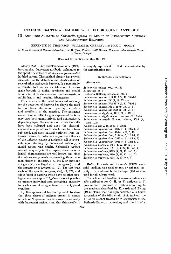
bredeney were agglutinated but not stained. Theappearance of a typical strain of S. typhosastained with labeled 0 (9, 12) globulin is shownin figure 1.
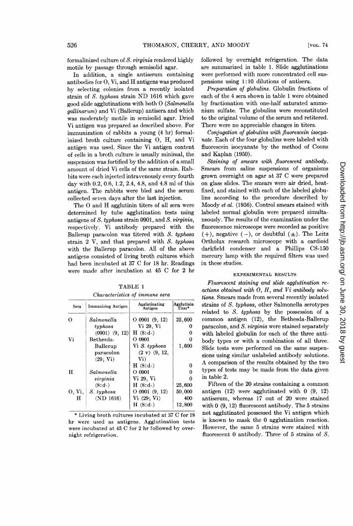
All strains containing Vi antigen were bothagglutinated and stained by Vi antibody solu-tions. Only negative reactions were obtainedwith strains not possessing Vi antigen. The Vicells of S. typhosa shown in figure 2 were stainedwith Vi globulin labeled with fluorescein.
Results of tests with H antiserum (table 2)showed that agglutination occurred with thosestrains possessing the H antigen. The fluorescencereaction was negative, since the H antigennecessary for the demonstration of staining bylabeled flagellar antiglobulin was lost during thepreparation of smears.
All cultures, with the exception of certain S.bredeney strains and S. virginia, reacted in bothfluorescent antibody and agglutination reactionsin which a single globulin solution containing 0,Vi, and H antibody was used. From the irregularresults obtained with the strains of S. bredeney,it might be suggested that this serotype is lessclosely related serologically to the typhoidorganism than are the other serotypes tested.The (d) antigen was the only component of S.virginia represented by specific antibody in thecombined (0, Vi, H) globulin solution. Since thesmear did not receive special handling, theflagella were lost and no staining could be demon-strated.
Selective fluorescent staining of cellular andflagellar portions of Salmonella organisms. Since
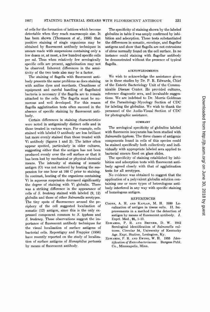
Figure 1. Salmonella typhosa treated withlabeled 0 (9, 12) antiglobulin. In figures 1, 2, 3,and 4 the photomicrographs were made throughthe Leitz Ortholux microscope using ultravioletillumination and a dark-field condenser.
Figure 2. Salmonella typhosa treated with labeledVi (Bethesda-Ballerup paracolon) antiglobulin.See legend under figure 1.
flagellar staining could not be demonstrated inthe above experiment, all strains of S. typhosaand of S. virginia were passaged serially throughsemisolid agar to enhance motility. Activelymotile, 4 to 6 hr broth cultures were prepared,formalin added to a final concentration of 0.3per cent, the suspension centrifuged, and theorganisms washed twice in distilled water. Sincethe flagella were easily dislodged from the cells,very careful handling of the cultures was required.Four sets of smears were made on acid-cleanedslides, air dried, and fixed in acetone. The first
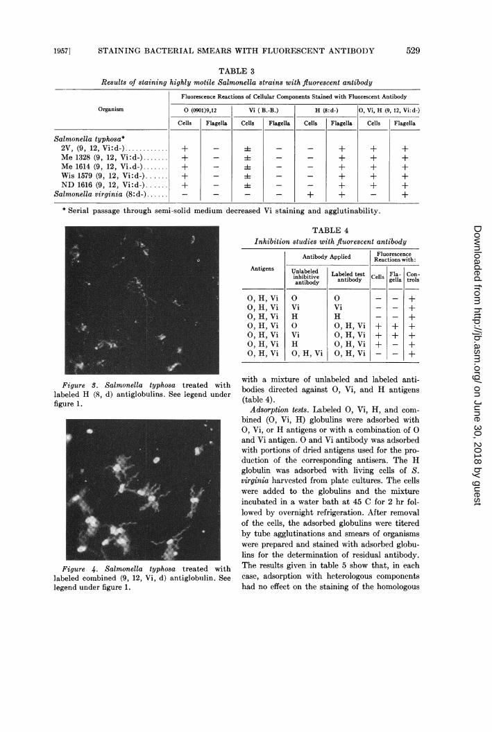
slide of each set was stained with 0, the secondwith Vi, the third with H, and the fourth withthe combined (0, Vi, H) antibody. The distribu-tion of fluorescent antibody on the cell body andflagella was noted under the fluorescence micro-scope. The results are shown in table 3. The cellbody, but not the flagella, of all strains of S.typhosa was stained by both the 0 and Vi globu-lins but not by the H globulin. Cells of S. virginia(8:d-) did not fluoresce when treated with 0 andVi globulin of S. typhosa but both the cells andflagella were stained by homologous globulin(8:d-). The flagella of all strains of S. typhosastained brilliantly with the H antibody (8:d-);the cell body was unstained. This is illustratedin figure 3. Both flagella and cells fluorescedwhen treated with the labeled combined (0, Vi,H) globulin. Only the flagella of S. virginia werestained by the (0, Vi, H) globulin prepared withS. typhosa. The organisms in figure 4 were stainedby combined (0, Vi, H) globulin (S. typhosa)resulting in fluorescence of both cells and flagella.
After organisms containing Vi had been passedseveral times through semisolid medium thequantity of Vi antigen had decreased consider-ably as revealed by the weak fluorescence seenfollowing treatment with Vi globulin. This changewas reflected in agglutination tests in whichsuspensions previously inagglutinable in 0 serumbecame agglutinable.
Inhibition tests. Further evidence of the speci-ficity of staining with fluorescent antibody wasobtained by the application of the one-phaseinhibition test (Moody et al., 1956). This con-sisted of treating dried smears of cells with amixture of appropriate dilutions of labeled andunlabeled antibody. In such a reaction the pres-ence of unlabeled homologous antibody resultsin inhibition of fluorescent staining. The datagiven in table 4 summarize the results obtained.It should be noted that staining of each of thecomponents of cells containing 0, Vi, and Hantigen could be inhibited by the use of un-labeled homologous antibody without interferingwith the staining of the remaining components.For example, when unlabeled 0 antibody wasmixed with labeled combined (0, Vi, H) anti-body, staining of only the 0 antigen was inhibitedwhile Vi and H antigens reacted with their respec-tive labeled antibodies as revealed by theirfluorescence. Blockage of all cellular attachmentsites occurred when the preparations were treated
Antigens UnableiUnlabeled Labeled test Cells Fla- Con-anhbtibod antibody Clsgella trols
O, H, Vi 0 0 - - +O, H,Vi Vi Vi _ - +O, H, Vi H H _ - +O, H, Vi 0 O, H, Vi + + +O, H,Vi Vi O, H, Vi + + +O, H, Vi H O, H, Vi + - +O, H, Vi O, H, Vi O, H, Vi - - +
Figure S. Salmonella typhosa treated withlabeled H (8, d) antiglobulins. See legend underfigure 1.
with a mixture of unlabeled and labeled anti-bodies directed against 0, Vi, and H antigens(table 4).Adsorption tests. Labeled 0, Vi, H, and com-
bined (0, Vi, H) globulins were adsorbed with0, Vi, or H antigens or with a combination of 0and Vi antigen. 0 and Vi antibody was adsorbedwith portions of dried antigens used for the pro-duction of the corresponding antisera. The Hglobulin was adsorbed with living cells of S.virginia harvested from plate cultures. The cellswere added to the globulins and the mixtureincubated in a water bath at 45 C for 2 hr fol-lowed by overnight refrigeration. After removalof the cells, the adsorbed globulins were titeredby tube agglutinations and smears of organismswere prepared and stained with adsorbed globu-lins for the determination of residual antibody.The results given in table 5 show that, in eachcase, adsorption with heterologous componentshad no effect on the staining of the homologous
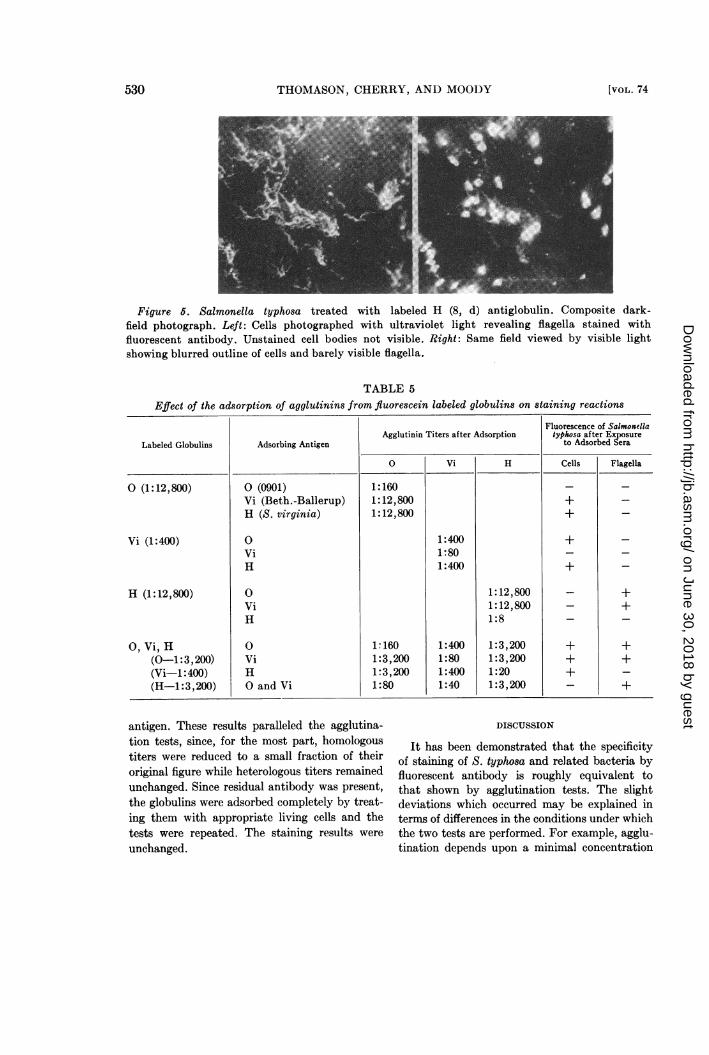
Figure 6. Salmonella typhosa treated with labeled H (8, d) antiglobulin. Composite dark-field photograph. Left: Cells photographed with ultraviolet light revealing flagella stained withfluorescent antibody. Unstained cell bodies not visible. Right: Same field viewed by visible lightshowing blurred outline of cells and barely visible flagella.
TABLE 5
Effect of the adsorption of agglutinins from fluorescein labeled globulins on staining reactions
Labeled Globulins
O (1:12,800)
Vi (1:400)
H (1:12,800)
0, Vi, H(0-1:3,200)(Vi-1:400)(H-1:3,200)
O (0901)Vi (Beth.-Ballerup)H (S. virginia)
0viH
0viH
0viHO and Vi
Agglutinin Titers after AdsorptionFluorescence of Salmonellatyphosa after Exposure
to Adsorbed Sera
0 Vi H Cells Flagella
1:1601:12,8001:12,800
1:1601:3,2001:3,2001:80
1:4001:801:400
1:4001:801:4001:40
1:12,8001:12,8001:8
1:3,2001:3,2001:201:3,200
+
+
+
+
+
antigen. These results paralleled the agglutina-tion tests, since, for the most part, homologoustiters were reduced to a small fraction of theiroriginal figure while heterologous titers remainedunchanged. Since residual antibody was present,the globulins were adsorbed completely by treat-ing them with appropriate living cells and thetests were repeated. The staining results were
unchanged.
DISCUSSION
It has been demonstrated that the specificityof staining of S. typhosa and related bacteria byfluorescent antibody is roughly equivalent tothat shown by agglutination tests. The slightdeviations which occurred may be explained interms of differences in the conditions under whichthe two tests are performed. For example, agglu-tination depends upon a minimal concentration
STAINING BACTERIAL SMEARS WITH FLUORESCENT ANTIBODY
of cells for the formation of lattices which becomedetectable when they reach macroscopic size. Ithas been shown (Thomason et al., 1956) thatpositive staining of single organisms may beobtained by fluorescent antibody techniques insmears made with suspensions containing only afew dozen or, at most, a few hundred specific cellsper ml. Thus when relatively few serologicallyspecific cells are present, agglutination may notbe observed. Inherent differences in the sensi-tivity of the two tests also may be a factor.The staining of flagella with fluorescent anti-
body presents the same problems as does stainingwith aniline dyes and mordants. Cleanliness ofequipment and careful handling of flagellatedbacteria is necessary if the flagella are to remainattached to the cells. The flagella must be nu-merous and well developed. For this reasonflagella agglutination tests often succeed in theabsence of specific staining by fluorescent anti-body.
Certain differences in staining characteristicswere noted in antigenically distinct cells and inthose treated in various ways. For example, cellsstained with labeled 0 antibody are less brilliantbut more evenly stained than those treated withVi antibody (figures 1 and 2). The latter oftenappear spotted, particularly in older cultures,suggesting either that the antigen has not beenproduced evenly over the cell surface or that ithas been lost by mechanical or physical-chemicalmeans. The intensity of staining of somaticantigen (0) was not reduced by heating the sus-pension for one hour at 100 C prior to staining.In contrast, heating of the organisms containingVi in aqueous suspension decreased significantlythe degree of staining with Vi globulin. Therewas a striking difference in the appearance ofcells of S. bredeney stained with labeled (9, 12)globulin and those of other Salmonella serotypes.The tiny spots of fluorescence around the pe-riphery of the cell suggested localization ofsomatic (12) antigen, since this is the only ex-pressed component common to S. typhosa andS. bredeney. These observations suggest the im-portance of fluorescent antibody techniques forthe visual localization of surface antigens ofbacterial cells. Repentigny and Frappier (1956)have recently reported on the study of localiza-tion of surface antigens of Hemophilus pertussisby means of fluorescent antibody.
The specificity of staining shown by the labeledglobulins in table 2 was amply confirmed by inhi-bition and adsorption. These tests substantiatedthe differences in somatic, envelope, and flagellarantigens and show that flagella are not extrusionsof slime normally found on the cell surface. In noinstance could staining with flagellar antibodybe demonstrated without the presence of typicalflagella.
ACKNOWLEDGMENTS
We wish to acknowledge the assistance givenus in these studies by Dr. P. R. Edwards, Chiefof the Enteric Bacteriology Unit of the Commu-nicable Disease Center. He provided cultures,reference diagnostic sera, and invaluable sugges-tions. We are indebted to Dr. Morris Goldmanof the Parasitology-Mycology Section of CDCfor labeling the globulins. We wish to thank thepersonnel of the Audio-Visual Section of CDCfor photographic assistance.
SUMMARY
The serological specificity of globulins labeledwith fluorescein isocyanate has been studied withSalmonella typhosa. The three classes of antigeniccomponents found in cells of this species couldbe stained specifically both collectively and indi-vidually with appropriate labeled sera applied tobacterial smears fixed on glass slides.The specificity of staining established by inhi-
bition and adsorption tests with fluorescent anti-body agreed closely with that of agglutinationtests for all serotypes.No evidence was obtained to suggest that the
application of a polyvalent globulin solution con-taining one or more types of heterologous anti-body interfered in any way with specific stainingof homologous antigen.
REFERENCESCOONS, A. H. AND KAPLAN, M. H. 1950 Lo-
calization of antigen in tissue cells. II. Im-provements in a method for the detection ofantigen by means of fluorescent antibody. J.Exptl. Med., 91, 1-13.
EDWARDS, P. R. AND BRUNER, D. W. 1942Serological identification of Salmonella cul-tures. Circuilar 54, University of KentuckyAgr. Expt. Station, Lexington, Ky.
EDWARDS, P. R. AND EWING, W. H. 1955 Iden-tification of Enterobacteriaceae. Burgess Publ.Co., Minneapolis, Minn.
MOODY, M. D., GOLDMAN, M., AND THOMASON,BERENICE M. 1956 Staining bacterialsmears with fluorescent antibody. I. Gen-eral methods for Malleomyces pseudomallei.J. Bacteriol., 72, 357-361.
DE REPENTIGNY, J. AND FRAPPIER, A. 1956Studies on Hemophilus pertussis liquid cul-tures. III. Localization of surface anti-
gens by means of fluorescent antibody.Can. J. Microbiol., 2, 677-683.
THOMASON, BERENICE M., MOODY, M. D., AND
GOLDMAN, M. 1956 Staining bacterialsmears with fluorescent antibody. II. Rapiddetection of varying numbers of Malleomycespseudomallei in contaminated materials andinfected animals. J. Bacteriol., 72, 366-367.