12
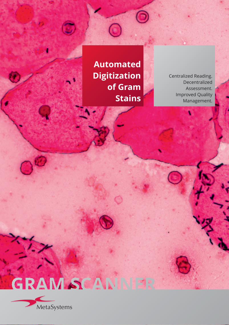
Automated Digitization of Gram Stains Centralized Reading. Decentralized Assessment. Improved Quality Management.
Automated
Digitization
of Gram
Stains
Centralized Reading.
Decentralized
Assessment.
Improved Quality
Management.
Gram staining is the rapid, easy, and
inexpensive method for the assessment of
specimen quality, providing early
information regarding potential patho-
gens present in patient samples. Serving
to guide initial infection therapy, the Gram
stain is one of the most commonly
performed tests in clinical microbiology.
The number of specimens needing to be
processed is growing steadily as a result of
the rising prevalence of infectious
diseases, an aging population with
increased susceptibility, and the imple-
mentation of Antibiotic Stewardship
Programs as main growth drivers.
Laboratories are facing increasing cost
pressures, intensified quality require-
ments and the constant demand to
provide test results as quickly as
possible; a considerable task, even
without taking into account the trend to
static, or even shrink, the number of
staff.
In response to this challenge many
laboratories automate microbial
workflows.
The Answer Gram staining has a high potential for
automation because Gram stained
samples are evaluated by manual
microscopy, even today. But the
introduction of automated microscopy
for Gram samples was hampered by
the lack of suitable system solutions.
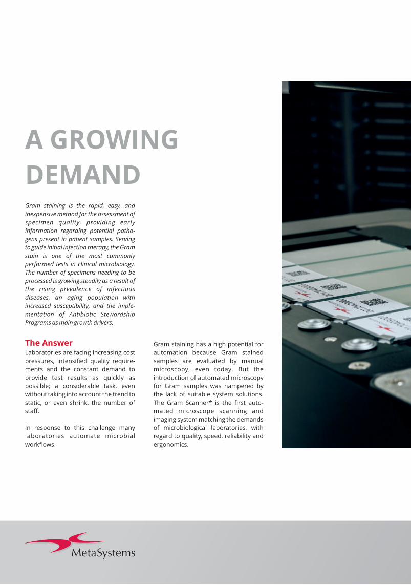
The Gram Scanner* is the first auto-
mated microscope scanning and
imaging system matching the demands
of microbiological laboratories, with
regard to quality, speed, reliability and
ergonomics.
A GROWING
DEMAND

Implementation of the Gram Scanner into
the laboratory workflow will revolutionize
one`s daily routine. The system scans,
digitizes and archives – where requested –
up to 800 slides in one batch.
Instead of spending time on slide
scanning, technicians can focus on
Gram stain assessment with improved
ergonomics. Moreover, all Gram
sample scans can be consolidated on
one workstation. Seamless integration
into the existing laboratory environ-
ment facilitates the exchange of images
and data with external systems via LIMs
to store Gram information with patient
files, or to upload pre-defined procedu-
res for reading and assessment.
Centralized Reading
GRAM READING
AS NEVER BEFORE
Decentral AssessmentImage digitization ensures maximum
efficiency of Gram sample evaluation
because data can be accessed from
multiple users at the same time in the
hospital / laboratory network.
Ease and speed of second opinion
requests is ensured, as experts can
access images via the network or can
receive digital data electronically.
Gram sample digitization could there-
fore support more timely therapy
decisions and interdisciplinary collabo-
rations for better patient outcomes.
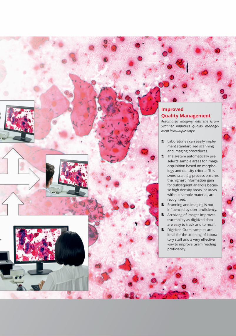
Improved
Quality ManagementAutomated imaging with the Gram
Scanner improves quality manage-
ment in multiple ways:
a Laboratories can easily imple-
ment standardized scanning
and imaging procedures.
a The system automatically pre-
selects sample areas for image
acquisition based on morpho-
logy and density criteria. This
smart scanning process ensures
the highest information gain
for subsequent analysis becau-
se high density areas, or areas
without sample material, are
recognized.
a Scanning and imaging is not
influenced by user proficiency.
a Archiving of images improves
traceability as digitized data
are easy to track and to recall.
a Digitized Gram samples are
ideal for the training of labora-
tory staff and a very effective
way to improve Gram reading
proficiency.
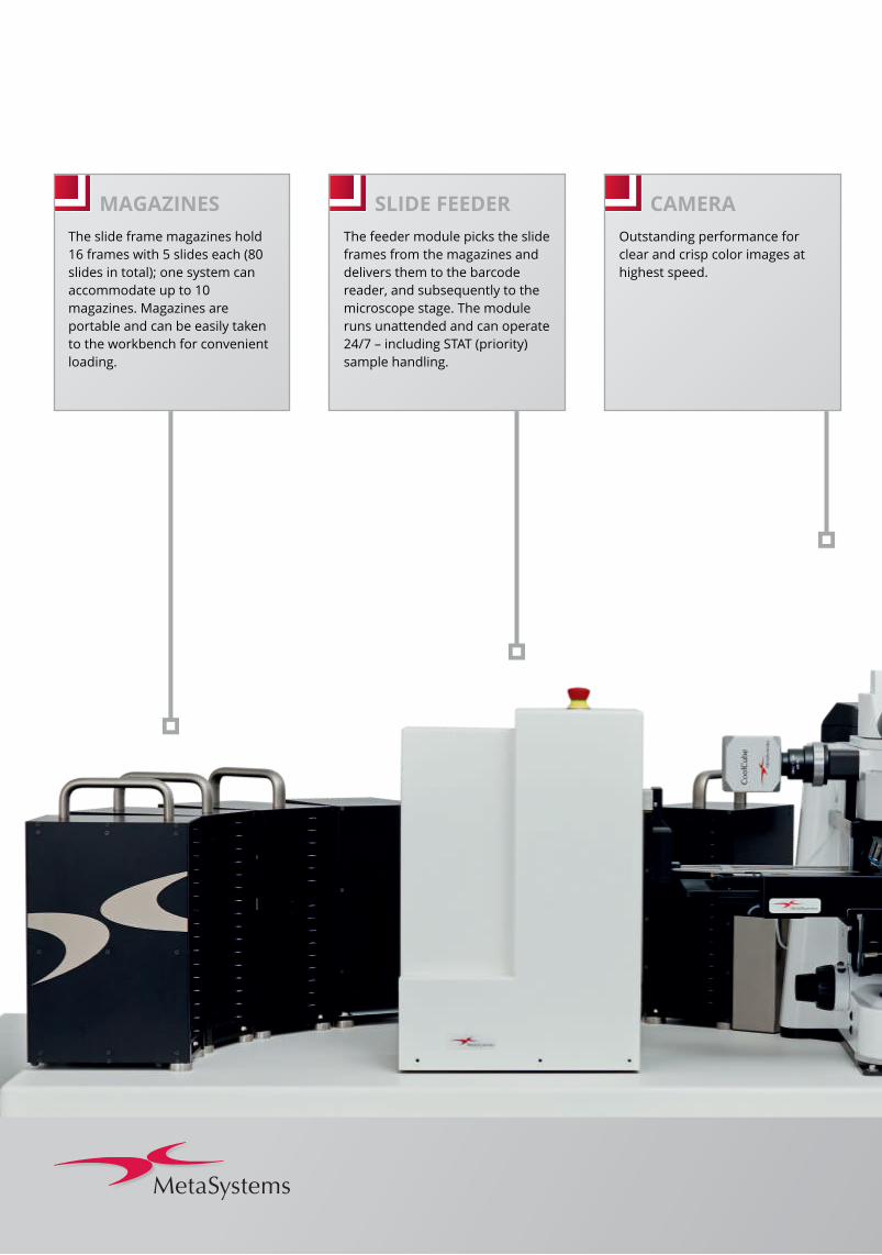
MAGAZINES
The slide frame magazines hold
16 frames with 5 slides each (80
slides in total); one system can
accommodate up to 10
magazines. Magazines are
portable and can be easily taken
to the workbench for convenient
loading.
SLIDE FEEDER
The feeder module picks the slide
frames from the magazines and
delivers them to the barcode
reader, and subsequently to the
microscope stage. The module
runs unattended and can operate
24/7 – including STAT (priority)
sample handling.
CAMERA
Outstanding performance for
clear and crisp color images at
highest speed.
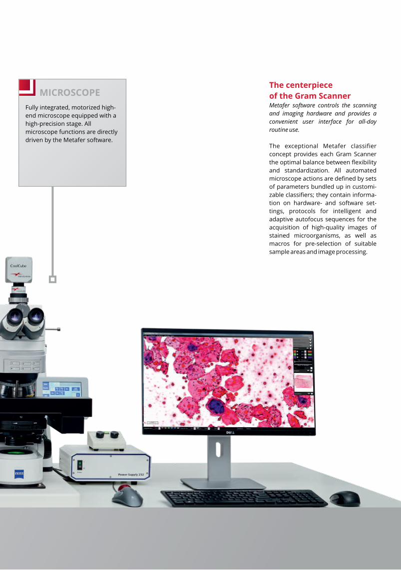
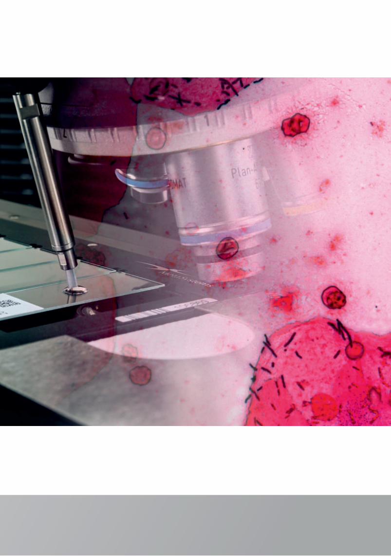
MICROSCOPE
Fully integrated, motorized high-
end microscope equipped with a
high-precision stage. All
microscope functions are directly
driven by the Metafer software.
The centerpiece
of the Gram ScannerMetafer software controls the scanning
and imaging hardware and provides a
convenient user interface for all-day
routine use.
The exceptional Metafer classifier
concept provides each Gram Scanner
the optimal balance between flexibility
and standardization. All automated
microscope actions are defined by sets
of parameters bundled up in customi-
zable classifiers; they contain informa-
tion on hardware- and software set-
tings, protocols for intelligent and
adaptive autofocus sequences for the
acquisition of high-quality images of
stained microorganisms, as well as
macros for pre-selection of suitable
sample areas and image processing.
HOW IT
WORKS
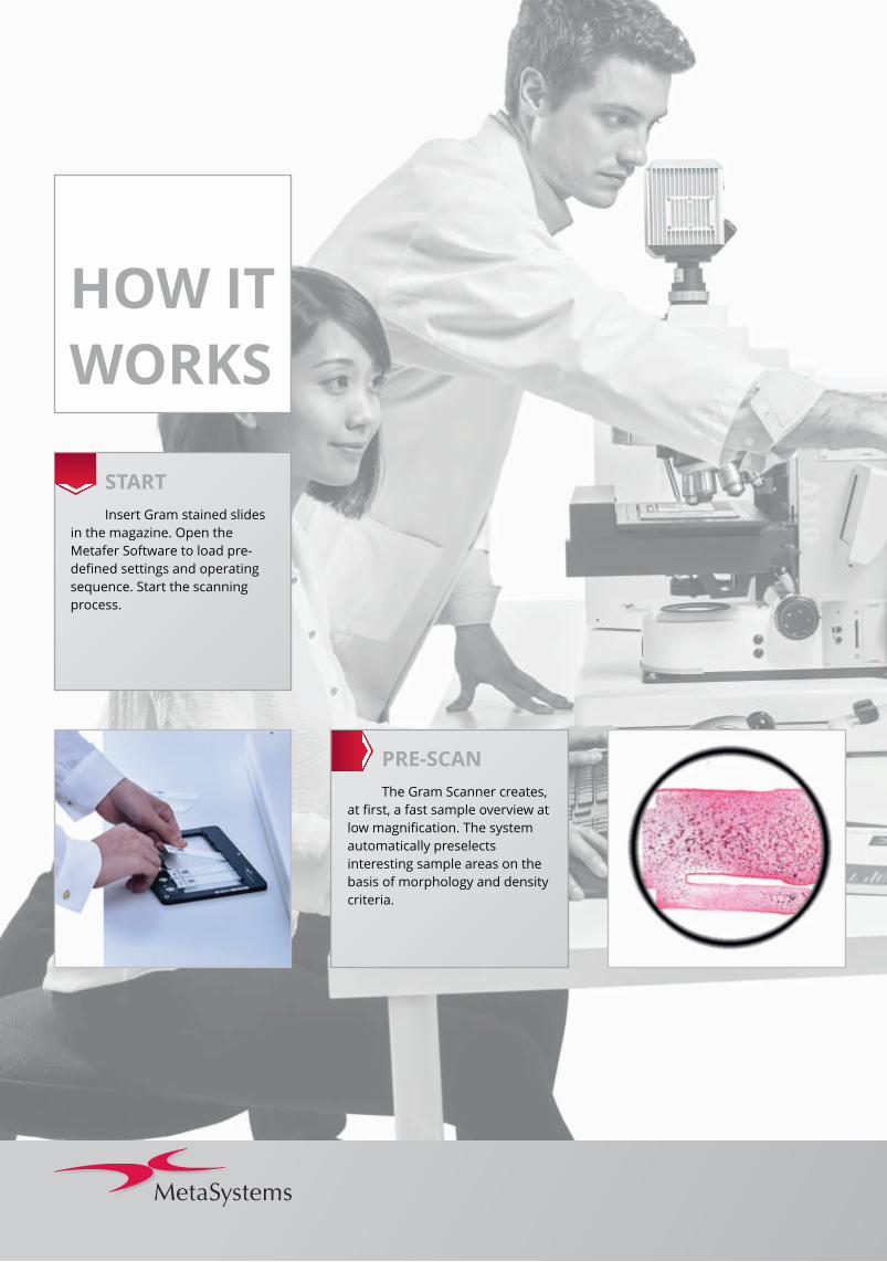
PRE-SCAN
The Gram Scanner creates,
at first, a fast sample overview at
low magnification. The system
automatically preselects
interesting sample areas on the
basis of morphology and density
criteria.
START
Insert Gram stained slides
in the magazine. Open the
Metafer Software to load pre-
defined settings and operating
sequence. Start the scanning
process.
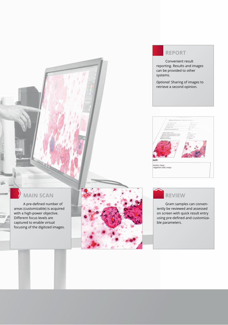
REPORT
Convenient result
reporting. Results and images
can be provided to other
systems.
Optional: Sharing of images to
retrieve a second opinion.
REVIEW
Gram samples can conven-
iently be reviewed and assessed
on screen with quick result entry
using pre-defined and customiza-
ble parameters.
MAIN SCAN
A pre-defined number of
areas (customizable) is acquired
with a high-power objective.
Different focus levels are
captured to enable virtual
focusing of the digitized images.
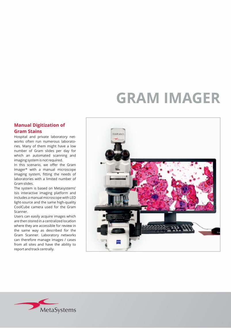
Manual Digitization of
Gram StainsHospital and private laboratory net-
works often run numerous laborato-
ries. Many of them might have a low
number of Gram slides per day for
which an automated scanning and
imaging system is not required.
In this scenario, we offer the Gram
Imager* with a manual microscope
imaging system, fitting the needs of
laboratories with a limited number of
Gram slides.
The system is based on Metasystems'
Isis interactive imaging platform and
includes a manual microscope with LED
light-source and the same high-quality
CoolCube camera used for the Gram
Scanner.
Users can easily acquire images which
are then stored in a centralized location
where they are accessible for review in
the same way as described for the
Gram Scanner. Laboratory networks
can therefore manage images / cases
from all sites and have the ability to
report and track centrally.
GRAM IMAGER

The Open and
Flexible PlatformAutomated scanning and imaging of
Gram stained slides with the Gram
Scanner is one of the microbiological
applications based on MetaSystems'
Metafer slide scanning platform.
Further applications for Metafer are the
Mycobacteria Scanner for automated
scanning and imaging of Tuberculosis
Sputum smears, and Direct Multiplex
Imaging (DMI) for identification of
pathogens directly from patient sam-
ples.
Due to Metafer´s modular and flexible architecture, it is possible to consoli-date several applications on one workstation. For example, one can combine the Gram Scanner with the Mycobacteria Scanner to process Gram stained slides during the day and TB sputum smear slides overnight.
For more information, see separate
product information or online using:
www.metasystems-international.com
METAFER
www.metasystems-international.com
MetaSystems products are used in many countries worldwide. Depending on the
regulations of the respective country or region, some products may not be used for
clinical diagnostic use.
*In the United States of America (USA) and Canada, the MetaSystems Gram Scanner
and Mycobacteria Scanner are intended for research purposes only.
* In Europe, MetaSystems devices are CE-marked in-vitro diagnostics (IVD) devices.
MetaSystems Hard & Software GmbH (Headquarters)
Robert-Bosch-Str. 6, 68804 Altlussheim, Germany
tel +49 6205 396 10 | fax +49 6205 322 70 | [email protected]
MetaSystems Group, Inc.
70 Bridge Street, Newton, MA 02458, USA
tel +1 6179 2499 50 | fax +1 6179 2499 54 | [email protected]
MetaSystems S.r.l.
Via Gallarate 80, 20151 Milano, Italy
tel +39 0236 7587 51 | fax +39 0245 3753 03 | [email protected]
MetaSystems India Pvt., Ltd.
No. 1/1, 2nd Floor, 1st Main Rd., 2nd cross, Thimmaiah Garden, R T Nagar, Bangalore Karnataka, 560 032, India
tel +91 9535 7788 01 | [email protected]
MetaSystems Asia Co., Ltd.
Unit 108, 1/F, Bio-Informatics Centre, No. 2 Science Park, West Avenue, Hong Kong Science Park, Shatin, New Territories, Hong Kong
tel +852 2587 8333 | fax +852 2587 8334 | [email protected]
MetaSystems Worldwide
MetaSystems believes that a global company should foster a community of partners
and customers to achieve a climate of collaboration and friendship. Our headquarters
and the subsidiary offices in USA, Italy, India, and Hong Kong with their trained and
motivated staff allow us to work very close to our customers. We know that communi-
cation is crucial to achieve our main goal: to continuously improve our understanding
of demands, requirements, and wishes of lab professionals everywhere in the world.
MetaSystems is very proud having established a worldwide network of competent
sales and servicing partners. Please feel free to contact your local MetaSystems
representative or MetaSystems directly to learn more about the community.
Do
cum
en
t N
o.
BR
O-G
ram
Sca
nn
er-
20
18
-01
-01
-P
© 2
01
8 b
y M
eta
Syst
em
s


