Page 1
Standard Operating Procedure for:
Anatoxin (ELISA)
University of Missouri Limnology Laboratory
Prepared by: Anthony Thorpe
Date: March 15, 2021
Approved by: Christopher Brunet
Date: May 25, 2021
Page 2
ID: ANA
Revision: 1.1
May 2021
Page: 2 of 12
Table of Contents 1 Identification of the method ................................................................................................. 3
2 Applicable matrix or matrices .............................................................................................. 3
3 Detection limit ..................................................................................................................... 3
4 Scope of the method ........................................................................................................... 3
5 Summary of the method ...................................................................................................... 3
6 Interferences ....................................................................................................................... 3
7 Health and Safety ............................................................................................................... 3
8 Personnel qualifications ...................................................................................................... 4
9 Equipment and supplies ...................................................................................................... 4
10 Reagents and standards ..................................................................................................... 4
11 Quality Control .................................................................................................................... 5
12 Analysis .............................................................................................................................. 5
13 Method performance ........................................................................................................... 7
14 Data assessment and acceptable criteria for quality control measures ............................... 7
15 Pollution prevention ............................................................................................................ 7
16 Waste management ............................................................................................................ 7
17 References ......................................................................................................................... 8
18 Additional materials ............................................................................................................. 8
Page 3
ID: ANA
Revision: 1.1
May 2021
Page: 3 of 12
1 Identification of the method
1.1 Measurement of Anatoxin-a via Eurofins Abraxis immunoassay for quantitative and
sensitive screening.
2 Applicable matrix or matrices
2.1 This method is suitable for the analysis of environmental samples.
3 Detection limit
3.1 Method Detection Limit: 0.10 µg/L.
3.2 This Method Detection Limit was determined by the manufacturer, Eurofins Abraxis.
4 Scope of the method
4.1 This standard operating procedure is intended to provide MU Limnology operators,
technicians, and analysts with guidance on the analysis of anatoxin-a in water samples
using the Eurofins Abraxis ELISA test 520060. This document is not intended to replace
individual training in this method by an experienced MU Limnology technician.
5 Summary of the method
5.1 ELISA (enzyme-linked immunosorbent assay) is a plate-based assay technique designed for
detecting and quantifying soluble substances such as peptides, proteins, antibodies, and
hormones.
5.2 Operating Range: 0.1 to 5 µg/L
6 Interferences
6.1 Freshwater samples must be preserved (and lysed) prior to filtration or Anatoxin-a may
bind to the filter, removing it from the sample, and producing falsely low results.
6.2 Immediately upon collection, freshwater samples must be preserved using the Sample
Diluent (10X) Concentrate (1 mL of 10X Sample Diluent Concentrate per 9 mL of water
sample), to prevent degradation of Anatoxin-a. Samples must be adjusted to between pH
5 and pH 7 and protected from exposure to natural and artificial light, as exposure to light
and/or high pH will cause degradation of Anatoxin-a.
7 Health and Safety
7.1 These analyses involve handling freshwater samples that may contain live microorganisms
and therefore pose some threat of infection. Laboratory personnel who are routinely
exposed to such water samples are encouraged to protect themselves from water borne
illnesses by wearing clean disposable gloves and washing hands frequently.
Page 4
ID: ANA
Revision: 1.1
May 2021
Page: 4 of 12
7.2 Wear protective gloves, lab coats, and other appropriate PPE when handling all chemical
substances used in this method. All operators and technicians performing this method
should review the MSDS for additional information and safety concerns regarding the
chemical substances used throughout these procedures.
8 Personnel qualifications
8.1 Samples will be analyzed by MU Limnology staff who have been trained to the operators
or technician level in this method and who are familiar with all of the MU Limnology
sampling handling and labeling procedures and appropriate SOPs.
9 Equipment and supplies
9.1 0.5 meter sampling tube
9.2 0.5 L PETG container (e.g., Fisher #0992316B)
9.3 Amber glass vial with PTFE lined cap (e.g., Fisher #14955332)
9.4 Sample diluent concentrate (from ELISA kit, see below)
9.5 Eurofins Abraxis Anatoxin-a ELISA kit (product# 520011)
9.6 Stepping pipette, 50 µL pipette, associated tips
9.7 Syringe with Luer-lock tip, inline 0.45 µm glass fiber filters (e.g., Fisher # 501049931)
9.8 500 ml squirt bottle
9.9 Vortex mixer
9.10 96 well microplate reader, computer, software
10 Reagents and standards
All reagents and standards used in this method are included with the kit and do not require further
preparation.
10.1 Microtiter plate coated with a secondary antibody (anti-mouse), in a re-sealable aluminum
pouch.
10.2 Lyophilized Anatoxin-a-HRP Enzyme Conjugate, 3 vials.
10.3 Conjugate Diluent, 12 mL.
10.4 Lyophilized Anti-Anatoxin-a Antibody, 3 vials.
10.5 Antibody Diluent, 12 mL.
10.6 Empty clear and amber HDPE bottles for combining reconstituted Enzyme Conjugate and
Antibody (if necessary).
10.7 (+)Anatoxin-a Standards (6): 0, 0.15, 0.40, 1.0, 2.5, 5.0 ppb; 1.5 mL each.
10.8 Control at 0.75 ± 0.185 ppb; 1.5 mL.
10.9 Sample Diluent (10X) Concentrate, 2 X 25 mL.
10.10 Wash Buffer (5X) Concentrate, 100 mL, must be diluted before use.
Page 5
ID: ANA
Revision: 1.1
May 2021
Page: 5 of 12
10.11 Substrate (Color) Solution (TMB), 12 mL.
10.12 Stop Solution, 12 mL (handle with care and appropriate PPE).
11 Quality Control
11.1 Positive Control
A positive control with a known concentration is included with each run and analyzed.
12 Analysis
12.1 Freeze/Thaw Lysis
Prior to analysis, samples should undergo a total of 3 freeze/thaw cycles to lyse cells and
release cell-bound toxins.
• Remove vials from freezer and place into test tube racks. Place test tube rack into plastic
tub.
• Slowly begin to fill plastic tub with cool water. When water slightly covers bottom of vials,
remove rack from water and inspect for cracked vials. Do this several times as vials begin
to thaw. If there are no broken vials, fill plastic tub until approximately half of each vial is
submerged. Allow to thaw, typically 15 minutes.
• If a crack is discovered (usually the bottom of the vial will break loose entirely),
immediately remove the vial from the rack and place into a clean glass test tube of
suitable size. Allow ice in broken vial to thaw inside test tube. After thawing, pour contents
into new vial. Reuse the same cap as it will have identifying information.
• Once thawed, vials should be re-frozen, again on their sides to reduce the risk of breakage.
A total of 3 freeze-thaw cycles needs to be completed before samples are ready for
analysis.
12.2 Filtering:
After lysing, samples should be filtered through an inline 0.45 µm GFF to remove
particulates that may interfere with the analysis.
• Remove syringe from packaging, remove plunger from syringe. Set both on clean paper
towel to avoid contamination from lab surfaces.
• Screw inline syringe filter to syringe body.
• Pour sample from vial into syringe, tap empty vial on another clean paper towel to remove
remaining water droplets. Put open end of filter into vial and insert plunger into syringe.
Depress plunger until filtered sample is returned to vial. Recap vial and mark a line on the
cap with a colored Sharpie marker. This mark indicates the sample has been filtered.
Discard syringe and filter into temporary waste bin.
12.3 Sample analysis
• Samples and ELISA kit should be at room temperature prior to analysis.
Page 6
ID: ANA
Revision: 1.1
May 2021
Page: 6 of 12
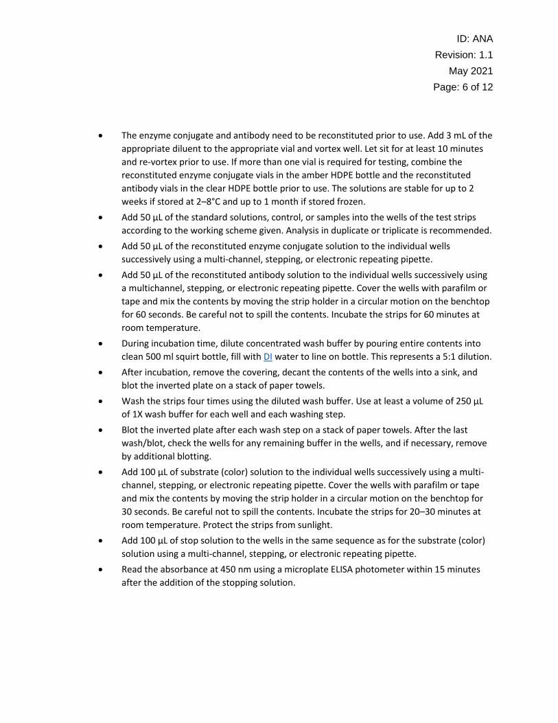
• The enzyme conjugate and antibody need to be reconstituted prior to use. Add 3 mL of the
appropriate diluent to the appropriate vial and vortex well. Let sit for at least 10 minutes
and re-vortex prior to use. If more than one vial is required for testing, combine the
reconstituted enzyme conjugate vials in the amber HDPE bottle and the reconstituted
antibody vials in the clear HDPE bottle prior to use. The solutions are stable for up to 2
weeks if stored at 2–8°C and up to 1 month if stored frozen.
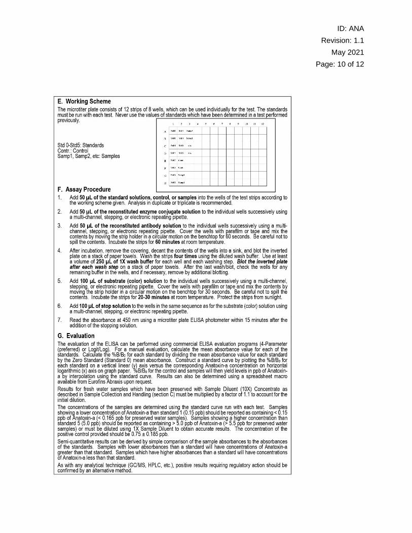
• Add 50 μL of the standard solutions, control, or samples into the wells of the test strips
according to the working scheme given. Analysis in duplicate or triplicate is recommended.
• Add 50 μL of the reconstituted enzyme conjugate solution to the individual wells
successively using a multi-channel, stepping, or electronic repeating pipette.
• Add 50 μL of the reconstituted antibody solution to the individual wells successively using
a multichannel, stepping, or electronic repeating pipette. Cover the wells with parafilm or
tape and mix the contents by moving the strip holder in a circular motion on the benchtop
for 60 seconds. Be careful not to spill the contents. Incubate the strips for 60 minutes at
room temperature.
• During incubation time, dilute concentrated wash buffer by pouring entire contents into
clean 500 ml squirt bottle, fill with DI water to line on bottle. This represents a 5:1 dilution.
• After incubation, remove the covering, decant the contents of the wells into a sink, and
blot the inverted plate on a stack of paper towels.
• Wash the strips four times using the diluted wash buffer. Use at least a volume of 250 μL
of 1X wash buffer for each well and each washing step.
• Blot the inverted plate after each wash step on a stack of paper towels. After the last
wash/blot, check the wells for any remaining buffer in the wells, and if necessary, remove
by additional blotting.
• Add 100 μL of substrate (color) solution to the individual wells successively using a multi-
channel, stepping, or electronic repeating pipette. Cover the wells with parafilm or tape
and mix the contents by moving the strip holder in a circular motion on the benchtop for
30 seconds. Be careful not to spill the contents. Incubate the strips for 20–30 minutes at
room temperature. Protect the strips from sunlight.
• Add 100 μL of stop solution to the wells in the same sequence as for the substrate (color)
solution using a multi-channel, stepping, or electronic repeating pipette.
• Read the absorbance at 450 nm using a microplate ELISA photometer within 15 minutes
after the addition of the stopping solution.
Page 7
ID: ANA
Revision: 1.1
May 2021
Page: 7 of 12
• Samples with concentrations exceeding the high standard should be diluted with the
included diluent and reanalyzed. The diluent included with the kit is a concentrate and
must be diluted prior to use. Dilute the Sample Diluent (10X) Concentrate at a ratio of 1:10
with deionized or distilled water (i.e., 1 mL of Sample Diluent (10X) Concentrate into 9 mL
of deionized water) as needed for sample dilutions. The Sample Diluent is now ready for
use. Samples are typically diluted at 10:1 (i.e., 4.5 mL of diluted diluent to 0.5 mL of
sample).
13 Method performance
13.1 Desired Performance Criteria
• Method Detection Limit: 0.10 µg/L
• Precision: CV < 15% for ≤ 0.4 µg/L and CV < 10% for > 0.4 µg/L
• Calibration r2 > 0.99
14 Data assessment and acceptable criteria for quality control measures
14.1 Standard curve should have an r2 value of at least 0.99.
14.2 Standard curve replicates should have an absorbance CV >10%. One standard may have an
absorbance CV of up to 15%, if no other standard CV exceeds 10%.
14.3 The concentration of the positive control should be 0.75 ± 0.185 µg/L (between 0.565 and
0.935 µg/L).
14.4 Samples with concentrations ≤ 0.4 µg/l may have a CV up to 15%.
14.5 Samples with concentrations > 0.4 µg/l may have a CV up to 10%.
14.6 Samples not meeting the above criteria should be reanalyzed.
14.7 Samples with replicates having concentrations < 0.10 µg/L and > 0.15 µg/L should be
reanalyzed.
14.8 Samples with concentrations > 5.0 µg/L should be diluted and reanalyzed.
14.9 Results from subsequent sample analyses will be combined with initial analysis to test for
outliers and determine further actions.
14.10 Samples not meeting criteria outlined above should be reanalyzed.
15 Pollution prevention
15.1 All reagents and standards will be prepared in appropriate volumes to reduce waste.
15.2 All sample and reagents will be handled according to MU EHS policies in order to ensure
proper disposal.
16 Waste management
16.1 All waste generated is considered hazardous.
Page 8
ID: ANA
Revision: 1.1
May 2021
Page: 8 of 12
16.2 All analyzed standards, and reagents should be treated as waste upon completion of the
run.
16.3 Waste should be kept in an approved container with proper labeling.
16.4 Waste will not be held for longer than 6 months and MU Environmental Health and Safety
(EHS) will be notified an appropriate time before this point so that waste can be collected
and disposed of.
16.5 Empty temporary waste bin directly in dumpster, not into lab trash cans. Syringes (even
without needles) discarded in the lab trash may pose a risk to custodial staff.
17 References
17.1 Anatoxin-a ELISA, Eurofins 520060 User Guide. Eurofins Abraxis.
18 Additional materials
18.1 Eurofins 520060 User Guide (downloaded November 2020)
Page 9
ID: ANA
Revision: 1.1
May 2021
Page: 9 of 12
Page 10
ID: ANA
Revision: 1.1
May 2021
Page: 10 of 12
Page 11
ID: ANA
Revision: 1.1
May 2021
Page: 11 of 12
Page 12
ID: ANA
Revision: 1.1
May 2021
Page: 12 of 12