PNL03 Last Updated July 2017 Page 1 Standard Operating Procedure for PulseNet PFGE of Campylobacter jejuni Purpose To describe the One-Day (24-26 hour) Standardized Laboratory Protocol for Molecular Subtyping of Campylobacter jejuni by Pulsed-field Gel Electrophoresis (PFGE). Scope To provide the PulseNet participants with a standardized procedure for performing PFGE of Campylobacter jejuni, thus ensuring inter-laboratory comparability of the generated results. Definitions and Terms 1. PFGE: Pulsed-field Gel Electrophoresis 2. DNA: Deoxyribonucleic acid 3. CDC: Centers for Disease Control and Prevention 4. CLRW: Clinical Laboratory Reagent Water 5. TE: Tris-EDTA 6. EDTA: Ethylenediaminetetraacetic acid 7. PBS: Phosphate Buffered Saline 8. TBE: Tris borate-EDTA 9. BHI-RB: Brain Heart Infusion + 5% rabbit blood agar 10. Microaerobic: an environment containing low levels of oxygen (specifically, 3.5% oxygen and 2-10% carbon dioxide for Campylobacter jejuni Biosafety Warning Please read all instructions carefully before starting protocol. It is recommended to plate cultures, prepare cell suspensions and cast plugs in a Class II Biosafety Cabinet (BSC), if available. Treat all plasticware, glassware, pipets, spatulas, etc. that come in contact with the cell suspensions or plugs as contaminated materials and dispose of or disinfect according to your institutional guidelines.
Transcript
PNL03 Last Updated July 2017 Page 1
Standard Operating Procedure for PulseNet PFGE of Campylobacter jejuni
Purpose
To describe the One-Day (24-26 hour) Standardized Laboratory Protocol for Molecular Subtyping of Campylobacter jejuni by Pulsed-field Gel Electrophoresis (PFGE).
Scope
To provide the PulseNet participants with a standardized procedure for performing PFGE of Campylobacter jejuni, thus ensuring inter-laboratory comparability of the generated results.
Definitions and Terms
1. PFGE: Pulsed-field Gel Electrophoresis2. DNA: Deoxyribonucleic acid3. CDC: Centers for Disease Control and Prevention4. CLRW: Clinical Laboratory Reagent Water5. TE: Tris-EDTA6. EDTA: Ethylenediaminetetraacetic acid7. PBS: Phosphate Buffered Saline8. TBE: Tris borate-EDTA9. BHI-RB: Brain Heart Infusion + 5% rabbit blood agar10. Microaerobic: an environment containing low levels of oxygen (specifically, 3.5% oxygen and 2-10% carbon dioxide for
Campylobacter jejuni
Biosafety Warning
Please read all instructions carefully before starting protocol. It is recommended to plate cultures, prepare cell suspensions and cast plugs in a Class II Biosafety Cabinet (BSC), if available. Treat all plasticware, glassware, pipets, spatulas, etc. that come in contact with the cell suspensions or plugs as contaminated materials and dispose of or disinfect according to your institutional guidelines.
PNL03 Last Updated July 2017 Page 2
Day 0
Plating for confluent growth
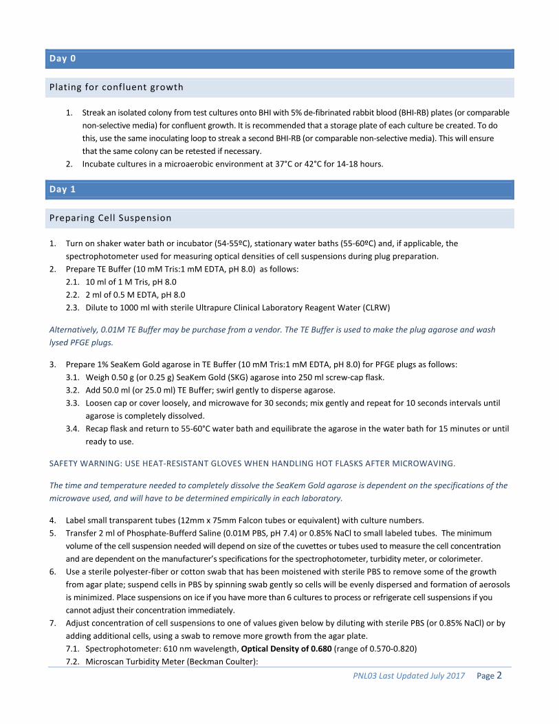
1. Streak an isolated colony from test cultures onto BHI with 5% de-fibrinated rabbit blood (BHI-RB) plates (or comparablenon-selective media) for confluent growth. It is recommended that a storage plate of each culture be created. To dothis, use the same inoculating loop to streak a second BHI-RB (or comparable non-selective media). This will ensurethat the same colony can be retested if necessary.
2. Incubate cultures in a microaerobic environment at 37°C or 42°C for 14-18 hours.
Day 1
Preparing Cell Suspension
1. Turn on shaker water bath or incubator (54-55ºC), stationary water baths (55-60ºC) and, if applicable, thespectrophotometer used for measuring optical densities of cell suspensions during plug preparation.
2. Prepare TE Buffer (10 mM Tris:1 mM EDTA, pH 8.0) as follows:2.1. 10 ml of 1 M Tris, pH 8.02.2. 2 ml of 0.5 M EDTA, pH 8.02.3. Dilute to 1000 ml with sterile Ultrapure Clinical Laboratory Reagent Water (CLRW)
Alternatively, 0.01M TE Buffer may be purchase from a vendor. The TE Buffer is used to make the plug agarose and wash lysed PFGE plugs.
3. Prepare 1% SeaKem Gold agarose in TE Buffer (10 mM Tris:1 mM EDTA, pH 8.0) for PFGE plugs as follows:3.1. Weigh 0.50 g (or 0.25 g) SeaKem Gold (SKG) agarose into 250 ml screw-cap flask.3.2. Add 50.0 ml (or 25.0 ml) TE Buffer; swirl gently to disperse agarose.3.3. Loosen cap or cover loosely, and microwave for 30 seconds; mix gently and repeat for 10 seconds intervals until
agarose is completely dissolved. 3.4. Recap flask and return to 55-60°C water bath and equilibrate the agarose in the water bath for 15 minutes or until
ready to use.
SAFETY WARNING: USE HEAT-RESISTANT GLOVES WHEN HANDLING HOT FLASKS AFTER MICROWAVING.
The time and temperature needed to completely dissolve the SeaKem Gold agarose is dependent on the specifications of the microwave used, and will have to be determined empirically in each laboratory.
4. Label small transparent tubes (12mm x 75mm Falcon tubes or equivalent) with culture numbers.5. Transfer 2 ml of Phosphate-Bufferd Saline (0.01M PBS, pH 7.4) or 0.85% NaCl to small labeled tubes. The minimum
volume of the cell suspension needed will depend on size of the cuvettes or tubes used to measure the cell concentration and are dependent on the manufacturer’s specifications for the spectrophotometer, turbidity meter, or colorimeter.
6. Use a sterile polyester-fiber or cotton swab that has been moistened with sterile PBS to remove some of the growthfrom agar plate; suspend cells in PBS by spinning swab gently so cells will be evenly dispersed and formation of aerosolsis minimized. Place suspensions on ice if you have more than 6 cultures to process or refrigerate cell suspensions if you cannot adjust their concentration immediately.
7. Adjust concentration of cell suspensions to one of values given below by diluting with sterile PBS (or 0.85% NaCl) or byadding additional cells, using a swab to remove more growth from the agar plate.7.1. Spectrophotometer: 610 nm wavelength, Optical Density of 0.680 (range of 0.570-0.820)7.2. Microscan Turbidity Meter (Beckman Coulter):
PNL03 Last Updated July 2017 Page 3
• 0.35-0.45 (measured in Falcon 2054 tubes)• 0.52-0.64 (measured in Falcon 2057 tubes)
The values in steps 7.1-7.3 give satisfactory results at CDC; each laboratory may need to establish the optimal concentration needed for satisfactory results.
Casting Plugs
The preparation of cell suspensions and subsequent casting of plugs should be performed as rapidly as possible in order to minimize premature cell lysis and solidification of agarose in the pipette tips/microcentrifuge tubes. If large numbers of samples are being prepared, it is recommended to process them in batches of ~10 samples at a time. Once the first batch of isolates are in the cell lysis incubation step, then the cell suspensions can be prepared for the next group of samples and so on. All batches can be lysed and washed together since additional lysis time will not affect the initial batches. Unused plug agarose can be kept at room temperature and reused 1 or 2 times. Microwave on low-medium power for 10-15 sec and mix; repeat for 5-10 sec intervals until agarose is completely melted.
1. Label wells of PFGE plug molds with culture number. When reusable plug molds are used, put strip of tape on lowerpart of reusable plug mold before labeling wells.
2. Transfer 400 µl adjusted cell suspensions to labeled 1.5-ml microcentrifuge tubes.3. Add 20 µl of Proteinase K (20 mg/ml stock) to each tube and mix gently with pipet tip.
Proteinase K solutions (20 mg/ml) are available commercially. Alternatively, a stock solution of Proteinase K can be prepared from powder in sterile Ultrapure water (CLRW; see PNL02). Just before use, thaw appropriate number of vials needed for the samples; keep Proteinase K solutions on ice. If the Proteinase K stock solution was prepared from powder, discard any thawed solution at the end of the work day. Store commercially prepared Proteinase K solutions according to directions provided by the supplier.
4. Add 400 µl melted 1% SeaKem Gold agarose to 400 µl cell suspension; mix by gently pipetting mixture up and downtwo or three times. Over-pipetting can cause DNA shearing. Maintain temperature of melted agarose by keepingflask in beaker of warm water (55-60°C).
5. Immediately, dispense part of mixture into appropriate well(s) of reusable plug mold without introducing bubbles.Two plugs of each sample can be made from these amounts of cell suspension and agarose and are useful if repeattesting is required. Allow plugs to solidify at room temperature for 10-15 minutes. They can also be placed in therefrigerator (4°C) for 5 minutes
If disposable plug molds are used, combine 200 μl cell suspension, 10 μl of Proteinase K (20 mg/ml stock) and 200 μl of agarose; up to 4 plugs can be made from the smaller volumes.
Lysis of Cells in Agarose Plugs
Two plugs (reusable molds) or 3 – 4 plugs (disposable molds) of the same strain can be lysed in the same 50ml tube.
1. Label 50ml polypropylene screw-cap or 50ml Oak Ridge tubes with culture numbers.2. Calculate the total volume of Cell Lysis Buffer/Proteinase K master mix needed as follows:
2.1. 5 ml stock Cell Lysis Buffer (CLB; 50 mM Tris:50 mM EDTA, pH 8.0 + 1% Sarcosyl per tube as prepared in“Formulas of PFGE Reagents” section)
• e. g., 5 ml x 10 tubes = 50 ml 2.2. 25 µl Proteinase K stock solution (20 mg/ml) per tube of the cell lysis buffer
PNL03 Last Updated July 2017 Page 4
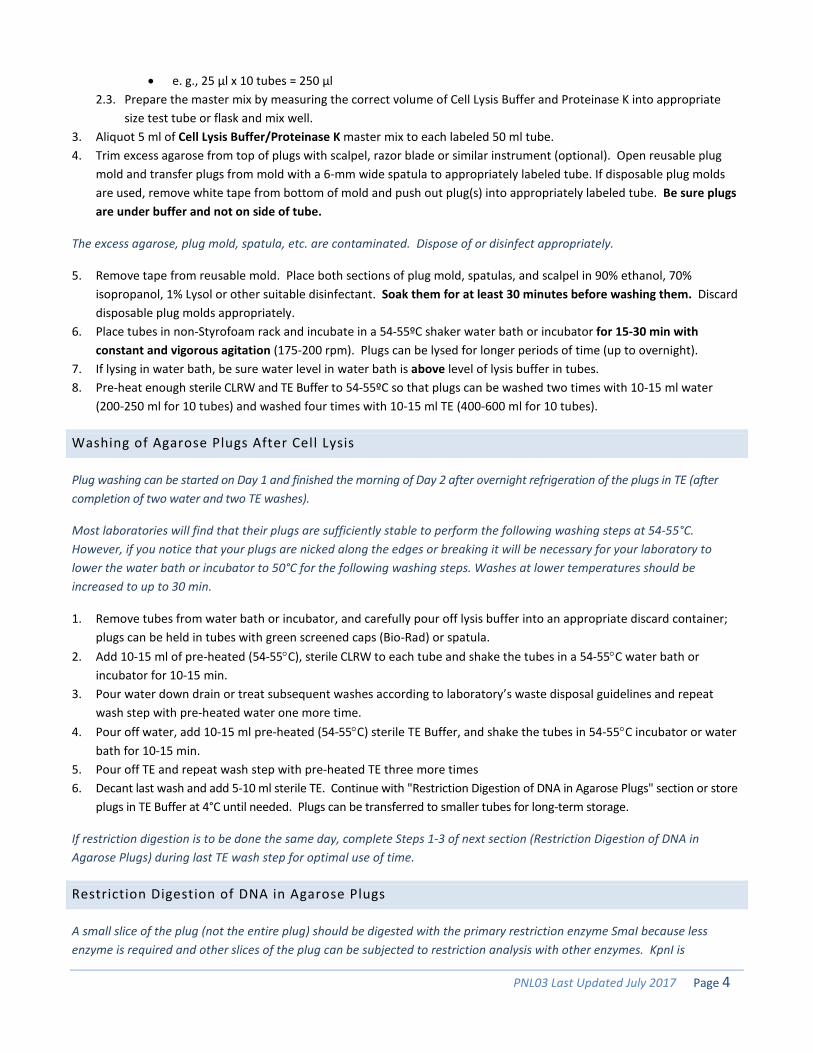
• e. g., 25 µl x 10 tubes = 250 µl 2.3. Prepare the master mix by measuring the correct volume of Cell Lysis Buffer and Proteinase K into appropriate
size test tube or flask and mix well. 3. Aliquot 5 ml of Cell Lysis Buffer/Proteinase K master mix to each labeled 50 ml tube.4. Trim excess agarose from top of plugs with scalpel, razor blade or similar instrument (optional). Open reusable plug
mold and transfer plugs from mold with a 6-mm wide spatula to appropriately labeled tube. If disposable plug moldsare used, remove white tape from bottom of mold and push out plug(s) into appropriately labeled tube. Be sure plugsare under buffer and not on side of tube.
The excess agarose, plug mold, spatula, etc. are contaminated. Dispose of or disinfect appropriately.
5. Remove tape from reusable mold. Place both sections of plug mold, spatulas, and scalpel in 90% ethanol, 70%isopropanol, 1% Lysol or other suitable disinfectant. Soak them for at least 30 minutes before washing them. Discarddisposable plug molds appropriately.
6. Place tubes in non-Styrofoam rack and incubate in a 54-55ºC shaker water bath or incubator for 15-30 min withconstant and vigorous agitation (175-200 rpm). Plugs can be lysed for longer periods of time (up to overnight).
7. If lysing in water bath, be sure water level in water bath is above level of lysis buffer in tubes.8. Pre-heat enough sterile CLRW and TE Buffer to 54-55ºC so that plugs can be washed two times with 10-15 ml water
(200-250 ml for 10 tubes) and washed four times with 10-15 ml TE (400-600 ml for 10 tubes).
Washing of Agarose Plugs After Cell Lysis
Plug washing can be started on Day 1 and finished the morning of Day 2 after overnight refrigeration of the plugs in TE (after completion of two water and two TE washes).
Most laboratories will find that their plugs are sufficiently stable to perform the following washing steps at 54-55°C. However, if you notice that your plugs are nicked along the edges or breaking it will be necessary for your laboratory to lower the water bath or incubator to 50°C for the following washing steps. Washes at lower temperatures should be increased to up to 30 min.
1. Remove tubes from water bath or incubator, and carefully pour off lysis buffer into an appropriate discard container;plugs can be held in tubes with green screened caps (Bio-Rad) or spatula.
2. Add 10-15 ml of pre-heated (54-55°C), sterile CLRW to each tube and shake the tubes in a 54-55°C water bath orincubator for 10-15 min.
3. Pour water down drain or treat subsequent washes according to laboratory’s waste disposal guidelines and repeatwash step with pre-heated water one more time.
4. Pour off water, add 10-15 ml pre-heated (54-55°C) sterile TE Buffer, and shake the tubes in 54-55°C incubator or waterbath for 10-15 min.
5. Pour off TE and repeat wash step with pre-heated TE three more times6. Decant last wash and add 5-10 ml sterile TE. Continue with "Restriction Digestion of DNA in Agarose Plugs" section or store
plugs in TE Buffer at 4°C until needed. Plugs can be transferred to smaller tubes for long-term storage.
If restriction digestion is to be done the same day, complete Steps 1-3 of next section (Restriction Digestion of DNA in Agarose Plugs) during last TE wash step for optimal use of time.
Restriction Digestion of DNA in Agarose Plugs
A small slice of the plug (not the entire plug) should be digested with the primary restriction enzyme SmaI because less enzyme is required and other slices of the plug can be subjected to restriction analysis with other enzymes. KpnI is
PNL03 Last Updated July 2017 Page 5
recommended as the secondary enzyme for analysis of Campylobacter jejuni isolates. The use of a secondary enzyme is useful in situations where the PFGE patterns obtained with the primary enzyme from two or more isolates are indistinguishable.
1. Label 1.5-ml microcentrifuge tubes with culture numbers; label 3 (10-well gel) or 4 (15-well gel) tubes for Salmonellaser. Braenderup H98121 standards.
The number of the Salmonella serotype Braenderup H9812 standards is dependent on the number of samples being run on the gel. Standards should be placed on the ends and between samples as needed; no more than 4 samples should be placed between the standard lanes.
1.1. Pre-Restriction Incubation Step (highly-recommended): Prepare a master mix by diluting the appropriate 10X restriction buffer (Roche/Sigma, New England Biolabs or equivalent) 1:10 with sterile CLRW according to the following table:
The appropriate restriction buffer varies between vendors and may differ between enzymes from the same vendor. Always use the restriction buffer recommended by the vendor for the particular restriction enzyme.
2. Add 200 µl diluted restriction buffer (1X) to labeled 1.5-ml microcentrifuge tubes.3. Carefully remove plug from TE with spatula and place in a sterile disposable Petri dish or on large glass slide.4. Cut a 2-2.5 mm wide slice from each test sample and the appropriate number of slices of H9812 standard with a scalpel
(or single edge razor blade, cover slip, etc.) and transfer to tube containing diluted restriction buffer. Be sure plug sliceis under buffer. Replace rest of plug in original tube (short-term storage) that contains at least 5 ml TE buffer orsmaller tube containing TE buffer (long-term storage). Plugs should be stored at 4°C.
PulseNet recommends that the combs with larger teeth (10 mm wide teeth) be used to cast the gels because computeranalysis of the gel lanes is more accurate and less tedious than analysis of gel lanes cast with combs with the smallerteeth (<10 mm). Using combs with smaller teeth is not advised. The number of slices that can be cut from the plugs willdepend on the skill and experience of the operator, integrity of the plug, and whether the slices are cut vertically orhorizontally (plugs made in disposable molds).
5. Incubate sample and control plug slices in the appropriate water bath for 5-10 minutes or at room temperature for 10-15 minutes.5.1. Incubate samples digested with SmaI at 25°C or room temperature.5.2. Incubate samples digested with KpnI or XbaI at 37°C.
6. After incubation, remove buffer from plug slice using a pipet fitted with 200-250 µl tip all the way to bottom of tubeand aspirate buffer. Be careful not to damage the plug slice with pipet tip and that plug slice is not discarded withpipet tip.
7. Prepare the restriction enzyme master mix according to the following table. Keep vials of restriction enzyme on ice or inan insulated storage box (-20°C) at all times.
1 Directions for making and testing PFGE plugs of Salmonella ser. Braenderup H9812 are in PNL05
PNL03 Last Updated July 2017 Page 6
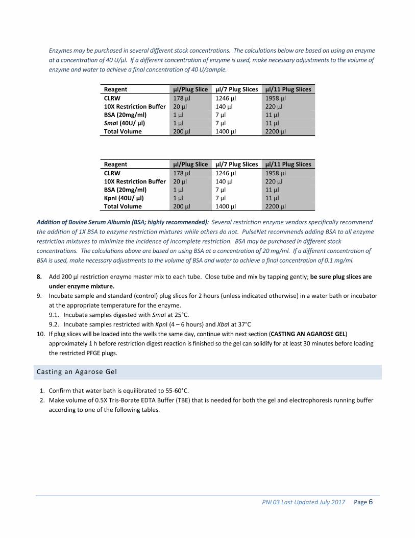
Enzymes may be purchased in several different stock concentrations. The calculations below are based on using an enzyme at a concentration of 40 U/μl. If a different concentration of enzyme is used, make necessary adjustments to the volume of enzyme and water to achieve a final concentration of 40 U/sample.
Addition of Bovine Serum Albumin (BSA; highly recommended): Several restriction enzyme vendors specifically recommend the addition of 1X BSA to enzyme restriction mixtures while others do not. PulseNet recommends adding BSA to all enzyme restriction mixtures to minimize the incidence of incomplete restriction. BSA may be purchased in different stock concentrations. The calculations above are based on using BSA at a concentration of 20 mg/ml. If a different concentration of BSA is used, make necessary adjustments to the volume of BSA and water to achieve a final concentration of 0.1 mg/ml.
8. Add 200 µl restriction enzyme master mix to each tube. Close tube and mix by tapping gently; be sure plug slices areunder enzyme mixture.
9. Incubate sample and standard (control) plug slices for 2 hours (unless indicated otherwise) in a water bath or incubatorat the appropriate temperature for the enzyme.9.1. Incubate samples digested with SmaI at 25°C.9.2. Incubate samples restricted with KpnI (4 – 6 hours) and XbaI at 37°C
10. If plug slices will be loaded into the wells the same day, continue with next section (CASTING AN AGAROSE GEL)approximately 1 h before restriction digest reaction is finished so the gel can solidify for at least 30 minutes before loadingthe restricted PFGE plugs.
Casting an Agarose Gel
1. Confirm that water bath is equilibrated to 55-60°C.2. Make volume of 0.5X Tris-Borate EDTA Buffer (TBE) that is needed for both the gel and electrophoresis running buffer
according to one of the following tables.
PNL03 Last Updated July 2017 Page 7
5X TBE:
Reagent
Volume in milliliters (ml)
(DR-II) Running buffer + 10 well gel prep
(DR-II) Running buffer + 15 well gel prep
(CHEF Mapper/DRIII) Running buffer + 10 well gel prep
(CHEF Mapper/DRIII) Running buffer + 15 well gel prep
5X TBE 210 215 230 235 CLRW 1890 1935 2070 2115 Total Volume of 0.5X TBE 2100 2150 2300 2350
10X TBE:
Reagent
Volume in milliliters (ml)
(DR-II) Running buffer + 10 well gel prep
(DR-II) Running buffer + 15 well gel prep
(CHEF Mapper/DRIII) Running buffer + 10 well gel prep
(CHEF Mapper/DRIII) Running buffer + 15 well gel prep
10X TBE 105 107.5 115 117.5 CLRW 1995 2042.5 2185 2232.5 Total Volume of 0.5X TBE 2100 2150 2300 2350
3. Make 1% SeaKem Gold (SKG; the only acceptable agarose to be used for PulseNet PFGE) agarose in 0.5X TBE asfollows:
3.1 Weigh appropriate amount of SKG into 500 ml screw-cap flask. 3.2 Add appropriate amount of 0.5X TBE prepared according to tables above; swirl gently to disperse agarose.
3.2.1 Mix 1.0 g agarose with 100 ml 0.5X TBE for 14 cm wide gel form (10 wells) 3.2.2 Mix 1.5 g agarose with 150 ml 0.5X TBE for 21 cm wide gel form (15 wells)
3.3 Loosen cap or cover loosely and microwave for 60 seconds; mix gently and repeat for 15 second intervals until agarose is completely dissolved.
3.4 Return flask to 55-60°C water bath and equilibrate the agarose in the water bath for 15 minutes or until ready to use.
SAFETY WARNING: USE HEAT-RESISTANT GLOVES WHEN HANDLING HOT FLASKS AFTER MICROWAVING.
Loading Restricted Plug Slices on the Comb (Option 1)
1. Remove restricted plug slices from 37°C water bath. Remove enzyme/buffer mixture and add 200 µl 0.5X TBE.Incubate at room temperature for 5 minutes. Alternatively, digested plug slices can be kept in the refrigerator for upto three days if they are stored in 0.5X TBE.
2. Assemble gel form, place on a leveling table and adjust until perfectly leveled. Place the comb holder so the frontpart (side with small metal screws) and teeth face the bottom of gel frame. Make sure the comb teeth touch thegel platform, when comb is upright.
3. Remove plug slices from tubes; lay comb flat on gel form and load plug slices on the bottom edge of the teeth,including Salmonella serotype Braenderup H9812 standards in outside lanes (teeth) and between samples asneeded (no more than 4 samples between standards).
4. Load samples on remaining teeth of the comb and note locations.5. Remove excess buffer with a kimwipe. Allow plug slices to air dry on the comb for 3-5 minutes or seal them to the
comb with 1% SKG agarose (55-60°C).6. Position comb in gel form and confirm that the plugs slices are correctly aligned on the bottom of the comb teeth,
that the lower edge of the plug slice is flush against the black platform.
PNL03 Last Updated July 2017 Page 8
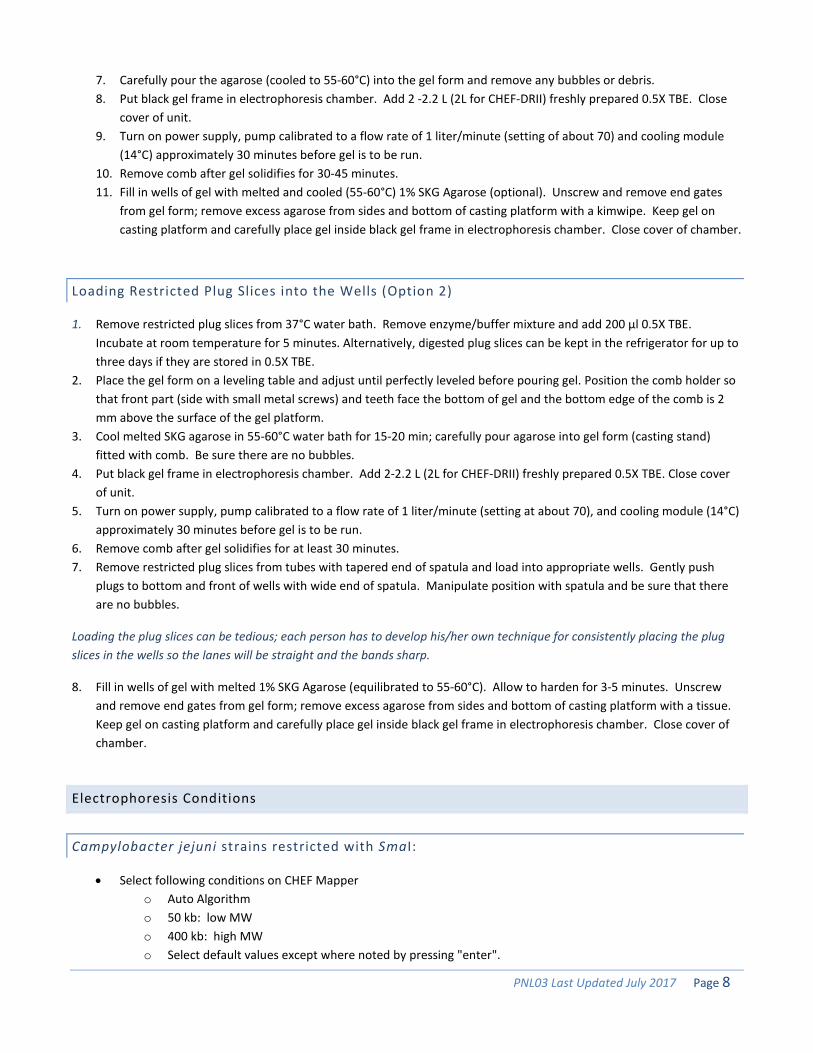
7. Carefully pour the agarose (cooled to 55-60°C) into the gel form and remove any bubbles or debris.8. Put black gel frame in electrophoresis chamber. Add 2 -2.2 L (2L for CHEF-DRII) freshly prepared 0.5X TBE. Close
cover of unit.9. Turn on power supply, pump calibrated to a flow rate of 1 liter/minute (setting of about 70) and cooling module
(14°C) approximately 30 minutes before gel is to be run.10. Remove comb after gel solidifies for 30-45 minutes.11. Fill in wells of gel with melted and cooled (55-60°C) 1% SKG Agarose (optional). Unscrew and remove end gates
from gel form; remove excess agarose from sides and bottom of casting platform with a kimwipe. Keep gel oncasting platform and carefully place gel inside black gel frame in electrophoresis chamber. Close cover of chamber.
Loading Restricted Plug Slices into the Wells (Option 2)
1. Remove restricted plug slices from 37°C water bath. Remove enzyme/buffer mixture and add 200 µl 0.5X TBE.Incubate at room temperature for 5 minutes. Alternatively, digested plug slices can be kept in the refrigerator for up tothree days if they are stored in 0.5X TBE.
2. Place the gel form on a leveling table and adjust until perfectly leveled before pouring gel. Position the comb holder sothat front part (side with small metal screws) and teeth face the bottom of gel and the bottom edge of the comb is 2mm above the surface of the gel platform.
3. Cool melted SKG agarose in 55-60°C water bath for 15-20 min; carefully pour agarose into gel form (casting stand)fitted with comb. Be sure there are no bubbles.
4. Put black gel frame in electrophoresis chamber. Add 2-2.2 L (2L for CHEF-DRII) freshly prepared 0.5X TBE. Close coverof unit.
5. Turn on power supply, pump calibrated to a flow rate of 1 liter/minute (setting at about 70), and cooling module (14°C)approximately 30 minutes before gel is to be run.
6. Remove comb after gel solidifies for at least 30 minutes.7. Remove restricted plug slices from tubes with tapered end of spatula and load into appropriate wells. Gently push
plugs to bottom and front of wells with wide end of spatula. Manipulate position with spatula and be sure that thereare no bubbles.
Loading the plug slices can be tedious; each person has to develop his/her own technique for consistently placing the plug slices in the wells so the lanes will be straight and the bands sharp.
8. Fill in wells of gel with melted 1% SKG Agarose (equilibrated to 55-60°C). Allow to harden for 3-5 minutes. Unscrewand remove end gates from gel form; remove excess agarose from sides and bottom of casting platform with a tissue.Keep gel on casting platform and carefully place gel inside black gel frame in electrophoresis chamber. Close cover ofchamber.
Electrophoresis Conditions
Campylobacter jejuni strains restricted with SmaI:
• Select following conditions on CHEF Mappero Auto Algorithmo 50 kb: low MWo 400 kb: high MWo Select default values except where noted by pressing "enter".
PNL03 Last Updated July 2017 Page 9
o Change run time to 17-20 hours (See note below)o (Default values: Initial switch time = 6.76 s; Final switch time = 35.38 s)
• Select following conditions on CHEF-DR IIIo Initial switch time: 6.8 so Final switch time: 35.4 so Voltage: 6 Vo Included Angle: 120°o Run time: 17-20 hours (See note below)
• Select following conditions on CHEF-DR IIo Initial A time: 6.8 so Final A time: 35.4 so Start ratio: 1.0 (if applicable)o Voltage: 200 Vo Run time: 17-20 hours (See note below)
Campylobacter jejuni strains restricted with KpnI:
• Select following conditions on CHEF Mappero Auto Algorithmo 50 kb: low MWo 475 kb: high MWo Select default values except where noted by pressing "Enter."o Change run time to 17-20 hours (See note below)o Change Initial Switch Time to 5.2s; Accept default Final Switch Time = 42.34s
• Select following conditions on CHEF DR-IIIo Initial switch time: 5.2 so Final switch time: 42.3 so Voltage: 6 Vo Included Angle: 120°o Run time: 17-20 hours (See note below)
• Select following conditions on CHEF DR-II.o Initial A time: 5.2so Final A time: 42.3 so Start Ratio: 1.0 (if applicable)o Voltage: 200 Vo Run time: 17-20 hours (See note below)
The electrophoresis running times recommended above are based on the equipment and reagents used at the CDC. The 21 cm wide (15-well) gels require ~1 hr longer than 14 cm wide (10-well) gels. Run times may be different in your laboratory and will have to be optimized for your gels so that the lowest band in the S. ser. Braenderup H9812 standard migrates 1.0-1.5 cm from the bottom of the gel.
Make note of the initial milliamp (mA) reading on the instrument. The initial mA should be between 110-150 mA. A reading outside of this range may indicate that the 0.5X TBE buffer was prepared improperly and the buffer should be remade.
Day 2
Staining and Documentation of an Agarose Gel
PNL03 Last Updated July 2017 Page 10
The following staining procedure describes the use of ethidium bromide to stain PFGE gels. Alternate DNA stains may be used. Please see the QuickTip “20140218_Staining” within the Library of PulseNet Documents forum (QuickTips/Wet Lab/PFGE) on the SharePoint site for additional information.
1. When electrophoresis run is over, turn off equipment; remove and place gel in an approximately 14 cm x 24 cmcovered container with 400 ml ethidium bromide or other approved stain (see PNL02). Larger or smaller volumes maybe used for different sized containers. Stain gel, gently rocking for 20-30 min in covered container.
Ethidium bromide (EtBr) is toxic and a mutagen. Stock solutions of 10 mg/ml EtBr in water are available from several commercial companies (see PNL01). The diluted solution can be kept in the dark at room temperature and re-used up to 15 times within 3 weeks before discarding according to your institution's guidelines for hazardous waste; do not pour down the drain. Aqueous solutions containing EtBr can be treated using de-staining bags from Amresco (E732-25), which effectively and safely remove EtBr from solutions. Once the EtBr is removed, the treated aqueous solutions can be discarded down the drain. Refer to the Safety Data Sheets (SDS) provided by the vendor for more details.
Currently, the acceptable alternative stain options are GelRedTM (Biotium, 41002), SYBR® Safe (ThermoFisher Scientific S33102) and SYBR® Gold (ThermoFisher Scientific S11494). Labs are strongly encouraged to follow manufacturer’s instructions and test stains in their labs before adopting them for routine use. If one of the alternative stains is used, the de-staining steps should be omitted. However, the gels can be briefly rinsed with CLRW before imaging. Diluted GelRedTM solution can be kept in the dark at room temperature and re-used up to 10 times within 10 days before discarding down the drain. Usage parameters have not been established for other alternative stains.
2. De-stain gel in approximately 500 ml CLRW for 60-90 min, changing water every ~20 minutes (at least 3 times). Captureimage on GelDoc XR, XR+ or equivalent documentation system. If background interferes with resolution, de-stain foran additional 30-60 min (2 or 3 more washes).
3. Follow directions from the imaging equipment to save gel image as an *.1sc (QuantityOne software) or *.scn (ImageLabsoftware) file; convert this file to *.tif file for analysis with BioNumerics software program. The gel image should fill theentire window of the imaging equipment (computer) screen (without cutting off wells or lower bands). Ensure that theimage is in focus and that there is little to no saturation (over-exposure) in the bands (signified by red pixilation in thesoftware). Additional instructions are provided in PNL07 (Image Acquisition) of the PulseNet QA/QC Manual.
4. Drain buffer from electrophoresis chamber and discard. Rinse chamber with ~2 L CLRW or, if unit is not going to beused for several days, flush lines with water by letting pump run for 5-10 min before draining water from chamber andtubing.
USE OF TRADE NAMES AND COMMERCIAL SOURCES IS FOR IDENTIFICATION PURPOSES ONLY AND DOES NOT IMPLY ENDORSEMENT BY CDC OR THE U.S. DEPARTMENT OF HEALTH AND HUMAN SERVICES.
CLIA Laboratory Procedure Manual Requirements
Efforts have been made to assure that the procedures described in this protocol have been written in accordance with the 1988 Clinical Laboratory Improvement Amendments (CLIA) requirements for a procedure manual (42 CFR 493.1211). However, due to the format required for training, the procedures will require some modifications and additions to customize them for your particular laboratory operation.
Any questions regarding the CLIA requirements for a procedure manual, quality control, quality assurance, etc., should be directed to the agency or accreditation organization responsible for performing your laboratory's CLIA inspection. In addition, some states and accreditation organizations may have more stringent requirements that will need to be addressed.
1. Establish a record book or electronic log (for an example, see Appendix PNL03-1: PFGE Isolates and Plug Run Log)for tracking isolates, including but not limited to, the following information:1.1. Sample number (state public health lab identifier or similar)1.2. Date the isolates were received in the PFGE lab1.3. Date the plugs were made1.4. Location (box and slot) where the plugs are kept1.5. Gel and lane numbers for each sample
2. Establish a record (for an example, see Appendix PNL03-02: PFGE Plug Preparation Worksheet) of plugpreparation, including but not limited to, the following information:2.1 Organism and number of samples2.2 Date plugs were made and initials of who made them2.3 Lot number and expiration dates of reagents used
3. Establish a record (for an example, see Appendix PNL03-3: PFGE Enzyme Master Mix and Gel Setup Worksheet) foreach gel, including but not limited to, the following information:3.1 Gel number and date gel run3.2 Electrophoresis equipment used3.3 Running conditions/times3.4 Lot number and expiration dates of reagents used3.5 Restriction temperatures and times3.6 Order of isolates on a gel
Formulas of PFGE Reagents
TRIS: EDTA BUFFER, PH 8.0: (TE, 10 MM TRIS: 1 MM EDTA, PH 8.0)
10 ml of 1 M Tris, pH 8.0
2 ml of 0.5 M EDTA, pH 8.0
Dilute to 1000 ml with sterile Ultrapure water (CLRW)
CELL LYSIS BUFFER: (50 MM TRIS: 50 MM EDTA, PH 8.0 + 1% SARCOSINE + 0.1 MG/ML PROTEINASE K)
50 ml of 1 M Tris, pH 8.0
100 ml of 0.5 M EDTA, pH 8.0
100 ml 10% N-Lauroylsarcosine, Sodium salt (Sarcosyl) OR 10 g of N-Lauroylsarcosine, Sodium salt (Sarcosyl).
Dilute to 1000 ml with Sterile Ultrapure water (CLRW)
Add 25 µl Proteinase K stock solution (20 mg/ml) per 5 ml of cell lysis buffer just before use for a final concentration in the lysis buffer of 0.1 mg/ml Proteinase K.
If Sarcosyl powder is added directly to the other components of this reagent, warm the solution to 50- 60ºC for 30-60 minutes, or leave at room temperature for about 2 hours to completely dissolve the Sarcosyl.
PNL03 Last Updated July 2017 Page 12
Contacts
CDC PulseNet MS-C03 1600 Clifton Rd. NE Atlanta, GA 30329 Phone (404) 639-4558 Fax (404) 630-3333 [email protected]
Amendments
1. The phrase “Type I Water” has been changed to “Ultrapure Clinical Laboratory Reagent Water (CLRW).”The water composition is the same, but this reflects a change in the terminology used by the ClinicalLaboratory Standards Institute (CLSI).
2. 2011-08 changes:− The wording for programming electrophoresis conditions was updated to standardize this section and
make it the same as other PFGE laboratory SOPs. − The wording for washing the agarose plugs after cell lysis was also updated to standardize the section.
3. 2013-03 changes:− Corrected formula for TE buffer. TE used at CDC is 10 mM for Tris and 1 mM for EDTA.− Recommended disinfectant changed from 10% bleach to 1% Lysol/Amphyll or 90% ethanol.− Volume of TE needed for washing plugs was correted from 300 – 350 ml to 400 – 600 ml.− A statement was added to clarify that using combs with small teeth (5.5 mm) was not advised.− Use of pre-restriction step and BSA was changed from optional to highly recommended.
Calculation for including BSA in restriction enzyme master mix was added. − Statement allowing of Megbase agarose (BioRad) was deleted. Additional testing revealed run
time and normalization were negatively impacted by this agarose. − A statement was included to allow the use of alternative DNA stains that are equivalent to EtBr.
Labs are strongly urged to follow manufacturer’s instructions as well as test stains in their own labs to gain experience using alternative agarose stains. Additional stain alternatives may be tested and deemed acceptable at a later date.
− The option to allow incubation times for restriction digestion to be increased longer than recommended was deleted.
4. 2017-01 changes:− Reformatted to current SOP numbering system. − Added Definitions and updated tables. − Reformatted header and removed footer in accordance with new document layout. − Updated Contact Information. − Added “Approval Signatures” section. − Added “Records Management” section by merging PNL08. − Statement allowing use of Amresco LF agarose (Amresco) was deleted. Additional testing
revealed run time and normalization were negatively impacted by this agarose. − Added Appendices PNL03-1 through PNL03-3.
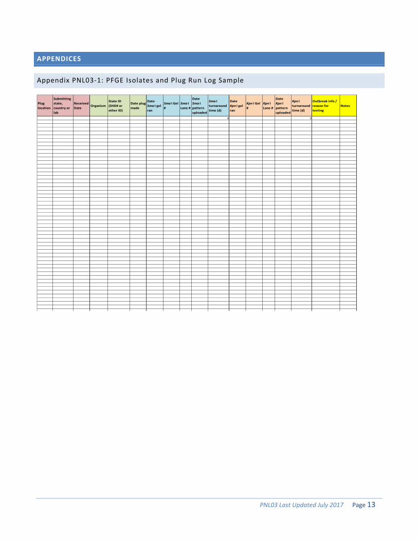
Appendix PNL03-1: PFGE Isolates and Plug Run Log Sample
Pluglocation
Submittingstate, country orlab
Received Date Organism
State ID (SHD# or other ID)
Date plugmade
Date Sma I gel ran
Sma I Gel #
Sma I Lane #
Date Sma I pattern uploaded
Sma I turnaroundtime (d)
Date Kpn I gel ran
Kpn I Gel #
Kpn I Lane #
Date Kpn I pattern uploaded
Kpn I turnaroundtime (d)
Outbreak info /reason fortesting
Notes
0 0
PNL03 Last Updated July 2017 Page 14
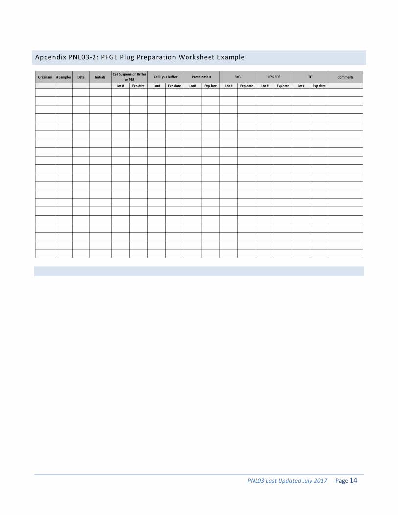
Appendix PNL03-2: PFGE Plug Preparation Worksheet Example
Organism # Samples Date Initials Comments
Lot # Exp date Lot# Exp date Lot# Exp date Lot # Exp date Lot # Exp date Lot # Exp date
TESKG 10% SDSCell Suspension Buffer
or PBSCell Lysis Buffer Proteinase K
PNL03 Last Updated July 2017 Page 15
Appendix PNL03-3: PFGE Enzyme Master Mix and Gel Setup Worksheet Example
Purpose:Gel #: Isolates Received from:Date Gel Run:
Set Temp. Actual Temp.Start time End timeXba I Digestion 37oCSma I Digestion 25oC
Volt. Grad. 6V/cmIncl. Angle 120° 1X 2 1X 2Ramping Linear Switch times Water 180 360 Water 174 348Low MW 50 kb 6.75 s 10X Buffer 20 40 10X Buffer H 20 40High MW 400 kb 35.38 s Total 200 400 BSA (20mg/ml) 1 2Run Time Enzyme (10U/µl) 5 10Initial mA Total 200 400Mapper 1X 2Delayed start? Water 180 360
10X Buffer 20 40 1X 2Total 200 400 Water 178 356
10X Buffer A 20 40Exp date BSA (20mg/ml) 1 2
Enzyme (40U/µl) 1 2Total 200 400
10X TBE BufferGelRed/Ethidium Bromide
Well Buffer Enzyme123456789
101112131415
Plugs made (initial, date)Isolate Comments / State ID / Serotype