1 Standard preanalytical coding for biospecimens: Review and implementation of the Sample PREanalytical Code (SPREC) Sabine Lehmann 1 , Fiorella Guadagni 2 , Helen Moore 3 , Garry Ashton 4 , Michael Barnes 5 , Erica Benson 6 , Judith Clements 7 , Iren Koppandi 8 , Domenico Coppola 9 , Sara Yasemin Demiroglu 10 , Yvonne DeSouza 11 , Annemieke De Wilde 12 , Jacko Duker 13 , James Eliason 14 , Barbara Glazer 15 , Keith Harding 6 , Jae Pil Jeon 16 , Joseph Kessler 17 , Theresa Kokkat 18 , Umberto Nanni 19 , Kathi Shea 20 , Amy Skubitz 21 , Stella Somiari 22 , Gunnel Tybring 23 , Elaine Gunter 24 , Fotini Betsou 1 [International Society for Biological and Environmental Repositories (ISBER) Working Group on Biospecimen Science] 1 Integrated Biobank of Luxembourg, 6 rue Nicolas Ernest Barblé, L-1210 Luxembourg 2 Interinstitutional Multidisciplinary Biobank (BioBIM), Department of Laboratory Medicine and Advanced Biotechnologies, IRCCS San Raffaele Pisana, Via della Pisana 235, 00163 Rome, Italy 3 OBBR, NCI, 11400 Rockville Pike, Suite 700, Bethesda, MD 20892-2590 4 Manchester Cancer Research Centre Biobank, Paterson Institute for Cancer Research, WilmslowRoad , Manchester M20 4BX, UK 5 Cincinnati Children's Hospital Medical Center, 3333 Burnet Avenue, Mail Location E4010, Cincinnati, OH 45229 6 Damar Research Scientists, Damar, Drum Road, Cuparmuir, Cupar, Fife, KY15 5RJ, Scotland UK 7 Australian Prostate Institute of Health and Biomedical Innovation, Queensland University of Technology, 60 Musk Avenue Kelvin Grove QLD 4059, Australia 8 Cellular Technology Limited, 20521 Chagrin Boulevard, Shaker Heights, OH 44122-5350 9 Moffitt Cancer Center, 12902 Magnolia Drive, Tampa, FL 33612 10 University Medical Center Göttingen, Dept of Medical Informatics, Robert-Koch-Strasse 40, 37075 Göttingen, Germany 11 University of California, San Francisco, UCSF AIDS Specimen Bank, 513 Parnassus Ave., San Francisco, CA 94143-0422 12 UZA, Wilrijkstraat 10, B-2650 Edegem, Belgium 13 UMCG Lifelines, Cohort Study, Postbus 30.001, 9700RB Groningen, The Netherlands 14 Great Lakes Stem Cell Innovation Center, 440 Burroughs, Detroit, MI 48202 15 Quintiles Laboratories, 1600 Terrell Mill Road, Marietta, GA 30067 16 National Biobank of Korea, Korea National Institute of Health, 200 Osongsaengmyung-2-ro, Osong, Chungbuk, Korea, 363-700 17 PPD Vaccines and Biologics Lab, 466 Devon Park Drive, Wayne, PA 19087 18 CHTN, University of Pennsylvania 19 Dipartimento di Ingegneria Informatica, Automatica e Gestionale, Università di Roma “La Sapienza”, Rome, Italy 20 SeraCare Life Sciences, 217 Perry Parkway, Gaithersburg, MD 20877 21 Department of Laboratory Medicine and Pathology, BioNet, Tissue Procurement Facility, University of Minnesota, 420 Delaware St SE, Minneapolis, MN 55455 22 Windber Research Institute, 620 Seventh St, Windber, PA 15963 23 Karolinska Institutet Biobank, BBMRI.se Nobels väg 12A, SE-17177 Stockholm, Sweden 24 Specimen Solutions LLC, 3939 LaVista Road, Tucker, GA 30084 Running title: SPREC: Sample PREanalytical Code version 2.0
Transcript
1
Standard preanalytical coding for biospecimens:
Review and implementation of the Sample PREanalytical Code
(SPREC)
Sabine Lehmann1, Fiorella Guadagni2, Helen Moore3, Garry Ashton4, Michael Barnes5, Erica Benson6, Judith Clements7, Iren Koppandi8, Domenico Coppola9, Sara Yasemin Demiroglu10, Yvonne DeSouza11, Annemieke De Wilde12, Jacko Duker13, James Eliason14, Barbara Glazer15, Keith Harding6, Jae Pil Jeon16, Joseph Kessler17, Theresa Kokkat18, Umberto Nanni19, Kathi Shea20, Amy Skubitz21, Stella Somiari22, Gunnel Tybring23, Elaine Gunter24, Fotini Betsou1 [International Society for Biological and Environmental Repositories (ISBER) Working Group on Biospecimen Science] 1Integrated Biobank of Luxembourg, 6 rue Nicolas Ernest Barblé, L-1210 Luxembourg
2Interinstitutional Multidisciplinary Biobank (BioBIM), Department of Laboratory Medicine and Advanced Biotechnologies, IRCCS San Raffaele Pisana, Via della Pisana 235, 00163 Rome, Italy 3OBBR, NCI, 11400 Rockville Pike, Suite 700, Bethesda, MD 20892-2590
4Manchester Cancer Research Centre Biobank, Paterson Institute for Cancer Research, WilmslowRoad , Manchester M20 4BX, UK 5Cincinnati Children's Hospital Medical Center, 3333 Burnet Avenue, Mail Location E4010, Cincinnati, OH
45229 6Damar Research Scientists, Damar, Drum Road, Cuparmuir, Cupar, Fife, KY15 5RJ, Scotland UK
7Australian Prostate Institute of Health and Biomedical Innovation, Queensland University of Technology,
60 Musk Avenue Kelvin Grove QLD 4059, Australia 8Cellular Technology Limited, 20521 Chagrin Boulevard, Shaker Heights, OH 44122-5350
9Moffitt Cancer Center, 12902 Magnolia Drive, Tampa, FL 33612
10University Medical Center Göttingen, Dept of Medical Informatics, Robert-Koch-Strasse 40, 37075
Göttingen, Germany 11
University of California, San Francisco, UCSF AIDS Specimen Bank, 513 Parnassus Ave., San Francisco, CA 94143-0422 12
Great Lakes Stem Cell Innovation Center, 440 Burroughs, Detroit, MI 48202 15
Quintiles Laboratories, 1600 Terrell Mill Road, Marietta, GA 30067 16
National Biobank of Korea, Korea National Institute of Health, 200 Osongsaengmyung-2-ro, Osong,
Chungbuk, Korea, 363-700 17
PPD Vaccines and Biologics Lab, 466 Devon Park Drive, Wayne, PA 19087 18
CHTN, University of Pennsylvania 19
Dipartimento di Ingegneria Informatica, Automatica e Gestionale, Università di Roma “La Sapienza”, Rome, Italy 20
SeraCare Life Sciences, 217 Perry Parkway, Gaithersburg, MD 20877 21
Department of Laboratory Medicine and Pathology, BioNet, Tissue Procurement Facility, University of Minnesota, 420 Delaware St SE, Minneapolis, MN 55455 22
Windber Research Institute, 620 Seventh St, Windber, PA 15963 23
Karolinska Institutet Biobank, BBMRI.se Nobels väg 12A, SE-17177 Stockholm, Sweden 24
Specimen Solutions LLC, 3939 LaVista Road, Tucker, GA 30084
Running title: SPREC: Sample PREanalytical Code version 2.0
2
Abstract
The first version of the Standard PREanalytical Code (SPREC) was developed in 2009
by the International Society for Biological and Environmental Repositories (ISBER)
Biospecimen Science Working Group to facilitate documentation and communication of
the most important preanalytical quality parameters of different types of biospecimens
used for research. This same Working Group has now updated the SPREC to version
2.0, presented here, so that it contains more options to allow for recent technological
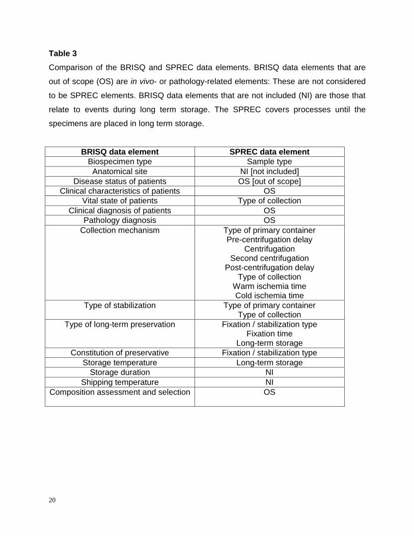
developments. Existing elements have been fine tuned. An interface to the Biospecimen
Reporting for Improved Study Quality (BRISQ) has been defined, and informatics
solutions for SPREC implementation have been developed. A glossary with SPREC-
related definitions has also been added.
3
Introduction
The SPREC (Standard PREanalytical Code) was developed in 2009 to provide a
comprehensive and easy-to-implement tool to document the in vitro preanalytical
(collection, processing and storage) details (1) of biospecimens. The objective of the
SPREC is to facilitate annotation of biospecimens with preanalytical factors that fulfill
two criteria: (a) their variation is known or highly suspected to impact the results of
downstream analyses, and (b) they are within the control of the biobank and thus can
be anticipated and standardized in standard operating procedures (SOPs). The original
SPREC is applicable to animal biospecimens, and more specifically to mammals.
SPREC development in botanical collections and storage has also been explored (2).
Since then and as originally anticipated, new technologies have been developed for
specimen collection (e.g. new anticoagulants), processing (e.g. new tissue stabilization
methods) and storage (e.g. new dry, room temperature storage media) necessitating an
update of the original codes. Furthermore, with the implementation of SPREC version
1.0 in different biobanks, feedback has been received and suggestions on possible
improvements considered. We are aware of at least thirteen biobanks or biobank
networks in the US, Europe, Korea and Australia (3) and of at least three commercial
biobank LIMS who have already implemented the SPREC (personal communications).
The SPREC has been incorporated in the “Minimum data set for sharing biobank
samples, information, and data” (MIABIS), developed by the Biobanking and
Biomolecular Resources Research Infrastructure Sweden (BBMRI.se) (4) and is also
being implemented as a national standard both for healthcare and research in
BBMRI.se (personal communication). An application programming interface (API)
module has been developed which allows SPREC to be defined from sample collection
and processing protocols, submitted to the Molecular Methods database
(www.molmeth.org) which is supported by BBMRI (5). Furthermore, SPREC is
mentioned in the College of American Pathologists (CAP) biobank accreditation
checklist, which is being tested in pilot audits (personal communication). Currently, the
application of SPREC to stem cell biobanks is being evaluated by Demiroglu and
colleagues at the University Medical Center in Göttingen, Germany (manuscript in
4
preparation). Thus, this new version 2.0 has been developed, and is expected to be
more comprehensive and easier to implement.
SPREC version 2.0
In updating SPREC 1.0, and for the sake of continuity, no significant changes to codes
were made, while new options have been added as detailed in Tables 1 and 2. The new
options in SPREC 2.0 include the following:
For fluid samples:
Sample types: Dried whole blood (e.g. Guthrie cards) and red blood cell fraction were
added. A distinction was made between random, timed and first morning urine
specimen.
Types of primary containers: Sodium-heparin collection tube, EDTA and heparin
collection tubes with a gel separation plug and cell preparation tubes (CPT®) were
added. A distinction was made between serum separation tubes with silica clot activator
and tubes without additive. No distinction is now made between evacuated
(Vacutainer®-type) and non-evacuated (Monovette®-type) collection tubes.
Polyethylene containers (often used for urine collection) and additives (often used in
urine collection) were added.
Centrifugation conditions: Initial centrifugation conditions of 30 min at room temperature
(RT) with a relative G-force <1000g and without braking used (most appropriate for
density centrifugation and isolation of mononuclear cells) were added. For all the
centrifugation options, a 10-min centrifugation time was replaced by 10-15 min to
provide more flexibility. A “not applicable” option in the post-centrifugation element was
added.
Storage conditions. Long-term storage options were included for liquid nitrogen
preceded by temporary -80 °C storage (in either cryotubes or straws). The temperature
options for paraffin blocs were revised from “RT” only to “RT or 2 to 10°C”. Other new
storage options include a dry technology medium at RT, bag storage, original primary
container, and tubes from 40 to 500 L sizes. Storage at refrigerated temperatures has
been harmonized throughout the SPREC and corresponds to 2°C – 10°C.
For solid samples,
5
Sample types: Placenta was added as a new sample type. The abbreviation LCM
appeared twice in the sample type element in version 1.0. In version 2.0, LCM
corresponds to cells from laser capture microdissected tissue, while TCM corresponds
to cells from mechanically disrupted tissue.
For biopsies and surgical excisions, options for collection (and subsequent transport) in
either saline, culture media or low-temperature transport media (e.g. AQIX,
Hyperthermosol, Unisol, Thermo-ROS) or in vacuum containers were added.
“Fixation” type became “fixation / stabilization” type and now includes heat stabilization,
PAXgene® tissue fixation and Allprotect® tissue fixation / stabilization options.
A “not applicable” option was added in the Fixation time element.
Support tools for implementation of SPREC version 2.0
The integration of SPREC into biobank databases generally requires the inclusion of
SPREC drop-lists among the “tables” in the software. This is relatively easy with
customized software, but the supplier would need to make a customized development
for off-the-shelf software. Two tools have been developed in order to facilitate SPREC
implementation and interfacing with either customized or off-the-shelf biobank software.
A default SPREC can be set per sample type according to the SOPs of the collection
project. Thus, only deviations from the SOP SPREC need to be documented. These
can be highlighted, making any deviation from pre-defined sample quality or any non-
conforming in processing directly visible. However, active generation of the SPREC for
each individual sample is to be preferred as it enforces traceability.
Although the use of the SPREC requires an additional initial investment of time, it is
expected that this will be more than repaid by the ease and accuracy of subsequent
sample search and selection.
SPRECware
The first tool [SPRECware] requires entry of each samples’ SPREC elements.
SPRECware (6) is an IT architecture, i.e., a collection of software and communication
tools which have been developed in order to foster the adoption of SPREC by biobanks
and biolaboratories. Basic information concerning preanalytical processing of a given
sample is selected from drop-down menus. The resulting SPREC is generated together
6
with the corresponding barcode, and all the preanalytical data with the SPREC are
stored in a local database.
One component, SPRECbase has been recently released and is freely downloadable
from http://www.sprecware.org. This software supports both coding / printing barcode
labels and decoding in text of a sample's SPREC, then storing all the preanalytical data
in a local database. These barcodes simplify storing and exchanging corresponding
information about the samples.
In a working environment, SPRECware is intended to be connected to the
laboratory/biobank information system (LIS/BIS). The required data can then be loaded
from the originating biobank’s LIS/BIS database and translated into the SPREC.
Samples are thus labeled with both the human-readable and barcoded format. The
barcode provides the receiver organization with the data documenting the specimen
and can be stored in the LIS/BIS. This approach provides fully automated data transfer
without manual writing or reading when the organizations exchanging specimens both
use SPREC. In the case that the receiving organization has not adopted the SPREC, it
is still possible to read the SPREC (e.g. by a barcode scanner) and download the
explicit preanalytical data by means of an internet connection.
Since the SPREC does not encapsulate any personal data, it can be used for searches
on sample availability. This possibility of a preliminary check would allow a researcher
to select material with the most appropriate preanalytical characteristics for a planned
experiment (6). Even in the case of derivatives (e.g. DNA, RNA), the SPREC
information on the collected samples, from which derivatives were produced, is highly
valuable.
SPRECalc
Unlike the SPRECware which requires entry of the SPREC options (A, B, X…), the
second tool [SPRECalc] requires entry of the corresponding preanalytical variables
themselves (temperature, time…); this tool then performs automatic calculation of the
time-associated elements and generation of SPREC.
The Excel-based SPRECalc automatically calculates SPREC time-associated elements
for solid and fluid samples, using the data logic depicted in Figures 1 and 2. Data entry
is supported by pull-down lists (blue-colored fields in the Excel tool) to minimize
7
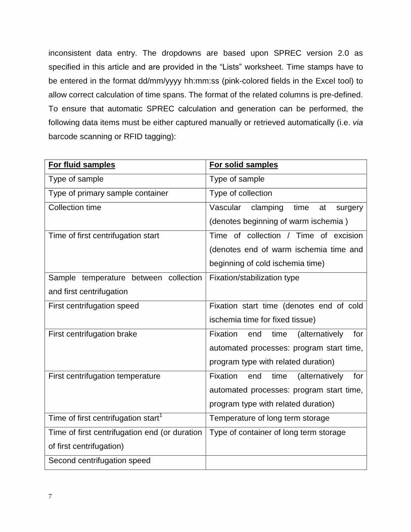
inconsistent data entry. The dropdowns are based upon SPREC version 2.0 as
specified in this article and are provided in the “Lists” worksheet. Time stamps have to
be entered in the format dd/mm/yyyy hh:mm:ss (pink-colored fields in the Excel tool) to
allow correct calculation of time spans. The format of the related columns is pre-defined.
To ensure that automatic SPREC calculation and generation can be performed, the
following data items must be either captured manually or retrieved automatically (i.e. via
barcode scanning or RFID tagging):
For fluid samples For solid samples
Type of sample Type of sample
Type of primary sample container Type of collection
Collection time Vascular clamping time at surgery
(denotes beginning of warm ischemia )
Time of first centrifugation start Time of collection / Time of excision
(denotes end of warm ischemia time and
beginning of cold ischemia time)
Sample temperature between collection
and first centrifugation
Fixation/stabilization type
First centrifugation speed Fixation start time (denotes end of cold
ischemia time for fixed tissue)
First centrifugation brake Fixation end time (alternatively for
automated processes: program start time,
program type with related duration)
First centrifugation temperature Fixation end time (alternatively for
automated processes: program start time,
program type with related duration)
Time of first centrifugation start1 Temperature of long term storage
Time of first centrifugation end (or duration
of first centrifugation)
Type of container of long term storage
Second centrifugation speed
8
Second centrifugation brake
Second centrifugation temperature
Time of second centrifugation start
Time of second centrifugation end2 (or
duration of second centrifugation)
Time of putting into temporary storage
Temperature of temporary storage
Time of putting into long term storage
Sample Temperature between end of last
centrifugation and putting into long term
storage
Temperature of long term storage
Type of container of long term storage
1time of freezing in the case of PAXgene®
2time of last washing centrifugation in the case of viable cells
Grey fields contain automatic calculations / translations, displaying either the time
period (e.g. warm/cold ischemia time, fixation time), or “help” columns for identification
of the applicable SPREC.
For solid samples, the fixation time can be determined in two different ways. Option 1 is
designed for use with automated fixation systems (fixation start time, program type, and
program start time need to be recorded). Option 2 is targeted for non-automated fixation
steps (fixation start and end time need to be entered).
With respect to option 1, the “Fixation Programs” worksheet allows the user to predefine
fixation programs and their respective duration. Selection of the applicable program
during data entry automatically uses the related duration to calculate the correct fixation
time, provided fixation start time and program start time have been entered as well. For
each sample, SPREC is then automatically displayed following the coding rules
specified in this article. All of these fields are protected against changes.
9
The tool also contains the following quality-related features. The data entry worksheet
provides its own “quality control” features, since Excel-specific error messages in the
calculated columns (e.g. ######, or #REF) indicate data inconsistency, e.g. the fixation
start time entered is before the collection time. Systematic investigation of such errors
supports quality checks to improve data quality and consistency.
Default SPRECs can be defined for each collection according to established SOPs for
sample collection. Non-conformities can be detected automatically by the system and
flagged in dedicated columns, with a flexible way of setting target parameters (e.g.
fixation time shall be <= 24h). Alternatively the same non-conformities can be
specifically custom-programmed directly in the final SPREC columns, using the
“conditional formatting” functionalities of Excel. These non-conformities should then be
investigated as required by the organization’s Quality Management System (QMS) as
part of biobank accreditation.
In addition to columns with a “Z” option (“Other”), text columns are provided, allowing
recording of more detailed information in cases where other specifications than those
listed in the SPREC were applied. If a value is unknown, then “unknown” should be
entered.
The “Statistics” worksheet displays summary statistics about the sample collection (e.g.
number and percentage of samples per code type, average, minimum, maximum and
median for selected time spans) related to selected SPREC items (e.g. warm/cold
ischemia time and fixation time for solid samples). This summary sheet also allows the
continuous automated calculation of key performance indicators (KPI), as required by
quality norms. An example is shown for the percentage of solid samples in the collection
with warm ischemia times below 30 minutes (Figure 3). For example, a report can be
generated stating the percentage of solid samples in the collection with warm ischemia
times below 30 minutes. Reports of variables of interest can be programmed as
required by the user. The Excel tool is available on
http://www.isber.org/SPRECtools.cfm
Although, SPRECalc is a standalone tool, the final SPREC can be imported into a
biobank database at regular intervals. This solution provides a simple way of integrating