517
Stanniocalcin 2 expression is regul
ated by hormone signallingand negatively affects breast cancer cell viability in vitroSanda Raulic1,3, Yudith Ramos-Valdes1 and Gabriel E DiMattia1,2,3
1London Regional Cancer Program, 790 Commissioners Road, Room A4-921, London, Ontario, N6A 4L6 Canada
Departments of 2Oncology and 3Biochemistry, The University of Western Ontario, London, Ontario, Canada
(Correspondence should be addressed to G E DiMattia; Email: [email protected])
Abstract
Stanniocalcin 1 (STC1) and STC2 are secreted, homodimeric
glycoproteins that share 30% amino acid sequence identity.
Breast tumour gene profiling studies have demonstrated
significantly upregulated STC2 expression in hormone-
responsive positive breast tumours; therefore, the purpose of
this study was to investigate STC2 hormonal regulation and
function in breast cancer cells. Here we report that STC2 is
expressed in a number of human breast cancer cell lines,
regardless of their oestrogen (E2) and progesterone (P4)
receptor status, and its expression is readily detectable in
human and mouse mammary gland tumours. Besides E2,
retinoic acid (RA) and P4 play an important role in the
regulation of STC2 expression, not only in MCF-7 but also
in other breast cancer and non-breast cell lines. The
expression of the related hormone, STC1, is not affected by
Journal of Endocrinology (2008) 197, 517–5290022–0795/08/0197–517 q 2008 Society for Endocrinology Printed in Great
the above hormones in breast and endometrial cancer cell
lines implying a fundamental difference in regulation in
cancer cell lines. The induction of STC2 expression by E2 and
RA occurs at the transcriptional level but through inter-
mediary transcription factors. The STC2 proximal promoter
region is not responsible for hormonal induction, but exhibits
a high basal transcriptional activity. Constitutive STC2
expression in human breast cancer cell lines resulted in
significant impairment of cell growth, migration and cell
viability after serum withdrawal. In conclusion, STC2 is a
downstream target of E2, P4 and RA signalling pathways. In
hormone receptor negative cell lines it can function in a
paracrine/autocrine fashion to reduce cell proliferation.
Journal of Endocrinology (2008) 197, 517–529
Introduction
Stanniocalcin 1 (STC1) and STC2 constitute a small family of
secreted, homodimeric glycoproteins that have been impli-
cated in the physiology of phosphate regulation (Wagner et al.
1997, Madsen et al. 1998, Yoshiko et al. 2007), metabolism
(Wagner & DiMattia 2006), reproduction (Deol et al. 2000,
Varghese et al. 2002, Luo et al. 2004, 2005), stress response
(Sheikh-Hamad et al. 2000, Anderson 2003, Ito et al. 2004)
and development (Stasko & Wagner 2001, Varghese et al.
2002, Gagliardi et al. 2005). Moreover, the expression of
STCs has been recognised as notably altered in a variety of
cancers suggesting that they play a role in tumorigenesis
(Chang et al. 2003). This is based on gene profiling studies
where STC2 expression is significantly elevated in a specific
subset of breast tumours (Gruvberger et al. 2001, Wilson et al.
2002, Kun et al. 2003, Amatschek et al. 2004, Yu et al. 2004,
Zucchi et al. 2004, Esseghir et al. 2006, 2007) or elevated
upon oestrogen (E2) treatment of human breast cancer cell
lines (Charpentier et al. 2000, Bouras et al. 2002). However,
whether STC2 is responsive to other hormones that can
regulate growth and what effect it might have on breast cancer
cells have not been investigated.
It has been well established that oestrogens can regulate
processes critical to breast tumorigenesis including cell
proliferation and migration (Katzenellenbogen & Frasor
2004, Yager & Davidson 2006, Jordan 2007); therefore,
those proteins regulated by E2 may play a role in the aetiology
of breast cancer. The clinical significance of breast tumour
STC2 expression was described by Iwao et al. (2002); they
reported that the expression of 21 genes was prognostic for
breast cancer and that the low expression of these genes,
including STC2, was associated with poor prognosis.
Yamamura et al. (2004) reported that high STC2 mRNA
steady-state levels were significantly associated with good
prognosis in oestrogen (ER)- and progesterone receptor
(PR)-positive breast cancer patients. More recently, a
tissue microarray screen found that STC2 expression was
highly predictive for longer disease-free survival (Esseghir
et al. 2007). Taken together, these studies open a new niche
for utilising STC2 as a potentially useful breast cancer
molecular marker.
The purpose of this study was twofold. Our first objective
was to expand on the mechanism of STC2 regulation by E2and determine whether it is regulated by other growth
regulatory hormones. We also asked whether STC2
DOI: 10.1677/JOE-08-0043Britain Online version via http://www.endocrinology-journals.org
Downloaded from Bioscientifica.com at 04/02/2022 07:10:53AMvia free access
S RAULIC and others . STC2 in breast cancer cells518
regulation by specific hormones was breast cancer-specific
and whether the upregulation of the STC2 expression also
extended to murine models of mammary tumorigenesis.
Secondly, we addressed the function of STC2 in human breast
cancer cells by examining the phenotypic effects of de novo
production in two human breast cancer cell lines. Presently,
little is known regarding the function of STC2. A persistent
exposure to human STC2 in transgenic mice results in severe
growth retardation and female reproductive deficits (Gagliardi
et al. 2005). Moreover, the exogenous expression of STC2
can produce a cytoprotective effect (Ito et al. 2004). These
data suggest widespread effects of STC2 and that it can have
autocrine and paracrine effects in breast cancer (Gagliardi et al.
2005). Here, we show that STC2 production can be regulated
by hormones other than E2 and that STC1 expression is
unaffected. We also demonstrate that Stc2 expression is
strongly upregulated in mouse mammary tumours.
To begin to address the function of STC2 in breast cancer
cells, we chose the approach described by Ito et al. (2004) and
generated STC2-stable transfectants in human breast cancer
cell lines. We found that STC2 expression can result in
significantly reduced cell growth, motility and viability
implying that the loss of STC2 expression in breast tumours
contributes to a more aggressive phenotype.
Materials and Methods
Cell culture and hormonal treatment
The human breast cancer cell lines T-47D, MCF-7, HCC
1937, HCC 1500, MDA-MB-435, MDA-MB-231 and
MDA-MB-468 (obtained from ATCC, Manassas, VA, USA)
and the endometrial adenocarcinoma (Ishikawa) cell line were
cultured in Dulbecco’s modified Eagle’s medium (DMEM)
(Wisent, St. Bruno, Canada) with 10% fetal bovine serum
(FBS) (Wisent) and 50 mg/ml penicillin–streptomycin (Invi-
trogen). The cells were seeded atw60% density and allowed to
attach overnight. Before hormonal treatment, they were
washed twice with 1! PBS and cultured in an oestrogen-
depletedmedium: phenol red-freeDMEM-F12mediumwith
10% charcoal-stripped FBS (Wisent) and 50 mg/ml penicillin–
streptomycin, for 24 h. For gene regulation studies in MCF-7
cells, each hormonewas applied at 10K6Mfinal concentration
for 0, 1, 3 and 24 h, and Ishikawa and T-47D cell lines were
treated for 0, 1, 3, 8, 24, 48 and 72 h.
Human and mouse tissues
Human breast tumour tissue samples were obtained from the
London Health Sciences Tumour Bank (London, ON,
Canada). Normal breast tissue was obtained by reduction
mammoplasty from the Cooperative Human Tissue Network
(Midwestern Division, The Ohio State University). Trans-
genic MMTV/activated Erb-2 (NDL) (Siegel et al. 1999) and
MMTV/PyV MT (Guy et al. 1992) mouse mammary gland
Journal of Endocrinology (2008) 197, 517–529
tumour tissues were generously provided by Dr William
Muller (McGill University, Montreal).
RNA extraction and northern blot analysis
Total RNA was isolated using TRIzol (Invitrogen). For
northern blot experiments, 30–50 mg total RNA per lane was
used, as described previously (Gagliardi et al. 2005). A Pst
I/Sty I human STC2 cDNA fragment (617 bp of coding
sequence) and Pst I/Sph I human STC1 cDNA fragment
(417 bp of coding region) were used as probes. Murine Stc2
northerns were performed with an 896 bp coding sequence
Stc2 cDNA fragment. The whey acid protein cDNA probe
was generated from the mouse IMAGE clone 5249612. To
normalise for RNA loading or to demonstrate RNA
integrity, blots were hybridised with an 18S rDNA fragment
or with a human acidic ribosomal phosphoprotein PO (36B4)
cDNA fragment. The resultant hybridisation signal was
quantified using a PhosphorImager and ImageQuant software
(GE Healthcare, Baie d’Urfe, Canada).
Western blot
Conditioned cell culturemedia fromhormone-treatedT-47D,
Ishikawa and the pcDNA3/STC2 stably transfected
MDA-MB-231 and MDA-MB-435 cell lines were collected
at the indicated time points and concentrated sixfold using
CentriconYM-10 centrifugal filters according to the supplier’s
instructions (Fisher Scientific, Ottawa, Canada). The total
protein concentrationwas determined using theBradford assay
(Bradford 1976). About 100 mg total protein were loaded
per lane for 12% PAGE and transferred to the polyvinylidene
fluoride (PVDF) membrane (Roche). The blots were reacted
with polyclonal STC2 antisera (1:5000) raised in rabbits as
described previously (Gagliardi et al. 2005). Immunoreactive
bands were detected using the ECL Plus western blotting
detection system (GE Healthcare) with subsequent exposure
to Kodak Bio Max XAR film.
Generation of the pGL2-STC2 luciferase reporter gene plasmidconstructs
To generate pGL2-STC2-luciferase promoter constructs, an
STC2 135A09 BAC clone from RPC11H library was used to
amplify a 1.382 kb STC2 gene promoter fragment. The PCR
product was cloned into the pCR-BluntII-TOPO plasmid
according to the supplier’s instructions (Invitrogen). A 916 bp
BamH I fragment of the STC2 promoter was ligated to the
Bgl II site of pGL2-Basic (Promega) in both orientations. The
5 0 truncations of the luciferase reporter were produced using
convenient restriction sites.
Transient transfection and hormone treatment
Human embryonic kidney, endometrial adenocarcinoma
(Ishikawa) and breast cancer T-47D cells were cultured as
www.endocrinology-journals.org
Downloaded from Bioscientifica.com at 04/02/2022 07:10:53AMvia free access
STC2 in breast cancer cells . S RAULIC and others 519
described previously. The following day, the cells were
transfected with 1 mg pGL2-STC2-667 plasmid and molar
equivalents of pGL2-STC2-337, pGL2-STC2-172, pGL2-
STC2-112, pGL2 basic and pBKSII. Co-transfection with a
molar equivalent of pCMV-lacZ was done as a measure of
transfection efficiency. Transfection was performed using
ExGen according to the supplier’s instructions (MBI
Fermentas, Burlington, Canada) and the luciferase assay
performed 24 h later. The hormonal responsivity of pGL2-
STC2-667 was tested by transfection of T-47D cells and
compared with molar equivalents of the negative control,
pGL2-basic and pERE-Luc as an oestrogen response plasmid
(generously provided by Dr J Torchia, London Regional
Cancer Program). One day after transfection, cells were
cultured in an oestrogen-depleted medium with E2 (10K6),
RA (10K6) or an equivalent volume of solvent, for an
additional 24 h before measuring the luciferase activity.
b-Galactosidase assays were performed on transfected cells
to normalise values for luciferase assays (Gaasenbeek et al.
1999).
Stable transfection of the MDA-MB-231 and MDA-MB-435cell lines
The human breast cancer MDA-MB-231 and MDA-MB-435
cell lines were cultured and transfected with pcDNA3 or
pcDNA3/STC2 using ExGen transfection agent as described
previously. After transfection, they were maintained in serum
and antibiotic free-Opti-MEM medium for 24 h which was
subsequently replaced with complete media. The cells were
allowed to recover for 24 h after transfection and a selection
pressure was applied using 800 mg/ml Geneticin (Sigma–
Aldrich) for 2 weeks.
Chemical treatments and quantitative real-time RT-PCR(qPCR) analysis
MCF-7 or T-47D cells were cultured as described above. For
antioestrogen ICI 182 780 (ICI) (Sigma–Aldrich) treatment,
T-47D cells were seeded in six-well cluster plates at w80%
density and allowed to attach overnight. The cells were then
washed twice with 1! PBS and incubated for 48 h in an
oestrogen-depleted medium. The cells were treated overnight
(18 h) with E2 (10K8) M (Sigma–Aldrich) or following a 2 h
pre-treatment with ICI. Treatments with ICI alone or solvent
were used as controls. The level of STC2 mRNA was
determined by qPCR using 1.5 mg total RNA and the STC2
primers described below. For actinomycin D (5 mg/ml) or
cycloheximide (30 mg/ml) treatments, the cells were seeded in
60 mmdishes atw60% density and allowed to attach overnight.
Subsequently, the cells were washed twice with 1! PBS and
cultured for 24 h in an oestrogen-depleted medium. The cells
were pretreated for 1 h with actinomycin D (5 mg/ml) or
cycloheximide (30 mg/ml) and after addingE2 orRA(10K6)M,
harvested at 0 and 3 h. The treatment with actinomycin D or
cycloheximide alonewas used as a control for the effects of E2 or
www.endocrinology-journals.org
RA. Total RNAwas isolated and 1 mg reverse-transcribed usingSuperscript III RNase H reverse transcriptase kit (Invitrogen).
qPCR was carried out using the Stratagene SYBER Green
PCR Master Mix kit according to the manufacturer’s
instructions and the MxPro-3000 real-time PCR detection
system. The following sets of primers were used: human STC2
sense (5 0-GTTCATGACCCTGGCTTTGGGTT-3 0) and
antisense (5 0-AAACAGTGCTGGATCTCCGCTGTA-3 0);
human b-actin sense (50-ACCAACTGGGACGACATGGA-
GAA-3 0) and antisense (5 0-TAGCACAGCCTGGATAG-
CAACGTA-30).
Assessment of cell proliferation using the alamarBlue assayand cell counting
For the alamarBlue assay, MDA-MB-231 and MDA-MB-435
cells (stably transfected with pcDNA3 or pcDNA3/STC2)
were seeded in 24-well plates at a density of 2!104 cells per
well in quintuplicate, and allowed to attach overnight.
Subsequently, the cells were incubated with 1:20 dilution of
alamarBlue (Biosource, Camarillo, CA, USA) stock solution
in culture media and placed in a humidified incubator for 4 h.
The fluorescence measurement was obtained at 580 nm in a
Wallac Victor2-1420 microplate reader (Perkin–Elmer,
Woodbridge, Canada) after 1 and 7 days of culture.
For cell counting, MDA-MB-231 cell lines expressing
STC2 (lines 3 and 5) or control vector (line 6) were seeded in
24-well plates at a density of 5!103 cells per well in
quadruplicate. The cells were trypsinised, stained with Trypan
blue and counted at day 3 and 7 using a haemocytometer.
Cell survival, colony-forming and scratch-wound motility assays
The breast cancer MDA-MB-231 cell lines expressing STC2
(lines 3 and 5) or control vector (line 6) were plated in 24-well
plates at a density of 1.6!105 cells per well in triplicate. The
next day, designated as day 0, cell counts were determined to
provide the starting cell number, which represented the 100%
value for cell number. After washing with 1! PBS, the cells
were cultured in serum-free DMEM and alamarBlue assay
performed for 3, 5 and 7 days of culture. For colony-forming
assay, the cells were seeded in 60 or 100 mm plates at a density
of 150 or 250 cells per plate respectively in quadruplicate.
After 6 days in culture, colonies were fixed and stained using
the Hema 3 staining system (Fisher Scientific) according to
the supplier’s instructions. The colonies were counted
manually using the Olympus SZ-40 microscope (Olympus,
Canada). The scratch-wound assays were done in six-well
plates at confluence. Twenty-four hours later, two scratches
were made in the monolayer of each well using a 1000 mlplastic pipette tip. Four images were captured along each
scratch using the Olympus AX-70 microscope and Olympus
DP71 camera at 0, 24 and 48 h after the wound infliction.
The cell migration was assessed based on the area covered by
cells between 0 and 24 h using ImageJ software (http://www.
uhnresearch.ca/facilities/wcif/fdownload.html).
Journal of Endocrinology (2008) 197, 517–529
Downloaded from Bioscientifica.com at 04/02/2022 07:10:53AMvia free access
S RAULIC and others . STC2 in breast cancer cells520
Statistical analysis
Experimental data were analysed with the unpaired Student’s
t-test and by one-way ANOVA with the Dunnett’s post-test
(when indicated) using PRISM 3.0a software (GraphPad
Software, San Diego, CA, USA). Statistically significant
differences were assumed if P!0.05 for all experiments.
Results
STC2 expression in various human breast cancer cell linesand breast tumour tissue
Our screen of w40 different human cancer cell lines for
STC1 and STC2 expression, at the protein or mRNA level,
showed that STC2 expression was highest amongst human
breast cancer cell lines (unpublished results). To expand on
our preliminary observations and determine whether ER
signalling was essential for STC2 gene expression in human
breast cancer cells, we examined STC2 expression in a variety
of human breast cancer cell lines. Northern blot analysis
detected STC2 mRNAs in the form of w4, w3.6 and
w2 kb transcripts in all tested cell lines. Additionally, an
w1.8 kb transcript was detected in MCF-7, MDA-MB-468
and HCC 1500 cell lines (Fig. 1A). While others had
previously indicated that the STC2 gene expression in human
breast cancer cell lines was linked to the presence of ER
(Charpentier et al. 2000), we showed that the STC2 gene
expression was easily detectable in cell lines such as HCC
1937, MDA-MB-468 and HCC 1806 which do not contain
ER or PR (Neve et al. 2006) (Fig. 1A). Moreover, we have
detected multiple STC2 mRNA species whereas others have
reported only the presence of thew2 kb transcript in human
breast cancer cell lines (Charpentier et al. 2000).
To determine whether the STC2 mRNA species seen in
human breast cancer cell lines are also present in vivo, we
investigated the STC2 expression in a small number of
normal human breast samples obtained from reduction
mammoplasty and breast tumours. All three STC2 mRNAs
previously detected in human breast cancer cell lines were
present (Fig. 1B), suggesting that the breast cancer cell line
STC2 gene accurately represents the in vivo STC2
transcription unit.
Stc2 expression is activated in mouse mammary gland tumours
As a logical extension of the above studies, we investigated the
expression of Stc2 in the mouse mammary gland. Normal
mouse mammary gland tissue was harvested from post partum
females on different days of lactation and involution.
Northern blot analysis did not detect a Stc2 mRNA signal
in these tissues (Fig. 1C).
To determine whether Stc2 expression is altered in murine
mammary gland tumours relative to its normal counterpart,
we assessed the Stc2 expression in two different types of
Journal of Endocrinology (2008) 197, 517–529
transgenic mouse mammary gland tumours. Northern blot
analysis of transgenic MMTV/PyV MT and MMTV/acti-
vated Erb-2 (NDL) mouse mammary gland tumours revealed
the presence of w4 and w2 kb Stc2 mRNA species in all
tumour samples (Fig. 1C). This is in sharp contrast to the
normal mammary gland where STC2 expression is undetec-
table by northern.
Hormonal regulation of STC2 gene expression in human breastcarcinoma T-47D and MCF-7 cell lines
As mentioned previously, recent gene profiling studies
reported an enhanced STC2 expression, primarily in
MCF-7 cells treated with E2, implying that E2 is the
primary regulator of STC2 expression in breast cancer cells
(Charpentier et al. 2000, Gruvberger et al. 2001, Bouras
et al. 2002, Frasor et al. 2003). To determine whether STC2
expression was linked to other growth-modulating hor-
mones, we investigated the role P4, RA and glucocorticoid,
on STC2 mRNA levels. We chose these hormones because
MCF-7 and T-47D breast cancer cell lines express a
relatively high level of E2, P4, RA and glucocorticoid
receptors (Sutherland et al. 1988, Hall et al. 1990, Roman et
al. 1992, Rishi et al. 1996). As shown in Fig. 2A, the w2
and 4 kb STC2 mRNA species in T-47D cells were
maximally induced by E2 (mean 17.3-fold increase), and
RA (mean 4.0-fold increase) by the 24 h time point
(Fig. 2A and B). An increase in STC2 mRNA levels was
discernable 3 h after hormonal treatment when compared
with vehicle-treated cells. Similar results were obtained
using qPCR analysis (data not shown). Experiments
examining P4 regulation of STC2 mRNA levels using
northern blot showed a low level of induction and were
therefore repeated using qPCR. We observed a modest
twofold increase in T-47D cell STC2 mRNA levels, and
unlike E2 and RA, P4 induction of STC2 transcripts
occurred only after a 24-h treatment (data not shown).
We also analysed STC1 mRNA levels in the same samples
because it is a highly related to STC2 and its function may
overlap with STC2. Moreover, others have shown the
expression of STC1 in human breast tumours and that
STC1 expression is also linked to ER expression in breast
tumours and cells lines (Bouras et al. 2002, McCudden et al.
2004). Interestingly, STC1 basal expression was low and not
changed by any of the hormonal treatments of T-47D cells
(data not shown).
We confirmed that STC2 protein levels, in the conditioned
culture media, also increased with time and correlated with
the increase in STC2 mRNA levels. Western blot analysis
showed STC2 protein accumulation during the late phase of
the time course, reaching a maximum at 72 h for all E2 and
RA treatments (Fig. 2C). Similar experiments were carried
out with MCF-7 cells to determine whether the effects we
observed were cell line-specific. Treatment of MCF-7 cells
with E2 and RA induced mean STC2 mRNA steady-state
levels (mean 12- and 21-fold respectively) (Fig. 2). P4 and
www.endocrinology-journals.org
Downloaded from Bioscientifica.com at 04/02/2022 07:10:53AMvia free access
Figure 1 Northern blot analysis of STC2 expression in human and mouse tissues and cell lines. (A) STC2transcript sizes in human breast cancer cell lines determined by comparison with 18S (1.9 kb) and 28S(5 kb) units of rRNAs. HT1080 cell line RNA was used as a positive control. (B) STC2 mRNA levels innormal human breast tissue and breast tumours. Fifty microgrammes of breast total RNA was loaded perlane and 20 mg T-47D total RNA was loaded in the (C) lane. (C) STC2 mRNA levels in normal mousemammary gland and tumour tissue. Mouse mammary gland RNAs from different days of lactation and post-lactation as indicated by lane labelling in the panel on the left (mouse pituitary GC cell line RNA was thepositive control). The WAP mRNA signal confirmed that mammary glands were from lactating females.STC2 mRNAs of w4 and 2 kb were seen in all transgenic MMTV/activated ErbB-2 (NDL) and MMTV/PyVMT mouse mammary tumours samples. In all the blots, either 36B4 mRNA or 18S rRNA hybridisationdemonstrates RNA integrity and relative lane loading.
STC2 in breast cancer cells . S RAULIC and others 521
dexamethasone treatments did not change MCF-7 cell STC2
expression. Moreover, as we observed with T-47D cells,
STC1 mRNA steady-state levels in MCF-7 cells were not
affected by the above hormones (data not shown).
To determine whether regulation of STC2 expression by
steroid hormones and RA can be extended to other steroid-
responsive cancers, we tested responses in the Ishikawa
www.endocrinology-journals.org
endometrial cancer cell line. Treatment with E2, P4 and RA
resulted in a strong activation of STC2 at the mRNA and
protein level, starting at 24 h and reaching a maximum at 72 h
(data not shown). Similar to human breast cancer cell lines,
these treatments, including dexamethasone, did not induce
STC1 mRNA steady-state levels in the Ishikawa cells (data
not shown).
Journal of Endocrinology (2008) 197, 517–529
Downloaded from Bioscientifica.com at 04/02/2022 07:10:53AMvia free access
Figure 2 Regulation of STC2 gene expression by E2 and RA in theT-47D human breast cancer cell line. (A) Representative autoradio-grams of northern blots showing the effect of E2 and RA, over a 24-htreatment period, on STC2 mRNA levels. STC2 mRNA levels weremaximally induced by the 24-h time point for each treatment.The 18S rRNA signal shows equal RNA loading. (B) Bar graphsdepicting the induction of STC2 mRNA levels by E2 and RA in T47-Dcells. The STC2 mRNA signal intensity was normalised to the 18SrRNA signal obtained from each blot to control for variations in RNAloading. The expression of STC2 in vehicle-treated cells was set to 1for comparison of different blots. Data are indicated as themeanGS.E.M. from three independent experiments. (C) Western blotanalysis of STC2 accumulation in the conditioned culture mediaof hormone-treated cells. Two immunoreactive STC2 bands(arrows) were routinely obtained as described previously(Gagliardi et al. 2005). Blots were stained with Ponceau S to showrelative lane loading.
Figure 3 qPCR analysis of STC2 expression in MCF-7 cells treatedwith inhibitors of transcription or translation in the presence of E2
or RA. (A) Treatment with the type II antioestrogen, ICI, resulted in asignificant reduction in E2-inducedSTC2expression indicating that ERmediates this effect. The induction of STC2 mRNA levels by E2 or RAwas inhibited in the presence of the transcriptional inhibitor,actinomycin D (Act. D), B and C respectively. (D) Treatment with aninhibitor of de novo protein synthesis, cycloheximide (CHX), showeda partial inhibition of E2-induced STC2 mRNA levels. Cycloheximidealone significantly increased STC2 mRNA levels when comparedwithcontrol vehicle-treated (Con.) cells. Bars represent meansGS.E.M. ofthree independent experiments, each done in triplicate and significantdifferences (indicated by asterisks) between groups were determinedby Student’s t-test and one-way ANOVA (P!0.0001).
S RAULIC and others . STC2 in breast cancer cells522
E2 and RA regulation of STC2 gene expression is a secondaryresponse
To further examine the mechanism by which the above
hormones induce STC2 gene expression in human breast
cancer cell lines, we used chemical inhibitors of transcription,
translation and ER signalling. To confirm that E2 induction of
STC2 requires ligand-activated ER we treated cells with the
antioestrogen ICI 182 780 (ICI). This compound is able to
inhibit oestrogen-induced transcriptional activation by comp-
eting with E2 for binding to ER but inhibiting homodimerisa-
tion resulting in the degradation of ER (Dauvois et al. 1992).
The ICI treatment significantly blunted the E2 induction of
T-47D STC2 mRNA levels from 7.5- to a threefold inductionafter an 18-h co-treatment indicating that ligand-bound ER
mediates the E2 induction of STC2 transcript levels (Fig. 3A).
Next, we asked whether STC2 was a primary E2- and
RA-responsive gene in human breast cancer cells by using a
transcriptional inhibitor (actinomycinD) and a protein synthesis
inhibitor (cycloheximide). As shown in Fig. 3B, a 3-h treatment
Journal of Endocrinology (2008) 197, 517–529
of MCF-7 cells with E2 alone resulted in a 3.1-fold increase inSTC2 mRNA levels in comparison with vehicle-treated cells.
In the presence of a transcriptional inhibitor, the E2 induction of
STC2 mRNA levels was blocked, driving the level of its
expressionbelow that of untreated cells (Fig. 3B).Weperformed
the same experiment with T-47D cells and obtained a similar
result with a threefold induction at 3 h that was completely
abrogated by actinomycin D treatment (data not shown). This
mRNA synthesis inhibitor also abrogated RA induction of
STC2 mRNA levels after a 3-h treatment (Fig. 3C).
If induction of STC2 mRNA levels in human breast cancer
cells by these hormones is a primary transcriptional response,
then co-treatment with an inhibitor of protein translation
should not affect the upregulation of STC2 transcript levels.
Treatment with E2 alone resulted in a significant 2.8-foldincrease of STC2 mRNA compared with untreated MCF-7
cells (Fig. 3D). Unexpectedly, we found that cycloheximide
treatment in combination with E2, partially blocked the
induction of STC2 mRNA levels by E2 (Fig. 3D). Also,
treatment with cycloheximide alone resulted in a small but
significant increase in STC2mRNA levels in comparisonwith
control cells, suggesting that STC2 transcript levels can be
enhanced by inhibiting the synthesis of proteins that repress
STC2 transcription or decrease the stability of STC2 mRNA.
www.endocrinology-journals.org
Downloaded from Bioscientifica.com at 04/02/2022 07:10:53AMvia free access
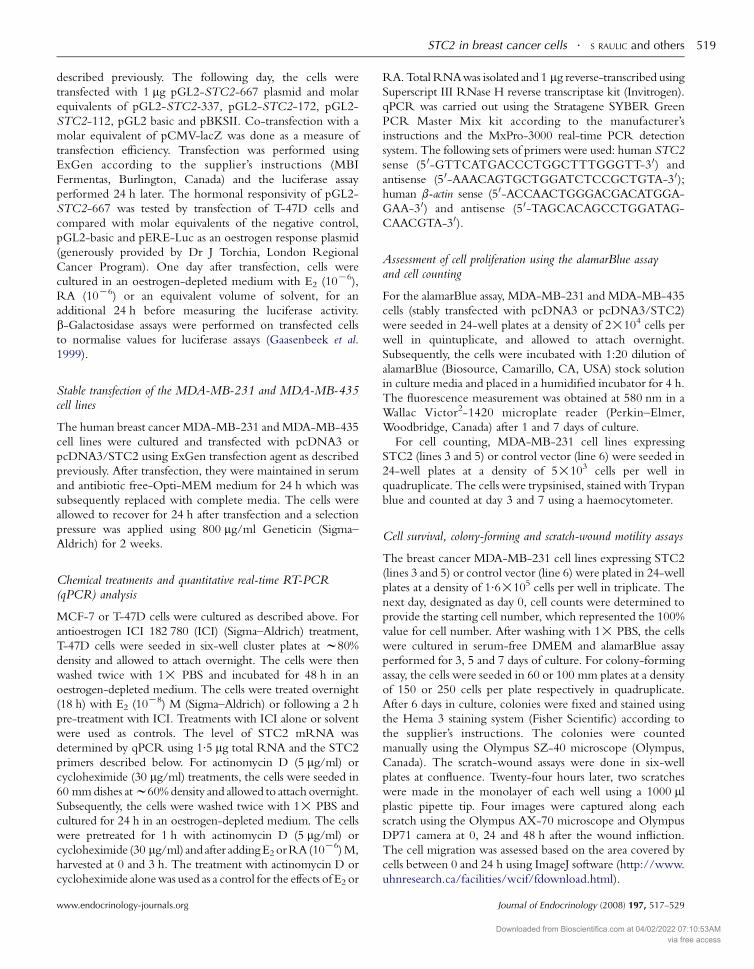
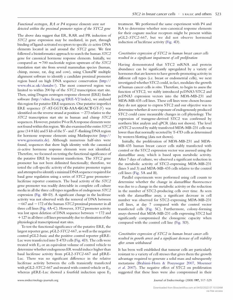
STC2 in breast cancer cells . S RAULIC and others 523
Functional oestrogen, RA or P4 response elements were notdetected within the proximal promoter region of the STC2 gene
The above data suggest that ER, RAR and PR induction of
STC2 gene expression may be mediated, in part, through
binding of ligand-activated receptors to specific cis-active DNA
elements located in and around the STC2 gene. We first
followed a bioinformatics approach to search the human STC2
gene for canonical hormone response elements. Initially, we
compared an w700 nucleotide region upstream of the STC2
translation start site from several mammalian species (human,
chimp, mouse, rat, dog and cow), using ClustalW multiple
alignment software to identify a candidate proximal promoter
region based on high DNA sequence conservation (http://
www.ebi.ac.uk/clustalw/). The most conserved region was
limited to within 200 bp of the STC2 transcription start site.
Then, using Dragon oestrogen response element (ERE) finder
software (http://sdmc.lit.org.sg/ERE-V2/index), we analysed
this region for putative ERE sequences. One putative imperfect
ERE sequence (50-AT-GGTCG-AAA-GGACT-GT-30) was
identified on the reverse strand at positionK273 relative to the
STC2 transcription start site in human and chimp STC2
sequences.However, putative P4orRAresponse elementswere
not foundwithin this region.We also examined the entireSTC2
gene (14.8 kb) and 5 kb of the 50- and 30-flanking DNA region
for hormone response elements using MatInspector (http://
www.genomatix.de). Although numerous half-sites were
found, sequences that show high identity with the canonical
cis-active hormone response elements were not identified.
Therefore, we focussed on testing the functional significance of
the putative ERE by transient transfection. The STC2 gene
promoter has not been delimited functionally; therefore, we
tested the cell-specific activity of the putative promoter region
and attempted to identify aminimalDNA sequence required for
basal gene regulation using a series of STC2 gene promoter–
luciferase reporter constructs. The basal activity of the STC2
gene promoter was readily detectable in complete cell culture
media in all the three cell types regardless of endogenous STC2
expression (Fig. 4B–D). A significant change in the luciferase
activity was not observed with the removal of DNA between
K667 andK172 of the human STC2 proximal promoter in all
three cell lines (Fig. 4A–C). However, STC2 promoter activity
was lost upon deletion of DNA sequence between K172 and
C127 in all three cell lines presumably due to elimination of the
physiological transcriptional start sites.
To test the functional significance of the putative ERE, the
largest reporter gene, pGL2-STC2-667, as well as the negative
control pGL2-basic and the positive control reporter pERE-
Lucwere transfected into T-47D cells (Fig. 4D). The cells were
treated with E2 or an equivalent volume of control vehicle to
determinewhether endogenous ERwould induce higher than
basal luciferase activity from pGL2-STC2-667 and pERE-
Luc. There was no significant difference in the relative
luciferase activity between the cells transiently transfected
with pGL2-STC2-667 and treated with control vehicle or E2,
whereas pERE-Luc showed a fourfold induction upon E2
www.endocrinology-journals.org
treatment. We performed the same experiment with P4 and
RA to determine whether non-canonical response elements
for their cognate nuclear receptors might be present within
pGL2-STC2-667, but we did not observe hormonal
induction of luciferase activity (Fig. 4D).
Constitutive expression of STC2 in human breast cancer cellsresulted in a significant impairment of cell proliferation
Having demonstrated that STC2 mRNA and protein
abundance can be significantly upregulated by a variety of
hormones that are known to have growth-promoting activity in
different cell types (i.e. breast or endometrial cells), we next
investigated whether STC2 could, in fact, modulate the growth
of human cancer cells in vitro. Therefore, to begin to assess the
function of STC2, we stably introduced pcDNA3/STC2 and
pcDNA3 expression vectors into the MDA-MB-231 and
MDA-MB-435 cell lines. These cell lines were chosen because
they do not appear to express STC2 and our objective was to
determinewhether de novo exposure of endogenously produced
STC2 could cause measurable changes in cell physiology. The
expression of transgene-derived STC2 was confirmed by
northern blot analysis and qPCR (data not shown). The level
of STC2 secreted by stably transfectedMDA-MB-231 cells was
lower than that normally secreted by T-47D cells as determined
by western blotting (data not shown).
Initially, the proliferation of MDA-MB-231 and MDA-
MB-435 human breast cancer cells stably transfected with
control or the STC2 expression vector was assessed using the
alamarBlue assay, which is based upon metabolic activity.
After 7 days of culture, we observed a significant reduction in
the metabolic activity of STC2-expressing MDA-MB-231
(lines 5 and 3) and MDA-MB-435 cells relative to the control
cell lines (Fig. 5A and B).
Parallel experiments were performed using cell counts to
determine whether the change in alamarBlue fluorescence
was due to a change in the metabolic activity or the reduction
in the number of STC2-producing cells over time. As seen
with the alamarBlue assay, a significant decrease in cell
number was observed for STC2-expressing MDA-MB-231
cell lines, at day 7 compared with the control vector
transfected cells (Fig. 5C). Furthermore, colony-forming
assays showed that MDA-MB-231 cells expressing STC2 had
significantly compromised the clonogenic capacity when
compared with the control cell line (Fig. 5D).
Constitutive expression of STC2 in human breast cancer cellsresulted in growth arrest and a significant decrease of cell viabilityafter serum withdrawal
It has been well established that tumour cells are particularly
resistant to a variety of cell stresses that gives them the growth
advantage required to generate a solid mass and subsequently
metastasise (Brahimi-Horn & Pouyssegur 2007, Moenner
et al. 2007). The negative effect of STC2 on proliferation
suggested that these lines were also compromised in their
Journal of Endocrinology (2008) 197, 517–529
Downloaded from Bioscientifica.com at 04/02/2022 07:10:53AMvia free access
Figure 4 Human STC2 gene basal and hormone-inducible promoter activity in human cell lines. Three differenthuman cell lines (HEK-293 embryonic kidney cells (A) T-47D breast cancer cells (B) and Ishikawa endometrialcancer cells (C) were transiently transfected with molar equivalent quantities of different 5 0-truncatedSTC2/luciferase reporter constructs. The number in bps (bp) for each construct indicates the size of the proximalpromoter. The results are shown as the fold increase of normalised luciferase activity over the promoterless controlvector, pGL2-Basic. The results for three independent experiments are depicted on each graphGS.E.M..(D) Hormonalresponsivity of pGL2-STC2-677 was tested in T-47D cells with pGL2-Basic as the negative controland pERE as the positive E2-responsive promoter construct. E2, RA, P4 or control vehicle (equivalent volume)treatment of cells transfected with the pGL2-STC2-677 construct did not result in significant induction of luciferaseactivity. These results represent meanGS.E.M. of three independent experiments, each done in triplicate.
S RAULIC and others . STC2 in breast cancer cells524
response to nutrient deprivation as a form of stress. Serum
withdrawal is a well-documented condition that leads to
decreased cell viability through induction of apoptosis (Xu
et al. 2002). We observed two different patterns of response to
serum deprivation (Fig. 6A and B). The control MDA-MB-
231 cell line and the STC2-expressing lines exhibited an
increase in alamarBlue fluorescence over the initial 3 days of
the experiment indicating cell proliferation. However, this
increase was significantly lower in the STC2-expressing lines
compared with the control cell line at day 3, 5 and 7.
Moreover, while the control cell line maintained a constant
level of metabolic activity, both STC2-expressing lines
showed a significant decrease from day 3 to day 7. For
STC2-expressing line 5, this resulted in day 7 values
significantly below those generated by the initial number of
cells seeded (day 0) for these experiments (Fig. 6A). For line
3, the alamarBlue activity at day 7 was not significantly
Journal of Endocrinology (2008) 197, 517–529
different from day 0, which may be related to the lower level
of STC2 production by this line (Fig. 6B).
Ectopic expression of STC2 resulted in compromised cell motility
Certainly, one of the most lethal characteristics of an aggressive
cancer phenotype is the ability of these cells to move through
dense tissue, extravasate and form new tumours at distant
locations (Kedrin et al. 2007, Sahai 2007). To test the effects of
de novo expression of STC2 on the motility of cancer cells,
we utilised the conventional monolayer scratch-wound assay
(Fig. 6C and D). The STC2-expressing cells (line 5 and line 3)
migrated less efficiently into the wound-cleared area compared
with control cells (line 6), during the first 24 h after the injury
(Fig. 6C). However, this difference was not maintained
after another 24 h where all cells lines had filled the gap
(Fig. 6D). The results of several alamarBlue and cell-counting
www.endocrinology-journals.org
Downloaded from Bioscientifica.com at 04/02/2022 07:10:53AMvia free access
Figure 5 Constitutive STC2 expression in the MDA-MB-231 andMDA-MB-435 cancer cell lines reduces cell proliferation. (A)Analysis of proliferation/viability of human breast cancer MDA-MB-231 cells stably transfected with pcDNA3/STC2 (lines 5 and 3) orpcDNA3 control vector (line 6) using the alamarBlue assay. Barsrepresent meanGS.E.M. of three independent experiments done inquintuplicate. (B) The same experiment performed with MDA-MB-435 cells stably transfected with pcDNA3/STC2 showing a similardecrease in proliferation after 7 days in the cultures producingSTC2. (C) An assessment of MDA-MB-231 cell growth by cellcounting. Data represent meansGS.E.M. of three independentexperiments done in quadruplicate confirming the alamarBlueassay results. (D) A representative quantitative analysis ofclonogenic assay indicating significantly reduced colony formationby MDA-MB-231 cells expressing STC2 (lines 5 and 3) incomparison with control cell line 6. Bars represent meansGS.E.M. ofthree independent experiments done in quadruplicate. Significantdifferences (indicated by asterisks) between groups weredetermined by Student’s t-test and one-way ANOVA (P!0.0001).
STC2 in breast cancer cells . S RAULIC and others 525
experiments showedno significant difference in cell proliferation
between MDA-MB-231 STC2-expressing and non-expressing
cells during the first 2 days of culture. These data support the
notion that the difference in gap closure was due to impaired
migrationcausedbySTC2expressionandnotdue to a difference
in proliferation.
Discussion
Our studies of STC2 regulation and function in human
tumour cell lines are based on the fact that it is an extracellular
messenger produced by cancer cells and upregulated by
hormones that can regulate proliferation, suggesting that
STC2 can indirectly influence tumorigenesis. The objective
of this study was to determine whether STC2 gene expression
www.endocrinology-journals.org
was linked to hormones that can positively regulate cancer cell
growth and whether de novo STC2 expression could alter the
growth properties of breast cancer cells in vitro.
STC2 expression in breast cancer
Our northern blot analysis revealed STC2 expression in
ER-positive as well as ER-negative human breast cancer cell
lines in contrast to Charpentier et al. (2000). Clearly, STC2
expression is not exclusively dependent on ER signalling in
breast cancer cells and may have different effects in
ER-positive and -negative cells. We also analysed human
breast RNA for STC2 expression. Previously, STC2
expression data were derived from commercially available
northern blots that utilised polyAC enriched mRNA and did
not include human breast tissue (Chang & Reddel 1998,
DiMattia et al. 1998, Moore et al. 1999). Our data agree with
previously published data showing several STC2 transcripts
in human tissue and cell line RNAs (Chang & Reddel
1998, DiMattia et al. 1998, Moore et al. 1999) likely arising
from the use of alternative polyadenylation signals in the
O3 kb 3 0-untranslated region of the mRNA as predicted by
Aceview (Thierry-Mieg & Thierry-Mieg 2006). Those
earlier experiments indicated that STC2 expression in
human tissues is low; therefore, it was surprising to find that
all three species of STC2 mRNAwere detectable by northern
blot in normal human breast and breast tumour tissue RNAs.
Therefore, it would appear that breast is a major site of STC2
expression implying that it plays a physiological role in the
mammary gland.
Given the high level of STC2 expression in human breast
tissue and breast cancer cell lines, we investigated its expression
in normal and tumorigenic mouse mammary gland. This was
important because of the potential utility of mutant mouse
mammary tumour models to study the role of STC2 in cancer.
Unlike human breast tissue, Stc2 expression was not detectable
in normal mouse mammary gland RNA using northern
analysis. However,we readily detected the expression of Stc2 in
MMTV/ERBB2/Neu and MMTV/PyVMT transgenic
mammary tumours. This suggests that signalling mechanisms
involved in the tumorigenic pathways initiated by the above
transgenes regulate Stc2 expression. Whether or not STC2
plays a positive or negative role in this process requires further
investigation with Stc2 null and STC2-overexpressing mouse
strains (Varghese et al. 2002, Chang et al. 2005, Gagliardi et al.
2005). A model of familial adenomatous polyposis is the only
other mouse tumour model that showed elevated Stc2
expression specifically in adenomatous lesions, which corre-
lated with the STC2 expression in human colorectal cancers
with Apc gene mutations (Andreu et al. 2006). Our data imply
that specific transgenic murine models of mammary gland
tumour formation may be helpful in understanding the role of
STC2 in cancer and that the molecular mechanisms
controlling tumour cell STC2 expression may be conserved
between mouse and human cells.
Journal of Endocrinology (2008) 197, 517–529
Downloaded from Bioscientifica.com at 04/02/2022 07:10:53AMvia free access
Figure 6 Constitutive expression of STC2 in MDA-MB-231 cells significantly decreased the cell viability after serum withdrawal and negativelyaffected cell movement. STC2-expressing and control vector-transfected MDA-MB-231 cell line viability was determined by alamarBlue assayat the indicated time points. Over the 7 days in culture, a significant decrease in cell viability was observed for STC2-expressing line 5 (A), andline 3 (B) comparedwith the control cell line. The viability of the control cell line (line 6) was not significantly affected by serum deprivation overthe assay period. Asterisks indicate statistically significant differences between the control cells and the STC2 expressing by one-way ANOVA(P!0.0001). (C) The scratch-wound assay was used to assess the effect of STC2 expression on random cell movement. The MDA-MB-231 linesexpressing STC2 closed the gap less efficiently than the control cell line at the 24-h time point. Data are presented as the percentage of areapopulated by cells between 0 and 24 hGS.E.M. from four independent experiments done in octoplicate. (D) Representative images of scratch-wound area at different time points showing that by 48 h all three lines had completely filled the gap.
S RAULIC and others . STC2 in breast cancer cells526
Hormonal regulation of STC2 and STC1 expressionin human cancer cell lines
Others have reported the induction of STC2 expression in
response to E2 treatment in human breast cancer cells
(Charpentier et al. 2000, Bouras et al. 2002). Our results
revealed thatSTC2 represents a downstream target of E2, P4 and
RA signalling pathways in human breast cancer cell lines (MCF-
7, T-47D) and a human endometrial cancer cell line (Ishikawa).
We observed some differences in the level of STC2 induction
Journal of Endocrinology (2008) 197, 517–529
and in the temporal pattern of expression amongst the different
cell lines. For example, P4 treatment of MCF-7 or T-47D cells
did not result in strong upregulation of STC2 steady-state
mRNA levels, whereas a significant induction was seen in the
Ishikawa cells. This could be due to the fact that nuclear
receptor-mediated gene expression can be differentially
modulated in the different types of human cancer cells under
identical hormone treatments due to cell-specific differences in
the level of nuclear receptor co-regulators (Hyder et al. 1998,
Magklara et al. 2000, Liang et al. 2005). Additionally, we found
www.endocrinology-journals.org
Downloaded from Bioscientifica.com at 04/02/2022 07:10:53AMvia free access
STC2 in breast cancer cells . S RAULIC and others 527
that dexamethasone had no effect on the STC2 mRNA levels
suggesting that STC2 is not under the control of this hormone,
at least not in these cell types. This is not the case for Stc1, which
showed potent downregulation when exposed to glucocorti-
coids in a murine pituitary cell line (Groves et al. 2001).
Interestingly, none of the hormonal treatments applied in this
study influenced STC1 expression in Ishikawa, MCF-7 or
T-47D cells. This agrees with Frasor et al. (2003) and
Katzenellenbogen & Frasor (2004) where they showed that
STC1 was actually downregulated by E2 in MCF-7 cells.
Collectively, our results indicate that STC2 is a hormonally
responsive gene in breast cancer and non-breast cancer cell
lines implying that it plays a general function in cell
homeostasis that is associated with a variety of nuclear
receptor signalling pathways. In addition, the lack of STC1
and STC2 co-regulation in our studies may indicate that
STC1 and STC2 play distinct roles in human tumour cells
with STC2 function linked to nuclear receptor signalling.
Transcriptional regulation of STC2 expression by oestrogenand RA
We used chemical inhibitors of transcription or translation to
determinewhetherSTC2was a direct E2 orRARtarget gene in
breast cancer cells. The transcriptional block eliminated the
upregulation of STC2mRNAabundance at the 3-h time point;
however, we also observed that blocking protein translation also
diminished E2-induced STC2 expression in two different breast
cancer cell lines. Based on Dean & Sanders’ (1996) proposal,
steroid-responsive genes can be classified into three main
categories: primary response genes, delayed primary response
genes and secondary response genes. The primary responsive
genes interact with steroid receptors directly and their response
usually occurs within minutes after exposure to steroids. The
secondary responsive genes generally represent downstream
targets of regulatory proteins synthesised by primary responsive
genes. They exhibit a time lag in response to steroid treatment
usually measured in hours. Our time-course experiments
showed that multi-fold upregulation of STC2 mRNA levels,
uponE2,RAorP4 treatment, occurredwith a time lag between
3 and 24 h. Based on the above discussion, and taking into
consideration the findings of Carroll et al. (2006), it is likely that
STC2 represents an E2 secondary responsive gene and that E2regulates STC2 expression at the transcriptional level through
ER, but this also requires intermediary proteins (i.e. newly
synthesised co-factors).
To further investigate whether STC2was a direct target for
the E2, P4 andRA receptors, we examined the STC2 gene for
the presence of canonical DNA-binding sites for these nuclear
receptors. One putative ERE was identified; however, this
sequence (5 0-AT-GGTCG-AAA-GGACT-GT-3 0) differs
from the consensus core ERE (5 0-CA-GGTCA-nnn-
TGACC-CT-3 0) at three nucleotide positions in both arms
of the consensus sequence. Several studies have shown that
nucleotide alterations from the core sequence on each side of
an ERE palindrome, results in a significant decrease in
www.endocrinology-journals.org
ER-binding affinity and transcriptional activation (Klinge
2001). This lack of sequence identity with a canonical ERE
does not preclude direct ligand-activated ER regulation as
demonstrated for the imperfect ERE present in the proximal
promoter of the human WISP2 gene (5 0-GGTCA-CAC-
CCACC-3 0) (Fritah et al. 2006). Therefore, we directly tested
the hormone inducibility of the STC2 proximal promoter
region; however, E2, RA or P4 treatments did not induce the
STC2 promoter. These data suggest that the putative ERE, we
identified in the proximal promoter region is not responsive to
E2 and that putative cis-active regulatoryelements responsive to
RA and P4 are not present within the STC2 proximal
promoter region, lending support to the hypothesis that
regulation of STC2 by these hormones is indirect. Moreover,
we did not uncover perfect or imperfect response elements for
the E2, P4 andRA receptors in other regions of the STC2 gene
suggesting that these hormones function indirectly to regulate
STC2 expression. This is not surprising, given that Kwon et al.
(2007) found that only 6% of E2-regulated genes were bound
by ER in the proximal promoter region. In addition, a
genome-wide screen for ER-binding sites in MCF-7 cells
found that themajority of the high-confidence sites are located
outside of proximal promoter regions and they did not identify
a functional ERE within 100 kb of the STC2 gene (Lin et al.
2007). Therefore, it seems likely that for E2 regulation of the
STC2 gene, other transcription factors may be mediating the
effect as has been documented for the Sp1, AP-1 and nuclear
factor-kB (Chambliss & Shaul 2002,Wintermantel et al. 2005,
McDevitt et al. 2007). Therefore, functional identification of
hormone-responsive regions of the STC2 gene will require a
systematic testing of the entire locus through a combination of
transfection assays coupled with ChIP-chip assays.
Constitutive expression of STC2 in human breast cancer cellsalters cell growth
Given that STC2 expression can be significantly increased in
cancer cells by a variety of growth-promoting hormones, we
asked fundamental questions regarding the role of STC2 in
breast cancer cell biology. As demonstrated by alamarBlue assay,
cell-counting and colony-forming assay, de novo production of
STC2byMDA-MB-231 andMDA-MB-435breast cancer cells
resulted in a significant impairment of cell growth. It is
important to note that the level of STC2 produced by the stably
transfected cellswas substantially lower than produced byT-47D
cells indicating that overexpression of the protein is not likely
responsible for the growth impairment effect of STC2. This is
the first study to show that STC2can exert a growth-suppressive
effect on human breast cancer cells or any cell type in vitro.
Recently, Gagliardi et al. (2005) demonstrated that constitutive
expression of human STC2 in transgenic mice, results in a
significant reduction of intramembranous and endochondral
bone development, as well as high neonatal morbidity,
suggesting that STC2 can act as a potent growth inhibitor
in vivo. The mechanism responsible for the STC2 growth
inhibitory effect is not known, although it seems to be
Journal of Endocrinology (2008) 197, 517–529
Downloaded from Bioscientifica.com at 04/02/2022 07:10:53AMvia free access
S RAULIC and others . STC2 in breast cancer cells528
independent ofGH/IGF I axis andmay be linked to ametabolic
disturbance (Gagliardi et al. 2005). Whether STC2 is having a
negative effect on MDA-MB-231 cell metabolism and thus
reducing cell proliferation will require further study to evaluate
ATP production and mitochondrial function.
Our results are in contrast to those of Ito et al. (2004) since
they reported that STC2 overexpression resulted in selective
protection of HeLa cells against endoplasmic reticulum stress-
induced cell death. A significant decrease in cell death was
observed in STC2-overexpressing HeLa cells treated with
thapsigargin, while STC2 overexpression did not affect
tunicamycin-treated HeLa cell viability (Ito et al. 2004).
These findings suggest that STC2 expression is differentially
responsive to stress stimuli and that the activation of distinct
signalling pathways could implicate STC2 in a cytoprotective
or pro-apoptotic role. Collectively, our data suggest that
STC2 expression could be detrimental to cell viability in vitro
and that human breast cancer MDA-MB-231 cells may be a
good model to study its mechanism of action.
In summary, our data indicate that STC2 expression can be
induced through a variety of nuclear receptor signalling
pathways in different cell types through an indirect
mechanism. Functionally, de novo STC2 production in
aggressive human cancer cell lines decreases cell viability.
This coupled with clinical data indicating that loss of STC2
expression positively correlates with a more aggressive breast
tumour phenotype suggests that STC2 may act as an
autocrine or paracrine brake on cell growth and its expression
is associated with a more differentiated phenotype (Iwao et al.
2002, Yamamura et al. 2004, Esseghir et al. 2007).
Acknowledgements
This research was supported by funds from the Canadian
Institutes of Health Research Grant MOP 53128 and the
London Regional Cancer Program Small Grants Competition
LHRF 7626, specifically, the Pamela Greenaway-Kohlmeier
Translational Breast Cancer Research Unit. We are grateful to
Drs Chris Pin, Trevor Shepherd and Bonnie Deroo for their
helpful suggestions and for a comprehensive and critical reading
of this manuscript and to Dr William Muller for generously
providing us the mouse transgenic mammary gland tumour
samples. The authors declare that there is no conflict of interest
that would prejudice the impartiality of this study.
References
Amatschek S, Koenig U, Auer H, Steinlein P, Pacher M, Gruenfelder A,
Dekan G, Vogl S, Kubista E, Heider KH et al. 2004 Tissue-wide expression
profiling using cDNA subtraction and microarrays to identify tumor-
specific genes. Cancer Research 64 844–856.
Anderson HC 2003 Matrix vesicles and calcification. Current Rheumatology
Reports 5 222–226.
AndreuP,Colnot S,GodardC,Laurent-PuigP, LamarqueD,KahnA,PerretC&
Romagnolo B 2006 Identification of the IFITM family as a new molecular
marker in human colorectal tumors. Cancer Research 66 1949–1955.
Journal of Endocrinology (2008) 197, 517–529
Bouras T, Southey MC, Chang AC, Reddel RR, Willhite D, Glynne R,
HendersonMA, Armes JE & Venter DJ 2002 Stanniocalcin 2 is an estrogen-
responsive gene coexpressed with the estrogen receptor in human breast
cancer. Cancer Research 62 1289–1295.
Bradford MM 1976 A rapid and sensitive method for the quantitation of
microgram quantities of protein utilizing the principle of protein-dye
binding. Analytical Biochemistry 72 248–254.
Brahimi-Horn MC & Pouyssegur J 2007 Hypoxia in cancer cell metabolism
and pH regulation. Essays in Biochemistry 43 165–178.
Carroll JS, Meyer CA, Song J, LiW, Geistlinger TR, Eeckhoute J, Brodsky AS,
Keeton EK, Fertuck KC, Hall GF et al. 2006 Genome-wide analysis of
estrogen receptor binding sites. Nature Genetics 38 1289–1297.
Chambliss KL & Shaul PW 2002 Estrogen modulation of endothelial nitric
oxide synthase. Endocrine Reviews 23 665–686.
Chang AC & Reddel RR 1998 Identification of a second stanniocalcin
cDNA in mouse and human: stanniocalcin 2. Molecular and Cellular
Endocrinology 141 95–99.
Chang AC, Jellinek DA & Reddel RR 2003 Mammalian stanniocalcins
and cancer. Endocrine-Related Cancer 10 359–373.
Chang AC, Cha J, Koentgen F & Reddel RR 2005 The murine stanniocalcin
1 gene is not essential for growth and development. Molecular and Cellular
Biology 25 10604–10610.
Charpentier AH, Bednarek AK, Daniel RL, Hawkins KA, Laflin KJ, Gaddis S,
MacLeod MC & Aldaz CM 2000 Effects of estrogen on global gene
expression: identification of novel targets of estrogen action. Cancer Research
60 5977–5983.
Dauvois S, Danielian PS, White R & Parker MG 1992 Antiestrogen ICI
164,384 reduces cellular estrogen receptor content by increasing its
turnover. PNAS 89 4037–4041.
Dean DM & Sanders MM 1996 Ten years after: reclassification of steroid-
responsive genes. Molecular Endocrinology 10 1489–1495.
Deol HK, Varghese R, Wagner GF & DiMattia GE 2000 Dynamic regulation
of mouse ovarian stanniocalcin expression during gestation and lactation.
Endocrinology 141 3412–3421.
DiMattia GE, Varghese R & Wagner GF 1998 Molecular cloning and
characterization of stanniocalcin-related protein. Molecular and Cellular
Endocrinology 146 137–140.
Esseghir S, Reis-Filho JS, Kennedy A, JamesM,O’HareMJ, Jeffery R, Poulsom
R & Isacke CM 2006 Identification of transmembrane proteins as potential
prognosticmarkers and therapeutic targets in breast cancer bya screen for signal
sequence encoding transcripts. Journal of Pathology 210 420–430.
Esseghir S, Kennedy A, Seedhar P, Nerurkar A, Poulsom R, Reis-Filho JS &
Isacke CM 2007 Identification of NTN4, TRA1, and STC2 as prognostic
markers in breast cancer in a screen for signal sequence encoding proteins.
Clinical Cancer Research 13 3164–3173.
Frasor J, Danes JM, Komm B, Chang KC, Lyttle CR & Katzenellenbogen BS
2003 Profiling of estrogen up- and down-regulated gene expression in
human breast cancer cells: insights into gene networks and pathways
underlying estrogenic control of proliferation and cell phenotype.
Endocrinology 144 4562–4574.
Fritah A, Redeuilh G & Sabbah M 2006 Molecular cloning and
characterization of the human WISP-2/CCN5 gene promoter reveal its
upregulation by oestrogens. Journal of Endocrinology 191 613–624.
Gaasenbeek M, Gellersen B & DiMattia GE 1999 DNase I hypersensitivity
analysis of non-pituitary human prolactin gene expression. Molecular and
Cellular Endocrinology 152 147–159.
Gagliardi AD, Kuo EY, Raulic S, Wagner GF & DiMattia GE 2005 Human
stanniocalcin-2 exhibits potent growth-suppressive properties in transgenic
mice independently of growth hormone and IGFs. American Journal of
Physiology. Endocrinology and Metabolism 288 E92–105.
Groves TC, Wagner GF & DiMattia GE 2001 cAMP signaling can antagonize
potent glucocorticoid post-transcriptional inhibition of stanniocalcin gene
expression. Journal of Endocrinology 171 499–516.
Gruvberger S, Ringner M, Chen Y, Panavally S, Saal LH, Borg A, Ferno M,
Peterson C & Meltzer PS 2001 Estrogen receptor status in breast cancer is
associated with remarkably distinct gene expression patterns. Cancer
Research 61 5979–5984.
www.endocrinology-journals.org
Downloaded from Bioscientifica.com at 04/02/2022 07:10:53AMvia free access
STC2 in breast cancer cells . S RAULIC and others 529
Guy CT, Cardiff RD & Muller WJ 1992 Induction of mammary tumors by
expression of polyomavirus middle Toncogene: a transgenic mouse model
for metastatic disease. Molecular and Cellular Biology 12 954–961.
Hall RE, Lee CS, Alexander IE, Shine J, Clarke CL & Sutherland RL 1990
Steroid hormone receptor gene expression in human breast cancer cells:
inverse relationship between oestrogen and glucocorticoid receptor
messenger RNA levels. International Journal of Cancer 46 1081–1087.
Hyder SM, Murthy L & Stancel GM 1998 Progestin regulation of vascular
endothelial growth factor in human breast cancer cells. Cancer Research 58
392–395.
Ito D, Walker JR, Thompson CS, Moroz I, Lin W, Veselits ML, Hakim AM,
Fienberg AA & Thinakaran G 2004 Characterization of stanniocalcin 2, a
novel target of the mammalian unfolded protein response with
cytoprotective properties. Molecular and Cellular Biology 24 9456–9469.
Iwao K, Matoba R, Ueno N, Ando A, Miyoshi Y, Matsubara K, Noguchi S &
Kato K 2002 Molecular classification of primary breast tumors possessing
distinct prognostic properties. Human Molecular Genetics 11 199–206.
Jordan VC 2007 SERMs: meeting the promise of multifunctional medicines.
Journal of National Cancer Institute 99 350–356.
Katzenellenbogen BS & Frasor J 2004 Therapeutic targeting in the estrogen
receptor hormonal pathway. Seminars in Oncology 31 28–38.
Kedrin D, van Rheenen J, Hernandez L, Condeelis J & Segall J 2007 Cell
motility and cytoskeletal regulation in invasion and metastasis. Journal of
Mammary Gland Biology and Neoplasia 12 143–152.
Klinge CM 2001 Estrogen receptor interaction with estrogen response
elements. Nucleic Acids Research 29 2905–2919.
Kun Y, How LC, Hoon TP, Bajic VB, Lam TS, Aggarwal A, Sze HG, BokWS,
YinWC&TanP2003Classifying the estrogen receptor status of breast cancers
by expression profiles reveals a poor prognosis subpopulation exhibiting high
expression of the ERBB2 receptor. Human Molecular Genetics 12 3245–3258.
Kwon YS, Garcia-Bassets I, Hutt KR, Cheng CS, Jin M, Liu D, Benner C,
Wang D, Ye Z, Bibikova M et al. 2007 Sensitive ChIP-DSL technology
reveals an extensive estrogen receptor alpha-binding program on human
gene promoters. PNAS 104 4852–4857.
Liang Y, Wu J, Stancel GM & Hyder SM 2005 p53-dependent inhibition of
progestin-induced VEGF expression in human breast cancer cells. Journal of
Steroid Biochemistry and Molecular Biology 93 173–182.
Lin CY, Vega VB, Thomsen JS, Zhang T, Kong SL, Xie M, Chiu KP, Lipovich
L, Barnett DH, Stossi F et al. 2007 Whole-genome cartography of estrogen
receptor alpha binding sites. PLoS Genetics 3 e87.
Luo CW, Kawamura K, Klein C & Hsueh AJ 2004 Paracrine regulation of
ovarian granulosa cell differentiation by stanniocalcin (STC) 1: mediation
through specific STC1 receptors. Molecular Endocrinology 18 2085–2096.
Luo CW, Pisarska MD & Hsueh AJ 2005 Identification of a stanniocalcin
paralog, stanniocalcin-2, in fish and the paracrine actions of stanniocalcin-2
in the mammalian ovary. Endocrinology 146 469–476.
MadsenKL,TaverniniMM,YachimecC,MendrickDL,AlfonsoPJ,BuerginM,
OlsenHS, AntonaccioMJ, ThomsonAB& FedorakRN1998 Stanniocalcin:
a novel protein regulating calcium and phosphate transport across mammalian
intestine. American Journal of Physiology 274 G96–102.
MagklaraA,GrassL&Diamandis EP2000Differential steroidhormone regulation
of humanglandular kallikrein (hK2) andprostate-specific antigen (PSA) inbreast
cancer cell lines. Breast Cancer Research and Treatment 59 263–270.
McCudden CR, Majewski A, Chakrabarti S & Wagner GF 2004
Co-localization of stanniocalcin-1 ligand and receptor in human breast
carcinomas. Molecular and Cellular Endocrinology 213 167–172.
McDevittMA,Glidewell-KenneyC,Weiss J,ChambonP, Jameson JL&Levine JE
2007 Estrogen response element-independent estrogen receptor (ER)-asignaling does not rescue sexual behavior but restores normal testosterone
secretion in male ERalpha knockout mice. Endocrinology 148 5288–5294.
MoennerM,PluquetO,BouchecareilhM&ChevetE2007 Integratedendoplasmic
reticulum stress responses in cancer.Cancer Research 67 10631–10634.
Moore EE, Kuestner RE, Conklin DC, Whitmore TE, Downey W, Buddle
MM, Adams RL, Bell LA, Thompson DL, Wolf A et al. 1999 Stanniocalcin
2: characterization of the protein and its localization to human pancreatic
alpha cells. Hormone and Metabolic Research 31 406–414.
www.endocrinology-journals.org
Neve RM, Chin K, Fridlyand J, Yeh J, Baehner FL, Fevr T, Clark L, Bayani N,
Coppe JP, Tong F et al. 2006 A collection of breast cancer cell lines for the
study of functionally distinct cancer subtypes. Cancer Cell 10 515–527.
RishiAK,GeraldTM,ShaoZM,LiXS,BaumannRG,DawsonMI&Fontana JA
1996Regulation of the human retinoic acid receptor alpha gene in the estrogen
receptor negative human breast carcinoma cell lines SKBR-3 and
MDA-MB-435. Cancer Research 56 5246–5252.
Roman SD, Clarke CL, Hall RE, Alexander IE & Sutherland RL 1992
Expression and regulation of retinoic acid receptors in human breast cancer
cells. Cancer Research 52 2236–2242.
Sahai E2007 Illuminating themetastatic process.NatureReviews.Cancer7737–749.
Sheikh-Hamad D, Rouse D & Yang Y 2000 Regulation of stanniocalcin in
MDCK cells by hypertonicity and extracellular calcium. American Journal of
Physiology. Renal Physiology 278 F417–F424.
Siegel PM, Ryan ED, Cardiff RD & Muller WJ 1999 Elevated expression of
activated forms of Neu/ErbB-2 and ErbB-3 are involved in the induction
of mammary tumors in transgenic mice: implications for human breast
cancer. EMBO Journal 18 2149–2164.
Stasko SE & Wagner GF 2001 Possible roles for stanniocalcin during early
skeletal patterning and joint formation in the mouse. Journal of Endocrinology
171 237–248.
Sutherland RL, Hall RE, Pang GY, Musgrove EA & Clarke CL 1988 Effect of
medroxyprogesterone acetate on proliferation and cell cycle kinetics of
human mammary carcinoma cells. Cancer Research 48 5084–5091.
Thierry-Mieg D & Thierry-Mieg J 2006 AceView: a comprehensive cDNA-
supported gene and transcripts annotation. Genome Biology 7 S12 11–14.
Varghese R, Gagliardi AD, Bialek PE, Yee SP, Wagner GF & DiMattia GE
2002 Overexpression of human stanniocalcin affects growth and
reproduction in transgenic mice. Endocrinology 143 868–876.
Wagner GF &DiMattia GE 2006 The stanniocalcin family of proteins. Journal of
Experimental Zoology AComparative Experimental Biology 305 769–780.
WagnerGF,VozzoloBL, JaworskiE,HaddadM,KlineRL,OlsenHS,RosenCA,
DavidsonMB&Renfro JL 1997Human stanniocalcin inhibits renal phosphate
excretion in the rat. Journal of Bone and Mineral Research 12 165–171.
Wilson KS, Roberts H, Leek R, Harris AL & Geradts J 2002 Differential gene
expression patterns in HER2/neu-positive and -negative breast cancer cell
lines and tissues. American Journal of Pathology 161 1171–1185.
Wintermantel TM, Berger S, Greiner EF & Schutz G 2005 Evaluation of
steroid receptor function by gene targeting in mice. Journal of Steroid
Biochemistsry and Molecular Biology 93 107–112.
Xu Z, Stokoe D, Kane LP & Weiss A 2002 The inducible expression of the
tumor suppressor gene PTEN promotes apoptosis and decreases cell size by
inhibiting the PI3K/Akt pathway in Jurkat T cells. Cell Growth and
Differentiation 13 285–296.
Yager JD & Davidson NE 2006 Estrogen carcinogenesis in breast cancer. New
England Journal of Medicine 354 270–282.
Yamamura J, Miyoshi Y, Tamaki Y, Taguchi T, Iwao K, Monden M, Kato K &
Noguchi S 2004 mRNA expression level of estrogen-inducible gene, alpha
1-antichymotrypsin, is a predictor of early tumor recurrence in patients
with invasive breast cancers. Cancer Science 95 887–892.
Yoshiko Y, Candeliere GA, Maeda N & Aubin JE 2007 Osteoblast
autonomous Pi regulation via Pit1 plays a role in bone mineralization.
Molecular and Cellular Biology 27 4465–4474.
Yu K, Lee CH, Tan PH & Tan P 2004 Conservation of breast cancer
molecular subtypes and transcriptional patterns of tumor progression across
distinct ethnic populations. Clinical Cancer Research 10 5508–5517.
Zucchi I, Mento E, Kuznetsov VA, Scotti M, Valsecchi V, Simionati B,
Vicinanza E, Valle G, Pilotti S, Reinbold R et al. 2004 Gene expression
profiles of epithelial cells microscopically isolated from a breast-invasive
ductal carcinoma and a nodal metastasis. PNAS 101 18147–18152.
Received in final form 18 March 2008Accepted 27 March 2008Made available online as an Accepted Preprint27 March 2008
Journal of Endocrinology (2008) 197, 517–529
Downloaded from Bioscientifica.com at 04/02/2022 07:10:53AMvia free access