Department of Medicine Division of Infectious Diseases Helsinki University Central Hospital Helsinki, Finland Staphylococcus aureus bacteraemia - Disease progression and prognosis Erik Sebastian Forsblom Academic Dissertation To be presented, with permission of the Faculty of Medicine, University of Helsinki, for public examination in Auditorium 3, Biomedicum Helsinki, Haartmaninkatu 8, on September 6 th , 2014, at 12 noon. Helsinki 2014
Transcript
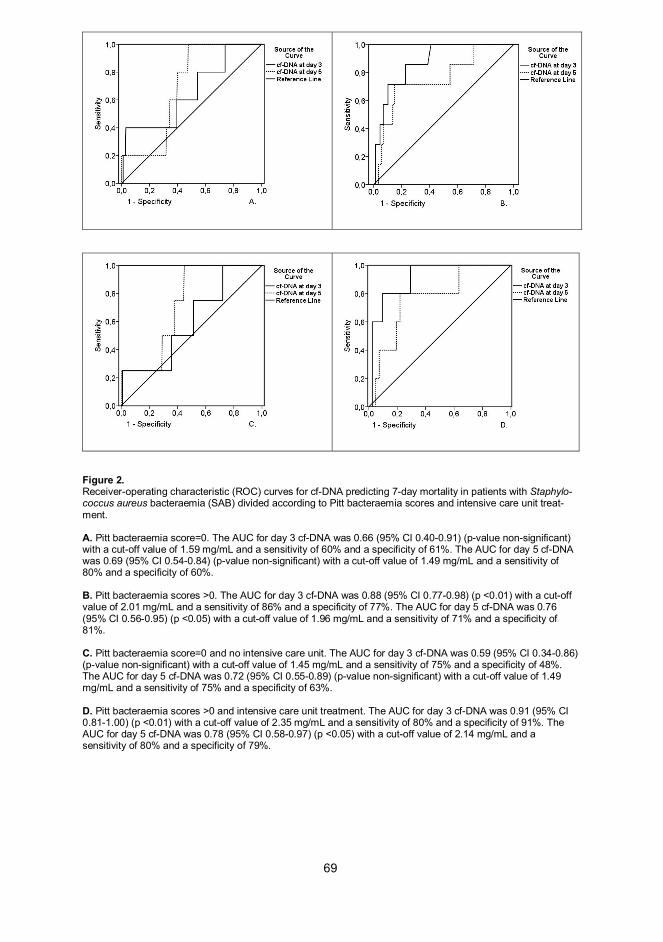
Department of Medicine
Division of Infectious Diseases
Helsinki University Central Hospital
Helsinki, Finland
Staphylococcus aureus bacteraemia -
Disease progression and prognosis
Erik Sebastian Forsblom
Academic Dissertation
To be presented, with permission of the Faculty of Medicine,
University of Helsinki, for public examination in Auditorium 3,
Biomedicum Helsinki, Haartmaninkatu 8, on September 6 th, 2014,
at 12 noon.
Helsinki 2014
SupervisorsDocent Asko Järvinen, MD, PhD
Department of Medicine, Division of Infectious Diseases
Helsinki University Central Hospital
Helsinki, Finland
Eeva Ruotsalainen, MD, PhD
Department of Medicine, Division of Infectious Diseases
Helsinki University Central Hospital
Helsinki, Finland
ReviewersDocent Pertti Arvola, MD, PhD
Department of Medicine, Division of Infectious Diseases
Tampere University Hospital
Tampere, Finland
Docent Timo Hautala, MD, PhD
Department of Medicine, Division of Infectious Diseases
Oulu University Hospital
Oulu, Finland
OpponentDocent Jaana Syrjänen, MD, PhD
Department of Medicine, Division of Infectious Diseases
Tampere University Hospital
Tampere, Finland
ISBN 978-951-51-0079-5 (pbk)
ISBN 978-951-51-0080-1 (pdf)
UNIGRAFIA
HELSINKI 2014
To my family, friends and colleagues
5
ContentsLIST OF ORIGINAL PUBLICATIONS....................................................................................8ABBREVIATIONS..................................................................................................................9ABSTRACT .........................................................................................................................101. INTRODUCTION..............................................................................................................122. REVIEW OF THE LITERATURE......................................................................................15
2.1. Incidence of Staphylococcus aureus bacteraemia.....................................................15
2.2. Predisposing factors for Staphylococcus aureus bacteraemia...................................15
2.2.1. Microbiology and carriage of Staphylococcus aureus bacteria................................ 15
2.2.1.1. Microbiological aspects of Staphylococcus aureus..............................................15
2.2.1.2. Colonization with Staphylococcus aureus ...........................................................16
2.2.1.3. Clinical impact of nasal Staphylococcus aureus carriage ....................................16
2.2.1.4. Decolonization of Staphylococcus aureus carriage .............................................16
2.2.2. Patient-related underlying factors for Staphylococcus aureus bacteraemia............ 17
2.2.2.1. Impact of age and gender ...................................................................................17
2.2.2.2. Impact of underlying diseases.............................................................................17
2.2.2.3. Impact of substance abuse .................................................................................18
2.2.3. Destruction of skin and mucous membrane............................................................. 18
2.3. Clinical picture of Staphylococcus aureus bacteraemia.............................................18
2.7. Prognosis and mortality in Staphylococcus aureus bacteraemia ...............................50
2.7.1. Impact of host-related factors................................................................................... 50
2.7.2. Impact of community or health care acquisition on mortality.................................... 51
2.7.3. Impact of methicillin resistance on mortality............................................................. 51
2.7.4. Impact of clinical manifestations on mortality........................................................... 52
3. AIMS OF THE STUDY .....................................................................................................554. MATERIALS AND METHODS .........................................................................................56
4.1. Study populations .....................................................................................................56
4.2. Study designs ...........................................................................................................57
4.3. Definitions of terminology..........................................................................................59
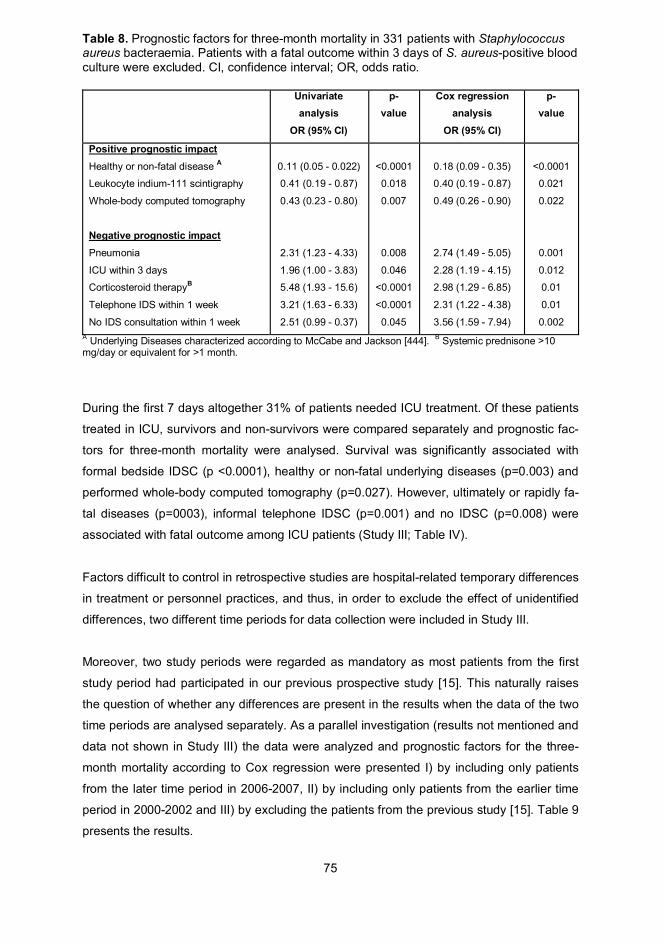
5.4.4. Effect of rifampicin treatment on outcome................................................................ 78
6. DISCUSSION...................................................................................................................816.1. Health care- and community-associated Staphylococcus aureus bacteraemia..........81
6.2. Cell-free DNA as a biomarker in Staphylococcus aureus bacteraemia......................84
6.3. Bedside and telephone infectious diseases specialist consultation in Staphylococcus
Methicillin, a semisynthetic penicillin derivative, was presented in 1959 against penicillin-
resistant S. aureus strains. However, in 1961 the first MRSA strains emerged in the United
Kingdom [143], after which MRSA has steadily become more common and is now encoun-
tered worldwide [1]. The mechanism for methicillin resistance in S. aureus is the mecA
20
gene, located on an DNA region named the staphylococcal cassette chromosome mec
(SCCmec element, found also in coagulase-negative staphylococci), encoding the penicil-
lin-binding protein 2a (PBP2a) [144,145]. PBPs are required for bacterial cell wall synthe-
sis. However, PBP2a differs from regular PBPs as it does not bind methicillin or other -
lactam antibiotics, hence, PBP2a can function despite the presence of methicillin or other
-lactam antibiotics. The mechanism for MRSA is the expression of PBP2a, which is not
inhibited by methicillin or other -lactam antibiotics at concentrations that inhibit other PBPs
[146,147].
MRSA was for decades regarded as a health care-associated challenge, but in the 1990s
community-associated MRSA spread rapidly [148,149]. During the past decades the overall
incidence of SAB due to MRSA has increased without any corresponding decline in MSSA
bacteraemia [150,151]. Hence, the overall impact of SAB has increased. In the United
States, health care-associated MRSA bacteraemia increased from 35% in 1991 to 45% in
1997-1999 [152,153], whereas the overall MRSA bacteraemia incidence in the United
Kingdom and Wales increased from 4% in 1993 to 43% in 2002 [154]. The aetiology behind
rising MRSA rates is complex and probably multifactorial. However, the increase in MRSA
prevalence is associated with underlying diseases and comorbidity, prolonged hospitaliza-
tion and poor adherence to infection control precautions [5,155].
Patients with MRSA bacteraemia have been reported to be older and to more often have
previous MRSA colonization and a longer duration of hospitalization than patients with
MSSA bacteraemia [156,157,158]. Higher mortality in MRSA relative to MSSA bacteraemia
is a common observation – a topic discussed more in detail in section 2.7.3. As community-
associated MRSA infections emerged in the 1990s, several studies reported that CA-
MRSA frequently causes severe skin and soft tissue infections and severe necrotising
pneumonia linked to Panton-Valentine leukocidin (PVL) toxins [159,160,161,162, 163]. CA-
MRSA bacteraemias are increasing and have been associated with necrotizing pneumonia
and cutaneous abscesses, although no mortality difference relative to MSSA bacteraemias
has been observed [120].
2.3.3. Classification, characteristics and prevalence of infection foci
2.3.3.1. Categorization of infection foci
Infection foci in SAB are mostly defined as primary (i.e. cutaneous or portal of entry) or
secondary (i.e. deep or metastatic) [164,165,166] (see Tables 1 and 2a-b). Moreover, an
unknown portal of entry is defined as primary SAB [27]. Some authors classify SAB simply
as complicated or uncomplicated [21]. Furthermore, some authors have used definitions
21
such as deep-seated foci [120], whereas others have only mentioned the foci with most
clinical relevance, e.g. endocarditis [167], and some report infection foci when they are
eradicable and eradicated [19,22,168]. Categorization as primary, secondary, cutaneous,
deep or metastatic foci are the most common categorization types [164,165,166], whereas
complicated and uncomplicated SAB are seldom used [21,30,136]. Many reports list the
occurrence of primary SAB, i.e. cases where the portal of entry or primary source of SAB is
unknown [27] (Table 1). The variable classification of infection foci in the literature makes
comparison of different patient materials cumbersome.
2.3.3.2. Primary infection foci
Various body locations may function as the primary site of infection. The recognition of SAB
foci as either primary or secondary was first introduced in 1976 [164], and SAB patients
were divided according to recognizable primary infection lesions. The primary S. aureus in-
fection focus was viewed as a potential portal of entry for the SAB if the clinical picture of
the primary focus preceded SAB, whereas secondary foci were viewed as metastatic infec-
tions. When no primary focus is found, the presence of an intravascular catheter or a post-
operative wound may be the primary focus [19]. Also the urinary tract may serve as a portal
of entry and a primary focus for SAB. Patients with urological challenges, such as long-
term care patients with frequent urine catheterization, often have S. aureus isolated from
urine samples. However, although the urinary tract may function as a primary focus, in
most cases simultaneous S. aureus bacteruria is a result of haematogenous spread and is
secondary to SAB [169,170,171,172]. The respiratory tract is identified as a primary source
in many reports [12,19,40].
2.3.3.3. Secondary, metastatic or deep infection foci
Due to haematogenous spread in SAB, virtually any organ may be infected [173] and the
infections are defined as secondary, metastatic or deep foci. However, S. aureus infections
are seldom the result of bacterial inoculation due to trauma or an iatrogenic process, e.g.
joint puncture, surgery or arthroscopy [12,44,173,174]. Foreign body infections and deep-
seated abscesses are often of haematogenous origin, although foreign body infections may
be postoperative without a bacteraemia phase [121,136,175,176]. Prosthetic joint infections
are commonly classified as early (i.e. development within 3 months of surgery), as delayed
(i.e. development within 3-24 months of surgery) and as late (i.e. development later than 24
months after surgery) [177,178]. Early and delayed prosthetic joint infections are mostly
achieved during the prosthesis implantation process, whereas late prosthetic joint infec-
tions are commonly of haematogenous origin where the skin, dental region or respiratory or
22
urinary tract are frequent sources of SAB [179]. S. aureus pneumonia is predominantly of
haematogenous origin and may be due to release of infected tricuspidal vegetations or re-
lease of infected thrombotic material in the venous system [19,180]. S. aureus meningitis is
most often postoperative and on rare occasions haematogenous (due to massive S. aureus
bacteraemia load and usually a high number of other deep foci present) [181,182]. Most
studies report endocarditis, osteomyelitis, abscesses and pneumonia or respiratory infec-
tion, whereas septic arthritis and foreign body infections are rarely described (Table 2a).
Some report bone and joint infections together (Table 2a), whereas some mention only
specific abscesses, e.g. epidural [29,183], psoas [184] or abdominal abscesses [16].
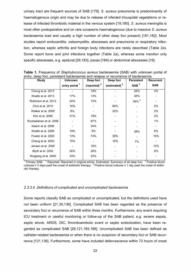
Table 1. Frequency of Staphylococcus aureus bacteraemia (SAB) with unknown portal ofentry, deep foci, persistent bacteraemia and relapse or recurrence of bacteraemia.
Study Unknown
entry portal 1
Deep foci
(reported) 2
Deep foci
(estimated) 2
Persistent
SAB 3
RecurrentSAB
Chong et al. 2013
Khatib et al. 2013
Robinson et al. 2012
Choi et al. 2010
Walker et al. 2009
Kim et al. 2008
Ruotsalainen et al. 2006
Kaech et al. 2006
Khatib et al. 2006
Fowler et al. 2003
Chang et al. 2003
Jensen et al. 2002
Blyth et al. 2002
Ringberg et al. 2000
-
17%
24%
16%
8%
31%
-
-
19%
13%
15%
-
26%
33%
19%
13%
13%
-
-
19%
87%
24%
9%
74%
-
16%
26%
53%
-
-
-
66%
32%
-
-
-
-
39%
16%
-
-
-
26%
36%
56% 4
-
-
-
-
-
38%
-
7%
-
-
-
4%
-
-
5%
2%
2%
1%
-
6%
16%
-
12%
6%
-1 Primary SAB. 2 Reported: Reported in original article. Estimated: Summary of all deep foci. 3 Positive bloodcultures 3 days past the onset of antibiotic therapy. 4 Positive blood cultures 1 day past the onset of antibi-otic therapy.
2.3.3.4. Definitions of complicated and uncomplicated bacteraemia
Some reports classify SAB as complicated or uncomplicated, but the definitions used have
not been uniform [21,30,136]. Complicated SAB has been regarded as the presence of
secondary foci or recurrence of SAB within three months. Furthermore, any event requiring
ICU treatment or careful monitoring or follow-up of the SAB patient, e.g. severe sepsis,
septic shock, ARDS, DIC, thromboembolic event or septic embolization, have been re-
garded as complicated SAB [28,121,185,186]. Uncomplicated SAB has been defined as
catheter-related bacteraemia or when there is no suspicion of secondary foci or SAB recur-
rence [121,136]. Furthermore, some have included defervescence within 72 hours of onset
23
of appropriate antibiotic therapy in the criteria [30]. A very recent report form 2013 defined
complicated SAB as persistent bacteraemia (duration three days), SAB relapse and/or
secondary foci, whereas uncomplicated SAB was defined as bacteraemia duration two
days, no foreign device and/or secondary foci [21].
2.3.3.5. Risk factors associated with complicated bacteraemia
Persistent bacteraemia or fever for longer than 72-96 hours have been identified as risk
factors for complicated SAB [14,40,121,187]. Furthermore, CA-SAB, underlying diseases
and especially haemodialysis and unremoved infected catheters have been connected to
complicated SAB [42,136,166]. Several reports have failed in connecting MRSA bacterae-
mia to complicated SAB [121,188], although one report associated intravascular catheter-
related MRSA bacteraemia with complicated SAB [136]. Vancomycin therapy in SAB, re-
gardless of MSSA or MRSA, has been recognized as an independent risk factor for recur-
rence, treatment failure and mortality [37,41,189,190], whereas high vancomycin minimum
inhibitory concentration (MIC) (>1.5 ug/mL) has been presented as an independent predic-
tor for complicated bacteraemia [186]. Some reports associate primary SAB with higher oc-
currence of secondary deep foci [2]. A thorough prospective report from 2003 identified four
risk factors to be significantly associated with the risk for complicated SAB: persistent bac-
teraemia and persistent fever (positive blood cultures > 72-96 hours and fever > 72 hours)
subsequent to onset of appropriate antibiotic therapy, CA-SAB and presence of skin le-
sions suggesting acute systemic infection. The lack of any of these risk factors gave a
probability of 16% for complicated SAB, whereas the presence of three risk factors had a
probability of 70% for complicated SAB [121].
2.3.3.6. Prevalence of infection foci
The prevalence of infection foci in SAB differs widely in studies due to usage of various
definitions and likely underdiagnosis [191] (Table 1).
SAB with unknown portal of entry or unknown primary source (i.e. primary SAB) [27] varies
between 8% and 33% (Table 1), whereas some report up to 50% [192]. The occurrence
among HA-SAB patients is much less frequent [2,192]. A primary focus (i.e. cutaneous or
portal of entry) has been identified in 37-88% of SAB cases [2,3,19,136]. The term secon-
dary foci is used by a few studies, with an occurrence of up to 16% [19,21], whereas the
term metastatic or deep foci is commonly applied (9-87%) (Tables 1 and 2a).
24
The occurrence of endocarditis has varied from only 2% to up to 39% (Table 2a). Most
studies present endocarditis en bloc [3,12,21,57,121] and a few specify left- or right-sided
or prosthetic valve endocarditis [2]. Osteomyelitis is often reported and the frequency var-
ies between 2% and 14%. It is noteworthy that many authors present osteomyelitis and
septic arthritis together, whereas deep-seated abscesses have been reported in only a few
studies, with an occurrence of 1-24% in SAB patients. The prevalence of SAB pneumonia
has varied from 5% to 30% (Table 2a).
Table 2a. Frequency of deep infection foci in Staphylococcus aureus bacteraemia (SAB).
Study Endo-carditis
Osteo-myelitis
Deep-seatedabscesses
Pneumonia Septicarthritis
Foreign bodyinfection
Deepfoci
Robinson et al.2012
Nagao et al.2010
Choi et al.2010
Walker et al.2009
Lahey et al.2009
Rieg et al.2009
Wang et al.2008
Jacobsson et al.2008 3
Jacobsson et al.2007 3
Ruotsalainen et al.2009
Kaech et al.2006
Khatib et al.2006
Fowler et al.2003
Jensen et al.2002
Blyth et al.2002
Mylotte et al.2000
Ringberg et al.2000
Fowler et al.1998
10%
5%
2%
2%
12%
11%
26% 3
-
-
18%
17%
8%
39%
8%
4%
6%
33%
13%
11% 1
5% 7
10% 1
2%
10%
10% 1
20% 1
12%
14%
34%
6% 2
10%
10%
7%
11%
2%
-
6%
-
1%
7%
-
3% 6
15%
24%
1% 4
1% 4
44%
-
-
10%
-
10%
1%
-
2% 5
9%
6%
30%
6%
15%
-
13%
5%
5%
40%
-
5%
-
15%
5%
14%
-
-
-
1%
-
2%
5%
-
-
14%
15%
13%
-
-
24%
-
5%
-
-
6%
-
-
-
-
2%
-
-
-
-
18%
12%
-
-
-
-
-
-
-
13%
10%
-
-
-
36%
-
-
-
87%
24%
9%
74%
16%
26%
15%
53%
-
1 Bone and joint infections together. 2 Endocarditis or mycotic aneurysm. 3 Patients with invasive Staphylococ-cus aureus infections. 4 Only epidural abscesses. 5 Only psoas abscesses. 6 Abdominal abscesses. 7 Onlyvertebral osteomyelitis.
25
2.3.4. Characteristics of the most common deep infection foci
2.3.4.1. Endocarditis
Historically, endocarditis has been found predominantly in community-associated bacte-
raemia cases, with rheumatic heart disease as a common predisposing valvular abnormal-
ity, and streptococcal bacteria has accounted for up to 60-80% of the microbiological aeti-
ology [193,194]. However, in recent decades the characteristics of endocarditis have
changed; the prevalence of rheumatic heart disease has decreased and new risk factors,
such as degenerative valve diseases among the elderly population, prosthetic valves, in-
travascular catheters, nosocomial bacteraemia and an increasing IDU incidence, have
emerged [19,195, 196,197]. Moreover, S. aureus is replacing streptococcal bacteria as an
aetiological pathogen in endocarditis [198,199], and several reports indicate increasing in-
cidences of SAB and S. aureus-related endocarditis over the last decades [4,5,7]. A thor-
ough prospective study reported a nearly 7-fold increase in S. aureus-related endocarditis
in the 1990s in the United States [198]. However, the absolute incidence of infective endo-
carditis has not increased [20].
The presence of SAB, in combination with some predisposing cardiac disease, constitutes
the basis for endocarditis development [191]. The combination of improved diagnostics, in-
creasing incidence of nosocomial SAB, the ever-increasing usage of invasive procedures
and intravascular catheters as well as more frequent injection drug abuse are presented as
explanations for the increase of S. aureus endocarditis [5,20,34,199]. Major cardiac risk
factors are degenerative valve sclerosis associated with older age, prosthetic valves, mitral
valve prolapse, valvular diseases in general, previous endocarditis and injection drug
abuse [14,31,166, 191,198,200]. Furthermore, risk factors predisposing to SAB endocardi-
tis are persistent bacteraemia, fever [14,121,200,201] CA-SAB [14,121] and unknown
source of bacteraemia [14,200].
2.3.4.2. Pneumonia
Pneumonia due to S. aureus constitutes 1-10% of cases of community-acquired pneumo-
nia and up to 50% of cases of health care-associated pneumonia [202]. The aetiology of S.
aureus pneumonia may be aspiration or haematogenous spread due to release of infected
thrombotic material from the venous system or from tricuspidal vegetations (tricuspidal en-
docarditis) [19,180]. S. aureus pneumonia may eventually become complicated leading to
lung abscess in 19% [203] or pleural empyema in 11-15% of cases [204,205].
26
Community-acquired necrotizing pneumonia due to the Panton-Valentine leukocidin (PVL)
toxin secreted by S. aureus is highly fatal, affecting previously healthy individuals and
young people [163]. The association between PVL toxin secreting S. aureus and necrotiz-
ing pneumonia was demonstrated in 1999 [206], and the clinical picture involves leuko- and
thrombocytopenia, severe respiratory distress, airway haemorrhage, multilobar necrosis
and rapid septic shock development with a high mortality ranging from 40% to 60%
[163,207,208,209,210]. However, two recent studies presented outcome and mortality
rates for health care-associated S. aureus pneumonia that were irrespective of PVL even
after adjusting for confounding factors [211,212]. A recent meta-analysis associated PVL-
positive S. aureus strains more commonly with skin and soft tissue infections than with
pneumonia and demonstrated either no evidence or an uncertain indication (due to con-
founding factors) that PVL-positive S. aureus strains were associated with poorer outcome
[213].
2.3.4.3. Septic arthritis
S. aureus is the most common causative organism, accounting for 40-60% of all cases of
septic arthritis [214,215,216,217]. In specific patient subgroups, e.g. diabetes or rheuma-
toid arthritis, S. aureus is found in as many as 80% of cases [218]. Risk factors for septic
arthritis are rheumatoid arthritis, gout, osteoarthritis and HIV infection [218].
Throughout recent decades, the predominance of S. aureus as the leading cause of septic
arthritis has remained unchanged [219]. In SAB, the occurrence of septic arthritis has been
up to 24% (Table 2a) [2,13,16,121]. A prospective study, including S. aureus as a causa-
tive organism in 44% of cases, investigated the source of infection in septic arthritis and
concluded that 67% were of haematogenous origin and 33% non-haematogenous origin
[220]. Septic arthritis is rarely the result of an iatrogen joint intervention and has been esti-
mated to occur in < 0.5% of arthroscopies [221]. Most septic arthritis affects a single joint,
with 50% afflicting the knee and most of the rest the hip or shoulder [218], whereas the pu-
bic symphysis or sacroiliac joint is affected in only 5% of cases [222]. Septic arthritis due to
S. aureus is an emergency due to the high risk of non-reversible and rapid joint destruction
[223].
2.3.4.4. Osteomyelitis
S. aureus as a causative pathogen accounts for more than 50% of osteomyelitis cases.
The classical picture of osteomyelitis involves infection, destruction and necrosis of bone
and potentially new bone formation [224]. Osteomyelitis is encountered in 2-34% of SAB
patients (Table 2a). Many reports apply the Waldvogel classification of osteomyelitis ac-
27
cording to aetiology: 1) haematogenous osteomyelitis (due to haematogenous spread), 2)
contiguous focus osteomyelitis (infection spreading from nearby structures, e.g. joint or soft
tissue infections or infection spread due to S. aureus implantation as a result of trauma or
surgery) and 3) osteomyelitis due to vascular insufficiency (most commonly diabetics or
patients with peripheral vascular disease) [225]. Haematogenous osteomyelitis predomi-
nates among paediatric patients, and 85% of haematogenous osteomyelitis is diagnosed
among children < 17 years of age [226]. A study from 2003 investigating osteomyelitis due
to various pathogens (54% S. aureus) reported 6% haematogenous osteomyelitis, 90%
contiguous osteomyelitis, 2% vascular osteomyelitis and 2% other forms [224]. Some stud-
ies use the categorization of acute and chronic osteomyelitis, but there is no strict time ref-
erence for the separation of these two [227]. The clinical presence of a new bone infection
in combination with the lack of bone necrosis and devascularized bone are viewed as acute
osteomyelitis. Histopathologically, acute osteomyelitis correlates with clinical symptoms
that have been present for less than 10-14 days [228]. Chronic osteomyelitis is defined as
long-term bone infection, including low-grade inflammation in pathological analysis and
possible presence of devascularized necrotic bone and new bone formation [229]. Specific
osteomyelitis sites are associated with certain SAB patient subgroups, e.g. clavicular or
sternal osteomyelitis, and are reported more frequently among IDUs than non-IDUs [230].
S. aureus osteomyelitis of the vertebral column (i.e. spondylitis) with or without interverte-
bral disc space affision (i.e. spondylodiscitis) is a continuous clinical challenge. A thorough
Danish nationwide report concluded that 82% of S. aureus spondylitis patients were related
to CA-SAB. Only 39% of the patients had a diagnosis at admission that suggested an ac-
tive vertebral column process, such as back pain, prolapse suspicion or fracture, and only
5% were admitted due to suspicion of osteomyelitis. Altogether 53% had an unknown por-
tal of entry (primary SAB) for the SAB. The spondylitis in 70% of patients was located in the
lumbar part of the vertebral column [231].
2.3.4.5. Foreign body infection
S. aureus is presently ranked as the second most common causative pathogen after co-
agulase-negative staphylococci in foreign body infections [227], accounting for 12-23% of
prosthetic joint infections [232,233,234]. Patients with foreign body devices are at high risk
of device-related infections in SAB; these are encountered in 2-18% of patients (Table 2a).
Two prospective studies investigating patients with a foreign body device and SAB con-
cluded that over 42% of orthopaedic devices, 34% of prosthetic joints and 45% of cardiac
devices became infected [175,235]. Another prospective study observed that 45% of pa-
28
tients with permanent pacemakers or implantable cardioverter-defibrillators developed car-
diac device infections as a result of SAB [236].
During the last decades increasingly more foreign body devices, e.g. orthopaedic or car-
diac devices, are inserted [178,237] and device-related infections are receiving more atten-
tion [168,175,236]. Foreign body infections and especially prosthetic joint infections are
categorized according to the time-point of infection onset after insertion as early, delayed,
late or acute haematogenous [178,238]. However, the exact time references vary in differ-
ent reports. According to Zimmerli et al. [178], early infections develop within 3 months of
surgery, delayed infections within 3-24 months of surgery and late infections 24 months or
more after surgery [178]. The categorization according to Zimmerli et al. is the one most
commonly used in clinical practice (discussion with Dr. Kaisa Huotari, Helsinki University
Central Hospital). Some authors use different time references and define early infections
(onset within one month) and haematogenous infections (rapid onset after one month) as
mostly caused by S. aureus, whereas late infections (onset after one month) are usually
caused by coagulase-negative staphylococci [227,238].
2.3.4.6. Meningitis
S. aureus meningitis as a result of SAB is rare and encountered in 0.1-5% of cases
[21,29,121]. SAB with subsequent meningitis mostly reflects a very complicated situation,
with vast infection spread and high probability of other secondary foci. The prognosis is of-
ten poor [239]; one report states a mortality rate of 56% [181].
2.3.4.7. Role of bacteruria
S. aureus bacteruria is very uncommon among the healthy population, except for patients
with urological challenges, such as catheterization or urologic procedures, and usually rep-
resents secondary haematogenous spreading for patients with bacteraemia symptoms. S.
aureus seldom causes urinary tract infections [240]. S. aureus bacteraemia and bacteruria
are observed in 7-10% of patients [241,242]. A case-controlled study including 58% MSSA
bacteraemia cases found that patients with S. aureus bacteraemia and bacteruria (SABU)
had an almost 3-fold increased mortality risk (OR 2.9) as compared with SAB patients
without bacteruria even after adjusting for factors known to increase the risk for S. aureus-
Primary SAB (i.e. unknown portal of entry or unknown focus) is more common among com-
munity-associated cases as compared with HA-SAB, but one report presented the opposite
results, with primary SAB in 12% of CA-SAB cases and 57% of HA-SAB cases [85] (Table
2b). When comparing occurrences of primary foci, CA-SAB presents more often with skin
infections and soft tissue infections and IDU. Skin infections were present as a primary fo-
cus in 13-40% of CA-SAB [2,7,19,28] and in 3-4% of HA-SAB, whereas soft tissue infec-
tions were observed in 53% of CA-SAB and 23% of HA-SAB [29], and catheter-related
34
SAB occured in only 1-17% of CA-SAB [2,3,28,29], 21-64% of HA-SAB [2,3,28,29] and
37% of COHA-SAB [3]. The same trend applies to wounds and surgical infections, with oc-
currences of 6-16% for HA-SAB and 0-2% for CA-SAB [7,19,28]. Hence, the primary foci of
HA-SAB are mostly iatrogenic and related to invasive procedures or catheter use, whereas
the primary foci for CA-SAB are often unknown or related to IDU or skin and soft tissue in-
fections.
Metastatic, secondary or deep foci are observed more often in CA-SAB than in HA-SAB
(Table 2b). Generally, all deep foci occur more frequently in CA-SAB, with the exception of
foreign body infections. Endocarditis is diagnosed in 7-29% of CA-SAB, 0-5% of HA-SAB
and 10% of COHA-SAB, and both native and artificial valve endocarditis are more common
in CA-SAB (Table 2b). Moreover, CA-SAB patients have been reported to receive more
echocardiography than HA-SAB patients [21]. The occurrence of osteomyelitis is 13-16%
for CA-SAB and 2-4% for HA-SAB [7,19,28], and many studies report septic arthritis and
osteomyelitis together under the term bone and joint infections, with a presence of 11-47%
in CA-SAB and 0-17% in HA-SAB [2,85]. Pneumonia and respiratory infection are reported
in 4-18% of CA-SAB and 1-16% of HA-SAB [7,12,19,28], whereas some report explicitly
more respiratory infections among HA-SAB [29,85]. Furthermore, deep-seated abscesses,
S. aureus-related meningitis and CNS infections are reported more often among CA-SAB
[7,28,85]. However, foreign body infections occur more frequently in HA cases, with fre-
quencies of 0% for CA-SAB and 11% for HA-SAB [2], whereas surgical site infections with
no foreign body are reported in 0% of CA-SAB and 9-20% of HA-SAB [2,28]. Persistent
SAB is reported more often in CA-SAB [30], whereas recurrent SAB is seen in 5% of CA-
SAB and 11% of HA-SAB [19]. However, no significant difference in recurrence prevalence
with respect to acquisition was seen in one report [40].
Several studies have reported no significant difference in mortality between CA-SAB, HA-
SAB and COHA-SAB at 28-day or 30-day [12,18] or three-month follow-up [3,19]. However,
discrepant results have also been presented, with higher mortality in CA-SAB [2] or HA-
SAB [37]. A thorough Danish study reported overall declined trends in mortality for both
CA- and HA-SAB during the last decades [7]. The impact of SAB acquisition on mortality is
discussed in more detail in Section 2.7.2.
35
Table 2b Frequency of Staphylococcus aureus bacteraemia (SAB) with unknown portal ofentry, various primary infection foci and deep foci according to community-associated (CA)and healthcare-associated (HA) acquisition.
Unknown
portal of entry 1
Primary foci
(reported) 2
Deep foci
(reported) 2
Endocarditis Mortality 3Study
CA HA CA HA CA HA CA HA CA HA
Laupland et al.2008
Jacobsson et al.2007
Benfield et al.2007
Kaech et al.2006
Johnson et al.2003
Jensen et al.2002
Blyth et al.2002
Mylotte et al.2000
12%
44%
61%
52%
-
20%
22%
42%
57%
36%
53%
3% ¤¤
-
4% ¤¤
3%
44%
88%
56%
39%
48%
-
80%
78%
58%
43%
64%
47%
97% ¤¤
-
96% ¤¤
97%
56%
-
-
31%
43%
-
29%
35%
-
-
-
6%
5% ¤¤
-
5% ¤¤
12% ¤
-
-
-
12%
29%
-
14%
7%
-
-
-
2%
5% ¤¤
-
3% ¤¤
0
-
-
-
-
26%
24%
40%
-
23%
-
-
-
13% ¤
43% ¤
29%
-
23%
1 Primary SAB. 2 As reported in the original article. 3 Mortality at 3-month follow-up. ¤ p<0.05 and ¤¤ p<0.001.
2.4. Treatment of Staphylococcus aureus bacteraemia
2.4.1. Standard antibiotic therapy
Countries with low MRSA prevalence, such as Finland, use semisynthetic penicillin (i.e.
cloxacillin) as the standard antimicrobial therapy in SAB [15] and for patients with penicillin
allergy either clindamycin or first, or second-generation cephalosporins [31,33,34]. Several
older reports observe that semisynthetic penicillin might be superior to cephalosporines
such as cefazolin (first-generation cephalosporin) [281,282], cefonicid (second-generation
cephalosporin) and ceftazidime (third-genertaion cephalosporin) [283,284]. In contrast, a
recent study concluded that cefazolin and cloxacillin therapy did not differ with respect to
outcome in MSSA bacteraemia and both were associated with a lower 30-day mortality
than second- (cefuroxime) and third-generation cephalosporins (ceftriaxone and cefo-
taxime) [285]. However, the bacteriostatic nature of clindamycin may increase the risk for
relapses, and there are recommendations to avoid clindamycin in SAB with endocarditis,
whereas in osteomyelitis clindamycin is often recommended due to its excellent tissue
penetration [35,165,286]. Alternatively, MSSA bacteraemia patients with severe allergy to
penicillins or cephalosporins may be treated with vancomycin [36,37].
For MRSA, vancomycin is viewed as the drug of choice, although newer antibiotics like
daptomycin or linezolid have been presented as alternatives (with the exception of left-
sided endocarditis). Daptomycin has been reported to be non-inferior to standard an-
tistaphylococcal therapy in SAB and in right-sided endocarditis due to MSSA or MRSA
36
[276], whereas one meta-analysis found no outcome difference between linezolid and van-
comycin therapy [287] and another meta-analysis showed higher success with linezolid,
ableit without improved survival compared with -lactam or glycopeptide therapy [288].
2.4.2. Duration of antimicrobial therapy and aminoglycoside combination
Short duration of therapy
Short parenteral antibiotic therapy (10-14 days) is usually regarded as sufficient for uncom-
plicated SAB and, in particular, for most cases of catheter-related SAB [44]. Several stud-
ies have demonstrated that in uncomplicated catheter-related SAB the risk of secondary
foci is low and 10-14 days of parenteral therapy is sufficient when the catheter is removed
[42,43,44,45], whereas two reports show rising complications among patients receiving
shorter than 14 days parenteral therapy [19,289,290]. However, in catheter-related SAB
with persistent bacteraemia, prolonged fever (> 72 hours), predisposing factors for endo-
carditis, e.g. valvular abnormalities, and in some subgroups of patients e.g. rheumatologic
diseases or malignancies, the risk for complicated SAB is increased and long parenteral
therapy may be needed [121,290,291,292]. For uncomplicated non-catheter-related SAB,
the recommendation has been 14 days of parenteral therapy with subsequent 14 days of
oral therapy [293,294]. Moreover, some reports indicate that 14 days of parenteral therapy
may be sufficient for uncomplicated cases of right-sided endocarditis [295,296,297,298].
Long duration of therapy
Patients with deep or metastatic infection foci, left-sided endocarditis, non-eradicable pri-
mary focus or signs of a complicated catheter-related SAB after catheter removal (e.g. per-
sistent bacteraemia, prolonged fever, predisposing factors for endocarditis and some pa-
tients with severe underlying diseases) are considered to need parenteral therapy for 4 (-6)
weeks [46,47,48]. Most SAB-related deep infection foci (i.e. septic arthritis, osteomyelitis,
deep-seated abscesses and foreign body infections) require at least 4 or even 6 weeks of
standard parenteral antibiotic therapy [299,300,301]. However, there is scant evidence to
support the standard parenteral antibiotic therapy of 4 (-6) weeks.
One randomized controlled trial investigated the impact of 2 versus 4 weeks of intravenous
antimicrobial therapy for adult SAB patients. Endocarditis developed in one patient in the 2-
week group, whereas the 4-week group no endocarditis was observed [294]. Recom-
mended antimicrobial therapy differs considerably for left-sided native valve, prosthetic
valve and right-sided endocarditis. For left-sided native valve endocarditis, standard par-
enteral therapy of (4) -6 weeks in uncomplicated cases [20,48] and 6 weeks in complicated
cases [48] is recommended (IA strength of recommendation according to the Infectious
37
Diseases Society of America, IDSA) [48]. In both cases, a combination with the first 3-5
days on an aminoglycoside is suggested in most guidelines [1,20,48], although no recom-
mendation strength has been established according to IDSA [48]. The antimicrobial therapy
for prosthetic valve endocarditis resembles that of left-sided native valve endocarditis, al-
though the standard parenteral therapy is recommended to continue 6 weeks (IB strength
of recommendation according to the IDSA) [48] with initial aminoglycoside therapy contin-
ued for 14 days [1,20,48]; no strength of recommendation has been established by to the
IDSA, however [48].
The pathophysiology of right-sided endocarditis differs from other forms of endocarditis and
is frequently encountered among IDUs. The recommendation is a standard antibiotic ther-
apy and aminoglycoside combination, and in uncomplicated right-sided endocarditis 14
days of parenteral therapy may be sufficient [295,296,297,298,301]. However, in compli-
cated right-sided endocarditis, including extracardiac infections, vegetations of consider-
able magnitude (>2 cm), MRSA cases, immunosuppression or slow response to initial ther-
apy, 4 weeks of parenteral therapy is recommended [301,302,303].
The current role of aminoglycosides in SAB endocarditis is controversial. Experimental set-
tings have demonstrated -lactam and gentamicin synergy [304], although only one clinical
study has reported reduced defervescence and reduced duration of bacteraemia (by one
day) when 2 weeks of gentamicin was combined with nafcillin in SAB endocarditis [305]. In
2006, a meta-analysis observed no improved treatment success and no mortality reduction
as a result of -lactam and aminoglycoside combination relative to -lactam alone for na-
tive valve SAB endocarditis [306]. However, aminoglycoside combination therapy was as-
sociated significantly with nephrotoxicity. In 2009, one study concluded that addition of low-
dose gentamicin in native valve SAB endocarditis is an independent predictor for renal tox-
icity and should not be routinely used [307]. The recommendation not to routinely add gen-
tamicin to SAB endocarditis treatment has been supported by other authors [46].
Continuous debate exists as to whether parenteral and oral therapy are equally sufficient in
some subgroups of SAB patients. Two reports, one comparing per os rifampicin and cipro-
floxacin with standard parenteral therapy for right-sided endocarditis in IDUs [80] and the
other comparing per os rifampicin and fleroxacin with standard parenteral therapy for SAB
patients with bone, joint or catheter-related infections [308] presented equal clinical cure
rates in both groups.
38
2.4.3. Role of rifampicin adjunctive therapy
The role of rifampicin in SAB and, in particular, in deep infections has been debated for
decades. Recommendations suggest combining rifampicin with standard therapy in foreign
body infections [178], osteomyelitis [77] and deep-seated abscesses [308]. Rifampicin has
potentially valuable antimicrobial characteristics such as high intracellular concentrations,
bactericidal and high antistaphylococcal activity for MSSA and MRSA, penetration of
biofilms [59,60,61,62,63,64,65] and eradication of S. aureus in both non-phagocytic cells
[309] and cells in sessile and planktonic growth phases [310]. Monotherapy with rifampicin
results in rapid resistance development, and thus, combination therapy is a prerequisite for
rifampicin use [36,66,67,68]. However, the exact role of rifampicin in SAB management
remains to be elucidated.
2.4.3.1. Rifampicin studies in vitro
In vitro studies have investigated the efficacy and interactions of rifampicin combined with
other antimicrobial agents – with contradictory results. Rifampicin combined with oxacillin
has shown antagonistic or indifferent interactions [69], antagonistic (at high oxacillin con-
centrations) and synergistic (at low oxacillin concentrations) interactions [71] or no antago-
nism [70]. Rifampicin and ciprofloxacin in vitro combinations have demonstrated antago-
sults have been presented also for rifampicin and vancomycin combinations, with reported
indifference [314], antagonism [315] or synergy [316]. Several reports have noted that
changes in antibiotic concentrations affected the interaction [70,317,318]. Some reports
have suggested that the interaction between rifampicin and other antimicrobial agents may
be method-dependent, e.g. time-kill curve assay versus checkerboard microdilution assay
[71,319,320,321]. However, contradictory results have been achieved also in cases where
the same research methodology has been applied, e.g. time-kill curve assay (rifampicin-
oxacillin combinations) [69,70,71]. A recent thorough review summarizing the results of al-
together 72 reports concludes that in vitro studies are heavily method-dependent and ques-
tions whether in vitro studies have any relevance in exploring the efficacy of rifampicin
combination therapy for clinical infections [72].
2.4.3.2. Rifampicin studies with animal models
Animal models have investigated monotherapy versus rifampicin combination therapy in
various study settings. Mouse models with penicillin-susceptible SAB have demonstrated
higher (p <0.001) survival rates for rifampicin in combination with penicillin or methicillin
39
than for penicillin or methicillin alone [322]. Rat and rabbit models of osteomyelitis have
demonstrated reduced colony-forming units in pefloxacin-rifampicin [73] and clindamycin-
rifampicin [323] and higher sterile bone cultures in vancomycin-rifampicin (p <0.01) [74],
cephalothin-rifampicin (p <0.001) [75] and trimethoprim-rifampicin [324] combinations com-
pared with pefloxacin, clindamycin, vancomycin, cephalothin or trimetophrim alone. Rabbit
and rat models of endocarditis treated with monotherapy versus rifampicin combination
therapy have demonstrated enhanced valve sterilization or reduced colony-forming units as
a result of cloxacillin-rifampicin [325] or vancomycin-rifampicin [326] versus non-rifampicin
monotherapy alone. However, contradicting these are reports of an indifferent impact of
vancomycin-rifampicin therapy versus vancomycin alone in rat endocarditis models
[327,328].
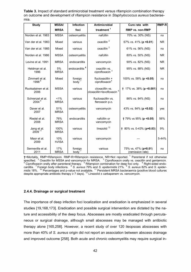
2.4.3.3. Clinical studies with rifampicin combination therapy
During 1983-2011 the clinical effect of rifampicin was evaluated in 16 reports. The vast ma-
jority of these studies were prospective, whereas three of the most recent ones were retro-
spective [82,83,176]. The studies differ widely with respect to MRSA occurrence. Some re-
port no MRSA [15,77], whereas others report high (76-100%) MRSA prevalence
[81,82,83,329,330]. In addition, definitions and inclusions of deep infection foci vary con-
siderably. Some studies report only endocarditis [81] or right-sided endocarditis [80],
whereas others report only osteomyelitis [75,77], and one study presented various deep in-
fection foci [15]. The main results of the clinical rifampicin combination studies are summa-
rized in Table 3. These studies compare the clinical outcome of rifampicin combination
therapy against standard therapy alone. Most studies with low MRSA occurrence report
some degree of improved clinical outcome due to rifampicin combination therapy as com-
pared with standard therapy alone, whereas studies with high MRSA occurrence mostly re-
port adverse effects and negative prognostic impact of rifampicin combination therapy.
Several small prospective studies with 14-65 patients from the 1980s report higher cure
rate or lower mortality with rifampicin combination therapy than with standard therapy
alone, although statistical significance is not achieved in many studies due to small sample
size [66,75,76,77]. The end-points, the MRSA prevalence and the deep focus classification
differ between these studies. One study with right-sided endocarditis among IDUs reported
a 100% cure rate of rifampicin combination therapy among patients who managed to com-
plete the study, but no control group was included [78]. Some studies have either failed to
observe resistance development to rifampicin [75,76] or rifampicin resistance is not men-
tioned [66,77].
40
During the 1990s and 2000s several prospective studies of varying size (33-381 patients)
and mostly low MRSA occurrence (0-11%) have reported positive results with rifampicin
combination therapy relative to standard therapy (Table 3). One study of right- sided endo-
carditis among IDUs compared oral rifampicin-ciprofloxacin with intravenous oxacillin or
vancomycin (in addition to gentamicin) and noted no difference in clinical failures in the ri-
fampicin combination group relative to the standard therapy group (5% vs. 12%) [80]. An-
other study compared oral rifampicin-ciprofloxacin with ciprofloxacin-placebo in foreign
body infections and showed significantly higher cure rates among patients with rifampicin
combination therapy (100% vs. 58%, p <0.05) [79].
A post hoc analysis of 331 MSSA bacteraemia patients, including various deep infection
foci patients but no MRSA bacteraemia cases, demonstrated improved three-month out-
come for adjunctive rifampicin therapy [15]. A prospective randomized trial with MSSA bac-
teraemia (2% MRSA) and a high number of various deep infection foci compared fleroxacin
and rifampicin combination against conventional intravenous monotherapy with flucloxacil-
lin or vancomycin [308]. The study observed similar cure rates for both therapies, although
rifampicin therapy resulted in several adverse reactions such as hepatitis. Furthermore, a
retrospective study with 17% MRSA cases concluded that rifampicin-fluoroquinolone ther-
apy, compared with other antimicrobial regimens, was associated with improved outcome
in patients with total hip or knee prosthetic infections, with no differences in outcome be-
tween MSSA and MRSA infections [176]. Altogether, four studies with high (51-100%)
prevalence of MRSA bacteraemia have investigated rifampicin combination therapy in en-
docarditis [81,82], osteomyelitis [331] or various deep infection foci [83] or in persistent
MRSA bacteraemia [329]. Of these studies, one included 10% heteroresistant vancomycin-
intermediate S. aureus (hVISA) cases [83].
Development of rifampicin resistance in S. aureus is a well-known disadvantage [67,68]
and has been observed in studies with high MRSA prevalence [82,83,84,329,331]. These
studies have reported development of rifampicin resistance in 5-56% of cases [82,83,
84,329], whereas one study reported unspecified rifampicin resistance [331]. All of these
studies have reported poorer clinical outcome with rifampicin combination therapy. How-
ever, studies with MSSA cases only [15,75] or low (1-17%) MRSA occurrence [176,308]
have reported no rifampicin resistance. Moreover, one study with mixed MSSA and MRSA
cases (percentages not provided) [76] reported no rifampicin resistance.
Conflicting results have been obtained with rifampicin combination therapy for prolonged
bacteraemia. A prospective randomized study with 42 native valve endocarditis patients
compared vancomycin-rifampicin combination with vancomycin only and observed a non-
significantly prolonged bacteraemia rate due to vancomycin-rifampicin combination therapy
41
(7 days vs. 9 days) [81]. In another study, rifampicin combination therapy was observed to
lead more often to prolonged bacteraemia than vancomycin or nafcillin treatment alone
[82]. In the latter study, each rifampicin resistance case was associated with rifampicin ini-
tiation during the bacteraemia phase. A study comparing MRSA and hVISA bacteraemia
treated with a vancomycin-rifampicin combination demonstrated prolonged bacteraemia
and higher rifampicin resistance for hVISA cases [83]. The authors proposed that due to
hVISA the vancomycin serum concentration was below the required hVISA MIC, and
hence, rifampicin therapy might be viewed as monotherapy resulting in rifampicin resis-
tance. In the fourth study, 19 elderly patients with prolonged MRSA bacteraemia were
treated with a glycopeptide-rifampicin combination. Patients who developed rifampicin re-
sistance (30%) showed no higher mortality [329]. A retrospective study from 2009, includ-
ing 35 patients with persistent MRSA bacteraemia and various deep infection foci, com-
pared the effect of linezolid (with or without carbapenem) against vancomycin (with or with-
out aminoglycoside or rifampicin) and reported significantly more rapidly achieved early
microbiological response in the linezolid group than in the vancomycin-rifampicin group.
Moreover, significantly higher (80%) mortality rate for the vancomycin and aminoglycoside-
rifampicin combination therapy as compared with vancomycin alone (40% mortality) or
linezolid alone (0% mortality) or linezolid and carbapenem (22% mortality) was observed
[84].
42
Table 3. Impact of standard antimicrobial treatment versus rifampicin combination therapyon outcome and development of rifampicin resistance in Staphylococcus aureus bacterae-mia.
Study MSSA/MRSA
Infectionfoci
Antimicrobialtreatment 1
Cure rate withRMP vs. non-RMP
RMP-R
Norden et al. 1983
Van der et al. 1983
Van der et al. 1985
Norden et al. 1986
Levine et al. 1991
Heldman et al.1996
Zimmerli et al.1998 8
Ruotsalainen et al.2006
Schrenzel et al.2004 9
Daver et al.2007
Riedel et al.2008
Jang et al.2009 11
Maor et al.2009
Senneville et al.2011
MSSA
Mixed
Mixed
MSSA
MRSA
5%MRSA
Mixed
MSSA
<1%MRSA
51%MRSA
76%MRSA
100%MRSA
10%hVISA
17%MRSA
osteomyelitis
various
various
osteomyelitis
endocarditis
endocarditis 6
foreignbody 7
various
various
osteomyelitis
endocarditis
various
various
foreignbody 7
nafcillin
oxacillin 2
oxacillin 2
nafcillin
vancomycin
oxacillin vs.ciprofloxacin 3
flucloxacillin +ciprofloxacin4
cloxacillin vs.cloxacillin+levofloxacin 5
flucloxacillin vs. fleroxacin p.o.
vancomycin
nafcillin orvancomycin
linezolid 12
vancomycin
various
70% vs. 30% (NS)
67% vs. 41% (p <0.01)
61% vs. 56% (NS)
80% vs. 50% (NS)
90% vs. 82% (NS)
95% vs. 88% (NS)
100% vs. 58% (p <0.05)
† 17% vs. 38% (p <0.001)
86% vs. 84% (NS)
43% vs. 84% (p <0.02)
† 79% vs 95% (p <0.05)
† 80% vs. 0-43% (p=0.03)
---
75% vs. 47% (p=0.01)(remission rate)
no
NR
no
NR
NR
NR
no
no
no
yes
56%
9%
5-44%
no
†=Mortality. RMP=Rifampicin. RMP-R=Rifampicin resistance. NR=Not reported. 1 Parenteral if not otherwisespecified. 2 Oxacillin for MSSA and vancomycin for MRSA. 3 Ciprofloxacin orally vs. oxacillin and gentamicin.4 Ciprofloxacin orally after parenteral therapy. 5 Rifampicin combination for deep foci only. 6 Right-sided endo-carditis. 7 Foreign body infections. 8 S. aureus 79% and S. epidermidis 21%. 9 S. aureus 82% and S. epider-midis 18%. 10 Percentages and p-value not available. 11 Persistent MRSA bacteraemia (positive blood culturesdespite appropriate antibiotic therapy 7 days). 12 Linezolid ± carbapenem vs. vancomycin.
2.4.4. Drainage or surgical treatment
The importance of deep infection foci localization and eradication is emphasized in several
studies [19,168,173]. Eradication and possible surgical intervention are dictated by the na-
ture and accessibility of the deep focus. Abscesses are mostly eradicated through percuta-
neous or surgical drainage, although small abscesses may be managed with antibiotic
therapy alone [165,258]. However, a recent study of over 120 ileopsoas abscesses with
more than 40% of S. aureus origin did not report an association between abscess drainage
and improved outcome [258]. Both acute and chronic osteomyelitis may require surgical in-
43
tervention such as surgical decompression, debridement of the infected area and revascu-
larization [333].
The requirement for surgical intervention is high in endocarditis, with up to 45% of left-sided
native valve and virtually 100% of prosthetic valve endocarditis cases [129,334,335],
whereas only a small proportion of right-sided endocarditis requires surgery [34]. For left-
sided native valve endocarditis, the following conditions are generally considered to require
surgical intervention: valvular regurgitation of haemodynamic significance (New York Heart
Association stage III-IV congestive heart failure), mobile and/or large-sized vegetations,
vegetations > 1 cm on the anterior mitral valve area, vegetation causing mechanical ob-
struction of valves, sinus Valsalva rupture, infection extending to the paravalvular area or
paravalvular abscess formation and persistent SAB ( 7 days) despite appropriate antim-
icrobial therapy [336,337]. For right-sided endocarditis, persistent and recurrent SAB or
continuous septic embolic complications are indications for surgical intervention [34].
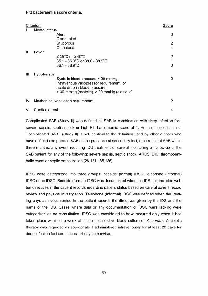
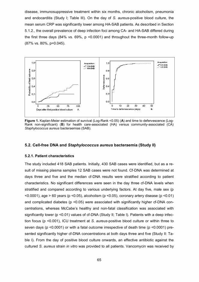
sion [2,13], diabetes [12] and multiple comorbidities [7,443] as independent predictors of
mortality. Many studies have applied the McCabe and Jackson criteria to categorize the
severity and prognosis of underlying diseases and comorbidity as healthy, non-fatal, ulti-
mately fatal or rapidly fatal [444]. Ultimately or rapidly fatal underlying diseases have been
shown to predict fatal outcome in many reports [3,22,188]. However, two studies with small
population sizes failed to detect an impact of comorbidities on outcome in SAB [120, 445].
2.7.2. Impact of community or health care acquisition on mortality
The impact of SAB acquisition on outcome has been controversial, with a trend in the last
decade of no significant association with mortality. SAB has traditionally been divided ac-
cording to acquisition into health care- (nosocomial) and community-associated cases.
Several studies in 1970-1990 found HA-SAB to be associated with higher mortality [51,427,
446], and the association of HA-SAB with higher age and comorbidity has been presented
as an explanation for the higher mortality. However, the majority of studies after 2000 have
not managed to detect any significant prognostic impact of SAB acquisition on outcome
[12,17,19,23,183,188,190,447], with the exception of one study connecting HA-SAB to
lower mortality [2]. HA-SAB has been observed to carry higher mortality than CA-SAB in
only two recent studies [442,448]. Hence, it appears that there might be a trend of diminish-
ing impact of SAB acquisition on outcome.
2.7.3. Impact of methicillin resistance on mortality
The relationship between MRSA and SAB prognosis has been thoroughly investigated, but
with conflicting results. Several studies have associated MRSA with a significantly higher
52
mortality rate in multivariate analyses [3,23,138,156,431]. Two meta-analyses in 2000 pre-
sented a significantly higher mortality rate in MRSA bacteraemia than in MSSA bacterae-
mia [49,449]. However, some studies have failed to connect MRSA bacteraemia to higher
mortality rates than MSSA bacteremia [12,168,188,450]. Hence, the results of the two
meta-analyses have been questioned due to lack of knowledge of hospital duration prior to
SAB in the original studies; when length of hospital stay was adjusted for, bacteraemias
with MRSA and MSSA presented similar mortality rates [451].
Several factors have been proposed to explain the higher mortality in MRSA bacteraemia.
Some reports have suggested that in patients with MRSA bacteremia higher mortality is
due to higher age [51,157], more severe underlying diseases [188], more severe illness at
bacteraemia onset (e.g. septic shock) and more complications such as pneumonia [51] as
compared with MSSA bacteraemia. One report states that higher mortality in MRSA bacte-
raemia than in MSSA bacteraemia is evident only in critically ill patients after adjustment for
disease severity and acute illness [138]. Various factors in MRSA treatment may be asso-
ciated with poorer outcome. MRSA has been connected to a delay in effective antibiotic
therapy onset [52,157], and vancomycin therapy has been regarded as having weaker effi-
cacy and a less effective blood-sterilizing effect, increasing the risk for persistence of SAB
relative to semi-synthetic penicillin or other -lactams [40,53,274,452,453]. Thus, although
vancomycin is the drug of choice, it has repeatedly been connected to treatment failure and
higher mortality than -lactams [22,38,39,40,41]. A prospective study of considerable size
(n=1865) presented glycopeptide (mostly vancomycin) therapy as an independent signifi-
cant mortality predictor [190]. Both pro- and retrospective reports have demonstrated a
connection between high vancomycin MIC and worse prognosis in patients with MRSA
bacteraemia [454,455], with MIC values 1.5-2 mg/L representing independent parameters
for treatment failure [454] and mortality [455]. Recently, a retrospective study reported high
vancomycin MIC ( 1.5 mg/L) as the only independent risk factor for complicated bacte-
raemia when MSSA bacteraemia patients were treated with vancomycin. However, MIC
1.5 mg/L was not associated with higher mortality [186]. Moreover, pathogen-related viru-
lence factors common in MRSA strains have been demonstrated, such as SCCmec type I
or agr (accessory gene regulator) group II polymorphism, which might be associated with
higher mortality or vancomycin treatment failure [456,457].
2.7.4. Impact of clinical manifestations on mortality
Severity of illness at Staphylococcus aureus bacteraemia onset
The severity of illness, particularly the presence of severe sepsis, septic shock or multi or-
gan failure, at onset of SAB are factors strongly predicting mortality [2,19,24,25,183,445].
53
Different scoring systems for assessment of severity of illness and outcome prediction have
been developed, e.g. the APACHE II, SOFA and PITT scores [139,140,142]. Severity of ill-
ness at S. aureus-positive blood culture, as evaluated by APACHE-scores [139], has been
shown to be significantly correlated with mortality [12,13,458,459]. However, among ICU
patients with sepsis, the Pitt bacteraemia score system has been observed to predict mor-
tality better than APACHE II with respect to sensitivity-specificity (67% and 74% for
APACHE II versus 68% and 82% for PITT scores) [141]. Other clinical conditions con-
nected to higher mortality have been acute organ dysfunction, need for mechanical ventila-
tion [431], acute renal failure [2], neutropenia [442] and thrombocytopenia [460]. Need for
ICU treatment [3,25,188,436] or ICU admission [3,443] has been observed to independ-
ently predict weaker outcome as compared with non-ICU patients.
Deep infection foci
The prognostic impact of deep infection foci in SAB has varied widely depending on the
type of deep focus. Several studies have presented pneumonia (OR 5.8-17.0) [12,17,51]
and endocarditis (OR 2.8-12.1) [3,24,456] as independent predictors for mortality. How-
ever, among IDUs endocarditis has been associated with significantly better outcome than
among non-IDUs [199]. In native valve infective endocarditis, factors such as age, perian-
nular abscess, heart failure, lack of surgical intervention and thromboembolic central nerv-
ous system event, have been associated with significantly weaker outcome [129]. One
study observed no association between deep infection foci and outcome [2], in contrast to
another that found metastatic foci to lead to weaker outcome [24].
Dosing and onset of antibiotic therapy
Several studies have demonstrated an adverse impact of delayed empiric antibiotic therapy
in both MSSA and MRSA bacteraemia [40,50,188,437]. The delay in time between S.
aureus-positive blood culture and initiation of empiric appropriate antibiotic administration,
after which mortality rises, has varied from 24 to 72 hours [40,50,188,461]. Contrary to this,
there are studies demonstrating a non-significant association between correctly timed ap-
propriate antibiotic therapy and survival rates in both MSSA and MRSA bacteraemia
[24,25,157]. One study concluded that only severely ill SAB patients with APACHE II points
> 15.5 gained from early onset of antibiotic therapy, whereas for SAB patients with
APACHE II < 15.5 delayed antibiotic therapy had no impact on mortality [50]. Some studies
have demonstrated the significance of appropriate dosing of antibiotic therapy. A prospec-
tive study of 278 cases of MSSA bacteraemia, including various deep infection foci, dem-
onstrated that a total daily dose of penicillinase-stable penicillin < 4 g was an independent
predictor for mortality and a total daily dose < 3 g an independent predictor for SAB recur-
rence [19]. Another study that included 87 cases of MRSA bacteraemia demonstrated in-
54
creased survival when vancomycin initiation took place within 48 hours of S. aureus-
positive blood culture results and the dose was 2.0 g/day [462].
Surgery and focus removal
SAB patients with non-eradicated and non-eradicable foci had higher mortality than pa-
tients who had their focus surgically (or by another intervention) removed (OR 4.17 vs. OR
3.75) [168]. An uneradicated focus was associated with significantly weaker outcome (OR
6.7) also in a study that included only 1% of patients with MRSA bacteraemia [19]. A large
retrospective study comparing vancomycin and -lactam therapy on outcome in solely
MSSA bacteraemia patients found eradicated infection foci to be an independent prognos-
tic factor for improved outcome (OR 0.3) [22]. Antibiotic therapy combined with early sur-
gery had significantly better outcome in native valve endocarditis in SAB as compared with
antibiotic therapy alone [463]. A study investigating the prognostic impact of IDS recom-
mendations on outcome of 244 SAB patients found unremoved, infected intravascular de-
vices to be significantly associated with SAB relapse and mortality (OR 6.5) [184]. A very
recent prospective study, including 58% of patients with MRSA bacteraemia, found a three-
day delay in removing eradicable foci to be associated significantly with persistent SAB
(OR 2.18) [30]. However, no direct connection between delayed eradication and mortality
was presented.
55
3. AIMS OF THE STUDY
Specific objectives of this study were as follows:
I To compare predisposing factors, disease progression and outcome of health care-
and community-associated methicillin-sensitive Staphylococcus aureus bacteraemia.
II To evaluate the prognostic value of the biomarker cell-free DNA in methicillin-sensitive
Staphylococcus aureus bacteraemia patients with early intensive care unit treatment.
III To investigate the impact of formal bedside infectious diseases specialist consultation,
informal telephone consultation and no consultation on disease progression and prog-
nosis of methicillin-sensitive Staphylococcus aureus bacteraemia.
IV To evaluate the impact of early and late adjunctive rifampicin therapy onset on disease
progression and prognosis in methicillin-sensitive Staphylococcus aureus bacteraemia
patients with deep infection foci.
56
4. MATERIALS AND METHODS
4.1. Study populations
The study populations consisted of prospectively collected patient data (Studies I and II)
and retrospectively collected data (Studies III and IV).
Study I was a prospective, multicenter study carried out in all five university central hospi-
tals and in seven central hospitals in Finland throughout two time periods: January 1999 to
May 1999 and January 2000 to August 2002. Adult patients with at least one blood culture
positive for Staphylococcus aureus were prospectively followed from a median of three
days after blood culture collection. In total, 1226 SAB patients were identified during the
study period and after controlling for exclusion criteria and excluding patients unable to
provide an informed consent or patients who refused participation, altogether 430 cases
were included. The exclusion criteria were age < 18 years, imprisonment, pregnancy (sus-
pected or proven), breastfeeding, epilepsy, bacteraemia during previous 28 days, po-
lymicrobial bacteraemia ( 3 microbes), history of allergy to any quinolone antibiotic, previ-
ous tendinitis during fluoroquinolone therapy, prior fluoroquinolone use for more than five
days before randomization, positive culture for S. aureus only from a central intravenous
catheter, neutropenia (<0.5 x 10 9/L), patients with bacteraemia due to MRSA (n=6) and a
S. aureus strain resistant to any fluoroquinolone.
Study II included the same prospectively collected patient data as in Study I, although due
to missing study samples (n=12), only 418 SAB cases were included in the analysis.
Study III was retrospective with 342 SAB cases representing all adult patients from Hel-
sinki University Central Hospital in Finland with at least one blood culture positive for
Staphylococcus aureus during two time periods: 2000–2002 and 2006–2007. The earlier
time period 2000-2002 included the patients from Studies I and II. Through the use of a
unique personal number provided to all Finnish residents, S. aureus isolates and patients
were matched. The patient data were collected from written (2000–2002) and electronic
(2006–2007) patient records. Due to missing patient records, 7 patients had to be ex-
cluded. Two time periods were included in order to exclude the possible effect of unidenti-
fied temporal differences regarding personnel, treatment practices or any other factors diffi-
cult to control. All cases with MRSA bacteraemia were excluded (5 cases during 2000-
2002, but none during 2006-2007).
57
Study IV included all patient data collected for Studies I and III. Cases with MRSA bacte-
raemia were excluded (n=6)
4.2. Study designs
Data collection included basic patient characteristics: age, gender, underlying diseases and
predisposing factors. SAB acquisition, infection focus and antibiotic treatment were regis-
tered. Surgical procedures, duration of hospitalization and infection foci confirmed through
radiological, bacteriological or pathological research or clinical suspicion only were docu-
mented. Radiological investigations and time to defervescence (axillary temperature < 37.5oC) were recorded. Laboratory findings included plasma cf-DNA and CRP concentrations at
days three and five from the positive blood culture sampling. IDSC during the first week af-
ter the first blood culture positive for S. aureus was documented. SAB relapse within three
months was documented.
Study I was a prospective study. An IDS followed up each SAB patient for three months.
SAB cases were categorized according to acquisition into CA- and HA-SAB. The differ-
ences of CA- and HA-SAB regarding patient characteristics, underlying conditions, predis-
posing factors and prevalence of deep infection foci within three days and three months
were analysed with univariate analysis. Three-month survival of CA- and HA-SAB were es-
timated with the Kaplan-Meier method and prognostic factors analysed with multivariate
analysis. The primary end-point was case fatality at 28 days and at three months. Secon-
dary end-points were the time to defervescence, decrease in serum CRP concentration
and number of deep infection foci within three days and three months.
Study II was a prospective study. Plasma cf-DNA at days three and five from the positive
blood culture were stratified and compared according to patient demographics, underlying
conditions, severity of illness, deep infection foci, treatment in ICU and mortality for 1) the
whole patient population and 2) patients receiving ICU treatment within seven days of S.
aureus-positive blood culture. Receiver-operating characteristic (ROC) analyses for cf-DNA
and CRP were performed, and cut-off values for day three and five cf-DNA were calcu-
lated. The patient demographics, underlying conditions, severity of illness, deep infection
foci, treatment in ICU and mortality were then stratified and compared according to the cf-
DNA cut-off values of days three and five. Prognostic factors were analysed according to
the Cox regression model. The primary end-point was mortality at seven days, 28 days or
three months, and secondary end-points were deep infection foci localized during the
three-months follow-up.
58
Study III was a retrospective study. The SAB cases were categorized according to bedside
(formal), telephone (informal) or no IDSC within one week of S. aureus-positive blood cul-
ture. Patients with fatal outcome within three days after S. aureus-positive blood culture
were excluded to allow for the possibility of death occurring before IDSC, as the mean time
lapse between blood culture collection and IDSC was three days. Patient demographics,
underlying conditions, severity of illness, deep infection foci, treatment in ICU and mortality
were stratified and compared according to IDSC. Multinomial logistic regression analyses
were performed to simultaneously compare the three consultation groups. The Kaplan-
Meier method was used to compare the impact of various IDSC groups on mortality and
defervescence. Prognostic factors were analysed according to the Cox regression model
in order to determine the prognostic impact of IDSC. The primary end-point was case fatal-
ity at 28 days and three months. Secondary outcome measures were the time to deferves-
cence, any inadequate antibiotic therapy, duration of hospitalization, number of deep infec-
tion foci and any relapse of SAB within three months.
Study IV was a retrospective study. The patient population was categorized according to
whether rifampicin therapy was received, whether it was initiated within seven days (early)
or seven days past (late) positive blood culture and whether it was continued for at least 14
days. The main analyses were performed by excluding cases with a fatal outcome within
three days as well as excluding patients with alcoholism and acute or chronic liver disease
to allow for death before positive blood culture results (the mean time-lapse between blood
culture collection and positive blood culture results was three days) and to account for ri-
fampicin therapy contraindications (alcoholism and liver disease). Moreover, as a parallel
analysis, the patient population was analysed by excluding cases with a fatal outcome
within 14 days of blood culture collection to allow for death before completing 14 days of ri-
fampicin therapy.
Patient demographics, underlying conditions, severity of illness, deep infection foci, treat-
ment in ICU, antibiotic therapy and mortality were stratified and compared according to ri-
fampicin therapy 14 days or < 14 days. Cox regression analysis was performed to evalu-
ate the prognostic value of early and late rifampicin therapy for 1) the whole patient popula-
tion, 2) patients with a deep infection foci. The prognostic impact of early and late rifampicin
therapy for 14 days or < 14 days in the whole patient population and among patients with
deep infection foci was analysed using the Kaplan-Meier method. The primary end-point
was mortality at three months and the secondary end point deep infection foci during the
three-month follow-up. The rifampicin dose was 450 mg (< 50 kg of body weight) or 600 mg
(> 50 kg of body weight) given once daily.
59
4.3. Definitions of terminology
Prognosis or severity of underlying conditions was classified as healthy (no approximated
time period to death), non-fatal (no approximated time period to death), ultimately fatal (ap-
proximated death within 6 months - 5 years) or rapidly fatal (approximated death within 6
months) according to the McCabe and Jackson criteria [444]. SAB was considered HA if
the first positive S. aureus blood culture was received 48 hours after admission to hospi-
tal or within two days of admission when the patient had been discharged from a hospital
within seven days precedingly or when the patient was a long-term care facility resident
during the previous two months or was attending haemodialysis. SAB was considered CA
when the first positive S. aureus blood culture was received within 48 hours of hospital ad-
mission without any preceding hospitalization within seven days. Deep infection foci com-
prised deep-seated abscesses, endocarditis, foreign body infection, meningitis, mediastini-
tis, osteomyelitis, pneumonia and septic arthritis. Central venous catheter (CVC) infections
were defined according to the guidelines of the IDSA [42]. The modified Duke criteria were
used to define endocarditis as definite, possible or rejected according to the presence of
major and minor criteria [248]. SAB relapse was defined as the same pattern of resistance
and PFGE typing for the two S. aureus strains. Severe sepsis was regarded as sepsis in-
cluding 1) hypotension, i.e. systolic blood pressure < 90 mmHg, mean arterial blood pres-
sure < 70 mmHg, or a systolic blood pressure decrease > 40 mmHg in adults or < 2 SD be-
low normal for age or 2) hypoperfusion, i.e. hyperlactataemia (>1 mmol/L) or decreased
capillary refill or 3) organ failure, i.e. arterial hypoksaemia Pao2 / F102 < 300, acute oliguria
comparison of the three different IDSC processes (Study III).
Receiver-operating characteristic (ROC) curves were produced for cf-DNA and CRP to es-
timate the discriminative power of these two in predicting three-month mortality. For each
ROC curve, the area under the curve (AUC) was calculated. The Youden index was de-
fined from the ROC curves as either the ROC-curve point maximizing both sensitivity and
specificity values or the sensitivity and specificity sum with the highest value in order to lo-
cate the cut-off point.
Survival estimates and time to defervescence were presented with the Kaplan-Meier
method using the Log-Rank test to compare the graphs (Studies I, III and IV). The Kaplan-
Meier method was used to present survival estimates and time to defervescence (Studies I,
III and IV). The ROC curve cut-off points were used for the Kaplan-Meier survival estima-
tion (Study II). All tests were two-tailed and p <0.05 was considered significant. All analyses
were performed using SPSS software, version 12.0 (SPSS Inc., Chicago, IL, USA).
4.6. Ethical aspects
The study protocols were approved by the ethics committees of all study sites and by the
Institutional Review Board and the Ethics Committee of Helsinki University Central Hospi-
tal. In Study I, patients provided a signed informed consent. Severely ill patients, e.g. pa-
tients in an unconscious state with assisted ventilation, were included as well without a
signed informed consent, as these patients were presumed to gain from the study medica-
tion. A signed informed consent was provided by the patient or a representative as soon as
possible.
63
5. RESULTS
5.1. Community- and health care-associated bacteraemia (Study I)
5.1.1. Patient characteristics
The 430 SAB cases included 198 (46%) CA-SAB and 232 (54%) HA-SAB patients.
CA-SAB patients, as compared to HA-SAB patients, were significantly more often HIV-
positive (4% vs. <1%, p=0.018) and had more often chronic alcoholism (16% vs. 7%, p
=0.002), liver disease (25% vs. 7%, p <0.0001) and injection drug abuse (21% vs. 1%, p
<0.0001). HA-SAB patients underwent significantly more frequently a foreign body implan-
tation within one year preceding SAB (41% vs. 9%, p <0.0001), surgical procedures within
three months (44% vs. 8%, p <0.0001) or CVC application (23% vs. 1%, p <0.0001).
Wounds and chronic skin diseases were significantly more common among CA-SAB pa-
tients than among HA-SAB patients (59% vs. 46%, p=0.006) (Study I; Table I).
HA-SAB patients, as compared to HA-SAB patients, were significantly older (62.4 ± 15.2
vs. 52.9 ± 19.5 years, mean ± SD, p <0.0001), had more often an ultimately or rapidly fatal
underlying disease (41% vs. 12%, p <0.0001) and were more chronically ill with cardiovas-
cular disease (55% vs. 27%, p <0.0001), chronic renal failure (24% vs. 3%, p <0.0001), di-
alysis care (20% vs. <1%, p <0.0001), heart valve disease (22% vs. 7%, p <0.0001), ma-
lignancy (21% vs. 7%, p <0.0001), complicated diabetes (21% vs. 11%, p=0.003), haema-
tological malignancy (6% vs. <1%, p=0.001) or connective tissue or rheumatic disease
(15% vs. 8%, p=0.021) and more often received immunosuppressive treatment (19% vs.
7%, p <0.0001).
5.1.2. Clinical aspects
At S. aureus-positive blood culture and within the first week of treatment, no significant dif-
ferences between the patients with CA-SAB or HA-SAB were seen in severe sepsis (7%
vs. 6% at positive blood culture and 13% during the first week for both groups, respectively)
septic shock (3% for both groups and 7% vs. 4%, respectively) or need for ICU treatment
(16% vs. 15% and 23% vs. 21%). During the first three days of SAB, less deep infection
foci were localized among patients with HA-SAB relative to CA-SAB (69% vs. 84%, p
<0.0001), and throughout the three-month follow-up a slight increase was observed in the
number of patients with identified deep foci (80% vs. 87%, p=0.045) (Table 5). HA-SAB pa-
tients presented significantly more often mediastinitis or infection of CVC or peripheral
catheter or permanent foreign bodies, whereas osteomyelitis and deep-seated abscesses
were seen significantly more frequently in CA-SAB. No difference was observed in the oc-
64
currence of pneumonia, septic arthritis, endocarditis or cutaneous infections between CA-
and hA-SAB patients. The prevalence of CA- and HA-SAB patients without any diagnosed
infection focus was low at three days (6% vs. 3%) and remained low throughout the three-
month follow-up (5% vs. 2%); however, the difference at both time-points was non-
significant (Table 5).
Table 5. Comparison of infection focus and mortality in community- (CA-) and health care-(HA) associated Staphylococcus aureus bacteraemia at day three and at three months.Data are number (%) of patients.
From the first day of the positive blood culture, all patients with CA- and HA-SAB were
treated with an antibiotic effective against the isolated S. aureus strain. The vast majority of
patients received a -lactam antibiotic whereas only 12% were treated with vancomycin.
5.1.4. Outcome
The case fatality at 28 days did not differ between CA- and HA-SAB (11% vs. 14%),
whereas at three months it was significantly higher among the HA-SAB patients (22% vs.
13%, p=0.023). Overall, the case fatality at 28 days was 13% and at three months 18%.
The mortality difference between CA- and HA-SAB remained significant in Kaplan-Meier
analysis. However, no significant difference between CA- and HA-SAB was observed in
time to defervescence (Figure 1). Prognostic factors for a fatal outcome within 28 days ac-
cording to binary logistic regression analysis were age > 60 years, ultimately or rapidly fatal
65
disease, immunosuppressive treatment within six months, chronic alcoholism, pneumonia
and endocarditis (Study I; Table III). On the day of S. aureus-positive blood culture, the
mean serum CRP was significantly lower among HA-SAB patients. As described in Section
5.1.2., the overall prevalence of deep infection foci among CA- and HA-SAB differed during
the first three days (84% vs. 69%, p <0.0001) and throughout the three-month follow-up
(87% vs. 80%, p=0.045).
Figure 1. Kaplan-Meier estimation of survival (Log-Rank <0.05) (A) and time to defervescence (Log-Rank non-significant) (B) for health care-associated (HA) versus community-associated (CA)Staphylococcus aureus bacteraemias (SAB).
5.2. Cell-free DNA and Staphylococcus aureus bacteraemia (Study II)
5.2.1. Patient characteristics
The study included 418 SAB patients. Initially, 430 SAB cases were identified, but as a re-
sult of missing plasma samples 12 SAB cases were not found. Cf-DNA was determined at
days three and five and the median cf-DNA results were stratified according to patient
characteristics. No significant differences were seen in the day three cf-DNA levels when
stratified and compared according to various underlying factors. At day five, male sex (p
<0.0001), age > 60 years (p <0.05), alcoholism (p <0.05), coronary artery disease (p <0.01)
and complicated diabetes (p <0.05) were associated with significantly higher cf-DNA con-
centrations, whereas McCabe’s healthy and non-fatal classification was associated with
significantly lower (p <0.01) values of cf-DNA (Study II; Table I). Patients with a deep infec-
tion focus (p <0.001), ICU treatment at S. aureus-positive blood culture or within three to
seven days (p <0.0001) or with a fatal outcome irrespective of death time (p <0.0001) pre-
sented significantly higher cf-DNA concentrations at both days three and five (Study II: Ta-
ble I). From the day of positive blood culture onwards, an effective antibiotic against the
cultured S. aureus strain in vitro was provided to all patients. Vancomycin was received by
66
a minority (2%) of the patients and vancomycin as the only antibiotic was given to only 1%,
whereas the vast majority of patients were treated with a -lactam antibiotic (76%).
5.2.2. Treatment in intensive care unit and cell-free DNA
Within three days of S. aureus-positive blood culture, 87 patients (21%) and within seven
days 99 patients (24%) needed ICU treatment. Regarding demographics and underlying
conditions, the only difference was that ICU patients significantly more often suffered from
alcoholism (p <0.05) than non-ICU patients. However, deep infection foci and mortality at
seven days, 28 days and three months were significantly higher among ICU patients rela-
tive to their non-ICU counterparts. These results were similar when ICU treatment during
the first three days only or during the fiest seven days after positive blood culture were
taken into account (data not shown).
SAB patients who needed ICU treatment within seven days of positive blood culture were
analysed as a subgroup according to three-month survival. At day 3, the non-survivors in
the ICU group presented significantly higher cf-DNA values for age (p <0.01), healthy or
non-fatal disease, alcoholism and cardiovascular disease (p <0.05), ICU-specific character-
istics such as severe sepsis, need for mechanical ventilation or inotropia support or Pitt
bacteraemia scores 4 (p <0.05) as well as deep infection foci and complicated SAB (p
<0.01), whereas no difference was seen between CA- and HA-SAB acquisition. However,
at day 5, no significant difference between ICU survivors and non-survivors was observed
(Study II; Table II) and Table 6 (below).
67
Table 6. Plasma cf-DNA concentration ( g/mL) at days 3 and 5 from the positive blood cul-ture in 99 patients with Staphylococcus aureus bacteremia (SAB) in the intensive care unit(ICU) within 7 days of positive blood culture. Patients are divided according to survivorsand non-survivors at the three-months follow-up. Values are given as median (quartiles).NS=non significant.
cf-DNA at day 3 cf-DNA at day 5
ICUsurvivor
ICUnon-survivor
p-value X
ICUsurvivor
ICUnon-survivor
p-value X
DemographicsAge >60 years
HA-SAB 1
UnderlyingconditionsHealthy or nonfatal 2
Cardiovascular
Alcoholism
ICU characteristicsSevere sepsis
Mechanic ventilation
Inotropic support
Pitt scores 4
Infection focusAny deep infection
Complicated SAB 3
1.62(1.37-2.16)
1.60(1.37-2.13)
1.67(1.40-2.12)
1.65(1.38-2.13)
1.68(1.37-2.30)
1.69(1.40-2.11)
1.82(1.43-2.30)
1.77(1.50-2.23)
1.83(1.42-2.42)
1.66(1.40-2.15)
1.66(1.39-2.14)
3.97(2.55-9.46)
2.01(1.59-4.84)
2.37(1.80-7.92)
3.97(2.32-3.63)
5.96(2.37-11.0)
3.94(1.91-7.22)
3.60(1.91-7.22)
3.05(2.01-5.96)
3.05(2.01-5.96)
3.05(2.01-4.84)
3.07(2.09-5.68)
<0.01
NS
<0.05
<0.05
<0.05
<0.05
<0.05
<0.05
<0.05
<0.01
<0.01
1.57(1.31-2.46)
1.53(1.31-1.89)
1.53(1.29-1.93)
1.72(1.38-2.44)
1.81(1.34-1.93)
1.64(1.31-1.88)
1.90(1.48-2.66)
1.63(1.29-1.97)
1.76(1.45-2.45)
1.60(1.29-2.18)
1.59(1.29-2.12)
2.27(1.59-3.01)
2.27(1.14-2.26)
2.27(1.52-3.01)
2.30(1.19-2.26)
2.96(1.70-2.73)
2.66(1.72-3.48)
2.64(1.71-3.48)
2.31(1.89-3.33)
2.31(1.89-3.33)
2.27(1.56-2.66)
2.31(1.89-3.33)
NS
NS
NS
NS
NS
NS
NS
NS
NS
NS
NS
X Mann-Whitney U-test. 1 Health care-associated. 2 Classified according to McCabe and Jackson [444].3 Defined as SAB and deep infection focus, severe sepsis, septic shock or Pitt bacteraemia scores 4.
5.2.3. Sensitivity and specificity of cell-free DNA
Receiver-operating characteristic (ROC) curves were applied to estimate predictive value
of cf-DNA and CRP on the 28-day and three-month outcome in patients treated in the ICU
within 7 days of S. aureus-positive blood culture (n=99) and in non-ICU patients (n=319).
For ICU and non-ICU patients, the ROC analysis revealed that both the 3-day and the 5-
day cf-DNA significantly predicted fatal outcome. This finding held when the data were ana-
lysed separately for the 28-day and the three-month follow-up. CRP, however, had no pre-
dictive value for outcome.
The 3-day AUC for cf-DNA in ICU patients was 0.71 (95% CI 0.57-0.84, p <0.01) with a cut-
off value of 1.99 g/mL and a corresponding sensitivity of 67% and specificity of 77% in
predicting fatal outcome within three months. The corresponding 5-day cf-DNA AUC was
0.71 (95% CI 0.58-0.84, p <0.01) with a cut-off value of 1.69 g/mL and a sensitivity of 63%
68
and a specificity of 60%. For the 28-day mortality, the 3-day cf-DNA AUC was 0.66 (95% CI
0.52-0.81, p <0.05) with a cut-off value of 1.99 g/mL and a sensitivity of 60% and a speci-
ficity of 73% (data not shown). For CRP, the 3-day AUC was 0.46 (95% CI 0.32-0.59,
p=0.55) and the 5-day AUC 0.47 (95% CI 0.34-0.61, p=0.67). Among non-ICU patients,
AUC for 3-day cf-DNA was 0.64 (95% CI 0.55-0.74, p <0.01) with a cut-off value of 1.57
ug/mL and a sensitivity of 62% and a specificity of 61% in predicting a fatal outcome in the
three-month follow-up. The corresponding 5-day cf-DNA AUC was 0.68 (95% CI 0.59-0.77,
p <0.01) with a cut-off value of 1.49 g/mL and a sensitivity of 65% and a specificity of
61%. For CRP, the 3-day AUC was 0.46 (95% CI 0.36-0.57, p=0.48) and the 5-day AUC
0.51 (95% CI 0.41-0.62, p=0.80) (Study II; Figure 1a-b)
ROC analysis was applied to predict seven days mortality for day three and five cf-DNA in
patients with SAB divided according to Pitt bacteraemia scores and intensive care unit
treatment (see Figure 2a-d below).
69
Figure 2.Receiver-operating characteristic (ROC) curves for cf-DNA predicting 7-day mortality in patients with Staphylo-coccus aureus bacteraemia (SAB) divided according to Pitt bacteraemia scores and intensive care unit treat-ment.
A. Pitt bacteraemia score=0. The AUC for day 3 cf-DNA was 0.66 (95% CI 0.40-0.91) (p-value non-significant)with a cut-off value of 1.59 mg/mL and a sensitivity of 60% and a specificity of 61%. The AUC for day 5 cf-DNAwas 0.69 (95% CI 0.54-0.84) (p-value non-significant) with a cut-off value of 1.49 mg/mL and a sensitivity of80% and a specificity of 60%.
B. Pitt bacteraemia scores >0. The AUC for day 3 cf-DNA was 0.88 (95% CI 0.77-0.98) (p <0.01) with a cut-offvalue of 2.01 mg/mL and a sensitivity of 86% and a specificity of 77%. The AUC for day 5 cf-DNA was 0.76(95% CI 0.56-0.95) (p <0.05) with a cut-off value of 1.96 mg/mL and a sensitivity of 71% and a specificity of81%.
C. Pitt bacteraemia score=0 and no intensive care unit. The AUC for day 3 cf-DNA was 0.59 (95% CI 0.34-0.86)(p-value non-significant) with a cut-off value of 1.45 mg/mL and a sensitivity of 75% and a specificity of 48%.The AUC for day 5 cf-DNA was 0.72 (95% CI 0.55-0.89) (p-value non-significant) with a cut-off value of 1.49mg/mL and a sensitivity of 75% and a specificity of 63%.
D. Pitt bacteraemia scores >0 and intensive care unit treatment. The AUC for day 3 cf-DNA was 0.91 (95% CI0.81-1.00) (p <0.01) with a cut-off value of 2.35 mg/mL and a sensitivity of 80% and a specificity of 91%. TheAUC for day 5 cf-DNA was 0.78 (95% CI 0.58-0.97) (p <0.05) with a cut-off value of 2.14 mg/mL and asensitivity of 80% and a specificity of 79%.
70
5.2.4. Prognostic value of cell-free DNA relative to other prognostic factors
The prognostic value of cf-DNA was analysed with proportional hazards regression (Cox
regression model) for ICU and non-ICU patients by applying the cf-DNA cut-off values
(from Study II; Figure 1a-b) as one statistical parameter. In univariate analysis, factors
among ICU patients that were associated with three-month mortality were age > 60 years
chronic pulmonary disease (p <0.01), presence of a deep infection focus (p <0.05) and cf-
DNA > 1.57 g/mL (at day 3) (p <0.01) were all associated with fatal outcome within three
months, whereas healthy or non-fatal underlying disease had a protective prognostic im-
pact (p <0.0001) (Table 7).
In the Cox regression model for ICU patients only Pitt bacteraemia scores 4 (OR 4.47, CI
1.94-10.3) (p <0.0001) and cf-DNA > 1.99 g/mL (at day 3) (OR 3.56, CI 95% 1.69-7.59) (p
<0.001) predicted a fatal outcome whereas, healthy or non-fatal underlying disease (OR
0.34, CI 95% 0.15-0.77) (p <0.05) had a protective prognostic value. Among non-ICU pa-
tients, corticosteroid use (OR 2.89, CI 95% 1.39-6.07) (p <0.01), chronic pulmonary dis-
ease (OR 2.45, CI 95% 1.21-4.96) (p <0.05) and healthy or non-fatal underlying disease
(OR 0.26, CI 95% 0.11-0.63) (p <0.01) had a significant impact on prognosis (Table 7).
71
Table 7. Prognostic factors for three-month mortality in Staphylococcus aureus bacterae-mia patients with (n=99) or without (n=319) intensive care unit treatment within 7 days of S.aureus-positive blood culture. CI, confidence interval; OR, odds ratio; NS, non-significant.
Univariateanalysis
OR (95% CI)
p-value
Cox regressionanalysis
OR (95% CI)
p-value
Intensive care unitAge > 60 yearsHealthy or non-fatal A
Chronic alcoholismCorticosteroid use B
Chronic renal failure C
Dialysis (haemo- or peritoneal)Diabetes (and complications)Haematological malignancyAny deep infectionEndocarditisInotropia needMechanical ventilationPitt bacteraemia scores 4Pitt bacteraemia scores 3Pitt bacteraemia scores 2cf-DNA cut-off 1.99 g/mL D
Non-intensive care unitAge > 60 yearsHealthy or non-fatal A
Chronic alcoholismCorticosteroid use B
Dialysis (haemo- or peritoneal)Diabetes (and complications)Haematological malignancyChronic pulmonary diseaseAny deep infectionEndocarditiscf-DNA cut-off 1.57 g/mL D
A Classified according to McCabe and Jackson [444]. B In the 6 months preceding the positive blood culture.C Chronically elevated plasma creatinine (>180 mol/L). D Cut-off value at day 3 as in Figure 1a-b in Study II.
The Cox regression model above was performed using a day 3 cf-DNA cut-off value. When
performed with a day 5 cf-DNA cut-off value of 1.69 g/mL for ICU patients or 1.49 g/mL
for non-ICU patients, the prognostic value in univariate analysis was significant for both
ICU patients and non-ICU patients (p <0.05). However, in Cox regression model the 5-day
cf-DNA cut-off values had no significant prognostic value for either ICU or non-ICU pa-
tients.
The Kaplan-Meier method was applied to estimate the survival for ICU and non-ICU pa-
tients according to 3-day and 5-day cf-DNA cut-off values. According to the Kaplan-Meier
Log-Rank test, both 3-day and 5-day cf-DNA cut-off values presented significant differ-
ences in three-month survival both for ICU patients at day three (p <0.0001) and at day five
(p <0.05) and for non-ICU patients at days three and five (p <0.01) (data not shown).
72
5.3. Impact of infectious disease specialist consultation (IDSC) on Staphylo-coccus aureus bacteraemia outcome (Study III)
5.3.1. Patient characteristics
The vast majority of the SAB patients received formal bedside IDSC (72%), whereas infor-
mal telephone conversation-based IDSC was given much less often (18%) and a small
proportion of patients had no IDSC (10%). No significant differences emerged in sex, age,
nosocomial acquisition or underlying diseases between patients with formal bedside and in-
formal telephone IDSC. However, significantly more ultimately or rapidly fatal underlying
diseases were seen among SAB patients managed without any IDSC (p=0.008). The mean
time-lapse between blood culture collection and IDSC was 3 days for both formal bedside
and informal telephone groups (Study III; Table I). No significant differences in the occur-
rence of severe sepsis at S. aureus-positive blood culture were seen between the formal
bedside (7%) and the informal telephone consultation (10%) groups, whereas patients
without IDS consultation (31%) suffered from severe sepsis significantly more often than
bedside IDS consultation patients (p <0.0001). The need for ICU treatment among informal
telephone IDSC patients (34%) was more common than among formal bedside IDSC pa-
tients (21%) during the first 3 days after S. aureus-positive blood culture (p=0.037),
whereas within the first week the difference was non-significant (37% for informal tele-
phone IDSC and 29% for formal bedside IDSC, p=0.22) (Study III; Table I).
5.3.2. Impact on radiological diagnostics
The use of echocardiography - transthoracic (TTE) or transoesophageal (TEE) - did not dif-
fer between formal bedside and informal telephone IDSC (77% vs. 71% for TTE and 11%
vs. 3% for TEE). The use of whole-body computed tomography was less common among
informal telephone IDSC (55%) (p=0.049) and no IDSC (29%) (p <0.0001) than among for-
mal bedside IDSC (68%). The use of leukocyte indium-111 scintigraphy was more common
in formal bedside IDSC (43%) than in informal telephone (13%) or no IDSC (9%) (p <0.001)
(Study III; Table I). However, after adjustment for mortality before IDSC, leukocyte indium-
111 scintigraphy was the only radiological investigation provided less often to informal tele-
phone IDSC patients than to formal bedside IDSC patients (p=0.011) (Study III; Table II).
5.3.3. Impact on deep infection focus localization
Formal bedside IDSC resulted significantly more often in identification of a deep infection
focus (78%) than to informal telephone IDSC (53%) or no IDSC (29%), and this was seen
in all deep infection focus types, except endocarditis, for which the difference between for-
73
mal bedside (16%) and informal telephone IDSC (7%) was non-significant (p=0.055) (Study
III; Table I). According to multinomial logistic regression analysis, the odds ratio (OR) for
deep infection focus localization was only 0.15 (95% CI 0.06-0.38, p <0.0001) for informal
telephone IDSC and 0.13 (95% CI 0.03-0.54, p=0.005) for no IDSC relative to formal bed-
side IDSC (Study III; Table II).
5.3.4. Impact on antibiotic treatment
From the first day of positive blood culture, each patient received an antibiotic effective
against S. aureus. Most patients received a -lactam antibiotic (93%), whereas only 3%
were treated with vancomycin. Patients with formal bedside IDSC (85%) received signifi-
cantly more often proper length of antibiotic therapy relative to informal telephone IDSC
(63%) (p=0.008) or no IDSC patients (54%) (p=0.004) (Study III; Table I). However, after
adjustment for mortality before IDSC, no difference in number of patients with proper antibi-
otic length was observed (Study III: Table II).
5.3.5 Impact on outcome
Formal bedside IDSC resulted in longer mean duration of hospitalization (38.7 ± 21.7 days)
than either informal telephone IDSC (30.6 ± 23.0 days) (p=0.014) or no IDSC (24.9 ± 24.8)
(p=0.001) (Study III; Table I). Mean time to defervescence was shorter for formal bedside
IDSC (6.7 ± 9.7 days) than for informal telephone IDSC (12.6 ± 13.4) (p=0.001) or no IDSC
(13.4 ± 14.7) (p =0.003) (Study III; Table I). Similar results were obtained after early 3-day
mortality was omitted (Figure 3b) (Study III; Table II and Figure 1a). No difference in mor-
tality was seen between formal bedside and informal telephone IDSC within the first 3
days. However, mortality was lower among patients with bedside IDSC at 7 days (1% vs.
8%, p=0.001), at 28 days (5% vs. 16%%, p=0.002), and at three months (9% vs. 29%, p
<0.0001) than among patients treated with informal telephone IDSC (Study III; Table I). Pa-
tients who received no IDSC had a high mortality already at 3 days (26%) and had a higher
mortality throughout the three-month study period than patients receiving formal bedside
IDSC (9% vs 46%%, p <0.001) (Study III; Table I). After adjustment for early death, the 28-
day mortality for formal bedside IDSC (5%) as compared with informal telephone IDSC
(15%) (p=0.08) and no IDSC (12%) (p=0.68) became non-significant. However, the three-
month mortality remained significantly lower for formal bedside IDSC (9%) than for the
other two IDSC groups (28% for informal telephone IDSC and 27% for no IDSC) (Figure
3a) (Study III; Table II and Figure 1b).
74
Figure 3. Kaplan-Meier analysis for fatal outcome (3a) and time to defervescence (3b) in patients with Staphy-lococcus aureus bacteraemia according to telephone (informal) (n=62) or bedside (formal) (n=245) infectiousdisease specialist consultation. Log-Rank test for fatal outcome in informal versus formal consultation (p<0.0001) and for defervescence in informal versus formal consultation (p=0.001). Patients who died during thefirst 3 days of S. aureus-positive blood culture (n=11) were excluded.
Prognostic factors for three-month mortality were analysed with proportional hazards re-
gression (Cox regression model). When all prognostic determinants were taken into ac-
count, the factors associated with fatal outcome were informal telephone IDSC (p=0.01), no
IDSC (p=0.002), pneumonia (p=0.001), ICU within three days of S. aureus-positive blood
culture (p =0.012) and corticosteroid therapy (p=0.01), whereas healthy or non-fatal under-
lying disease (p <0.0001), leukocyte indium-111 scintigraphy (p=0.021) and whole-body
computed tomography (p=0.022) had positive prognostic value (Table 8). When compared
with formal bedside IDSC, the odds ratio (OR) for mortality in informal telephone IDSC was
2.31 (95% CI 1.22-4.38) (Study III; Table II).
75
Table 8. Prognostic factors for three-month mortality in 331 patients with Staphylococcusaureus bacteraemia. Patients with a fatal outcome within 3 days of S. aureus-positive bloodculture were excluded. CI, confidence interval; OR, odds ratio.
Univariateanalysis
OR (95% CI)
p-value
Cox regressionanalysis
OR (95% CI)
p-value
Positive prognostic impactHealthy or non-fatal disease A
Leukocyte indium-111 scintigraphy
Whole-body computed tomography
Negative prognostic impactPneumonia
ICU within 3 days
Corticosteroid therapyB
Telephone IDS within 1 week
No IDS consultation within 1 week
0.11 (0.05 - 0.022)
0.41 (0.19 - 0.87)
0.43 (0.23 - 0.80)
2.31 (1.23 - 4.33)
1.96 (1.00 - 3.83)
5.48 (1.93 - 15.6)
3.21 (1.63 - 6.33)
2.51 (0.99 - 0.37)
<0.0001
0.018
0.007
0.008
0.046
<0.0001
<0.0001
0.045
0.18 (0.09 - 0.35)
0.40 (0.19 - 0.87)
0.49 (0.26 - 0.90)
2.74 (1.49 - 5.05)
2.28 (1.19 - 4.15)
2.98 (1.29 - 6.85)
2.31 (1.22 - 4.38)
3.56 (1.59 - 7.94)
<0.0001
0.021
0.022
0.001
0.012
0.01
0.01
0.002A Underlying Diseases characterized according to McCabe and Jackson [444]. B Systemic prednisone >10mg/day or equivalent for >1 month.
During the first 7 days altogether 31% of patients needed ICU treatment. Of these patients
treated in ICU, survivors and non-survivors were compared separately and prognostic fac-
tors for three-month mortality were analysed. Survival was significantly associated with
formal bedside IDSC (p <0.0001), healthy or non-fatal underlying diseases (p=0.003) and
performed whole-body computed tomography (p=0.027). However, ultimately or rapidly fa-
tal diseases (p=0003), informal telephone IDSC (p=0.001) and no IDSC (p=0.008) were
associated with fatal outcome among ICU patients (Study III; Table IV).
Factors difficult to control in retrospective studies are hospital-related temporary differences
in treatment or personnel practices, and thus, in order to exclude the effect of unidentified
differences, two different time periods for data collection were included in Study III.
Moreover, two study periods were regarded as mandatory as most patients from the first
study period had participated in our previous prospective study [15]. This naturally raises
the question of whether any differences are present in the results when the data of the two
time periods are analysed separately. As a parallel investigation (results not mentioned and
data not shown in Study III) the data were analyzed and prognostic factors for the three-
month mortality according to Cox regression were presented I) by including only patients
from the later time period in 2006-2007, II) by including only patients from the earlier time
period in 2000-2002 and III) by excluding the patients from the previous study [15]. Table 9
presents the results.
76
Table 9. Prognostic factors for three-month mortality according to Cox regression analysisin patients with Staphylococcus aureus bacteraemia from different time periods. Patientswith a fatal outcome within 3 days of S. aureus-positive blood culture were excluded. CI,confidence interval; OR, odds ratio.
n(342 total)
Cox regression analysisOR (95% CI)
p-value
2006 - 2007Healthy or non-fatal disease A
Corticosteroid therapyB
Telephone IDSC within 1 week C
Whole-body computed tomography
Pneumonia
2000 - 2002Healthy or non-fatal disease A
Corticosteroid therapyB
Telephone IDSC within 1 week C
Whole-body computed tomography
Pneumonia
2000 - 2002 & 2006 - 2007 X
Healthy or non-fatal disease A
Corticosteroid therapyB
Telephone IDSC within 1 week C
Whole-body computed tomography
Pneumonia
139
48
187
0.35 (0.15 - 0.80)
---
3.33 (1.17 - 9.49)
0.42 (0.18 - 0.96)
2.80 (1.28 - 6.13)
Insufficient
statistical
power
0.24 (0.10 - 0.53)
3.13 (0.99 - 9.89)
3.48 (1.30 - 9.27)
---
---
< 0.05
---
< 0.05
< 0.05
=0.01
Insufficient
statistical
power
<0.0001
=0.052
<0.05
---
---A According to McCabe and Jackson [444]. B Systemic prednisone >10 mg/day or equivalent for >1 month. C
Infectious disease specialist consultation. X Patients from previous prospective study [15] excluded (n=155).
When analysing the earlier time period (2000 – 2002), the statistical power was insufficient
for Cox regression due to the low number of telephone consultations relative to formal bed-
side consultations; however in univariate analysis the factors predicting three-month mor-
tality were McCabe healthy or non-fatal classification (OR 0.09, 95% CI 0.03-0.25, p
<0.0001), pneumonia (OR 2.62, 95% CI 1.08-6.39, p <0.05) and formal bedside IDSC (OR
0.35, 95% CI 0.12-1.07, p=0.057).
By performing separate analyses that take into account only the 2000-2002 or the 2006-
2007 time periods or that exclude data from the earlier study [15], we receive results that
strongly resemble those of Study III. Thus, the two different time periods or the usage of
data from our earlier study [15] do not significantly alter the results.
77
5.4. Adjunctive rifampicin treatment in Staphylococcus aureus bacteraemia(Study IV)
5.4.1. Patient characteristics
Altogether 617 SAB patients were included, 291 (47%) of whom received rifampicin for at
least 14 days, with a mean (± SD) duration of 45.1 ± 24.7. Rifampicin for 1-13 days were
received by 72 patients (12%) and no rifampicin at all by 254 patients (41%).
The main analyses were then performed by excluding patients with a fatal outcome within
three days (n=8) of S. aureus-positive blood culture, alcoholism and acute or chronic liver
disease (n=128). When taking into account these exclusion criteria the patient cohort de-
creased to 475 patients of whom 240 (51%) received rifampicin for at least 14 days, 58
(12%) for 1-13 days and 177 (37%) received no rifampicin therapy. When the two groups
(rifampicin 14 days and rifampicin < 14 days) were compared, no difference in age > 60
years, underlying diseases including McCabe´s rapidly fatal disease classification, ICU
treatment or severe sepsis were seen. Male sex, however, associated to longer rifampicin
therapy (p<0.01) (Study IV; Table I).
As a parallel analysis, the patient population was analysed by excluding patients with a fa-
tal outcome within 14 days. When comparing patients with rifampicin 14 days and rifam-
picin < 14 days the patients with rifampicin 14 days were more likely to be of male gender
(p <0.05) and to be healthier and have less underlying diseases (more McCabe’s healthy or
2.34, p<0.01) (Study IV; Table 3). Table 10 presents these results of Study IV.
Kaplan-Meier analysis of the effect of onset time point of rifampicin treatment for at least 14
days on outcome demonstrated a significant survival benefit with early onset therapy as
compared to late onset among all SAB patients (Log Rank 0.001) and this difference was
even more accentuated among patients with a deep infection focus (Log-Rank 0.0001)
(Study IV; Figure 2a-b). Figure 4a-b below presents these results.
Lack of rifampicin therapy or rifampicin therapy for 1-7 days or 8-13 days had no positive
prognostic impact in Cox regression analysis. These results were achieved for both the
whole patient cohort and when analysing patients with a deep infection foci separately
(data not shown).
As a parallel analysis, patients with a fatal outcome within 14 days of S. aureus-positive
blood culture (n=49) were excluded, and the patient population (n=568) was analysed with
proportional hazards regression (Cox regression model) to address factors predicting
three-month mortality. In the Cox regression model, a significant positive prognostic impact
79
was seen for early onset of rifampicin therapy 14 days (OR 0.55, p <0.05) and for
McCabe’s healthy or non-fatal disease (OR 0.29, p <0.0001), whereas pneumonia (OR
3.99, p <0.0001), corticosteroid therapy (OR 2.91, p <0.01) and age > 60 years (OR 1.97, p
<0.05) had a negative prognostic impact. When only patients with a deep infection focus
(n=429) were included in the Cox regression model a positive prognostic impact was seen
for early onset of rifampicin therapy 14 days (OR 0.38, p <0.01) and for McCabe’s healthy
or non-fatal disease (OR 0.43, p <0.01), whereas pneumonia (OR 3.88, p <0.0001), corti-
costeroid therapy (OR 3.55, p <0.0001) and age > 60 years (OR 1.94, p <0.05) once again
predicted poor outcome (data not shown).
Table 10. Cox regression analysis for prognostic factors according to three-month mortalityof 475 patients (all patients) and of 357 (patients with a deep infection focus) with Staphy-lococcus aureus bacteraemia. Patients with a fatal outcome within the first 3 days (n=8), al-coholism and acute or chronic liver disease (n=128) were excluded. Data are given asnumber (%) of patients in each parameter and odds ratio for fatal outcome within threemonths. NS, non significant.
Univariate analysis Cox regression
Died Survived OR (95% CI) p-value OR (95% CI) p-value
All patients
Positive prognostic valueMale sexRifampicin 14 days early onset A
Rifampicin 14 days late onset A
Negative prognostic valueAge > 60 yearsRapidly fatal disease B
Cardiovascular diseaseCorticosteroid therapy C
Severe sepsis D
Intensive care unit D
Pneumonia E
Endocarditis E
Patients with a deep focus
Positive prognostic valueMale sexRifampicin 14 days early onset A
Rifampicin 14 days late onset A
Negative prognostic valueAge > 60 yearsRapidly fatal disease B
A Rifampicin for at least 14 days initiated early (within 7 days of S. aureus-positive blood culture) or late (7 dayspast S. aureus-positive blood culture). B According to McCabe and Jackson [444]. C Systemic prednisone >10mg/day for > 1 month. D Intensive care unit treatment or severe sepsis at time-point of S. aureus-positive bloodculture. E Pneumonia or endocarditis diagnosed within 90 days follow-up.
80
Figure 4. Kaplan-Meier analysis of rifampicin therapy on three-month survival of Staphylococcus aureus bac-taeremia. Rifampicin therapy was continued 14 days and divided according to onset time point: early onset(i.e. onset within 7 days of positive blood cultures) and late onset (i.e. onset 7 days after positive blood cul-tures). 4a Including all study patients (N=475). Log-Rank 0.001. 4b Including patients with a deep infection fo-cus (N=357). Log-Rank 0.0001.
81
6. DISCUSSION
6.1. Health care- and community-associated Staphylococcus aureus bacte-raemia
Disease progression and prognosis of SAB are positively impacted by various factors.
IDSC has been shown to improve identification of deep infection focus and endocarditis
[3,56,57,184,257], resulting in fewer episodes of persistent SAB [18] and reduced probabil-
ity for SAB relapse [184,353]. Some reports have viewed deep focus localization as a pre-
requisite for appropriate management of SAB and improved survival rate [3,16,18,167].
We observed that CA-SAB patients were significantly younger (52 vs. 62 years) and less
often chronically ill with significantly more McCabe’s healthy or non-fatal disease classifica-
tions (88% vs. 59%) than HA-SAB patients. Regarding underlying conditions, only alcohol-
ism, IDU and chronic liver disease were significantly more common among CA-SAB. These
observations are in agreement with previous reports [2,7,19,28]. Also consistent with an
earlier study is the lack of gender difference [19].
In most patients, deep infection foci were evident already within 3 days of positive blood
culture. Deep infection foci were diagnosed more frequently among CA-SAB than among
HA-SAB both within three days (84% vs. 69%) and at three months (87% vs. 80%). In
Study I, each SAB patient was treated and followed up by an IDS, which might explain the
higher number of infection foci localized in our study than in previous studies, but not the
early acquisition. The time-point for deep infection identification has not been reported in
most studies, but one older study observed that metastatic foci were verified within the ini-
tial two weeks of SAB [185], whereas another more recent study concluded that 74% of pa-
tients had a complicated infection (deep infection foci, including septic thrombophlebitis) at
the time of hospitalization [121]. The higher frequency of deep, metastatic or secondary in-
fection foci among CA-SAB than among HA-SAB has been well-documented [2,19,28,29],
with overall deep focus prevalence of 31-43% for CA-SAB and 5-12% for HA-SAB [2,7,28].
Regarding various deep infection foci, the trend with higher prevalence among CA-SAB
than among HA-SAB, was consistent - except for foreign body infections. CA-SAB patients
compared with HA-SAB patients were observed to have significantly more osteomyelitis at
three days (36% vs. 24%) and at three months (41% vs. 28%), both exceeding figures in
previous reports with overall osteomyelitis occurrence of 13-16% in CA-SAB and 2-4% in
HA-SAB [7,19,28]. The frequency of septic arthritis in CA-SAB and HA-SAB patients at
three days was 13% vs. 9% and at three months 17% vs. 11%, respectively, whereas pre-
vious reports have localized septic arthritis in only 5% of CA-SAB and 0% of HA-SAB pa-
tients [28]. Many authors report septic arthritis and osteomyelitis together with an overall
82
presence of 11-47% in CA-SAB and 0-17% in HA-SAB [2,85]. Occurrence of pneumonia
among CA-SAB and HA-SAB at three days was 31% and 25% and at three months 38%
and 39% in our patient population, which is more common than reports in earlier studies of
4-18% in CA-SAB and 1-16% in HA-SAB [7,12,19,28]. Endocarditis has been found in 7-
29% in CA-SAB and 0-5% in HA-SAB, with both native and artificial valve endocarditis be-
ing more common in CA-SAB [2,3,7,19,28]. We diagnosed endocarditis within three days in
15% and 11% and at three months in 20% and 15% of CA-SAB and HA-SAB patients, re-
spectively. In conclusion, Study I localized deep infection foci among 83% of patients alto-
gether, which is high considering that some studies report deep or secondary foci in only 5-
6% of HA-SAB and 29-31% of CA-SAB patients [7,19].
In Study I, the CA-SAB patients presented significantly higher mean CRP level on the day
of positive blood culture than HA-SAB patients. A probable explanation for this could be
longer bacteraemia duration, and thus, more time for metastatic spread (as seen in Table
5) and deep infection focus development in CA-SAB patients before arrival to hospital.
We observed that infection foci related to prior surgical interventions, or other invasive pro-
cedures were significantly less common among CA-SAB than among HA-SAB at both three
days and three months (data not shown here). Infections related to a foreign body were
more common in HA-SAB than in CA-SAB at three months for PVCs (peripheral venous
catheters) (2% vs. 14%, p <0.0001), for central venous catheters (0 vs. 19%, p <0.0001)
and for permanent foreign bodies (11% vs. 25%, p <0.0001). Various penetrating foreign
bodies are reported as common predisposing factors for HA-SAB, and catheter-related in-
fection has been reported behind 21-64% of HA-SAB cases [2,3,28,29] and 1-22% of CA-
SAB cases [2,12,19,28]. Surgical infections or infected wounds have been observed in 0-
2% of CA-SAB and 6-16% of HA-SAB patients [7,19,28]. In our study, surgical procedures
during three months preceding SAB were less common in CA-SAB (8%) than in HA-SAB
(44%). A previous study reported 11% of SAB patients have undergone surgery in the pre-
vious month, although a clear division between CA-SAB and HA-SAB was not provided
[136]. The prevalence of permanent foreign body infections was lower for CA-SAB (11%)
than for HA-SAB (25%), but both exceeded an earlier report of 0% for CA-SAB and 11% for
HA-SAB [2].
No difference was seen in the occurrence of cutaneous infection foci throughout the three-
month study period (69% vs. 62%). Skin infections as primary foci have been reported in
earlier studies among 13-40% of CA-SAB and 3-4% of HA-SAB. Hence, Study I presents a
higher prevalence of skin foci in HA- and CA-SAB patients than in previous reports
[7,19,28].
83
Prospective studies have reported higher numbers of serious SAB cases [183], and fewer
patients (12-27%) have been classified as pirmary SAB or without infection focus
[19,137,183]. In contrast, retrospective studies have reported 3-61% of SAB patients to
present with primary SAB or without any infection focus [2,7,12,192]. Primary SAB is re-
ported among 20-61% of CA-SAB and 3-53% of HA-SAB cases [2,7,19,28,29]. All patients
in Study I were followed up by an IDS, which have been reported to result in more radio-
logical examinations, more echocardiography and more bone scans [3,18,56,257]. The pa-
tient population in Study I was examined by numerous radiological investigations, e.g.
echocardiography, whole-body computed tomography and leukocyte indium-111 scintigra-
phy. This is demonstrated by Study III, in which up to 45% of the patient population was the
same as in Study I. Thus, the prospective study design and IDS follow-up resulted in more
investigations performed and higher numbers of deep infection focus localization.
In Study I, no difference emerged between HA- and CA-SAB patients suffering from severe
sepsis (13% for both groups), septic shock (4% vs. 7%) or need for intensive care unit sur-
veillance (21% vs. 23%) within one week of S. aureus-positive blood culture. Likewise, no
difference was seen in severity of illnesses on the day of positive blood culture (data not
shown). However, the severity of illness reported here (severe sepsis and septic shock)
was far lower than in previous studies of septic shock (11-24% of CA-SAB and 7-26% of
HA-SAB) [2,19,28] or ICU stay (29% of CA-SAB and 18% of HA-SAB) [2].
Meticulous deep infection search has been shown to result in optimized treatment and im-
proved survival due to deep infection foci often needing eradication and longer antibiotic
treatment [19,22,167]. Our study revealed that most infection foci were present already
early during the first week of SAB and that a thorough search for deep infection foci is war-
ranted in most SAB patients.
The overall mortality of SAB in recent studies has varied between 14% and 32% for both
in-hospital and SAB-related mortality [2,3,12,13,14,15,16,17,18]. The overall mortality in
Study I, 13% at 28 days and 18% at three months, lies within the lower range of previous
findings. The mortality at three months was high for HA-SAB patients, which is in accor-
dance with several previous studies conducted in the 1970s - 1990s [51,427,446].
Due to the prospective nature of Study I, the possibility exists that critically ill patients may
have been missed. Possible reasons for this may be difficult recruitment processes among
very ill patients and severely ill patients with an expected rapid disease progression and fa-
tal outcome may be rejected. Originally, 1226 SAB episodes were identified during the
study period [15], and 430 patients were finally included.
84
Studies from the 1970s - 1990s have connected HA-SAB to higher mortality and explained
this link by higher age and more comorbidities in HA-SAB patients than in CA-SAB patients
[51,427,446], whereas more recent reports in the 2000s have failed to detect any signifi-
cant mortality difference between HA- and CA-SAB [12,17,19,23,183,188,190,447]. How-
ever, one study connected HA-SAB to significantly lower mortality [2] and two studies to
higher mortality relative to CA-SAB [442,448]. Higher mortality in CA-SAB has been ex-
plained by earlier detection of bloodstream infections among HA-SAB cases [2]
The higher 28-day mortality rate of HA-SAB than CA-SAB in our patient material was
probably due to severe underlying conditions and higher age among HA-SAB cases. These
factors have outweigted the negative prognostic impact of later hospital admission and
higher deep focus occurrence among CA-SAB patients. Further support for this interpreta-
tion is the similarity in occurrence of unstable haemodynamic status and ICU surveillance
between CA-SAB and HA-SAB; thus, severity of illness has not influenced the mortality
trend. This observation of Study I is in contradiction with many previous studies associating
CA-SAB with more severe illness at S. aureus-positive blood culture with higher occurrence
of septic shock [2,28], ARDS [2,28], DIC [2,28], ICU need [2], mechanical ventilation [2]
and renal failure [2,28].
6.2. Cell-free DNA as a biomarker in Staphylococcus aureus bacteraemia
Cell-free DNA (cf-DNA) is formed from DNA fragments released into the circulation from
apoptotic cells [415]. Cf-DNA has been observed to serve as a biomarker for fatal outcome
in septic and critically ill patients [402,409,410,411,412,413,414,422]. However, only two
reports have evaluated the predictive value of cf-DNA in bacteraemic patients [412,414],
and no studies with S. aureus as the only causative bacteraemia pathogen have been per-
formed. High cf-DNA values in ICU-treated patients were reported in a previous study that
included solely bacteraemic patients with various causative pathogens (S. aureus, Strepto-
coccus pneumoniae, ß-haemolytic streptococcae or Escherichia coli). However, in this
study, separate prognostic cf-DNA cut-off values were not evaluated for individual patho-
gens and no specific comparison of predictive values of cf-DNA between ICU and non-ICU
patients on outcome was performed [412].
We investigated the prognostic value of cf-DNA among SAB patients treated in the general
ward and in ICU (Study II). Plasma cf-DNA levels were found to be significantly higher at
both day 3 and day 5 among ICU patients than among non-ICU patients, and the high cf-
DNA levels predicted fatal outcome, especially in ICU patients, during the first week, 28
days or three months, i.e. irrespective of death time. When accounting for all prognostic
markers, Pitt bacteraemia scores 4 points, the day 3 cf-DNA cut-off value and McCabe’s
85
healthy or non-fatal classification were observed to have the strongest association with fa-
tal outcome among ICU patients. However, day 5 plasma cf-DNA was not a significant
prognostic marker and was more dependent on patients age and underlying diseases.
In Study II, in 99 ICU patients cf-DNA predicted fatal outcome with a sensitivity of 67% and
a specificity of 77% and AUC of 0.71 in ROC analysis at day 3 from positive blood culture.
These results are comparable with those seen in studies with other types of patient co-
horts, but are clearly lower than the highest reported ones [402,409,411,412,413,422]. In
previous studies, cf-DNA as a predictor of mortality has been determined within 0 - 72
hours of ICU admission and the sensitivity and specificity have ranged from 60% to 92%
and 67% to 93%, with ROC analysis AUC values of 0.70 - 0.97 [409,411,414,422]. It
seems that larger studies may reveal a lower specificity and sensitivity since cf-DNA de-
termined at admission and at 48 hours in 580 mechanically ventilated critically ill patients
had a predictive value for fatal outcome with sensitivity of 53% and specificity of 69% and a
ROC analysis AUC of 0.62 [413]. Furthermore, in a recent study, cf-DNA predicted the
presence of infection among febrile patients with AUC of 0.99 and 95% sensitivity and 96%
specificity and among sepsis patients with AUC of 0.95 and 77% sensitivity and 94% speci-
ficity [402].
The lower sensitivity and specificity observed in Study II relative to earlier studies maybe
explained by several factors. Most previous cf-DNA studies have determined cf-DNA at
ICU admission or subsequent to ICU admission, whereas in Study II cf-DNA was measured
in relation to positive blood culture [409,411,413,414,422]. As Study II correlated cf-DNA
measurement with a specific time-point of disease progression, and not with clinical dete-
rioration (i.e. ICU admission), the patients who deteriorated later certainly presented with
lower cf-DNA levels. In Study II, only 4% of ICU patients presented with severe sepsis,
whereas in previous studies much higher percentages of severe sepsis have been re-
ported, with one study reporting 100% of patients suffering from severe sepsis with high
APACHE points and high sensitivity and specificity for the prognostic value of cf-DNA [414].
Thus, it is evident that some of the most severely ill SAB patients have been missed, which
is substantiated by a 28-day mortality of 12%, a much lower figure than in previous SAB
studies and clearly lower than the mortality of 25-34% in ICU studies only [51,137,184,
409,422]. Furthermore, 24% of the SAB patients in Study II required ICU surveillance within
one week and 8% of these ICU patients did not survive. Although these factors clearly re-
duced the prognostic value of cf-DNA in our study, the cut-off value of day 3 cf-DNA to-
gether with Pitt bacteraemia scores 4 points were the strongest factors predicting fatal
outcome among ICU patients when all prognostic markers were accounted for. The results
of Study II suggest that early apoptosis in SAB patients requiring ICU surveillance might
86
contribute to fatal outcome, as ICU non-survivors had significantly higher cf-DNA levels at
day 3, although non-significant differences were observed at day 5.
More studies on the use of cf-DNA as a biomarker in serious infections and in septic pa-
tients are needed before it can be used in everyday clinical practice. Different cf-DNA
measurement scales applied in various reports complicates the clinical usefulness of cf-
DNA, with some authors using cf-DNA qPCR quantification with results presented as ge-
nome equivalents per millilitre (GE/mL) [402,411,413,422] and others measuring cf-DNA
straight from plasma in micro- or nanograms per millilitre ( g/mL or ng/mL) [409,412].
Several studies with varying study settings and study populations have demonstrated a
positive impact of IDSC on SAB management and prognosis [2,3,12,16,18,56,57,58,
184,257,353]. IDSC and especially informal IDSC have become more common as a result
of the ever-deepening specialization in clinical medicine [340,345,346,347], and IDSs are
among the specialities most frequently consulted [344]. The value of informal consultations
in SAB was investigated in one study with a power of only six informal consultations out of
all 233 studied IDSC. This study came to the conclusion that informal consultations were
not associated with more SAB relapses or lower survival rate [353]. Furthermore, a pro-
spective post hoc study of 627 patients with various infections observed no significant dif-
ference between formal and informal IDSC regarding compliance with recommendations
for treatment, performing of diagnostic or monitoring tests, early clinical improvement, in-
hospital mortality or length of hospital stay. However, only 3% of the patients had received
ICU treatment and only 7% were defined as bacteraemic or septic. No causative pathogens
were reported for the bacteraemic or septic patients [343].
In Study III, the impact of formal bedside IDSC, informal telephone IDSC and no IDSC on
disease progression and prognosis of MSSA bacteremia was investigated. The main result
was a significantly poorer prognosis of SAB patients treated with informal telephone IDSC
as compared with formal bedside IDSC. Informal telephone IDSC was associated with an
over twofold higher mortality than formal bedside IDSC when all prognostic factors were
adjusted for. Altogether, informal telephone IDSC, as compared with formal bedside IDSC,
was associated with less frequently performed radiological investigations, fewer deep infec-
tion focus localized, fewer patients with proper duration of antibiotic therapy, prolonged du-
ration of fever and shorter hospitalization time. Moreover, the poorer outcome among in-
87
formal telephone IDSC patients was not explainable by differences in underlying conditions
or severity of illness as compared with patients provided with formal bedside IDSC.
Delayed onset of appropriate antibiotic therapy impairs prognosis in both MSSA and MRSA
bacteraemia [40,50,188,437]. MRSA bacteraemia has been connected to poorer prognosis
and delayed onset of correct antibiotic therapy [49,51,52,58]. Vancomycin, the first-line
drug for MRSA infections, has been associated with higher occurrence of persistent and
recurrent SAB than the staphylococcal penicillin cloxacillin [53]. However, invasive and
bacteraemic MRSA infections are rare in Finland, with a recent prevalence below 3% [54],
and no MRSA cases were included in Study III. Moreover, effective empiric antibiotic ther-
apy was provided to each patient on the day of first positive blood culture, and only 3% of
SAB patients were treated with vancomycin. Hence, the study setting enabled analyses of
impact of various IDSC types on disease progression and prognosis without any bias or
disturbance from MRSA or differences in antibiotic selection prior to the IDSC.
The nature of Study II was retrospective, which has been linked to risk for various biases,
such as failing to detect serious S. aureus cases, as compared with prospective studies
[183]. In Study III, no significant difference was observed in underlying diseases (McCabe’s
classification) or severe sepsis at S. aureus-positive blood culture in patients receiving for-
mal bedside IDCS and informal telephone IDSC. Thus, underlying diseases and severity of
illness at S. aureus-positive blood culture have most probably not influenced the results be-
tween formal bedside and informal telephone IDSC.
Factors associated with poor prognosis that were identified in Study III have been reported
previously: need for ICU surveillance [3,18], corticosteroid therapy (>10 mg/day for > 1
month) [58] and pneumonia [7,12,51]. Informal telephone IDSC was linked more often to
ICU treatment within the first three days, but not within the first week, than formal bedside
IDSC. This might have contributed to a higher mortality rate in the informal telephone IDSC
group. ICU treatment within the first three days was significantly more common among pa-
tients presenting with other factors associated with poor prognosis, such as severe sepsis
at S. aureus-positive blood culture (OR 10.2, p <0.001) and acute congestive heart failure
(OR 5.94, p <0.001), than among patients managed outside the ICU. When the patients
with ICU treatment were analysed separately, informal telephone IDSC remained one of
the strongest prognostic factors for poor outcome (OR 4.87, p=0.001).
IDSC has been observed to result in more diagnosed endocarditis and deep infection focus
[3,56,57,184,257] and improved selection and duration of antibiotic therapy. Furthermore,
IDSC has resulted in more appropriate timing of MRSA therapy and in use of -lactam an-
tibiotics whenever possible [3,18,56,57,58,257,353], longer mean duration of therapy [16,
88
257] and longer hospital treatment than non-IDSC cases [57]. Interestingly, these differ-
ences were observed also between formal bedside and informal telephone IDSC in our
study. Formal bedside IDSC resulted more often in proper duration of antibiotic therapy and
longer hospitalization duration than informal telephone IDSC. However, in multinomial lo-
gistic regression analysis, when the various prognostic factors were adjusted for, no differ-
ences were seen between formal bedside and informal telephone IDSC with regard to
proper duration of antibiotic treatment or duration of hospitalization. The presence of deep
infection foci was linked to proper length of antibiotic therapy. Less radiological examina-
tions were made based on informal telephone IDSC, which resulted in fewer deep infection
foci localized. Therefore, one key factor behind better outcome among formal bedside
IDSC than among informal telephone IDSC seemed to be the more thorough search for
deep infection foci.
Previous reports have concluded that IDSC results in more radiological investigations, in-
cluding both echocardiography and bone scans [3,18,56,257]. Study III presented, surpris-
ingly, leukocyte indium-111 scintigraphy as an independent positive prognostic factor,
which has not been reported previously. Leukocyte indium-111 scintigraphy is uncommon
in the clinical management of bacteraemic infections, but it was provided significantly more
often to formal bedside IDSC patients than to the other two SAB patient groups. Despite
the independent nature in statistical analysis, the independent prognostic nature of leuko-
cyte indium-111 scintigraphy must be considered carefully and further validation and inves-
tigation of its prognostic nature are needed.
The number of potentially missed informal telephone IDSC cases was low as only 10% of
all SAB patients had no mention of any IDSC in their patient records and all S. aureus
blood culture isolates could be linked to patient identification, verifying that no SAB patients
were missed. Furthermore, patient records were unavailable for only seven patients. During
office hours the same IDS consultant performed informal telephone and formal bedside
IDSC. However, outside office hours and on weekends, the IDS or resident on call received
the informal telephone consultation calls. The specific time-point of the consultations could
not be retrieved from the patient records and might potentially explain the high number of
informal telephone IDSCs among ICU patients, and to some extent also provide a reason
for the improved outcome of formal bedside IDSC relative to informal telephone IDSC. The
risk of insufficient information being provided or important information being missed in in-
formal consultations [344] or inaccurate or incomplete information being presented in in-
formal consultations resulting in inappropriate management advice [350] has been reported
previously. Hence, it is advisable that informal telephone IDSC in SAB should be comple-
mented by formal bedside IDSC as soon as possible.
89
Study III demonstrated that formal bedside IDSC, compared with informal telephone IDSC,
associated with improved outcome. This trend was observed also in Study IV, as telephone
IDSC patients had higher risk for fatal outcome in univariate analysis, but not in Cox re-
gression analysis. However, Study IV analysed a much larger patient number than Study III
(617 vs. 342), and patients with a fatal outcome within 14 days were excluded to allow for a
fatal outcome before completing at least 14 days of rifampicin therapy. When Cox regres-
sion analysis was performed excluding only patients with a fatal outcome within 3 days as
in Study III, the negative prognostic value of telephone consultation became significant (OR
1.72, p<0.05) (data not shown).
Hospital-related temporary differences in treatment or personnel practices are factors diffi-
cult to control for in retrospective studies, and thus, to exclude the effect of unidentified dif-
ferences, two different time periods for data collection were included in Study III. Moreover,
two study periods were regarded as mandatory, as most patients from the first study period
had participated in our previous prospective study [15]. When the two study periods were
analysed separately, no significant difference in the results was observed. The electronic
patient records available in the later study period enabled retrieval of patient records and
laboratory results during the ongoing informal telephone IDSC. However, the degree to
which electronic patient records were retrieved during consultations could not be assessed
and further investigations on possible benefits of electronic patient records during the con-
sultation process are necessary.
The results of Study III differ from those of an earlier study [353] and are completely oppo-
site to those demonstrated by another study [343], in which no significant difference was
observed between formal and informal IDSC. However, only 7% of the patients were bacte-
raemic or septic and only 3% required ICU in the latter report. Hence, it is reasonable to
assume that a positive prognostic impact of formal IDSC, as compared with informal IDSC,
is not observed until the patient population reaches a considerable size and a certain de-
gree of severity of illness.
6.4. Rifampicin in Staphylococcus aureus bacteraemia with deep infectionfoci
Few clinical studies have evaluated the impact of rifampicin combination therapy on dis-
ease progression and prognosis in SAB and S. aureus-related deep infection foci. In gen-
eral, studies with low MRSA bacteraemia occurrence have reported some degree of im-
proved clinical outcome due to rifampicin combination therapy [15,66,75,76,77,78,
79,80,176]. Studies with high MRSA occurrence, however, have reported such adverse ef-
90
fects as prolonged bacteraemia, rifampicin resistance development and negative prognos-
tic impact to be associated with rifampicin combination therapy [82,83,84,329]. Rifampicin
resistance development in S. aureus infections has been well-characterized [67,68] and
has especially been described in studies with high (76-100%) MRSA prevalence, where ri-
fampicin resistance has developed in 5-56% of cases [82,83,84,329,465]. Most authors
conclude that initiation of rifampicin therapy during the MRSA bacteraemic phase [82,329]
and especially with suboptimal vancomycin effect creates a setting resembling rifampicin
monotherapy against MRSA [83,465].
The Infectious Diseases Society of America guidelines regarding management of MRSA in-
fections recommend rifampicin and vancomycin combination therapy with level A-II evi-
dence for device-related osteoarticular infections, level B-III evidence for prosthetic valve
infective endocarditis, osteomyelitis, meningitis and abscesses and level C-III evidence for
recurrent skin and soft tissue infections [36]. Recently, rifampicin-vancomycin combination
therapy was associated with higher cure rates than vancomycin alone for HA MRSA pneu-
monia [330].
However, no studies have evaluated the optimal onset time-point for rifampicin therapy in
MRSA bacteraemia. Some studies recommend rifampicin initiation after clearance of bacte-
raemia based on observations that rifampicin onset during ongoing MRSA bacteraemia re-
sults in significantly prolonged bacteraemia and significantly poorer outcome [82]. No re-
ports are available regarding the optimal rifampicin therapy onset time-point for MSSA bac-
teremia.
Study IV included only MSSA bacteraemia cases, and 99% of patients had appropriate
empirical antibiotic therapy initiated at blood culture collection. Vancomycin therapy was
received by only 2% of patients. MRSA has been associated with delayed effective antim-
icrobial therapy, which in turn has been identified as a major risk factor for poor prognosis
[49,50,51,52]. In Study IV, the impact of rifampicin adjunctive therapy on disease progres-
sion and prognosis could be evaluated without disturbance from delayed empirical antibi-
otic therapy.
Study IV investigated the optimal time-point for onset of adjunctive rifampicin therapy and
the minimal rifampicin therapy duration needed for a positive impact on prognosis in 475
patients. Originally, the patient cohort included 617 patients, however, after taking into ac-
count exclusion criteria, 475 patients were included in the study. The primary finding of this
study was the positive prognostic impact of rifampicin adjunctive therapy on SAB patients
with a deep infection focus. The positive impact was seen when rifampicin was initiated
within one week of S. aureus-positive blood culture and continued for at least 14 days. As a
91
result of early adjunctive rifampicin therapy for at least 14 days, the risk of fatal outcome
decreased (OR 0.38) in the whole patient population and this positive prognostic impact
was further accentuated in patients with a deep infection focus (OR 0.29). Study IV is the
first to demonstrate a positive prognostic impact of early initiation of rifampicin adjunctive
therapy on MSSA bacteraemia.
Adjunctive rifampicin therapy has had a positive impact on disease progression and prog-
nosis in SAB patients with a deep infection focus and a low (0-13%) MRSA prevalence
[15,66,75,76,77,78,79,80,176,308]. In two prospective studies, a mainly orally given com-
bination of a fluoroquinolone, ciprofloxacin, together with rifampicin has been compared
with intravenous conventional antistaphylococcal therapy (oxacillin, flucloxacillin or vanco-
mycin) [79,80]. In these studies, a tendency for less clinical failures in endocarditis (5% vs.
12%, non-significant) [80] and a higher cure rate in foreign body infection (100% vs. 58%,
p<0.05) [79] were observed with the combination therapy. However, the cure rate was not
significantly different in a prospective randomized trial with a fleroxacin-rifampicin combina-
tion compared with conventional intravenous monotherapy in patients with mostly MSSA
bacteraemia and a high number of deep infection foci [308]. Recently, a retrospective re-
port that included patients with total hip or knee prosthetic infections and 17% MRSA asso-
ciated rifampicin-fluoroquinolone therapy with improved outcome relative to other antim-
icrobial regimens, with no outcome difference between MSSA and MRSA infections [176].
No prospective studies with rifampicin as an adjunctive therapy to standard -lactam-based
intravenous therapy in MSSA bacteraemia have been done. We observed in a post hoc
analysis of our previous prospective study in MSSA bacteraemia that patients with a deep
infection focus who also received rifampicin had lower three-month mortality than patients
treated without rifampicin. However, no randomization for rifampicin was performed and the
rifampicin therapy onset time-point was not analysed [15].
Most commonly, the combination of vancomycin and rifampicin has been investigated.
Many studies involving a high MRSA prevalence (76-100%) have reported poorer clinical
outcome due to rifampicin combination therapy in endocarditis, various deep infection foci
or persistent bacteraemia [81,82,83,84,329]. A retrospective randomized cohort study of 42
native valve endocarditis patients compared rifampicin-vancomycin with vancomycin only;
the combination treatment group demonstrated prolonged bacteraemia [81]. Resistance
development to rifampicin is a common finding, occurring in 56% of cases in a retrospec-
tive study in 2008 that investigated the benefit of rifampicin adjunctive therapy in native
valve infective endocarditis with high (76%) MRSA prevalence and showed prolonged bac-
teraemia with rifampicin treatment [82]. Another retrospective report followed rifampicin-
vancomycin therapy in MRSA and hVISA bacteraemia patients and demonstrated pro-
longed bacteraemia and higher rifampicin resistance development (5% vs. 44%) for hVISA
92
cases [83]. A recent study investigated prolonged MRSA bacteraemia in elderly (> 65
years) patients and demonstrated rifampicin resistance in 36% of cases due to glycopep-
tide-rifampicin combination therapy and patients with rifampicin resistance presented
higher, although not significant, MRSA-related and 30-day mortality [329]. Thus, it is rea-
sonable to assume that resistance development to rifampicin may de due to prolonged bac-
teraemia during vancomycin therapy rather than to the methicillin resistance of the staphy-
lococcal strain [53]. No prospective studies evaluating the optimal onset of rifampicin ther-
apy in SAB are available in the literature. Some reports recommend onset of rifampicin ad-
junctive therapy after clearance of bacteraemia [36,82]. These recommendations, however,
are based on reports including high MRSA occurrence. No guidelines exist regarding ad-
junctive rifampicin therapy in MSSA bacteraemia.
In Study IV, the median time from blood culture to clinical report of S. aureus as the
causative bacteraemic pathogen was three days. Hence, rifampicin adjunctive therapy was
initiated at the earliest at three days subsequent to onset of appropriate empirical antibiotic
therapy, i.e. mostly a -lactam antibiotic. As Study IV did not observe a single case of
rifampicin resistance during follow-up, it is reasonable to assume that the three days of
appropriate -lactam antibiotic therapy prior to rifampicin onset prevented resistance
development among the MSSA bacteraemia patients. Moreover, only 1% of patients
experienced a SAB relapse during the three-month follow-up, which is far lower than
reported in previous studies of 2-16% of SAB patients suffering from SAB recurrence,
reinfection or relapse [3,19,22,41,121,123]. The low SAB relapse percentage supports the
observation of successful antimicrobial therapy for each SAB patient and the lack of any
rifampicin resistance development. However, repeated blood culture during antimicrobial
therapy is not a routine procedure in Finland, and thus, some cases of prolonged
bacteraemia or even rifampicin resistance might have been missed.
Study IV clearly indicates that rifampicin adjunctive therapy should be initiated within the
first week of positive blood culture. A subanalysis (Study IV; Table 3) included 26 SAB
patients with various deep infection foci with adjunctive rifampicin therapy initiated 7 days
after S. aureus-positive blood culture and with rifampicin therapy duration for at least 14
days. Interestingly, no positive impact on prognosis was observed among these patients as
a result of rifampicin adjunctive therapy. Rifampicin adjunctive therapy is recommended to
be continued for several weeks [36]. Study IV did not investigate the optimal duration of
rifampicin treatment, however, patients with < 14 days of rifampicin therapy received
rifampicin with a mean treatment period of 7.68 ± 3.8 days (mean ±SD). This short
rifampicin treatment was not associated with improved prognosis.
93
Patients with a history of alcoholism or patients suffering from acute or chronic liver
diseases were excluded in Study IV. These exclusion criteria reduced the patient number
by 128 cases. Alcoholism and acute or chronic liver failure were viewed as
contraindications for rifampicin therapy as the risk for liver failure, as a complication of
rifampicin therapy, is accentuated in patients with these conditions. Patients with
alcoholism and liver diseases unavoidably contributes to a statistical bias due to the fact
that patients with these conditions are unlike to be treated with rifampicin.
When comparing age, gender, underlying conditions, severity of illness at blood culture
time point and antibiotic therapy, no difference was seen between patients with rifampicin
14 days and patients with rifampicin < 14 days with the exception of male sex associating
to longer rifampicin therapy. Similar patterns were seen when comparing the patient groups
receiving shorter duration of rifampicin therapy. Thus, rifampicin therapy duration could be
compared without disturbance from confounding differences in age, underlying diseases,
severity of illness or standard antibiotic therapy. These are parameters difficult to control for
in statistical analyses.
It is well documented that rifampicin is a potent CYP3A4 liver enzyme inducer (including
other hepatic CYP- liver enzymes such as CYP2C8, CYP2C9 and 2C19 as well). Thus, the
metabolism of drugs administered simultaneously may be enhanced. Furthermore,
rifampicin carries the risk of hepatitis and this risk is increased by alcoholism [61,62].
Underlying conditions (as well as rapidly fatal McCabe´s classification), did not differ
between patients receiving shorter and longer rifampicin therapy, and thus, it may be
assumed that background medication of the different patient groups were approximately
the same. Hence, the risk and possibility for pharmacological interactions with rifampicin as
a potential contraindication for rifampicin therapy was not accentuated in any patient group.
Several factors associated with poor prognosis in Study IV have been reported earlier, e.g.
McCabe’s healthy or non-fatal diseases [3,7], high age [7,12,19] and pneumonia [7,12].
Rifampicin adjunctive therapy for at least 14 days with early onset within one week of
bacteraemia remained a favourable prognostic factor with lower likelihood for fatal outcome
(OR 0.38, 95% CI 0.23-0.64) (p<0.01) when controlling for all of these factors. Furthermore,
when only patients with a diagnosed deep infection focus were included, the positive
prognostic value was intensified (OR 0.29, 95% CI 0.17-0.52) (p<0.01).
The overall mortality in Study IV was 13% at 30 days and 17% at three months, which is at
the lower end compared to the mortality of 14-32% reported in several recent studies from
the 2000s and 2010s [2,3,12,13,14,16,17,18]. The power to detect a positive prognostic
effect of rifampicin has most probably been reduced by the low overall mortality rate.
94
6.5. Limitations due to designs in Studies III and IV
Several notable limitations in the designs of Study III and IV warrant discussion.
Study III
Study III lacked documentation of the precise timing and content of the telephone IDSC
provided. The study design could not determine the extent to which the informal IDSC was
telephone-based or an informal sidewalk discussion between the treating physician and the
IDS. However, outside office hours and during the weekend, the on-call IDS is physically
located at the Division for Infectious Diseases at Aurora Hospital, which is located
separately from Helsinki University Central Hospital in Meilahti - this setting enables only
telephone consultations outside office hours, and thus, it is reasonable to assume that the
vast majority of informal IDSCs were in fact telephone-based. We could not document I)
whether the advice given by the IDS via the telephone conversation was correct, II)
whether the treating physician requesting advice understood the advice received and III)
whether the advice received was properly documented by the treating physician. Hence,
misconceptions taking place during the telephone conversation were not documented.
Moreover, Study III did not document the type of physician requesting an informal IDSC,
and thus, we do not know the proportions of internists, neurologists, surgeons and
intensivists requesting informal IDSC. This information would have been valuable as it
might have enlightened the fact that ICU patients, during the initial 3 days, received more
telephone IDSCs.
In Study III, rigorous adjustments were made for confounding factors, however, the
adjustments may have been insufficient. High age [12,22,23,156,168,431,432,433,434,
435], ultimately or rapidly fatal underlying diseases [3,22,188] and severe sepsis at S.
aureus-positive blood culture [25,183,445] were associated independently with higher
mortality. When comparing background patient characteristics and severity of illness at S.
aureus-positive blood culture in patients who received bedside and telephone IDSC, the
differences regarding age, McCabe’s classification and severe sepsis were statistically
non-significant. However, the telephone IDSC patients were somewhat older (54.8 ± 16.5
vs. 53.2 ± 17.7 years, mean ±SD), had somewhat more ultimately or rapidly fatal underlying
diseases (42% vs. 29%) and slightly more severe sepsis at S. aureus-positive blood culture
(10% vs. 7%).
Moreover, telephone IDSC patients, as compared with bedside IDSC patients, received
significantly more ICU treatment (34% vs. 21%) during the initial 3 days. Previous reports
have independently associated need for ICU treatment [3,25,188,436] and ICU admission
[3,443] with weaker outcome relative to non-ICU patients. However, when analysing
95
separately patients treated in the ICU by survival rate, telephone IDSC was one of the most
important prognostic markers for fatal outcome, with an OR of 4.87 (p=001).
Study III was retrospective. Retrospective design, compared with prospective design, may
fail to detect serious S. aureus cases [183]. A prospective study could have been designed
to document the time, content and type of treating physician requesting a consultation, and
thus, many of the limitations discussed above would have been avoided.
The limitations of Study III, especially the lack of documentation of exact timing, content
and possible misconceptions about informal IDSC, make it difficult to understand the
precise reason for the poorer outcome of informal telephone IDSC patients. However,
these limitations do not invalidate the major finding of Study III, the significantly better
prognosis of bedside IDSC, relative to telephone IDSC, in the management of SAB. This
conclusion was reached also by Chu and Sexton in the editorial commentary connected to
Study III [466].
Study IV
The major limitation of Study IV was its retrospective design. Randomized controlled trials
are viewed as the gold standard for clinical trials and medical interventions. Hence, the
patient cohort in Study IV could not be randomized and controlled with respect to
rifampicin.
Study IV applied dichotomization of rifampicin therapy duration, according to rifampicin
therapy 14 days or < 14 days, which may appear artificial and arbitrary. However,
approximately on third of the patients received no rifampicin therapy and more than half of
patients received rifampicin 14 days. Few received rifampicin for only a few days.
Categorization of rifampicin therapy, according to duration 14 days or < 14 days, in the
present study was performed enable statistical analyses. A valid continuous statistical
analysis estimating specific cut-off values (in days) after which rifampicin therapy would
impact the prognosis positively was not possible with the present patient cohort. The
present study, however, shows a positive prognostic impact in Cox regression for patients
with early onset of rifampicin therapy for at least 14 days. This observation was not seen
among patients receiving shorter durations of rifampicin therapy.
Study IV excluded cases with a fatal outcome within three days. This was made to allow for
death prior to positive blood culture results and the possibility for rifampicin therapy. One
may argue that patients with early death (e.g. within 14 days) were sicker and thus more
likely to be treated without rifampicin. Thus, a parallel analysis was performed by excluding
patients with a fatal outcome within 14 days - with results very similar to those reported
96
above. When excluding patients with a fatal outcome within 14 days, early onset of
rifampicin therapy for at least 14 days was associated to a positive prognostic impact in
Cox regression when taking into account only patients with a deep infection foci (OR 0.54,
p <0.01).
97
7. CONCLUSIONS
Results of Studies I - IV summarized as follows:
Study I
The community-associated (CA) SAB patients, compared with the health care-associated
(HA) SAB patients, were younger and more often classified as healthy or non-fatal due to
underlying conditions (88% vs. 59%, p <0.0001). Regarding underlying conditions, only al-
coholism, IDU and chronic liver disease were significantly more common among CA-SAB
than among HA-SAB. Deep infection foci were more common than reported in previous
studies, and they were diagnosed more frequently in CA-SAB than in HA-SAB both within
three days (84% vs. 69%, p <0.0001) and at three months (87% vs. 80%, p <0.05). At 28
days, no difference was present in mortality between CA- and HA-SAB (11% vs. 14%), but
at three months the mortality among HA-SAB patients was significant higher (13% vs. 22%,
p <0.05). Factors independently predicting outcome were higher age, alcoholism, immuno-
suppressive treatment, ultimate or rapidly fatal underlying diseases, severe sepsis at S.
aureus-positive blood culture, S. aureus pneumonia and endocarditis. The prospective
study design and infectious disease specialist (IDS) surveillance of each patient and the in-
tensive deep infection focus search resulted in a high percentage of SAB patients having
deep infection focus diagnosed already within three days of S. aureus-positive blood cul-
ture and overall low mortality rates.
Study II
SAB patients with high Pitt bacteraemia scores and need for ICU surveillance had signifi-
cantly higher plasma cf-DNA levels at both day 3 and day 5 after positive blood culture as
compared with non-ICU SAB patients. Moreover, significantly higher cf-DNA values among
ICU non-survivors than among ICU survivors were observed at days 3 and 5. When all
prognostic factors were controlled for, Pitt bacteraemia scores 4 and day 3 cf-DNA were
the strongest mortality predictors among ICU patients, whereas day 5 cf-DNA was not a
significant prognostic marker and it depended more on patients age and underlying dis-
eases.
Study III
Formal bedside IDSC significantly improved patient outcome in SAB as compared with in-
formal (telephone) IDSC. Patients treated according to formal bedside IDSC more often
had proper length of antibiotic treatment, more often radiological investigations performed,
more frequently localized deep infection focus (78% vs. 53%) and shorter duration of fever.
Formal bedside IDSC was associated with significantly lower mortality at seven days (1%
vs. 8%), 28 days (5% vs. 16%) and three months (9% vs. 29%). When all prognostic mark-
98
ers were noted, patients receiving informal telephone IDSC had over twofold higher odd ra-
tio of fatal outcome relative to formal bedside IDSC. Our results indicate that informal tele-
phone IDSC was inferior to formal bedside IDSC and that it might be reasonable to com-
plement and complete informal telephone IDSC either with thorough patient record retrieval
or formal bedside consultation.
Study IVAdjunctive rifampicin therapy initiated within seven days of S. aureus-positive blood culture
and continued for at least 14 days had a significant positive prognostic impact on SAB pa-
tients and this positive impact was further accentuated when taking into account only pa-
tients with a deep infection focus. As a result of early rifampicin adjunctive therapy for at
least 14 days, the risk of a fatal outcome was more than halved in the whole patient popu-
lation. When continued for less than 14 days or, initiated seven days past S. aureus-
positive blood culture, adjunctive rifampicin therapy demonstrated no positive impact on
prognosis.
The results of Study IV encourage a recommendation of rifampicin adjunctive therapy al-
ready during the first week in bacteraemia due to MSSA and with a suspicion of a deep in-
fection focus. However, these recommendations apply solely to MSSA bacteraemia and
are not applicable to MRSA bacteraemia and MRSA infections.
99
8. ACKNOWLEDGEMENTS
These studies were carried out at the Department of Medicine, Division of Infectious
Diseases, Helsinki University Central Hospital, during 2009 - 2014.
My sincere gratitude is owed to my supervisor Docent Asko Järvinen, Head of Division of
the Infectious Diseases, for encouraging supervision, great enthusiasm and constructive
criticism throughout this work. His professionalism and vast experience in the field of infec-
tious diseases, both clinically, and academically, have made a lasting impression on me. I
look forward to future collaboration with him.
I am very grateful to my other supervisor, Dr Eeva Ruotsalainen, for her friendly support
and scientific advice. This work is partly a continuation of the prospective studies per-
formed by Dr Eeva Ruotsalainen, Docent Asko Järvinen and Professor Ville Valtonen in
1999-2006. Hence, I am deeply thankful for having been provided with the opportunity and
the privilege of continuing the work with the largest prospective S. aureus bacteraemia pa-
tient cohort database in Finland - originally gathered by Dr Eeva Ruotsalainen.
This work would not have been possible without the advice and efforts of the other mem-
bers of our study group. My warmest thanks go to each co-author and collaborator: Profes-
sor Outi Lyytikäinen, MD Tomi Mölkänen, Statistician Jukka Ollgren, Professor Mikko
Hurme, Docent Janne Aittoniemi, Docent Reetta Huttunen, Dr Juulia Jylhävä and medical
student Visa Helmijoki.
Professor Outi Lyytikäinen, at the National Institute for Health and Welfare, offered valuable
scientific advice regarding the epidemiological aspects of S. aureus bacteraemia. MD Tomi
Mölkänen, at the Division of Infectious Diseases, generously assisted in the statistical
analyses at the initial phase of this work. Statistician Jukka Ollgren, at the National Institute
for Health and Welfare, provided invaluable statistical advice on several occasions.
The second article, the cell-free DNA biomarker work, was done in co-operation with the
following institutions connected to the University of Tampere: the Department of Microbiol-
ogy and Immunology, the Department of Clinical Microbiology, the Department of Internal
Medicine, the Fimlab Laboratories and the School of Medicine at Tampere University. I am
grateful to Professor Mikko Hurme and Docent Janne Aittoniemi for providing scientific ad-
vice, financial support and laboratory facilities that enabled the analyses of the cell-free
DNA biomarker. My warmest thanks go to medical student Visa Helmijoki for carrying out
the laboratory procedures and to Dr Juulia Jylhävä and Docent Reetta Huttunen for insight-
ful advice and constructive criticism.
100
I warmly thank Docent Anu Kantele for providing important guidance in practical matters
connected to this thesis and Docent Pentti Kuusela for his contribution regarding the meth-
odology of bacterial blood cultures.
I am deeply grateful to my reviewers, Docent Timo Hautala and Docent Pertti Arvola, for
their thorough review and insightful comments that markedly improved the manuscript.
My author-editor Carol Ann Pelli is thanked for editing the language of the manuscript.
My colleagues at the Division of Infectious Diseases at Aurora Hospital I thank for their
friendly, warm and supportive attitude throughout the study period. Special thanks go to
secretary Minna Ollikainen for invaluable help with practical matters.
Finally, heartfelt thanks go to my dear family, my parents, my sister and, most of all, my
wife Jutta and our son lilla Wilton.
This study was financially supported by grants from the following foundations: Finska Läka-
resällskapet (The Medical Society of Finland ), Biomedicum Helsinki Säätiö (Biomedicum
Helsinki Foundation), Suomen Infektiolääkärit ry, Stiftelsen Perkléns Minne, Suomen
Lääketieteen Säätiö (The Finnish Medical Foundation), Maud Kuistilan Muistosäätiö (The
Maud Kuistila Memorial Foundation), Nylands Nation, Wilhelm och Else Stockmanns
Stiftelse, Svenska Kulturfonden (The Swedish Cultural Foundation in Finland), Infektiotau-
tien tutkimusyhdistus (The Finnish Society for Study of Infectious Diseases), Medicinska
Understödsföreningen Liv och Hälsa, Oskar Öflunds Stiftelse, Paulon Säätiö (The Paulo
Foundation) and The SSAC Foundation (Scandinavian Society for Antimicrobial Chemo-
therapy).
The study also received financial support from the Valtakunnallinen Kliininen Tutkijakoulu
(The National Graduate School of Clinical Investigation) and the Faculty of Medicine at
University of Helsinki.
Helsinki,
Erik Sebastian Forsblom
August 2014
101
9. REFERENCES
1. Lowy FD. Staphylococcus aureus infections. N Engl J Med 1998; 339: 520 - 532.
2. Kaech C, Elzi L, Sendi P, et al. Course and outcome of Staphylococcus aureus bacteraemia: a retrospective analysis of 308 episodes in a Swiss tertiary-care centre. ClinMicrobiol Infect 2006; 12: 345 - 352.
3. Rieg S, Peyerl-Hoffmann G, de With K, et al. Mortality of S. aureus bacteremia and infectious diseases specialist consultation - a study of 521 patients in Germany. J Infect2009; 59: 232 - 239.
4. Steinberg JP, Clark CC, Hackman BO. Nosocomial and community-associatedStaphylococcus aureus bacteremias from 1980 to 1993: impact of intravascular devices and methicillin resistance. Clin Infect Dis 1996; 23: 255 - 9.
5. Petti CA, Fowler VG Jr. Staphylococcus aureus bacteremia and endocarditis. InfectDis Clin North Am 2002; 16: 413 - 435.
6. Lyytikäinen O, Ruotsalainen E, Järvinen A, et al. Trends and outcome of nosocomialand community-associated bloodstream infections due to Staphylococcus aureus inFinland, 1995-2001. Eur J Clin Microbiol Infect Dis 2005; 24: 399 - 404.
7. Benfield T, Espersen F, Frimodt-Møller N, et al. Increasing incidence but decreasingin-hospital mortality of adult Staphylococcus aureus bacteremia between 1981 and2000. Clin Microbiol Infect 2007; 13: 257 - 63.
9. Skogberg K, Lyytikainen O, Ollgren J, et al. Population-based burden of bloodstreaminfections in Finland. Clin Microbiol Inf 2012; 18: E170-E6.
10. Wilson J, Elgohari S, Livermore DM, et al. Trends among pathogens reported ascausing bacteremia in England 2004-2008. Clin Microbiol Infect 2011; 7: 451 - 8.
11. Diekema DJ, Beekmann SE, Chapin KC, et al. Epidemiology and outcome of nosocomial and community-onset bloodstream infection. J Clin Microbiol 2003; 41: 3655-60.
12. Mylotte JM, Tayara A. Staphylococcus aureus bacteremia: predictors of 30-day mortality in a large cohort. Clin Infect Dis 2000; 31: 1170 - 1174.
13. Hill PC, Birch M, Chambers S, et al. Prospective study of 424 cases of Staphylococcusaureus bacteremia: determination of factors affecting incidence and mortality. InternMed J 2001; 31: 97 - 103.
14. Chang FY, MacDonald BB, Peacock JE Jr, et al. A prospective multicenter study ofStaphylococcus aureus bacteremia: incidence of endocarditis, risk factors for mortality,and clinical impact of methicillin resistance. Medicine (Baltimore) 2003; 82: 322 - 32.
15. Ruotsalainen E, Järvinen A, Koivula I, et al. Levofloxacin does not decrease mortalityin Staphylococcus aureus bacteremia when added to the standard treatment: a prospective and randomized clinical trial of 381 patients. J Intern Med 2006; 259: 179 -190.
16. Lahey T, Shah R, Gittzus J, et al. Infectious disease consultation lowers mortality fromStaphylococcus aureus bacteremia. Medicine 2009; 88: 263 - 7.
102
17. Nickerson EK, Wuthiekanun V, Wongsuvan G, et al. Factors predicting and reducingmortality in patients with invasive Staphylococcus aureus disease in a developingcountry. PLoS One 2009; 4: e6512.
18. Honda H, Krauss MJ, Jones JC, et al. The value of infectious diseases consultation inStaphylococcus aureus bacteremia. Am J Med 2010; 123: 631 - 7.
19. Jensen AG, Wachmann CH, Espersen F, et al. Treatment and outcome of Staphylococcus aureus bacteremia: a prospective study of 278 cases. Arch Intern Med 2002;162: 25 - 32.
21. Khatib R, Sharma M. Echocardiography is dispensable in uncomplicated Staphylococcus aureus bacteremia. Medicine (Baltimore) 2013; 92: 182 - 8.
22. Kim SH, Kim KH, Kim Hb, et al. Outcome of vancomycin treatment in patients with methicillin-susceptible Staphylococcus aureus bacteremia. Antimicrob Agents Chemother2008; 52: 192 - 197.
23. Kang C-I, Song JH, Chung Dr, et al. Clinical impact of methicillin resistance on outcome of patients with Staphylococcus aureus infection: a stratified analysis accordingto underlying diseases and sites of infection in a large prospective cohort. J Infect2010; 61: 299 - 306.
24. Libert M, Elkholti M, Massaut J, et al. Risk factors for meticillin resistance and outcomeof Staphylococcus aureus bloodstream infection in a Belgian university hospital. JHosp Infect 2008; 68: 17 - 24.
25. Ammerlaan H, Seifert H, Harbarth S, et al. Adequacy of antimicrobial treatment andoutcome of Staphylococcus aureus bacteremia in 9 Western European countries. ClinInfect Dis 2009; 49: 997 - 1005.
26. Liao C-H, Chen S-Y, Huang Y-T, et al. Outcome of patients with meticillin-resistantStaphylococcus aureus bacteremia at anemergency department of a medical centrein Taiwan. Int J Antimicrob Agents 2008; 32: 326 - 332.
27. Lautenschlager S, Herzog C, Zimmerli W. Course and outcome of bacteremia due toStaphylococcus aureus: evaluation of different clinical case definitions. Clin Infect Dis1993; 16: 567 - 573.
28. Blyth CC, Darragh H, Whelan A, et al. Evaluation of clinical guidelines for the management of Staphylococcus aureus bacteremia. Intern Med J 2002; 32: 224 - 232.
29. Jacobsson G, Dashti S, Wahlberg T, et al. The epidemiology of and risk factors for invasive Staphylococcus aureus infections in western Sweden. Scand J Infect Dis 2007;39: 6 -13.
30. Chong YP, Park SJ, Kim HS, et al. Persistent Staphylococcus aureus bacteremia: aprospective analysis of risk factors, outcomes, and microbiologic and genotypic characteristics of isolates. Medicine (Baltimore) 2013; 92: 98 - 108.
31. Mitchell DH, Howden BP. Diagnosis and management of Staphylococcus aureus bacteraemia. Intern Med J 2005; 35: S17 - 24.
32. Rayner C, Munckhof WJ. Antibiotics currently used in the treatment of infectionscaused by Staphylococcus aureus. Intern Med J 2005; 2: S3 - 16.
103
33. Karchmer AW. Infective endocarditis. In: Braunwald E, Zipes DP, Libby P, eds. HeartDisease: A Textbook of Cardiovascular Medicine. 6th ed. W.B. Saunders Company:Philadelphia 2001; 1723 - 1750.
34. Murray RJ. Staphylococcus aureus infective endocarditis: diagnosis and managementguidelines. Intern Med J 2005; 2: S25 - 44.
35. Watanakunakorn C. Clindamycin therapy of Staphylococcus aureus endocarditis.Clinical relapse and development of resistance to clindamycin, lincomycin and erythromycin. Am J Med 1976; 60: 419 - 425.
36. Liu C, Bayer A, Cosgrove SE, et al. Clinical practice guidelines by the Infectious Diseases Society of America for the treatment of methicillin-resistant Staphylococcusaureus infections in adults and children. Clin Infect Dis 2011; 52: e18 - e55.
37. Johnson LB, Almoujahed MO, Ilg K, et al. Staphylococcus aureus bacteremia: compliance with standard treatment, long-term outcome and predictors of relapse. Scand JInfect Dis 2003; 35: 782 - 789.
38. Stryjewski ME, Szczech LA, Benjamin DK Jr, et al. Use of vancomycin or first generation cephalosporins for the treatment of hemodialysis-dependent patients with methicillin-susceptible Staphylococcus aureus bacteremia. Clin Infect Dis 2007; 44: 190 - 196.
39. Lodise TP Jr, McKinnon PS, Levine DP, et al. Impact of empirical-therapy selection onoutcomes of intravenous drug users with infective endocarditis caused by methicillin-susceptible Staphylococcus aureus. Antimicrob Agents Chemother 2007; 51: 3731 -33.
40. Khatib R, Johnson LB, Fakih MG, et al. Persistence in Staphylococcus aureus bacteremia: incidence, characteristics of patients and outcome. Scand J Infect Dis 2006;38: 7 - 14.
41. Chang FY, Peacock JE Jr, Musher DM, et al. Staphylococcus aureus bacteremia: recurrence and the impact of antibiotic treatment in a prospective multicenter study. Medicine (Baltimore) 2003; 82: 333 - 39.
42. Mermel LA, Farr BM, Sherertz RJ, et al. Guidelines for the management of intravascular catheter-related infections. Clin Infect Dis 2001; 32: 1249 - 72.
43. Ehni WF, Reller LB. Short-course therapy for catheter-associated Staphylococcusaureus bacteremia. Arch Intern Med 1989; 149: 533 - 36.
44. Mylotte JM, McDermott C, Spooner JA. Prospective study of 114 consecutive episodes of Staphylococcus aureus bacteremia. Rev Infect Dis 1987; 9: 891 - 907.
45. Mylotte JM, McDermott C. Staphylococcus aureus bacteremia caused by infected intravenous catheters. Am J Infect Control 1987; 15: 1 - 6.
46. Elliott TS, Foweraker J, Gould FK, et al. Guidelines for the antibiotic treatment of endocarditis in adults: report of the Working Party of the British Society for AntimicrobialChemotherapy. J Antimicrob Chemother 2004; 54: 971 - 81.
47. Habib G, Hoen B, Tornos P, et al. Guidelines on the prevention, diagnosis, and treatment of infective endocarditis (new version 2009): the Task Force on the Prevention,Diagnosis, and Treatment of Infective Endocarditis of the European Society of Cardiology (ESC). Eur Heart J 2009; 30: 2369 - 413.
104
48. Baddour LM, Wilson WR, Bayer AS, et al. Infective endocarditis: diagnosis, antimicrobial therapy, and management of complications: a statement for healthcare professionals from the Committee on Rheumatic Fever, Endocarditis, and Kawasaki Disease, Council on Cardiovascular Disease in the Young, and the Councils on ClinicalCardiology, Stroke, and Cardiovascular Surgery and Anesthesia, American Heart Association: endorsed by the Infectious Diseases Society of America. Circulation 2005;111: e394 - 434.
49. Cosgrove SE, Sakoulas G, Perencevich EN, et al. Comparison of mortality associatedwith methicillin- resistant and methicillin-susceptible Staphylococcus aureus bacteremia: a meta-analysis. Clin Infect Dis 2003; 36: 53 - 59.
50. Lodise TP, McKinnon PS, Swiderski L, et al. Outcomes analysis of delayed antibiotictreatment for hospital-associated Staphylococcus aureus bacteremia. Clin Infect Dis2003; 36: 1418 - 23.
51. Conterno LO, Wey SB, Castelo A. Risk factors for mortality in Staphylococcus aureusbacteremia. Infect Control Hosp Epidemiol 1998; 19: 32 - 7.
52. Pfaller MA, Jones RN, Doern GV, et al. Survey of blood stream infections attributableto gram-positive cocci: frequency of occurrence and antimicrobial susceptibility of isolates collected in 1997 in the United States, Canada, and Latin America from theSENTRY Antimicrobial Surveillance Program. SENTRY Participants Group. Diagn Microbiol Infect Dis 1999; 33: 283 - 97.
53. Siegman-Igra Y, Reich P, Orni-Wasserlauf R, et al. Schwartz D, The role of vancomycin in the persistence or recurrence of Staphylococcus aureus bacteremia. Scand JInfect Dis 2005; 37: 572 - 78.
54. Finnish National Institute for Health and Welfare. Report 17/2011. Page 28.http://www.thl.fi/thl-client/pdfs/1d73f597-8188-4ff5-b33c-101d7e1c3e90.
55. Botelho-Nevers E, Thuny F, Casalta JP, et al. Dramatic reduction in infective endocarditis- related mortality with a management-based approach. Arch Intern Med 2009;169: 1290 - 8.
56. Nagao M, Iinuma Y, Saito T, et al. Close cooperation between infectious disease physicians and attending physicians results in better outcomes for patients with Staphylococcus aureus bacteremia. Clin Microbiol Infect 2009; 16:1783 - 8.
57. Robinson JO, Pozzi-Langhi S, Phillips M, et al. Formal infectious diseases consultationis associated with decreased mortality in Staphylococcus aureus bacteremia. Eur JClin Microbiol Infect Dis 2012; 31: 2421 - 8.
58. Choi SH, Cho SY, Park JH, et al. Impact of infectious-disease specialist consultationson outcomes of Staphylococcus aureus bacteremia in a hospital with a low volume ofpatients with S. aureus bacteremia. J Infect 2011; 62: 181 - 5.
59. Darouiche RO, Hamill RJ. Antibiotic penetration of and bactericidal activity within endothelial cells. Antimicrob Agents Chemother 1994; 38: 1059 - 64.
60. Zimmerli W, Frei R, Widmer AF, et al. Microbiological tests to predict treatment outcome in experimental device-related infections due to Staphylococcus aureus. J Antimicrob Chemother 1994; 33: 959 - 67.
61. Calfee DP. Rifamycins. In: Mandell GL, Bennett JE, Dolin R, eds. Mandell, Douglas,and Bennett`s Principles and Practice of Infectious Diseases. 6th ed. Vol. 1. Philadelphia: Churchill Livingstone 2005; 374 - 387.
105
62. Craig WA. Rifampin and related drugs. In: Gorbach SL, Bartlett JG, Blacklow NR, eds.Infectious Diseases. 3rd ed. Lippincott Williams & Wilkins: Philadelphia 2004; 277 -280.
63. Bahl D, Miller DA, Leviton I, et al. In vitro activities of ciprofloxacin and rifampin aloneand in combination against growing and nongrowing strains of methicillin-susceptibleand methicillinresistant Staphylococcus aureus. Antimicrob Agents Chemother 1997;41: 1293 - 97.
64. Bamberger DM, Fields MT, Herndon BL. Efficacies of various antimicrobial agents intreatment of Staphylococcus aureus abscesses and correlation with in vitro tests of antimicrobial activity and neutrophil killing. Antimicrob Agents Chemother 1991; 35: 2335- 39.
65. Seligman SJ. Current concepts of Staphylococcus aureus infection with emphasis ontreatment. Compr Ther 1983; 9: 27 - 32.
66. Van der Auwera P, Meunier-Carpentier F, Klastersky J. Clinical study of combinationtherapy with oxacillin and rifampin for staphylococcal infections. Rev Infect Dis 1983;5: S515 - S522.
67. Spratt BG. Resistance to antibiotics mediated by target alterations. Science 1994; 264:388 - 93.
68. Kaye KS, Engemann JJ, Fraimow HS, et al. Pathogens resistant to antimicrobialagents: epidemiology, molecular mechanisms, and clinical management. Infect DisClin North Am 2004; 18: 467 - 511.
69. Watanakunakorn C, Tisone JC. Antagonism between nafcillin or oxacillin and rifampinagainst Staphylococcus aureus. Antimicrob Agents Chemother 1982; 22: 920 - 22.
70. Brandt CM, Rouse MS, Tallan BM, et al. Failure of time-kill synergy studies usingsubinhibitory antimicrobial concentrations to predict in vivo antagonism of cephalosporin - rifampin combinations against Staphylococcus aureus. Antimicrob AgentsChemother 1994; 38: 2191 - 93.
71. Zinner SH, Lagast H, Klastersky J. Antistaphylococcal activity of rifampin with otherantibiotics. J Infect Dis 1981; 144: 365 - 71.
72. Perlroth J, Kuo M, Tan J, et al. Adjunctive use of rifampin for the treatment of Staphylococcus aureus infections: a systematic review of the literature. Arch Intern Med2008; 168: 805 - 19.
73. Dworkin R, Modin G, Kunz S, et al. Comparative effi cacies of ciprofl oxacin, pefl oxacin, and vancomycin in combination with rifampin in a rat model of methicillin- resistant Staphylococcus aureus chronic osteomyelitis. Antimicrob Agents Chemother1990; 34: 1014 - 16.
74. Norden CW, Shaffer M: Treatment of experimental chronic osteomyelitis due toStaphylococcus aureus with vancomycin and rifampin. J Infect Dis 1983; 147: 352 -57.
76. Van der Auwera P, Klastersky J, Thys JP, et al. Double-blind, placebocontrolled studyof oxacillin combined with rifampin in the treatment of staphylococcal infections. Antimicrob Agents Chemother 1985; 28: 467 - 72.
77. Norden CW, Bryant R, Palmer D, et al. Chronic osteomyelitis caused by Staphylococcus aureus: controlled clinical trial of nafcillin therapy and nafcillin-rifampin therapy.South Med J 1986; 79: 947 - 51.
78. Dworkin RJ, Lee BL, Sande MA, et al. Treatment of right-sided Staphylococcus aureusendocarditis in intravenous drug users with ciprofloxacin and rifampicin. Lancet 1989;2: 1071 - 73.
79. Zimmerli W, WidmerAF, Blatter M, et al. Foreign-Body Infection (FBI) Study Group.Role of rifampin for treatment of orthopedic implant–related staphylococcal infections:a randomized controlled trial. JAMA 1998; 279: 1537 - 41.
80. Heldman AW, Hartert TV, Ray SC, et al. Oral antibiotic treatment of right-sided staphylococcal endocarditis in injection drug users: prospective randomized comparison withparenteral therapy. Am J Med 1996; 101: 68 - 76.
81. Levine DP, Fromm BS, Reddy BR. Slow response to vancomycin or vancomycin plusrifampin in methicillin-resistant Staphylococcus aureus endocarditis. Ann Intern Med1991; 115: 674 - 80.
82. Riedel DJ, Weekes E, Forrest GN. Addition of rifampin to standard therapy for treatment of native valve infective endocarditis caused by Staphylococcus aureus. Antimicrob Agents Chemother 2008; 52: 2463 - 7.
83. Maor Y, Hagin M, Belausov N, et al. Clinical features of heteroresistant vancomycin-intermediate Staphylococcus aureus bacteremia versus those of methicillin-resistantS. aureus bacteremia. J Infect Dis 2009; 199: 619 - 24.
84. Jang HC, Kim SH, Kim KH, et al. Salvage treatment for persistent methicillin-resistantStaphylococcus aureus bacteremia: efficacy of linezolid with or without carbapenem.Clin Infect Dis 2009; 1:395 - 401.
85. Laupland KB, Ross T, Gregson DB. Staphylococcus aureus bloodstream infections:risk factors, outcomes, and the influence of methicillin resistance in Calgary, Canada,2000-2006. J Infect Dis 2008; 1: 336 - 43.
86. Jensen VF. DANMAP 2009 - use of antimicrobial agents and occurrence of antim-icrobial resistance in bacteria from food animals, foods and humans in Denmark. 2009.http://www.danmap.org /Downloads/Rep.aspx.
87. Laupland KB, Church DL, Mucenski M, et al. Population-based study of the epidemiology of and the risk factors for invasive Staphylococcus aureus infections. J Infect Dis2003; 187: 1452 - 9.
88. Public Health Laboratory Service Communicable Diseases Surveillance Centre 2003.Staphylococcus aureus bacteremia: England, Wales, and Northern Ireland, Januaryto December 2002. CDR Wkly. 13: 1203.http://www.hpa.org.uk/cdr/archives/2003/cdr1203.pdf.
89. El Atrouni WI, Knoll BM, Lahr BD, et al. Temporal trends in the incidence of Staphylococcus aureus bacteremia in Olmsted County, Minnesota, 1998 to 2005: a population-based study. Clin Infect Dis 2009; 49: 130 - 8.
91. Uslan DZ, Crane SJ, Steckelberg JM, et al. Age- and sex-associated trends in bloodstream infection: a population-based study in Olmsted County, Minnesota. Arch InternMed 2007; 167: 834 - 9.
92. Griffiths C, Lamagni TL, Crowcroft NS, et al. Trends in MRSA in England and Wales:analysis of morbidity and mortality data for 1993-2002. Health Stat Q 2004; Spring:15 -22.
93. Laupland KB, Lyytikäinen O, Søgaard M et al. The changing epidemiology of Staphylococcus aureus bloodstream infection: a multinational population-based surveillancestudy. Clin Microbiol Infect 2013; 19: 465 - 71.
95. Ryan KJ, Ray CG. Sherris Medical Microbiology, 4th ed., 2004. McGraw Hill.
96. Dinges MM, Orwin PM, Schlievert PM. Exotoxins of Staphylococcus aureus. Clin Microbiol Rev 2000; 13: 16 - 34.
97. Becker K, Friedrich AW, Lubritz G. Prevalence of genes encoding pyrogenic toxin superantigens and exfolative toxins among strains of Staphylococcus aureus isolatedfrom blood and nasal specimens. J Clin Microbiol 2003; 41: 1434 - 1439.
98. Wertheim HF, Vos MC, Ott A, et al. Mupirocin prophylaxis against nosocomial Staphylococcus aureus infections in nonsurgical patients: a randomized study. Ann InternMed 2004; 140: 419 - 425.
99. Wertheim HF, Melles DC, Vos MC, et al. The role of nasal carriage in Staphylococcusaureus infections. Lancet Infect Dis 2005; 5: 751 - 62.
100. Wertheim HF, Vos MC, Ott A, et al. Risk and outcome of nosocomial Staphylococcusaureus bacteremia in nasal carriers versus non-carriers. Lancet 2004; 364: 703 - 705.
101. Kluytmans J, van Belkum A, Verbrugh H. Nasal carriage of Staphylococcus aureus:epidemiology, underlying mechanisms, and associated risks. Clin Microbiol Rev 1997;10: 505 - 520.
102. Bogaert D, van Belkum A, Sluijter M, et al. Colonisation by Streptococcus pneumoniaeand Staphylococcus aureus in healthy children. Lancet 2004; 5: 1871 - 2.
103. Peacock SJ, Justice A, Griffiths D, et al. Determinants of acquisition and carriage ofStaphylococcus aureus in infancy. J Clin Microbiol 2003; 41: 5718 - 25.
104. Aiello AE, Lowy FD, Wright LN, et al. Meticillin-resistant Staphylococcus aureusamong US prisoners and military personnel: review and recommendations for futurestudies. Lancet Infect Dis 2006; 6: 335 - 41.
105. Padoveze MC, de Jesus Pedro R, Blum-Menezes D, et al. Staphylococcus aureusnasal colonization in HIV outpatients: persistent or transient? Am J Infect Control 2008; 36: 187 - 91.
106. Wenzel RP, Perl TM. The significance of nasal carriage of Staphylococcus aureus andthe incidence of postoperative wound infection. J Hosp Infect 1995; 31:13 - 24.
108
107. Perl TM, Golub JE. New approaches to reduce Staphylococcus aureus nosocomial infection rates: treating S. aureus nasal carriage. Ann Pharmacother 1998; 32: S7 - S16.
108. von Eiff C, Becker K, Machka K, et al. Nasal carriage as a source of Staphylococcusaureus bacteremia. Study Group. N Engl J Med 2001; 344: 11 - 16.
109. Toshkova K, Annemüller C, Akineden O, et al. The significance of nasal carriage ofStaphylococcus aureus as risk factor for human skin infections. FEMS MicrobiolLett 2001; 7: 17 - 24.
110. Nouwen JL, Fieren MW, Snijders S, et al. Persistent (not intermittent) nasal carriage ofStaphylococcus aureus is the determinant of CPD-related infections. Kidney Int 2005;67: 1084 - 92.
111. Perl TM, Cullen JJ, Wenzel RP. Intranasal mupirocin to prevent postoperative Staphylococcus aureus infections. N Engl J Med 2002; 346: 1871 - 77.
112. Kalmeijer MD, Coertjens H, van Nieuwland-Bollen E et al. Surgical site infections in orthopedic surgery: the effect of mupirocin nasal ointment in a double-blind, randomized,placebo-controlled study. Clin Infect Dis 2002; 35: 353 - 358.
113. Boelaert JR, De Baere YA, Geernaert MA, et al. The use of nasal mupirocin ointmentto prevent Staphylococcus aureus bacteremias in haemodialysis patients: an analysisof cost-effectiveness. J Hosp Infect 1991; 19: 41 - 46.
114. Perez-Fontan M, Garcia-Falcon T, Rosales M. Treatment of Staphylococcus aureusnasal carriers in continuous ambulatory peritoneal dialysis with mupirocin: long-termresults. Am J Kidney Dis 1993; 22: 708 - 12.
115. Climo MW, Sepkowitz KA, Zuccotti G, The effect of daily bathing with chlorhexidine onthe acquisition of methicillin-resistant Staphylococcus aureus, vancomycin-resistantEnterococcus, and healthcare-associated bloodstream infections: Results of a quasi-experimental multicenter trial. Crit Care Med 2009; 37: 1858 - 65.
116. Gould IM, MacKenzie FM, MacLennan G, et al. Topical antimicrobials in combinationwith admission screening and barrier precautions to control endemic methicillin-resistant Staphylococcus aureus in an Intensive Care Unit. Int J Antimicrob Agents2007; 29: 536 - 43.
117. Klevens RM, Morrison MA, Nadle J, et al. Invasive methicillin-resistant Staphylococcusaureus infections in the United States. JAMA 2007; 298: 1763 - 71.
118. Morin CA, Hadler JL. Population-based incidence and characteristics of community-onset Staphylococcus aureus infections with bacteremia in 4 metropolitan Connecticutareas, 1998. J Infect Dis 2001; 184: 1029 - 34.
119. Kuikka A, Valtonen VV. Improved outcome of Staphylococcus aureus bacteremia. Infectious Diseases in Clinical Practice 1994; 3: 282 - 87.
120. Wang JL, Chen SY, Wang JT et al. Comparison of both clinical features and mortalityrisk associated with bacteremia due to community-associated methicillin-resistantStaphylococcus aureus and methicillin-susceptible S. aureus. Clin Infect Dis 2008; 15:799 - 806.
121. Fowler Jr VG, Olsen MK, Corey GR, et al. Clinical identifiers of complicated Staphylococcus aureus bacteremia. Arch Intern Med 2003; 163: 2066-72.
109
122. Miller LG, Perdreau-Remington F, Bayer AS, et al. Clinical and epidemiologic characteristics cannot distinguish community-associated methicillin-resistant Staphylococcusaureus infection from methicillin-susceptible S. aureus infection: a prospective investigation. Clin Infect Dis 2007; 15: 471 - 82.
123. Walker TM, Bowler IC, Bejon P. Risk factors for recurrence after Staphylococcusaureus bacteremia. A retrospective matched case-control study. J Infect 2009; 58:411 - 6.
124. Price J, Baker G, Heath I, et al. Clinical and Microbiological Determinants of Outcomein Staphylococcus aureus Bacteremia. Int J Microbiol 2010; 2010: 654858.
125. Moss M. Epidemiology of sepsis: race, sex, and chronic alcohol abuse. Clin InfectDis 2005; 15: S490 - 7.
126. Pavia CS, La Mothe M, Kavanagh M. Influence of alcohol on antimicrobial immunity.Biomed Pharmacother 2004; 58: 84 - 9.
127. Laupland KB, Gregson DB, Zygun DA, et al. Severe bloodstream infections: a population-based assessment. Crit Care Med 2004; 32: 992 - 7.
128. O'Brien JM Jr, Lu B, Ali NA, et al. Alcohol dependence is independently associatedwith sepsis, septic shock, and hospital mortality among adult intensive care unit patients. Crit Care Med 2007; 35: 345 - 50.
129. Miro JM, Anguera I, Cabell CH, et al. Staphylococcus aureus native valve infectiveendocarditis: report of 566 episodes from the International Collaboration on Endocarditis Merged Database. Clin Infect Dis 2005; 41: 507 - 514.
130. Gordon RJ, Lowy FD. Bacterial infections in drug users. N Engl J Med 2005; 353:1945-54.
131. Kluytmans J, van Belkum A, Verbrugh H. Nasal carriage of Staphylococcus aureus:epidemiology, underlying mechanisms, and associated risks. Clin Microbiol Rev 1997;10: 505-20.
132. Murphy EL, DeVita D, Liu H, et al. Risk factors for skin and soft-tissue abscessesamong injection drug users: a case-control study. Clin Infect Dis 2001; 33: 35 - 40.
133. Binswanger IA, Kral AH, Bluthenthal RN, et al. High prevalence of abscesses and cellulitis among community recruited injection drug users in San Francisco. Clin Infect Dis2000; 30: 579-81.
134. Fleisch F, Zbinden R, Vanoli C, et al. Epidemic spread of a single clone of methicillinresistant Staphylococcus aureus among injection drug users in Zurich, Switzerland.Clin Infect Dis 2001; 32: 581 - 6.
135. Espersen F. Identifying the patient risk for Staphylococcus aureus bloodstream infections. J Chemother 1995; 3: 11 - 17.
136. Fowler VG Jr, Justice A, Moore C, et al. Risk factors for hematogenous complicationsof intravascular catheter-associated Staphylococcus aureus bacteremia. Clin Infect Dis2005; 40: 695 - 703.
137. Fowler Jr VG, Sanders LL, Kong LK, et al. Infective endocarditis due to Staphylococcus aureus: 59 prospectively identified cases with follow-up. Clin Infect Dis 1999;28:106 - 114.
110
138. Blot SI, Vandewoude KH, Hoste EA, et al. Outcome and attributable mortality in critically Ill patients with bacteremia involving methicillin-susceptible and methicillin-resistant Staphylococcus aureus. Arch Intern Med 2002; 162: 2229 - 35.
139. Knaus WA, Draper EA, Wagner DP, et al. APACHE II: a severity of disease classification system. Crit Care Med 1985; 13: 818 - 29.
140. Vincent JL, Moreno R, Takala J, et al. The SOFA (Sepsis-related Organ Failure Assessment) score to describe organ dysfuntion /failure. On behalf of the Working Groupon Sepsis-Related Problems of the European Society of Intensive Care Medicine. Intensive Care Med 1996; 22: 707 - 10.
141. Rhee JY, Kwon KT, Ki HK, et al. Shin SY, Jung DS, et al. Scoring systems for prediction of mortality in patients with intensive care unit-acquired sepsis: a comparison ofthe Pitt bacteremia score and the Acute Physiology and Chronic Health Evaluation IIscoring systems. Shock 2009; 31: 146 - 50.
142. Paterson DL, Ko WC, Von Gottberg A, et al. Mohapatra S, Casellas JM, et al. International prospective study of Klebsiella pneumoniae bacteremia: Implications of extended spectrum beta-lactamase production in nosocomial infections. Ann Intern Med2004; 6: 26 - 32.
144. Katayama Y, Ito T, Hiramatsu K. A new class of genetic element, Staphylococcus cassette chromosome mec, encodes methicillin resistance in Staphylococcus aureus.Antimicrob Agents Chemother 2000; 44: 1549 - 55.
145. Hartman BJ, Tomasz A. Low-affinity penicillin-binding protein associated with beta-lactam resistance in Staphylococcus aureus. J Bacteriol 1984; 158: 513 - 16.
146. Llarrull LI, Fisher JF, Mobashery S. Molecular basis and phenotype of methicillin resistance in Staphylococcus aureus and insights into new beta-lactams that meet the challenge. Antimicrob Agents Chemother 2009; 53: 4051 - 63.
147. Lim D, Strynadka NC. Structural basis for the beta lactam resistance of PBP2a frommethicillin-resistant Staphylococcus aureus. Nat Struct Biol 2002; 9:870 - 76.
148. Chambers HF, DeLeo FR. Waves of resistance: Staphylococcus aureus in the antibiotic era. Nat Rev Microbiol 2009; 7: 629 - 41.
149. DeLeo FR, Otto M, Kreiswirth BN, et al. Community-associated meticillin-resistantStaphylococcus aureus. Lancet 2010; 375: 1557 - 68.
153. Diekema DJ, Pfaller MA, Schmitz FJ, et al. Survey of infections due to Staphylococcusspecies: frequency of occurrence and antimicrobial susceptibility of isolates collectedin the United States, Canada, Latin America, Europe, and the Western Pacifi c region
111
for the SENTRY Antimicrobial Surveillance Program, 1997-1999. Clin Infect Dis 2001;2: S114 - 132.
154. Johnson AP, Pearson A, Duckworth G. Surveillance and epidemiology of MRSAbacteremia in the UK. J Antimicrob Chemother 2005; 56: 455 - 62.
156. Shurland S, Zhan M, Bradham DD, et al. Comparison of mortality risk associated withbacteremia due to methicillinresistant and methicillin-susceptible Staphylococcusaureus. Infect Control Hosp Epidemiol 2007; 28: 273 - 79.
157. Roghmann MC. Predicting methicillin resistance and the effect of inadequate empirictherapy on survival in patients with Staphylococcus aureus bacteremia. Arch InternMed 2000; 160: 1001 - 04.
158. Romero-Vivas J, Rubio M, Fernandez C, et al. Mortality associated with nosocomialbacteremia due to methicillin-resistant Staphylococcus aureus. Clin Infect Dis 1995;21:1417 - 23.
159. Francis JS, Doherty MC, Lopatin U, et al. Severe community-onset pneumonia inhealthy adults caused by methicillin-resistant Staphylococcus aureus carrying the Panton-Valentine leukocidin genes. Clin Infect Dis 2005; 40:100 - 7.
160. Miller LG, Perdreau-Remington F, Rieg G, et al. Necrotizing fasciitis caused by community-associated methicillin-resistant Staphylococcus aureus in Los Angeles. N EnglJ Med 2005; 352: 1445 - 53.
162. Naimi TS, LeDell KH, Como-Sabetti K, et al. Comparison of community- and healthcare-associated methicillin-resistant Staphylococcus aureus infection. JAMA 2003;290: 2976 - 84.
163. Gillet Y, Issartel B, Vanhems P, et al. Association between Staphylococcus aureusstrains carrying gene for Panton-Valentine leukocidin and highly lethal necrotisingpneumonia in young immunocompetent patients. Lancet 2002; 359: 753 - 59.
164. Nolan CM, Beaty HN: Staphylococcus aureus bacteremia. Current clinical patterns.Am J Med 1976; 60: 495 - 500.
165. Jensen AG. Staphylococcus aureus bacteremia. Dan Med Bull 2003; 50: 423 - 38.
166. Lesens O, Hansmann Y, Storck D, et al. Risk factors for metastatic infection in patientswith Staphylococcus aureus bacteremia with and without endocarditis. Eur J InternMed 2003, 14: 227 - 31.
167. Ringberg H, Thorén A, Lilja B. Metastatic complications of Staphylococcus aureussepticemia. To seek is to find. Infection 2000; 28: 132 - 6.
168. Kim SH, Park WB, Lee KD, et al. Outcome of Staphylococcus aureus bacteremia inpatients with eradicable foci versus noneradicable foci. Clin Infect Dis 2003; 37: 794 -99.
112
169. Muder RR, Brennen C, Rihs JD, et al. Isolation of Staphylococcus aureus from the urinary tract: association of isolation with symptomatic urinary tract infection and subsequent staphylococcal bacteremia. Clin Infect Dis 2006; 42: 46 - 50.
170. Chihara S, Popovich KJ, Weinstein RA, et al. Staphylococcus aureus bacteriuria as aprognosticator for outcome of Staphylococcus aureus bacteremia: a case-controlstudy. BMC Infect Dis 2010; 10: 225.
171. Huggan PJ, Murdoch DR, Gallagher K, et al. Concomitant Staphylococcus aureusbacteriuria is associated with poor clinical outcome in adults with S. aureus bacteraemia. JHosp Infect 2008; 69: 345 - 49.
172. Perez-Jorge EV, Burdette SD, Markert RJ, et al. Staphylococcus aureus bacteremia(SAB) with associated S. aureus bacteriuria (SABU) as a predictor of complicationsand mortality. J Hosp Med 2010; 5: 208 - 211.
173. Jensen AG. Importance of focus identification in the treatment of Staphylococcusaureus bacteremia. J Hosp Infect 2002; 52: 29 - 36.
174. Goldenberg DL, Cohen AS. Acute infectious arthritis. A review of patients with nongonococcal joint infections (with emphasis on therapy and prognosis). Am J Med 1976;60: 369 - 377.
175. Murdoch DR, Roberts SA, Fowler JV Jr, et al. Infection of orthopedic prostheses afterStaphylococcus aureus bacteremia. Clin Infect Dis 2001; 32: 647 - 649.
176. Senneville E, Joulie D, Legout L et al. Outcome and predictors of treatment failure intotal hip/knee prosthetic joint infections due to Staphylococcus aureus. Clin Inf Dis2011; 53: 334 - 40.
177. Schafroth M, Zimmerli W, Brunazzi M, et al. Infections. In: Ochsner PE, ed. Total hipreplacement. Berlin: Springer- Verlag, 2003: 65 - 90.
178. Zimmerli W, Trampuz A, Ochsner PE. Prosthetic-joint infections. N Engl J Med 2004;351: 1645 - 54.
179. Maderazo EG, Judson S, Pasternak H. Late infections of total joint prostheses: a review and recommendations for prevention. Clin Orthop 1988; 229: 131 - 42.
180. Tuazon CU, Cardella TA, Sheagreen JN. Staphylococcal endocarditis in drug users.Clinical and microbiological aspects. Arch Intern Med 1975; 135: 1555 - 61.
181. Jensen AG, Espersen F, Skinhùj P, et al. Staphylococcus aureus meningitis. A reviewof 104 nationwide, consecutive cases. Arch Intern Med 1993; 153: 1902 - 08.
182. Kim JH, van der Horst C, Mulrow CD, et al. Staphylococcus aureus meningitis: reviewof 28 cases. Rev Infect Dis 1989; 11: 698 - 706.
183. Jacobsson G, Gustafsson E, Andersson R. Outcome for invasive Staphylococcusaureus infections. Eur J Clin Microbiol Infect Dis 2008; 27: 839 - 48.
184. Fowler VG Jr, Sanders LL, Sexton DJ, et al. Outcome of Staphylococcus aureus bacteremia according to compliance with recommendations of infectious diseases specialists: experience with 244 patients. Clin Infect Dis 1998; 27: 478 - 86.
185. Libman H, Arbeit RD. Complications associated with Staphylococcus aureus bacteremia. Arch Intern Med 1984; 144: 541 - 45.
113
186. Aguado JM, San-Juan R, Lalueza A et al. High vancomycin MIC and complicated methicillin-susceptible Staphylococcus aureus bacteremia. Emerg Infect Dis 2011; 17:1099 - 1102.
187. Lesens O, Hansmann Y, Brannigan E, et al. Positive surveillance blood culture is apredictive factor for secondary metastatic infection in patients with Staphylococcusaureus bacteremia. J Infect 2004; 48: 245 - 52.
188. Soriano A, Martínez JA, Mensa J, et al. Pathogenic significance of methicillin resistance for patients with Staphylococcus aureus bacteremia. Clin Infect Dis 2000; 30:368 - 73.
189. Kreisel K, Boyd K, Langenberg P, et al. Risk factors for recurrence in patients withStaphylococcus aureus infections complicated by bacteremia. Diagn Microbiol InfectDis 2006; 55: 179 - 84.
190. Turnidge JD, Kotsanas D, Munckhof W, et al. Staphylococcus aureus bacteremia: amajor cause of mortality in Australia and New Zealand. Med J Aust 2009; 191: 368 -73.
191. Espersen F, Frimodt-Moller N. Staphylococcus aureus endocarditis. A review of 119cases. Arch Intern Med 1986; 146: 1118 - 21.
192. Cuijpers ML, Vos FJ, Bleeker-Rovers CP, et al. Complicating infectious foci in patientswith Staphylococcus aureus or Streptococcus species bacteremia. Eur J Clin Microbiol Infect Dis 2007; 26: 105 - 13.
193. Rabinovich S, Evans J, Smith IM, et al. A long-term view of bacterial endocarditis. 33cases 1924 to 1963. Ann Intern Med 1965; 63: 185 - 98.
195. Kaye D. Changing pattern of infective endocarditis. Am J Med 1985; 28: 157 - 62.
196. Garvey GJ, Neu HC. Infective endocarditis: An evolving disease. A review of endocarditis at the Columbia-Presbyterian Medical Center 1968 - 1973. Medicine (Baltimore)1978; 57: 105 - 27.
197. Julander I, Svanbom M. Prediction of staphylococcal etiology among patients with septicemia with or without endocarditis by multivariate statistical methods. Scand J InfectDis 1985; 17: 37 - 46.
198. Cabell CH, Jollis JG, Peterson GE, et al. Changing patient characteristics and the effect on mortality in endocarditis. Arch Intern Med 2002; 162: 90 - 94.
199. Fowler VG Jr, Miro JM, Hoen B, et al. Staphylococcus aureus endocarditis: a consequence of medical progress. JAMA 2005; 293: 3012 - 21.
200. Hill EE, Vanderschueren S, Verhaegen J, et al. Risk factors for infective endocarditisand outcome of patients with Staphylococcus aureus bacteremia. Mayo Clin Proc2007; 82: 1165 - 9.
201. El-Ahdab F, Benjamin DK Jr, Wang A, et al. Risk of endocarditis among patients withprosthetic valves and Staphylococcus aureus bacteremia. Am J Med 2005; 118:225 -9.
114
202. Howard LS, Sillis M, Pasteur MC, et al. Microbiological profile of community-acquiredpneumonia in adults over the last 20 years. J Infect 2005; 50: 107 - 113.
203. Vos FJ, Kullberg BJ, Sturm PD, et al. Metastatic infectious disease and clinical outcome in Staphylococcus aureus and Streptococcus species bacteremia. Medicine(Baltimore) 2012; 91: 86 - 94.
204. Maskell NA, Davies CW, Nunn AJ, et al. U.K. Controlled trial of intrapleural streptokinase for pleural infection. N Engl J Med 2005; 3: 865 - 74.
205. Marks DJ, Fisk MD, Koo CY, et al. Thoracic empyema: a 12-year study from a UK tertiary cardiothoracic referral centre. PLoS One 2012; 7: e30074.
206. Lina G, Piemont Y, Godail-Gamot F, et al. Involvement of Panton-Valentine leukocidinproducing Staphylococcus aureus in primary skin infections and pneumonia. Clin Infect Dis 1999; 29: 1128 - 32.
207. Li HT, Zhang TT, Huang J, et al. Factors associated with the outcome of life-threatening necrotizing pneumonia due to community-acquired Staphylococcus aureusin adult and adolescent patients. Respiration 2011; 81: 448 - 60.
208. Vardakas KZ, Matthaiou DK, Falagas ME. Comparison of communityacquired pneumonia due to methicillin-resistant and methicillinsusceptible Staphylococcus aureusproducing the Panton-Valentine leukocidin. Int J Tuberc Lung Dis 2009; 13: 1476 - 85.
209. Gillet Y, Vanhems P, Lina G, et al. Factors predicting mortality in necrotizing community-acquired pneumonia caused by Staphylococcus aureus containing Panton- Valentine leukocidin. Clin Infect Dis 2007; 45: 315 - 21.
210. Kallen AJ, Brunkard J, Moore Z, et al. Staphylococcus aureus community-acquiredpneumonia during the 2006 to 2007 influenza season. Ann Emerg Med 2009; 53: 358 -65.
211. Sharma-Kuinkel BK, Ahn SH, Rude TH, et al. Presence of genes encoding panton-valentine leukocidin is not the primary determinant of outcome in patients with hospital-acquired pneumonia due to Staphylococcus aureus. J Clin Microbiol 2012; 50: 848- 56.
212. Peyrani P, Allen M, Wiemken TL, et al. Severity of disease and clinical outcomes inpatients with hospital-acquired pneumonia due to methicillin-resistant Staphylococcusaureus strains not influenced by the presence of the Panton-Valentine leukocidingene. Clin Infect Dis 2011; 53: 766 - 71.
213. Shallcross LJ, Fragaszy E, Johnson AM, et al. The role of the Panton-Valentine leucocidin toxin in staphylococcal disease: a systematic review and meta-analysis. LancetInfect Dis 2013; 13: 43 - 54.
214. Ryan M, Kavanaugh R,Wall PG, et al. Bacterial joint infections in England and Wales:analysis of bacterial isolates over a four year period. Br J Rheumatol 1997; 36: 370 - 3.
215. Morgan D, Fisher D, Merianos A, et al. An 18 year clinical review of septic arthritisfrom tropical Australia. Epidemiol Infect 1996; 117: 423 - 8.
216. Le Dantec L, Maury F, Flipo RM. Peripheral pyogenic arthritis. A study of one hundredseventy-nine cases. Rev Rheum 1996; 63: 103 - 10.
115
217. Gupta M, Sturrock R, Field M. Prospective comparative study of patients with cultureproven and high suspicion of adult onset septic arthritis. Ann Rheum Dis 2003; 62: 327- 31.
219. Dubost JJ, Soubrier M, De Champs C, et al. No changes in the distribution of organisms responsible for septic arthritis over a 20 year period. Ann Rheum Dis 2002; 61:267 - 9.
220. Kaandorp CJ, Dinant HJ, van de Laar MA, et al. Incidence and sources of native andprosthetic joint infection: a community based prospective survey. Ann Rheum Dis1997; 56: 470 - 75.
221. Babcock H, Matava M, Fraser V. Postarthroscopy surgical site infections: review of theliterature. Clin Infect Dis 2002; 34: 65 - 71.
222. Ross J, Hu L. Septic arthritis of the pubic symphysis: review of 100 cases. Medicine(Baltimore) 2003; 82: 340 - 5.
223. Shirtliff M, Mader J. Acute septic arthritis. Clin Microbiol Rev 2002; 15: 527 - 44.
224. Tice AD, Hoagland P, Shoultz DA. Outcomes of osteomyelitis among patients treatedwith outpatient parenteral antimicrobial therapy. Am J Med 2003; 114: 723 - 8.
225. Waldvogel F, Medoff G, Swartz MN. Osteomyelitis: a review of clinical features, therapeutic considerations, and unusual aspects. N Engl J Med 1970; 282: 316 - 22.
226. David R, Barron BJ, Madewell JE. Osteomyelitis, acute and chronic. Radiol Clin NorthAm 1987; 25: 1171 - 1201.
227. Davis JS. Management of bone and joint infections due to Staphylococcus aureus. Intern Med J 2005; 35: S79 – 96.
228. Lew DP, Waldvogel FA. Osteomyelitis. N Engl J Med 1997; 336: 999 - 1007.
233. Segawa H, Tsukayama DT, Kyle RF, et al. Infection after total knee arthroplasty: a retrospective study of the treatment of eighty-one infections. J Bone Joint Surg Am 1999;81: 1434 - 45.
234. Steckelberg JM, Osmon DR. Prosthetic joint infections. In: Waldvogel FA, Bisno AL,eds. Infections associated with indwelling medical devices. 3rd ed. Washington, D.C.:American Society for Microbiology, 2000; 173 - 209.
235. Chu VH, Crosslin DR, Friedman JY, et al. Staphylococcus aureus bacteremia in patients with prosthetic devices: costs and outcomes. Am J Med 2005; 118: 1416.
116
236. Chamis AL, Peterson GE, Cabell CH, et al. Staphylococcus aureus bacteremia in patients with permanent pacemakers or implantable cardioverter-de brillators. Circulation2001; 104: 1029 - 33.
237. Cabell CH, Heidenreich PA, Chu VH, et al. Increasing rates of cardiac device infections among Medicare bene ciaries: 1990-1999. Am Heart J 2004; 147: 582 - 86.
238. Tsukayama D, Estrada R, Gustilo R. Infection after total hip arthroplasty. A study ofthe treatment of one hundred and six infections. J Bone Joint Surg Am 1996; 78: 512 -23.
239. Bohr V, Hansen B, Jessen O et al. Eight hundred and seventy-five cases of bacterialmeningitis. Part I of a three-part series: clinical data, prognosis, and the role of specialized hospital departments. J Infect 1983; 7: 21 - 30.
241. Lee BK, Crossley K, Gerding DN. The association between Staphylococcus aureusbacteremia and bacteriuria. Am J Med 1978; 65: 303 - 06.
242. Ekkelenkamp MB, Verhoef J, Bonten MJ. Quantifying the relationship betweenStaphylococcus aureus bacteremia and S. aureus bacteriuria: a retrospective analysis in a tertiary care hospital. Clin Infect Dis 2007; 44: 1457 - 59.
243. Nadji G, Remadi JP, Coviaux F, et al. Comparison of clinical and morphological characteristics of Staphylococcus aureus endocarditis with endocarditis caused by otherpathogens. Heart 2005; 91: 932 - 937.
244. Sjursen H. In: Espersen F, Heström SÅ, Solberg CO, eds. The Ever Present Pathogens: An update on staphylococci and staphylococcal infections. The Authors andPharmacia & Upjohn: Rosell & Co, 1999; 210 - 236.
245. Vilacosta I, Graupner C, San Roman JA, et al. Risk of embolization after institution ofantibiotic therapy for infective endocarditis. J Am Coll Cardiol 2002; 39: 1489 - 95.
246. Mylonakis E, Calderwood SB. Infective endocarditis in adults. N Engl J Med 2001;345: 1318 - 30.
247. Fowler VG Jr, Li J, Corey R et al. Role of echocardiography in evaluation of patientswith Staphylococcus aureus bacteremia: experience in 103 patients. J Am Coll. Cardiol 1997; 30: 1072 - 1078.
248. Li JS, Sexton DJ, Mick N, et al. Proposed modifications to the Duke criteria for the diagnosis of infective endocarditis. Clin Infect Dis 2000; 30: 633 - 38.
249. Von Reyn CF, Levy BS, Arbeit RD, et al. Infective endocarditis: an analysis based onstrict case definitions. Ann Intern Med 1981; 94: 505 - 518.
250. Durack DT, Lukes AS, Bright DK. New criteria for diagnosis of infective endocarditis:utilization of specific echocardiographic findings. Duke Endocarditis Service. Am JMed 1994; 96: 200 - 209.
251. Shively BK, Gurule FT, Roldan CA, et al. Diagnostic value of transesophageal compared with transthoracic echocardiography in infective endocarditis. J Am Coll Cardiol1991; 18: 391 - 97.
117
252. Purnell PW, O’Brien DP, Appelbe AF, et al. Nosocomial Staphylococcus aureus bacteraemia: a high incidence of endocarditis found on transesophageal echocardiography.Intern Med J 2001; 31: A23.
253. Erbel R, Rohmann S, Drexler M, et al. Improved diagnostic value of echocardiographyin patients with infective endocarditis by transoesophageal approach. A prospectivestudy. Eur Heart J 1988; 9: 43 - 53.
254. Lowry RW, Zoghbi WA, Baker WB, et al. Clinical impact of transesophageal echocardiography in the diagnosis and management of infective endocarditis. Am J Cardiol1994; 73: 1089 - 91.
255. Daniel WG, Mugge A, Grote J, et al. Comparison of transthoracic and transesophagealechocardiography for the detection of abnormalities of prosthetic and bioprostheticvalves in the mitral and aortic positions. Am J Cardiol 1993; 71: 210 - 15.
256. Birmingham GD, Rahko PS, Ballantyne F, et al. Improved detection of infective endocarditis with transesophageal echocardiography. Am Heart J 1992; 123: 774 - 81.
257. Jenkins TC, Price CS, Sabel AL,´et al. Impact of routine infectious diseases serviceconsultation on the evaluation, management, and outcomes of Staphylococcus aureusbacteremia. Clin Infect Dis 2008; 46: 1000 - 8.
258. Navarro López V, Ramos JM, Meseguer V, et al. Microbiology and outcomeof iliopsoas abscess in 124 patients. Medicine (Baltimore) 2009; 88: 120 - 30.
259. Krey PR, Bailen DA. Synovial fluid leukocytosis. A study of extremes. Am J Med 1979;67: 436 - 442.
260. Chhem RK, Kaplan PA, Dussault RG. Ultrasonography of the musculoskeletal system.Radiol Clin North Am 1994; 32: 275 – 89.
261. Ross J, Shamsuddin H. Sternoclavicular septic arthritis: review of 180 cases. Medicine(Baltimore) 2004; 83: 139 - 48.
262. Brower AC. Septic arthritis. Radiol Clin North Am 1996; 34: 293 - 309.
264. Kaim AH, Gross T, von Schulthess GK. Imaging of chronic posttraumatic osteomyelitis. Eur Radiol 2002; 12: 1193 - 202.
265. Santiago Restrepo C, Gimenez CR, McCarthy K. Imaging of osteomyelitis and musculoskeletal soft tissue infections: current concepts. Rheum Dis Clin North Am 2003; 29:89 - 109.
266. Golimbu C, Firooznia H, Rafii M. CT of osteomyelitis of the spine. AJR Am J Roentgenol 1984; 142: 159 - 63.
267. Williamson M, Quenzer R, Rosenberg R, et al. Osteomyelitis: sensitivity of 0.064 TMRI, three-phase bone scanning and indium scanning with biopsy proof. Magn ResonImaging 1991; 9: 945 - 8.
268. Vogel WV, van Dalen JA, Schinagl DA, et al. Correction of an image size differencebetween positron emission tomography (PET) and computed tomography (CT) improves image fusion of dedicated PET and CT. Nucl Med Commun 2006; 27: 515 - 19.
118
269. Vogel WV, Oyen WJ, Barentsz JO, et al. PET/CT: panacea, redundancy, or somethingin between? J Nucl Med 2004; 45: 15S - 24S.
270. Bleeker-Rovers CP, Vos FJ, Wanten GJ, et al. 18F-FDG PET in detecting metastaticinfectious disease. J Nucl Med 2005; 46: 2014 - 19.
271. Bleeker-Rovers CP, Vos FJ, Mudde AH, et al. A prospective multicentre study of thevalue of FDG-PET as part of a structured diagnostic protocol in patients with fever ofunknown origin. Eur J Nucl Med Mol Imaging 2007; 34: 694 - 703.
272. Vos FJ, Bleeker-Rovers CP, Sturm PD et al. 18 F-FDG PET/CT for detection of metas-tatic infection in gram-positive bacteremia. J Nucl Med 2010; 51: 1234 - 40.
273. Hawkins C, Huang J, Jin N, et al. Persistent Staphylococcus aureus bacteremia: ananalysis of risk factors and outcomes. Arch Intern Med 2007; 167: 1861 - 67.
274. Khatib R, Johnson LB, Sharma M, et al. Persistent Staphylococcus aureus bacteremia: incidence and outcome trends over time. Scand J Infect Dis 2009; 41: 4 - 9.
275. Neuner EA, Casabar E, Reichley R, et al. Clinical, microbiologic, and genetic determinants of persistent methicillinresistant Staphylococcus aureus bacteremia. Diagn Microbiol Infect Dis 2010; 67: 228 - 33.
276. Fowler VG Jr, Boucher HW, Corey GR, et al. Daptomycin versus standard therapy forbacteremia and endocarditis caused by Staphylococcus aureus. N Engl J Med 2006;355: 653 - 65.
277. Leibovici L, Shraga I, Drucker M, et al. The benefit of appropriate empirical antibiotictreatment in patients with bloodstream infection. J Intern Med 1998; 244: 379 - 86.
278. Patel N, Pai MP, Rodvold KA, et al. Vancomycin: we can’t get there from here. Clin Infect Dis 2011; 52: 969 - 74.
279. Friedman ND, Kaye KS, Stout JE, et al. Health care--associated bloodstream infections in adults: a reason to change the accepted definition of community-associated infections. Ann Intern Med 2002; 137: 791 - 7.
280. Crowe M, Ispahani P, Humphreys H, et al. Bacteremia in the adult intensive care unitof a teaching hospital in Nottingham, UK, 1985-1996. Eur J Clin Microbiol Infect Dis1998; 17: 377 - 384.
281. Quinn EL, Pohlod D, Madhavan T, et al. Clinical experiences with cefazolin and othercephalosporins in bacterial endocarditis. J Infect Dis 1973; 128: S386 - 89.
282. Bryant RE, Alford RH. Unsuccessful treatment of staphylococcal endocarditis with cefazolin. JAMA 1977; 237: 569 - 70.
283. Francioli P, Clement M, Geroulanos S, et al. Ceftazidime in severe infections: a Swissmulticentre study. J Antimicrob Chemother 1983; 12: 139 - 46.
284. Chambers HF, Mills J, Drake TA, et al. Failure of a once-daily regimen of cefonicid fortreatment of endocarditis due to Staphylococcus aureus. Rev Infect Dis 1984; 6: S870- 74.
285. Paul M, Zemer-Wassercug N, Talker O, et al. Are all beta-lactams similarly effective inthe treatment of methicillin-sensitive Staphylococcus aureus bacteremia? Clin Microbiol Infect 2011; 17: 1581 - 86.
119
286. Frimodt-Moller N, Espersen F, Skinhoj P, et al. Epidemiology of Staphylococcusaureus bacteremia in Denmark from 1957 to 1990. Clin Microbiol Infect 1997; 3: 297 -305.
287. Shorr AF, Kunkel MJ, Kollef M. Linezolid versus vancomycin for Staphylococcusaureus bacteremia: pooled analysis of randomized studies. J Antimicrob Chemother2005; 56: 923 - 29.
288. Falagas ME, Siempos II, Vardakas KZ. Linezolid versus glycopeptide or beta-lactamfor treatment of Gram-positive bacterial infections: meta-analysis of randomised controlled trials. Lancet Infect Dis 2008; 8: 53 - 66.
289. Malanoski GJ, Samore MH, Pefanis A, et al. Staphylococcus aureus catheter-associated bacteremia. Minimal effective therapy and unusual infectious complicationsassociated with arterial sheath catheters. Arch Intern Med 1995; 155: 1161 - 66.
290. Raad II, Sabbagh MF. Optimal duration of therapy for catheter-related Staphylococcusaureus bacteremia: a study of 55 cases and review. Clin Infect Dis 1992; 14: 75 - 82.
291. Ghanem GA, Boktour M, Warneke C, et al. Catheter-related Staphylococcus aureusbacteremia in cancer patients: high rate of complications with therapeutic implications.Medicine (Baltimore) 2007; 86: 54 - 60.
292. Kim AI, Adal KA, Schmitt SK. Staphylococcus aureus bacteremia: using echocardiography to guide length of therapy. Cleve Clin J Med 2003; 70: 520-521, 525 - 526.
294. Rahal JJ Jr, Chan YK, Johnson G. Relationship of staphylococcal tolerance, teichoicacid antibody, and serum bactericidal activity to therapeutic outcome in Staphylococcus aureus bacteremia. Am J Med 1986; 81: 43 - 52.
295. DiNubile MJ. Short-course antibiotic therapy for right-sided endocarditis caused byStaphylococcus aureus in injection drug users. Ann Intern Med 1994; 121: 873 - 76.
296. Chambers HF, Miller RT, Newman MD. Right-sided Staphylococcus aureus endocarditis in intravenous drug abusers: two-week combination therapy. Ann Intern Med 1988;109: 619 - 24.
297. Torres-Tortosa M, de Cueto M, Vergara A, et al. Prospective evaluation of a two-weekcourse of intravenous antibiotics in intravenous drug addicts with infective endocarditis. Eur J Clin Microbiol Infect Dis 1994; 13: 559 - 64.
298. Fortun J, Navas E, Martinez-Beltran J, et al. Short-course therapy for right-side endocarditis due to Staphylococcus aureus in drug abusers: cloxacillin versus glycopeptides in combination with gentamicin. Clin Infect Dis 2001; 33: 120 - 25.
299. Bamberger DM. Diagnosis and treatment of osteomyelitis. Compr Ther 2000; 26: 89 -95.
300. Darley ES, MacGowan AP. Antibiotic treatment of gram-positive bone and joint infections. J Antimicrob Chemother 2004; 53: 928 - 935.
301. Moreillon P, Que YA, Glauser MP. Staphylococcus aureus (including staphylococcaltoxic shock). In: Mandell GL, Bennett JE, Dolin R, eds. Mandell, Douglas, and Bennett`s Principles and Practice of Infectious Diseases. 6th ed. Vol. 2. Philadelphia:Churchill Livingstone, 2005; 2321 - 2351.
120
302. Moss R, Munt B. Injection drug use and right sided endocarditis. Heart 2003; 89: 577 -81.
303. Miro JM, Moreno A, Mestres CA. Infective endocarditis in intravenous drug abusers.Curr Infect Dis Rep 2003; 5: 307 - 16.
304. Sande MA, Courtney KB. Nafcillin-gentamicin synergism in experimental staphylococcal endocarditis. J Lab Clin Med 1976; 88: 118 - 24.
305. Korzeniowski O, Sande MA. Combination antimicrobial therapy for Staphylococcusaureus endocarditis in patients addicted to parenteral drugs and in nonaddicts: a prospective study. Ann Intern Med 1982; 97: 496 - 503.
306. Falagas ME, Matthaiou DK, Bliziotis IA. The role of aminoglycosides in combinationwith a beta-lactam for the treatment of bacterial endocarditis: a meta-analysis of comparative trials. J Antimicrob Chemother 2006; 57: 639 - 47.
307. Cosgrove SE, Vigliani GA, Fowler VG Jr, et al. Initial low-dose gentamicin for Staphylococcus aureus bacteremia and endocarditis is nephrotoxic. Clin Infect Dis 2009; 48:713 - 21.
308. Schrenzel J, Harbarth S, Schockmel G, et al. A randomized clinical trial to comparefleroxacin-rifampicin with flucloxacillin or vancomycin for the treatment of staphylococcal infection. Clin Infect Dis 2004; 39: 1285 - 92.
309. Krut O, Sommer H, Kronke M. Antibiotic-induced persistence of cytotoxic Staphylococcus aureus in non-phagocytic cells. J Antimicrob Chemother 2004; 53: 167 - 73.
310. Schierholz JM, Beuth J, Pulverer G. Killing effects of antibiotics and two-fold antimicrobial combinations on proliferating and non growing staphylococci. Zentralbl Bakteriol 1998; 288: 527 - 39.
311. Kaatz GW, Seo SM, Barriere SL, et al. Ciprofloxacin and rifampin, alone and in combination, for therapy of experimental Staphylococcus aureus endocarditis. AntimicrobAgents Chemother 1989; 33: 1184 - 87.
312. Gradelski E, Kolek B, Bonner DP, et al. Activity of gatifloxacin and ciprofloxacin incombination with other antimicrobial agents. Int J Antimicrob Agents 2001; 17: 103 -07.
313. Røder BL, Forsgren A, Gutschik E. The effect of antistaphylococcal agents used aloneand in combinations on the survival of Staphylococcus aureus ingested by human polymorphonuclear leukocytes. APMIS 1991; 99: 521 - 29.
314. Tsuji BT, Rybak MJ. Etest synergy testing of clinical isolates of Staphylococcus aureusdemonstrating heterogeneous resistance to vancomycin. Diagn Microbiol Infect Dis2006; 54: 73 - 77.
315. Mercier RC, Kennedy C, Meadows C. Antimicrobial activity of tigecycline (GAR-936)against Enterococcus faecium and Staphylococcus aureus used alone and in combination. Pharmacotherapy 2002; 22: 1517 - 23.
316. Saleh-Mghir A, Ameur N, Muller-Serieys C, et al. Combination of quinupristin-dalfopristin (Synercid) and rifampin is highly synergistic in experimental Staphylococcus aureus joint prosthesis infection. Antimicrob Agents Chemother 2002; 46: 1122 -24.
121
317. Van der Auwera P, Joly P. Comparative in-vitro activities of teicoplanin, vancomycin,coumermycin and ciprofloxacin, alone and in combination with rifampicin or LM 427,against Staphylococcus aureus. J Antimicrob Chemother 1987; 19: 313 - 20.
318. Drugeon HB, Caillon J, Juvin ME. In-vitro antibacterial activity of fusidic acid alone andin combination with other antibiotics against methicillinsensitive and -resistant Staphylococcus aureus. J Antimicrob Chemother 1994; 34: 899 - 907.
319. Kang SL, Rybak MJ, McGrath BJ, et al. Pharmacodynamics of levofloxacin, ofloxacin,and ciprofloxacin, alone and in combination with rifampin, against methicillinsusceptible and -resistant Staphylococcus aureus in an in vitro infection model. AntimicrobAgents Chemother 1994; 38: 2702 - 09.
320. Norden CW. Experimental osteomyelitis, IV: therapeutic trials with rifampin alone andin combination with gentamicin, sisomicin, and cephalothin. J Infect Dis 1975; 132: 493- 99.
321. Grif K, Dierich MP, Pfaller K, et al. In vitro activity of fosfomycin in combination withvarious antistaphylococcal substances. J Antimicrob Chemother 2001; 48: 209 - 17.
322. Mandell GL,Moorman DR. Treatment of experimental staphylococcal infections: effectof rifampin alone and in combination on development of rifampin resistance. Antimicrob Agents Chemother 1980; 17: 658 - 62.
323. O’Reilly T, Kunz S, Sande E, et al. Relationship between antibiotic concentration inbone and efficacy of treatment of staphylococcal osteomyelitis in rats: azithromycincompared with clindamycin and rifampin. Antimicrob Agents Chemother 1992; 36:2693 - 97.
324. Norden CW, Keleti E. Treatment of experimental staphylococcal osteomyelitis with rifampin and trimethoprim, alone and in combination. Antimicrob Agents Chemother1980; 17: 591 - 94.
325. Zak O, Scheld WM, Sande MA. Rifampin in experimental endocarditis due to Staphylococcus aureus in rabbits. Rev Infect Dis 1983; 5: S481 - S490.
326. Bayer AS, Lam K. Efficacy of vancomycin plus rifampin in experimental aortic-valveendocarditis due to methicillin-resistant Staphylococcus aureus: in vitro - in vivocorrelations. J Infect Dis 1985; 151: 157 - 65.
327. Hessen MT, Pitsakis PG, Kaye D. Oral temafloxacin versus vancomycin for therapy ofexperimental endocarditis caused by methicillinresistant Staphylococcus aureus. Antimicrob Agents Chemother 1990; 34: 1143 - 45.
328. Perdikaris G, Giamarellou H, Pefanis A, et al. Vancomycin or vancomycin plus netilmicin for methicillin- and gentamicinresistant Staphylococcus aureus aortic valve experimental endocarditis. Antimicrob Agents Chemother 1995; 39: 2289 - 94.
329. Lai CC, Tan CK, Lin SH, et al. Emergence of rifampicin resistance during rifampicin-containing treatment in elderly patients with persistent methicillin-resistant Staphylococcus aureus bacteremia. J Am Geriatr Soc 2010; 58: 1001 - 03.
330. Jung YJ, Koh Y, Hong SB, et al. Effect of vancomycin plus rifampicin in the treatmentof nosocomial methicillin-resistant Staphylococcus aureus pneumonia. Crit Care Med2010; 38: 175-80.
122
331. Daver NG, Shelburne SA, Atmar RL, et al. Oral step-down therapy is comparable to intravenous therapy for Staphylococcus aureus osteomyelitis. J Infect 2007; 54: 539 -44.
332. Mwangi MM, Wu SW, Zhou Y, et al. Tracking the in vivo evolution of multidrug resistance in Staphylococcus aureus by whole-genome sequencing. Proc Natl Acad SciUSA 2007; 104: 9451 - 56.
334. Sexton DJ, Spelman D. Current best practice and guidelines: assessment and management of complications in infective endocarditis. Infect Dis Clin North Am 2002; 16:507 - 21.
335. Hasbun R, Vikram HR, Barakat LA, et al. Complicated left-sided native valve endocarditis in adults: risk classifi cation for mortality. JAMA 2003; 289: 1933 - 40.
336. Bayer AS, Scheld WM. Endocarditis and intravascular infection. In: Mandell GL, Bennett JE, Dolin R, editors. Principles and practice of infectious diseases. 5th edition.Philadelphia: Churchill Livingstone; 2000; 857 - 902.
337. Hendren WG, Morris AS, Rosenkranz ER, et al. Mitral valve repair for bacterial endocarditis. J Thorac Cardiovasc Surg 1992; 103: 124 - 9.
338. Yinnon AM. Whither infectious diseases consultations? Analysis of 14,005 consultations from a 5-year period. Clin Infect Dis 2001; 15: 1661 - 7.
339. Vick A, Estrada CA, Rodriguez JM. Clinical reasoning for the infectious disease specialist: a primer to recognize cognitive biases. Clin Infect Dis 2013; 57: 573 - 8.
340. Grace C, Alston WK, Ramundo M, et al. The complexity, relative value, and financialworth of curbside consultations in an academic infectious disease unit. Clin Infect Dis2010; 51: 651 - 5.
341. Petrak RM, Sexton DJ, Butera ML, et al. The value of an infectious diseases specialist.Clin Infect Dis 2003; 15: 1013 - 7.
342. Fluckiger U, ZimmmerliW, Sax H, et al. Clinical impact of an infectious disease serviceon the management of bloodstream infection. Eur J Clin Microbiol Infect Dis 2000; 19:493 - 500.
343. Sellier E, Labarère J, Gennai S, et al. Complance with recommendations and clinicaloutcomes for formal and informal infectious disease specialist consultations. Eur J ClinMicrobiol Infect Dis 2011; 30: 887 - 94.
344. Kuo D, Gifford DR, Stewin MD. Curbside consultations practices and attitudes amongprimary care physicians and medical specialists. JAMA 1998; 280: 905 - 9.
346. Leblebicioglu H, Akbulut A, Ulusory M, et al. Informal consultations in infectious diseases and clinical microbiology practice. Clin Microbiol Infect 2003; 9: 724 - 6.
347. Bal G, Sellier E, Gennai S, et al. Infectious disease specialist telephone consultationsrequested by general practitioners. Scand J Infect Dis 2011; 43: 912 - 7.
123
348. Gorman PN, Ash J, Wykoff L. Can primary care physicians' questions be answered using the medical journal literature? Bull Med Libr Assoc 1994; 82: 140 - 6.
349. Wegner SE, Humble CG, Feaganes J, et al. Estimated savings from paid telephonecon-sultations between subspecialists and primary care physicians. Pediatrics 2008;122: 1136 - 40.
350. Burden M, Sarcone E, Keniston A, et al. Prospective comparison of curbside versusformal consultations. J Hosp Med 2013; 8: 31 - 5.
351. Cotton VR. Legal risks of "curbside" consults. Am J Cardiol 2010; 106: 135 - 8.
354. Sellier E, Pavese P, Gennai S, et al. Factors and outcomes associated with physicians’ adherence to recommendations of infectious disease consultations for inpatients. J Antimicrob Chemother 2010; 65: 156 - 62.
355. McQuillen DP, Petrak RM, Wasserman RB, et al. The value of infectious diseasesspecialists: non-patient care activities. Clin Infect Dis 2008; 47: 1051 - 63.
356. Borer A, Gilad J, Meydan N, et al. Impact of regular attendance by infectious diseasespecialists on the management of hospitalized adults with community-acquired febrilesyndromes. Clin Microbiol Infect 2004; 10: 911 - 6.
357. Gomez J, Conde Cavero SJ, Hernandez JL, et al. The influence of the opinion of aninfectious disease consultant on the appropriateness of antibiotic treatment in a general hospital. J Antimicrob Chemother 1996; 38: 309 - 14.
358. Turner BJ, McKee L, Fanning T, et al. AIDS specialist versus generalist ambulatorycare for advanced HIV infection and impact on hospital use. Med Care 1994; 32:902 -16.
359. Takakura S, Fujihara N, Saito T, et al. Improved clinical outcome of patients with Candida bloodstream infections through direct consultation by infectious diseases physicians in a Japanese university hospital. Infect Control Hosp Epidemiol 2006; 27: 964 -8.
360. Marrie TJ, Wu L. Factors influencing in-hospital mortality in community-associatedpneumonia: a prospective study of patients not initially admitted to the ICU. Chest2005; 127: 1260 - 70.
361. Lobati F, Herndon B, Bamberger D. Osteomyelitis: etiology, diagnosis, treatment andoutcome in a public versus a private institution. Infection 2001; 29: 333 - 6.
362. Nathwani D, Davey P, France AJ, et al. Impact of an infection consultation service forbacteremia on clinical management and use of resources. QJM 1996; 89: 789 - 97.
363. Byl B, Clevenbergh P, Jacobs F et al. Impact of infectious diseases specialists and microbiological data on the appropriateness of antimicrobial therapy for bacteremia. ClinInfect Dis 1999; 29: 60-6 and discussion 67-8.
124
364. Schmitt S, McQuillen DP, Nahass R, et al. Infectious diseases spcialty intervention isassociated with decreased mortality and lower healthcarecosts. Clin Infect Dis 2014;58: 22 - 8.
365. Lever A, Mackenzie I. Sepsis: definition, epidemiology, and diagnosis. BMJ 2007; 335:879 - 83.
366. Marshall JC, Reinhart K. Biomarkers of sepsis. Crit Care Med 2009; 37: 2290 - 98.
367. Povoa P, Coelho L, Almeida E, et al. C-reactive protein as a marker of infection incritically ill patients. Clin Microbiol Infect 2005; 11: 101 - 08.
368. Nakamura A, Wada H, Ikejiri M, et al. Efficacy of procalcitonin in the early diagnosis ofbacterial infections in a critical care unit. Shock 2009; 31: 591.
369. Clyne B, Olshaker JS. The C-reactive protein. J Emerg Med 1999; 17: 1019 - 25.
370. Tang BM, Eslick GD, Craig JC, et al. Accuracy of procalcitonin for sepsis diagnosis incritically ill patients: systematic review and metaanalysis. Lancet Infect Dis 2007; 7:210 - 17.
371. Pierrakos C, Vincent JL. Sepsis biomarkers: a review. Crit Care 2010; 14: R15.
372. Dellinger RP, Levy MM, Carlet JM, et al. Surviving Sepsis Campaign: internationalguidelines for management of severe sepsis and septic shock: 2008. Crit Care Med2008; 36: 296 - 27.
373. Verbrugh HA, Peters R, Goessens WHF, et al. Distinguishing complicated from uncomplicated bacteremia caused by Staphylococcus aureus: the value of ‘new’ and ‘old’ serological tests. J Infect Dis 1986; 153: 109 - 15.
374. Wheat J, Kohler RB, White A, et al. IgM and IgG antibody response to teichoic acid ininfections due to Staphylococcus aureus. J Infect Dis 1983; 147: 1101.
375. Espersen F, Schiotz PO. Normally-occurring precipitating antibodies against Staphylococcus aureus in human serum and colostrum. Acta Pathol Microbiol Scand 1981; 89:93 - 8.
376. Bell JA, Pennington TH, Petrie DT. Western blot analysis of staphylococcal antibodiespresent in human sera during health and disease. J Med Microbiol 1987; 23: 95 - 9.
377. Tuazon CU, Sheagren JN, Choa MS, et al. Staphylococcus aureus bacteremia: relationship between formation of antibodies to teichoic acid and development of metastatic abscesses. J Infect Dis 1978; 137: 57 - 62.
378. Crowder JG, White A. Teichoic acid antibodies in staphylococcal and nonstaphylococcal endocarditis. Ann Intern Med 1972; 77: 87 - 90.
379. Larinkari UM, Valtonen MV, Sarvas M, et al. Teichoic acid antibody test: its use in patients with coagulase-positive staphylococcal bacteremia. Arch Intern Med 1977; 137:1522 - 25.
380. Bayer AS, Lam K, Ginzton L, et al. Staphylococcus aureus bactere mia. Clinical, serologic, and echocardiographic findings in patients with and without endocarditis. ArchIntern Med 1987; 147: 457 - 62.
125
381. Kaplan JE, Palmer DL, Tung KS. Teichoic acid antibody and circulating immune complexes in the management of Staphylococcus aureus bacteremia. Am J Med 1981; 70:769 - 74.
382. Granstrom M, Julander I, Mollby R. Serological diagnosis of deep Staphylococcusaureus infections by enzyme-linked immunosorbent assay (ELISA) for staphylococcalhemolysins and teichoic acid. Scand J Infect Dis 1983; 41: 132 - 39.
383. White A, Wheat LJ, Kohler RB. Diagnostic and therapeutic signifi cance of staphylococcal teichoic acid antibodies. Scand J Infect Dis 1983; 41: 105 - 16.
384. Larinkari U. Serum antibody to staphylococcal teichoic acid and alpha-haemolysin indermatological patients. Br J Dermatol 1982; 107: 53 - 8.
385. Mustakallio KK. Antistaphylolysin (ASta) level of the blood in relation to barrier functionof the skin. Multivariate analysis of survey data of 593 hospitalized patients with Besnier’s prurigo (atopic eczema). Ann Med Exp Biol 1966; 7: 1 - 53.
386. Larinkari U, Valtonen VV. Comparison of anti-alpha-hemolysin and teichoic acid antibody tests in patients with endocarditis and septicaemia caused by Staphylococcusaureus. Scand J Infect 1983; 41: 144 - 147.
387. Larinkari U, Valtonen VV. Comparison of anti-alpha-haemolysin and teichoic acid antibody tests in patients with Staphylococcus aureus endocarditis or bacteremia. J Infect 1984; 8: 221 - 26.
388. Christensson B. Serological and other non-culture diagnostic assays. In: Espersen F,Heström SÅ, Solberg CO, eds. The Ever Present Pathogens: An update on staphylococci and staphylococcal infections. The Authors and Pharmacia & Upjohn: Rosell &Co, 1999; 166 - 83.
389. Ruotsalainen E, Kardén-Lilja M, Kuusela P, et al. Methicillin-sensitive Staphylococcusaureus bacteremia and endocarditis among injection drug users and nonaddicts: hostfactors, microbiological and serological characteristics. J Infect 2008; 56: 249-56.
390. Mölkänen T, Ruotsalainen E, Thorball CW, et al. Elevated soluble urokinase plasminogen activator receptor (suPAR) predicts mortality in Staphylococcus aureus bacteremia. Eur J Clin Microbiol Infect Dis 2011; 30: 1417 - 24.
391. Hoenigl M, Raggam RB, Wagner J, et al. Diagnostic accuracy of soluble urokinaseplasminogen activator receptor (suPAR) for prediction of bacteremia in patients withsystemic inflammatory response syndrome. Clin Biochem 2013; 46: 225 - 9.
392. Huttunen R, Syrjänen J, Vuento R, et al. Plasma level of soluble urokinase-type plasminogen activator receptor as a predictor of disease severity and case fatality in patients with bacteremia: a prospective cohort study. J Intern Med 2011; 270: 32 - 40.
393. Plesner T, Behrendt N, Ploug M. Structure, function and expression on blood andbone marrow cells of the urokinase-type plasminogen activator receptor, uPAR. StemCells 1997:15: 398 - 408.
394. Blasi F, Carmeliet P. uPAR: a versatile signalling orchestrator. Nat Rev Mol Cell Biol2002; 3: 932 - 43.
395. De Witte H, Sweep F, Brünner N, et al. Complexes between urokinase-type plasminogen activator and its receptor in blood as determined by enzyme-linked immunosorbent assay. Int J Cancer 1998; 77: 236 - 42.
126
396. Sier CF, Sidenius N, Mariani A, et al. Presence of urokinase-type plasminogen activator receptor in urine of cancer patients and its possible clinical relevance. Lab Invest1999; 79: 717 - 22.
397. Ostrowski SR, Piironen T, Høyer-Hansen G, et al. High plasma levels of intact andcleaved soluble urokinase receptor reflect immune activation and are independentpredictors of mortality in HIV-1-infected patients. J Acquir Immune Defic Syndr 2005;39: 23 - 31.
398. Dziarski R. Comparison of in vitro and in vivo mitogenic and polyclonal antibody andautoantibody responses to peptidoglycan, LPS, protein A, PWM, PHA and Con A innormal and autoimmune mice. J Clin Lab Immunol 1985; 16: 93 - 109.
399. Chau TA, McCully ML, Brintnell W, et al. Toll-like receptor 2 ligands on the staphylococcal cell wall downregulate superantigen-induced T cell activation and prevent toxicshock syndrome. Nat Med 2009; 15: 641 - 8.
400. Frodermann V, Chau TA, Sayedyahossein S, et al. A modulatory interleukin-10 response to staphylococcal peptidoglycan prevents Th1/Th17 adaptive immunity toStaphylococcus aureus. J Infect Dis 2011; 204: 253 - 62.
401. Rose WE, Eickhoff JC, Shukla SK, et al. Elevated serum interleukin-10 at time of hospital admission is predictive of mortality in patients with Staphylococcus aureusbacteremia. J Infect Dis 2012; 206: 1604 - 11.
402. Moreira VG, Prieto B, Rodríguez JS, et al. Usefulness of cell-free plasma DNA, procalcitonin and C-reactive protein as markers of infection in febrile patients. Ann ClinBiochem 2010; 47: 253 - 8.
403. Shomali W, Hachem R, Chaftari AM, et al. Can procalcitonin differentiate Staphylococcus aureus from coagulase-negative staphylococci in clustered gram-positivebacteremia? Diagn Microbiol Infect Dis 2013; 76: 158 - 61.
404. Liaudat S, Dayer E, Praz G, et al. Usefulness of procalcitonin serum level for the diagnosis of bacteremia. Eur J Clin Microbiol Infect Dis 2001; 20: 524 - 7.
405. Charles PE, Ladoire S, Aho S, et al. Serum procalcitonin elevation in critically ill patients at the onset of bacteremia caused by either Gram negative or Gram positivebacteria. BMC Infect Dis 2008; 8: 38.
406. Schuetz P, Mueller B, Trampuz A. Serum procalcitonin for discrimination of blood contamination from bloodstream infection due to coagulase-negative staphylococci. Infection 2007; 35: 352 - 5.
407. Knudsen JB, Fuursted K, Petersen E, et al. Procalcitonin in 759 patients clinically suspected of infective endocarditis. Am J Med 2010; 123: 1121 - 7.
408. Cuculi F, Toggweiler S, Auer M, et al. Serum procalcitonin has the potential to identify Staphylococcus aureus endocarditis. Eur J Clin Microbiol Infect Dis 2008; 27: 1145- 9.
409. Rhodes A, Wort SJ, Thomas H, et al. Plasma DNA concentration as a predictor ofmortality and sepsis in critically ill patients. Crit Care 2006; 10: R60.
410. Saukkonen K, Lakkisto P, Varpula M, et al. Association of cell-free plasma DNA withhospital mortality and organ dysfunction in intensive care unit patients. Intensive CareMed 2007; 33: 1624 - 7.
127
411. Saukkonen K, Lakkisto P, Pettilä V, et al. Cell-free plasma DNA as a predictor of outcome in severe sepsis and septic shock. Clin Chem 2008; 54: 1000 - 7.
412. Huttunen R, Kuparinen T, Jylhävä J, et al. Fatal outcome in bacteremia is characterized by high plasma cell free DNA concentration and apoptotic DNA fragmentation: aprospective cohort study. PLoS One 2011; 6: e21700.
413. Okkonen M, Lakkisto P, Korhonen AM, et al. Plasma cell-free DNA in patients needingmechanical ventilation. Crit Care 2011; 15: R196.
414. Dwivedi DJ, Toltl LJ, Swystun LL, et al. Prognostic utility and characterization of cell-free DNA in patients with severe sepsis; the Canadian Critical Care Translational Biology Group. Crit Care 2012; 16: R151.
415. Jahr S, Hentze H, Englisch S, et al. DNA fragments in the blood plasma of cancer patients: quantitations and evidence for their origin from apoptotic and necrotic cells.Cancer Res 2001; 61: 1659 - 65.
416. Wu TL, Zhang D, Chia JH, et al. Cell-free DNA: measurement in various carcinomasand establishment of normal reference range. Clin Chim Acta 2002; 321: 77 - 87.
417. Zeerleder S, Zwart B, Wuillemin WA, et al. Elevated nucleosome levels in systemicinflammation and sepsis. Crit Care Med 2003; 31: 1947 - 51.
418. Tsumita T, Iwanagam M. Fate of injected deoxyribosnucleic acid in mice. Nature 1963;198: 1088 - 89.
419. Hehlgans T, Pfeffer K. The intriguing biology of the tumour necrosis factor/tumournecrosis factor receptor superfamily: players, rules and the games. Immunology 2005;115: 1 - 20.
420. Hotchkiss RS, Nicholson DW. Apoptosis and caspases regulate death and inflammation in sepsis. Nat Rev Immunol 2006; 6: 813 - 22.
421. Martins GA, Kawamura MT, Carvalho Mda G. Detection of DNA in the plasma ofseptic patients. Ann NY Acad Sci 2000; 906: 134 - 40.
422. Wijeratne S, Butt A, Burns S, et al. Cell-free plasma DNA as a prognostic marker in intensive treatment unit patients. Ann N Y Acad Sci 2004; 1022: 232 - 8.
423. Skinner D, Keefer CS. Significance of bacteremia caused by Staphylococcus aureus.Arch Intern Med 1941; 68: 851 - 75.
424. MacNeal WJ, Frisbee FC. One hundred patients with Staphyloccocus septicemia receiving bacteriophage service. Am J Med Sci 1936; 191: 179 - 89.
425. Faber V, Jessen O, Rosendal K, et al. Staphylococcal bacteremia. Clinical and bacteriological observations in 201 cases. Br Med J 1960; 5216: 1832 - 36.
426. Mylotte JM, Beam TR Jr, Allen JC. Staphylococcus aureus bacteremia: a prospectivestudy. South Med J 1983; 76: 1131 - 35.
429. Cooper G, Platt R. Staphylococcus aureus bacteremia in diabetic patients. Endocarditis and mortality. Am J Med 1982; 73: 658 - 62.
430. van Hal SJ, Jensen SO, Vaska VL, et al. Predictors of mortality in Staphylococcusaureus bacteremia. Clin Microbiol Rev 2012; 25: 362 - 86.
431. Wang F-D, Chen Y-Y, Chen T-L, et al. Risk factors and mortality in patients with nosocomial Staphylococcus aureus bacteremia. Am J Infect Control 2008; 36:118 - 22.
432. Allard C, Carignan A, Bergevin M, et al. Secular changes in incidence and mortalityassociated with Staphylococcus aureus bacteremia in Quebec, Canada, 1991- 2005.Clin Microbiol Infect 2008; 14: 421 - 28.
433. Chia JW, Hsu LY, Chai LY, et al. Epidemiology and outcomes of community-onsetmethicillin-susceptible Staphylococcus aureus bacteremia in a university hospital inSingapore. BMC Infect Dis 2008; 8: 14.
434. Cosgrove SE, Qi Y, Kaye KS, et al. The impact of methicillin resistance in Staphylococcus aureus bacteremia on patient outcomes: mortality, length of stay, and hospitalcharges. Infect Control Hosp Epidemiol 2005; 26: 166 - 74.
436. Seybold U, Kourbatova EV, Johnson JG, et al. Emergence of community-associatedmethicillin- resistant Staphylococcus aureus USA300 genotype as a major cause ofhealth care-associated blood stream infections. Clin Infect Dis 2006; 42: 647 - 56.
437. Paul M, Kariv G, Goldberg E, et al. Importance of appropriate empirical antibiotictherapy for methicillin-resistant Staphylococcus aureus bacteremia. J AntimicrobChemother 2010; 65: 2658 - 65.
438. Tong SY, Bishop EJ, Lilliebridge RA, et al. Community-associated strains of methicillinresistant Staphylococcus aureus and methicillin-susceptible S. aureus in indigenousNorthern Australia: epidemiology and outcomes. J Infect Dis 2009; 199: 1461 - 70.
439. Lamagni TL, Potz N, Powell D, et al. Mortality in patients with meticillin-resistantStaphylococcus aureus bacteremia, England 2004-2005. J Hosp Infect 2011; 77: 16 -20.
440. Bagger JP, Zindrou D, Taylor KM. Postoperative infection with meticillin-resistantStaphylococcus aureus and socioeconomic background. Lancet 2004; 363: 706 - 08.
441. Huggan PJ, Wells JE, Browne M, et al. Population-based epidemiology of Staphylococcus aureus bloodstream infection in Canterbury, New Zealand. Intern Med J 2010;40: 117 - 25.
442. Kang CI, Song JH, Ko KS, et al. Clinical features and outcome of Staphylococcusaureus infection in elderly versus younger adult patients. Int J Infect Dis 2011; 15: e58- e62.
443. van Hal SJ, Jones M, Gosbell IB, et al. Vancomycin heteroresistance is associatedwith reduced mortality in ST239 methicillin- resistant Staphylococcus aureus bloodstream infections. PLoS One 2011; 6: e21217.
444. McCabe WR, Jackson GG. Gram negative bacteremia. Etiology and ecology. ArchIntern Med 1962; 110: 847 - 55.
129
445. Guilarde AO, Turchi MD, Martelli CM, et al. Staphylococcus aureus bacteremia: incidence, risk factors and predictors for death in a Brazilian teaching hospital. J Hosp Infect 2006; 63: 330 - 36.
446. McGowan JE Jr, Barnes MW, Finland M. Bacteremia at Boston City Hospital: Occurrence and mortality during 12 selected years (1935-1972), with special reference tohospital-acquired cases. J Infect Dis 1975; 132: 316 - 35.
447. Park SY, Son JS, Oh IH, et al. Clinical impact of methicillin-resistant Staphylococcusaureus bacteremia based on propensity scores. Infection 2011; 39: 141 - 47.
448. Holmes NE, Turnidge JD, Munckhof WJ, et al. Antibiotic choice may not explain pooreroutcomes in patients with Staphylococcus aureus bacteremia and high vancomycinminimum inhibitory concentrations. J Infect Dis 2011; 204: 340 - 47.
449. Whitby M, McLaws ML, Berry G. Risk of death from methicillinresistant Staphylococcus aureus bacteremia: a meta-analysis. Med J Aust 2001; 175: 264 - 67.
450. Lodise TP, McKinnon PS. Clinical and economic impact of methicillin resistance in patients with Staphylococcus aureus bacteremia. Diagn Microbiol Infect Dis 2005; 52:113 - 22.
451. Wolkewitz M, Frank U, Philips G, et al. Mortality associated with in-hospital bacteraemia caused by Staphylococcus aureus: a multistate analysis with follow-up beyondhospital discharge. J Antimicrob Chemother 2011; 66: 381 - 86.
453. LaPlante KL, Rybak MJ. Impact of high-inoculum Staphylococcus aureus on the activities of nafcillin, vancomycin, linezolid, and daptomycin, alone and in combination withgentamicin, in an in vitro pharmacodynamic model. Antimicrob Agents Chemother2004; 48: 4665 - 72.
454. Lodise TP, Graves J, Evans A, et al. Relationship between vancomycin MIC andfailure among patients with methicillin-resistant Staphylococcus aureus bacteremiatreated with vancomycin. Antimicrob Agents Chemother 2008; 52: 3315 - 20.
455. Soriano A, Marco F, Martinez JA, et al. Influence of vancomycin minimum inhibitoryconcentration on the treatment of methicillin-resistant Staphylococcus aureus bacteremia. Clin Infect Dis 2008; 46: 193 - 200.
456. Ganga R, Riederer K, Sharma M, et al. Role of SCCmec type in outcome of Staphylococcus aureus bacteremia in a single medical center. J Clin Microbiol 2009; 47: 590 –95.
457. Moise-Broder PA, Sakoulas G, Eliopoulos GM, et al. Accessory gene regulator groupII polymorphism in methicillin-resistant Staphylococcus aureus is predictive of failure ofvancomycin therapy. Clin Infect Dis 2004; 38: 1700 - 05.
458. Yzerman EPF, Boelens HA, Tjhie JH, et al. APACHE II for predicting course and outcome of nosocomial Staphylococcus aureus bacteremia and its relation to host defense. J Infect Dis 1996; 173: 914 - 19.
460. Gafter-Gvili A, Mansur N, Bivas A, et al. Thrombocytopenia in Staphylococcus aureusbacteremia: risk factors and prognostic impotance. Mayo Clin Proc 2011; 86: 389 - 96.
461. Schramm GE, Johnson JA, Doherty JA, et al. Methicillin-resistant Staphylococcusaureus sterile-site infection: the importance of appropriate initial antimicrobial treatment. Crit Care Med 2006; 34: 2069 - 74.
462. Shime N, Kosaka T, Fujita N. The importance of a judicious and early empiric choiceof antimicrobial for methicillin-resistant Staphylococcus aureus bacteremia. Eur J ClinMicrobiol Infect Dis 2010; 29: 1475 - 79.
463. Lalani T, Cabell CH, Benjamin DK, et al. Analysis of the impact of early surgery on inhospital mortality of native valve endocarditis: use of propensity score and instrumental variable methods to adjust for treatment-selection bias. Circulation 2010; 121: 1005- 13.
464. Levy MM, Fink MP, Marshall JC, et al. 2001 SCCM/ESICM/ACCP/ ATS/SIS International Sepsis Definitions Conference. Crit Care Med 2003; 31: 1250–6.
465. Ju O, Woolley M, Gordon D. Emergence and spread of rifampicin resistant, methicillin-resistant Staphylococcus aureus during vancomycin-rifampicin combination therapy inan intensive care unit. Eur J Clin Microbiol Infect Dis 2006; 25: 61 - 62.