Subscriber access provided by CARDIFF UNIVERSITY Biochemistry is published by the American Chemical Society. 1155 Sixteenth Street N.W., Washington, DC 20036 Article Crystal Structure and Mechanism of the Staphylococcus cohnii Virginiamycin B Lyase (Vgb) Magdalena Lipka, Renata Filipek, and Matthias Bochtler Biochemistry, 2008, 47 (14), 4257-4265• DOI: 10.1021/bi7015266 • Publication Date (Web): 15 March 2008 Downloaded from http://pubs.acs.org on February 26, 2009 More About This Article Additional resources and features associated with this article are available within the HTML version: • Supporting Information • Access to high resolution figures • Links to articles and content related to this article • Copyright permission to reproduce figures and/or text from this article
Transcript
Subscriber access provided by CARDIFF UNIVERSITY
Biochemistry is published by the American Chemical Society. 1155 SixteenthStreet N.W., Washington, DC 20036
Article
Crystal Structure and Mechanism of theStaphylococcus cohnii Virginiamycin B Lyase (Vgb)
†‡
Magdalena Lipka, Renata Filipek, and Matthias BochtlerBiochemistry, 2008, 47 (14), 4257-4265• DOI: 10.1021/bi7015266 • Publication Date (Web): 15 March 2008
Downloaded from http://pubs.acs.org on February 26, 2009
More About This Article
Additional resources and features associated with this article are available within the HTML version:
• Supporting Information• Access to high resolution figures• Links to articles and content related to this article• Copyright permission to reproduce figures and/or text from this article
Crystal Structure and Mechanism of the Staphylococcus cohnii Virginiamycin BLyase (Vgb)†,‡
Magdalena Lipka,§,| Renata Filipek,§,| and Matthias Bochtler*,§,|,⊥
International Institute of Molecular and Cell Biology, Trojdena Street 4, 02-109 Warsaw, Poland, Max-Planck-Institute ofMolecular Cell Biology and Genetics, Pfotenhauerstrasse 108, 01309 Dresden, Germany, and Schools of Chemistry and
Biosciences, Cardiff UniVersity, Main Building, Park Place, Cardiff CF10 3AT, United Kingdom
ReceiVed July 31, 2007; ReVised Manuscript ReceiVed February 1, 2008
ABSTRACT: The semisynthetic streptogramin antibiotic quinupristin/dalfopristin (trade name Synercid,Aventis Pharma) is a mixture of the A-type streptogramin dalfopristin and the B-type streptograminquinupristin, a capped hexapeptide macrolactone. Quinupristin/dalfopristin was developed to combatmultidrug resistant pathogens, but suffers from its own problems with drug resistance. Virginiamycin Blyase (Vgb) inactivates the quinupristin component of Synercid by lactone ring opening. Remarkably, theenzyme promotes this reaction by intramolecular �-elimination without the involvement of a water molecule.Recently, structures of S. aureus Vgb in the presence and absence of substrate were reported and usedtogether with detailed mutagenesis data to suggest a catalytic mechanism. Here, we report an independentdetermination of the S. cohnii Vgb crystal structure and a biochemical characterization of the enzyme. Asexpected, the S. cohnii and S. aureus Vgb structures and active sites are very similar. Moreover, bothenzymes catalyze quinupristin lactone ring opening with similar rate constants, albeit perhaps with differentdependencies on divalent metal ions. Replacement of the conserved active site residues His228, Glu268,or His270 with alanine reduces or abolishes S. cohnii Vgb activity. Residue Lys285 in S. cohnii Vgb isspatially equivalent to the S. aureus Vgb active site residue Glu284. A glutamate but not an alanineresidue can substitute for the lysine without significant loss of activity.
Quinupristin/dalfopristin (trade name Synercid, AventisPharma), a 70:30 mixture of the A-type streptogramindalfopristin and the B-type streptogramin quinupristin, hasrecently been licensed for the treatment of serious and life-threatening infections caused by vancomycin-resistant En-terococcus faecium and for complicated skin and soft tissueinfections caused by susceptible pathogens, including me-thicillin-resistant strains of Staphylococcus aureus (1–3).Orally available successors to the parenterally active Synercidare currently in development (4). Quinupristin/dalfopristintargets bacterial ribosomes: its dalfopristin component in-terferes with the correct positioning of substrates for theribosomal A- and P-sites, and its quinupristin componentblocks access to the tunnel through which nascent peptideswould normally travel (5–7).
Quinupristin/dalfopristin was developed to combat mul-tidrug resistant pathogens, but suffers from its own problemswith drug resistance (8). As quinupristin/dalfopristin relatedstreptogramin antibiotics have been extensively used inveterinary medicine for the prevention and treatment ofenteric diseases of farm animals (9), several resistancemechanisms have evolved and are already characterized atthe molecular level: the Vat and Vga genes encode adalfopristin-inactivating acetyltransferase (10–12) and anefflux pump for A-type streptogramins (13, 14), respectively.The products of erm gene are methyltransferases, whichmodify ribosomes and render them resistant to quinupristin(15, 16). Moreover, the Vgb gene product was found toinactivate B-type streptogramins such as quinupristin by alinearization of the cyclic depsipeptides (11, 17).
Originally, Vgb1 was thought to linearize streptogramin Bby a hydrolysis reaction of the ester bond that links athreonine side chain of the linear peptide to the carboxy-terminus. However, a careful examination of the Strepto-myces liVidans and S. aureus Vgb catalyzed reactionsrevealed that the linearization of the depsipeptide proceedsinstead by intramolecular �-elimination leading to theformation of an N-terminal dehydrobutyrine group (18, 19)
† This work was supported by the Polish Ministry of Science andHigher Education (MNiSW, decisions 1789/E-529/SPB/5.PR UE/DZ600/2002–2005, 158/E-338/SPB/5.PR UE/DZ 19/2003 and PBZ-KBN-88/PO4/2003) and by the Commission of the European Communities,specific RTD program “Quality of Life and Management of LivingResources”, QLRT-2001-01250, “Novel non-antibiotic treatment ofstaphylococcal diseases”. R.F. acknowledges the fellowship from theFoundation for Polish Science (FNP). M.B. is grateful for YoungInvestigator Support from the European Molecular Biology Organization(EMBO) and the Howard Hughes Medical Institute (HHMI).
‡ The coordinates of the structure reported in this manuscript havebeen deposited with the PDB with accession code 2QC5.
* To whom correspondence should be adressed. Tel: 0048 225970732. Fax: 0048 22 5970715. E-mail: [email protected].
§ International Institute of Molecular and Cell Biology.|Max-Planck-Institute of Molecular Cell Biology and Genetics.⊥ Cardiff University.
10.1021/bi7015266 CCC: $40.75 2008 American Chemical SocietyPublished on Web 03/15/2008
(Figure 1A). The S. aureus Vgb catalyzed reaction proceedsas an anti-elimination that is strictly dependent on thepresence of divalent metal cations. Although the Vgbcatalyzed reaction is remarkable, it is not unprecedented: the7-bladed �-propeller protein 3-carboxy-cis,cis-muconate lac-tonizing enzyme (CMLE), which equilibrates 3-carboxy-cis,cis-muconate and the 3-carboxymuconolactone, catalyzesa similar reaction in the ring-opening direction (20) (Figure1B). However, in contrast to Vgb, CMLE catalyzes a syn-elimination reaction and does not require metal ion assistance(20). At the outset of this work, CMLE was the closeststructurally characterized enzyme to S. cohnii Vgb. Whilethis work was in progress, the crystal structures of S. aureusVgb (67% sequence identity to S. cohnii Vgb) in the presenceand absence of substrate were published (21). On the basisof these data and the characterization of a set of S. aureusVgb mutants, a detailed catalytic mechanism was proposed(21). Here, we report our independent crystal structuredetermination of S. cohnii Vgb and the analysis of a similarset of active site mutants. Our data confirm the keyconclusions of the study of S. aureus Vgb, but also indicatethat some residues, which are essential for S. aureus Vgbmediated catalysis, play only auxiliary roles in S. cohnii Vgb.
EXPERIMENTAL PROCEDURES
Quinupristin Purification. Synercid was purchased fromAventis Pharma. Preliminary thin layer chromatography(TLC) experiments (silica gel 60, F254 Merck) were run with
methanol as the mobile phase. Dalfopristin migrated closeto the solvent front (Rf ≈ 0.83). Quinupristin did not migrateat all with pure methanol as the mobile phase (Rf ≈ 0), butcould be eluted with a 4:1 mixture of methanol and aceticacid (Rf ≈ 0.24). Quinupristin spots on TLC plates could bevisualized under UV light with a maximum at 254 or at 360nm. Dalfopristin was visible only under 254 nm illumination.
On the basis of these findings, a preparative separation ofquinupristin and dalfopristin was set up. A glass column ofheight 40 cm and diameter 1.5 cm was packed with silicagel 100 (particle size 0.063 – 0.200 mm, 70–230 mesh,Fluka) in n-hexane and loaded with 25 mg of Synercid in100 µL of methanol. Development of the column withmethanol eluted dalfopristin as a yellow solution. Quinu-pristin was recovered from the column with a 4:1 mixtureof methanol and acetic acid and appeared orange. Organicsolvents were evaporated under reduced pressure. Dalfo-pristin was dissolved in 20% DMSO in water, quinupristinin double distilled water. Mass spectrometry confirmed theexpected molecular masses.
Cloning. Plasmid pIP1714 (4,978 bp), which confersresistance to streptogramins A and B and the mixture of thesecompounds, was isolated by Allignet et al. from a Staphy-lococcus cohnii subsp. cohnii strain found in the environmentof a hospital where pristinamycin was extensively used (11).Plasmid pIP1714, which contains the S. cohnii Vgb gene,was transferred experimentally to S. aureus, creating S.aureus subsp. aureus strain BM12392-I1877, which we
FIGURE 1: Intramolecular elimination reactions. (A) Vgb catalyzed reaction. R is 3-hydroxypicolinyl, R2 is 2-amino-butanoyl, R3 is prolyl,R4 is N-methyl-4N-dimethylphenylalanyl and R5 is 5-[3-quinuclidinylthiomethyl]-4-oxopipecolyl. Light shading highlights threonyl residuein the quinupristin substrate (left) and dehydrobutyrine group in the ring-opened product (right). Dark shading marks the phenylglycylresidue. Mukhtar et al. have shown that elimination results in the Z-isomer (NHR and CH3 groups on the same side of the double bond)of ring-opened quinupristin (19); therefore, the reaction must be an anti-elimination as shown in the figure. (B) N. crassa CMLE catalyzedreaction. Reported 1H NMR coupling constants are consistent with the assumption that the dominant conformation of 3-carboxymuconolactonein solution is the one with the bulkiest substituents anti-periplanar (far left). A detailed study by Kirby et al. has shown that in the ring-opening direction, CMLE catalyzes a syn-elimination, which can only proceed from the less favorable, eclipsed conformation of themuconolactone (36).
4258 Biochemistry, Vol. 47, No. 14, 2008 Lipka et al.
obtained from the Pasteur Institute collection. Lysate ofBM12392-I1877 was prepared according to technical infor-mation sheet number 12 of the Deutsche Sammlung vonMikroorganismen und Zellkulturen GmbH (unpublished;available on the Internet). Cells were grown to OD600 ) 0.6in 10 mL BHI medium (Fluka), harvested by centrifugation,resuspended in 0.5 mL solution A (7.5 mM NaCl, 50 mMEDTA at pH 7.0) and incubated with 10 µg of lysostaphin(Sigma) with gentle mixing, first for 0.5 h at 37 °C and thenfor 0.5 h at 4 °C after the addition of 0.75 mL solution B(0.4% deoxycholate, 0.3 M EDTA at pH 8.0). Insolubledebris was removed by centrifugation. Finally, 1.25 mL ofH2O was added to the supernatant, and the mixture wasincubated with 4 µg of RNase at 37 °C for 1 h. One microliterof template was used to amplify the Vgb gene by standardPCR (Pfu polymerase, Fermentas) with HindIII and XhoIcleavable primers. Using these restriction sites, the fragmentwas then cloned into pGEX-5T (22). The resulting plasmidpGEX-5T-Vgb was ampicillin-selectable, and directed theheterologous expression of Vgb with an N-terminal, thrombin-cleavable GST-tag in Escherichia coli. Site directed muta-tions in the expression construct were introduced by theQuickChange method (Stratagene), essentially according tothe manufacturer’s protocol, but with either Pfu Plus poly-merase (EURx) or WALK polymerase (A&A Biotechnol-ogy).
Expression and Purification. pGEX-5T-Vgb was trans-formed into E. coli BL21 (DE3). Expression of the wild-type protein was done at the 9 L scale in LB medium in aTechfors S biofermentor. Bacteria were grown at 37 °C toA600) 0.9, cooled to 16 °C and induced with 0.1 mMisopropyl-�-D-thiogalactopyranoside (IPTG). Twenty-fourhours after induction, cells were harvested by centrifugation.Mutant proteins were expressed similarly on a 2 L scale inshaker flask cultures.
Protein Purification. Cells were resuspended in a bufferC (50 mM HEPES at pH 7.5, 200 mM NaCl, and 10%glycerol) and treated with lysozyme, DNase I, and phenyl-methylsulfonyl fluoride for 3 h at 4 °C. After sonication andcentrifugation at 145,000g, the supernatant was applied to aGlutathione Sepharose 4B column (Amersham Bioscience)equilibrated with buffer C. The column was washed withbuffer C, and the bound protein was eluted with buffer D(10 mM glutathione, 50 mM HEPES at pH 7.5, 100 mMNaCl, and 10% glycerol). To cleave off the GST-tag, themixture was incubated in the presence of 10 mg of thrombinand 1 mM CaCl2 at 4 °C for 48 h. The digestion was stoppedby adding 1 mM benzamidine, and the sample was concen-trated by ultrafiltration (Vivaspin, 10 kDa cutoff). Theconcentrated sample was subjected in several portions to gelfiltration in buffer E (50 mM HEPES at pH 7.5, 10%glycerol) on a Sephacryl S-300 column (Amersham Bio-science). Although the Vgb peak of the eluate was alreadylargely free of GST contamination, any residual GST wasremoved by applying the sample to a column with a freshbatch of Glutathione Sepharose 4B. The flow-through of thiscolumn was concentrated again and then subjected to a finalround of gel filtration, this time on Sephacryl S-200 column(Amersham Bioscience). The peak fractions were concen-trated by ultrafiltration (Vivaspin, 10 kDa cutoff) to a final
concentration of 10 mg/mL. Gel filtration experiments weredone at 18 °C; all other purification steps were carried outat 7 °C.
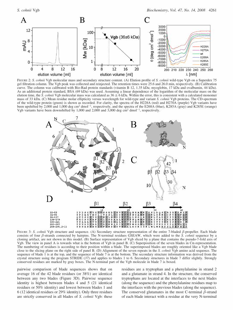
Analytical Gel Filtration. The molecular mass of S. cohniiVgb was estimated by analytical gel filtration on a Superdex75 10/300 GL column in buffer E. The experiment wasrepeated twice. The Vgb peak was collected and reinjected.The column was calibrated with Bio-Rad protein standards(vitamin B 12, 1.35 kDa; myoglobin, 17 kDa and ovalbumin44 kDa). As an additional protein standard, BSA (69 kDa)was used.
CD-Spectroscopy. The effect of point mutations on proteinfolding was assessed by circular dichroism spectroscopy.Spectra of the wild-type and mutant proteins were recordedwith a Jasco J-810 spectrometer equipped with a thermo-statted cell holder and 0.02 cm sample cell. For every proteinvariant, three spectra (in the wavelength range from 260 to200 nm) were recorded and averaged.
In Vitro ActiVity Assay. Spectrophotometric and fluoro-metric assays of S. aureus streptogramin lyase activity havebeen reported previously (19, 21). The fluorometric assay(21) with minor modifications was used. A Shimadzuspectrofluorophotometer (Model RF-5301 PC) and 1 mLcuvette were used throughout. Quinupristin absorption wasmaximal at 333 nm; therefore, the fluorescence was excitedat this wavelength (slit settings 3 nm). Vgb catalyzedquinupristin linearization was monitored by a decrease inthe fluorescence emission at 406 nm (slit settings 10 nm).As some decrease of fluorescence with time was observedalso in the absence of Vgb, a constant background drift hadto be subtracted from all readings.
Reactions for Michaelis–Menten kinetics were done at37 °C in buffer E supplemented with 1 mM MgCl2 oralternatively with 10 mM EDTA. Vgb and its variants wereused at 15 nM concentration (except for the H228A mutantwhere 30 nM of enzyme was applied). The Km values werederived by hyperbolic regression analysis from the depen-dence of the initial velocity on substrate concentration(program Hyper, version 1.1s, 1996). For the determinationof kcat values, rates of fluorescence change were convertedto reaction rates. The calibration curve was based on theassumption that the reaction had run to completion (allquinupristin was linearized) when the fluorescence changedno further. Values for kcat were then calculated from the initialvelocity and the known concentration of S. cohnii Vgb.
Protein Crystallization, Crystal DeriVatization, and DataCollection. S. cohnii Vgb crystals were grown by the vapordiffusion method in sitting drops at 6 °C. Reservoir bufferF (0.1 M HEPES at pH 7.5, 0.2 M CaCl2, 25% PEG 3350),1.2 µL, was mixed with 0.4 µL 10 mM L-cysteine and 2 µLof 10 mg/mL Vgb in buffer E. Needle-like crystals appearedafter 3 days. Native data were collected at the synchrotronbeamline BW6 at the Deutsches ElektronensynchrotronDESY in Hamburg and were integrated and scaled withDENZO and SCALEPACK (23). For derivatization, nativecrystals were either soaked for 7 days with 2 mM HgCl2 orfor 1 min with 0.35 M KI at 6 °C. Diffraction data for thesecrystals were collected on an in-house rotating anodegenerator equipped with a MAR345 image plate. These datawere processed with MOSFLM (24) and SCALA (25). Thedata for the mercury derivative extended only to 2.7 Å, butthe data for the iodide soak reached to 1.8 Å and were of
S. cohnii Vgb Biochemistry, Vol. 47, No. 14, 2008 4259
better quality than synchrotron data for the native crystal.Therefore, the diffraction data for the KI soak was used forthe final refinement (Table 1).
Structure Determination. Difference Patterson maps forthe KI soak showed clear peaks in the Harker sections, whichwere readily interpreted by the RSPS program (26) in termsof two I- sites located at the fractional coordinates (0.781,0.204, 0.226) and (0.968, 0.253, 0.115), respectively. Theresulting single isomorphous replacement (SIR) phases werethen used for cross-phasing to interpret the HgCl2 versusnative difference Fourier maps in terms of two confidentHg2+ sites at (0.310, 0.263, 0.778) and (0.037, 0.751, 0.316).Joint MIR phasing with the program MLPHARE (25) (upto a resolution of 2.5 Å) indicated that for this resolutionrange, the average figure of merit (FOM) for centricreflections was 0.60 and for acentric reflections was 0.38,which resulted in an average figure of merit of 0.41 for allreflections in this resolution range. The phases were thenextended to the full 1.8 Å resolution using the densitymodification program DM (25). In this procedure, FOM (forthe full resolution range up to 1.8 Å) went up from 0.14 to0.64 (corresponding to an increase in the correlation of themap with the final 2Fo - Fc map from 0.43 to 0.55).Nevertheless, the phases after solvent flattening were stillof insufficient quality for automatic model building. There-fore, the modeling program O (27, 28) was used to assemblea first version of the S. cohnii Vgb structure manually.Although the resulting crude model had very poor stereo-chemistry and could not be refined further, it was alreadysufficiently accurate to scan the Protein Data Bank (PDB)for similar structures with the help of the DALI Web-server(29, 30). At the time (October 22nd, 2006), our preliminaryS. cohnii Vgb model was most similar to the structure of a7-bladed �-propeller fragment of the transcriptional cosup-pressor protein TUP1 (residues 333–383, 441–565, 573–604and 621–710). Therefore, a polyalanine version of this modelwas first manually superimposed on the S. cohnii Vgb modeland then broken down into short, rigid segments. Thefragments, which contained only a few consecutive �-strandseach, were manually adjusted for an optimal fit to the density.
After the correction of some loops, this database-guidedmodel of S. cohnii Vgb was sufficiently accurate for furtherautomatic density improvement and model building usingthe ARP/wARP protocol (31). The model was completedmanually and refined with the REFMAC program (32).
RESULTS AND DISCUSSION
S. cohnii Vgb Expression and Purification. A Staphylo-coccus strain harboring the S. cohnii Vgb gene was obtainedfrom the collection of the Pasteur Institute in Paris, France.The gene was amplified by PCR and cloned into pGEX-5T.The resulting plasmid, pGEX-5T-Vgb, was ampicillin-selectable and directed the heterologous expression of solubleVgb with an N-terminal, thrombin-cleavable GST-tag in E.coli. The recombinant protein was purified by glutathione-S-transferase (GST) affinity chromatography and gel filtra-tion. Thrombin-cleavage of the N-terminal GST-tag leftresidues GSEAW as a cloning artifact at the N-terminus.Analytical gel filtration experiments indicated a molecularmass of 36 ( 6 kDa, consistent within the error limit witha calculated Vgb monomer mass of 33 kDa (Figure 2A andB).
S. cohnii Vgb Crystallization and Structure Determination.Crystals of S. cohnii Vgb were grown by the sitting dropvapor diffusion technique. Orthorhombic crystals in spacegroup P2(1)2(1)2(1) appeared in the presence or absence ofquinupristin and/or milimolar concentrations of divalentmetal cations and contained a single molecule of Vgb in theasymmetric unit. As the S. aureus Vgb structure was notavailable at the time, attempts to solve the S. cohnii Vgbstructure by molecular replacement (MR) were unsuccessful.The structure was eventually solved by a combination ofmultiple isomorphous replacement (MIR) and database-guided manual model building (see Experimental Proce-dures), but could of course have been solved more easily byconventional molecular replacement after the model for S.aureus Vgb became available. The final refinement statisticsare summarized in Table 1.
A SeVen-Bladed �-Propeller with a Velcro. On the basisof the presence of six readily detectable repeats in the Vgbsequence and weak similarity to the sensor domain of theMycobacterium tuberculosis receptor serine/threonine proteinkinase PknD, modeling programs predicted Vgb to form asix-bladed �-propeller. However, it became clear early inmodel building that there was an extra blade, which consistedof the first N-terminal and the three last C-terminal �-strandswith their connecting loops (Figure 3A). In retrospect, thislatched or Velcro arrangement could have been anticipatedbecause it was previously observed for many other �-propel-ler proteins (33). The seven blades in the S. cohnii Vgb�-propeller are connected by hairpins (except for the Velcro).Within each blade, the distance of strands from the pseudo-7-fold axis increases from the N-terminus to the C-terminus(Figure 3A). Superposition shows that the seven propellerblades of S. cohnii Vgb are structurally very similar (Figure3C). Excluding the discontinuous seventh blade, the averagedistance between CR-atoms for an optimal superposition ofblades is between 0.49 Å (blades 4 and 5) and 1.20 Å (blades4 and 6), with an average of 0.80 Å over all pairs of blades.At least in part, this high structural similarity is attributableto the high sequence similarity between the blades. A
Table 1: Data Collection and Refinement Statistics
Data Collection Statistics (12.0 – 1.8 Å)
space group P2(1)2(1)2(1)a (Å) 60.5b (Å) 68.1c (Å) 73.2wavelength (Å) 1.54total reflections 96, 127unique reflections 27, 602completeness (%) (last shell) 96.6 (92.8)I/σ (last shell) 16.9 (5.3)R(sym) (%) (last shell) 3.9 (13.9)B(iso) from Wilson (Å2) 13.5
Refinement Statistics (12.0 - 1.8 Å)
protein atoms (excluding H) 2315solvent molecules 261R-factor (%) 18.0R-free (%) 21.5rmsd bond lengths (Å) 0.014rmsd angles (°) 1.3Ramachandran core region (%) 90.2Ramachandran allowed region (%) 9.4Ramachandran additionally allowed region (%) 0.4Ramachandran disallowed region (%) 0.0
4260 Biochemistry, Vol. 47, No. 14, 2008 Lipka et al.
pairwise comparison of blade sequences shows that onaverage 16 of the 42 blade residues (or 38%) are identicalbetween any two blades (Figure 3D). Pairwise sequenceidentity is highest between blades 4 and 5 (21 identicalresidues or 50% identity) and lowest between blades 1 and6 (12 identical residues or 29% identity). Only three residuesare strictly conserved in all blades of S. cohnii Vgb: these
residues are a tryptophan and a phenylalanine in strand 2and a glutamate in strand 4. In the structure, the conservedtryptophans are located at the interfaces to the next blades(along the sequence) and the phenylalanine residues map tothe interfaces with the previous blades (along the sequence).The conserved glutamates in the most C-terminal �-strandof each blade interact with a residue at the very N-terminal
FIGURE 2: S. cohnii Vgb molecular mass and secondary structure content. (A) Elution profile of S. cohnii wild-type Vgb on a Superdex 75gel filtration column. The Vgb peak was collected and reinjected. The retention times were 25.6 and 26.0 min, respectively. (B) Calibrationcurve. The column was calibrated with Bio-Rad protein standards (vitamin B 12, 1.35 kDa; myoglobin, 17 kDa and ovalbumin, 44 kDa).As an additional protein standard, BSA (69 kDa) was used. Assuming a linear dependence of the logarithm of the molecular mass on theelution time, the S. cohnii Vgb molecular mass was calculated as 36 ( 6 kDa. Within the error, this is consistent with a calculated monomermass of 33 kDa. (C) Mean residue molar ellipticity versus wavelength for wild-type and variant S. cohnii Vgb proteins. The CD-spectrumof the wild-type protein (green) is shown as recorded. For clarity, the spectra of the H228A (red) and H270A (purple) Vgb variants havebeen upshifted by 2,000 and 1,000 deg cm2 dmol-1, respectively, and the spectra of the E268A (blue), K285A (gray) and K285E (orange)Vgb variants have been downshifted by 1,000 and 2,000 and 3,000 deg cm2 dmol-1, respectively.
FIGURE 3: S. cohnii Vgb structure and sequence. (A) Secondary structure representation of the entire 7-bladed �-propeller. Each bladeconsists of four �-strands connected by hairpins. The N-terminal residues GSEAW, which were added to the S. cohnii sequence by acloning artifact, are not shown in this model. (B) Surface representation of Vgb sliced by a plane that contains the pseudo-7-fold axis ofVgb. The view in panel A is towards what is the bottom of Vgb in panel B. (C) Superposition of the seven blades in CR-representation.The numbering of residues is according to their position within a blade. The superimposed blades are roughly oriented like a Vgb bladeclose to the slicing plane on the right side of panel B. (D) Alignment of the seven repeats in the S. cohnii Vgb amino acid sequence. Thesequence of blade 1 is at the top, and the sequence of blade 7 is at the bottom. The secondary structure information was derived from thecrystal structure using the program STRIDE (37) and applies to blades 1 to 6. Secondary structures in blade 7 differ slightly. Stronglyconserved residues are marked by gray boxes. The N-terminal part of the Vgb molecule in blade 7 is boxed.
S. cohnii Vgb Biochemistry, Vol. 47, No. 14, 2008 4261
end of the blade (or for other possible choices of the bladeboundary, with a residue at the very C-terminal end of theprevious blade).
ActiVe Site Near the Pseudo-7-fold Axis, on the Flat Sideof Vgb. Like many other �-propeller proteins, Vgb is cup-shaped with a deep central channel on the propeller axis,which is open to one (bottom of Figure 3B), but not the otherside (top of Figure 3B, see also Supporting Information,Figure 1). At first, the deep channel on the propeller axislooked like a probable quinupristin binding site (SupportingInformation, Figure 1A and B). However, residue conserva-tion scores mapped to the protein surface (compare Sup-porting Information, Figure 1C and D) suggested thatquinupristin was more likely to bind to the opposite side ofVgb, which is fairly flat with only a shallow depression(Supporting Information, Figure 1C and D). A literaturesurvey confirmed that this binding mode was well-docu-mented for six-bladed �-propeller proteins (34) and impor-tantly also for Neurospora crassa CMLE, a seven-bladed�-propeller protein that catalyzes the conversion between3-carboxy-cis,cis-muconate and its lactone form. Therefore,we expected Vgb to bind substrate near the pseudo-7-foldaxis on the flat side of the molecule, which was confirmedwhen the cocrystal structure of an inactive S. aureus Vgbvariant with quinupristin was published (21). In the S. cohniicrystals, the Vgb active site is blocked by residues GSEA-WMNFY (residues GSEAW are only present in the recom-binant protein) of the N-terminus of a neighboring moleculein the crystal (Supporting Information, Figure 2). In retro-spect, it is clear that this crystal contact interferes withquinupristin binding, and explains the failure of our quinu-pristin soaking experiments. As Mg2+ binding to Vgb iscomediated by the Vgb substrate (21), it is also clear why
soaking experiments with either Mg2+, Mn2+ or Ca2+ ionswere unsuccessful.
Predicted ActiVe Site of S. cohnii Vgb. Substantialmechanistic similarities between the Vgb and CMLE cata-lyzed reactions in the ring opening direction (ignoringdifferent metal dependencies and stereochemical courses ofthe �-elimination reactions) (Figure 1) and quantitativesimilarities between the S. cohnii Vgb and CMLE structures(DALI Z-score 27) suggested possible mechanistic similari-ties. In CMLE, His148 catalyzes the abstraction/addition ofa CR-proton in the ring opening/closing direction of thereaction. CMLE His148 has no direct counterpart in theequivalent blade of S. cohnii Vgb, but we noted twohistidines, His228 and His270, in roughly equivalent loca-tions in other blades of S. cohnii Vgb and found catalyticdefects when we changed these residues to alanines (seebelow). Although it is now clear that the similarity of theVgb and CMLE active sites is not due to divergent evolution(21), the functional equivalence of CMLE His148 and VgbHis270 was confirmed when the a cocrystal structure anddetailed biochemical characterization of S. aureus Vgbappeared in press (21). This work also showed that His228had a separate catalytic role and described additional activesite residues in S. aureus Vgb. With the exception of Glu284(Lys285 in S. cohnii), all identified S. aureus Vgb activesite residues have direct counterparts (with identical residuenumbers) in S. cohnii Vgb. Superposition of the S. cohniiand S. aureus Vgb structures further shows that conservedresidues are present in spatially conserved locations (Figure4). Therefore, the catalytic roles of active site residuesproposed by Korczynska et al. for S. aureus Vgb (21) arelikely to apply to S. cohnii Vgb as well. According toKorczynska et al., His270 abstracts a proton from the
FIGURE 4: Superposition of the S. cohnii and S. aureus Vgb active sites in stereo representation. The S. cohnii Vgb active site residues(green) were optimally superimposed on their counterparts in the complex of inactive S. aureus Vgb protein variant (orange) with quinupristin(black). Quinupristin is shown in wireframe representation, with the usual color code (red for oxygen atoms, blue for nitrogen atoms).3-HyPic stands for 3-hydroxypicolinyl, Thr stands for threonyl, and PheGly stands for phenylglycyl. The Cγ atom of threonine is difficultto see because it points away from the viewer and into the page. The CR and C� carbon atoms of the threonyl residue are labeled R and�, respectively. Hydrogen atoms directly connected to these two carbon atoms are shown in gray in their inferred positions; all otherhydrogen atoms are implicit. The black triangle marks the carbon-oxygen bond that breaks during catalysis. The dashed black line representsfour amino acid residues of quinupristin that connect the threonyl residue to the phenylglycyl residue. Dotted gray lines indicate metal–ligandinteractions or hydrogen bonds for which a role in catalysis has been suggested. Note that the H270A mutant of S. aureus Vgb was usedfor cocrystallization with quinupristin and that in wild-type S. aureus Vgb, a histidine residue is present just as in S. cohnii Vgb. The Mg2+
ion, which is represented by a gray ball, is present only in the S. aureus Vgb-quinupristin complex, but not in the S. cohnii Vgb structure.Also note that the model is consistent with anti-elimination and the formation of the Z-product (methyl and amino groups on the same sideof the double bond), as required by the experimental data for S. aureus Vgb (19). The residues that are different for the two structures aremarked according to the colors of the C-C bonds for the appropriate structure (green for S. cohnii Vgb and orange for the S. aureus Vgb);the labels that apply to both structures are black.
4262 Biochemistry, Vol. 47, No. 14, 2008 Lipka et al.
threonyl CR-atom when the reaction proceeds in the ring-opening direction. His228 donates a hydrogen bond fromits Nδ-atom to the carbonyl oxygen atom of the phenylglycylresidue in quinupristin, which should lower the pKa of the(conjugate acid of) the phenylglycyl residue and make it abetter leaving group. Tyr18 donates a hydrogen bond to thecarbonyl oxygen atom of the threonyl residue in quinupristinto stabilize the negative charge that delocalizes in part tothe oxygen atom after abstraction of the CR-proton. Glu268is involved in metal chelation in the S. aureus enzyme andis predicted to have an analogous role in the S. cohnii enzyme(Figure 4). Another S. aureus Vgb active site residue,Glu284, is also involved in metal chelation and cannot bemutated to alanine without drastic loss of activity. Althoughthis residue is highly conserved in other Vgb sequences, itis not present in S. cohnii Vgb. Becaue of insertions anddeletions in flanking loops, the spatial equivalent of S. aureusVgb Glu284 is S. cohnii Vgb Lys285, a residue not suitablefor direct metal chelation (Figure 4). The substitution isunlikely to be a cloning artifact because wild-type S. cohniiVgb with the lysine has kinetic parameters very similar tothose of wild-type S. aureus Vgb, despite the substitution(see below).
S. cohnii Vgb ActiVe Site Mutations. Alteration of S. aureusHis228 or His270 reduces the activity to undetectable levels,and alterations of other active site residues result in at leasta 40-fold reduction of the kcat/Km value (21). We therefore
tested whether equivalent mutations in S. cohnii had similareffects on the activity. The H228A, E268A, H270A, K285Aand K285E Vgb variants were expressed and purified in amanner analogous to that used with the wild-type protein.Circular dichroism spectra of all mutant proteins resembledthe spectrum of the wild-type protein, indicating that themutant proteins did not have gross folding defects (Figure2C). Quinupristin lactone ring opening at 37 °C was followedby monitoring the accompanying decrease in fluorescenceintensity essentially as previously described (19, 21). Forall measurements, a reproducible background decrease influorescence had to be subtracted. Rates of fluorescencechange were converted to reaction rates using the calibrationcurves recorded in the appropriate buffer (SupportingInformation, Figure 3). For the determination of these curves,it was assumed that the reaction had proceeded to completionwhen no further change of fluorescence was observed. Inthe presence of 1 mM Mg2+, the activity of 15 nM wild-type S. cohnii Vgb was readily detectable on a time scale ofminutes (Figure 5A and B). In contrast, no activity wasobserved for the H270A variant of S. cohnii Vgb, even whenthe concentration of the mutant protein was increased farbeyond the concentration required to observe a robustreaction for the wild-type protein. In order to distinguishwhether the H270A variant was defective in substrate bindingor catalysis, a competition experiment with the mutant andwild-type protein was set up. High concentrations of the S.
FIGURE 5: Characterization of S. cohnii Vgb. The S. cohnii Vgb mediated quinupristin linearization reduces the fluorescence of the depsipeptide.(A) Raw recordings of fluorescence versus time for the quinupristin linearization by wild-type Vgb (green), the H228A (red), H270A(purple), E268A (blue), K285A (gray), and K285E (orange) Vgb variant or buffer alone (black). (B-F) Dependence of the initial reactionvelocity on the substrate concentration. Experiments were done with 15 nM of the enzyme, except for the H228A mutant protein, whichwas used in twice higher concentration. Reaction mixtures contained either 1 mM Mg2+ (squares) or 10 mM EDTA (inverted triangles). Asquinupristin fluorescence is expected to be metal dependent, separate calibration curves were obtained for the conversion of fluorescenceunits (FU) to concentrations of linearized quinupristin for Mg2+ and for EDTA (Supporting Information, Figure 3).
S. cohnii Vgb Biochemistry, Vol. 47, No. 14, 2008 4263
cohnii Vgb H270A variant but not of the control proteinbovine serum albumin protected quinupristin against theactivity of the wild-type protein (Supporting Information,Figure 4). This result is consistent with a catalytic defect ofthe H270A variant, but could also be explained by a directinhibitory effect of the mutant protein on the wild-typeprotein, especially in light of the crystallographic information.With the exception of the S. cohnii Vgb H270A variant, themutant proteins had sufficient activity for the determinationof Km and kcat values assuming standard Michaelis–Mentenkinetics (Figure 5 and Table 2). The Km and kcat values forwild-type S. cohnii Vgb (Figure 5B) were very similar tothe previously reported values for S. aureus Vgb (21). Thesame was true for the S. cohnii Vgb K285E variant (Figure.5F), which was made to mimic the active site of wild-typeS. aureus Vgb. As expected, all other mutations impairedcatalytic activity (Figures 5C-E and Table 2). The H270Amutation abolished activity entirely, as previously reportedfor the S. aureus enzyme by Korczynska et al. (21). All othermutations had milder effects in our assays than those reportedby Korczynska et al. for the S. aureus enzyme, perhapsindicating a genuine difference between the S. aureus andS. cohnii enzymes. Contamination of our enzyme prepara-tions with background lyase activity might have been analternative explanation, but was unlikely because no residualactivity was detected for the H270A mutant of S. cohnii Vgb,which was purified according to the same protocol.
In contrast to the strict metal dependence of S. aureus Vgb(19), a substantial residual activity of S. cohnii wild-typeVgb was detectable in the absence of exogenously addedMg2+ ions. This residual activity persisted when quinupristinand Vgb were separately preincubated with 10 mM EDTAat 4 °C for at least 12 h (Figure 5B). This result could indicatethat Mg2+ ions remained chelated by quinupristin, explainingwhy against expectation (35) similar calibration curves wereobtained in the presence of 1 mM Mg2+ and in the presenceof 10 mM EDTA (Supporting Information, Figure 3).Alternatively, S. cohnii Vgb is less metal dependent thanS. aureus Vgb, perhaps because the charged ε-amino groupof Lys285 could functionally substitute for the metal ion.This interpretation is supported by the strong metal depen-dence of the S. aureus Vgb-like S. cohnii K285E variant(Figure 5F) but remains to be reconciled with the substantialresidual activity of the S. cohnii Vgb K285A variant in thepresence of EDTA (Figure 5E). Together, our data suggestthat S. cohnii Vgb is more tolerant toward modifications ofthe metal chelating residues than S. aureus Vgb because itsactivity is only partially dependent on the presence ofdivalent metal ions. This difference aside, our data supportthe novel catalytic mechanism that has been proposed (21)
to account for the mechanistically remarkable opening of alactone-ring by �-elimination.
ACKNOWLEDGMENT
We thank the staff of the Pasteur Institute for S. aureussp. aureus strain BM12392-I1877 that contains plasmidpIP1714 with the Vgb gene, Henryk Korza for his early workon the project, and Dr. Honorata Czapinska and Dr. AnetaKaczmarczyk for proofreading the manuscript.
SUPPORTING INFORMATION AVAILABLE
Surface representation of S. cohnii Vgb colored accordingto electrostatic potential and amino acid conservation. Stereoview of the S. cohnii Vgb active site with quinupristinmodeled into the crystal structure of S. cohnii Vgb withoutsubstrate and with the N-terminus of a neighboring moleculein the crystal inserted into the active site. Calibration curvefor kinetic measurements. Competition of the S. cohnii VgbH270A mutant with wild-type enzyme. This material isavailable free of charge via the Internet at http://pubs.acs.org.
REFERENCES
1. Livermore, D. M. (2000) Quinupristin/dalfopristin and linezolid:where, when, which and whether to use. J. Antimicrob. Chemother.46, 347–350.
2. Johnson, A. P., and Livermore, D. M. (1999) Quinupristin/dalfopristin, a new addition to the antimicrobial arsenal. Lancet354, 2012–2013.
3. Rubinstein, E., Prokocimer, P., and Talbot, G. H. (1999) Safetyand tolerability of quinupristin/dalfopristin: administration guide-lines. J. Antimicrob. Chemother. 44, 37–46.
4. Pankuch, G. A., Kelly, L. M., Lin, G., Bryskier, A., Couturier, C.,Jacobs, M. R., and Appelbaum, P. C. (2003) Activities of a neworal streptogramin, XRP 2868 compared to those of other agentsagainst Streptococcus pneumoniae and Haemophilus species.Antimicrob. Agents Chemother. 47, 3270–3274.
5. Tu, D., Blaha, G., Moore, P. B., and Steitz, T. A. (2005) Structuresof MLSBK antibiotics bound to mutated large ribosomal subunitsprovide a structural explanation for resistance. Cell 121, 257–270.
6. Harms, J. M., Schlunzen, F., Fucini, P., Bartels, H., and Yonath,A. (2004) Alterations at the peptidyl transferase centre of theribosome induced by the synergistic action of the streptograminsdalfopristin and quinupristin. BMC Biol. 2, 4.
7. Mukhtar, T. A., and Wright, G. D. (2005) Streptogramins,oxazolidinones, and other inhibitors of bacterial protein synthesis.Chem. ReV. 105, 529–542.
8. Allington, D. R., and Rivey, M. P. (2001) Quinupristin/dalfopristin:a therapeutic review. Clin. Ther. 23, 24–44.
9. Cocito, C. (1979) Antibiotics of the virginiamycin family, inhibitorswhich contain synergistic components. Microbiol. ReV. 43, 145–192.
10. Allignet, J., Loncle, V., Simenel, C., Delepierre, M., and el Solh,N. (1993) Sequence of a staphylococcal gene, vat, encoding anacetyltransferase inactivating the A-type compounds of virginia-mycin-like antibiotics. Gene 130, 91–98.
11. Allignet, J., Liassine, N., and el Solh, N. (1998) Characterizationof a staphylococcal plasmid related to pUB110 and carrying two
Table 2: Kinetic Constants for Wild-Type and Mutant Vgb Proteins in the Presence of 1 mM Mg2+ Ions or 10 mM EDTA
1 mM MgCl2 10 mM EDTA
Km [µM] kcat [1/s] kcat/Km [M-1s-1] Km [µM] kcat [1/s] kcat/Km [M-1s-1]
4264 Biochemistry, Vol. 47, No. 14, 2008 Lipka et al.
novel genes, vatC and vgbB, encoding resistance to streptograminsA and B and similar antibiotics. Antimicrob. Agents Chemother.42, 1794–1798.
12. Kehoe, L. E., Snidwongse, J., Courvalin, P., Rafferty, J. B., andMurray, I. A. (2003) Structural basis of Synercid (quinupristin-dalfopristin) resistance in Gram-positive bacterial pathogens.J. Biol. Chem. 278, 29963–29970.
13. Allignet, J., and El Solh, N. (1999) Comparative analysis ofstaphylococcal plasmids carrying three streptogramin-resistancegenes: vat-vgb-vga. Plasmid 42, 134–138.
14. Allignet, J., Loncle, V., and el Sohl, N. (1992) Sequence of astaphylococcal plasmid gene, vga, encoding a putative ATP-bindingprotein involved in resistance to virginiamycin A-like antibiotics.Gene 117, 45–51.
15. Monod, M., Denoya, C., and Dubnau, D. (1986) Sequence andproperties of pIM13, a macrolide-lincosamide-streptogramin Bresistance plasmid from Bacillus subtilis. J. Bacteriol. 167, 138–147.
16. Bussiere, D. E., Muchmore, S. W., Dealwis, C. G., Schluckebier,G., Nienaber, V. L., Edalji, R. P., Walter, K. A., Ladror, U. S.,Holzman, T. F., and Abad-Zapatero, C. (1998) Crystal structureof ErmC′, an rRNA methyltransferase which mediates antibioticresistance in bacteria. Biochemistry 37, 7103–7112.
17. Allignet, J., Loncle, V., Mazodier, P., and el Solh, N. (1988)Nucleotide sequence of a staphylococcal plasmid gene, vgb,encoding a hydrolase inactivating the B components of virginia-mycin-like antibiotics. Plasmid 20, 271–275.
18. Bateman, K. P., Yang, K., Thibault, P., White, R. L., and Vining,L. C. (1996) Inactivation of etamycin by a novel eliminationmechanism in Streptomyces liVidans. J. Am. Chem. Soc. 118, 5335–5338.
19. Mukhtar, T. A., Koteva, K. P., Hughes, D. W., and Wright, G. D.(2001) Vgb from Staphylococcus aureus inactivates streptograminB antibiotics by an elimination mechanism not hydrolysis. Bio-chemistry 40, 8877–8886.
20. Kajander, T., Merckel, M. C., Thompson, A., Deacon, A. M.,Mazur, P., Kozarich, J. W., and Goldman, A. (2002) The structureof Neurospora crassa 3-carboxy-cis,cis-muconate lactonizingenzyme, a beta propeller cycloisomerase. Structure 10, 483–492.
21. Korczynska, M., Mukhtar, T. A., Wright, G. D., and Berghuis,A. M. (2007) Structural basis for streptogramin B resistance inStaphylococcus aureus by virginiamycin B lyase. Proc. Natl. Acad.Sci. U.S.A. 104, 10388–10393.
22. Berthold, H., Frorath, B., Scanarini, M., Abney, C. C., Ernst, B.,and Northemann, W. (1992) Plasmid pGEX-5T: An alternativesystem for the expression and purification of recombinant proteins.Biotechnol. Lett. 14, 245–250.
23. Otwinowski, Z., and Minor, W. (1997) Processing of X-raydiffraction data collected in oscillation mode. Methods Enzymol.276, 307–326.
24. Leslie, A. W. G. (1992) Recent changes to the MOSFLM packagefor processing film and image plate data, Joint CCP4 + ESF-EAMCB Newsletter on Protein Crystallography 26.
25. Collaborative Computational Project Number 4. (1994) The CCP4Suite: Programs for Protein Crystallography, Acta Crystallogr.,Sect. D 50, 760–763.
26. Knight, S. D. (2000) RSPS version 4.: a semi-interactive vector-search program for solving heavy-atom derivatives. Acta Crystal-logr., Sect. D 56, 42–47.
27. Jones, T. A., Zou, J. Y., and Cowan, S. W. (1991) Improvedmethods for building protein models in electron density maps andthe location of errors in these models. Acta Crystallogr., Sect. A47, 110–119.
28. Kleywegt, G. J., and Jones, T. A. (1998) Databases in proteincrystallography. Acta Crystallogr., Sect. D 54, 1119–1131.
29. Holm, L., and Sander, C. (1993) Protein structure comparison byalignment of distance matrices. J. Mol. Biol. 233, 123–138.
30. Holm, L., and Sander, C. (1995) Dali: a network tool for proteinstructure comparison. Trends Biochem. Sci. 20, 478–480.
31. Morris, R. J., Perrakis, A., and Lamzin, V. S. (2003) ARP/wARPand automatic interpretation of protein electron density maps.Methods Enzymol. 374, 229–244.
32. Murshudov, G. N., Vagin, A. A., and Dodson, E. J. (1997)Refinement of macromolecular structures by the maximum-likelihood method. Acta Crystallogr., Sect. D 53, 240–255.
33. Fulop, V., and Jones, D. T. (1999) Beta propellers: structural rigidityand functional diversity. Curr. Opin. Struct. Biol. 9, 715–721.
34. Good, M. C., Greenstein, A. E., Young, T. A., Ng, H. L., andAlber, T. (2004) Sensor domain of the Mycobacterium tuberculosisreceptor Ser/Thr protein kinase, PknD, forms a highly symmetricbeta propeller. J. Mol. Biol. 339, 459–469.
35. Di Giambattista, M., Engelborghs, Y., Nyssen, E., Clays, K., andCocito, C. (1991) Interaction between virginiamycin S and ribo-somes is partly provided by a salt bridge with a Mg2+ ion.Biochemistry 30, 7277–7282.
36. Kirby, G. W., Loughlin, G. J., and Robins, D. J. (1975) Thestereochemistry of the enzymatic cyclisation of 3-carboxymuconicacid to 3-carboxymuconolactone. J. Chem. Soc., Chem. Commun.402–403.
37. Frishman, D., and Argos, P. (1995) Knowledge-based proteinsecondary structure assignment. Proteins 23, 566–579.
BI7015266
S. cohnii Vgb Biochemistry, Vol. 47, No. 14, 2008 4265