+ All Categories
Home > Documents > Stem Cell-Derived Extracellular Vesicles and Immune … · Studies reporting the role of EVs on T...

Stem Cell-Derived Extracellular Vesicles and Immune … · Studies reporting the role of EVs on T...

Date post: 05-Jun-2018
Category:
Upload: dangtuyen
View: 214 times
Download: 0 times
Share this document with a friend
10
REVIEW published: 22 August 2016 doi: 10.3389/fcell.2016.00083 Frontiers in Cell and Developmental Biology | www.frontiersin.org 1 August 2016 | Volume 4 | Article 83 Edited by: Kaushik Choudhuri, University of Michigan Health System, USA Reviewed by: Todd R. Graham, Vanderbilt University, USA Alissa Weaver, Vanderbilt University, USA *Correspondence: Giovanni Camussi [email protected] Specialty section: This article was submitted to Membrane Traffic, a section of the journal Frontiers in Cell and Developmental Biology Received: 19 May 2016 Accepted: 02 August 2016 Published: 22 August 2016 Citation: Burrello J, Monticone S, Gai C, Gomez Y, Kholia S and Camussi G (2016) Stem Cell-Derived Extracellular Vesicles and Immune-Modulation. Front. Cell Dev. Biol. 4:83. doi: 10.3389/fcell.2016.00083 Stem Cell-Derived Extracellular Vesicles and Immune-Modulation Jacopo Burrello, Silvia Monticone, Chiara Gai, Yonathan Gomez, Sharad Kholia and Giovanni Camussi * Stem Cell Laboratory, Department of Medical Sciences, University of Torino, Torino, Italy Extra-cellular vesicles (EVs) are bilayer membrane structures enriched with proteins, nucleic acids, and other active molecules and have been implicated in many physiological and pathological processes over the past decade. Recently, evidence suggests EVs to play a more dichotomic role in the regulation of the immune system, whereby an immune response may be enhanced or supressed by EVs depending on their cell of origin and its functional state. EVs derived from antigen (Ag)-presenting cells for instance, have been involved in both innate and acquired (or adaptive) immune responses, as Ag carriers or presenters, or as vehicles for delivering active signaling molecules. On the other hand, tumor and stem cell derived EVs have been identified to exert an inhibitory effect on immune responses by carrying immuno-modulatory effectors, such as transcriptional factors, non-coding RNA (Species), and cytokines. In addition, stem cell-derived EVs have also been reported to impair dendritic cell maturation and to regulate the activation, differentiation, and proliferation of B cells. They have been shown to control natural killer cell activity and to suppress the innate immune response (IIR). Studies reporting the role of EVs on T lymphocyte modulation are controversial. Discrepancy in literature may be due to stem cell culture conditions, methods of EV purification, EV molecular content, and functional state of both parental and target cells. However, mesenchymal stem cell-derived EVs were shown to play a more suppressive role by shifting T cells from an activated to a T regulatory phenotype. In this review, we will discuss how stem cell-derived EVs may contribute toward the modulation of the immune response. Collectively, stem cell-derived EVs mainly exhibit an inhibitory effect on the immune system. Keywords: extracellular vesicles, exosomes, stem cells, immune system, immuno-modulation INTRODUCTION Extra-cellular vesicles (EVs) are bilayer membranal structures released by cells as a means of transferring their content to and from other cells, now acknowledged as a novel mechanism of intercellular communication. These vesicles comprise of both membrane and cytoplasmic components including: proteins, nucleic acids, and other active molecules and can be sub classified according to size, biogenesis, and composition into exosomes or microvesicles (also defined as shedding vesicles; Théry et al., 2009; EL Andaloussi et al., 2013; Yáñez-Mó et al., 2015; see Figure 1). The term “microvesicle” generally refers to both vesicles released by healthy cells as well as pre-apoptotic vesicles. They are commonly heterogeneous in size ranging from 50 to 1000 nm in diameter depending on the state of the cell during release by either direct shedding or budding
Transcript

REVIEWpublished: 22 August 2016

doi: 10.3389/fcell.2016.00083

Frontiers in Cell and Developmental Biology | www.frontiersin.org 1 August 2016 | Volume 4 | Article 83

Edited by:

Kaushik Choudhuri,

University of Michigan Health System,

USA

Reviewed by:

Todd R. Graham,

Vanderbilt University, USA

Alissa Weaver,

Vanderbilt University, USA

*Correspondence:

Giovanni Camussi

[email protected]

Specialty section:

This article was submitted to

Membrane Traffic,

a section of the journal

Frontiers in Cell and Developmental

Biology

Received: 19 May 2016

Accepted: 02 August 2016

Published: 22 August 2016

Citation:

Burrello J, Monticone S, Gai C,

Gomez Y, Kholia S and Camussi G

(2016) Stem Cell-Derived Extracellular

Vesicles and Immune-Modulation.

Front. Cell Dev. Biol. 4:83.

doi: 10.3389/fcell.2016.00083

Stem Cell-Derived ExtracellularVesicles and Immune-ModulationJacopo Burrello, Silvia Monticone, Chiara Gai, Yonathan Gomez, Sharad Kholia and

Giovanni Camussi *

Stem Cell Laboratory, Department of Medical Sciences, University of Torino, Torino, Italy

Extra-cellular vesicles (EVs) are bilayer membrane structures enriched with proteins,

nucleic acids, and other active molecules and have been implicated in many physiological

and pathological processes over the past decade. Recently, evidence suggests EVs to

play a more dichotomic role in the regulation of the immune system, whereby an immune

response may be enhanced or supressed by EVs depending on their cell of origin and its

functional state. EVs derived from antigen (Ag)-presenting cells for instance, have been

involved in both innate and acquired (or adaptive) immune responses, as Ag carriers or

presenters, or as vehicles for delivering active signaling molecules. On the other hand,

tumor and stem cell derived EVs have been identified to exert an inhibitory effect on

immune responses by carrying immuno-modulatory effectors, such as transcriptional

factors, non-coding RNA (Species), and cytokines. In addition, stem cell-derived EVs

have also been reported to impair dendritic cell maturation and to regulate the activation,

differentiation, and proliferation of B cells. They have been shown to control natural

killer cell activity and to suppress the innate immune response (IIR). Studies reporting

the role of EVs on T lymphocyte modulation are controversial. Discrepancy in literature

may be due to stem cell culture conditions, methods of EV purification, EV molecular

content, and functional state of both parental and target cells. However, mesenchymal

stem cell-derived EVs were shown to play a more suppressive role by shifting T cells

from an activated to a T regulatory phenotype. In this review, we will discuss how

stem cell-derived EVs may contribute toward the modulation of the immune response.

Collectively, stem cell-derived EVs mainly exhibit an inhibitory effect on the immune

system.

Keywords: extracellular vesicles, exosomes, stem cells, immune system, immuno-modulation

INTRODUCTION

Extra-cellular vesicles (EVs) are bilayer membranal structures released by cells as a means oftransferring their content to and from other cells, now acknowledged as a novel mechanismof intercellular communication. These vesicles comprise of both membrane and cytoplasmiccomponents including: proteins, nucleic acids, and other active molecules and can be sub classifiedaccording to size, biogenesis, and composition into exosomes or microvesicles (also defined asshedding vesicles; Théry et al., 2009; EL Andaloussi et al., 2013; Yáñez-Mó et al., 2015; see Figure 1).

The term “microvesicle” generally refers to both vesicles released by healthy cells as well aspre-apoptotic vesicles. They are commonly heterogeneous in size ranging from 50 to 1000 nm indiameter depending on the state of the cell during release by either direct shedding or budding

Burrello et al. Extracellular Vesicles and Immune-Modulation

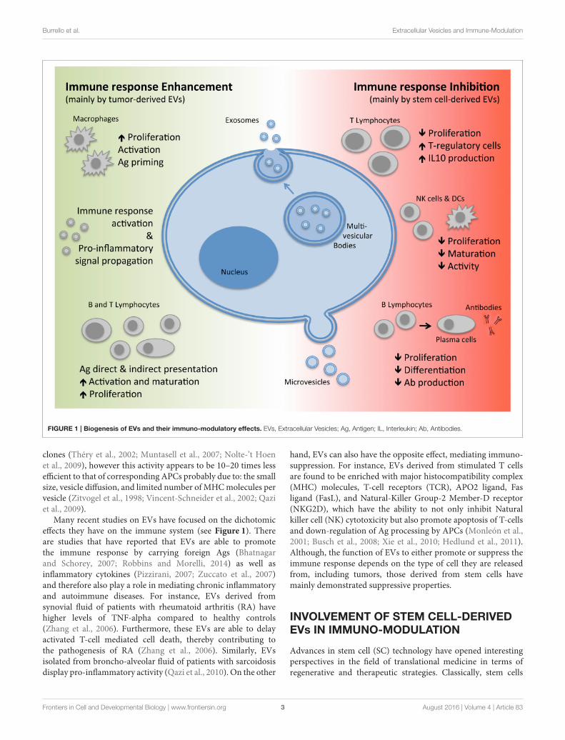
from the plasma membrane (Figure 1). Exosomes, on theother hand, are generated through an invagination process ofthe endosomal membrane of multi-vesicular bodies (MVBs)found within the cells forming vesicles. This is followed byfusion of the MVBs with the plasma membrane leading toexocytosis and release of exosomes which are homogenous insize with a diameter ranging from 40 to 200 nm (Figure 1;Yáñez-Mó et al., 2015). Since exosomes and microvesicles shareseveral molecular and functional characteristics and are releasedconcomitantly by the same cell types, the term EVs will beused to collectively indicate the two subtypes throughout thisreview.

EVs carry a plethora of molecules that influence theirmode of action. These include a variety of receptors, adhesionmolecules, proteins involved in cell trafficking, and/or intra-cellular signal transduction, cytoskeletal proteins, cytoplasmicenzymes, cytokines, chemokines, as well as cell-specific antigens(Ags). Moreover, they are also enriched with a range ofnucleic acids, including mRNA, long non-coding RNAs,microRNAs (miRNA), and even extra-chromosomal DNA(Robbins and Morelli, 2014; Yáñez-Mó et al., 2015). Thecontent carried however may vary depending on the typeof cell and the state of activation. Once released, these EVsmay interact with neighboring cells or diffuse through andcirculate in the bloodstream or other organic fluids suchas: breast milk, semen, saliva, urine, and sputum. Theirstate of ubiquity makes them important effectors of cell-to-cell communication, which may occur in a paracrine,autocrine, exocrine, and or endocrine manner (Yáñez-Mó et al.,2015).

Virtually all cell types have the ability to release EVs includingcells of the immune system such as: macrophages, antigenpresenting cells (APCs), dendritic cells (DCs), B and T cells(both CD4+ T helper and CD8+ cytotoxic T lymphocytes),natural killer cells (NKs), as well as cells complementingthe immune system such as platelets, mast cells, fibroblasts,epithelial cells, and stem cells. Recently, there has been enoughgrowing evidence to suggest that EVs play an essential role inimmuno-modulation (Robbins and Morelli, 2014), influencingboth the activation and suppression of the immune response.Furthermore, a role of EVs has also been implicated ininflammation, autoimmune, infectious, and cancer diseases(Yáñez-Mó et al., 2015). This review will however focus onthe interactions of stem-cell derived EVs with the immunesystem.

Abbreviations: EVs, Extra-cellular vesicles; Ag(s), Antigen(s); miRNA,

MicroRNA; APCs, Antigen presenting cells; DCs, Dendritic cells; NKs, Natural

killer cells; IIR, Innate immune response; AIR, Acquired immune response;

TGF-β, Transforming Growth factor-β; CTLs, Cytotoxic T-lymphocytes; RA,

Rheumatoid arthritis; SC(s), Stem cell(s); ESCs, Embryonic stem cells; MSCs,

Mesenchymal stem/stromal cells; iPS, Induced pluripotent stem cells; IFN-γ,

Interferon-γ; TNF-α, Tumor Necrosis Factor-α; HGF, Hepatocyte Growth

Factor; NO, Nitric oxide; IDO, Indoleamine 2,3-dioxygenase; HLA-G, Human

Leukocyte Antigen-G; PG, Prostaglandin; IL, Interleukin; CXCR4/CXCR5/CCR7,

Chemokine Receptor Type 4/5–7; PBMCs, Peripheral Blood Mononuclear Cells;

GAD65, Glutamic Acid Decarboxylase 65; NKG2D, Natural-Killer Group-2

Member-D receptor; MHC, Major Histocompatibility Complex; siRNA, Small

interfering RNA; BM, Bone Marrow; ASCs, Adipose Stem Cells.

REGULATION OF IMMUNE RESPONSE BYEVs

The immune system can be divided into two branches: theinnate immune response (IIR)—an evolutionary conservedsystem common to all multicellular organisms, and the acquiredor adaptive immune response (AIR)—an exclusive featuredeveloped in vertebrates (Sirisinha, 2014).

Within the IIR system, EVs act as paracrine messengers,allowing the propagation of pro-inflammatory signals(Mastronardi et al., 2011; Wang et al., 2011; Prakash et al., 2012;Cloutier et al., 2013). However, they have also been reported tocontribute as negative regulators of the inflammatory responseprimarily by carrying molecules such as Transforming Growthfactor-β (TGF-β), and other immuno-suppressive mediators(Gasser and Schifferli, 2004). The role of EVs in the regulationof IIR is complex and has not yet been fully elucidated. Manystudies so far have investigated the composition and function ofEVs from innate immune cells cultured in-vitro. For instance,macrophages treated in vitro with EVs isolated from cellsinfected with Mycobacterium tuberculosis released cytokinesand chemokines that contributed toward the activation of theimmune response (Walters et al., 2013). On the other hand,macrophages infected with the Leishmania parasite secretedEVs enriched with the Leishmania surface protein gp63, whichdown-regulated the inflammatory response, favoring parasiteinvasion (Hassani and Olivier, 2013).

Whereas, IIR is a non-specific first line of defense againstmicrobial pathogens and other tissue injuries, AIR is a specificresponse induced after Ag recognition by adaptive immune cellsfollowed by activation and clonal expansion of immune cellscarrying the recognized Ag-specific receptors (Schenten andMedzhitov, 2011; Zhang et al., 2014). In this setting, EVs mayact not only as Ag carriers (since they may transfer bacterial,viral, and tumoral components to APCs; O’Neill and Quah, 2008;Walker et al., 2009; Testa et al., 2010), but also as modulators ofdirect and indirect Ag presentation. Furthermore, this propertyof EVs to carry Ags from parental cells can allow them to actas reporters of foreign agents in the organism both for thehost immune system as well as from a diagnostic point of view(Yáñez-Mó et al., 2015). For example, tumor-derived EVs carrytumor-Ags, which can be taken up and processed by DCs andthen cross-presented to tumor-specific cytotoxic T-lymphocytes(CTLs; Wolfers et al., 2001; Andre et al., 2002). This has beendemonstrated for EVs isolated from ascites of tumoral patientsas well as other tumoral cell lines (Wolfers et al., 2001; Andreet al., 2002; Morelli et al., 2004). This hypothesis is supported bythe fact that vaccination of mice with tumor peptide-pulsed DC-derived EVs induces a potent CD8+ T cell-mediated anti-tumoraleffect (Wolfers et al., 2001). On the basis of these findings, itcan be speculated that tumor-derived EVs carry tumor-specificAgs and that they could be used to stimulate or inhibit theimmune anti-tumoral surveillance (Robbins and Morelli, 2014).In this regard, ongoing studies are exploring their potential rolein the field of anti-tumor vaccination, as reviewed by Kunigeliset al. (Kunigelis and Graner, 2015). Furthermore, APC-derivedEVs can also act as “Ag-presenting vesicles” for in vitro T-cell

Frontiers in Cell and Developmental Biology | www.frontiersin.org 2 August 2016 | Volume 4 | Article 83

Burrello et al. Extracellular Vesicles and Immune-Modulation

FIGURE 1 | Biogenesis of EVs and their immuno-modulatory effects. EVs, Extracellular Vesicles; Ag, Antigen; IL, Interleukin; Ab, Antibodies.

clones (Théry et al., 2002; Muntasell et al., 2007; Nolte-’t Hoenet al., 2009), however this activity appears to be 10–20 times lessefficient to that of corresponding APCs probably due to: the smallsize, vesicle diffusion, and limited number of MHCmolecules pervesicle (Zitvogel et al., 1998; Vincent-Schneider et al., 2002; Qaziet al., 2009).

Many recent studies on EVs have focused on the dichotomiceffects they have on the immune system (see Figure 1). Thereare studies that have reported that EVs are able to promotethe immune response by carrying foreign Ags (Bhatnagarand Schorey, 2007; Robbins and Morelli, 2014) as well asinflammatory cytokines (Pizzirani, 2007; Zuccato et al., 2007)and therefore also play a role in mediating chronic inflammatoryand autoimmune diseases. For instance, EVs derived fromsynovial fluid of patients with rheumatoid arthritis (RA) havehigher levels of TNF-alpha compared to healthy controls(Zhang et al., 2006). Furthermore, these EVs are able to delayactivated T-cell mediated cell death, thereby contributing tothe pathogenesis of RA (Zhang et al., 2006). Similarly, EVsisolated from broncho-alveolar fluid of patients with sarcoidosisdisplay pro-inflammatory activity (Qazi et al., 2010). On the other

hand, EVs can also have the opposite effect, mediating immuno-suppression. For instance, EVs derived from stimulated T cellsare found to be enriched with major histocompatibility complex(MHC) molecules, T-cell receptors (TCR), APO2 ligand, Fasligand (FasL), and Natural-Killer Group-2 Member-D receptor(NKG2D), which have the ability to not only inhibit Naturalkiller cell (NK) cytotoxicity but also promote apoptosis of T-cellsand down-regulation of Ag processing by APCs (Monleón et al.,2001; Busch et al., 2008; Xie et al., 2010; Hedlund et al., 2011).Although, the function of EVs to either promote or suppress theimmune response depends on the type of cell they are releasedfrom, including tumors, those derived from stem cells havemainly demonstrated suppressive properties.

INVOLVEMENT OF STEM CELL-DERIVEDEVs IN IMMUNO-MODULATION

Advances in stem cell (SC) technology have opened interestingperspectives in the field of translational medicine in terms ofregenerative and therapeutic strategies. Classically, stem cells

Frontiers in Cell and Developmental Biology | www.frontiersin.org 3 August 2016 | Volume 4 | Article 83

Burrello et al. Extracellular Vesicles and Immune-Modulation

TABLE 1 | Potential EV-derived effectors reported to play a role in immuno-modulation.

Effector Type Cell source Function References

Nanog Transcriptional factor ESCs Tumoral cells immune-escape Ratajczak et al., 2006b; Yáñez-Mó et al., 2015

Oct-4 Transcriptional factor ESCs Innate immune suppression Ratajczak et al., 2006b; Lee et al., 2012

HoxB4 Transcriptional factor ESCs Through WNT signaling affects DCs maturation and

T-cells proliferation, differentiation, and activation

Ratajczak et al., 2006b; Staal et al., 2008

Rex-1 Transcriptional factor ESCs Through PIP3 signaling stimulate innate immune

response and neutrophils activity

Weiner, 2002; Ratajczak et al., 2006b

Oct4 mRNA ESCs Innate immune suppression Katsman et al., 2012; Noh et al., 2012

Sox2 mRNA ESCs Initiate innate response against microbial infection

through neutrophils activation

Katsman et al., 2012; Xia et al., 2015

CD81 CD9 Membrane protein iPS Immune system cell adhesion, motility, activation,

and signal transduction

Levy et al., 1998; Hu et al., 2015

miR-21 miRNA iPS Inhibiting effect on granulopoiesis O’Connell et al., 2010; Wang et al., 2015b

TGF-β1 Cytokine MSCs Maintain tolerance through regulation of lymphocyte

proliferation and survival

Bruno et al., 2015

IL-6 Cytokine MSCs Interfere effect in DCs maturation Djouad et al., 2007

IL-10 Cytokine MSCs Suppress T lymphocytes proliferation Hwu et al., 2000

PGE-2 Prostaglandin MSCs Inhibit NKs activity and proliferation Aggarwal and Pittenger, 2005; Spaggiari et al., 2006

Suppress T lymphocytes proliferation

Interfere effect in DCs maturation

IDO Enzyme MSCs Inhibit NKs activity and proliferation Hwu et al., 2000

miR-155 miR-146 miRNA MSCs miR-155 promotes, while miR-146 reduce,

inflammatory reaction in mice

Di Trapani et al., 2016

HGF Growth factor MSCs Suppress T lymphocytes proliferation Di Nicola et al., 2002

ESCs, Embryonic Stem Cells; iPS, induced Pluripotent Stem Cells; MSCs, Mesenchymal Stromal/Stem Cells; IL, Interleukin; TGF, Transforming Growth Factor; PG, Prostaglandin; IDO,

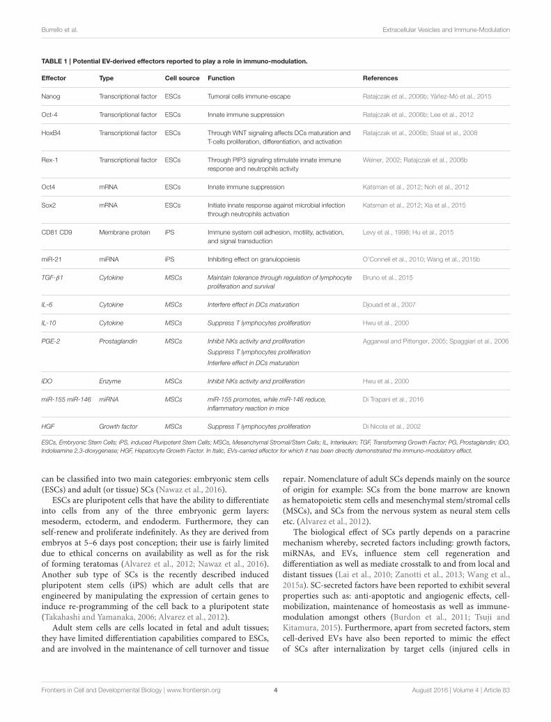
Indoleamine 2,3-dioxygenase; HGF, Hepatocyte Growth Factor. In Italic, EVs-carried effector for which it has been directly demonstrated the immuno-modulatory effect.

can be classified into two main categories: embryonic stem cells(ESCs) and adult (or tissue) SCs (Nawaz et al., 2016).

ESCs are pluripotent cells that have the ability to differentiateinto cells from any of the three embryonic germ layers:mesoderm, ectoderm, and endoderm. Furthermore, they canself-renew and proliferate indefinitely. As they are derived fromembryos at 5–6 days post conception; their use is fairly limiteddue to ethical concerns on availability as well as for the riskof forming teratomas (Alvarez et al., 2012; Nawaz et al., 2016).Another sub type of SCs is the recently described inducedpluripotent stem cells (iPS) which are adult cells that areengineered by manipulating the expression of certain genes toinduce re-programming of the cell back to a pluripotent state(Takahashi and Yamanaka, 2006; Alvarez et al., 2012).

Adult stem cells are cells located in fetal and adult tissues;they have limited differentiation capabilities compared to ESCs,and are involved in the maintenance of cell turnover and tissue

repair. Nomenclature of adult SCs depends mainly on the sourceof origin for example: SCs from the bone marrow are knownas hematopoietic stem cells and mesenchymal stem/stromal cells(MSCs), and SCs from the nervous system as neural stem cellsetc. (Alvarez et al., 2012).

The biological effect of SCs partly depends on a paracrinemechanism whereby, secreted factors including: growth factors,miRNAs, and EVs, influence stem cell regeneration anddifferentiation as well as mediate crosstalk to and from local anddistant tissues (Lai et al., 2010; Zanotti et al., 2013; Wang et al.,2015a). SC-secreted factors have been reported to exhibit severalproperties such as: anti-apoptotic and angiogenic effects, cell-mobilization, maintenance of homeostasis as well as immune-modulation amongst others (Burdon et al., 2011; Tsuji andKitamura, 2015). Furthermore, apart from secreted factors, stemcell-derived EVs have also been reported to mimic the effectof SCs after internalization by target cells (injured cells in

Frontiers in Cell and Developmental Biology | www.frontiersin.org 4 August 2016 | Volume 4 | Article 83

Burrello et al. Extracellular Vesicles and Immune-Modulation

the context of regeneration), mainly through receptor–ligandmediated interactions and/or direct fusion, leading to horizontaltransfer of proteins and nucleic acids (including mRNA and non-coding RNA; Ratajczak et al., 2006a; Valadi et al., 2007; Camussiet al., 2010).

Many authors in the recent years have focused their researchin studying the role of EVs (from various cellular origins)in immuno-modulation, however, very little studies have beenreported that address the role of SC-derived EVs in the immunesystem particularly on IRR and AIR. Table 1 shows referencesof studies that have reported potential effectors identified withinEVs that exhibit immuno-modulatory properties. The reason forsuch limited studies in the literature may partly be due to the factthat the role of SC-EVs as potential immune-modulatory effect isstill novel (Nawaz et al., 2016) and partly due to the reporting ofcontroversial results. Nevertheless, in order to elucidate the novelrole(s) of SC-EVs on the immune system as well as to clarifythe reported controversies in the results, further studies in thissubject are only warranted.

Ratajczak et al. first reported that EVs released by ESCs mightmodulate the phenotype of target cells, supporting self-renewalof hematopoietic progenitors and multi-potency by transfer ofgrowth factors and messenger-RNA (mRNA) (Ratajczak et al.,2006b). Subsequent studies confirmed this horizontal transfer ofgenetic material in other contexts, such as endothelial progenitorinduced angiogenesis and modulation of bone-marrow cellphenotype by EVs released by injured tissues (Deregibus et al.,2007; Aliotta et al., 2010). Furthermore, ESC-derived EVs maynot only contribute to cell-fate determination but also to tissuerepair and various other physio-pathological processes, but theirrole as immuno-modulators has not yet been fully elucidated.Nonetheless, ESC-derived EVs have been reported to carrytranscriptional factors and mRNAs such as: Nanog, octamer-binding transcription factor 4 (Oct-4), HoxB4, Rex-1, Oct4, andSox2 that have immuno-modulatory properties (see Table 1). Forinstance, Nanog has been described in tumor cell immuno-escape(Ratajczak et al., 2006b; Noh et al., 2012), Oct-4 in the inhibitionof IIR (Ratajczak et al., 2006b; Lee et al., 2012), and HoxB4 inimpairment of DC maturation, and T-cell proliferation througha WNT-mediated mechanism (Ratajczak et al., 2006b; Staal et al.,2008). Whereas, Rex-1 and Sox2 have been shown to stimulateIIR (Weiner, 2002; Ratajczak et al., 2006b; Katsman et al., 2012;Xia et al., 2015). However, a direct role of ESC-derived EVs on theimmune system through the above described effectors has yet tobe reported. Similar to ESCs, little is known about the functionsof iPS-derived EVs on the immune system. The available studiesshow that membrane proteins, such as CD81 and CD9, and miR-21 are carried by iPS-derived EVs and that these effectors mayaffect the immune response by regulating: cell adhesion, motility,activation and signal transduction (see Table 1; Levy et al., 1998;O’Connell et al., 2010; Hu et al., 2015; Wang et al., 2015b).

Amongst adult SCs, MSCs are certainly the most studiedtype (Pittenger et al., 1999; Bruno et al., 2015). In addition toregenerative effects in animal models (Syed and Evans, 2013),there is increasing evidence describing their role in both IIRand AIR modulation (MacDonald et al., 2011; Dalal et al.,2012). For instance, MSCs are able to not only inhibit NK

proliferation and activity but also suppress T-/B-cell proliferationand DC maturation (Keating, 2012; Bruno et al., 2015).Furthermore, high levels of IFN-γ and TNF-α influence MSCsto develop immuno-suppressive properties toward IIR (affectingneutrophils, monocytes, and NKs) and AIR (T- and B-cells)(Corcione et al., 2006; Krampera et al., 2006; Spaggiari et al., 2006;Uccelli et al., 2008; Le Blanc andMougiakakos, 2012). MSCs havealso been reported to have a marked immuno-regulatory effectagainst autoimmune disorders and promising results have beenobserved in clinical trials on patients with Crohn’ s disease andgraft-vs.-host disease (Pittenger, 2009; Caplan and Correa, 2011).

It is still widely debated as to whether the immuno-modulatory action of MSCs relies on direct cell-to-cellinteraction or on the paracrine action of soluble mediatorsreleased by these cells. Either one or both mechanisms are apossibility. Both, in vivo and in vitro studies have demonstratedthat MSCs exhibit an immuno-suppressive role by suppressingT lymphocyte proliferation (Krampera et al., 2003; Aggarwaland Pittenger, 2005). A possible explanation for this rolecould be the presence of proteins and factors such as: TGF-β,hepatocyte growth factor (HGF), nitric oxide (NO), indoleamine2,3-dioxygenase (IDO), human leukocyte antigen G (HLA-G),prostaglandin E2 (PGE-2), Inter Leukin (IL)-6, and IL-10 (Hwuet al., 2000; Aggarwal and Pittenger, 2005; Spaggiari et al., 2006;Djouad et al., 2007; Sato et al., 2007; English et al., 2009; Caplanand Correa, 2011) that are expressed highly by MSCs. Forinstance, It has been suggested, that IDO, PGE-2, and TGF-β1are involved not only in the inhibition of NK cells but also in theinhibition of T-cell proliferation and activation (Aggarwal andPittenger, 2005; Spaggiari et al., 2006). Furthermore, IL-6 has alsobeen attributed to interfere with DC maturation (English et al.,2009). Finally, with a mechanism not completely known, MSCshave been reported to inhibit B-cells proliferation through thedown-regulation of chemokine receptor CXCR4, CXCR5, andchemokine receptor type 7 (CCR7) (Corcione et al., 2006). Theimmuno-modulatory effects of MSCs can therefore be speculatedto be achieved either through the release of the above mentionedfactors and proteins directly into the extracellular milieu assoluble molecules, or they may be packaged into EVs togetherwith nucleic acids and other post-transcriptional modulatorswhich could influence the inflammatory response when released(Robbins and Morelli, 2014).

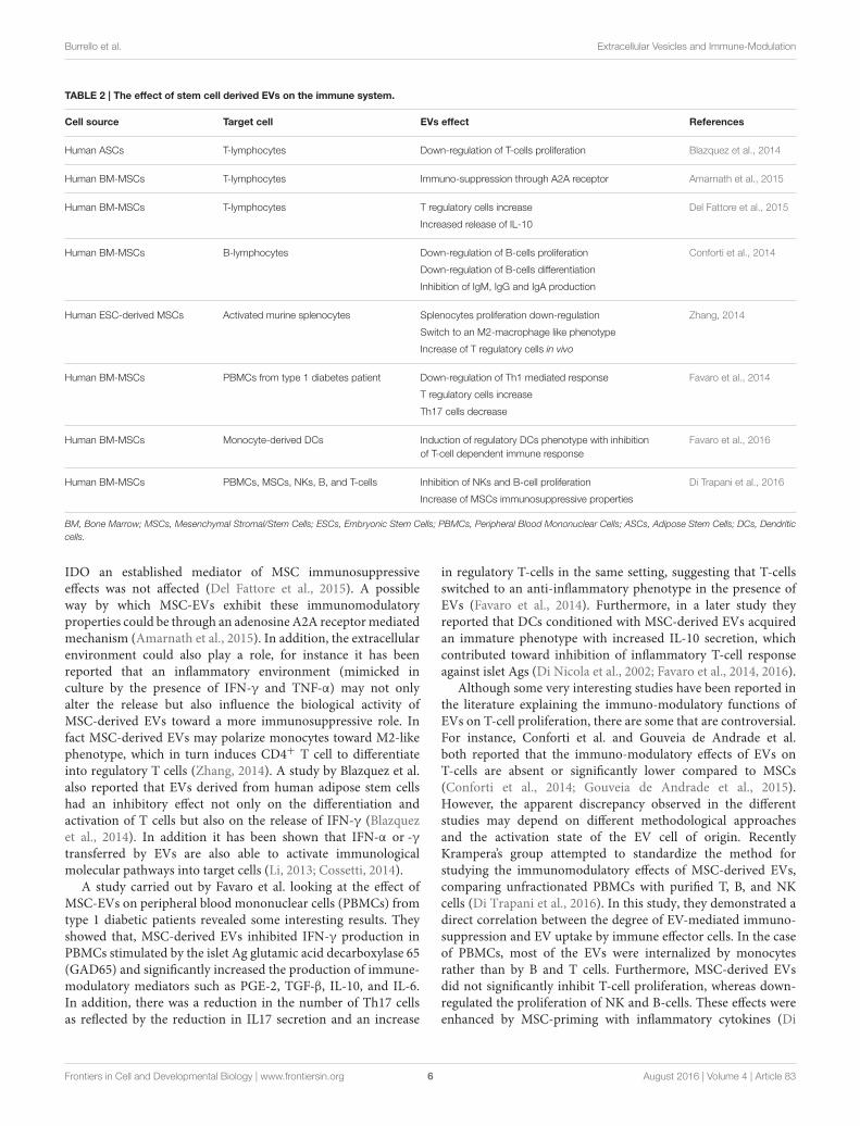
The ability of MSC-derived EVs to mimic the effect of the cellof origin has been studied on various different effector cells ofthe immune system (Blazquez et al., 2014; Conforti et al., 2014;Favaro et al., 2014, 2016; Zhang, 2014; Amarnath et al., 2015; DelFattore et al., 2015; Gouveia de Andrade et al., 2015; Di Trapaniet al., 2016; see Table 2).

The inhibitory effect of MSC-derived EVs on the activation ofcells of the immune system has been demonstrated by variousstudies. For example, Conforti et al. reported an inhibitoryactivity of MSCs and MSC-derived EVs on B-cell proliferation,which was further confirmed by a study conducted by DiTrapani et al. (Conforti et al., 2014). Del Fattore et al. alsodemonstrated that MSC-derived EVs not only increased the ratiobetween regulatory T-cells and effector T-cells, but also increasedthe release of the immunosuppressive cytokine IL-10, however,

Frontiers in Cell and Developmental Biology | www.frontiersin.org 5 August 2016 | Volume 4 | Article 83

Burrello et al. Extracellular Vesicles and Immune-Modulation

TABLE 2 | The effect of stem cell derived EVs on the immune system.

Cell source Target cell EVs effect References

Human ASCs T-lymphocytes Down-regulation of T-cells proliferation Blazquez et al., 2014

Human BM-MSCs T-lymphocytes Immuno-suppression through A2A receptor Amarnath et al., 2015

Human BM-MSCs T-lymphocytes T regulatory cells increase Del Fattore et al., 2015

Increased release of IL-10

Human BM-MSCs B-lymphocytes Down-regulation of B-cells proliferation Conforti et al., 2014

Down-regulation of B-cells differentiation

Inhibition of IgM, IgG and IgA production

Human ESC-derived MSCs Activated murine splenocytes Splenocytes proliferation down-regulation Zhang, 2014

Switch to an M2-macrophage like phenotype

Increase of T regulatory cells in vivo

Human BM-MSCs PBMCs from type 1 diabetes patient Down-regulation of Th1 mediated response Favaro et al., 2014

T regulatory cells increase

Th17 cells decrease

Human BM-MSCs Monocyte-derived DCs Induction of regulatory DCs phenotype with inhibition

of T-cell dependent immune response

Favaro et al., 2016

Human BM-MSCs PBMCs, MSCs, NKs, B, and T-cells Inhibition of NKs and B-cell proliferation Di Trapani et al., 2016

Increase of MSCs immunosuppressive properties

BM, Bone Marrow; MSCs, Mesenchymal Stromal/Stem Cells; ESCs, Embryonic Stem Cells; PBMCs, Peripheral Blood Mononuclear Cells; ASCs, Adipose Stem Cells; DCs, Dendritic

cells.

IDO an established mediator of MSC immunosuppressiveeffects was not affected (Del Fattore et al., 2015). A possibleway by which MSC-EVs exhibit these immunomodulatoryproperties could be through an adenosine A2A receptormediatedmechanism (Amarnath et al., 2015). In addition, the extracellularenvironment could also play a role, for instance it has beenreported that an inflammatory environment (mimicked inculture by the presence of IFN-γ and TNF-α) may not onlyalter the release but also influence the biological activity ofMSC-derived EVs toward a more immunosuppressive role. Infact MSC-derived EVs may polarize monocytes toward M2-likephenotype, which in turn induces CD4+ T cell to differentiateinto regulatory T cells (Zhang, 2014). A study by Blazquez et al.also reported that EVs derived from human adipose stem cellshad an inhibitory effect not only on the differentiation andactivation of T cells but also on the release of IFN-γ (Blazquezet al., 2014). In addition it has been shown that IFN-α or -γtransferred by EVs are also able to activate immunologicalmolecular pathways into target cells (Li, 2013; Cossetti, 2014).

A study carried out by Favaro et al. looking at the effect ofMSC-EVs on peripheral blood mononuclear cells (PBMCs) fromtype 1 diabetic patients revealed some interesting results. Theyshowed that, MSC-derived EVs inhibited IFN-γ production inPBMCs stimulated by the islet Ag glutamic acid decarboxylase 65(GAD65) and significantly increased the production of immune-modulatory mediators such as PGE-2, TGF-β, IL-10, and IL-6.In addition, there was a reduction in the number of Th17 cellsas reflected by the reduction in IL17 secretion and an increase

in regulatory T-cells in the same setting, suggesting that T-cellsswitched to an anti-inflammatory phenotype in the presence ofEVs (Favaro et al., 2014). Furthermore, in a later study theyreported that DCs conditioned with MSC-derived EVs acquiredan immature phenotype with increased IL-10 secretion, whichcontributed toward inhibition of inflammatory T-cell responseagainst islet Ags (Di Nicola et al., 2002; Favaro et al., 2014, 2016).

Although some very interesting studies have been reported inthe literature explaining the immuno-modulatory functions ofEVs on T-cell proliferation, there are some that are controversial.For instance, Conforti et al. and Gouveia de Andrade et al.both reported that the immuno-modulatory effects of EVs onT-cells are absent or significantly lower compared to MSCs(Conforti et al., 2014; Gouveia de Andrade et al., 2015).However, the apparent discrepancy observed in the differentstudies may depend on different methodological approachesand the activation state of the EV cell of origin. RecentlyKrampera’s group attempted to standardize the method forstudying the immunomodulatory effects of MSC-derived EVs,comparing unfractionated PBMCs with purified T, B, and NKcells (Di Trapani et al., 2016). In this study, they demonstrated adirect correlation between the degree of EV-mediated immuno-suppression and EV uptake by immune effector cells. In the caseof PBMCs, most of the EVs were internalized by monocytesrather than by B and T cells. Furthermore, MSC-derived EVsdid not significantly inhibit T-cell proliferation, whereas down-regulated the proliferation of NK and B-cells. These effects wereenhanced by MSC-priming with inflammatory cytokines (Di

Frontiers in Cell and Developmental Biology | www.frontiersin.org 6 August 2016 | Volume 4 | Article 83

Burrello et al. Extracellular Vesicles and Immune-Modulation

Trapani et al., 2016). On investigating they observed that primingwith MSCs increased the level of immunomodulatory miRNAs,such as miRNA-155 andmiRNA-146 and that EVs obtained fromprimed MSCs potentiated the immunosuppressive properties ofresting MSCs on T-cells with a mechanism that was independentto a direct EV inhibition of T-cell proliferation (Di Trapani et al.,2016).

Taken together these studies suggest that EVs derived fromMSCs are less effective than the cells themselves, as the latter mayact either by direct cell-to-cell interaction as well as by releasingseveral active soluble factors. Moreover, the biological effect ofEVs may vary depending extracellular micro-environment. Apro-inflammatory environment for instance may modify thecomposition of EVs and the consequent biological activities aswell as the activation state of immune effector cells targeted bythese EVs.

POTENTIAL THERAPEUTICAPPLICATIONS AND CONCLUDINGREMARKS

The ability of EVs to either enhance or suppress the immuneresponse may be exploited in immuno-therapy. This dualisticeffects of EVs makes them very flexible and lucrative, but, italso increases the risk of unpredictable adverse effects (Zhanget al., 2014). Several studies have reported how EV-dependentimmuno-modulation can be correlated to the source type and thestate of the cell of origin (activation and maturation degree). Forexample: APC and DC derived EVs can regulate Ag-specific andnon-specific immune responses, both positively and negatively(Robbins and Morelli, 2014). The immunogenicity depends onthe expression levels of MHC-class I and II, as well as co-stimulatory molecules such as CD80 and CD86 (Thomson andRobbins, 2008).

Although tumor-derived EVs seem to be mainly involvedin immune-escape mechanisms, it has been demonstratedthat APCs pulsed with EVs derived from tumor cells may

cross-present Ags and stimulate the activation of an Ag-specificCTL-mediated anti-tumoral response (Escudier, 2005; Morseet al., 2005). This was also confirmed in a clinical trial conductedby Bu et al. on malignant gliomas (Bu et al., 2011). However,other clinical trials have also revealed the low immunogenicityof tumor-derived EVs, which represents the main limitation forthis therapeutic strategy (Kunigelis and Graner, 2015).

The horizontal transfer of nucleic acid by EVs can beequally exploited in several therapeutic approaches (Robbinsand Morelli, 2014). For example engineering EVs with miRNAor small interfering RNA (siRNA), capable of promoting orsilencing the expression of particular transcripts, could beadopted in the future as therapeutic strategies.

Immune-cell-based and stem cell based therapies currentlypresent several risks and complications as well as technicalproblems such: culture-induced senescence, immune-mediatedrejection, genetic instability, loss of functional properties, andmalignant transformation that need to be solved to make themsuccessful (Nawaz et al., 2016). EVs which mimic in part the

effect of the stem cells from which they are derived mayrepresent a novel cell-free solution, which could overcome someof the limitations mentioned above linked to cell based therapies(Zhang, 2014). Giebel et al. in a case report demonstrated thefeasibility of an in vivo EV approach for the therapy of refractorygraft vs. host disease (Kordelas et al., 2014). The results of thisstudy suggested a beneficial effect of MSC-derived EVs as anti-inflammatory and immune-modulatory mediators in the contextof a severe inflammatory and immune disease. Nevertheless, inorder to develop an effective and successful immuno-therapybased on stem cell derived EVs warrants not only furtherstudies to clarify the mechanism of action of EVs, but also thestandardization of protocols for isolation and characterization(Witwer et al., 2013).

AUTHOR CONTRIBUTIONS

All authors listed, have made substantial, direct and intellectualcontribution to the work, and approved it for publication.

REFERENCES

Aggarwal, S., and Pittenger, M. F. (2005). Human mesenchymal stem cells

modulate allogeneic immune cell responses. Blood 105, 1815–1822. doi:

10.1182/blood-2004-04-1559

Aliotta, J. M., Pereira, M., Johnson, K. W., de Paz, N., Dooner, M. S., Puente,

N., et al. (2010). Microvesicle entry into marrow cells mediates tissue-

specific changes in mRNA by direct delivery of mRNA and induction

of transcription. Exp. Hematol. 38, 233–245. doi: 10.1016/j.exphem.2010.

01.002

Alvarez, C. V., Garcia-Lavandeira, M., Garcia-Rendueles, M. E., Diaz-Rodriguez,

E., Garcia-Rendueles, A. R., Perez-Romero, S., et al. (2012). Defining

stem cell types: understanding the therapeutic potential of ESCs, ASCs,

and iPS cells. J. Mol. Endocrinol. 49, R89–R111. doi: 10.1530/JME-

12-0072

Amarnath, S., Foley, J. E., Farthing, D. E., Gress, R. E., Laurence, A., Eckhaus,

M. A., et al. (2015). Bone marrow-derived mesenchymal stromal cells harness

purinergenic signaling to tolerize human Th1 cells in vivo. Stem Cells 33,

1200–1212. doi: 10.1002/stem.1934

Andre, F., Schartz, N. E., Movassagh, M., Flament, C., Pautier, P., Morice, P.,

et al. (2002). Malignant effusions and immunogenic tumour-derived exosomes.

Lancet 360, 295–305. doi: 10.1016/S0140-6736(02)09552-1

Bhatnagar, S., and Schorey, J. S. (2007). Exosomes released from infected

macrophages contain mycobacterium avium glycopeptidolipids and

are proinflammatory. J. Biol. Chem. 282, 25779–25789. doi: 10.1074/

jbc.M702277200

Blazquez, R., Sanchez-Margallo, F. M., de la Rosa, O., Dalemans, W., Alvarez, V.,

Tarazona, R., et al. (2014). Immunomodulatory potential of human adipose

mesenchymal stem cells derived exosomes on in vitro stimulated T cells. Front.

Immunol. 5:556. doi: 10.3389/fimmu.2014.00556

Bruno, S., Deregibus, M. C., and Camussi, G. (2015). The secretome of

mesenchymal stromal cells: role of extracellular vesicles in immunomodulation.

Immunol. Lett. 168, 154–158. doi: 10.1016/j.imlet.2015.06.007

Bu, N., Wu, H., Sun, B., Zhang, G., Zhan, S., Zhang, R., et al. (2011). Exosome-

loaded dendritic cells elicit tumor-specific CD8+ cytotoxic T cells in patients

with glioma. J. Neurooncol. 104, 659–667. doi: 10.1007/s11060-011-0537-1

Burdon, T. J., Paul, A., Noiseux, N., Prakash, S., and Shum-Tim, D. (2011).

Bone marrow stem cell derived paracrine factors for regenerative medicine:

Frontiers in Cell and Developmental Biology | www.frontiersin.org 7 August 2016 | Volume 4 | Article 83

Burrello et al. Extracellular Vesicles and Immune-Modulation

current perspectives and therapeutic potential. Bone Marrow Res. 12:7326. doi:

10.1155/2011/207326

Busch, A., Quast, T., Keller, S., Kolanus, W., Knolle, P., Altevogt, P., et al. (2008).

Transfer of T cell surface molecules to dendritic cells upon CD4+ T cell

priming involves two distinct mechanisms. J. Immunol. 181, 3965–3973. doi:

10.4049/jimmunol.181.6.3965

Camussi, G., Deregibus, M. C., Bruno, S., Cantaluppi, V., and Biancone, L. (2010).

Exosomes/microvesicles as amechanism of cell-to- cell communication.Kidney

Int. 78, 838–848. doi: 10.1038/ki.2010.278

Caplan, A. I., and Correa, D. (2011). The MSC: an injury drugstore. Cell Stem Cell

9, 11–15. doi: 10.1016/j.stem.2011.06.008

Cloutier, N., Tan, S., Boudreau, L. H., Cramb, C., Subbaiah, R., Lahey, L.,

et al. (2013). The exposure of autoantigens by microparticles underlies the

formation of potent inflammatory components: the microparticle-associated

immune complexes. EMBO Mol. Med. 5, 235–249. doi: 10.1002/emmm.2012

01846

Conforti, A., Scarsella, M., Starc, N., Giorda, E., Biagini, S., Proia, A., et al. (2014).

Microvescicles derived from mesenchymal stromal cells are not as effective as

their cellular counterpart in the ability to modulate immune responses in vitro.

Stem Cell Dev. 23, 2591–2599. doi: 10.1089/scd.2014.0091

Corcione, A., Benvenuto, F., Ferretti, E., Giunti, D., Cappiello, V., Cazzanti, F.,

et al. (2006). Human mesenchymal stem cells modulate B-cell functions. Blood

107, 367–372. doi: 10.1182/blood-2005-07-2657

Cossetti, C. (2014). Extracellular vesicles from neural stem cells transfer IFN-

gamma via Ifngr1 to activate Stat1 signaling in target cells. Mol. Cell 11,

589–593. doi: 10.1016/j.molcel.2014.08.020

Dalal, J., Gandy, K., and Domen, J. (2012). Role of mesenchymal stem cell therapy

in Crohn’s disease. Pediatr. Res. 71, 445–451. doi: 10.1038/pr.2011.56

Del Fattore, A., Luciano, R., Pascucci, L., Goffredo, B. M., Giorda, E., Scapaticci,

M., et al. (2015). Immunoregulatory effects of mesenchymal stem cell-derived

extracellular vesicles on T lymphocytes. Cell Transplant. 24, 2615–2627. doi:

10.3727/096368915X687543

Deregibus, M. C., Cantaluppi, V., Calogero, R., Lo Iacono, M., Tetta, C., Biancone,

L., et al. (2007). Endothelial progenitor cell-derived microvesicles activate an

angiogenic program in endothelial cells by a horizontal transfer of mRNA.

Blood 7, 2440–2448. doi: 10.1182/blood-2007-03-078709

Di Nicola, M., Carlo-Stella, C., Magni, M., Milanesi, M., Longoni, P. D., Matteucci,

P., et al. (2002). Human bone marrow stromal cells suppress T-lymphocyte

proliferation induced by cellular or nonspecific mitogenic stimuli. Blood 99,

3838–3843. doi: 10.1182/blood.V99.10.3838

Di Trapani, M., Bassi, G., Midolo, M., Gatti, A., Kamga, P. T., Cassaro, A.,

et al. (2016). Differential and transferable modulatory effects of mesenchymal

stromal cell-derived extracellular vesicles on T, B and NK cell functions. Sci.

Rep. 6:24120. doi: 10.1038/srep24120

Djouad, F., Charbonnier, L. M., Bouffi, C., Louis-Plence, P., Bony, C., Apparailly,

F., et al. (2007). Mesenchymal stem cells inhibit the differentiation of

dendritic cells through an interleukin-6-dependent mechanism. Stem Cells 25,

2025–2032. doi: 10.1634/stemcells.2006-0548

EL Andaloussi, S., Mäger, I., Breakefield, X. O., and Wood, M. J. (2013).

Extracellular vesicles: biology and emerging therapeutic opportunities. Nat.

Rev. Drug Discov. 12, 347–357. doi: 10.1038/nrd3978

English, K., Ryan, J. M., Tobin, L., Murphy, M. J., Barry, F. P., Mahon, B. P., et al.

(2009). Cell contact, prostaglandin E(2) and transforming growth factor beta 1

play non-redundant roles in human mesenchymal stem cell induction of CD4

+ CD25 (High) forkhead box P3+ regulatory T cells. Clin. Exp. Immunol. 156,

149–160. doi: 10.1111/j.1365-2249.2009.03874.x

Escudier, B. (2005). Vaccination of metastatic melanoma patients with autologous

dendritic cell (DC) derived-exosomes: results of the first phase I clinical trial.

J. Transl. Med. 3, 10–17. doi: 10.1186/1479-5876-3-10

Favaro, E., Carpanetto, A., Caorsi, C., Giovarelli, M., Angelini, C., Cavallo-

Perin, P., et al. (2016). Human mesenchymal stem cells and derived

extracellular vesicles induce regulatory dendritic cells in type 1 diabetic patients.

Diabetologia 59, 325–333. doi: 10.1007/s00125-015-3808-0

Favaro, E., Carpanetto, A., Lamorte, S., Fusco, A., Caorsi, C., Deregibus,

M. C., et al. (2014). Human mesenchymal stem cell-derived microvesicles

modulate T cell response to islet antigen glutamic acid decarboxylase in

patients with type 1 diabetes. Diabetologia 57, 1664–1673. doi: 10.1007/s00125-

014-3262-4

Gasser, O., and Schifferli, J. A. (2004). Activated polymorphonuclear neutrophils

disseminate anti-inflammatory microparticles by ectocytosis. Blood 104,

2543–2548. doi: 10.1182/blood-2004-01-0361

Gouveia de Andrade, A. V., Bertolino, G., Riewaldt, J., Bieback, K., Karbanová, J.,

Odendahl, M., et al. (2015). Extracellular vesicles secreted by bonemarrow- and

adipose tissue-derived mesenchymal stromal cells fail to suppress lymphocyte

proliferation. Stem Cells Dev. 6, 345–351. doi: 10.1089/scd.2014.0563

Hassani, K., and Olivier, M. (2013). Immunomodulatory impact of leishmania-

induced macrophage exosomes: a comparative proteomic and functional

analysis. PLoS Negl. Trop. Dis. 7:e2185. doi: 10.1371/journal.pntd.0002185

Hedlund, M., Nagaeva, O., Kargl, D., Baranov, V., and Mincheva-Nilsson, L.

(2011). Thermal- and oxidative stress causes enhanced release of NKG2D

ligand-bearing immuno-suppressive exosomes in leukemia/lymphoma T and

B cells. PLoS ONE 6:e16899. doi: 10.1371/journal.pone.0016899

Hu, G. W., Li, Q., Niu, X., Hu, B., Liu, J., Zhou, S. M., et al. (2015). Exosomes

secreted by human-induced pluripotent stem cell-derived mesenchymal stem

cells attenuate limb ischemia by promoting angiogenesis in mice. Stem Cell Res.

Ther. 6, 10. doi: 10.1186/scrt546

Hwu, P., Du, M. X., Lapointe, R., Do, M., Taylor, M. W., and Young, H. A.

(2000). Indoleamine 2,3-dioxygenase production by human dendritic cells

results in the inhibition of T cell proliferation. J. Immunol. 164, 3596–3599.

doi: 10.4049/jimmunol.164.7.3596

Katsman, D., Stackpole, E. J., Domin, D. R., and Farber, D. B. (2012). Embryonic

stem cell-derived microvesicles induce gene expression changes in Müller cells

of the retina. PLoS ONE 7:e50417. doi: 10.1371/journal.pone.0050417

Keating, A. (2012). Mesenchymal stromal cells: new directions. Cell Stem Cell 10,

709–716. doi: 10.1016/j.stem.2012.05.015

Kordelas, L., Rebmann, V., Ludwig, A. K., Radtke, S., Ruesing, J., Doeppner, T. R.,

et al. (2014). MSC-derived exosomes: a novel tool to treat therapy-refractory

graft-versus-host disease. Leukemia 28, 970–973. doi: 10.1038/leu.2014.41

Krampera, M., Cosmi, L., Angeli, R., Pasini, A., Liotta, F., Andreini, A., et al.

(2006). Role for interferon-gamma in the immunomodulatory activity of

human bone marrow mesenchymal stem cells. Stem Cells 24, 386–398. doi:

10.1634/stemcells.2005-0008

Krampera, M., Glennie, S., Dyson, J., Scott, D., Laylor, R., Simpson, E., et al.

(2003). Bone marrow mesenchymal stem cells inhibit the response of naive and

memory antigen-specific T cells to their cognate peptide. Blood 101, 3722–3729.

doi: 10.1182/blood-2002-07-2104

Kunigelis, K. E., and Graner, M. W. (2015). The Dichotomy of Tumor Exosomes

(TEX) in cancer immunity: is it all in the ConTEXt? Vaccines 3, 1019–1051. doi:

10.3390/vaccines3041019

Lai, R. C., Arslan, F., Lee, M. M., Sze, N. S., Choo, A., Chen, T. S., et al. (2010).

Exosome secreted by MSC reduces myocardial ischemia/reperfusion injury.

Stem Cell Res. 4, 214–222. doi: 10.1016/j.scr.2009.12.003

Le Blanc, K., and Mougiakakos, D. (2012). Multipotent mesenchymal stromal

cells and the innate immune system. Nat. Rev. Immunol. 12, 383–396. doi:

10.1038/nri3209

Lee, J., Sayed, N., Hunter, A., Au, K. F., Wong, W. H., Mocarski, E. S., et al. (2012).

Activation of innate immunity is required for efficient nuclear reprogramming.

Cell 151, 547–558. doi: 10.1016/j.cell.2012.09.034

Levy, S., Todd, S. C., and Maecker, H. T. (1998). CD81 (TAPA-1): a molecule

involved in signal transduction and cell adhesion in the immune system. Annu.

Rev. Immunol. 16, 89–109. doi: 10.1146/annurev.immunol.16.1.89

Li, J. (2013). Exosomes mediate the cell-to-cell transmission of IFN-α-induced

antiviral activity. Nat. Immunol. 45, 456–465. doi: 10.1038/ni.2647

MacDonald, G. I., Augello, A., and De Bari, C. (2011). Role of mesenchymal

stem cells in reestablishing immunologic tolerance in auto-immune Rheumatic

diseases. Arthritis Rheum. 63, 2547–2557. doi: 10.1002/art.30474

Mastronardi, M. L., Mostefai, H. A., Meziani, F., Martinez, M. C., Asfar, P.,

and Andriantsitohaina, R. (2011). Circulating microparticles from septic

shock patients exert differential tissue expression of enzymes related to

inflammation and oxidative stress. Crit. Care Med. 39, 1739–1748. doi:

10.1097/CCM.0b013e3182190b4b

Monleón, I., Martínez-Lorenzo, M. J., Monteagudo, L., Lasierra, P., Taulés, M.,

Iturralde, M., et al. (2001). Differential secretion of Fas ligand- or APO2

ligand/TNF-related apoptosis-inducing ligand-carrying microvesicles during

activation-induced death of human T cells. J. Immunol. 167, 6736–6744. doi:

10.4049/jimmunol.167.12.6736

Frontiers in Cell and Developmental Biology | www.frontiersin.org 8 August 2016 | Volume 4 | Article 83

Burrello et al. Extracellular Vesicles and Immune-Modulation

Morelli, A. E., Larregina, A. T., Shufesky, W. J., Sullivan, M. L., Stolz, D. B.,

Papworth, G. D., et al. (2004). Endocytosis, intracellular sorting, and processing

of exosomes by dendritic cells. Blood 104, 3257–3266. doi: 10.1182/blood-2004-

03-0824

Morse, M. A., Garst, J., Osada, T., Khan, S., Hobeika, A., Clay, T. M., et al. (2005).

A phase I study of dexosome immunotherapy in patients with advanced non-

small cell lung cancer. J. Transl. Med. 3, 9–13. doi: 10.1186/1479-5876-3-9

Muntasell, A., Berger, A. C., and Roche, P. A. (2007). T cell-induced secretion of

MHC class II-peptide complexes on B cell exosomes. EMBO J. 26, 4263–4272.

doi: 10.1038/sj.emboj.7601842

Nawaz, M., Fatima, F., Vallabhaneni, K. C., Penfornis, P., Valadi, H., Ekström, K.,

et al. (2016). Extracellular vesicles: evolving factors in stem cell biology. Stem

Cells Int. 2016:1073140. doi: 10.1155/2016/1073140

Noh, K. H., Kim, B. W., Song, K. H., Cho, H., Lee, Y. H., Kim, J. H., et al. (2012).

Nanog signaling in cancer promotes stem-like phenotype and immune evasion.

J. Clin. Invest. 122, 4077–4093. doi: 10.1172/JCI64057

Nolte-’t Hoen, E. N., Buschow, S. I., Anderton, S. M., Stoorvogel, W., andWauben,

M. H. (2009). Activated T cells recruit exosomes secreted by dendritic cells via

LFA-1. Blood 113, 1977–1981. doi: 10.1182/blood-2008-08-174094

O’Neill, H. C., and Quah, B. J. C. (2008). Exosomes secreted by bacterially infected

macrophages are proinflammatory. Sci. Signal. 1:pe8. doi: 10.1126/stke.16pe8

O’Connell, R. M., Rao, D. S., Chaudhuri, A. A., and Baltimore, D. (2010).

Physiological and pathological roles formicroRNAs in the immune system.Nat.

Rev. Immunol. 10, 111–122. doi: 10.1038/nri2708

Pittenger, M. (2009). Sleuthing the source of regeneration by MSCs. Cell Stem Cell

5, 8–10. doi: 10.1016/j.stem.2009.06.013

Pittenger, M. F., Mackay, A. M., Beck, S. C., Jaiswal, R. K., Douglas, R., Mosca, J.

D., et al. (1999). Multilineage potential of adult humanmesenchymal stem cells.

Science 284, 143–147. doi: 10.1126/science.284.5411.143

Pizzirani, C. (2007). Stimulation of P2 receptors causes release of IL-1beta-

loaded microvesicles from human dendritic cells. Blood 109, 3856–3864. doi:

10.1182/blood-2005-06-031377

Prakash, P. S., Caldwell, C. C., Lentsch, A. B., Pritts, T. A., and Robinson, B. R. H.

(2012). Human microparticles generated during sepsis in patients with critical

illness are neutrophil-derived and modulate the immune response. J. Trauma

Acute Care Surg. 73, 401–406. doi: 10.1097/TA.0b013e31825a776d

Qazi, K. R., Gehrmann, U., Domange Jordö, E., Karlsson, M. C., and Gabrielsson,

S. (2009). Antigen-loaded exosomes alone induce Th1-type memory through a

B-cell-dependent mechanism. Blood 113, 2673–2683. doi: 10.1182/blood-2008-

04-153536

Qazi, K. R., Torregrosa Paredes, P., Dahlberg, B., Grunewald, J., Eklund, A.,

and Gabrielsson, S. (2010). Proinflammatory exosomes in bronchoalveolar

lavage fluid of patients with sarcoidosis. Thorax 65, 1016–1024. doi:

10.1136/thx.2009.132027

Ratajczak, J., Miekus, K., Kucia, M., Zhang, J., Reca, R., Dvorak, P., et al.

(2006b). Embryonic stem cell-derived microvesicles reprogram hematopoietic

progenitors: evidence for horizontal transfer of mRNA and protein delivery.

Leukemia 20, 847–856. doi: 10.1038/sj.leu.2404132

Ratajczak, J., Wysoczynski, M., Hayek, F., Janowska-Wieczorek, A., and

Ratajczak, M. Z. (2006a). Membrane-derived micro-vesicles: important and

underappreciated mediators of cell-to-cell communication. Leukemia 20,

1487–1495. doi: 10.1038/sj.leu.2404296

Robbins, P. D., and Morelli, A. E. (2014). Regulation of immune responses by

extracellular vesicles. Nat. Rev. Immunol. 14, 195–208. doi: 10.1038/nri3622

Sato, K., Ozaki, K., Oh, I., Meguro, A., Hatanaka, K., Nagai, T., et al. (2007). Nitric

oxide plays a critical role in suppression of T-cell proliferation by mesenchymal

stem cells. Blood 109, 228–234. doi: 10.1182/blood-2006-02-002246

Schenten, D., andMedzhitov, R. (2011). The control of adaptive immune responses

by the innate immune system. Adv. Immunol. 109, 87–124. doi: 10.1016/B978-

0-12-387664-5.00003-0

Sirisinha, S. (2014). Evolutionary insights into the origin of innate and adaptive

immune systems: different shades of grey. Asian Pac. J. Allergy Immunol. 32,

3–15.

Spaggiari, G. M., Capobianco, A., Becchetti, S., Mingari, M. C., and Moretta, L.

(2006). Mesenchymal stem cell-natural killer cell interactions: evidence that

activated NK cells are capable of killing MSCs, whereas MSCs can inhibit IL-2-

induced NK-cell proliferation. Blood 107, 1484–1490. doi: 10.1182/blood-2005-

07-2775

Staal, F. J., Luis, T. C., and Tiemessen, M. M. (2008). WNT signalling in the

immune system: WNT is spreading its wings. Nat. Rev. Immunol. 8, 581–593.

doi: 10.1038/nri2360

Syed, B. A., and Evans, J. B. (2013). Stem cell therapy market. Nat. Rev. Drug

Discov. 12, 185–186. doi: 10.1038/nrd3953

Takahashi, K., and Yamanaka, S. (2006). Induction of pluripotent stem cells from

mouse embryonic and adult fibroblast cultures by defined factors. Cell 126,

663–676. doi: 10.1016/j.cell.2006.07.024

Testa, J. S., Apcher, G. S., Comber, J. D., and Eisenlohr, L. C. (2010). Exosome-

driven antigen transfer for MHC class II presentation facilitated by the receptor

binding activity of influenza hemagglutinin. J. Immunol. 185, 6608–6616. doi:

10.4049/jimmunol.1001768

Théry, C., Duban, L., Segura, E., Véron, P., Lantz, O., and Amigorena, S. (2002).

Indirect activation of naive CD4+ T cells by dendritic cell-derived exosomes.

Nat. Immunol. 3, 1156–1162. doi: 10.1038/ni854

Théry, C., Ostrowski, M., and Segura, E. (2009). Membrane vesicles as conveyors

of immune responses. Nat. Rev. Immunol. 9, 581–593. doi: 10.1038/nri2567

Thomson, A. W., and Robbins, P. D. (2008). Tolerogenic dendritic

cells for autoimmune disease and transplantation. Ann. Rheum. Dis.

67(Suppl. 3):iii90–iii96. doi: 10.1136/ard.2008.099176

Tsuji, K., and Kitamura, S. (2015). Trophic factors from tissue stem cells for renal

regeneration. Stem Cells Int. 7:e537204. doi: 10.1155/2015/537204

Uccelli, A., Moretta, L., and Pistoia, V. (2008). Mesenchymal stem cells in health

and disease. Nat. Rev. Immunol. 8, 726–736. doi: 10.1038/nri2395

Valadi, H., Ekstrom, K., Bossios, A., Sjostrand, M., Lee, J. J., and Lotvall, J. O.

(2007). Exosome-mediated transfer of mRNAs and micro- RNAs is a novel

mechanism of genetic exchange between cells. Nat. Cell Biol. 9, 654–659. doi:

10.1038/ncb1596

Vincent-Schneider, H., Stumptner-Cuvelette, P., Lankar, D., Pain, S., Raposo,

G., Benaroch, P., et al. (2002). Exosomes bearing HLA-DR1 molecules need

dendritic cells to efficiently stimulate specific T cells. Int. Immunol. 14, 713–722.

doi: 10.1093/intimm/dxf048

Walker, J. D., Maier, C. L., and Pober, J. S. (2009). Cytomegalovirus-

infected human endothelial cells can stimulate allogeneic CD4+ memory

T cells by releasing antigenic exosomes. J. Immunol. 182, 1548–1559. doi:

10.4049/jimmunol.182.3.1548

Walters, S. B., Kieckbusch, J., Nagalingam, G., Swain, A., Latham, S. L., Grau,

G. E., et al. (2013). Microparticles from mycobacteria- infected macrophages

promote inflammation and cellular migration. J. Immunol. 190, 669–677. doi:

10.4049/jimmunol.1201856

Wang, J. G., Williams, J. C., Davis, B. K., Jacobson, K., Doerschuk, C. M., Ting,

J. P. Y., et al. (2011). Monocytic microparticles activate endothelial cells in an

IL-1 beta-dependent manner. Blood 118, 2366–2374. doi: 10.1182/blood-2011-

01-330878

Wang, K. X., Xu, L. L., Rui, Y. F., Huang, S., Lin, S. E., Xiong, J. H., et al. (2015a).

The effects of secretion factors from umbilical cord derived mesenchymal

stem cells on osteogenic differentiation of mesenchymal stem cells. PLoS ONE

10:e0120593. doi: 10.1371/journal.pone.0120593

Wang, Y., Zhang, L., Li, Y., Chen, L., Wang, X., Guo, W., et al. (2015b).

Exosomes/microvesicles from induced pluripotent stem cells deliver

cardioprotective miRNAs and prevent cardiomyocyte apoptosis in the ischemic

myocardium. Int. J. Cardiol. 192, 61–69. doi: 10.1016/j.ijcard.2015.05.020

Weiner, O. D. (2002). Rac activation: P-Rex1 - a convergence point for PIP3 and

Gβγ? Curr. Biol. 12:R429. doi: 10.1371/journal.pone.0050417

Witwer, K. W., Buzás, E. I., Bemis, L. T., Bora, A., Lässer, C., Lötvall, J.,

et al. (2013). Standardization of sample collection, isolation and analysis

methods in extracellular vesicle research. J. Extracell. Vesicles 27:2. doi:

10.3402/jev.v2i0.20360

Wolfers, J., Lozier, A., Raposo, G., Regnault, A., Théry, C., Masurier, C., et al.

(2001). Tumor-derived exosomes are a source of shared tumor rejection

antigens for CTL cross-priming. Nat. Med. 7, 297–303. doi: 10.1038/85438

Xia, P., Wang, S., Ye, B., Du, Y., Huang, G., Zhu, P., et al. (2015). Sox2

functions as a sequence-specific DNA sensor in neutrophils to initiate innate

immunity against microbial infection.Nat. Immunol. 16, 366–375. doi: 10.1038/

ni.3117

Xie, Y., Zhang, H., Li, W., Deng, Y., Munegowda, M. A., Chibbar, R., et al.

(2010). Dendritic cells recruit T cell exosomes via exosomal LFA-1 leading to

inhibition of CD8+ CTL responses through downregulation of peptide/MHC

Frontiers in Cell and Developmental Biology | www.frontiersin.org 9 August 2016 | Volume 4 | Article 83

Burrello et al. Extracellular Vesicles and Immune-Modulation

class I and Fas ligand-mediated cytotoxicity. J. Immunol. 185, 5268–5278. doi:

10.4049/jimmunol.1000386

Yáñez-Mó, M., Siljander, P. R., Andreu, Z., Zavec, A. B., Borràs, F. E., Buzas,

E. L., et al. (2015). Biological properties of extracellular vesicles and their

physiological functions. J. Extracell. Vesicles 4:27066. doi: 10.3402/jev.v4.27066

Zanotti, L., Sarukhan, A., Dander, E., Castor, M., Cibella, J., Soldani, C., et al.

(2013). Encapsulated mesenchymal stem cells for in vivo immunomodulation.

Leukemia 27, 500–503. doi: 10.1038/leu.2012.202

Zhang, B. (2014). Mesenchymal stem cells secrete immunologically active

exosomes. Stem Cells Dev. 2, 175–179. doi: 10.1089/scd.2013.0479

Zhang, B., Yin, Y., Lai, R. C., and Lim, S. K. (2014). Immunotherapeutic potential

of extracellular vesicles. Front. Immunol. 5:518. doi: 10.3389/fimmu.2014.00518

Zhang, H. G., Liu, C., Su, K., Yu, S., Zhang, L., Zhang, S., et al. (2006). A membrane

form of TNF-alpha presented by exosomes delays T cell activation-induced cell

death. J. Immunol. 176, 7385–7393. doi: 10.4049/jimmunol.176.12.7385

Zitvogel, L., Regnault, A., Lozier, A., Wolfers, J., Flament, C., Tenza, D.,

et al. (1998). Eradication of established murine tumors using a novel cell-

free vaccine: dendritic cell-derived exosomes. Nat. Med. 4, 594–600. doi:

10.1038/nm0598-594

Zuccato, E., Blott, E. J., Holt, O., Sigismund, S., Shaw, M., Bossi, G., et al.

(2007). Sorting of Fas ligand to secretory lysosomes is regulated by mono-

ubiquitylation and phosphorylation. J. Cell Sci. 120, 191–199. doi: 10.1242/jcs.

Conflict of Interest Statement: The authors declare that the research was

conducted in the absence of any commercial or financial relationships that could

be construed as a potential conflict of interest.

Copyright © 2016 Burrello, Monticone, Gai, Gomez, Kholia and Camussi. This

is an open-access article distributed under the terms of the Creative Commons

Attribution License (CC BY). The use, distribution or reproduction in other forums

is permitted, provided the original author(s) or licensor are credited and that the

original publication in this journal is cited, in accordance with accepted academic

practice. No use, distribution or reproduction is permitted which does not comply

with these terms.

Frontiers in Cell and Developmental Biology | www.frontiersin.org 10 August 2016 | Volume 4 | Article 83


Recommended