422 American Society of Hematology Stem Cell Transplantation: Supportive Care and Long-Term Complications John R. Wingard, Georgia B. Vogelsang, and H. Joachim Deeg With increasing hematopoietic stem cell transplant (HSCT) activity and improvement in outcomes, there are many thousands of HSCT survivors currently being followed by non-transplant clini- cians for their healthcare. Several types of late sequelae from HSCT have been noted, and aware- ness of these complications is important in mini- mizing late morbidity and mortality. Late effects can include toxicities from the treatment regimen, infections from immunodeficiency, endocrine disturbances, growth impairment, psychosocial adjustment disorders, second malignancies, and chronic graft-versus-host disease (GVHD). A variety of risk factors for these complications have been noted. The clinician should be alert to the potential for these health issues. Preventive and treatment strategies can minimize morbidity from these problems and optimize outcomes. I. OVERVIEW OF LATE COMPLICATIONS John R. Wingard, MD* Hematopoietic stem cell transplantation (HSCT) pro- vides effective therapy for patients with lympho- hematopoietic, immunologic, metabolic and other dis- orders. Both the annual number of HSCT procedures has increased dramatically and the number of diseases for which HSCT is considered appropriate have ex- panded over the years. Estimates suggest that 30,000– 40,000 HSCT procedures are performed annually world- wide. HSCT offers a potential for cure or long-term dis- ease control for a number of diseases where other treat- ment options fail or prognostic indicators suggest that durable control is unlikely. Outcomes have also gradu- ally improved. Data from the IBMTR, ABMTR, and EBMTR indicate gradual improvement in long-term outcomes with an average survival improvement of ap- proximately 10% per decade. With expanding applica- tions and increase in HSCT activity, along with techno- logical advances in supportive care, histocompatibility testing, safer conditioning regimens, and control of graft- versus-host disease (GVHD), growth in transplant ac- tivities is likely to occur, outcomes should improve, and increasing numbers of transplant survivors will be fac- ing life after the transplant experience. Many patients have now been followed for two or three decades post-transplant and are presumably cured. For many HSCT survivors, cure or control of the under- lying disease is not accompanied by full restoration of health. 1-7 Some patients develop long-term complications, the topic of this session. Both long-term physical and psychosocial morbid- ity can occur. The types of late sequelae include toxici- ties from the treatment regimen, immune deficiency, au- toimmune syndromes, infectious complications, endo- crine disturbances, growth impairment in children, cog- nitive dysfunction, second malignancies, chronic GVHD, and problems with psychosocial adjustment and quality of life. These late effects can negatively affect a patient’s performance of daily activities, interpersonal and fam- ily relationships, and sense of personal well being. The multifactorial etiologies of posttransplant com- plications are illustrated in Figure 1. Several factors act solely or in concert in causation. Some complications are due to the transplant procedure, such as GVHD, im- munodeficiency, infectious complications, and autoim- mune syndromes. Others are due to the conditioning regi- men or prior anti-neoplastic therapy, such as sterility, alopecia, endocrine disturbances, cardiorespiratory in- sufficiency, renal impairment, impaired growth, and cog- nitive disturbances. Some are due to the underlying dis- ease itself, such as recurrence. Some complications are multifactorial: for example, respiratory insufficiency may * University of Florida, HSC, College of Medicine, 1600 SW Archer Rd., Rm. R4-116, P.O. Box 100277, Gainesville, FL 32610-3001
Transcript
422 American Society of Hematology
Stem Cell Transplantation:Supportive Care and Long-Term Complications
John R. Wingard, Georgia B. Vogelsang, and H. Joachim Deeg
With increasing hematopoietic stem cell transplant(HSCT) activity and improvement in outcomes,there are many thousands of HSCT survivorscurrently being followed by non-transplant clini-cians for their healthcare. Several types of latesequelae from HSCT have been noted, and aware-ness of these complications is important in mini-mizing late morbidity and mortality. Late effects caninclude toxicities from the treatment regimen,
infections from immunodeficiency, endocrinedisturbances, growth impairment, psychosocialadjustment disorders, second malignancies, andchronic graft-versus-host disease (GVHD). A varietyof risk factors for these complications have beennoted. The clinician should be alert to the potentialfor these health issues. Preventive and treatmentstrategies can minimize morbidity from theseproblems and optimize outcomes.
I. OVERVIEW OF LATE COMPLICATIONS
John R. Wingard, MD*
Hematopoietic stem cell transplantation (HSCT) pro-vides effective therapy for patients with lympho-hematopoietic, immunologic, metabolic and other dis-orders. Both the annual number of HSCT procedureshas increased dramatically and the number of diseasesfor which HSCT is considered appropriate have ex-panded over the years. Estimates suggest that 30,000–40,000 HSCT procedures are performed annually world-wide. HSCT offers a potential for cure or long-term dis-ease control for a number of diseases where other treat-ment options fail or prognostic indicators suggest thatdurable control is unlikely. Outcomes have also gradu-ally improved. Data from the IBMTR, ABMTR, andEBMTR indicate gradual improvement in long-termoutcomes with an average survival improvement of ap-proximately 10% per decade. With expanding applica-tions and increase in HSCT activity, along with techno-logical advances in supportive care, histocompatibilitytesting, safer conditioning regimens, and control of graft-versus-host disease (GVHD), growth in transplant ac-tivities is likely to occur, outcomes should improve, and
increasing numbers of transplant survivors will be fac-ing life after the transplant experience.
Many patients have now been followed for two orthree decades post-transplant and are presumably cured.For many HSCT survivors, cure or control of the under-lying disease is not accompanied by full restoration ofhealth.1-7 Some patients develop long-term complications,the topic of this session.
Both long-term physical and psychosocial morbid-ity can occur. The types of late sequelae include toxici-ties from the treatment regimen, immune deficiency, au-toimmune syndromes, infectious complications, endo-crine disturbances, growth impairment in children, cog-nitive dysfunction, second malignancies, chronic GVHD,and problems with psychosocial adjustment and qualityof life. These late effects can negatively affect a patient’sperformance of daily activities, interpersonal and fam-ily relationships, and sense of personal well being.
The multifactorial etiologies of posttransplant com-plications are illustrated in Figure 1. Several factors actsolely or in concert in causation. Some complicationsare due to the transplant procedure, such as GVHD, im-munodeficiency, infectious complications, and autoim-mune syndromes. Others are due to the conditioning regi-men or prior anti-neoplastic therapy, such as sterility,alopecia, endocrine disturbances, cardiorespiratory in-sufficiency, renal impairment, impaired growth, and cog-nitive disturbances. Some are due to the underlying dis-ease itself, such as recurrence. Some complications aremultifactorial: for example, respiratory insufficiency may
* University of Florida, HSC, College of Medicine, 1600 SWArcher Rd., Rm. R4-116, P.O. Box 100277, Gainesville, FL32610-3001
Hematology 2002 423
Figure 1. Multifactorial etiology of post-transplantationcomplications.
Reprinted with permission from Deeg HJ. Chapter 66. Delayedcomplications after hematopoietic cell transplantation. In FormanSJ, Blume KG, Thomas ED, eds: Hematopoietic Cell Transplanta-tion, 3rd Ed. Boston: Blackwell Scientific Publications, Inc; 1999:776-806.
Abbreviations: TBI, total body irradiation; GVHD, graft-versus-hostdisease.
result from the collective effects of lung injury from theconditioning regimen, bronchiolitis obliterans fromGVHD, and a superimposed infection. Table 1 lists vari-ous types of complications, with risk factors and con-siderations for prevention and treatment.
A variety of health behaviors, both personal andphysician dependent, can improve survivors’ health andfunctioning and prevent some of these complications.Accordingly, it is increasingly important for non-trans-plant clinicians to be aware of these sequelae and beadvocates for health maintenance behaviors to optimizefunctioning and sense of well being.
ImmunodeficiencyAntigen-specific T and B cell responses are necessaryfor control of many infectious pathogens. Immune re-sponses are profoundly deficient early after HSCT;gradual restoration occurs during the first year follow-ing transplant.8-11 Factors that impede development ofprotective responses include GVHD, certain viral infec-tions, particularly those by the herpesvirus family, deple-tion of lymphocytes from the stem cell graft, or post-transplant immunotherapeutic maneuvers such as admin-istration of antibodies against T and B cells (either forcontrol of GVHD or malignancy). Donor source (pe-ripheral blood versus bone marrow) and the degree ofhistocompatibility between donor and recipient also af-fect the pace of immune reconstitution.
The major risk factor for late infections is chronicGVHD.12,13 Chronic GVHD is associated with suscepti-bility for infections by encapsulated bacteria (S.
pneumonaie, H. influenzae, and N. meningitidis), inva-sive candidiasis, aspergillosis, and herpesviruses, espe-cially cytomegalovirus (CMV) and varicella zoster vi-rus (VZV). Prophylaxis with an antibiotic with activityagainst gram positive organisms is recommended.14 Itshould be continued for as long as active therapy forGVHD is given. The duration of antibiotic prophylaxisbeyond active therapy is unclear.
Pneumocystis carinii infection (PCP) prophylaxiswith trimethaprin-sulfamethoxazole should be given forthe first 6 months and continue for those receiving ac-tive therapy for chronic GVHD. Immunizations with in-activated vaccines should be given starting at 12months.14 Live attenuated virus vaccines should beavoided during the first two years and should be avoidedin those with chronic GVHD.
An infrequent but severe manifestation of VZV in-fection is severe abdominal pain and a clinical syndromesuggesting a perforated intra-abdominal viscus, acutepancreatitis, or severe hepatitis. This can be associatedwith a rapidly progressive and life threatening course.Accordingly, VZV infections should be strongly consid-ered in the evaluation of a patient with acute abdominalpain. Acyclovir is effective therapy for VZV infection.
Autoimmune antibodies are detected in some pa-tients especially those with chronic GVHD. Often thisis without clinical consequence but autoimmune cyto-penias are occasionally problematic.15,16 Recipients ofABO incompatible donor grafts also can have persistenthemolysis for months after HSCT or longer.17 This isdue to persistent host isohemagglutinins, which may per-sist until full donor chimerism is established.
Endocrine DysfunctionHypothyroidism occurs in patients who have receivedprior cranial or mantle irradiation and in those in whichtotal body irradiation (TBI) was part of the conditioningregimen.18-20 Children appear to be more susceptible.Annual assessment of thyroid function is advised in allpatients who have received TBI or radiotherapy to thehead and neck. If hypothyroidism is noted, then replace-ment therapy is appropriate.
Hypoadrenalism may occur in patients on prolongedcorticosteroid therapy for GVHD treatment. Gradualtapering of corticosteroids is appropriate. For patientsrequiring surgical procedures or with acute medical ill-nesses, short-term “stress” replacement is warranted.
Gonadal function is impaired lifelong in the major-ity of patients receiving intensive chemotherapy or TBI-containing conditioning regimens.21-25 Lifelong infertil-ity is typical but not universal. Several studies have sug-gested recovery of spermatogenesis in a minority of menyears later (10-15%) and ovarian recovery has occurred
424 American Society of Hematology
Table 1. Types of late complications: tissues affected, risk factors, prevention, and treatment.
Tissue/Organs Late Complications Risk Factors Preventive Measures Treatment Options
Immunity Infections GVHD Antibiotic prophylaxis Targeted antimicrobialsT cell depletion Immunizations for specific infectiousHerpesvirus infection Optimization of matching pathogensDonor source PCP prophylaxisHistocompatibility of donor and recipient
Autoimmune syndromes GVHD Optimization of matching IVIG for autoimmunethrombocytopenia
Steroids for variousautoimmune phenomena
Endocrine glands Hypothyroidism Radiotherapy to head, Fractionation of TBI Thyroid replacement neck, & mantle Annual thyroid screeningTBI
Hypoadrenalism Prolonged corticosteroid use Replacement steroids for surgical procedures or acute medical conditions
Growth Short stature CNS irradiation Periodic assessment of Hormone replacementTBI (single dose rather than endocrine status fractionated)HypothyroidismCorticosteroid therapyGonadal insufficiency
Bladder Scarring after Cyclophosphamide, BK virus, Hyperhydration or mesna Antispasmodics for hemorrhagic cystitis adenovirus, CMV Cyclophosphamide symptomatic relief
administration
Kidneys Nephropathy TBI, prior platinum compounds Angiotension-converting Control of hypertensionenzyme inhibitors
Abbreviations: GVHD, graft-versus-host disease; PCP, Pneumocystis carinii pneumonia; TBI, total body irradiation; CNS, central nervoussystem; CMV, cytomegalovirus
Hematology 2002 425
in some women years later (5-10%). Accordingly, spermbanking is advisable for men in advance if possible. Simi-larly, once egg harvesting and cryopreservation tech-niques have been refined, this should also be consideredin advance. Testosterone levels in men typically are nor-mal, however estrogen levels are almost universally im-paired. After transplant, annual gynecologic evaluationis advisable. Because deficiency of estrogen at an earlyage can lead to osteopenia, cardiovascular complications,and lipid disorders, consideration for ovarian hormonereplacement should be given. Counseling should be of-fered as to benefits and risks and routine gynecologicevaluation is imperative. Typically, hormonal replace-ment is initiated 3-6 months following HSCT.
Skeletal DisordersOsteopenia is frequent, especially in patients given TBIin the conditioning regimen, those receiving prolongedcorticosteroids, patients who are inactive for prolongedperiods, and women with estrogen deficiency.26,27 Bonedensitometry studies are useful to monitor osteopeniaover time and response to treatment. Ovarian hormonalreplacement should be considered in women. Bis-phosphonates may also be useful in men or in women inwhom hormonal replacement is contraindicated. An ex-ercise program is also advisable.
Avascular necrosis (AVN) of the bone occurs aftercorticosteroid therapy.28-31 In one series, 8% of HSCTpatients at 5 years had AVN. The hip joint is most fre-quently affected, but humerus and other weight bearingbones can also be involved. Multiple joints are affectedin most patients. Males and adults are more susceptible.Minimization of corticosteroid therapy is paramount.Once AVN occurs, joint replacement may be necessary.
LiverHepatic involvement by chronic GVHD is the most com-mon cause of late hepatopathy. However, chronic hepa-titis from hepatitis B or C virus can occur due to trans-mission via the graft or from transfusion support. He-patic injury often coincides with tapering of immuno-suppressive therapy.32,33 Cirrhosis can result33,34 and maynot become manifest for ten or more years. Iron over-load resulting from red cell transfusions or altered ironabsorption35 and infrequently hemosiderosis have beenreported.36 Evaluation should include hepatitis serolo-gies, PCR for hepatitis C, and ferritin levels. Histopatho-logic examination of the liver is advisable unless con-traindicated. For patients with chronic hepatitis who areseronegative to hepatitis A, immunization with the hepa-titis A vaccine should be given since a superimposedhepatitis A infection can result in acute hepatic decom-pensation.37 For hepatitis B, lamivudine or foscarnet can
reduce hepatic injury.38 For hepatitis C, therapy with in-terferon plus ribavirin should be considered.39 The roleof chelating therapy for iron overload is uncertain.
Ophthalmologic ProblemsCataracts are frequent, particularly in those receivingTBI or those given corticosteroids.40-44 Both corticoster-oids and TBI are independent risk factors for the devel-opment of cataracts. When visual acuity is compromised,cataract extraction and implantation of an artificial lensis indicated. Keratoconjunctivitis sicca is most commonlya manifestation of chronic GVHD but can also occuroccasionally in the absence of GVHD.45 Artificial tearsolutions during the day and ointment at night can pro-vide symptomatic relief and protect the cornea fromabrasions or ulceration. Ligation of the canaliculi thatdrain the lacrimal fluid can also be performed to opti-mize lubrication of the eye.
Nervous System AbnormalitiesImpaired memory and short attention spans have beennoted in occasional patients and appear to be multifac-torial, but as yet poorly elucidated.46 Learning deficitsin children have also been described related to intensivecranioradiotherapy and intrathecal chemotherapy.47 Leu-koencephalopathy related to intensive cranial radiationand intrathecal chemotherapy has declined over the yearswith attempts to minimize therapies with overlappingtoxicities. However, with increasing use of fludarabinein conditioning regimens, another potential incitingagent, additional cases may be expected. Peripheral neu-ropathies have also been described due to chronicGVHD, or certain chemotherapeutic or immuno-suppressive medications.48,49 The neuropathy, if due toGVHD, is responsive to corticosteroids.
RespiratoryLung injury from the cytotoxic agents in the condition-ing regimen can lead to pulmonary interstitial fibrosis.With modern conditioning regimens and fractionationof TBI, this has become much less frequent today. Lateinterstitial pneumonitis occurs infrequently but moreoften in patients with chronic GVHD.50 With improvedcontrol of early CMV infection with ganciclovir duringthe first three months, late onset CMV pneumonitis hasbecome more frequent, especially in patients with earlyCMV infection and in those with delayed immune re-covery and chronic GVHD. Obstructive airway diseaseis more common but its etiology is poorly understood.51
One manifestation, bronchiolitis obliterans, occurs as amanifestation of chronic GVHD.52 Superinfections withrespiratory viruses, bacteria and fungi are frequent com-plications of chronic respiratory disease and acute ex-
426 American Society of Hematology
acerbations of pulmonary dysfunction should be aggres-sively investigated and when infection is found treatedappropriately.
Growth in ChildrenSequential assessment of growth should be performedin all children after HSCT. Several factors may impairgrowth.53-56 Hypothyroidism can result in reduced growthin children and should be evaluated in the assessment ofimpaired growth velocity. Growth hormone (GH) defi-ciency can occur following central nervous system (CNS)irradiation and the risk is dose dependent. Children givenTBI also are at greater risk, especially those given TBIin a single fraction. Damage to bone epiphyses may re-sult and can dampen response to GH. The use of chroniccorticosteroid therapy for chronic GVHD can also re-tard growth. Gonadal insuffiency may result in delayedpubescence, an impaired growth spurt, and a reductionin height. Careful endocrine assessment should be peri-odically performed. In children with delayed secondarysex characteristics development, consideration to sexhormonal replacement should be made. Children withGH deficiency should be considered for synthetic GHtherapy.
Psychosocial AdjustmentA number of studies have characterized psychosocialadjustment and quality of life (QOL) in long-term sur-vivors.57-63 For many HSCT survivors, relatively “nor-mal” physical and social functioning, perception ofhealth, sense of well being, and perception of QOL arereported. However, deficits occur in a substantial pro-portion. Such deficits include low self-esteem, psycho-logical distress, occupational disability, impaired social,marital, and family relationships, limitations of routinedaily tasks and recreational ac-tivities, unemployment, sexualdysfunction, cognitive impair-ment, and sleep difficulties. Sev-eral factors have been associatedwith susceptibility for adjustmentproblems. Less education hasbeen linked to poorer emotionalwell being, sexual dysfunction,and less ability to perform dailyactivities. Higher doses of TBIhave been linked to poorer cog-nitive function. Patients withchronic GVHD report lower lev-els of physical function. A num-ber of studies are underway tocharacterize the impact of per-sonal and social resources, cop-
ing strategies, and identifying patients at risk for poorpsychosocial adjustment. Evaluation of interventions thattarget at-risk patients are a high research priority.
Infrequent Late EffectsCardiomyopathy can occur as an acute effect of high-dose cyclophosphamide; chronic cardiac insufficiencyis infrequent. Likewise, coronary artery disease has notbeen described as a complication of HSCT. Myopathymay result from corticosteroid use and polymyocitis canbe an infrequent manifestation of chronic GVHD. Main-tenance of physical activity is important to optimize func-tional capacity. Caries are frequent in persons with poordental hygiene and chronic GVHD complicated by sali-vary gland inflammation and poor salivary production.Hygienic measures, fluoride treatment, and artificial sa-liva are important preventive measures. Acute hemor-rhagic cystitis from cyclophosphamide or infection fromseveral viruses occurs early after HSCT but scarring ofthe bladder may result and lead to chronic urinary fre-quency. There is no specific therapy. Acute renal dys-function is frequent early after transplant related to neph-rotoxic immunosuppressive or antimicrobial agents.Chronic nephropathy is generally infrequent but patientswith prior nephrotoxic chemotherapy (e.g., platinum) orthose receiving TBI are at greater risk. It has been sug-gested that angiotensin-converting enzyme inhibitorsmay have protective effects but this has not been for-mally evaluated. Control of hypertension is important.
Guidelines for Follow-upBecause of the various complications noted above, rou-tine follow-up of HSCT survivors should be performedon a periodic basis. Table 2 provides a schema for guide-lines for follow-up assessment. In Table 1 are a number
Table 2. Guidelines for follow-up assessment.
Assessment During First Year Subsequent Years
Control of underlying disease Per specific disease Continued
Engraftment Blood counts Blood counts
GVHD History and exam Continued
Infection screen PCP prophylaxis for first 6 months PCP prophylaxis duringImmunizations starting at 12 months GVHD therapyBacterial prophylaxis during GVHD
Thyroid function Thyroid function tests Continued
Height in children Growth chart Continued
Psychosocial Screen for depression Continued
Health maintenance Promote healthy behaviors Continued
Second cancers Screening exam and testing Continued
of suggested preventive and treatment suggestions toconsider during the follow-up assessments.
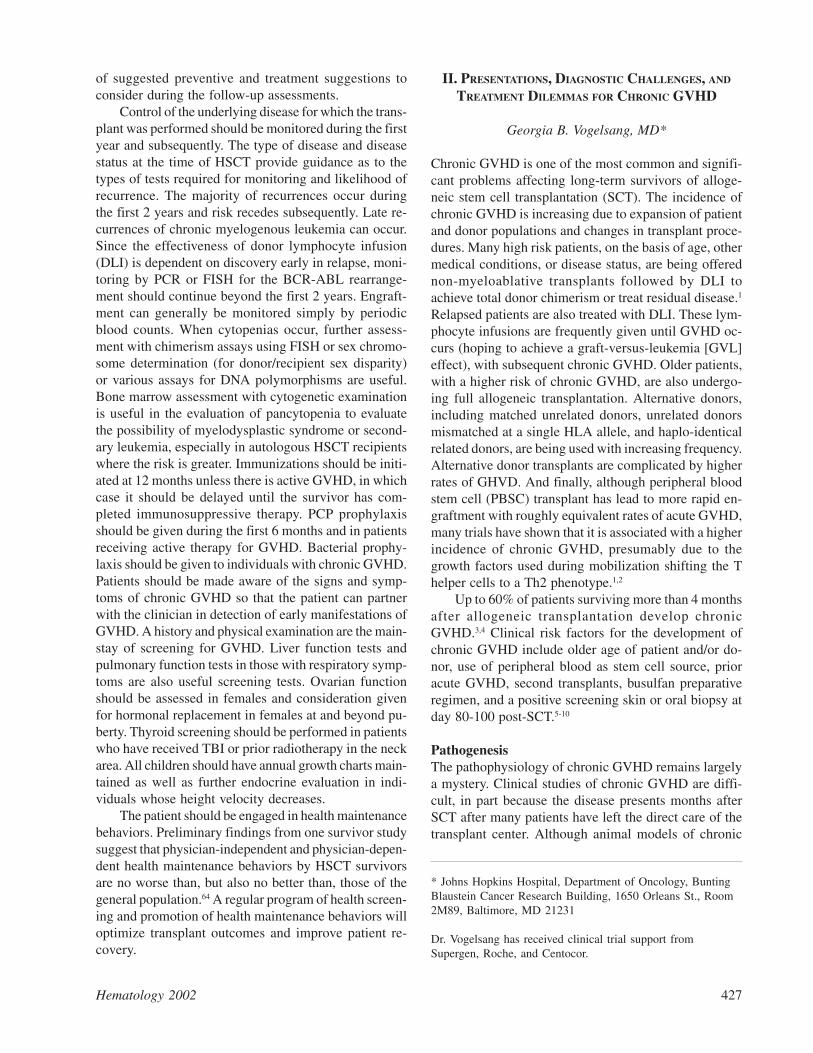
Control of the underlying disease for which the trans-plant was performed should be monitored during the firstyear and subsequently. The type of disease and diseasestatus at the time of HSCT provide guidance as to thetypes of tests required for monitoring and likelihood ofrecurrence. The majority of recurrences occur duringthe first 2 years and risk recedes subsequently. Late re-currences of chronic myelogenous leukemia can occur.Since the effectiveness of donor lymphocyte infusion(DLI) is dependent on discovery early in relapse, moni-toring by PCR or FISH for the BCR-ABL rearrange-ment should continue beyond the first 2 years. Engraft-ment can generally be monitored simply by periodicblood counts. When cytopenias occur, further assess-ment with chimerism assays using FISH or sex chromo-some determination (for donor/recipient sex disparity)or various assays for DNA polymorphisms are useful.Bone marrow assessment with cytogenetic examinationis useful in the evaluation of pancytopenia to evaluatethe possibility of myelodysplastic syndrome or second-ary leukemia, especially in autologous HSCT recipientswhere the risk is greater. Immunizations should be initi-ated at 12 months unless there is active GVHD, in whichcase it should be delayed until the survivor has com-pleted immunosuppressive therapy. PCP prophylaxisshould be given during the first 6 months and in patientsreceiving active therapy for GVHD. Bacterial prophy-laxis should be given to individuals with chronic GVHD.Patients should be made aware of the signs and symp-toms of chronic GVHD so that the patient can partnerwith the clinician in detection of early manifestations ofGVHD. A history and physical examination are the main-stay of screening for GVHD. Liver function tests andpulmonary function tests in those with respiratory symp-toms are also useful screening tests. Ovarian functionshould be assessed in females and consideration givenfor hormonal replacement in females at and beyond pu-berty. Thyroid screening should be performed in patientswho have received TBI or prior radiotherapy in the neckarea. All children should have annual growth charts main-tained as well as further endocrine evaluation in indi-viduals whose height velocity decreases.
The patient should be engaged in health maintenancebehaviors. Preliminary findings from one survivor studysuggest that physician-independent and physician-depen-dent health maintenance behaviors by HSCT survivorsare no worse than, but also no better than, those of thegeneral population.64 A regular program of health screen-ing and promotion of health maintenance behaviors willoptimize transplant outcomes and improve patient re-covery.
II. PRESENTATIONS, DIAGNOSTIC CHALLENGES, AND
TREATMENT DILEMMAS FOR CHRONIC GVHD
Georgia B. Vogelsang, MD*
Chronic GVHD is one of the most common and signifi-cant problems affecting long-term survivors of alloge-neic stem cell transplantation (SCT). The incidence ofchronic GVHD is increasing due to expansion of patientand donor populations and changes in transplant proce-dures. Many high risk patients, on the basis of age, othermedical conditions, or disease status, are being offerednon-myeloablative transplants followed by DLI toachieve total donor chimerism or treat residual disease.1
Relapsed patients are also treated with DLI. These lym-phocyte infusions are frequently given until GVHD oc-curs (hoping to achieve a graft-versus-leukemia [GVL]effect), with subsequent chronic GVHD. Older patients,with a higher risk of chronic GVHD, are also undergo-ing full allogeneic transplantation. Alternative donors,including matched unrelated donors, unrelated donorsmismatched at a single HLA allele, and haplo-identicalrelated donors, are being used with increasing frequency.Alternative donor transplants are complicated by higherrates of GHVD. And finally, although peripheral bloodstem cell (PBSC) transplant has lead to more rapid en-graftment with roughly equivalent rates of acute GVHD,many trials have shown that it is associated with a higherincidence of chronic GVHD, presumably due to thegrowth factors used during mobilization shifting the Thelper cells to a Th2 phenotype.1,2
Up to 60% of patients surviving more than 4 monthsafter allogeneic transplantation develop chronicGVHD.3,4 Clinical risk factors for the development ofchronic GVHD include older age of patient and/or do-nor, use of peripheral blood as stem cell source, prioracute GVHD, second transplants, busulfan preparativeregimen, and a positive screening skin or oral biopsy atday 80-100 post-SCT.5-10
PathogenesisThe pathophysiology of chronic GVHD remains largelya mystery. Clinical studies of chronic GVHD are diffi-cult, in part because the disease presents months afterSCT after many patients have left the direct care of thetransplant center. Although animal models of chronic
* Johns Hopkins Hospital, Department of Oncology, BuntingBlaustein Cancer Research Building, 1650 Orleans St., Room2M89, Baltimore, MD 21231
Dr. Vogelsang has received clinical trial support fromSupergen, Roche, and Centocor.
428 American Society of Hematology
GVHD do exist, the most commonly used model (a par-ent into F1 hybrid model) produces extensive antibody-mediated damage that more closely resembles lupus thanchronic GVHD.11 Another model of chronic GVHDcomes from the model of cyclosporine (CSA)-inducedautologous GVHD, which clinically resembles chronicGVHD.12 CSA inhibits thymic-dependent clonal dele-tion of autoreactive T cells, thereby paradoxically dis-rupting self-tolerance. Autologous GVHD is mediateday autoreactive T cells which recognize the CLIP re-gion of MHC class II molecules. The effector cells havebroad-based recognition of tissues and the clinical mani-festations when fully evolved are identical to chronicGVHD. Thymic damage is critical for expression ofautologous GVHD. Chronic GVHD may stem from aimmune injury similar to that of acute GVHD and SCT,which allows the development of autoreactive clones thatwould normally be deleted (see Figure 2).
Although the clinical findings in chronic GVHD maybe impressive, the main effect of chronic GVHD is onimmune function. Infections account for the majority ofdeaths in these patients. Because of decreased negativeselection, reduced extrathymic generation, and/or accel-eration of the normal thymic aging process with chronicGVHD, patients have an increase in peripheral auto-reactive T lymphocytes.13,14 These autoreactive Tlymphocytes act with interferon gamma to produce theincreased collagen deposition seen histopathologicallyin chronic GVHD.15
Classification of Chronic GVHDChronic GVHD can be classified according to the typeof onset, the clinical manifestations, or the extent of dis-ease. The majority of patients with chronic GVHD havehad prior acute GVHD. Their disease may evolve di-rectly from acute GVHD (progressive), which has a grimprognosis, or may follow a period of recovery GVHD(quiescent), with an intermediate prognosis. Patients maydevelop chronic GVHD with no history of prior acuteGVHD (de novo) and these patients have a relativelygood prognosis.
Patients may also be described as having lichenoidor sclerodermatous disease based on the cutaneous mani-festations. The lichenoid form occurs earlier after SCT,and may evolve into sclerodermatous GVHD. The mostcommonly employed staging system stratifies patientsinto limited or extensive disease based on the outcomeof 20 patients.16 Localized skin involvement, with orwithout hepatic dysfunction, is classified as limited dis-ease and does not require treatment. Patients with gen-eralized skin involvement or with limited skin involve-ment in association with eye involvement, oral involve-ment, hepatic dysfunction with abnormal liver histology,
or involvement of any other target organ are consideredto have extensive disease, which needs therapy. Althoughthis staging system is highly reproducible, it provideslittle information about prognosis and is of limited clini-cal utility.17
More recently, a grading system for chronic GVHDthat stratifies patients into risk categories according toclinical characteristics at diagnosis has been reported.18
Using a database of 151 patients with chronic GVHD,three variables were found to be risk factors for short-ened survival by multivariate analysis: extensive skinGVHD involving > 50% of the body surface area, plate-let count of < 100,000/µL, and progressive-type onset.As reported at this meeting last year, this model wasvalidated using data from 1108 patients from the IBMTR(n = 711), Fred Hutchinson Cancer Research Center (n= 188), University of Nebraska (n = 60), and the Uni-versity of Minnesota (n = 149). Despite significant het-erogeneity of the data, the proposed grading system iden-tified three prognostic groups, each with different sur-vival outcomes. Because this grading system is highlypredictive of outcome, it may help to improve clinicalmanagement, trial design, and communication amongtransplant centers.
Clinical ManifestationsThe diagnosis of chronic GVHD is traditionally madeafter day 100 post-transplant. The median time of diag-nosis is day 201 after HLA-identical sibling transplant,
Figure 2. Potential mechanism of chronic graft-versus-hostdisease (GVHD) induction.
Autoreactive T cells are generated by prior damage to the immunesystem from acute GVHD, host immune system factors such asage, and/or T helper cell population administered with the trans-plant. Effector cells help generate a graft versus tumor effect as wellas chronic GVHD. (Figure from Dr. A. Hess, Johns HopkinsOncology).
Hematology 2002 429
day 159 after mismatched related transplant, and day133 after an unrelated donor transplant.19 The clinicalmanifestations are summarized in Table 3.
Immune systemChronic GVHD causes profound immunosuppression.Most chronic GVHD deaths are attributable to infec-tion. Functional asplenia with an increased susceptibil-ity to encapsulated bacteria is common, and circulatingHowell-Jolly bodies may be seen on peripheral bloodsmear. Patients are also at risk for invasive fungal infec-tions and PCP.
SkinLichenoid chronic GVHD presents as an erythematous,papular rash that resembles lichen planus and has notypical distribution pattern. Scleradermatous GVHD mayinvolve the dermis and/or the muscular fascia and clini-cally resembles systemic sclerosis. The skin is thickened,tight, and fragile with very poor wound-healing capac-ity. Alteration in pigmentation, either hypo- or hyper-pigmentation, may occur. In severe cases the skin maybecome blistered from poor lymphatic drainage or ul-cerated from minor trauma. Because the sclerosis af-fects the dermis, hair loss and destruction of the sweatglands are common. Isolated fascial scleroderma presentswith decreased mobility with normal appearing skin. Theskin is fixed to the fascia below and careful examinationof the skin is critical in making the diagnosis.
Dermal appendagesFingernails and toenails may be affected by chronicGVHD. Nails develop vertical ridges and cracking andare very fragile. Nail problems may persist even afterskin changes have resolved. Hair loss in areas of affectedskin may also persist after treatment, although recoveryof hair is frequently a sign of recovery. Brittle hair oftenprecedes allopecia. Premature graying is often associ-ated with chronic GVHD, even in children, and may ef-fect hair and eyebrows.
Musculoskeletal systemFascial involvement in sclerodermatous GVHD is usu-ally associated with skin changes, but may develop withnormal, but fixed overlying skin. Fasciitis causes sig-nificant limitations in range of motion if joint areas areinvolved. Muscle cramps are a common complaint inpatients with chronic GVHD, although the pathophysi-ology is not understood. Myositis, with tender musclesand elevated muscle enzymes, is rare and does not ex-plain the frequent complaints of severe cramps. Myosi-tis, with tender muscles and elevated muscle enzymes,is rare and does not explain the frequent complaints of
severe cramps. Patients with sclerodermatous GVHD andrestricted range of motion may benefit from a regularprogram of physical therapy to help in recovery and toprovide functional recommendations for limited joints.
EyesOcular GVHD usually presents with irritation or dryeyes. Irreversible destruction of the lacrimal glands re-sults in dryness, photophobia, and burning. Conjuncti-val GVHD is a rare manifestation of severe chronicGVHD and is associated with a poor prognosis.
MouthOral mouth GVHD causes xerostomia and/or food sen-sitivity. More advanced disease may cause odynophagiadue to extension of damage. Rarely, patients will haveesophageal involvement without oral disease. Physicalexam in mild disease reveals erythema with whiteplaques, which may be confused with thrush or herpeticinfections. Lichenoid changes of more advanced diseasecause more extensive plaques. Secondary infections withviruses (especially herpes simplex) and yeasts are al-most universal and patients usually require treatment aslong as their oral disease persists and/or immunosup-pression is given. Changes in symptoms with little changein exam may occur with local infections.
Gastrointestinal tractMany patients with chronic GVHD have GI complaintsthat are not necessarily related to their chronic GVHD.These symptoms are often attributable to other diseasestates including acute GVHD, infection, dysmotility, lac-tose intolerance, pancreatic insufficiency, and drug-re-lated side effects.20 As many of these problems are verycorrectable, full evaluation of these symptoms is impor-tant. In a retrospective review of the intestinal biopsiesof 40 patients with chronic GVHD and persistent GIsymptoms, histopathologic evidence of chronic GVHDwas found in only 11 patients. The majority of thesepatients had evidence of both acute and chronic GVHD,with only 3 patients (7%) found to have isolated chronicGVHD. Wasting in patients with chronic GVHD is com-mon, with one recent report finding malnutrition in 43%of patients and severe malnutrition with body mass in-dex less than 18.5 in 14%.21 The mechanisms of wastingare not fully defined but may include increased cata-bolic rate due to elevated resting energy expenditure andhigh cytokine levels, especially tumor necrosis factor(TNF). Although full nutritional evaluation and inter-ventions are recommended, many patients with activeGVHD continue to lose weight despite adequate caloricintake. Symptoms often improve with successful treat-ment of GVHD.
430 American Society of Hematology
Tab
le 3
. C
linic
al m
anife
stat
ion
s o
f ch
ron
ic g
raft
-ver
sus-
ho
st d
isea
se (G
VH
D).
Org
anC
linic
al M
anif
esta
tio
nE
valu
atio
nIn
terv
enti
on
Ski
nE
ryth
emat
ous
papu
lar
rash
(lic
heno
id)
or th
icke
ned,
tig
ht, f
ragi
le s
kin
Clin
ical
and
bio
psy
to c
onfir
m th
e di
agno
sis
ofM
oist
uriz
e (p
etro
leum
jelly
), tr
eat l
ocal
infe
ctio
ns,
(scl
erod
erm
atou
s).
GV
HD
.pr
otec
t fro
m fu
rthe
r tr
aum
a. T
opic
al s
tero
idoi
ntm
ent m
ay b
e us
ed if
it g
ives
sym
ptom
atic
relie
f to
loca
lized
are
as.
Nai
lsV
ertic
al r
idgi
ng, f
ragi
le.
Clin
ical
.N
ail p
olis
h m
ay h
elp
to d
ecre
ase
furt
her
dam
age.
Sw
eat g
land
sD
estr
uctio
n le
adin
g to
ris
k of
hyp
erth
erm
ia.
Avo
id e
xces
sive
hea
t.
Hai
rS
calp
and
bod
y ha
ir is
thin
and
frag
ile, c
an b
e pa
rtia
lly o
rC
linic
al.
com
plet
ely
lost
.
Eye
sD
ryne
ss, p
hoto
phob
ia, a
nd b
urni
ng.
Reg
ular
oph
thal
mol
ogic
eva
luat
ion
incl
udin
gP
rese
rvat
ive
free
tear
s du
ring
the
day
and
Pro
gres
sion
to c
orne
al a
bras
ion.
Sch
irm
er’s
tes
t.pr
eser
vativ
e fr
ee o
intm
ent a
t nig
ht.
Mou
thD
ry; s
ensi
tivity
to m
int,
spic
y fo
od, t
omat
o. W
hitis
h la
ce-li
ke p
laqu
esR
egul
ar d
enta
l eva
luat
ion
(with
app
ropr
iate
Avo
id fo
ods
whi
ch a
re n
ot to
lera
ted.
Reg
ular
in th
e ch
eeks
and
tong
ue id
entic
al to
lich
en p
lanu
s. E
ryth
ema
and
endo
card
itis
prop
hyla
xis)
. Vira
l and
fung
al c
ultu
res
dent
al c
are
prec
eded
by
app
ropr
iate
end
ocar
di-
pain
ful u
lcer
atio
ns, m
ucos
al s
cler
oder
ma
with
dec
reas
edat
dia
gnos
is a
nd a
t any
wor
seni
ng.
tis p
roph
ylax
is. T
opic
al s
tero
id ri
nses
follo
wed
by
sens
itivi
ty to
tem
pera
ture
can
als
o ha
ppen
.an
ant
ifung
al a
gent
for s
ympt
omat
ic re
lief.
Res
pira
tory
trac
tB
ronc
hiol
itis
Obl
itera
ns c
an m
anife
st a
s dy
spne
a, w
heez
ing,
cou
ghP
ulm
onar
y fu
nctio
n te
sts
incl
udin
g F
EV
1 , F
VC
,In
vest
igat
iona
l the
rapy
.w
ith n
orm
al C
T s
can
and
mar
ked
obst
ruct
ion
at p
ulm
onar
y fu
nctio
nD
LCO
, hel
ium
lung
vol
umes
. CT
sca
n in
test
s. C
hron
ic s
inop
ulm
onar
y sy
mpt
oms
and/
or in
fect
ions
are
als
osy
mpt
omat
ic p
atie
nts.
com
mon
.With
abn
orm
al c
hest
CT,
mus
t rul
e ou
t inf
ectio
ns.
Lung
biop
sy if
clin
ical
ly in
dica
ted.
Gas
troi
ntes
tinal
Abn
orm
al m
otili
ty a
nd s
tric
ture
s. W
eigh
t los
s.S
wal
low
ing
stud
ies,
end
osco
py if
clin
ical
ly in
dica
ted.
Sys
tem
ic tr
eatm
ent o
f GV
HD
; end
osco
pica
l/N
utrit
iona
l eva
luat
ion.
surg
ical
trea
tmen
t of s
tric
ture
s. N
utrit
iona
lin
terv
entio
n.
Live
rC
hole
stas
is (
incr
ease
d bi
lirub
in, a
lkal
ine
phos
phat
ase)
. Iso
late
d liv
erLi
ver
func
tion
test
s. L
iver
bio
psy
if cl
inic
ally
indi
cate
d.N
o sp
ecifi
c th
erap
y is
pro
ven
supe
rior.
FK
506
invo
lvem
ent n
eeds
his
tolo
gic
conf
irm
atio
n.m
ay c
once
ntra
te in
the
liver
.
Mus
culo
skel
etal
Fasc
iitis
. Myo
sitis
is r
are.
Ost
eopo
rosi
s m
ay o
ccur
sec
onda
ry to
Per
iodi
cal p
hysi
cal t
hera
py e
valu
atio
n to
doc
umen
tA
ggre
ssiv
e ph
ysic
al th
erap
y pr
ogra
m.
horm
onal
def
icits
, use
of s
tero
ids,
dec
reas
ed a
ctiv
ity.
the
rang
e of
mot
ion.
Bon
e de
nsity
eva
luat
ion
espe
cial
ly in
pat
ient
s us
ing
ster
oids
.
Imm
une
syst
emP
rofo
und
imm
unod
efic
ienc
y. F
unct
iona
l asp
leni
a. H
igh
risk
ofA
ssum
e al
l pat
ient
s as
sev
erel
y im
mun
o-P
CP
pro
phyl
axis
(un
til 6
mon
ths
afte
r no
GV
HD
)pn
eum
ococ
cal s
epsi
s, P
CP,
and
inva
sive
fung
al in
fect
ions
.co
mpr
omis
ed a
nd a
sple
nic
to 6
mon
ths
afte
ran
d P
neum
ococ
cal p
roph
ylax
is (
lifet
ime)
. Del
ayV
aria
ble
IgG
leve
ls.
GV
HD
has
res
olve
d.va
ccin
atio
ns.
Hem
atop
oiet
ic s
yste
mC
ytop
enia
s. O
ccas
iona
l eos
inop
hilia
.C
ount
s. B
one
mar
row
asp
irate
and
bio
psy,
ant
i-S
yste
mic
trea
tmen
t of G
VH
D.
neut
roph
il an
d an
ti-pl
atel
et a
ntib
odie
s w
hen
indi
cate
d.
Oth
ers
Vir
tual
ly a
ll au
toim
mun
e di
seas
e m
anife
stat
ions
hav
e be
enA
s cl
inic
ally
indi
cate
d.de
scrib
ed in
ass
ocia
tion
with
chr
onic
GV
HD
.
Hematology 2002 431
LiverHepatic disease typically presents as cholestasis, withlaboratory evaluation revealing elevated alkaline phos-phatase and elevated serum bilirubin. Isolated hepaticchronic GVHD is being seen with increased frequencywith the use of DLI.22 Liver biopsy is required to con-firm the diagnosis and is especially important in patientswith no other symptoms of chronic GVHD, as viral in-fection and drug toxicity may mimic GVHD.
Respiratory tractBronchiolitis obliterans is a late and serious manifesta-tion of chronic GVHD. Patients typically present with acough or dyspnea.23 Many things may cause these symp-toms (infection, reactive airway disease, fluid overload,cardiac disease, etc.); therefore, the symptoms need tobe evaluated before labeling the disease as bronchiolitisobliterans. For example, patients with severe scleroticchest wall disease may have similar symptoms but haveno intrinsic pulmonary disease. Complete pulmonaryfunction tests are needed to localize the problem. ChestCT may be normal or may show hyperinflation. Patientswith bronchiolitis obliterans have minimal response totherapy and a very poor prognosis.
Patients with chronic GVHD are also at risk forchronic sinusitis, which may cause minimal symptoms.The sinuses are a frequent site of infection and shouldbe considered in fever workups.
Hematopoietic systemCytopenias are seen commonly in chronic GVHD pa-tients. This may be a result of stromal damage, but au-toimmune neutropenia, anemia, and/or thrombocytope-nia are also seen. Thrombocytopenia at the time ofchronic GVHD diagnosis has been associated with a poorprognosis.18,24-27 Eosinophilia may be seen and may trackwith disease activity.
Evaluation of Suspected Chronic GVHDThe accurate and timely diagnosis of chronic GVHD isan important step in its successful treatment. Most pa-tients have returned to the care of their primary oncolo-gist when chronic GVHD develops. Not every rash orgastrointestinal complaint represents GVHD. In a seriesof 123 patients referred to Johns Hopkins for the man-agement of refractory chronic GVHD, 9 patients hadnever had chronic GVHD and 26 patients had inactivedisease.26 Because the therapies for chronic GVHD arehighly immunosuppressive and must be continued for aprolonged time, it is important to confirm the diagnosisbefore initiating therapy. Conversely, subtle manifesta-tions of chronic GVHD may go undiagnosed for monthsand this delay may make successful treatment and reha-
bilitation difficult. The diagnosis of fasciitis without skinchanges, for example, may be a difficult diagnosis toestablish. If a diagnosis of chronic GVHD is suspected,histologic confirmation of at least one organ system isrecommended.
Treatment of Chronic GVHDAs the main manifestation of this disease is immuno-deficiency, patient education and infection prophylaxisare very important in this disease. Infection is the lead-ing cause of death among patients with chronic GVHD.Prophylaxis against Pneumocystis carinii should be ad-ministered to all patients undergoing treatment of chronicGVHD for six months after discontinuation of immuno-suppressive medications. These patients also have life-long splenic dysfunction and should therefore receiveprophylaxis against encapsulated bacteria for life. Theguidelines published by the American Heart Associa-tion for endocarditis prophylaxis should be followedwhen patients are undergoing dental or other invasiveprocedures. Patients receiving topical steroid therapy fororal GVHD should be treated with clotrimazole trochesor nystatin swishes. Patients at risk for CMV reactiva-tion should receive frequent monitoring with CMV sur-veillance cultures or antigenemia testing. A positive anti-genemia test should be treated preemptively with ganci-clovir, and patients with evidence of CMV disease shouldreceive both ganciclovir and CMV-specific immunoglo-bulin. Some centers administer intravenous IgG to pa-tients with hypogammaglobulinemia to keep IgG levels> 500 mg/dL. Vaccination series should be delayed un-til 1 year after the completion of GVHD therapy be-cause most patients will not mount an immune responsewith active disease or while receiving immunosuppres-sive medications. Posttransplant vaccination guidelinesare available on the Centers for Disease Control and Pre-vention web site (www.cdc.gov/mmwr/mmwr_rr.html).
The most widely employed first line therapy fortreatment of chronic GVHD is CSA and prednisone,administered on alternating days. Sullivan et al reportedthat prednisone alone is superior to prednisone plus aza-thioprine for primary treatment of patients with stan-dard-risk extensive chronic GVHD.25 However, in pa-tients classified as high-risk on the basis of platelet counts< 100,000/µL, treatment with prednisone alone resultedin only 26% 5-year survival. When a similar group ofpatients was treated with alternating day CSA and pred-nisone, 5-year survival exceeded 50%.27 After this en-couraging result, most centers adopted this regimen forinitial treatment of all patients. Patients are treated ini-tially with daily prednisone at 1 mg/kg per day and dailyCSA at 10 mg/kg per day divided BID. If disease is stableor improving after two weeks, prednisone is tapered by
432 American Society of Hematology
25% per week to a target dose of 1 mg/kg every otherday. After successful completion of this steroid taper,CSA is reduced by 25% per week to alternate day dos-ing of 10 mg/kg per day divided BID, every other day. Ifthe disease has completely resolved at their 9-monthevaluation, patients are slowly weaned from both medi-cations, with dose reductions approximately every twoweeks. Patients with incomplete response are kept ontherapy for 3 more months and then re-evaluated. If pa-tients fail to respond by 3 months or demonstrate pro-gressive disease, salvage regimens are warranted.28
Although this regimen of alternating CSA and pred-nisone is widely employed for the treatment of standard-risk (platelet count > 100,000/µL) extensive GVHD,there was no data on its effectiveness in standard riskpatients. The Seattle group has reported results on a studycomparing prednisone alone to prednisone plus CSA inpatients without thrombocytopenia in 307 patients withextensive GVHD. Two hundred eighty-seven patientswere evaluable. Paul Martin (personal communication)observed that:
The cumulative incidence of transplant-relatedmortality at 5 years from enrollment was 17% (95%CI, 11%-23%) in the cyclosporine plus prednisonearm and 13% (95% CI, 8%-19%) in the prednisonearm. The hazards of transplant-related mortality,overall mortality, recurrent malignancy, secondarytherapy and discontinuation of all immunosuppres-sive therapy were not significantly different be-tween the two arms, but survival without recurrentmalignancy was lower in the two-drug arm (P =0.03). Eighteen (13%) of the 142 patients in theCSA plus prednisone arm and 32 (22%) of the 145patients in the prednisone arm developed avascu-lar necrosis (P = 0.04). Treatment with CSA plusprednisone may reduce the risk of steroid-relatedtoxicity, but results of this study do not substanti-ate the hypothesis that administration of CSA re-duces transplant-related mortality among patientswith chronic GVHD.
This uncertainty regarding the choice of front-linetherapy emphasizes the importance of enrolling patientson clinical trials so that fundamental questions about thepathogenesis and treatment of chronic GVHD may beanswered. Currently two large randomized trials areplanned or underway for front-line therapy. One trialthrough COG is looking at the addition of hydroxycholo-roquine to prednisone. The other multicentered trialplanned by the Fred Hutchinson Cancer Research Cen-ter is examining the addition of mycophenolate mofetil(MMF) to cyclosporine or Tacrolimus (FK-506) plusprednisone in patients with extensive chronic GVHD.
Salvage therapiesTacrolimus or FK506 has been used in steroid refrac-tory patients.29 Six of 17 patients with extensive chronicGVHD showed response. FK506 concentrates in the liverand has a theoretical advantage over CSA for the treat-ment of hepatic GVHD. Although there have been anumber of reports of responses to FK 506 switched fromCSA, the similar mechanism of action of these drugsmakes this substitution, other than in the setting of liverGVHD, a modest change. FK 506 has been combinedwith MMF. A review of 26 patients treated with this regi-men showed a response rate of almost 50%.30 Becauseof the response rate to MMF and the rather disappoint-ing results reviewed above with cyclosporine and pred-nisone, a multicentered trial comparing cyclosporine/FK506 plus prednisone to the same combination with MMFhas been organized through the Fred Hutchinson Can-cer Research Center. This trial should answer the ques-tion of the activity of MMF in chronic GVHD. Thalido-mide has been reported to have immunosuppressive prop-erties and to be active against chronic GVHD, althoughmost studies have found that the side effects of sedationprevent many patients from continuing on the drug.31
Etretinate is a synthetic retinoid that has been used totreat patients with systemic scleroderma. Based on re-ports of response in this patient population, it has beenused to treat patients with sclerodermatous and fascialchronic GVHD. Of 27 patients completing a 3 monthtrial, 20 showed some improvement in skin lesions and/or range of motion in patients with refractory diseasetreated with etretinate.32 Etretinate is not currently com-mercially available, and acetretin, a more rapidly clearedderivative, has been used in its place. Acetretin may beadded to immunosuppressive medications to increase thecutaneous response in patients with sclerodermatousGVHD. Clofazimine, an antimycobacterial drug used totreat leprosy and Mycobacterium avium complex, hasanti-inflammatory activity in a number of chronic au-toimmune skin disorders. Based on the success of treat-ment in these disorders, it was studied in 22 patients withchronic GVHD.33 Over half of the patients with sclero-dermatous disease showed improvement in skin involve-ment, flexions contractures, or oral manifestations.Plaquenil (hydroxychloroquine) is an anti-malarial drugused in the treatment of autoimmune diseases. It inter-feres with antigen presentation and cytokine production,and is synergistic with CSA and tacrolimus in vitro.4
Based on promising results of 32 patients with over halfresponding, a large trial sponsored by COG is looking atthe addition of plaquenil to a steroids plus cyclosporinefor initial therapy. PUVA (8-methoxypsoralen plus ul-traviolet A irradiation) has been used for treatment ofsteroid-resistant lichenoid chronic GVHD and in patients
Hematology 2002 433
for whom steroids are contra-indicated.28 PUVA is verydifficult to administer to sclerodermatous GVHD. Photo-pheresis has also been reported to be useful in chronicGVHD, although the mechanics of this therapy, includ-ing the need for a pheresis catheter and the location ofthe machine to deliver the therapy, have made it difficultto do trials with this approach. Pentostatin is currentlybeing studied in several clinical trials, with early resultssuggesting that the drug is well tolerated, with responsesseen in heavily treated patients.34 Rapamycin is also be-ing explored. The antifibrotic properties of this drugmake it particularly attractive for sclerodermatousGVHD.35,36
Future DirectionsEfforts to prevent the development of chronic GVHDhave been unsuccessful. Attempts to prevent chronicGVHD including the use of IV IgG and thalidomide havebeen unsuccessful.37,38 A more recent trial of prolongedadministration of CSA found no difference in chronicGVHD or mortality when CSA was given for 24 monthsrather then six months.39 Current transplantation prac-tices, including the use of DLI and peripheral blood stemcells, older patient age, and the increasing use of unre-lated and mismatched marrow donors make it likely thatchronic GVHD is going to be a progressively more com-mon problem and more frequently in patient populationsthat are going to be very difficulty to treat. Ongoing re-search to further characterize the pathogenesis of thisdisease is crucial to the development of new therapeuticapproaches. Several new therapies are currently underevaluation, including the antineoplastic and immunosup-pressive drug pentostatin; daclizumab, a soluble IL-2receptor antagonist; and infliximab, an anti-TNF-αmonoclonal antibody.
III. MALIGNANCIES AFTER
HEMATOPOIETIC STEM CELL TRANSPLANTATION:WHY DO THEY OCCUR? CAN WE PREDICT THEM?
CAN WE PREVENT THEM?
H. Joachim Deeg, MD*
HSCT provides effective therapy for patients withlymphohemopoietic, immunologic, metabolic and otherdisorders. Many patients have now been followed fortwo or three decades post-transplant and are presum-ably cured. Some patients, however, develop long-termcomplications, including new malignancies, which maynot be unexpected. Secondary malignancies occur inpatients with Hodgkin disease or non-Hodgkin lym-phoma (NHL) treated with chemotherapy or radiochemo-therapy,1,2 and in recipients of solid organ transplants
treated with immunosuppressive therapy.Studies in the 1970s and 1980s showed a significant
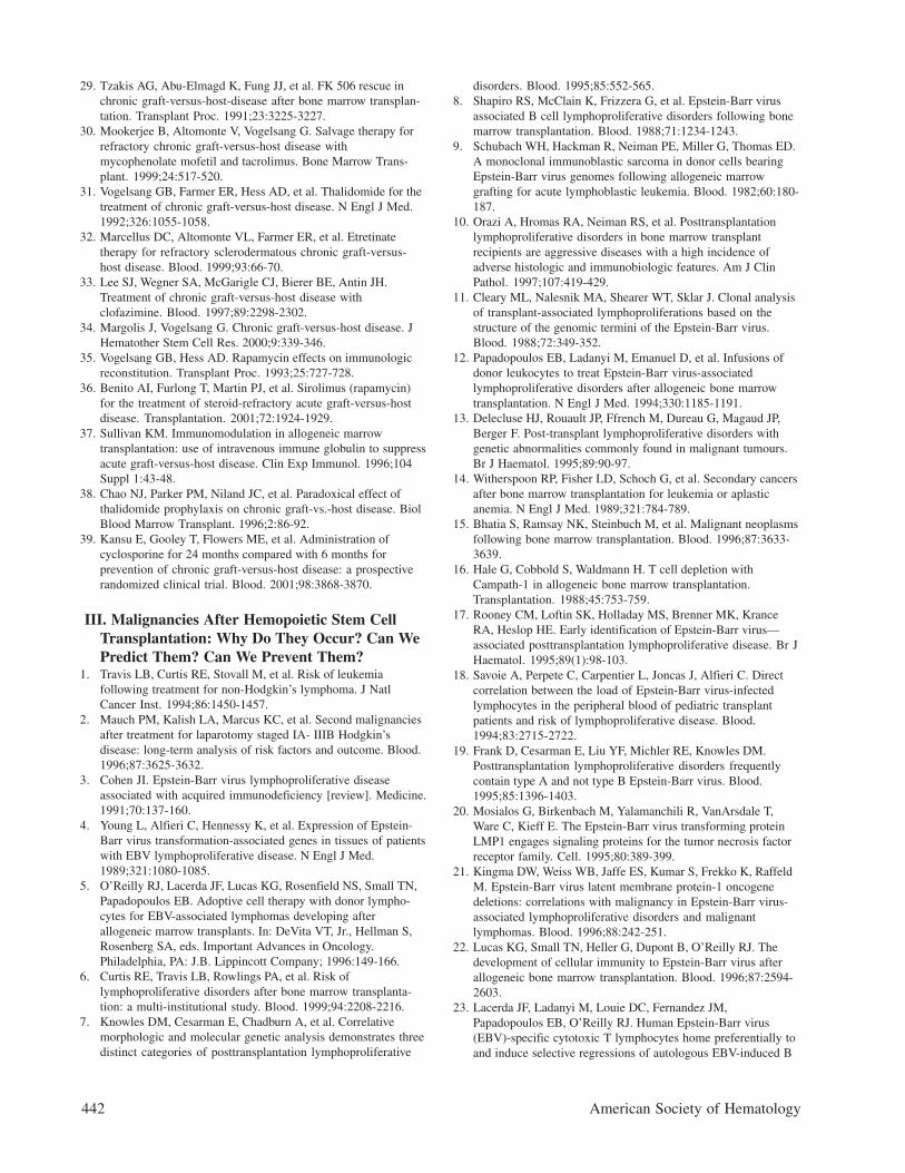
increase in the incidence of malignancies relative to con-trols in rhesus monkeys and dogs irradiated with lethaldoses of TBI and infused with autologous or allogeneicmarrow cells (radiation chimeras). Patients who undergoHSCT, in addition, may have genetic defects associatedwith their primary disease predisposing them to the de-velopment of new malignancies. Finally, viruses suchas Epstein-Barr virus (EBV), used to immortalize celllines in vitro, can transform cells in vivo, particularly inimmunodeficient patients.3,4 Table 4 lists factors that arethought to contribute to the development of new malig-nancies in HSCT recipients. Major categories of malig-nancies are described in Table 5. Figure 3 shows theincidence of PTLD and solid cancers over time post-transplant.
Lymphoid MalignanciesLymphoproliferative disorders after HSCT (PTLD) oc-cur mostly in allogeneic transplant recipients; autolo-gous cases are rare. Most PTLD are of B-cell lineage,3,5
although some T-cell PTLD have been reported. In ad-dition, lymphomas with typical characteristics of NHLor Hodgkin disease have occurred.
B-cell PTLDIncidence. Results in 18,014 allogeneic transplant re-cipients followed for up to 25 years revealed 78 cases ofpost-transplant lymphoid malignancies, a rather low in-cidence compared to solid organ transplant patients.6 Inagreement with an earlier review by Cohen,3 82% werediagnosed within 1 year of transplantation, with peakoccurrence (120 cases/10,000 patients/year) at 2–5months, and a decline to < 5 cases/10,000/year among1-year survivors. The incidence at 4–10 years was 1–2%, although figures of 10% have been reported in pa-tients transplanted for immunodeficiency disorders.
Clinical and pathologic features. B-cell PTLD areclinically and morphologically heterogeneous, usuallyassociated with EBV and developing in a milieu of T-cell dysfunction.3,7 Patients frequently present with fe-ver, even a sepsis-like syndrome, and lymphadenopa-
* Fred Hutchinson Cancer Research Center, 1100 FairviewAvenue, N., D1-100, P.O. Box 19024, Seattle, WA 98109-1024
This work was supported by PHS grants CA18029, CA15704,HL36444, and CA87948.
Acknowledgments: I thank R. Curtis, G. Gilliland, W.Schubach, H.J. Kolb, G. Socié, and D. Rizzo for their com-ments and contributions, and B. Larson and H. Crawford forhelp with manuscript preparation.
434 American Society of Hematology
thy. Intra-abdominal lymphadenopathy, splenomegaly,hepatomegaly, or bowel involvement may cause abdomi-nal pain, vomiting, and diarrhea. Often lungs, kidneys,and the central nervous system (CNS) are involved. B-cell PTLD after allogeneic HSCT are almost always ofdonor origin. Few studies have examined in detail thehistologic features of PTLD in HSC recipients.8,9 Someare similar to the monomorphic or polymorphic, diffuselarge-cell lymphomas of B-cell origin observed aftersolid organ transplantation, while at least half show ag-gressive features of immunoblastic lymphoma.5,10 MostPTLD after HSCT are oligoclonal or monoclonal, asdetermined by analysis of immunoglobulin gene rear-rangement or fused termini of episomal EBV DNA.5,7,11,12
There is no strong correlation between clonality andmorphology.10 Cytogenetic abnormalities occur, usually
in aggressive monoclonal lesions. Exceptional casesshow the characteristic t(8;14) translocation of Burkitt’slymphoma.13
Risk factors. Risk factors5,6,8,14 are listed in Table 6:Use of antithymocyte globulin (ATG) (relative risk [RR]6.4) or anti-CD3 MAB (RR 43.2) for acute GVHD pro-phylaxis (RR 5.9) or in the preparative regimen (RR 3.1),use of TBI in the conditioning regimen (RR 2.9), T-celldepletion of donor marrow (RR 11.9;12.7),15 unrelateddonor or HLA non-identical related donor (RR 4.1; 8.9),and primary immune deficiency disease (RR 2.5), oc-currence of acute GVHD (grades III-IV) (RR 1.9), andtreatment of acute GVHD with ATG or monoclonal anti-T cell antibody. However, while in patients transplantedwith marrow depleted of T-cells with specific anti-CD3monoclonal antibodies, the incidence of EBV-positive
Table 5. New malignancies after hemopoietic stem celltransplantation.
1. Malignancies of the lymphoid systema) B-cell PTLDb) T-cell lymphomasc) Hodgkin disease and other late lymphomas
2. Hematologic malignanciesa) Leukemia recurrence in donor cellsb) New leukemias in host cellsc) Myelodysplastic syndrome/AML
Figure 3. Cumulative incidence (%) of posttransplantlymphoproliferative disorders (PTLD) and invasive solidcancers following allogeneic marrow transplantation at 235centers worldwide.
Reproduced with permission from Curtis RE, Travis LB, RowlingsPA, et al. Risk of lymphoproliferative disorders after bone marrowtransplantation: a multi-institutional study. Blood. 1999;94:2208-2216.
Hematology 2002 435
PTLD was 11% to 25%, the incidence was < 1% withtechniques removing both T and B lymphocytes (e.g.,soybean agglutinin or Campath-1).5,16 The risk of PTLDis particularly high in EBV-negative patients transplantedfrom EBV-positive donors.
The impact of risk factors is additive (or synergis-tic). In one analysis the risk of B-cell PTLD in patientswith primary immune deficiency given T-cell–depletedHLA non-identical transplants was 64.8% ± 17.7% at 4years, versus 0.9 ± 0.2% (P < 0.001) in patients givenunmanipulated HLA identical marrow. In one large studythe incidence of PTLD was 8% ± 2.9% with one or tworisk factors, and 22% ± 17.9% with three or more riskfactors present. The role of HLA-mismatching in thepathogenesis of B-cell PTLD is not clear but may consistin chronic antigenic stimulation, or delayed immune re-constitution. There is a direct correlation between viral load
in peripheral blood and the development of PTLD.17,18
Pathogenesis. EBV, a herpes virus, is present asa latent virus (in B lymphocytes and certain epithe-lial cells) in 95% of individuals by adulthood. EBVtype A (distinguished from type B by sequence di-vergence in the EBNA-2 gene) is present in mostcases of PTLD after solid organ transplantation;19
several strains have been identified. Whether thesame applies to HSCT recipients remains to be de-termined.
The oncogenic potential of EBV is due to itsability to immortalize B cells. The same viral genesthat drive proliferation in vitro, EBNA-1 and 2,LMP-1, and EBNA-3A, -3B, and -3C, are expressedin PTLD.4,5 LMP-1 acts as a constitutively activatedTNF receptor-like protein, induces gene expressionthrough NFκB, and acts as a transforming onco-protein in certain experimental systems.20 Deletionsnear the 3′ end of the LMP-1 gene appear to corre-late with EBV malignancy.21 Other viral proteins(ENV-2A, -3A, -3B, and -3C) control transcriptionof viral and cellular genes. The result is expressionof IL-1β, IL-6, IL-10, and TNFβ, which act asgrowth factors for EBV-infected cells, as well assoluble CD23 and low affinity IgE receptor.
Prophylaxis and therapy. Investigators at Me-morial Sloan-Kettering Cancer Center22 used limit-ing dilution analysis to quantify anti-EBV specificcytotoxic T-lymphocyte precursor (CTLp) frequen-cies in recipients of unmodified or T-cell depletedgrafts from EBV-positive donors. At 3 months (in-terval of peak incidence of B-cell PTLD) only 20%of patients had EBV CTLp frequencies in the rangeof seropositive controls, while at 6 months 70% werenormal. Based on studies in xenografted SCID mice23
the same investigators12 used unirradiated donor leu-kocytes (1.0 × 10⁶ CD3+ T cells/kg) to treat PTLD. Theinitial 5 patients had complete responses but 3 devel-oped chronic GVHD, and 2 died of respiratory failurewith no evidence of PTLD. An update showed 14 of 15patients responded and 6 of 12 evaluable patients devel-oped GVHD. Gene-marked EBV-specific cytotoxic T-lymphocytes persisted in vivo and restored cellular im-munity against EBV.
The St. Jude’s team used rising titers of EBV DNAin patient plasma as a criterion to institute pre-emptivetherapy with EBV-specific T-cell clones in 25 high-riskpatients. None developed PTLD. Among 6 patients whorefused CTL therapy or were ineligible, 2 developedPTLDs that were successfully treated with CTL.24 Boniniet al showed that HSV-TK gene-modified donor lym-phocytes can be used effectively and can be inactivatedby ganciclovir if GVHD develops.25 Rooney et al26 and
Table 6. Risk factors.
Type ofAllogeneic HSCT Secondary Malignancy Risk Factor
*In particular, mechlorethamine and chlorambucil have been implicated(C. Metayer et al., unpublished).†Available data suggest a higher incidence with the use of peripheralblood stem cells, particularly after mobilization with topoisomeraseinhibitors.
436 American Society of Hematology
Heslop et al24 confirmed the efficacy of gene-markedEBV-specific T lymphocytes and showed long-term res-toration of anti-EBV immunity.
While EBV-transformed B cells contain a circularviral DNA that is not susceptible to inhibition by thymi-dine kinase (TK) antagonists, tumor regression withacyclovir or ganciclovir therapy was noted in anecdotalreports (reviewed in ref. 27). The proliferative programof EBV-infected cells is restricted to latently infectedcells. That treatment directed at the lytic cycle shouldbe effective may depend on productive infection in othertissues that contribute to PTLD.
Anti-CD21 and anti-CD24 antibodies were testedin a multicenter trial.28 Among 19 marrow transplant re-cipients, 10 achieved complete remissions, and 6, all witholigoclonal disease, survived at a median follow-up of20 months.27 However, studies in SCID mice29 indicatethat residual EBV-positive B cells persist and can pro-voke a second tumor in the absence of efficient cyto-toxic T cells.
Based on in vitro data showing antitumor effects ofanti-IL-6 antibody,30 neutralization of IL-6 may also bea strategy worth exploring.
Chemotherapy, irradiation, and surgical resection areuseful in selected cases in solid organ recipients.3 Thebest approach in patients after HSCT is close monitor-ing of plasma EBV DNA and pre-emptive therapy inpatients with rising EBV DNA titers.
T-cell lymphoproliferative disordersRare T-cell proliferative disorders with or without EBVassociation occur, usually more than 1 or 2 years aftertransplantation. After HSCT only few such cases havebeen reported,31 none associated with HTLV1, HIV, orHHV6 infection.
Hodgkin disease and other late-onset lymphomasSeveral late occurring lymphomas have been reported,32
some linked to EBV (just as early onset PTLD), and oth-ers associated with T-cell depletion of the graft. Clinicalpresentation was like ordinary non-Hodgkin lymphomawith lymph node enlargement with or without general-ized symptoms.
A recent collaborative study of 18,531 transplant re-cipients (covering more than 42,000 patient years), found8 cases of Hodgkin disease 2.9–9.1 years after HSCT(observed/expected ratio 6.2).33 Five cases (67%) showedmixed cellularity subtype, and 5 of 6 cases studied con-tained the EBV genome. Two patients were also posi-tive for HIV. Risk factors identified for early PTLDs weregenerally absent. Patients with Hodgkin disease weremore likely to have acute GVHD and require therapyfor chronic GVHD (RR in one study 4.0). These data
add support to the theory that links overstimulation ofcellular immunity and exposure to EBV to various sub-types of Hodgkin disease.34
Hematologic Malignancies
After allogeneic HSCTIncidence. Lymphoblastic leukemias in donor cells werefirst recognized 30 years ago.35,36 The incidence was es-timated at 3–5%. More recent data suggest a lower inci-dence.37 In addition, new leukemias in patient cells, i.e.,leukemias of a different morphology or lineage than thepatient’s primary disease, have also been described (re-viewed in ref. 38).
Pathogenesis. The mechanism remains unclear.Transformation of donor cells via antigenic stimulationby host tissue, a leukemogenic host environment, fusionof normal cells with leukemic cells still residing in therecipient, or transfection with a host oncogene or virushave been proposed. Studies using molecular techniques(e.g., microsatellite analysis) to determine host-versus-donor origin of abnormal cells post-transplant indicatethat recurrence in donor-derived cells is infrequent.37
However, cases of leukemia or MDS transplanted fromthe donor into the patient have been reported.39,40
Recent work suggests that “replicative stress” aftertransplantation may result in accelerated telomere short-ening of donor HSC.41,42 This in turn might lead to chro-mosomal instability and increased probability of MDSor leukemia. While telomere shortening occurs in HSCTrecipients, the concept has remained controversial. Nev-ertheless, several cases of MDS/AML in donor cells pre-senting 5, 10 years or even later after HSCT have beenobserved (unpublished).
Therapy. There is no known prophylaxis, and notherapeutic standards have been established. Some pa-tients have recently been treated with second transplantsusing myeloablative or non-myeloablative protocols.
After Autologous HSCT“Secondary” MDS and AML occur after conventionalchemotherapy, and to a lesser extent after radiotherapy forHodgkin disease and NHL, as well as some solid tumors;1,43-
46 they also are a major complication after autologous HSCT(reviewed in refs. 32,47-50).
Incidence. In studies involving more than 1200 pa-tients the incidence of MDS was 4–18% at 3–6 years,with the post-transplant interval ranging from 2.5 to 8.5years.51-54 A case control study revealed 12 cases of MDS/AML in 511 patients after autologous transplants forHodgkin disease (n = 249) or NHL (n = 262) for a cu-mulative incidence of 4% at 5 years. Another reportshowed clonal chromosomal abnormalities in 10 of 275
Hematology 2002 437
patients 1.8–6.5 years after chemotherapy, and 0.5–3.1years after autologous transplant for Hodgkin disease orNHL,54 but only 5 patients had morphological evidenceof MDS or AML. The cumulative probability of devel-oping clonal chromosomal abnormalities reached 9% ±4.7% at 3 years after transplantation.
Risk factors. In one analysis a prolonged pre-transplant interval, and use of radiotherapy, especiallypelvic irradiation, were significant pretransplant vari-ables. In several studies the risk was higher with periph-eral blood cell transplants (e.g., 31% ± 33% versus 10.5%± 12% with marrow; RR 5.8; similar in another study55),in patients more than 35 (40) years of age at transplan-tation (RR 3.5), and with the use of TBI.51
A case control study under the auspices of the NCIanalyzed data on 56 patients who developed myelo-dysplastic syndrome (MDS)/leukemia, and 168 controlswithin a cohort of 2739 patients with Hodgkin diseaseor NHL transplanted at 12 institutions (Metayer, Curtiset al, unpublished data). MDS/AML was significantlycorrelated with the intensity of pre-transplant chemo-therapy, specifically mechlorethamine (RR 2.0; 4.3 fordoses of < 50; ≥ 50 mg/m2), and chlorambucil (RR 3.8;8.4 for duration < 10; ≥ 10 months; P = 0.0009) com-pared to cyclophosphamide. Also, higher doses of TBI(> 1200 cGy) used for transplant conditioning tended tocarry a higher risk (RR 4.7). The difference betweenmarrow and peripheral blood stem cells was not signifi-cant in this analysis.
Pathogenesis. Whether MDS/AML arises from in-fused HSC or from residual cells in the patient is con-troversial. If the former were true, then the type of con-ditioning therapy given for transplant should not be arisk factor—unless we postulate that conditioning (TBI)modifies the microenvironment and enhances the riskof leukemogenesis. If MDS/AML is related to transplan-tation, then the culprit could be the procedure itself orthe status of immunoincompetence after transplantation(Table 4). In some patients, cells harvested pre-trans-plant contain cytogenetic abnormalities, while in othersthey don’t. However, posttransplant MDS/AML is aclonal disorder, and in most patients cytogenetic abnor-malities (e.g., -7, +8, 7q-, 20q-, 11q23) are present, evenif they are not detected in cells used for transplantation.In some female patients without cytogenetic abnormali-ties clonality has been documented by X-inactivationbased clonality assays,56 and mutations of RAS, FLT3,AML 1, CBF among others, have been recognized (re-viewed in ref. 57).
More than one mutagenic/leukemogenic event is re-quired for MDS or AML to develop. Gene fusion prod-ucts recognized as leukemogenic (e.g., BCR/ABL, TEL/AML 1) are present even in normal individuals with the
use of sensitive PCR technology. Thus, such clones mayexist in patients pre-transplant, and a “second hit” mightoccur during or after transplantation. It has also beenargued that many patients may not have MDS but rather“disordered engraftment.”49
Prophylaxis and therapy. These data suggest thatreducing pretransplant exposure to alkylating agents,topoisomerase inhibitors, and irradiation and shorten-ing the duration of therapy should reduce the risk ofMDS.58 Alkylators and topoisomerase inhibitors shouldprobably not be used for stem cell mobilization.53 If cy-togenetics are abnormal at the time of stem cell harvest,an allogeneic transplant should be considered. In addi-tion to standard cytogenetics, interphase FISH, determi-nation of loss of heterozygosity or point mutations, andX-inactivation-based clonality assays are useful. OnceMDS/AML has evolved, the options are limited. Che-motherapy is often not well tolerated, and remissionsare of short duration. Allogeneic HSCT with standardor reduced intensity conditioning is a realistic option forsome patients.59
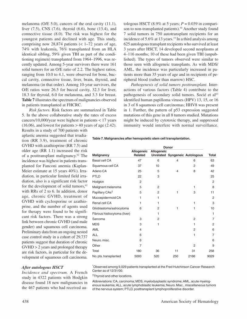
Solid TumorsIn irradiated and transplanted rhesus monkeys solid tu-mors developed 7.5 to 15 (median 11.5) years afterx-irradiation, and 4 to 15 (median 8) years after fissionneutrons. The time interval in gamma-irradiated dogswas 1.6 to 10.5 (median 8) years. Extrapolation to hu-mans suggests that post-transplant solid tumors mightdevelop after a decade or more.38
After allogeneic HSCTIncidence and spectrum of tumors. Among 2145 patientstransplanted from 1970-1987 13 solid tumors were ob-served, including glioblastoma, melanoma, squamouscell carcinoma, adenocarcinoma, hepatoma, and basalcell carcinoma.14 In another cohort of 2150 patients, 15developed solid tumors (8 of 1400 allogeneic, and 7 of750 autologous transplant recipients)15 for an incidenceof 5.6% at 13 years. In a third study of 1211 patientswho had survived at least 5 years after HSCT,60 47 de-veloped malignancies, including squamous cell carci-noma, breast cancer, glioblastoma, and lymphoma. Incomparison to normal controls, the incidence was in-creased significantly for malignancies of the oral cavity,skin, esophagus, uterine cervix, and brain.
A collaborative study analyzed results in 19,220patients (97.2% allogeneic, 2.8% syngeneic recipients)transplanted between 1964 and 1992.61 There were 80solid tumors for an observed/expected (O/E) ratio of 2.7(P < 0.001). In 10-year survivors, the risk was increased8-fold. The tumor incidence was 2.2% at 10, and 6.7%at 15 years. The risk was increased significantly for
438 American Society of Hematology
melanoma (O/E 5.0), cancers of the oral cavity (11.1),liver (7.5), CNS (7.6), thyroid (6.6), bone (13.4), andconnective tissue (8.0). The risk was highest for theyoungest patients and declined with age. This study,comprising now 28,874 patients (< 1–72 years of age,74% with leukemia, 76% transplanted from an HLAidentical sibling, 59% given TBI as part of the condi-tioning regimen) transplanted from 1964–1996, was re-cently updated. Among 5-year survivors there were 161solid tumors for an O/E ratio of 2.2. The highest ratios,ranging from 10.0 to 4.1, were observed for bone, buc-cal cavity, connective tissue, liver, brain, thyroid, andmelanoma (in that order). Among 10-year survivors, theO/E ratios were 26.5 for buccal cavity, 32.3 for liver,18.3 for thyroid, 6.0 for melanoma, and 3.3 for breast.Table 7 illustrates the spectrum of malignancies observedin patients transplanted at FHCRC.
Risk factors. Risk factors are summarized in Table5. In the above collaborative study the rates of excesscancers/10,000/year were highest in patients < 17 years(16.06), and lowest for patients > 40 years of age (2.42).Results in a study of 700 patients withaplastic anemia suggested that irradia-tion (RR 3.9), treatment of chronicGVHD with azathioprine (RR 7.5) andolder age (RR 1.1) increased the riskof a posttransplant malignancy.62 Theincidence was highest in patients trans-planted for Fanconi anemia (Kaplan-Meier estimate at 15 years 40%). Irra-diation, in particular limited field irra-diation, also is a significant risk factorfor the development of solid tumors,62
with RRs of 2 to 6. In addition, donorage, chronic GVHD, treatment ofGVHD with cyclosporine or azathio-prine, and the number of agents usedfor therapy were found to be signifi-cant risk factors. There was a stronglink between chronic GVHD (and malegender) and squamous cell carcinoma.Preliminary data from an ongoing nestedcase control study in a cohort of 29,737patients suggest that duration of chronicGVHD > 2 years and prolonged therapyare risk factors, in particular for the de-velopment of squamous cell carcinoma.
After autologous HSCTIncidence and spectrum. A Frenchstudy in 4322 patients with Hodgkindisease found 18 new malignancies inthe 467 patients who had received au-
tologous HSCT (8.9% at 5 years; P = 0.039 in compari-son to non-transplanted patients).55 Another study found7 solid tumors in 750 autotransplant recipients for anincidence of 5.6% at 13 years.15 In a third analysis among625 autologous transplant recipients who survived at least3 years after HSCT, 14 developed second neoplasms at4–116 months; 10 of these had been given TBI (unpub-lished). The types of tumors observed were similar tothose seen with allogeneic transplants. As with MDS/AML, the incidence was particularly increased in pa-tients more than 35 years of age and in recipients of pe-ripheral blood (rather than marrow) HSC.
Pathogenesis of solid tumors posttransplant. Inter-actions of various factors (Table 4) contribute to thepathogenesis of secondary solid tumors. Socié et al63
identified human papilloma viruses (HPV) 13, 15, or 16in 3 of 8 squamous cell carcinomas; HHV8 was presentin 1. Further, the pattern of p53 expression suggestedmutations of this gene in all 8 tumors studied. Mutationsmight be induced by cytotoxic therapy, and suppressedimmunity would interfere with normal surveillance.
Table 7. Malignancies after hemopoietic stem cell transplantation.
DonorAllogeneic Allogeneic
Malignancy Related Unrelated Syngeneic Autologous Total
Basal cell CA 47 6 4 6 63
Squamous cell CA 37 10 2 49
Adeno CA 25 5 4 8 42
PTLD 22 3 25
Hodgkin 2 2
Malignant melanoma 5 2 1 8
Papillary CAs† 5 2 1 3 11
Mucoepidermoid CA 1 1 2
Renal cell CA 1 1 1 3
Glioblastoma/astrocytoma 6 2 1 1 10
Fibrous histiocytoma (liver) 1 1
Sarcoma 3 2 2 7
MDS 3 1 1 2
AML 4 2 6
ALL 6 1 7
Neuro. misc. 6 6
Other 7 2 9
Total 180 36 11 31 258
No. pts. transplanted 5093 520 250 2166 9029
*Observed among 9,029 patients transplanted at the Fred Hutchinson Cancer ResearchCenter as of 12/31/00.†Thyroid and other locations.
Abbreviations: CA, carcinoma; MDS, myelodysplastic syndrome; AML, acute myelog-enous leukemia; ALL, acute lymphoblastic leukemia; Neuro. Misc., miscellaneous tumorsof the nervous system; PTLD, posttransplant lymphoproliferative disorder.
Hematology 2002 439
There is also evidence that polymorphism at position 72in p53 (in particular homozygosity for arginine) mayconfer unusual susceptibility of p53 to inactivation byHPV. Chronic inflammation and impaired DNA repairare other factors. However, considerable work is neededfor a better understanding of those questions.
Observations in autologous patients will be of greatinterest because etiologic factors, such as chronicalloantigenic stimulation and GVHD are absent, thusallowing to focus on cytotoxic therapy and genetic pre-disposition (xenobiotic polymorphism).
Prophylaxis and therapyRisk factors varied, dependent upon the type of tumor.Omission of (high dose) irradiation from the condition-ing regimen should be beneficial, in particular for theprevention of melanomas, thyroid carcinomas, carcino-mas of the buccal cavity, and breast cancer. Prevention(or different therapy) of chronic GVHD should have aneffect on the development of buccal carcinomas.
Surgical resection, whenever possible, is the front-line therapy for solid tumors. Selective immuno-stimulation and measures aimed at scavenging free radi-cals have yielded some promising results in experimen-tal studies.
ConclusionsHSCT offers curative therapy for many patients. Cur-rently about 30,000 transplants are carried out annually,and most patients who do not relapse within a year ortwo of transplantation do well and lead productive lives.However, complications occur, one of them being thedevelopment of new malignancies. While the overallincidence is low, high-risk situations such as an under-lying diagnosis of immunodeficiency or other geneticdefects, high-dose irradiation for conditioning, T-celldepletion of the marrow, HLA nonidentity of the donor,and chronic GVHD have been identified. We have be-gun to develop an understanding of the mechanism in-volved in PTLD. Insights into the development of he-mopoietic disorders and solid tumors is more limited.Other potential factors, e.g., smoking before or afterHSCT, need to be examined further. Also, longer obser-vation is required before the full extent of the risk ofsolid tumors in particular can be assessed. While manyquestions remain, available data provide a basis on whichto develop pre-emptive approaches.
REFERENCES
I. Overview of Late Complications1. Kolb HJ, Bender-Gotze C. Late complications after allogeneic
bone marrow transplantation for leukaemia. Bone MarrowTransplant. 1990;6:61-72.
2. Deeg HJ, Delayed complications and long-term effects afterbone marrow transplantation. Hem Onc Clin N Am.1991;4:641-657.
3. Sullivan KM, Mori M, Sanders J, et al: Late complications ofallogeneic and autologous bone marrow transplantation. BoneMarrow Transplant. 1992;10:127-134.
4. Duell T, van Lint MT, Ljungman P, et al. Health and functionalstatus of long-term survivors of bone marrow transplantation.Ann Intern Med. 1997;126:184-192.
5. Wingard, JR. Functional ability and quality of life of patientsafter allogeneic marrow transplantation: factors affecting socialand occupational functions—strategies to improve social andjob reintegration [abstract]. Bone Marrow Transplant.1994;14(Suppl 4):S29-S33.
6. Socie G, Veum-Stone J, Wingard JR, et al. Long-term survivaland late deaths after allogeneic bone marrow transplantation:Implications for cure. N Engl J Med. 1999;341:14-21.
7. Michallet M, Thomas X, Vernant JP, et al. Long-term outcomesafter allogeneic hematopoietic stem cell transplantation foradvanced stage acute myeloblastic leukemia: a retrospectivestudy of 379 patients reported to the Societe Francaise deGreffe de Moelle (SFGM). Bone Marrow Transplant.2000;26:1157-1163.
8. Witherspoon RP, Storb R, Ochs HD, et al. Recovery of antibodyproduction in human allogeneic marrow graft recipients:influence of time post-transplantation, the presence or absenceof chronic graft-versus-host disease, and antithymocyteglobulin treatment. Blood. 1981;58:360-368.
9. Lum LG, Munn NA, Schanfield MS, et al. Detection of specificantibody formation to recall antigens after human bone marrowtransplantation. Blood. 1981;67:582-587.
10. Lum LG. Kinetics of immune reconstitution after humanmarrow transplantation. Blood. 1987;69:369-380.
11. Acouturier P, Barra A, Intrator L, et al. Long lasting IgGsubclass and antibacterial polysaccharide antibody deficiencyafter allogeneic bone marrow transplantation. Blood.1987;70:779-785.
12. Ochs L, Shu XO, Miller J, et al. Late infections after allogeneicbone marrow transplantation: comparison of incidence inrelated and unrelated donor transplant recipients. Blood.1995;86:3979-3986.
13. Roy V, Ochs L, Weisdorf D. Late infections followingallogeneic bone marrow transplantation: suggested strategiesfor prophylaxis. Leuk Lymphoma. 1997;26:1-15.
14. Dykewicz CA, Jaffe HW, Spira TJ, et al. Guidelines forpreventing opportunistic infections among hematopoietic celltransplant recipients: Recommendations of CDC, the InfectiousDisease Society of America, and the American Society ofBlood and Marrow Transplantation. MMWR. 2000;49(RR-10):1-125.
15. Sivakumaran M, Hutchinson RM, Pringle H, et al. Thrombocy-topenia following autologous bone marrow transplantation:Evidence for autoimmune aetiology and B cell clonal involve-ment. Bone Marrow Transplant. 1995;15:531-536.
16. De Lord C, Marsh JC, Smith JG, Singer CR, Gordon-Smith EC.Fatal autoimmune pancytopenia following bone marrowtransplantation for aplastic anaemia. Bone Marrow Transplant.1996;18:237-239.
440 American Society of Hematology
17. Sniecinski IJ, Oien L, Petz LD, Blume KG. Immunohemato-logic consequences of major ABO-mismatched bone marrowtransplantation. Transplantation. 1988;45:530-534.
18. Boulad F, Bromley M, Black P, et al. Thyroid dysfunctionfollowing bone marrow transplantation using hyperfractionatedradiation. Bone Marrow Transplant. 1995;15:71-76.
19. Toubert ME, Socie G, Gluckman E, et al. Short- and long-termfollow-up of thyroid dysfunction after allogeneic bone marrowtransplantation without the use of preparative total bodyirradiation. Br J Haematol. 1997;98:453-457.
20. Borgstrom B, Bolme P. Thyroid function in children afterallogeneic bone marrow transplantation. Bone MarrowTransplant. 1994;13:59-64.
21. Baruch J, Benjamin S, Treleaven J, Wilcox AH, Barron JL,Powles R. Male sexual function following bone marrowtransplantation. Bone Marrow Transplant. 1991;7(2):52.
22. Sanders JE, Hawley J, Levy W, et al. Pregnancies followinghigh-dose cyclophosphamide with or with out high dosebusulfan or total body irradiation and bone marrow transplanta-tion. Blood. 1996;87:3045-3052.
23. Sander JE and the Seattle Marrow Transplant Team. The impactof marrow transplant preparative regimens on subsequentgrowth and development. Semin Hematol. 1991;28:244-249.
24. Sander JE, Buckner CD, Amos D, et al. Ovarian functionfollowing marrow transplantation for aplastic anemia orleukemia. J Clin Oncol. 1988;6:813-818.
25. Wingard JR, Curbow B, Baker F, et al. Sexual satisfaction insurvivors of bone marrow transplantation. Bone MarrowTransplant. 1992;9:185-190.
26. Castaneda S, Carmona L, Carvajal I, Arranz R, Diaz A, Garcia-Vadillo A. Reduction of bone mass in women after bonemarrow transplantation. Calcif Tissue Int. 1997;60:343-347.
27. Stern JM, chestnut CH, Bruemmer B, et al. Bone density lossduring treatment of chronic GVHD. Bone Marrow Transplant1996;17:395-400.
28. Socie G, Selimi F, Sedel L, et al. Avascular necrosis of boneafter allogeneic bone marrow transplantation: Clinical findings,incidence and risk factors. Br J Haematol. 1994;86:624-628.
29. Socie G, Cahn JY, Vernant JP, et al. Avascular necrosis of boneafter bone marrow transplantation: Analysis of risk factors for4388 patients by the Societe Francaise de Greffe de Moelle(SFGM). Br J Haematol. 1997;97:865-870.
30. Fink JC, Leisenring WM, Sullivan KM, Sherrard DJ, Weiss NS.Avascular necrosis following bone marrow transplantation: acase control study. Bone. 1998;22:67-71.
31. Fletcher BD, Crom DB, Krance RA, Kun LE. Radiation-inducedbone abnormalities after bone marrow transplantation forchildhood leukemia. Radiology. 1994;191:231-235.
32. Mertens T, Kock J, Hampl W, et al. Reactivated fulminanthepatitis B virus replication after bone marrow transplantation:Clinical course and possible treatment with ganciclovir. JHepatol. 1996;25:968-971.
33. Strasser SI, Myerson D, Spurgeon CL, et al. Hepatitis C virusinfection and bone marrow transplantation: a cohort study with10 year follow-up. Hepatology. 1999;29:1893-1899.
34. Ljungman P, Johansson N, Aschan J, et al. Long-term effects ofhepatitis C virus infection in allogeneic bone marrow transplantrecipients. Blood. 1995;86:1614-1618.
35. Wingard JR, Holland HK, Santos GW. Serum ferritin andtransferrin saturation after bone marrow transplantation[abstract]. Blood. 1990;76 (Suppl. 1):573a.
36. Mahendra P, Hood IM, Bass G, Patterson P, Marcus RE. Severehemosiderosis post allogenic bone marrow transplantation.Hematol Oncol. 1996;14:33-35.
37. Koff RS. Risks associated with hepatitis A and hepatitis B in
patients with Hepatitis C. J Clin Gastroenterol. 2001;33:20-26.38. Rivkina A, Rybalov S. Chronic hepatitis B: current and future
treatment options. Pharmacotherapy. 2002;22:721-737.39. Kjaergard LL, Krogsgaard K, Gluud C. Ribavirin with or
without alpha interferon for chronic hepatitis C (CochraneReview). Cochrane Database Syst Rev. CD002234, 2002.
40. Deeg HJ, Flournoy N, Sullivan KM, et al. Cataracts after totalbody irradiation and marrow transplantation: A sparing effectof dose fractionation. Int J Radiat Oncol Biol Phys.1984;10:957-964.
41. Dunn JP, Jabs DA, Wingard JR, Enger C, Vogelsang G, SantosGW. Bone marrow transplantation and cataract development.Arch Ophthalmol. 1993;11:1367-1372.
42. Benyunes MC, Sullivan KM, Deeg HJ, et al. Cataracts afterbone marrow transplantation: Long-term follow-up of adultstreated with fractionated total body irradiation. Int J RadiatOncol Biol Phys. 1995;32:661-670.
43. Belkacemi Y, Labopin M, Vernant JP, et al. Cataracts alter totalbody irradiation and bone marrow transplantation in patientswith acute leukemia in complete remission: a study of theEuropean Group for Blood and Marrow Transplantation. Int JRadiat Oncol Bio Phys. 1998;41:659-668.
44. Zierhut D, Lohr F, Schraube P, et al. Cataract incidence aftertotal body irradiation. Int J Radiat Oncol Biol Phys.2000;46:131-135.
45. Tichelli A, Duell T, Weiss M, et al. Late-onset keratoconjunc-tivitis sicca syndrome after bone marrow transplantation:Incidence and risk factor. European Group on Blood andMarrow Transplantation (EBMT) Working Party on LateEffects. Bone Marrow Transplant. 1996;17:1105-1111.
46. Sanders J, Flowers M, Siadek M, et al. Negative impact of priorcentral nervous system (CMS) irradiation on growth andneuropsychological function after total body irradiation (TBI)and bone marrow transplant (BMT). [abstract] Blood.1994;84:250a.
47. Chou RH, Wong JB, Kramer JH, et al. Toxicities of total bodyirradiation for pediatric bone marrow transplantation. Int JRadiat Oncol Bio Phys. 1996;34:843-851.
48. Amato AA, Barohn RJ, Sahenk Z, et al. Polyneuropathycomplicating bone marrow and solid organ transplantation.Neurology. 1993;43:1513-1518.
49. Greenspan A, Deeg HJ, Cottler-Fox M, et al. Incapacitatingperipheral neuropathy as a manifestation of chronic graft-versus-host disease. Bone Marrow Transplant. 1990;5:349-352.
51. Clark JG, Crawford SW, Madtes DK, Sullivan KM. The clinicalpresentation and course of obstructive lung disease afterallogeneic marrow transplantation. Ann Intern Med.1989;84:632-639.
52. Holland HK, Wingard JR, Beschorner WE, et al. Bronchiolitisobliterans in bone marrow transplantation and its relationship tochronic graft-versus-host disease and low-serum IgG. Blood.1988;72:621-627.
53. Cohen A, Ravelli A, Bakker B, et al. Final height of patientswho underwent bone marrow transplantation for hematologicaldisorders during childhood: a study by the Working Party forLate Effects-EBMT. Blood. 1999;93:4109-4115.
54. Bakker B, Massa GG, Oostdijk W, et al. Pubertal developmentand growth after total-body irradiation and bone marrowtransplantation for haematological malignancies. Eur J Pediatr.2000;159:31-37.
55. Wingard JR, Plotnick LP, Freemer CS, et al. Growth in children
Hematology 2002 441
after bone marrow transplantation: Busulfan plus cyclophos-phamide versus cyclophosphamide plus total body irradiation.Blood. 1992;79:1068-1073.
56. Brauner R, Adan L, Souberbielle JC, et al. Contribution ofgrowth hormone deficiency to the growth failure that followsbone marrow transplantation. J Pediatr. 1997;130:785-792.
57. Wingard JR, Curbow B, Baker F, Piantadosi S. Health,functional status, and employment of long-term survivors afterbone marrow transplantation. Ann Intern Med. 1991;114:113-118.
58. Baker F, Wingard JR, Curbow B, et al. Quality of life of bonemarrow transplant long-term survivors. Bone Marrow Trans-plant. 1994;13:589-596.
59. Andrykowski MA, Greiner CV, Altmaier EM, et al. Quality oflife following bone marrow transplantation: findings from amulticenter study. Br J Cancer. 1995;71:1322-1329.
60. Duell T, van Lint MT, Ljungman P, et al. Health and functionalstatus of long-term survivors of bone marrow transplantation.Ann Intern Med. 1997;126:184-192.
61. Schmidt GM, Niland JC, Fonbuena PP, et al. Extended follow-up in 212 long-term allogeneic bone marrow transplantsurvivors: addressing issues of quality of life. Transplantation.1993;5:551-557.
62. Zittoun R, Suciu S, Watson M, et al. Quality of life in patientswith acute myelogenous leukemia in prolonged first completeremission after bone marrow transplantation (allogeneic orautologous) or chemotherapy: a cross sectional study of theEORTC-GIMEMA AML 8A trial. Bone Marrow Transplant.1997;20:307-315.
63. Syrjala KL, Chapko MK, Vitaliano PP, et al. Recovery afterallogeneic marrow transplantation: prospective study ofpredictors of long-term physical and psychosocial functioning.Bone Marrow Transplant. 1993;11:319-327.
64. Lee SJ, Bishop MM, Odom LC. Preventive health behaviors ofstem cell transplant survivors. [abstract] Blood. 2001;98:402a.
II. Presentations, Diagnostic Challenges, andTreatment Dilemmas for Chronic GVHD
2. Korbling M, Anderlini P. Peripheral blood stem cell versus bonemarrow allotransplantation: does the source of hematopoieticstem cells matter? Blood. 2001;98:2900-2908.
3. Goerner M, Gooley T, Flowers ME, et al. Morbidity andmortality of chronic GVHD after hematopoietic stem celltransplantation from HLA-identical siblings for patients withaplastic or refractory anemias. Biol Blood Marrow Transplant.2002;8:47-56.
4. Gilman AL, Schultz KR. Treatment of chronic GVHD. BoneMarrow Transplant. 2000;26:460-462.
5. Tomonari A, Iseki T, Ooi J et al. Second allogeneic hematopoi-etic stem cell transplantation for leukemia relapse after firstallogeneic transplantation: outcome of 16 patients in a singleinstitution. Int J Hematol. 2002;75:318-323.
6. Schetelig J, Kroger N, Held TK, et al. Allogeneic transplanta-tion after reduced conditioning in high risk patients iscomplicated by a high incidence of acute and chronic graft-versus-host disease. Haematologica. 2002;87:299-305.
7. Godder KT, Metha J, Chiang KY, et al. Partially mismatchedrelated donor bone marrow transplantation as salvage forpatients with AML who failed autologous stem cell transplant.Bone Marrow Transplant. 2001;28:1031-1036.
8. Kollman C, Howe CW, Anasetti C, et al. Donor characteristicsas risk factors in recipients after transplantation of bone
marrow from unrelated donors: the effect of donor age. Blood.2001;98:2043-2051.
9. Wagner JL, Flowers ME, Longton G, et al. The development ofchronic graft-versus-host disease: an analysis of screeningstudies and the impact of corticosteroid use at 100 days aftertransplantation. Bone Marrow Transplant. 1998;22:139-146.
10. Niederwieser D, Pepe M, Storb R, et al. Factors predictingchronic graft-versus-host disease and survival after marrowtransplantation for aplastic anemia. Bone Marrow Transplant.1989;4:151-156.
11. Okamoto I, Kohno K, Tanimoto T, et al. IL-18 prevents thedevelopment of chronic graft-versus-host disease in mice. JImmunol. 2000;164:6067-6074.
12. Hess A, Thoburn C, Chen W, Horwitz L. Autoreactive T-Cellsubsets in acute and chronic syngeneic graft-versus-hostdisease. Transplant Proc. 2001;33:1754-1756.
13. Weinberg K, Blazar BR, Wagner JE, et al. Factors affectingthymic function after allogeneic hematopoietic stem celltransplantation. Blood. 2001;97:1458-1466.
14. Sullivan KM, Parkman R. The pathophysiology and treatmentof graft-versus-host disease. Clin Haematol. 1983;12:775-789.
15. Parkman R. Chronic graft-versus-host disease. Curr OpinHematol. 1998;5:22-25.
16. Shulman HM, Sullivan KM, Weiden PL, et al. Chronic graft-versus-host syndrome in man. A long-term clinicopathologicstudy of 20 Seattle patients. Am J Med. 1980;69:204-217.
17. Lee SJ, Vogelsang G, Gilman A, et al. A survey of diagnosis,management, and grading of chronic GVHD. Biol BloodMarrow Transplant. 2002;8:32-39.
18. Akpek G, Zahurak ML, Piantadosi S, et al. Development of aprognostic model for grading chronic graft-versus-host disease.Blood. 2001;97:1219-1226.
19 Sullivan KM, Agura E, Anasetti C, et al. Chronic graft-versus-host disease and other late complications of bone marrowtransplantation. Semin Hematol. 1991;28:250-259.
20 Akpek G, Valladares JL, Lee L, Margolis J, Vogelsang GB.Pancreatic insufficiency in patients with chronic graft-versus-host disease. Bone Marrow Transplant. 2001;27:163-166.
21 Jacobsohn DA, Margolis J, Doherty J, Anders V, Vogelsang GB.Weight loss and malnutrition in patients with chronic graft-versus-host disease. Bone Marrow Transplant. 2002;29:231-236.
22. Arai S, Lee L, Vogelsang G. A systematic approach to hepaticcomplications in hematopoietic stem cell transplantation. JHematother Stem Cell Res. 2002;11:215-230.
23. Clark JG, Crawford SW, Madtes DK, Sullivan KM. Obstructivelung disease after allogeneic marrow transplantation. Clinicalpresentation and course. Ann Intern Med. 1989;111:368-376.
24. Anasetti C, Rybka W, Sullivan KM, Banaji M, Slichter SJ.Graft-v-host disease is associated with autoimmune-likethrombocytopenia. Blood. 1989;73:1054-1058.
25. Sullivan KM, Witherspoon RP, Storb R, et al. Prednisone andazathioprine compared with prednisone and placebo fortreatment of chronic graft-v-host disease: prognostic influenceof prolonged thrombocytopenia after allogeneic marrowtransplantation. Blood. 1988;72:546-554.
26. Jacobsohn DA, Montross S, Anders V, Vogelsang GB. Clinicalimportance of confirming or excluding the diagnosis of chronicgraft-versus-host disease. Bone Marrow Transplant.2001;28:1047-1051.
27. Sullivan KM, Witherspoon RP, Storb R, et al. Alternating-daycyclosporine and prednisone for treatment of high-risk chronicgraft-v-host disease. Blood. 1988;72:555-561.
28. Vogelsang GB. How I treat chronic graft-versus-host disease.Blood. 2001;97:1196-1201.
442 American Society of Hematology
29. Tzakis AG, Abu-Elmagd K, Fung JJ, et al. FK 506 rescue inchronic graft-versus-host-disease after bone marrow transplan-tation. Transplant Proc. 1991;23:3225-3227.
30. Mookerjee B, Altomonte V, Vogelsang G. Salvage therapy forrefractory chronic graft-versus-host disease withmycophenolate mofetil and tacrolimus. Bone Marrow Trans-plant. 1999;24:517-520.
31. Vogelsang GB, Farmer ER, Hess AD, et al. Thalidomide for thetreatment of chronic graft-versus-host disease. N Engl J Med.1992;326:1055-1058.
32. Marcellus DC, Altomonte VL, Farmer ER, et al. Etretinatetherapy for refractory sclerodermatous chronic graft-versus-host disease. Blood. 1999;93:66-70.
33. Lee SJ, Wegner SA, McGarigle CJ, Bierer BE, Antin JH.Treatment of chronic graft-versus-host disease withclofazimine. Blood. 1997;89:2298-2302.
35. Vogelsang GB, Hess AD. Rapamycin effects on immunologicreconstitution. Transplant Proc. 1993;25:727-728.
36. Benito AI, Furlong T, Martin PJ, et al. Sirolimus (rapamycin)for the treatment of steroid-refractory acute graft-versus-hostdisease. Transplantation. 2001;72:1924-1929.
37. Sullivan KM. Immunomodulation in allogeneic marrowtransplantation: use of intravenous immune globulin to suppressacute graft-versus-host disease. Clin Exp Immunol. 1996;104Suppl 1:43-48.
39. Kansu E, Gooley T, Flowers ME, et al. Administration ofcyclosporine for 24 months compared with 6 months forprevention of chronic graft-versus-host disease: a prospectiverandomized clinical trial. Blood. 2001;98:3868-3870.
III. Malignancies After Hemopoietic Stem CellTransplantation: Why Do They Occur? Can WePredict Them? Can We Prevent Them?
1. Travis LB, Curtis RE, Stovall M, et al. Risk of leukemiafollowing treatment for non-Hodgkin’s lymphoma. J NatlCancer Inst. 1994;86:1450-1457.
2. Mauch PM, Kalish LA, Marcus KC, et al. Second malignanciesafter treatment for laparotomy staged IA- IIIB Hodgkin’sdisease: long-term analysis of risk factors and outcome. Blood.1996;87:3625-3632.
4. Young L, Alfieri C, Hennessy K, et al. Expression of Epstein-Barr virus transformation-associated genes in tissues of patientswith EBV lymphoproliferative disease. N Engl J Med.1989;321:1080-1085.
5. O’Reilly RJ, Lacerda JF, Lucas KG, Rosenfield NS, Small TN,Papadopoulos EB. Adoptive cell therapy with donor lympho-cytes for EBV-associated lymphomas developing afterallogeneic marrow transplants. In: DeVita VT, Jr., Hellman S,Rosenberg SA, eds. Important Advances in Oncology.Philadelphia, PA: J.B. Lippincott Company; 1996:149-166.
6. Curtis RE, Travis LB, Rowlings PA, et al. Risk oflymphoproliferative disorders after bone marrow transplanta-tion: a multi-institutional study. Blood. 1999;94:2208-2216.
7. Knowles DM, Cesarman E, Chadburn A, et al. Correlativemorphologic and molecular genetic analysis demonstrates threedistinct categories of posttransplantation lymphoproliferative
associated B cell lymphoproliferative disorders following bonemarrow transplantation. Blood. 1988;71:1234-1243.
9. Schubach WH, Hackman R, Neiman PE, Miller G, Thomas ED.A monoclonal immunoblastic sarcoma in donor cells bearingEpstein-Barr virus genomes following allogeneic marrowgrafting for acute lymphoblastic leukemia. Blood. 1982;60:180-187.
10. Orazi A, Hromas RA, Neiman RS, et al. Posttransplantationlymphoproliferative disorders in bone marrow transplantrecipients are aggressive diseases with a high incidence ofadverse histologic and immunobiologic features. Am J ClinPathol. 1997;107:419-429.
11. Cleary ML, Nalesnik MA, Shearer WT, Sklar J. Clonal analysisof transplant-associated lymphoproliferations based on thestructure of the genomic termini of the Epstein-Barr virus.Blood. 1988;72:349-352.
12. Papadopoulos EB, Ladanyi M, Emanuel D, et al. Infusions ofdonor leukocytes to treat Epstein-Barr virus-associatedlymphoproliferative disorders after allogeneic bone marrowtransplantation. N Engl J Med. 1994;330:1185-1191.
13. Delecluse HJ, Rouault JP, Ffrench M, Dureau G, Magaud JP,Berger F. Post-transplant lymphoproliferative disorders withgenetic abnormalities commonly found in malignant tumours.Br J Haematol. 1995;89:90-97.
14. Witherspoon RP, Fisher LD, Schoch G, et al. Secondary cancersafter bone marrow transplantation for leukemia or aplasticanemia. N Engl J Med. 1989;321:784-789.
15. Bhatia S, Ramsay NK, Steinbuch M, et al. Malignant neoplasmsfollowing bone marrow transplantation. Blood. 1996;87:3633-3639.
16. Hale G, Cobbold S, Waldmann H. T cell depletion withCampath-1 in allogeneic bone marrow transplantation.Transplantation. 1988;45:753-759.
17. Rooney CM, Loftin SK, Holladay MS, Brenner MK, KranceRA, Heslop HE. Early identification of Epstein-Barr virus—associated posttransplantation lymphoproliferative disease. Br JHaematol. 1995;89(1):98-103.
18. Savoie A, Perpete C, Carpentier L, Joncas J, Alfieri C. Directcorrelation between the load of Epstein-Barr virus-infectedlymphocytes in the peripheral blood of pediatric transplantpatients and risk of lymphoproliferative disease. Blood.1994;83:2715-2722.
19. Frank D, Cesarman E, Liu YF, Michler RE, Knowles DM.Posttransplantation lymphoproliferative disorders frequentlycontain type A and not type B Epstein-Barr virus. Blood.1995;85:1396-1403.
20. Mosialos G, Birkenbach M, Yalamanchili R, VanArsdale T,Ware C, Kieff E. The Epstein-Barr virus transforming proteinLMP1 engages signaling proteins for the tumor necrosis factorreceptor family. Cell. 1995;80:389-399.
21. Kingma DW, Weiss WB, Jaffe ES, Kumar S, Frekko K, RaffeldM. Epstein-Barr virus latent membrane protein-1 oncogenedeletions: correlations with malignancy in Epstein-Barr virus-associated lymphoproliferative disorders and malignantlymphomas. Blood. 1996;88:242-251.
22. Lucas KG, Small TN, Heller G, Dupont B, O’Reilly RJ. Thedevelopment of cellular immunity to Epstein-Barr virus afterallogeneic bone marrow transplantation. Blood. 1996;87:2594-2603.
23. Lacerda JF, Ladanyi M, Louie DC, Fernandez JM,Papadopoulos EB, O’Reilly RJ. Human Epstein-Barr virus(EBV)-specific cytotoxic T lymphocytes home preferentially toand induce selective regressions of autologous EBV-induced B
Hematology 2002 443
cell lymphoproliferations in xenografted C.B-17 scid/scid mice.J Exp Med. 1996;183:1215-1228.
24. Heslop HE, Ng CY, Li C, et al. Long-term restoration ofimmunity against Epstein-Barr virus infection by adoptivetransfer of gene-modified virus-specific T lymphocytes. NatMed. 1996;2:551-555.
25. Bonini C, Ferrari G, Verzeletti S, et al. HSV-TK gene transferinto donor lymphocytes for control of allogeneic graft-versus-leukemia. Science. 1997;276:1719-1724.
26. Rooney CM, Smith CA, Ng CY, et al. Use of gene-modifiedvirus-specific T lymphocytes to control Epstein-Barr-virus-related lymphoproliferation. Lancet. 1995;345:9-13.
27. Benkerrou M, Durandy A, Fischer A. Therapy for transplant-related lymphoproliferative diseases. [Review] Hematol OncolClin North Am. 1993;7:467-475.
28. Fischer A, Blanche S, Le Bidois J, et al. Anti-B-cell monoclonalantibodies in the treatment of severe B- cell lymphoproliferativesyndrome following bone marrow and organ transplantation. NEngl J Med. 1991;324:1451-1456.
29. Durandy A, Brousse N, Rozenberg F, de Saint Basile G, FischerAM, Fischer A. Control of human B cell tumor growth in severecombined immunodeficiency mice by monoclonal anti-B cellantibodies. J Clin Invest. 1992;90:945-952.
30. Tanner JE, Menezes J. Interleukin-6 and Epstein-Barr virusinduction by cyclosporine A: potential role inlymphoproliferative disease. Blood. 1994;84:3956-3964.
31. Zutter MM, Durnam DM, Hackman RC, et al. Secondary T-celllymphoproliferation after marrow transplantation. Am J ClinPathol. 1990;94:714-721.
32. Socié G. Secondary malignancies. Curr Opin Hematol.1996;3:468-470.
33. Rowlings PA, Curtis RE, Passweg JR, et al. Increased incidenceof Hodgkin’s disease after allogeneic bone marrow transplanta-tion. J Clin Oncol. 1999;17:3122-3127.
34. Mueller NE. Hodgkin’s disease. In: Schottenfeld D, FraumeniJF, eds. Cancer Epidemiology and Prevention. New York:Oxford University Press; 1996:893-919.
35. Fialkow PJ, Thomas ED, Bryant JI, Neiman PE. Leukaemictransformation of engrafted human marrow cells in vivo.Lancet. 1971;i:251-255.
36. Thomas ED, Bryant JI, Buckner CD, et al. Leukaemictransformation of engrafted human marrow cells in vivo.Lancet. 1972;i:1310-1313.
37. Radich J. Detection of minimal residual disease in acute andchronic leukemias. Curr Opin Hematol. 1996;3:310-314.
39. Niederwieser DW, Appelbaum FR, Gastl G, et al. Inadvertenttransmission of a donor’s acute myeloid leukemia in bonemarrow transplantation for chronic myelocytic leukemia. NEngl J Med. 1990;322:1794-1796.
40. Mielcarek M, Bryant E, Loken M, Torok-Storb B, Storb R.Haemopoietic reconstitution by donor-derived myelodysplasticprogenitor cells after haemopoietic stem cell transplantation. BrJ Haematol. 1999;105:361-365.
41. Wynn RF, Cross MA, Hatton C, et al. Accelerated telomereshortening in young recipients of allogeneic bone-marrowtransplants. Lancet. 1998;351:178-181.
42. Mathioudakis G, Storb R, McSweeney PA, et al. Polyclonalhematopoiesis with variable telomere shortening in humanlong-term allogeneic marrow graft recipients (Brief Report).Blood. 2000;96:3991-3994.
43. Haioun C, Lepage E, Gisselbrecht C, et al. Benefit of autolo-
gous bone marrow transplantation over sequential chemo-therapy in poor-risk aggressive non-Hodgkin’s lymphoma:updated results of the prospective study LNH87-2. Grouped’Etude des Lymphomes de l’Adulte. J Clin Oncol.1997;15:1131-1137.
44. Travis LB, Weeks J, Curtis RE, et al. Leukemia following low-dose total body irradiation and chemotherapy for non-Hodgkin’s lymphoma. J Clin Oncol. 1996;14:565-571.
45. Gale RE, Bunch C, Moir DJ, Patterson KG, Goldstone AH,Linch DC. Demonstration of developing myelodysplasia/acutemyeloid leukaemia in haematologically normal patients afterhigh-dose chemotherapy and autologous bone marrowtransplantation using X- chromosome inactivation patterns. Br JHaematol. 1996;93:53-58.
46. Roman-Unfer S, Bitran JD, Hanauer S, et al. Acute myeloidleukemia and myelodysplasia following intensive chemotherapyfor breast cancer. Bone Marrow Transplant. 1995;16:163-168.
47. Chao NJ, Nademanee AP, Long GD, et al. Importance of bonemarrow cytogenetic evaluation before autologous bone marrowtransplantation for Hodgkin’s disease. J Clin Oncol.1991;9:1575-1579.
48. Kumar L. Secondary leukaemia after autologous bone marrowtransplantation. Lancet. 1995;345:810.
49. Stone RM. Myelodysplastic syndrome after autologoustransplantation for lymphoma: the price of progress? Blood.1994;83:3437-3440.
50. Marolleau JP, Brice P, Morel P, Gisselbrecht C. Secondary acutemyeloid leukemia after autologous bone marrow transplantationfor malignant lymphomas [letter]. J Clin Oncol. 1993;11:590-591.
51. Darrington DL, Vose JM, Anderson JR, et al. Incidence andcharacterization of secondary myelodysplastic syndrome andacute myelogenous leukemia following high-dosechemoradiotherapy and autologous stem-cell transplantation forlymphoid malignancies. J Clin Oncol. 1994;12:2527-2534.
52. Stone RM, Neuberg D, Soiffer R, et al. Myelodysplasticsyndrome as a late complication following autologous bonemarrow transplantation for non-Hodgkin’s lymphoma. J ClinOncol. 1994;12:2535-2542.
53. Miller JS, Arthur DC, Litz CE, Neglia JP, Miller WJ, WeisdorfDJ. Myelodysplastic syndrome after autologous bone marrowtransplantation: an additional late complication of curativecancer therapy. Blood. 1994;83:3780-3786.
54. Traweek ST, Slovak ML, Nademanee AP, Brynes RK, NilandJC, Forman SJ. Clonal karyotypic hematopoietic cell abnor-malities occurring after autologous bone marrow transplanta-tion for Hodgkin’s disease and non-Hodgkin’s lymphoma.Blood. 1994;84:957-963.
55. Andre M, Henry-Amar M, Blaise D, et al. Incidence of secondcancers (SC) and causes of death after autologous stem celltransplantation (ASCT) for Hodgkin’s disease (HD) [abstract].Blood. 1995;86:460a, #1826.
56. Mach-Pascual S, Legare RD, Lu D, et al. Predictive value ofclonality assays in patients with non-|H|odgkin’s lymphomaundergoing autologous bone marrow transplant:a singleinstitution study. Blood. 1998;91:4496-4503.
57. Gilliland DG, Gribben JG. Evaluation of the risk of therapy-related MDS/AML after autologous stem cell transplantation.Biol Blood Marrow Transplant. 2002;8:9-16.
58. Govindarajan R, Jagannath S, Flick JT, et al. Preceding standardtherapy is the likely cause of MDS after autotransplants formultiple myeloma. Br J Haematol. 1996;95:349-353.
2001;19:2134-2141.60. Kolb HJ, Socie G, Duell T, et al. Malignant neoplasms in long-
term survivors of bone marrow transplantation. Late EffectsWorking Party of the European Cooperative Group for Bloodand Marrow Transplantation and the European Late EffectProject Group. Ann Intern Med. 1999;131:738-744.
61. Curtis RE, Rowlings PA, Deeg HJ, et al. Solid cancers afterbone marrow transplantation. N Engl J Med. 1997;336:897-904.
62. Deeg HJ, Socié G, Schoch G, et al. Malignancies after marrowtransplantation for aplastic anemia and Fanconi anemia: a jointSeattle and Paris analysis of results in 700 patients. Blood.1996;87:386-392.
63. Socie G, Scieux C, Gluckman E, et al. Squamous cell carcino-mas after allogeneic bone marrow transplantation for aplasticanemia: further evidence of a multistep process. Transplanta-tion. 1998;66:667-670.