62
Stertor, Stridor, and Squeaky Babies: A Practical Approach Carissa Wentland DO Pediatric Otolaryngology Fellow Massachusetts Eye and Ear Infirmary

Stertor, Stridor, and Squeaky Babies:A Practical Approach
Carissa Wentland DOPediatric Otolaryngology FellowMassachusetts Eye and Ear Infirmary
• None
Disclosures

Christopher J. Hartnick, MDMassachusetts Eye and Ear Infirmary
Boston, MA
Matthew T. Brigger, MD, MPHCDR MC USN
Naval Medical Center San DiegoSan Diego, CA
M. Pransky, MDRady Children’s Hospital San Diego,
San Diego, CA

• Adequately assess and characterize normal and abnormal pediatric airway sounds
• Form a differential diagnosis for children with abnormal airway sounds
• Understand diagnostic and practical approaches in evaluating and treating children with noisy breathing
• Review management of specific diagnoses
Objectives

Anatomy Prone to Obstruction
• Obligate nasal breathers• Prominent occiput
– Flexion of the neck ina supine position
• Low skull base• Immature support, tone and sensation• Large tongue• Anterior and cephalad larynx• Epiglottis
– Soft, omega shaped, vertically positioned• Developing lymphoid system• The cricoid is the narrowest portion
– Non‐distensible• THE PASSAGEWAYS ARE JUST SMALL!!!!

• Nose• Oral cavity/Oropharynx• Hypopharynx/Supraglottis• Subglottis• Trachea• Lower airway
So where do the sounds come from?

Airway Noises
• Stertor‐low pitched inspiratory • Supraglottic Lesions
– Inspiration • Tracheal/intrathoracic lesions
(Tracheomalacia)– Expiration
• Fixed (vocal fold paralysis, croup, laryngeal mass or web)– Biphasic
• Wheeze‐musical, high pitched, expiration

• A good ear• Watch the child• Pay attention to the clues
Where from here?


• Clinical exam is KEY– May not be time for labs, imaging or blood gasses
• HPI– Sudden/acute/intermittent– Relation to feeding or choking– Positional– Cry– Apnea– Cyanotic events
• Associated symptoms (fever, voice changes, swelling)• Allergen or smoke exposure
Workup

• Birth history– Gestation < 32 weeks– Intubation– Nasogastric feeding– Nasal CPAP– Cardiac
• Other problems– Bronchopulmonary Dysplasia– Syndromic– Craniofacial anomalies
• Surgery– PDA/Cardiac
PMH

• Respiratory rate• Posture• Drooling• Saturation• Craniofacial abnormalities• Neck masses or adenopathy• Retractions• Complete exam
– HEENT– Lungs– Heart
Exam

• Sudden onset: foreign body or anaphylaxis• Fever/toxic appearing/drooling/leaning forward: epiglottitis, tracheitis, retropharyngeal abscess
• Previous intubation: subglottic stenosis• Surgery: vocal fold immobility
Start thinking…..

Let parent hold child
Mirror for nasal airflow
Physical Examination

Physical Examination
• “Headless” stethoscope

Flexible Fiber optic Nasolaryngoscopy
• Gold standard of office evaluation– Dynamic assessment– Easy to do– Minimal morbidity– Well tolerated
Pearl: Let the parent do the holding. But always have an assistant available

Record if possible
Differential diagnosis

• Neonatal rhinitis• Nasolacrimal duct cyst• Low skull base/adenoid• Pyriform aperture stenosis• Choanal stenosis/atresia
Nasal
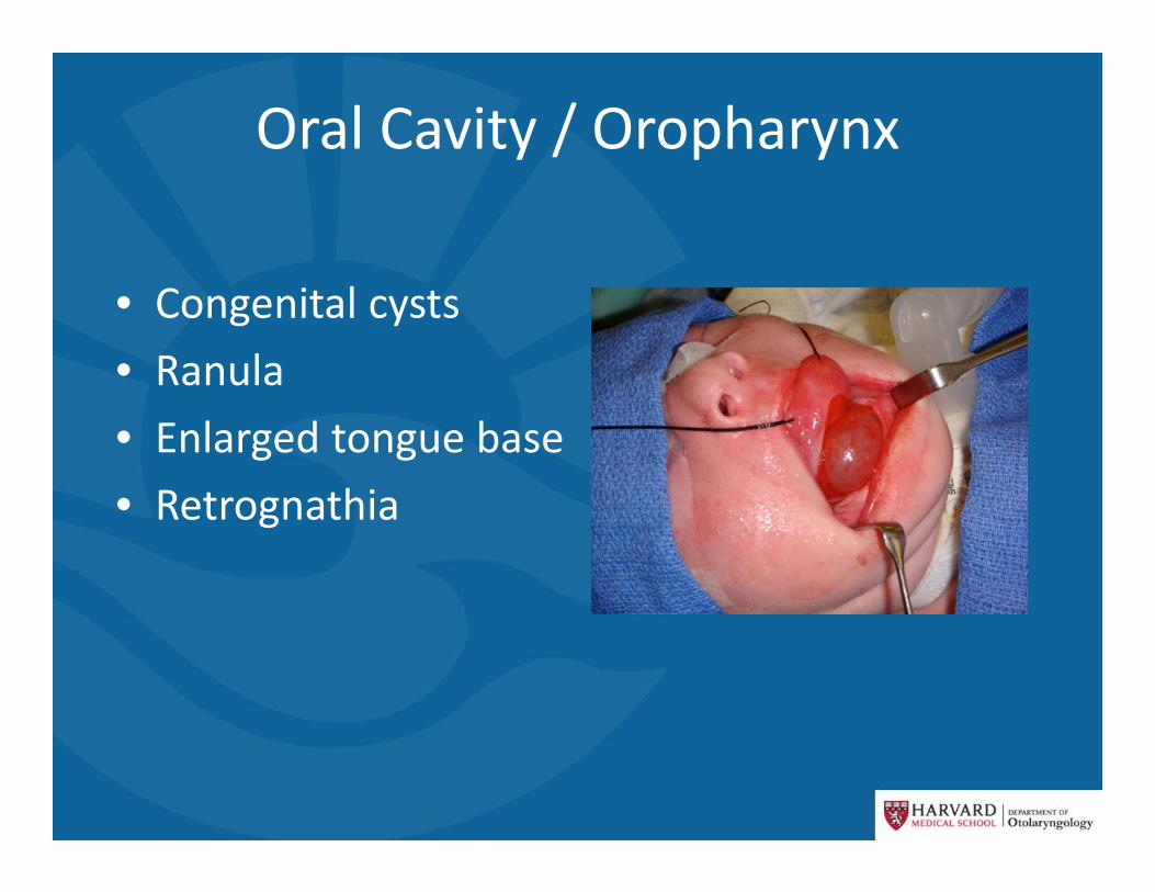
Oral Cavity / Oropharynx
• Congenital cysts• Ranula• Enlarged tongue base• Retrognathia

Vallecular Cyst

Hypopharynx/Supraglottis• Reflux• Laryngomalacia
– Floppers– Curlers

• Vocal Fold Immobility– Bilateral– Unilateral
• Webs• Congenital cyst• Trauma/granuloma• Neurologic
Glottis

Subglottis / trachea
• Croup• Subglottic stenosis• Subglottic hemangioma• Subglottic cyst• Tracheoesophageal fistula• Vascular compression

• Reactive airway disease• Asthma• Bronchopulmonary dysplasia• Restrictive lung disease
Lower airway

So what do you do?

The child does not look stable.Initial Management
• Keep the child comfortable• Avoid tongue depressor, IVs• Apply oxygen by the least threatening method• Wait for a secure environment before sedation
• Do not rely on pulse oximeter– Poor indicator of airway obstruction

• Anesthesia • Patient requires alveolar ventilation and adequate gas exchange via an (often) compromised airway
• Otolaryngology• Unobstructed view of airway• Surgical access to airway• Preservation of dynamic airway function
• Nurses and techs• Organized and available equipment• Focus on the patient
Special Challenges During Airway Procedures

Successful Airway Management
• Cooperation• Communication• Understanding
–Concerns/Needs– Techniques
• Proper Instrumentation

• Unable to explain based on physical exam• Out of proportion to physical exam• Risk factors for subglottic or tracheal obstruction by history
• You suspect lower airway pathology• What if the bronch is normal?
To bronch or not to bronch?
Pearl : Anxiety (yours or caretaker) is an indication for bronchoscopy

• Full monitoring especially while asleep and during feeds
• Polysomnography?• pH or impedance probe?• Interventions
– Nasal airway– Nasal cannula
• Does it improve safety?
Admit for an Airway Watch?
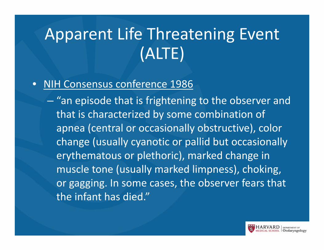
Apparent Life Threatening Event (ALTE)
• NIH Consensus conference 1986– “an episode that is frightening to the observer and that is characterized by some combination of apnea (central or occasionally obstructive), color change (usually cyanotic or pallid but occasionally erythematous or plethoric), marked change in muscle tone (usually marked limpness), choking, or gagging. In some cases, the observer fears that the infant has died.”

ALTE• Common
– 0.6 cases/1000 live births– 0.6%‐0.8% of ED visits for children under 1 year of age
• Definitive diagnosis in less than 50%• Numerous reports of airway abnormalities

ALTE• So who needs to see ENT
– Visibly obstructed breathing correlated with event– Risk factors for airway lesions
• Prematurity, intubations, syndromic

Disposition
• When can we go home?• What is normal?• Home apnea monitor?
– Diagnostic capability

• When can we go home?• What is normal?• Home apnea monitor?
– Diagnostic capability
Symptomatic Treatment

An Algorithm
• Overnight Airway Watch– Continuous cardiopulmonary monitoring
• Consideration for inpatient bronchoscopy• History and exam based imaging• Consideration for pH/impedence or PSG
High Severity (cyanosis, FTT, repeated ALTEs)

High Severity (cyanosis, FTT, repeated ALTEs)
• Events occurring during 24 hour period– Correlate to activity at that time– Cardiac concerns– Bronchoscopy
• No events– Education with family– Symptomatic treatment– Close follow up

Moderate Severity(prolonged feeding times, single ALTE type event, noisy
breathing without persistent distress)
• Imaging based on history• Consider outpatient bronchoscopy• Symptomatic treatment vs. “tincture of time”• Close follow up

Low Severity (noisy breathing in isolation, minimal feeding
issues, no ALTE events)
• Observation and reassurance

What can we do??
Infectious

Laryngotracheobronchitisaka Croup
• Most common infectious cause of upper airway obstruction in children
• Usually viral (parainfluenza)• Age 1‐6 (MC 12‐24mo)• Barking cough‐>stridor• Steeple sign• Tx:
– Steroids• Nebulized, oral, IM are equally effective• Dexamethasone 0.5mg/kg IM
– Nebulized epinephrine 1:1000• 0.3‐0.5 ml/kg (max 5 ml)

Epiglottitis
• Acute onset‐rapidly progressive• H. influenzae, Streptococcus and Staphylococcus• 4D’s
– Drooling, Dysphagia, Dysphonia, and Dyspnea• Tripod position, thumb sign• Transfer to PICU/OR asap
– Elective intubation• IV abx
– 3rd generation cephaolsporin (Ceftriaxone, Cefotaxime) 7‐10 days
– Add MRSA coverage in certain areas

Bacterial Tracheitisaka pseudomembranous croup
aka bacterial croup
• S. aureus• Median age 5 years• Croup‐>increasing respiratory distress‐>toxic appearance/dysphagia
• Diagnosis/treatment– Endoscopy under general anesthesia
• Remove exudative pseudomembrane – Intubation rates from 38‐100%– IV antibiotic (3rd generation cephalosporin and antistaphylococcal PCN)

TracheitisRepeat Bronchoscopy after 48 hours of Antibiotics
After 48 hours of antibiotics

Non‐Infectious

Foreign Body

Foreign Body
• MC <3• Male predilection• Food is most common (peanuts)• Witnessed choking episode 76‐92% sensitive• Aphonia with inability to cough=complete airway obstruction– Heimlich maueuver (older children) or alternate back blows (infants)
• Classic triad: cough, wheeze, unilateral diminished breath sounds observed in 57% of cases

Vocal Cord Paralysis
• Unilateral vs bilateral• Congenital vs acquired• Idiopathic, iatrogenic, neurological abnormality, birth trauma
• Bilateral present with early symptoms where unilateral may be missed
• Weak cry, stridor, recurrent aspiration, feeding problems

Vocal Cord Paralysis (cont)
• Imaging from brain stem to mediastinum– MRI for bilateral
• Arnold‐Chiari, tumors– Exclude intrathoracic lesions
for unilateral• Treatment
– Unilateral• Supportive with NG feeds until
safe swallow– Bilateral
• Tracheostomy, cordotomy (CO2 laser)
– Spontaneous recovery rates of up to 70%
– Avoid airway widening procedures for a year

Subglottic Stenosis
• Acquired is the most common form– History of endotracheal intubation or laryngeal trauma
• Congenital‐abnormality of cricoid cartilage• Presents with recurrent croup‐like illness


Laryngomalacia
• Most common congenital laryngeal abnormality• Most common cause of stridor in infancy• Onset usually in the first few weeks of life• Laryngoscopy to confirm diagnosis and rule out synchronous airway lesions‐Reports vary from 12‐64%
‐Skewed populations???‐Most commonly tracheomalacia, subglottic stenosis, and vocal cord paralysis‐4.7% required additional intervention (Mancuso, 1996)

• Flexible laryngoscopy• Magnified airway fluoroscopy• Barium swallow
– 65‐100% of infants have coexisting GERD– Breathing against an obstructed airway‐>negative intrathoracic pressure‐>higher likelihood of reflux through lower esophageal sphincter
– Edema from acid can worsen airway collapse• Direct laryngoscopy with rigid bronchoscopy• Polysomnogram• Echocardiogram

Management
• Reassurance for mild to moderate laryngomalacia without feeding difficulties– Monitor for appropriate weight gain or worsening of respiratory symptoms
• Feeding– Positional therapy– Thickened feeds– Treatment of GERD
• Average age of resolution is 7.6 months (Wright, 2012) with most resolution compete by 18‐24 months

• Surgical management in 10%– When they present with apparent life threatening events, severe dyspnea, recurrent cyanosis with feedings or failure to thrive
– Supraglottoplasty, aryepiglottoplasty, tracheostomy • Consider associated neurological or neuromuscular conditions
– Microlaryngeal instruments vs micro‐debrider vs CO2 laser
• Similar success rates regardless of technique (Rastatter, 2010)

Conclusions
• It is not as scary as it seems• Need a methodical approach• Balance between provider comfort, parental comfort and ensuring a child’s safety.
References
Mandal, Kabra, Lodha. Upper Airway Obstruction in Children. Indian J Pediatr. 82(8):737‐744. Mancuso R, Choi S, Zal G, et al. Laryngomalacia. The search for
the second lesion. Arch Otolaryngol Head Neck Surg.1996; 122(3):302‐306.
Wright CT, Goudy Sl. Congenital laryngomalacia: symptomduration and need for surgical intervention. Ann OtolRhinol Laryngol. 2012; 121(1):57‐60.
Rastatter JC, Schroeder JW, Hoff SR, Holinger LD. Aspirationbefore and after Supraglottoplasty regardless of Technique. Int J
Otolaryngol. 2010; 2010912814.Thorne MC, Garetz SL. Laryngomalacia: review and Summary of
Current Clinical Practice in 2015. Paediatric RespiratoryReview. 2015.

Questions???


