Page 1
THE DISTRIBUTION AND FUNCTION OF ELASTIN AND
ELASTIC FIBRES IN THE CANINE CRUCIATE LIGAMENT
COMPLEX
Thesis submitted in accordance with the requirements of the University of Liverpool for the
degree of Doctor in Philosophy
By
Kinley D. Smith
July 2010
Page 2
2
ABSTRACT
Anterior cruciate ligament rupture (ACL) is a major source of morbidity in the dog, leading to severe
osteoarthritis of the knee joint and marked lameness. Following rupture, the ACL will not heal and in
the dog, ACL rupture is thought to be the end stage of degenerative ligament disease (non-contact ACL
injury). The extracellular matrix (ECM) of CLs has been extensively studied but little is known of the
role of elastic fibres in the physiology of the ECM, the mechanics of CL function and in CL
degeneration. Elastic fibres include polymers of fibrillins (microfibrils), bundles of microfibrils
(oxytalan fibres) and elastin fibres (bundles of microfibrils with an elastin core). The hypothesis of this
thesis is that elastin has a mechanical and a biological role in the canine cruciate ligament complex. It is
further hypothesised that the distribution and function of elastic fibres will vary between three breeds of
dog with differing risk of ACL rupture are: the greyhound with a low risk, the beagle with a low-to-
moderate risk and the Labrador retriever with a high risk.
The distribution of elastic fibres, fibrillins and cells was investigated throughout the CL complex using
a combination of histochemical staining and immunofluorescence. CL microanatomy was studied using
Nomarski differential interference microscopy. Elastin was measured biochemically and compared to
histologic assessment of tissue architecture, elastic fibre staining and other biochemical parameters. The
biological effect of elastin degradation products (EDPs) was assessed in an in vitro ACL cell culture
model. A low risk dog breed to ACL rupture (greyhound) was used in all investigations and
comparisons were made with other breeds with regard to cellular and elastic fibre anatomy.
Differences in cell morphology between breeds with differing risk of ACL rupture may reflect
fundamental differences in CL physiology possibly through altered cell-to-cell communication. Cellular
and matrix changes, considered degenerative, were seen throughout the CL complex and may reflect
adaptation rather than degeneration in certain dog breeds such as the greyhound. Elastin content ranged
from 5.9 to 19.4% of ligament dry weight. This was a far greater proportion of canine CLs than
previously. Elastin fibres may have a mechanical role in bundle reorganization following ligament
deformation. The distribution of fibrillins 1 and 2 was different from the pattern previously reported in
tendon and may represent a fundamental difference between ligament and tendon. In the greyhound CL
there was a significant proportional increase in oxytalan fibre staining with advancing CL degeneration.
This response was seen also in the Labrador retriever and the beagle but the increase in oxytalan fibre
staining was less marked with advancing degeneration. Therefore production of oxytalan fibres may
reflect a healing response in damaged CL tissue in breeds at a low risk of ligament rupture. Fragments
of elastin containing the VGVAPG motif affect canine ACL cells in vitro resulting in increased
transcription of fibrillin 2 mRNA. Additionally, there was synergism with TGF-β1 resulting in
upregulation of the elastin laminin receptor 1, through which EDPs are transduced. EDPs may thus have
a role in response to injury in the CL.
Page 3
3
TABLE OF CONTENTS
ABSTRACT ........................................................................................................................ 2 TABLE OF CONTENTS .................................................................................................... 3 DEDICATION AND ACKNOWLEDGEMENTS ............................................................. 8 AUTHOR’S DECLARATION ........................................................................................... 9
LIST OF FIGURES .......................................................................................................... 10 LIST OF TABLES ............................................................................................................ 13 LIST OF ABBREVIATIONS ........................................................................................... 14
CHAPTER ONE: General introduction ............................................................................ 16 CANINE CRUCIATE LIGAMENTS .................................................................................. 16
1.1 Gross Anatomy ..................................................................................................... 16 1.1.1 Overview of the knee ................................................................................... 16 1.1.2 ACL anatomy ............................................................................................... 17 1.1.3 PCL anatomy ............................................................................................... 17 1.1.4 Blood supply ................................................................................................ 17 1.1.5 Neurology .................................................................................................... 18 1.1.6 Ultrastructural anatomy ................................................................................ 18
1.1.6.1 Fascicular subdivision ............................................................................. 18 1.1.6.2 Cellular organisation ................................................................................ 19
1.2 Functional anatomy of CLs ................................................................................... 21
CL ECM STRUCTURE ....................................................................................................... 22 1.3 Collagen ................................................................................................................ 22
1.3.1 Collagen: Classification ............................................................................... 22 1.3.2 Collagen: Structure ...................................................................................... 24 1.3.3 Collagen: Assembly ..................................................................................... 24 1.3.4 Collagen: Crosslinks .................................................................................... 25
1.4 Elastin, fibrillin and the elastic fibres ................................................................... 25 1.4.1 Overview ...................................................................................................... 25 1.4.2 Molecular composition ................................................................................. 26
1.4.2.1 Elastin core .............................................................................................. 26 1.4.2.2 Microfibrils .............................................................................................. 27 1.4.2.3 Elastic fibre interface molecules .............................................................. 28
1.4.3 Elastic fibre structure and assembly .............................................................. 31 1.4.3.1 Microfibril structure and assembly ........................................................... 31 1.4.3.2 Elastin fibre structure and assembly ........................................................ 31
1.4.4 Organisation of elastin fibres in tissue ........................................................ 32 1.4.5 Elastic fibre functions .................................................................................. 33
1.4.5.1 Elasticity .................................................................................................. 33 1.4.5.2 TGF-β family activation .......................................................................... 33 1.4.5.3. Cell adhesion .......................................................................................... 34
1.4.6 Elastic fibre production and degradation ...................................................... 34 1.5 Proteoglycans (PG) ............................................................................................... 35
1.5.1 Large aggregating PGs ................................................................................. 35 1.5.2 Non-aggregating PGs .................................................................................... 36
1.6 Glycosaminoglycans (GAGs) ............................................................................... 36 1.7 Other ECM components ........................................................................................ 37
Page 4
4
EXTRACELLULAR MATRIX PHYSIOLOGY ................................................................. 37 1.8 Metalloproteinases (MP) ....................................................................................... 38
1.8.1 Matrix metalloproteinases (MMPs) ............................................................. 38 1.9 Cysteine proteinases .............................................................................................. 39 1.10 Serine Proteases .................................................................................................... 39 1.11 Other collagenolytic agents ................................................................................... 39 1.12 Role of proteinases ................................................................................................ 40
1.12.1 Function and mode of action ..................................................................... 40 1.12.2 Involvement of proteinases in degenerative conditions .............................. 40
CURRENT THOUGHTS ON THE AETIOPATHOGENESIS OF CANINE CRUCIATE
LIGAMENT RUPTURE ...................................................................................................... 41 1.13 CL idiosyncrasies .................................................................................................. 41 1.14 Histologic and ultrastructural changes in CL disease ........................................... 41
1.14.1 Histologic overview .................................................................................... 41 1.14.2 Cell population changes ............................................................................... 43 1.14.3 Collagen changes ........................................................................................ 43 1.14.4 GAG and PG changes .................................................................................. 44 1.14.5 Gene expression profile ............................................................................... 44
1.15 Concepts of aetiology............................................................................................ 45 1.15.1 Weight, breed and age .................................................................................. 45 1.15.2 Compromise of blood supply .................................................................... 46 1.15.3 Cellular alterations .................................................................................... 46 1.15.4 Mechanobiological aetiologies .................................................................... 46 1.15.5 Ligament response to disease or injury ..................................................... 47
ELASTIC FIBRE -ASSOCIATED DISEASE ..................................................................... 48 1.16 Heritable disorders: ............................................................................................... 48
1.16.1 Marfan syndrome (MFS) and related fibrillinopathies ..................................... 48 1.16.2 Supravalvular aortic stenosis (SVAS) and Williams-Beuren syndrome (WBS)
..................................................................................................................................... 48 1.16.3 Cutis laxa ...................................................................................................... 49 1.16.4 Other conditions ............................................................................................ 49
1.17 Degenerative disorders ......................................................................................... 49 1.17.1 Vascular proliferative disease ....................................................................... 49 1.17.2 Chronic obstructive pulmonary disease (COPD) ......................................... 50 1.17.3 Intervertebral disc disease (IVDD) .............................................................. 50 1.17.4 Other articular structures .............................................................................. 50 1.17.5 Periodontal ligament .................................................................................... 51 1.17.6 Canine CLs ................................................................................................... 51
1.18 Hypothesis and aims ............................................................................................. 52 1.18.1 Mechanical role for elastin in the canine CL complex.................................. 52 1.18.2 Biological role for elastin fibres in the canine CL complex ......................... 52
Page 5
5
CHAPTER 2: Variations in cell morphology in the cruciate ligament complex .............. 53
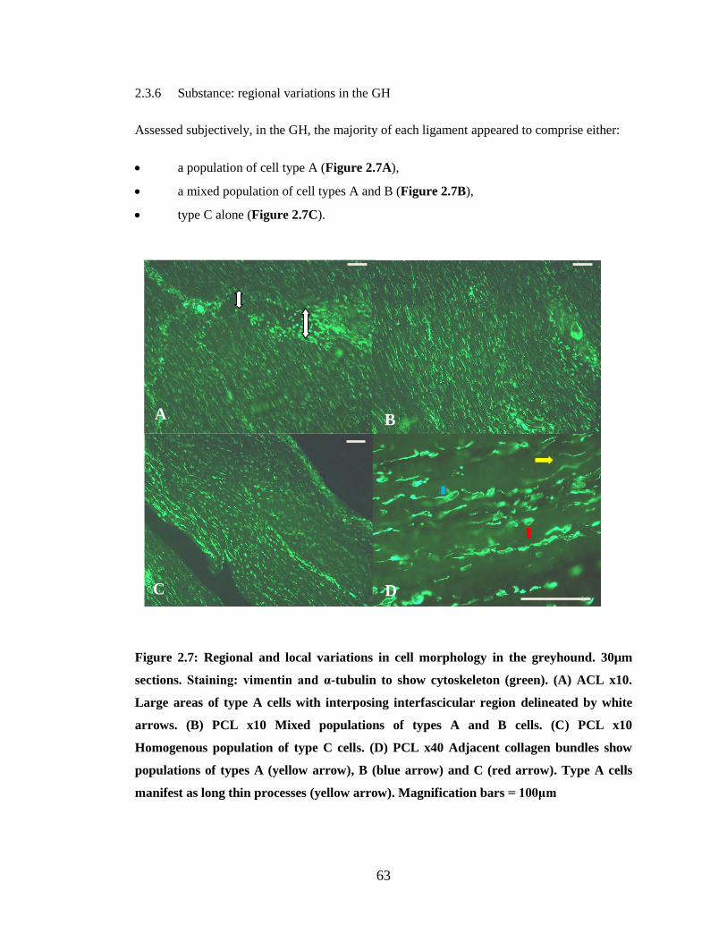
2.1 Introduction ........................................................................................................... 54 2.2 Materials and methods .......................................................................................... 55
2.2.1 Sample collection and preparation ................................................................ 55 2.2.2 Histology and immunofluorescence .............................................................. 55 2.2.3 Imaging ......................................................................................................... 56
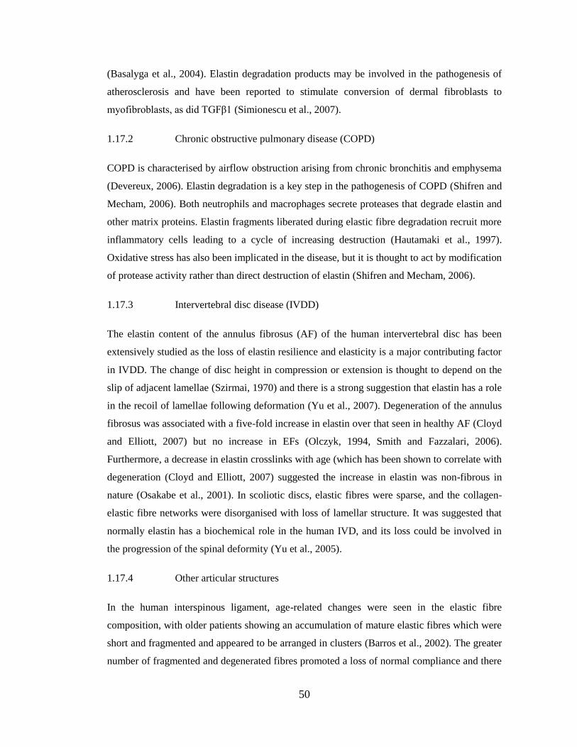
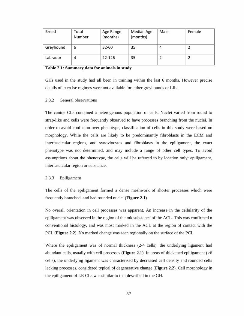
2.3 Results ................................................................................................................... 56 2.3.1 Animals ......................................................................................................... 56 2.3.2 General observations ..................................................................................... 57 2.3.3 Epiligament ................................................................................................... 57 2.3.4 Interfascicular regions ................................................................................... 59 2.3.5 Substance: variations in cell morphology ..................................................... 59 2.3.6 Substance: regional variations in the GH ...................................................... 63 2.3.7 Substance: regional variations in the LR ...................................................... 64
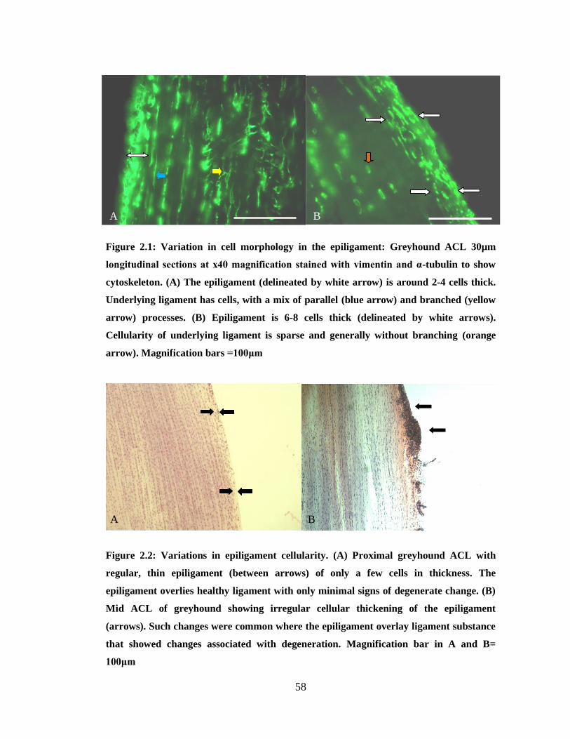
2.4 Discussion ............................................................................................................. 66
CHAPTER 3: The organisation of elastin and fibrillins 1 and 2 in the cruciate ligament
complex ............................................................................................................................. 70
3.1 Introduction ........................................................................................................... 71 3.2 Materials and methods .......................................................................................... 72
3.2.1 Sample collection and preparation ................................................................ 72 3.2.2 Histology ....................................................................................................... 72 3.2.3 Antibodies ..................................................................................................... 73 3.2.4 Immunofluorescence ..................................................................................... 73 3.2.5 Nomarski differential interference contrast optical microscopy (NDIC) ...... 74 3.2.6 Imaging ......................................................................................................... 74
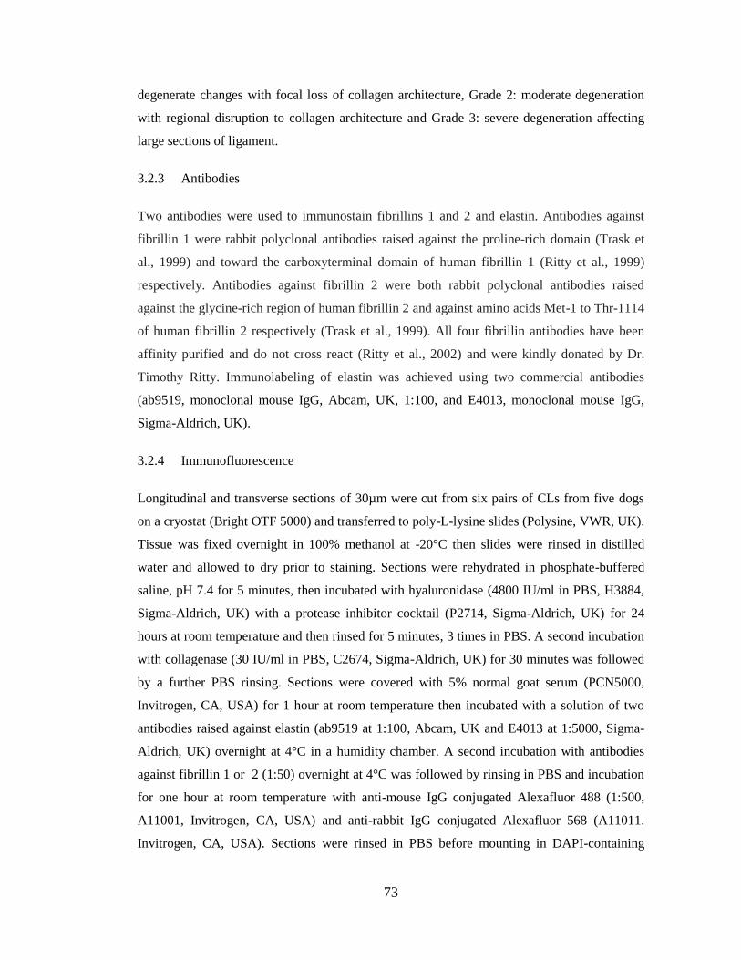
3.3 Results ................................................................................................................... 74 3.3.1 Animals ......................................................................................................... 74 3.3.2 H&E sections ................................................................................................ 75 3.3.3 Elastin fibres (EVH)...................................................................................... 75 3.3.4 Oxytalan fibres (Miller’s stain) ......................................................................... 76 3.3.5 Fibrillins ........................................................................................................ 77
3.4 Discussion ............................................................................................................. 82
CHAPTER 4: Elastin in the cruciate ligament complex: a correlative histological and
biochemical study.............................................................................................................. 85
4.1 Introduction ........................................................................................................... 86 4.2 Materials and methods .......................................................................................... 87
4.2.1 Tissue preparation ......................................................................................... 87 4.2.2 Histology: Staining ....................................................................................... 88 4.2.3 Histology: Scoring methods .......................................................................... 88 4.2.4 Biochemical analyses .................................................................................... 90 4.2.5 Data and statistical analysis .......................................................................... 91
4.3 Results ................................................................................................................... 91 4.3.1 Histology: H&E ............................................................................................ 91 4.3.2 Histology: Elastic fibre staining .................................................................... 92 4.3.3 Biochemical analyses .................................................................................... 94
Page 6
6
4.4 Discussion ............................................................................................................. 97
CHAPTER 5: The effect of elastin degradation peptides on canine anterior cruciate
ligament cell cultures ...................................................................................................... 102
5.1 Introduction ......................................................................................................... 103 5.2 Materials and methods ................................................................................... 104
5.2.1 Elastin peptides ........................................................................................... 104 5.2.2 Donors, extraction and preparation of cells ................................................ 105 5.2.3 Preparation of 6-well plates ........................................................................ 105 5.2.4 Harvesting of cells ...................................................................................... 105 5.2.5 mRNA extraction and real time RT-PCR ................................................... 106 5.2.6 Primer design .............................................................................................. 106 5.2.7 Absolute quantification of mRNA expression ............................................ 107 5.2.8 Statistical analysis ....................................................................................... 107
5.3 Results ................................................................................................................. 108 5.3.1 Animals ....................................................................................................... 108 5.3.2 Reference genes .......................................................................................... 108 5.3.3 Treatment with EDPs .................................................................................. 108 5.3.4 Treatment with TGF-β1 .............................................................................. 109 5.3.5 Treatment with TNF-α ................................................................................ 109
5.4 Discussion ........................................................................................................... 112
CHAPTER SIX: Comparison of elastic fibre distribution in the anterior cruciate
ligament in dogs at a differing risk of anterior cruciate ligament rupture ...................... 115
6.1 Introduction ......................................................................................................... 116 6.2 Material and methods .......................................................................................... 117
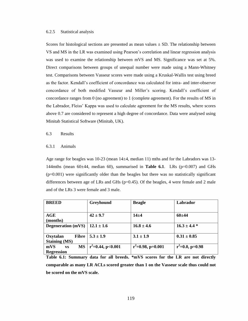
6.2.1 Animals ....................................................................................................... 117 6.2.2 Sample collection ........................................................................................ 118 6.2.3 Histology: Staining ..................................................................................... 118 6.2.4 Histology: Scoring methods ........................................................................ 118 6.2.5 Statistical analysis ....................................................................................... 119
6.3 Results ................................................................................................................. 119 6.3.1 Animals ....................................................................................................... 119 6.3.2 Degeneration Scoring .................................................................................. 120 6.3.3 Miller’s Scoring (Oxytalan Fibre Staining) ................................................ 120 6.3.4 Inter/intra-observer data .................................................................................. 122 6.3.5 Descriptive Histology ................................................................................. 122
6.4 Discussion ........................................................................................................... 125
CHAPTER 7:General Discussion ................................................................................... 129
7.1 Introduction ......................................................................................................... 129 7.2 Cell morphology ................................................................................................. 129 7.3 Elastic fibres and fibrillins .................................................................................. 130 7.4 Elastic fibres and CL degeneration ..................................................................... 131 7.5 Breed variation in elastic fibres .......................................................................... 132 7.6 Elastin degradation peptides ............................................................................... 133 7.7 Conclusions ......................................................................................................... 135
Page 7
7
CHAPTER 8: Future Studies .......................................................................................... 136
8.1 Cell morphology ................................................................................................. 136 8.2 Elastic fibres and fibrillins .................................................................................. 136 8.3 Elastic fibres and CL degeneration ..................................................................... 137 8.4 Breed variation in elastic fibres .......................................................................... 137 8.5 Elastin degradation peptides ............................................................................... 138
BIBLIOGRAPHY ........................................................................................................... 139
Page 8
8
DEDICATION AND ACKNOWLEDGEMENTS
Thanks must go foremost to my supervisors, Dr Eithne Comerford, who gave me the
opportunity to undertake this work, and to Professors Pete Clegg and John Innes. I
would like to acknowledge Dr Udo Hetzel and Dr David Spiller for their help with
pathology and confocal microscopy respectively. Many thanks also to Dr Nick Rhodes
for his help and advice. A great thank you goes to the members of the musculoskeletal
research group at Liverpool especially Dr Simon Tew and Dr Sarah Taylor for their
invaluable support with technical aspects of this study.
My family has been hugely supportive of my desire to undertake research training. My
wife Keeley has given me not only her unwavering support for my career but also a
happy home and two beautiful daughters, Heather and Eleanor. My mother and father
have encouraged me to work hard, be confident in what I do and not be afraid of
difficult decisions. I thank them for their continued support. Where would we be
without friends? Thanks especially to Chris, Alan, Steph, Gavin and my sister
Caroline, for keeping my feet firmly on the ground.
Dr Anne Vaughan-Thomas passed away in August 2009. Her supervision was filled
with enthusiasm and her office door always open. Her wisdom and support instilled in
me a passion for matrix biology. To her memory this thesis is dedicated
Dyfal donc a dyr y garreg
(Tapping persistently breaks the stone)
Page 9
9
AUTHOR’S DECLARATION
I declare that the work in this dissertation was carried out in accordance with the regulations of
the University of Liverpool. The work is original except where indicated by reference in the
text.
Any views expressed in this thesis are those of the author and in no way represent those of the
University of Liverpool.
This thesis has not been presented to any other university for examination in the United
Kingdom or overseas.
Page 10
10
LIST OF FIGURES
Figure 1.1: Cruciate ligament complex within the canine knee
Figure 1.2: Domain structures of elastin, fibrillin-1 and fibulin-5
Figure 1.3: Schematic diagram of the assembly of microfibrils and elastic fibres
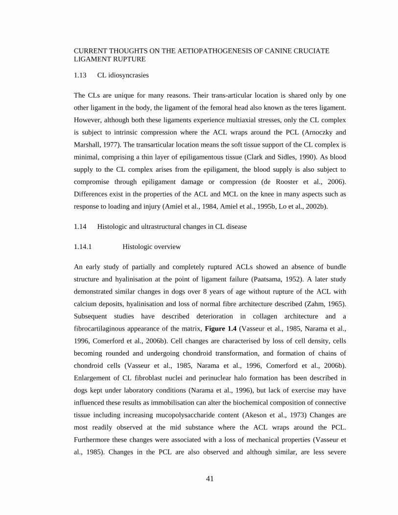
Figure 1.4: Typical changes associated with degeneration of the ACL
Figure 2.1: Variation in cell morphology in the epiligament
Figure 2.2: Mid ACL showing irregular cellular thickening of the epiligament
Figure 2.3: Confocal microscopy images of cells of the interfascicular region
Figure 2.4: Variations in cell morphology of the fascicular region of the CL complex
Figure 2.5: Relationships of transverse processes to collagen bundles
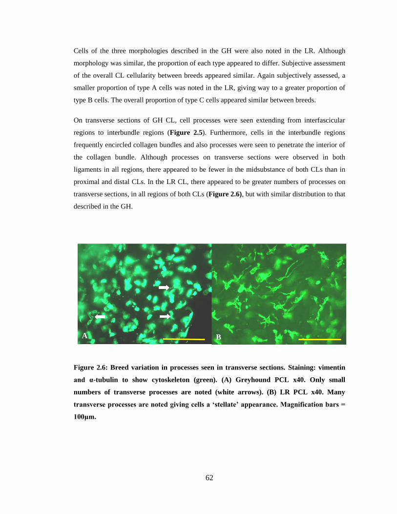
Figure 2.6: Breed variation in processes seen in transverse sections
Figure 2.7: Regional and local variations in cell morphology
Figure 2.8: Typical regional variation of cell morphology in the LR
Figure 2.9: Striking juxtaposition of regions of differing cell morphologies in the LR
Figure 2.10: Areas devoid of nuclear staining
Figure 3.1 Distribution of elastin fibres in the CL complex
Figure 3.2: Distribution of microfibrils in the canine CL complex
Figure 3.3: Distribution of fibrillin-1 in the canine CL complex
Figure 3.4: Elastin and fibrillin 1 rarely co-stain when in fibre form
Figure 3.5: Distribution of fibrillin-2 in the canine CL complex
Figure 3.6: Co-staining of elastin and fibrillin 2
Page 11
11
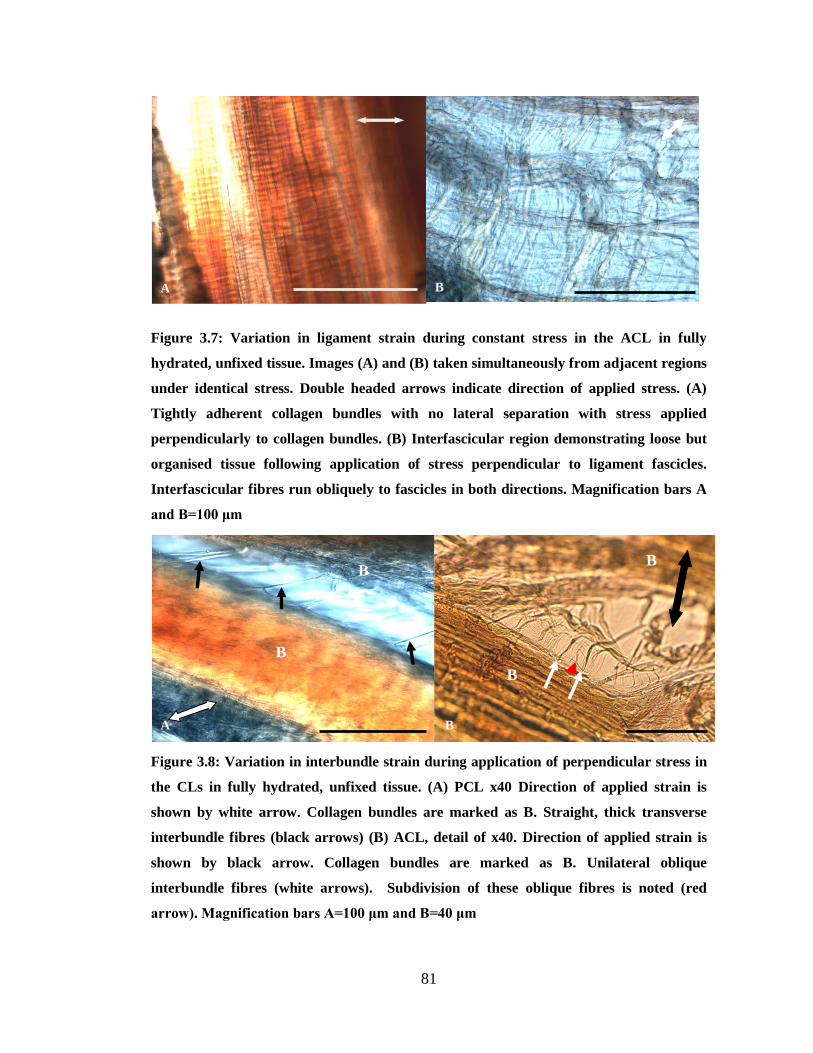
Figure 3.7: Variation in ligament strain during constant stress in the ACL in fully hydrated,
unfixed tissue
Figure 3.8: Variation in interbundle strain during application of perpendicular stress in the CLs
in fully hydrated, unfixed tissue
Figure 4.1: Miller’s stain scoring system sheet
Figure 4.2: Histologic changes in CLs with degeneration
Figure 4.3: Variation in interbundle staining
Figure 4.4: Boxplot summary of ACL and PCL results for Miller’s score
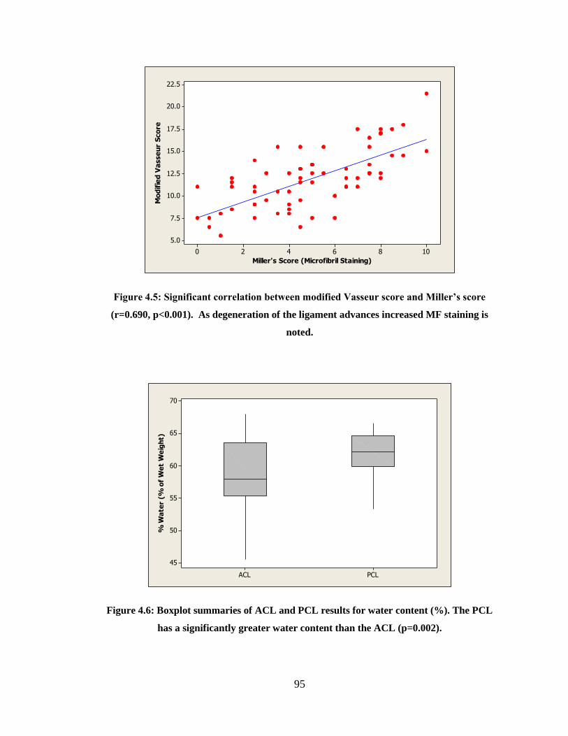
Figure 4.5: Significant correlation between modified Vasseur score and Miller’s score
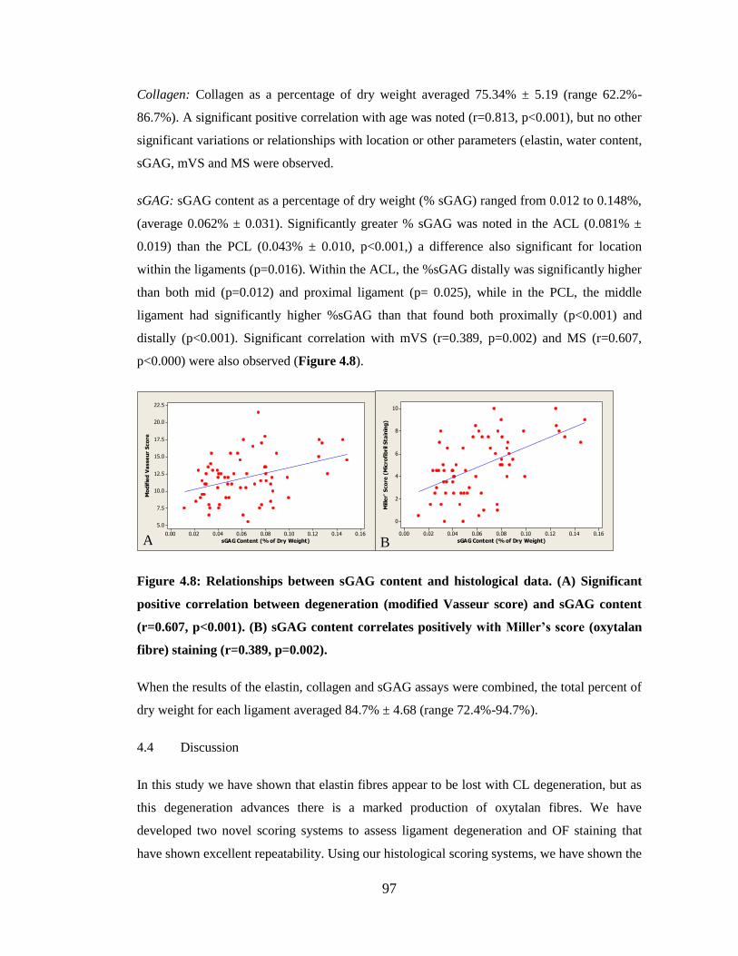
Figure 4.6: Boxplot summaries of ACL and PCL results for water content
Figure 4.7: Elastin content of canine CLs
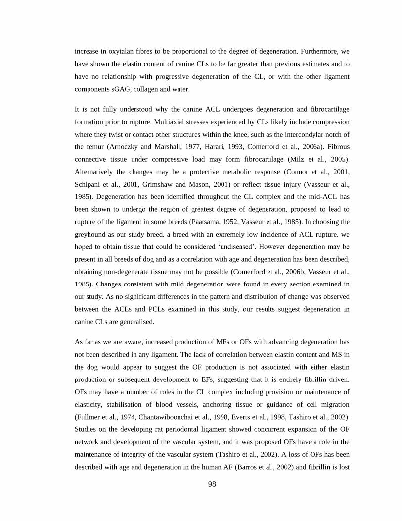
Figure 4.8: Relationships between biochemical and histological data
Figure 5.1: Fold change in mRNA transcription relative to GAPDH following 6 hours of
treatment with EDPs alone
Figure 5.2: Fold change in mRNA transcription relative to GAPDH following 6 hours of
treatment with TGF-β1 alone
Figure 5.3: Fold change in mRNA transcription relative to GAPDH following 6 hours of
treatment with TNF-α
Figure 5.4: Fold change in mRNA transcription relative to GAPDH following 24 hours of
treatment with TNF-α
Figure 5.5: Fold change in mRNA transcription relative to GAPDH following 6 hours of
treatment with TGF-β1 and EDPs in combination
Figure 5.6: Fold change in mRNA transcription relative to GAPDH following 24 hours of
treatment with TGF-β1 and EDPs in combination
Page 12
12
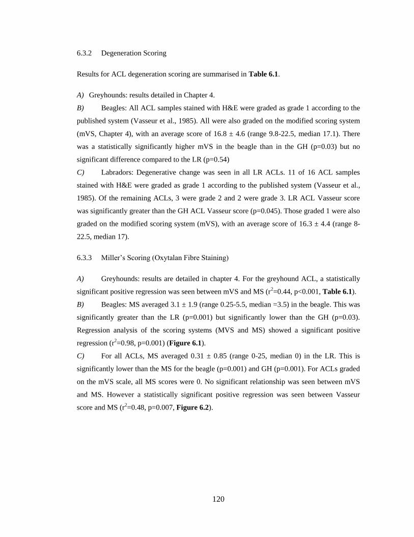
Figure 6.1: Relationship between ACL degeneration and oxytalan fibre staining in the beagle
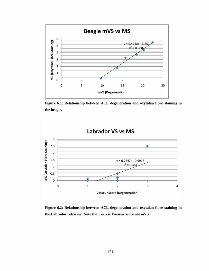
Figure 6.2: Relationship between ACL degeneration and oxytalan fibre staining in the
Labrador retriever
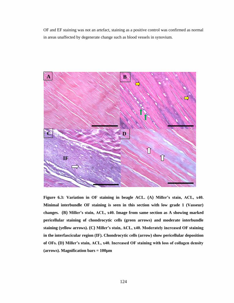
Figure 6.3: Variation in OF staining in beagle ACL
Figure 6.4: Minimal increase in OF staining in high grade degenerate LR CL
Page 13
13
LIST OF TABLES
Table 1.1: Collagen types, genes and supramolecular organisation and distribution
Table 1.2: Structural and associated molecules of microfibrils and elastic fibres
Table 2.1: Summary data for animals in study
Table 3.1: Summary data for animals in study
Table 4.1: Criteria for modified Vasseur scoring
Table 4.2: Summary data for animals in study
Table 4.3: Kendall’s coefficients of concordance for histology scoring methods.
Table 5.1: List of primer sequences used for reference and target genes
Table 6.1: Summary data for all breeds
Table 6.2: Intra- and inter-observer agreement in mVS and MS scoring
Page 14
14
LIST OF ABBREVIATIONS
ACL Anterior cruciate ligament
ADAM A disintegrin and metalloproteinase domain
ADAM-TS ADAM with a thrombospondin motif
ANOVA Analysis of variance
AP Anterior-posterior
ASMA Alpha smooth muscle actin
B2M Beta-2 macroglobulin
cbEGF Calcium binding epidermal growth factor
CCA Congenital contractile arachnodactyly
CL Cruciate ligament
CLSM Confocal laser scanning microscopy
DAPI 4', 6-diamidino-2-phenylindole
cDNA Cyclic deoxyribonucleic acid
Col1a2 Collagen type I (α2 chain) gene
Col2a1 Collagen type II (α1 chain) gene
Col3a1 Collagen type III (α1 chain) gene
CS Chondroitin sulphate
CTSB Cathepsin B
CTSK Cathepsin K
DS Dermatan sulphate
DMEM Dulbecco’s modified Eagle’s Medium
DMMB 1, 9-dimethylmethylene blue
ECM Extracellular matrix
EBP Elastin binding protein
EDP Elastin degradation peptides
EDTA Ethylenediaminetetraacetic acid
EF Elastin fibre
EGF Epidermal growth factor
Eln Elastin gene
ELR1 Elastin laminin receptor 1
EVH Verhoeff’s iodine-iron haematoxylin stain
Fbn1 Fibrillin 1 gene
Fbn2 Fibrillin 2 gene
FBS Foetal bovine serum
FGF Fibroblast growth factor
GAG Glycosaminoglycan
GAPDH Glyceraldehyde phosphate dehydrogenase
GH Greyhound
H&E Haematoxylin and eosin
HCl Hydrochloric acid
IGF Insulin-like growth factor
IL Interleukin
KS Keratin sulphate
LR Labrador retriever
LTBP-1 Latent transforming growth factor β binding protein 1
M Miller’s stain
MAGP-1 Microfibril associated glycoprotein 1
MCL Medial collateral ligament
Page 15
15
MF Microfibril
MFS Marfan syndrome M-MLV Moloney - murine leukaemia virus MMP Matrix metalloproteinase
mRNA Messenger ribonucleic acid
MS Miller’s score
mVS Modified Vasseur score
MT-MMP Membrane type-matrix metalloproteinase
NDIC Nomarski differential interference contrast (microscopy)
NO Nitric oxide
OA Osteoarthritis
OF Oxytalan fibre
OHPro Hydroxyproline
PBS Phosphate-buffered saline
PCL Posterior cruciate ligament
PG Proteoglycan
qPCR Quantitative polymerase chain reaction
RA Rheumatoid arthritis
RGD Arg-Gly-Asp peptide sequence
RT-PCR Reverse transcriptase polymerase chain reaction
SDFT Superficial digital flexor tendon
sGAG Sulphated glycosaminoglycans
SOX9 Sex determining region box 9 gene
TB 8-cysteine containing motif (thrombospondin motif)
TGF Transforming growth factor
TIMP Tissue inhibitor of metalloproteinase
TNF Tumour necrosis factor
TPA Tibial plateau angle
uPA Urokinase plasminogen activator
Vcan Versican gene
Page 16
16
CHAPTER ONE: General introduction
CANINE CRUCIATE LIGAMENTS
1.1 Gross Anatomy
1.1.1 Overview of the knee
The knee (stifle) is a condylar synovial joint consisting of articulations between the femur and
tibia and between the femur and patella. It is an extremely complex joint in both structure and
function and ligaments are essential for the maintenance of these articulations (Tirgari and
Vaughan, 1975b). The cruciate ligaments (CLs), anterior (ACL) and posterior (PCL), are the
primary stabilisers of the knee and serve to limit anterior translation and rotation of the tibia
(Arnoczky and Marshall, 1977, Moore and Read, 1996), (Figure 1.1). Within the knee joint,
the ACL and PCL are in intimate contact, with the ACL twisted around the PCL.
They are considered to function as a unit hence the term CL complex (Arnoczky and
Marshall, 1977, Harari, 1993).
Figure 1.1: Cruciate ligament complex within the canine knee
POSTERIOR CRUCIATE LIGAMENT
ANTERIOR CRUCIATE LIGAMENT
TIBIA
Page 17
17
1.1.2 ACL anatomy
The canine ACL runs anteriorly, medially, and distally in an outward spiral as it passes from
the medial aspect of the lateral femoral condyle to the anterior intercondyloid area of the tibial
plateau (Zahm, 1965, Haut and Little, 1969). Two separate bundles are observed in-situ in the
dog, termed anteriomedial and posteriolateral bands. The anteriomedial subdivision is the
longest, yet smaller component. It arises more proximally from the femur and inserts more
anteriorly on the tibial attachment area, compared with the posteriolateral subdivision
(Arnoczky and Marshall, 1977, Heffron and Campbell, 1978).
1.1.3 PCL anatomy
The PCL is a longer and wider structure than the ACL, narrowest at the midsection and
fanning toward origin and insertion (Arnoczky and Marshall, 1977). It is subdivided into two
bands, a larger anterior and smaller posterior, although these are often indistinct (Heffron and
Campbell, 1978). They have reciprocating functions through flexion and extension with the
anterior portion taut in flexion and loose in extension and the posterior portion taut in
extension only (Arnoczky and Marshall, 1977, Harari, 1993).
1.1.4 Blood supply
The main supply to the central knee is the middle genicular artery penetrating the posterior
joint capsule (Kobayashi et al., 2006). A well vascularised enveloping synovial tissue carries
the vessels, with minimal supply from origin and insertion (Arnoczky et al., 1979). The core
of the ACL is less well vascularised than the remainder (Tirgari, 1978, Vasseur et al., 1985,
Narama et al., 1996). Anastamoses exist between endo- and epiligamentous networks
(Kobayashi et al., 2006). The PCL may have a more substantial blood supply as there appears
to be more epiligamentous vessels (Arnoczky et al., 1979). CLs may also gain nutrition from
the synovial fluid (Kobayashi et al., 2006).
Although the ACL has been considered extra-articular due to the enveloping synovial
epiligament, free passage of macromolecules from intra-articular synovial fluid to the
substance of the ACL has been demonstrated (Kobayashi et al., 2006). As such free movement
exists between the synovial fluid and ACL substance, a fall in intra-articular pressure, as may
occur in osteoarthritis, is likely to adversely affect ACL blood flow.
Page 18
18
1.1.5 Neurology
Innervation of the canine knee joint is from three major articular nerves arising from the
saphenous, tibial and common peroneal nerve, although other nerves may contribute to a
variable extent (O'Connor et al., 1982). The main trunk of the nerve bundles is found at the
femoral origins of the CLs (Arnoczky, 1983). Nerves in the synovial envelope penetrate the
CLs radially, and endoligamentous nerves course with the vessels, where their function is
thought to be primarily associated with the regulation of blood flow and nociception (Kennedy
et al., 1974). Mechanoreceptors within the substance may activate local reflex patterns to
protect the ligament against tearing and warn against possible joint damage (Krauspe et al.,
1992, Biedert et al., 1992).
1.1.6 Ultrastructural anatomy
1.1.6.1 Fascicular subdivision
In man and dogs, CLs have been described as containing twisted collagenous fascicles and
fibre bundles that are subdivided into fascicles, subfascicular units, fibres, and fibrils
(Arnoczky, 1983, Yahia and Drouin, 1989). A simpler subdivision has been proposed with
only two divisions of bundles and fascicles with a fascicular membrane that does not
concentrically bound the fascicle (Clark and Sidles, 1990). Cells in the CL substance are
predominantly fibroblasts which may vary from a spindle to rounded-shape (Arnoczky, 1983).
These are isolated or in short rows of 2-3 cells and tightly applied to collagen bundles.
Both CLs are covered in a fold of synovial membrane, incompletely bisecting the joint in the
sagittal plane (Arnoczky and Marshall, 1977). The synovial envelope of the ACL originates at
the intercondylar notch and extends to the anterior tibia where it communicates with a fold of
the distal joint capsule (Alm and Stromberg, 1974). The PCL is ensheathed by two folds of
synovial membrane (Alm and Stromberg, 1974). These enveloping membranes are termed the
epiligament. It has been divided to an intima and subintima: the intima comprising a single
layer of synoviocytes and the subintima mainly areolar tissue containing small vessels,
fibroblasts and some adipocytes (Heffron and Campbell, 1978, Vasseur et al., 1985). The
synovial epiligament is far more cellular than the CL substance (Arnoczky and Marshall,
1977). The epiligament is an incomplete barrier and there is ready contact with synovial fluid
(Kobayashi et al., 2006, Tang et al., 2009).
Page 19
19
Each ligament component consists of multiple elliptical fascicles (Heffron and Campbell,
1978). The peripheral subunits follow a spiral path of waviness around the fascicle axis (Yahia
and Drouin, 1989, Amis and Dawkins, 1991, Kennedy et al., 1974). Each subfascicle contains
bundles of collagen fibres, but these are not orientated isometrically during knee motion (Amis
and Dawkins, 1991). Each change in knee position recruits fibres differently (Butler et al.,
1992). It has been postulated that individual fibres change their length by straightening their
crimp, a period banding of collagen observed under crossed polarizing filters (Amis and
Dawkins, 1991, Yahia and Drouin, 1989, Boorman et al., 2006).
The collagen fibres are formed by fibrils, themselves an organisation of repeated collagen
subunits. Fibrils have a uniform crimp parallel to the long axis of the fibril: the internal fibrils
are almost straight and those on the periphery undergo maximal crimp (Alm et al., 1974,
Hayashi et al., 2003a). The collagen fibrils of the PCL are larger (Brunnberg, 1989).
Organisation of the fascicles and smaller units has not been fully resolved. The difficulty in
separating individual fascicles in the gross specimen implies a firm structural association
(Frank, 2004). In the annulus fibrosus of the human intervertebral disc, it has been
demonstrated that structural cohesion between the fascicles is by complex linking elements
which control movement (Pezowicz et al., 2005). These are thought to contain elastin (Smith
and Fazzalari, 2006, Yu et al., 2007) and may have a role in fascicle realignment following
deformation (Szirmai, 1970). A lateral supporting structure for collagen fibres within bands of
the rabbit straight patellar ligament has been suggested as a mechanism for protecting against
ligament fibre damage (Boorman et al., 2006).
1.1.6.2 Cellular organisation
The cells in ligament have an important role in maintaining the extracellular matrix (ECM)
and controlling responses to altered mechanical load and injury (Frank, 2004). The ECM
determines the mechanical properties of a ligament. The mechanical environment of the cell
has been shown to influence cell morphology in other normal connective tissues such as
tendon and cartilage (Giori et al., 1993, Matyas et al., 1994, Ralphs et al., 1998). Cells in
tensile load in the absence of significant compressive load have cytoplasmic processes which
are frequently long and extend parallel perpendicularly or transversely to the collagen fibres
(Bruehlmann et al., 2002, Lo et al., 2002a). The detection of gap junctions in association with
these cell connections suggests the potential to coordinate cellular and metabolic responses
Page 20
20
throughout the tissue through cell-to-cell communication (Bruehlmann et al., 2002, McNeilly
et al., 1996, Lo et al., 2002b). This elaborate three-dimensional structure has been termed the
cell matrix, and has been described in tendon (Ralphs et al., 1998), meniscus (Hellio Le
Graverand et al., 2001a) as well as in CL (Lo et al., 2002b). The cell matrix is dynamic, and
changes have been noted in healing (Lo et al., 2002b), injury and degenerative joint disease
(Hellio Le Graverand et al., 2001d).
This cellular matrix has a number of possible functions:
a) Organisation of collagen
Classically the ECM has been thought of the scaffold in which cells proliferate. However, the
cellular matrix may itself act as the framework for ECM (Lo et al., 2002a). The processes
allow detection of collagen at some distance from the cell (Birk and Zycband, 1994). In
developing tissue, the cell number remains constant as the ECM is deposited in an orderly
manner around each cell (Lo et al., 2002a). Importantly, cell orientation often precedes matrix
deposition and alignment, particularly apparent in the intervertebral disc (Hayes et al., 1999).
b) Sensing the mechanical environment
The cellular network has the potential to connect the entire length of a ligament and may be
critical in maintaining and remodeling the ECM directly influencing its mechanics (Lo et al.,
2002a). Cells are thought to respond to mechanical environment in vivo (Banes et al., 1995).
In vitro, gap junction inhibitors affected calcium wave propagation and inhibited collagen type
I synthesis when tendon fibroblasts grown in monolayer culture were subjected to a
mechanical load (Waggett et al., 2006). This implies that gap junction inhibitors can affect
cellular response to mechanical stimulus, suggesting, at least in part, gap junctions regulate
this response. Ex-vivo studies in tendon fibroblasts have shown an altered DNA and collagen
synthesis pattern when gap junctions were inhibited (Banes et al., 1999). Taken together, these
results suggest gap junctions and cellular processes are important in sensing and responding to
the mechanical environment.
c) Injury
The effect of injury on connective tissue ECM has been studied, but little is known of changes
to the cellular matrix (Birch et al., 1998, Hellio Le Graverand et al., 2001b, Hellio Le
Graverand et al., 2001c). In the meniscus, the cell matrix in the periphery was disrupted in
Page 21
21
response to either apoptosis or cellular proliferation resulting in cells retracting their cellular
processes (Hellio Le Graverand et al., 2001b, Hellio Le Graverand et al., 2001d). This
retraction resulted in isolated islands of cells with changes to their phenotype. Cells in these
clusters further retracted the cytoplasmic processes and formed three types of morphological
phenotype: stellate, round or mixed. With time the clusters of round cells become more
prominent and increase in size possibly through proliferation. These changes resulted in
changes to cell-cell and cell-matrix interactions (Hellio Le Graverand et al., 2001d). A change
in the surrounding matrix was also reported, with increased matrix metalloproteinase (MMP)-
13 and collagen type II degradation products localised to meniscal tears (Hellio Le Graverand
et al., 2000). The failure or disruption of the cellular network may be the initiating event
leading to disruption of the ECM (Lo et al., 2002a).
d) Healing
The healing of the ovine medial collateral ligament (MCL, normal functional healing) and the
ACL (non-functional healing) have been compared (Lo et al., 2001, Lo et al., 2002b). At three
months, both ACL and MCL showed prominent discontinuities in the scar formation (area
devoid of nuclei). Scars in the MCL they were filled with cellular processes and were
connexin 43 positive, whereas in the ACL they were devoid of cells, processes and gap
junctions. Although these discontinuities may be a secondary change in the ACL, they are
likely to disrupt chemical and electrical signaling between cells in the scar and uninjured
tissue from communicating effectively. Thus a coordinated response to the mechanical
environment may not be possible further compromising ligament integrity. Differences in the
expression of α and β integrin subunits have also been noted in the MCL compared to the ACL
(Schreck et al., 1995). Thus differences in the healing capacity of the two ligament types may
be the result of complex changes in the relationships between cells and between cells and the
ECM.
1.2 Functional anatomy of CLs
Knee stability is a complex interaction of passive (bony and musculotendinous structures,
menisci and ligaments) and active (muscular forces and joint compression) forces (Slocum and
Slocum, 1993, Korvick et al., 1994). Other knee structures act as complementary constraints to
the CLs in various planes although differing in their primary functions (Markolf et al., 1981).
The ACL acts to restrain anterior translation of the tibia as well as to limit tibial rotation
Page 22
22
during flexion (Arnoczky and Marshall, 1977, Harari, 1993). The anteriomedial part of the
ACL is taut throughout flexion and extension and the posteriolateral part taut in extension
only. In extension, the collateral ligaments are the primary restraints as the CLs unravel in
external rotation, and so no restraint is provided in this direction (Vasseur et al., 1985).
Changes in joint flexion alters the tension in the separate bands as some fibres are stressed and
others relaxed, allowing the CLs to withstand multi-axial stresses of normal function and
range of motion (Kennedy et al., 1976, Takai et al., 1993). Muscular (active) support of the
knee is a function of the quadriceps and patellar tendon anteriorly and the popliteal and
hamstring muscles posteriorly (Markolf et al., 1981, Aron, 1988, Slocum and Slocum, 1993,
Korvick et al., 1994). The importance of the PCL in knee stability is less clear, and prevention
of posterior tibial translation appears to be the primary function (Arnoczky and Marshall,
1977).
CL ECM STRUCTURE
Overview
Ligaments are approximately two thirds water and one third solid (Boorman et al., 2006). The
solid components are principally collagen (85% type I, the rest III, VI, V, XI and XIV),
accounting for 75% of the dry weight (Frank, 2004). Collagen fibrils lie along the long axis of
a ligament and display crimp along their length (Amiel et al., 1984, Yahia and Drouin, 1989,
Amiel et al., 1995a). The remaining ligament consists of proteoglycans (PGs) (<1%), elastin,
glycoproteins such as laminin, actin and the integrins, and other components of the dry weight
which have not yet been identified (Frank, 2004).
1.3 Collagen
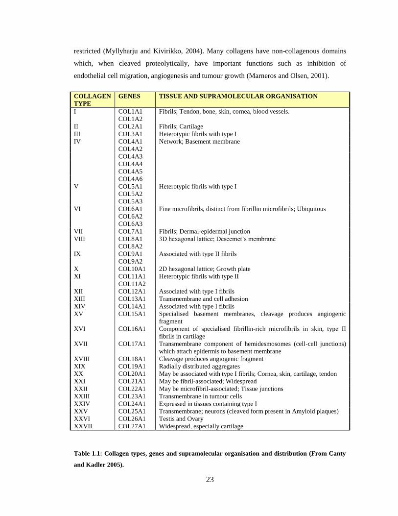
1.3.1 Collagen: Classification
Collagen is relatively inert, having a half life of 300-500 days, although this varies with tissue
(Neuberger and Slack, 1953) and may be much longer (Thorpe et al., 2010). To date, there
have been twenty seven collagens identified (Table 1.1) and they contribute to 30% of protein
mass in the human body (Canty and Kadler, 2005). They have many important functions such
as maintaining the structure of tissue, imparting tensile strength, cell adhesion, chemotaxis,
cell migration and regulation of matrix biology in growth, healing and disease (Myllyharju and
Kivirikko, 2004). Some collagens have a wide tissue distribution while others are very
Page 23
23
restricted (Myllyharju and Kivirikko, 2004). Many collagens have non-collagenous domains
which, when cleaved proteolytically, have important functions such as inhibition of
endothelial cell migration, angiogenesis and tumour growth (Marneros and Olsen, 2001).
COLLAGEN
TYPE
GENES TISSUE AND SUPRAMOLECULAR ORGANISATION
I COL1A1 Fibrils; Tendon, bone, skin, cornea, blood vessels.
COL1A2
II COL2A1 Fibrils; Cartilage
III COL3A1 Heterotypic fibrils with type I
IV COL4A1 Network; Basement membrane
COL4A2
COL4A3
COL4A4
COL4A5
COL4A6
V COL5A1 Heterotypic fibrils with type I
COL5A2
COL5A3
VI COL6A1 Fine microfibrils, distinct from fibrillin microfibrils; Ubiquitous
COL6A2
COL6A3
VII COL7A1 Fibrils; Dermal-epidermal junction
VIII COL8A1 3D hexagonal lattice; Descemet’s membrane
COL8A2
IX COL9A1 Associated with type II fibrils
COL9A2
X COL10A1 2D hexagonal lattice; Growth plate
XI COL11A1 Heterotypic fibrils with type II
COL11A2
XII COL12A1 Associated with type I fibrils
XIII COL13A1 Transmembrane and cell adhesion
XIV COL14A1 Associated with type I fibrils
XV COL15A1 Specialised basement membranes, cleavage produces angiogenic
fragment
XVI COL16A1 Component of specialised fibrillin-rich microfibrils in skin, type II
fibrils in cartilage
XVII COL17A1 Transmembrane component of hemidesmosomes (cell-cell junctions)
which attach epidermis to basement membrane
XVIII COL18A1 Cleavage produces angiogenic fragment
XIX COL19A1 Radially distributed aggregates
XX COL20A1 May be associated with type I fibrils; Cornea, skin, cartilage, tendon
XXI COL21A1 May be fibril-associated; Widespread
XXII COL22A1 May be microfibril-associated; Tissue junctions
XXIII COL23A1 Transmembrane in tumour cells
XXIV COL24A1 Expressed in tissues containing type I
XXV COL25A1 Transmembrane; neurons (cleaved form present in Amyloid plaques)
XXVI COL26A1 Testis and Ovary
XXVII COL27A1 Widespread, especially cartilage
Table 1.1: Collagen types, genes and supramolecular organisation and distribution (From Canty
and Kadler 2005).
Page 24
24
1.3.2 Collagen: Structure
Collagens are trimers of polypeptide chains, called α-chains. Each chain comprises a repeating
Gly-A-B triplet, where A and B can be any residue, but usually proline and hydroxyproline
respectively (Myllyharju and Kivirikko, 2004). This triplet motif results in a left-handed helix
that can intertwine with two others to form a right-handed triple helix structure (van der Rest
and Garrone, 1991).
Collagen fibrils are the principle source of tensile strength in mammalian tissue. They have a
67nm axial periodicity, are up to several millimetres long and have a wide range of widths
from a few nanometers to 500nm (Canty and Kadler, 2005). They have a wide variety of
tissue-specific arrangements, and in tendon and ligament they are organised in parallel arrays
(Boot-Handford et al., 2003, Myllyharju and Kivirikko, 2004). In tendon, ligament, skin and
bone, type I predominates and in cartilage type II is most abundant. Fibrils are usually
heterotypic which is achieved through different α chains in a molecule: the six chains of type
IV form at least three different molecules (Borza et al., 2001).
1.3.3 Collagen: Assembly
Formation of the collagen fibril begins with the synthesis of polypeptide chains on membrane
bound ribosomes and secreted into the lumen of the endoplasmic reticulum (Myllyharju and
Kivirikko, 2004). Here the main steps in biosynthesis take place:
1) Cleavage of the signal peptides
2) Hydroxylation of certain proline and lysine residues
3) Glycosylation of some of the hydroxylysine residues
4) Glycosylation of certain asparagine residues in the C, or C and N terminal propeptides
5) Association of three C propeptides, by specific recognition sequences
6) Formation of intra- and inter-molecular disulphide bonds
The C propeptide association forms the nucleus for triple helix formation, and following
hydroxylation of around 100 proline residues, the triple helix is propagated toward the N-
terminus (Canty and Kadler, 2005). The procollagen molecules are transported from the
endoplasmic reticulum through the Golgi apparatus where they aggregate to form secretory
vesicles. Following secretion, the N and C propeptides are cleaved and the molecules
Page 25
25
spontaneously self-assemble into fibrils and form covalent crosslinks (Canty and Kadler,
2005).
1.3.4 Collagen: Crosslinks
Following fibril formation, crosslinking imparts mechanical strength and stability to the
fibrils. The pattern of crosslinking varies between tissue and collagen types, and the pattern of
strain and pathology of that tissue (Eyre et al., 1984, Paul and Bailey, 1996). There are three
types: immature enzymatic, mature enzymatic and non-enzymatic (Kielty et al., 1993,
Yamauchi et al., 1988, Kuypers et al., 1992).
1.4 Elastin, fibrillin and the elastic fibres
1.4.1 Overview
Although collagen provides tensile strength to ligament, other components may contribute to
the overall mechanical function of the complex (Frank, 2004, Ujiie et al., 2008). Elastin fibres
comprise a central cross-linked core of highly extensible elastin surrounded by a supporting
sheath of microfibrils, with many other associated molecules (Kielty, 2006). Microfibrils
(MFs) are polymers of fibrillins 1 and 2 and are considered to have a structural role in
ligament and tendon. Bundles of MFs are known as oxytalan fibres. Collectively, oxytalan and
elastin fibres are referred to as elastic fibres. Elastin has traditionally been considered a minor
component of ligament tissue (Frank, 2004).
Elastic fibres have important mechanical, biochemical and cell-regulatory functions in tissue
namely vascular and connective tissues such as intervertebral disc (Kielty, 2006). Reversible
elasticity is a function of both elastin and oxytalan fibres (Eriksen et al., 2001) where elastic
fibre distribution is considered to reflect function (Kielty et al., 2002a). MFs may have a key
role in the extracellular regulation of transforming growth factor (TGF) β (Charbonneau et al.,
2004, Feng and Derynck, 2005) as well as regulation of cell-matrix interactions (Ito et al.,
1997, Wendel et al., 2000).
A wide distribution of elastic fibres in the human ACL has been described, with abundant
elastin fibres and oxytalan fibres running with collagen bundles described using electron
microscopy (Strocchi et al., 1992). In canine CLs, elastin fibres have only been reported at low
levels (Paatsama, 1952, Vasseur et al., 1985). The presence of oxytalan fibres or microfibrils
Page 26
26
has not been determined in the canine CL complex and the role of elastin fibres in the CL
complex has not been determined in any species.
1.4.2 Molecular composition
1.4.2.1 Elastin core
Elastin is the most abundant component of elastic fibres (around 90% of the fibre) and has a
very low turnover in healthy tissue (Petersen et al., 2002, Kielty, 2006). In man, coding for
elastin is by a single copy gene on chromosome 7q11.2 and secreted as a 65-70kDa soluble
tropoelastin precursor (Tamburro et al., 2005). Tissue specific functional properties may be
achieved by splice variants of the original transcript (Kielty, 2006). Tropoelastin has a multi-
domain structure with repeating hydrophobic and lysine-rich crosslinking domains each
encoded by separate exons (Figure 1.2). The unique C-terminal domain contains two cysteine
residues and may play a key role in elastin assembly. Crosslinked elastin is formed under the
direction of lysyl oxidase (LOX) and other members of this enzyme family (Szauter et al.,
2005). Proteoglycans, including biglycan, have been detected in the core, and heparan sulphate
may also be present (Kielty, 2006).
Page 27
27
Figure 1.2: Domain structures of elastin, fibrillin-1 and fibulin-5. Abbreviations: calcium
binding epidermal growth factor (cbEGF), Arg-Gly-Asp peptide sequence (RGD), eight-
cysteine-containing motif (TB, also known as thrombospondin motif), epidermal growth
factor (EGF). Adapted from Kielty 2006
1.4.2.2 Microfibrils
The structural framework of microfibrils (MFs) is comprised of fibrillins. These are large
multi-domain glycoproteins of around 350kDa, and comprise 47 epidermal growth factor-like
domains (EGF-like); 43 of which are calcium-bonding epidermal growth factor (cbEGF)-like
domains (Figure 1.2). These domains are interspersed with 7 eight-cysteine-containing (TB)
motifs and two hybrid motifs containing elements of both EGF-like and TB. There are 14-N-
glycosylation sites, and the N terminus is a unique cysteine-containing motif (Kielty et al.,
Signal
Peptide *
Domain affected by
transcript
alternative splicing
KP-rich
crosslinking
domain
KA-rich
crosslinking
domain
Hydrophobic
domain C-terminal
sequence
Fibrillin-1
Human Tropoelastin
RGD
Fibulin-5
TB Motif Hybrid 8-
Cys Motif
cbEGF-like
Domain
Fibulin C-
terminal
module
EGF-like
Domain
Terminal
Sequence
Proline-rich
Domain
RGD Cell
Adhesion
Motif
2 4 6 8 10 12 14 16 18 20 22 24 26 26a 28 30 32 36
* * * * * * * *
Page 28
28
2005). In man there are three fibrillin isoforms with fibrillins 1 and 2 having partially
overlapping expression patterns. Fibrillin 1 is expressed throughout life and fibrillin 2 is
expressed mainly in foetal tissues (Zhang et al., 1994, Charbonneau et al., 2003). Fibrillins 1
and 2 have been shown to co-localise within MFs and may overlap in function (Carta et al.,
2006). There are many MF associated molecules, of which microfibril associated glycoprotein
MAGP-1 is the most likely to have a structural component (Cain et al., 2006). (MAGP)-2 and
latent TGF-β binding protein (LTBP)-1 also co-localise in certain tissues (Kielty, 2006).
1.4.2.3 Elastic fibre interface molecules
A number of molecules are found at the pericellular-elastic fibre or microfibril-elastin
interface and are listed in Table 1.2 (Reinhardt et al., 1996, Isogai et al., 2002). Collagen type
VIII may also co-localise in vascular tissues forming hexagonal basement membrane
associated networks.
Fibulin-5 is expressed by vascular smooth muscle cells and endothelial cells. It is a
glycoprotein of around 55 kDa containing five cbEGF-like domains and an Arg-Gly-Asp
(RGD) motif (Figure 2). It is involved in mediation of vascular cell adhesion through integrin
receptors, smooth muscle cell proliferation and migration and in regulation of elastic fibre
formation (Chu and Tsuda, 2004). Its expression is down-regulated in adult arteries, but
markedly up-regulated in vascular cells following injury, angioplasty in neointimal cells, and
in atherosclerotic cells. It is localised to the elastic lamina surfaces adjacent to endothelial cells
and throughout the aortic media. Fibulin-4 is another factor essential for elastogenesis
(McLaughlin et al., 2006)
Page 29
29
Molecule Elastic Fibre Location
Fibrillin-1 Microfibrils
Fibrillin-2 Microfibrils
Fibrillin-3 Unknown – microfibrils likely
MAGP-1 Microfibrils
MAGP-2 Some microfibrils
LTBP-1 Some microfibrils; also fibronectin
LTBP-2 Microfibrils, elastic fibres
LTBP-3 Fibrillar structures
LTBP-4 Fibrillar structures, fibrillin
Decorin Microfibrils, microfibril-elastic fibre interface
Biglycan Elastic fibre core
Versican Some microfibrils
Heparin Sulphate Microfibrils, elastic fibre core
Perlecan Microfibrils
MFAP-1 Some microfibrils
MFAP-3 Some microfibrils
MFAP-4 (MAGP-36) Some microfibrils
βlgH3 Elastic fibre-collagen interface
Tropoelastin Elastic fibre core
LOX Newly secreted tropoelastin, microfibril-elastin interface
LOXL Microfibril-fibulin-5-elastin interface
Fibulin-1 Elastic fibre core
Fibulin-2 Elastin-microfibril interface
Fibulin-4 Unknown – likely in elastic fibre core
Fibulin-5 Elastic fibre-cell interface
Emilin-1 Elastin-microfibril interface
Emilin-2 Elastin-microfibril interface
Elastin binding protein Newly secreted tropoelastin
Vitronectin Some microfibrils in dermal tissues
Amyloid Some microfibrils in dermal tissues
Collagen VIII Vascular elastic tissues
Collagen XVI Dermal microfibrils
Endostatin (C-terminus of
collagen XVIII
Vascular elastic fibres
Collagen VI Some microfibrils
Table 1.2: Structural and associated molecules of microfibrils and elastic fibres.
(Adapted from Kielty 2006). Abbreviations: βlgH3, also known as transforming growth
factor-β-inducible gene-H3 and as keratoepithelin; LOX, lysyl oxidase; LOXL, lysyl
oxidase-like; LTBP, latent transforming growth factor-β-binding protein; MAGP,
microfibril associated glycoprotein; MFAP, microfibrillar associated protein.
Page 30
30
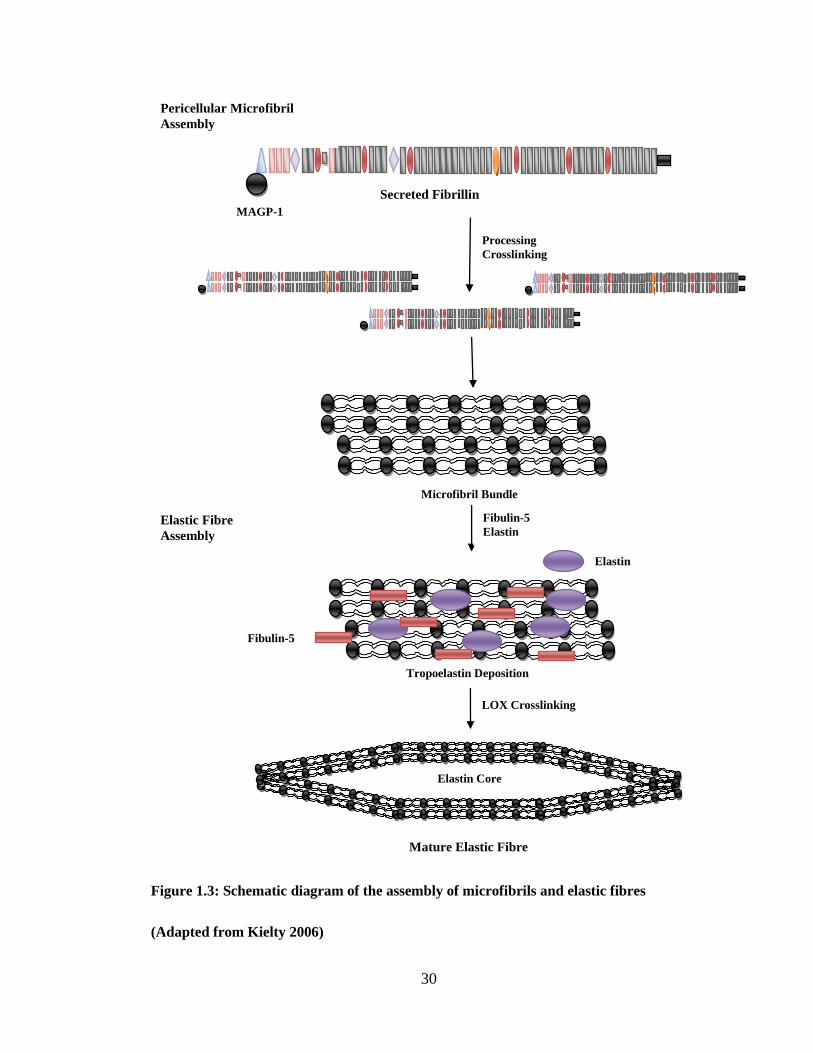
Figure 1.3: Schematic diagram of the assembly of microfibrils and elastic fibres
(Adapted from Kielty 2006)
Secreted Fibrillin
MAGP-1
Processing
Crosslinking
Microfibril Bundle
Pericellular Microfibril
Assembly
Fibulin-5
Elastin Elastic Fibre
Assembly
Elastin
Fibulin-5
Tropoelastin Deposition
LOX Crosslinking
Elastin Core
Mature Elastic Fibre
Page 31
31
1.4.3 Elastic fibre structure and assembly
1.4.3.1 Microfibril structure and assembly
Pericellularly secreted fibrilin-1 molecules assemble into beaded microfibrils linearly and
laterally through specific N- and C-terminal interactions (Figure 1.3) (Cain et al., 2006,
Kinsey et al., 2008, Ashworth et al., 1999a). The assembled microfibrils are then stabilised by
transglutaminase crosslinks. Each microfibril probably has eight fibrillin-1 molecules in cross
section (in Figure 1.3 only two are shown for clarity).
Microfibril assembly involves interaction with many different molecules. MAGP-1 strongly
bonds at the N-terminal controlling N- and C-terminal interactions (Ramirez and Sakai, 2009).
Heparin sulphate may have an in vivo role in regulating microfibril assembly by competing
with MAGP-1 at the N-terminus and with tropoelastin at one of the central sites. Fibrillin-1
has been shown to interact with other molecules such as LTBP-1, fibulin-2, versican and with
small chondroitin sulphate proteoglycans.
Microfibrils have been shown to be repeating globules on filamentous linear arrays using
atomic force microscopy (Baldock et al., 2001, Sherratt et al., 2001), but more recent work has
suggested in the physiological state they are cylindrical structures rather than beaded arrays
and have inter-microfibril links (Davis et al., 2002).
1.4.3.2 Elastin fibre structure and assembly
Elastin fibres are deposited in early post-natal life in elastic connective tissues such as skin,
lung, ligaments, articular cartilage, aorta and other elastic arteries (Kielty et al., 2005, Kielty et
al., 2002b, Mithieux and Weiss, 2005). Tropoelastin is deposited into a fibrillin microfibril
bundle template in the extracellular space (Czirok et al., 2006, Kozel et al., 2006). Elastin first
appears inside microfibril bundles before coalescing to form the crosslinked elastin core of
mature elastic fibres (Pasquali-Ronchetti and Baccarani-Contri, 1997, Kielty et al., 2002b).
Elastin is then stabilised through formation of the elastin-specific crosslinks desmosine and
isodesmosine to its insoluble form (Mithieux and Weiss, 2005). The widely accepted model is
shown in Figure 1.3. Real time microscopy on an in-vitro cell culture has shown elastin
globules, possibly associated with microfibrils may aggregate to form larger fibrillar structures
(Kozel et al., 2006, Czirok et al., 2006). This process is coupled to cell motion, possibly
through integrin bonding at the C-terminal and is known as coacervation (Ostuni et al., 2007).
Page 32
32
Although mature elastic fibres have an outer mantle of microfibrils, some microfibrils appear
to lie within the elastin core (Kielty, 2006).
Many other molecules have been associated with elastin fibres (Table 1.2). Lysyl oxidase
(LOX) and a second isoform lysyl oxidase-like (LOXL) play significant roles in the
integration process by mediating enzyme and tropoelastin binding (Thomassin et al., 2005).
Cell binding may also play a role in fibre assembly (Broekelmann et al., 2005). There are two
high-affinity binding sites for tropoelastin in fibrillin-1 but elastin also interacts with other
molecules including MAGP-1 and biglycan (Figure 1.3). Fibulins 4 and 5 have critical but
poorly understood roles in vascular elastic fibre formation in mice (Yanagisawa et al., 2002,
Nakamura et al., 2002, McLaughlin et al., 2006). Fibulin-4 may be required to recruit LOX to
facilitate tropoelastin crosslinking (Horiguchi et al., 2009). Fibulin-5 co-localises and binds
with LOXL and may also have a role in elastin crosslinking (Yanagisawa et al., 2002).
1.4.4 Organisation of elastin fibres in tissue
The functional properties of elastin fibres strongly reflect the tissue-specific architecture
(Kielty, 2006). In turn, this is dictated by the organisation of the microfibril template, the
orientation of the cells that deposit them and the forces acting on the tissue. In the lung, elastic
fibres are present throughout the respiratory tree in a fine highly branched network (Kielty et
al., 2002b). In the aorta and elastic arteries, the elastic fibres form concentric fenestrated
lamellar layers that intercalate with the smooth muscle. In the developing aorta, subendothelial
microfibrils provide anchorage for endothelial cells and are aligned with the flow of blood
(Davis, 1993a, Davis, 1993b). The skin contains an elastic fibre network varying from
microfibril bundles at the dermal epidermal junction, elaunin fibres (small amounts of elastin)
in the papillary dermis to thick horizontal elastic fibres in the reticular dermis (Kielty, 2006).
Regions of tendon that undergo the greatest flex and strain deformation have the highest
elastin content (Ritty et al., 2002). In ligament, elastin is abundant running parallel to collagen
fibrils, but sparse in tendon. In cartilage, fibres surround chondrocyte lacunae and form a thin
mesh running with interterritorial collagen fibrils (Kielty, 2006).
Thus the precise mechanical role of elastic fibres is thought to be a function of their chemistry,
ultrastructure, arrangement and collective density relative to other ECM constituents
Page 33
33
1.4.5 Elastic fibre functions
Elastic fibres have three recognised functions: 1) elastic recoil and resilience in dynamic
connective tissue, 2) regulation of the activity of the TGF-β family of growth factors and 3)
regulation of cell migration, survival and differentiation.
1.4.5.1 Elasticity
Elastin fibres comprised of both microfibrils and cross-linked elastin were an essential
evolutionary advance to support vertebrate high-pressure circulatory systems and other elastic
functions (Faury, 2001). Elastin fibres influence mechanical properties such as resilience and
low-strain stiffness (Kielty et al., 2002c). Deformation of elastin acts as an energy store that is
then used to drive recoil to the resting state (Gosline et al., 2002). Elastin is extremely
insoluble due to extensive lysyl-derived crosslinks, and the crosslinked elastin core of the fibre
provides the major contribution to tissue elasticity (Mithieux and Weiss, 2005).
Individual microfibrils and microfibril bundles also have elastic function (Sherratt et al.,
2003). Individual microfibrils were shown to have a Young’s modulus approximately two
orders of magnitude stiffer than elastin and are highly resistant to axial tension (Glab and
Wess, 2008). Elasticity in microfibril-containing tissue may arise primarily from reversible
alterations in microfibril bundle reorganisations while individual microfibrils might act as
reinforcing fibres (Sherratt et al., 2003). Untensioned microfibrils have a regular periodicity of
approximately 56 nm, but isolated extended microfibrils up to 150nm have also been observed
(Keene et al., 1991, Baldock et al., 2001). One study suggested a reversible extension of 56 to
100nm with irreversible extension above this (Baldock et al., 2001). Calcium and water are
important in reversible elasticity (Haston et al., 2003, Wright et al., 1999).
1.4.5.2 TGF-β family activation
Fibrillin microfibrils may play a key role in the extracellular regulation of TGF-β activation
and signaling (Charbonneau et al., 2004). The TGF-β family of growth factors are powerful
regulators of cell survival, proliferation and differentiation, tissue morphogenesis and cellular
responses to injury (Feng and Derynck, 2005). It remains unclear precisely how microfibrils
regulate TGF-β. Current ideas include structural relationships with LTBPs, emilin-1 and
possibly direct growth factor binding (BMPs).
Page 34
34
1.4.5.3. Cell adhesion
Elastic fibres play an important role in cell-matrix interactions in elastic connective tissues.
Interactions are mediated mainly through integrins, which are transmembrane receptors, that
recognise RGD motifs within matrix molecules and link the vascular ECM directly to the
cellular cytoskeletal framework (Mould and Humphries, 2004). Such integrin-mediated cell-
elastic fibre interactions influence cell survival, phenotype, proliferation, migration and ECM
expression and deposition. Major elastic fibre molecules involved are
a) Elastin: Cells bind through elastin binding protein which binds the tropoelastin hexapeptide
VGVAPG. Interactions of VGVAPG peptides with this G-protein-coupled receptor stimulate
actin polymerisation thereby influencing cell proliferation and migration (Karnik et al., 2003a,
Karnik et al., 2003b). Certain elastin proteolytic fragments are highly chemotactic.
b) Fibrillin-1: A single RGD motif on fibrillin-1 mediates adhesion, cell behaviour and gene
expression through integrin binding of human smooth muscle (Kielty, 2006). An RGD motif
in fibrillin-2 is similarly active.
c) Fibulin-5: Fibulin-5 interacts directly with vascular cells in a RGD and cation-dependent
manner which may contribute to its roles in elastic fibre deposition and modulation of smooth
muscle phenotype (Chu and Tsuda, 2004).
1.4.6 Elastic fibre production and degradation
Fibrillins -1 and -2 are produced at low levels throughout life in human skin (Ashcroft et al.,
1997). Elastin production peaks near birth and is nearly completely repressed by maturity
(Perrin and Foster, 1997). Production is repressed in the adult cell mainly through post-
transcritpional regulation. Expression of tropoelastin can be promoted (IL-1β, IL-10, IGF-1) or
repressed (TNF-α, bFGF) at the transcriptional level, as well as being promoted through
stabilisation at the post-transcriptional level (TGF-β1) (Duca et al., 2004). Strong evidence
now exists that EFs are not replaced in adult life, and must function for the lifetime of the
organism (Sherratt, 2009). As a result of this longevity, EFs are vulnerable to age-related
damage (Ritz-Timme et al., 2003).
Crosslinked elastin is highly insoluble and resistant to degradation. However insoluble elastin
is readily degraded by the serine proteinases (Novinec et al., 2007) and matrix
metalloproteinase (MMP) classes especially MMPs -2, -7, -9 and -12 and neutrophil elastase
Page 35
35
(Mecham et al., 1997, Ashworth et al., 1999b). Furthermore, EF morphology may be
significantly altered through the action of MMP-14, leading to a loss of mechanical function
(Sherratt, 2009). Elastin peptides produced enzymatically are able to influence the behaviour
of a wide variety of cells including fibroblasts, macrophages and neutrophils (Duca et al.,
2004). Elastin peptides are transduced through the elastin laminar receptor 1 (ELR1), a spliced
variant of β-galactosidase (Hinek et al., 1993). Peptides containing VGVAPG and GXXPG
sequences have been shown to bind to ELR1 (Brassart et al., 2001, Duca et al., 2004). They
can directly influence chemotaxis, proliferation, protease release and even induce apoptosis
(Duca et al., 2004, Privitera et al., 1998). Elastin degradation products and TGP-β1 promoted
myofibroblastic and osteogenic differentiation in dermal fibroblasts (Simionescu et al., 2007).
Degraded elastin fragments have been suggested to contribute to the degenerative cascade
through activation of matrix MMPs (Yu et al., 2007).
1.5 Proteoglycans (PG)
PGs make up around 1% of the dry weight of ligaments (Frank, 2004). PGs are proteins
containing one or more glycosaminoglycan (GAG) chains. Most exist as aggregates and are
non-covalently bound to a long chain of hyaluronate with link-protein (Heinegard and Hascall,
1974) They can also be small and non-aggregating.
1.5.1 Large aggregating PGs
The large aggregating PGs create a large osmotic swelling within cartilage, creating a water-
swollen matrix critical to the properties of cartilage (Kiani et al., 2002). Aggrecan is the most
abundant PG in cartilage and is required for chondrocyte survival and attachment in vitro (Lee
et al., 2000). Versican is another important large proteoglycan with a wide range of actions.
Versican forms an integral part of a pericellular matrix that organises the tendon cells in linear
arrays between collagen fascicles (Ritty et al., 2003). It has a significant role in regulating cell
phenotype (Kinsella et al., 2004) and may be involved in chondrogenic change in tissue
(Zhang et al., 2001, Erdelyi et al., 2005).
In human Achilles tendon aggrecan mRNA was expressed in fibrocartilage and versican
mRNA in the tendon midsubstance. This finding suggests the differing expression is a result
of differing forces on the tendon: tension in the substance and compression in the insertion
(Waggett et al., 1998). Versican expression in tendon has been shown to be site specific;
predominant in tensile regions, while aggrecan is seen more in fibrocartilaginous regions or
Page 36
36
where tendon wraps around bone (Waggett et al., 1998, Robbins and Vogel, 1994). The
formation of fibrocartilage in ligaments and tendon has been characterised by the upregulation
of aggrecan, versican, biglycan and type II collagen (Benjamin and Ralphs, 1998, Vogel and
Meyers, 1999). Versican has been found to interact with fibrillin-1 and co-localisation of
versican with microfibrils within the interlamellar space has been noted. Versican, along with
other proteoglycans such as decorin and biglycan may play a critical role in the integration of
microfibrils into the ECM (Kielty et al., 2002a).
1.5.2 Non-aggregating PGs
Small leucine-rich proteoglycans: This family of PGs are involved in the organisation of
tissue through orientating and ordering collagen fibrils and elastic fibres during ontogeny,
wound healing, tissue repair and tumour stroma formation (Iozzo, 1997). They are
characterised by a core protein dominated by leucine-rich repeats, although they are
genetically distinct (Heinegard, 2009, Neame et al., 2000). Decorin, biglycan and
fibromodulin are all associated with elastic fibre assembly (Kielty, 2006).
Heparan sulphate proteoglycans: These may be involved in mediating interactions between
cells and matrix molecules (Hook et al., 1986). One member, perlecan, is found in the
basement membrane and may play a role in charge selectivity in glomerular filtration and is
associated with microfibrils (Yanagishita, 1993).
1.6 Glycosaminoglycans (GAGs)
GAGs are generally comprised of repeating disaccharide units containing an uronic acid
(glucuronic acid or iduronic acid) and an N-acetylated sugar (N-acetyl glucosamine or N-
acetyl galactosamine) (Funderburgh, 2000, Trowbridge and Gallo, 2002). At least one of the
repeating units has a carboxylate or sulphate group, except in hyaluronan, which is not
sulphated. They are synthesised intracellularly and sulphated prior to excretion.
Hyaluranon: Hyaluranon serves not only as a backbone for the large aggregating PGs, but aids
in tissue hydration and reducing friction in moving surfaces. The smaller glycoproteins bind
and link collagens to the cell surface and may also have an important role regulation of growth
factors.
Chondroitin Sulphate (CS): This GAG is involved in articular and bone metabolism by
controlling cartilage matrix integrity and bone mineralisation. It is comprised of disaccharide
Page 37
37
repeats of glucuronic acid and N-acetyl glucosamine. The sulphate is present in every carbon 4
or 6 of the galactosamine of every disaccharide. Each side chain is 40-50 disaccharides,
although there is some tissue variation. The disaccharide chain is O-linked to protein core via
a serine-xylose-galactose-galactose structure (Bali et al., 2001).
Dermatan Sulphate (DS): This is a variant of chondroitin sulphate in which some of the
glucuronic acid residues have become epimerised to iduronate. DS containing proteoglycans
(DS-PG) are distributed in the ECM of tendon, cartilage, other connective tissues, skin and
sclera (Rosenberg et al., 1986).
Keratin Sulphate (KS): KS comprises disaccharide repeats of galactose and N-acetyl
glucosamine. Glucosamine is sulphated, but additional groups may be present. The length of
the side chains varies. KS differs from other GAGs in that it does not contain an uronic acid.
1.7 Other ECM components
Water forms two thirds of a ligament and is essential for cell and matrix function, lubrication
and viscoelastic properties (Amiel et al., 1995a).
EXTRACELLULAR MATRIX PHYSIOLOGY
The ECM undergoes continuous synthesis and degradation of its components. The balance
achieved can reflect the mechanical and physiological environment as well as any tissue
pathology. ECM degradation can be physiological in the case of uterine and mammary gland
involution or adaptation of connective tissues to mechanical environment (Alexander and
Werb, 1991). Degradation occurs in many pathological processes, and is a complex enzymatic
process (Ries and Petrides, 1995). ECM remodeling is a fine balance between activation and
inhibition of proteolytic and synthetic enzymes (Mauviel, 1993).
Proteolytic enzymes are the primary cause of ligament, cartilage and subchondral bone
degeneration in joint disease (Cawston, 1998). Based on the amino acid or chemical group at
the catalytic domain, the proteinases can be divided into four main groups, the
metalloproteinases and the cysteine-, aspartate and serine-dependent proteases (Barrett, 1994).
Collagen is very resistant to proteolytic attack. As crosslinks and close association with
proteoglycans make it a poor target. Lysis is thought to begin extracellularly at a neutral pH
through the action of the serine and metalloproteinases. This renders it susceptible to
Page 38
38
proteolytic attack from other enzymes or phagocytosis and intracellular destruction (Klein and
Bischoff, 2010). In this introduction we will concentrate on the MPs and cysteine proteases.
1.8 Metalloproteinases (MP)
MPs are part of a superfamily of zinc dependent proteases known as metzincins named after
the conserved Met residue and zinc ion at the active site (Klein and Bischoff, 2010). Although
there are many subfamilies, the matrix metalloproteinases, membrane-type MMPs and
adamalysins are the most relevant in connective tissue.
1.8.1 Matrix metalloproteinases (MMPs)
MMPs are grouped by common properties as they share common sequences of amino acids,
are secreted by inactive zymogens and are activated either proteolytically or by mercurial
agents (Cawston, 1998). They are categorised into five subgroups:
a) Collagenases (1, 8 and 13),
b) Gelatinases (2 and 9),
c) Stromelysins (3, 10 and 11),
d) Membrane type-matrix metalloproteinases (MT-MMPs) (14-17, 24 and 25),
e) A heterogenous group including matrilysin (7) and macrophage metalloelastase (12).
The important groups in ligament ECM turnover are the collagenases, gelatinases,
stromelysins and MT-MMPs (Harper et al., 1989, Foos et al., 2001). Regulation of MMPs is a
complex and poorly understood interaction of inflammatory cells, cytokines, growth factors
and ECM molecules. Regulation of MMPs can occur at several stages including transcription,
activation of proMMPs (Murphy and Docherty, 1992) and inhibition of active MMPs (Nagase,
1997).
Gelatinases (MMP-2 and -9) can cleave collagens type IV, V, VII and X, denatured collagens
(gelatins) and elastin, but not fibronectin or laminin (Tyagi, 1997). MMP-2 is produced by all
connective tissue cells making it the most widespread of all MMPs (Murphy and Crabbe,
1995). It is thought to be involved in ECM turnover through remodeling or removal of
denatured collagen (Creemers et al., 1998, Kerkvliet et al., 1999, Yoshizaki et al., 2002,
Morgunova et al., 2002). MMP-9 is of a similar structure to MMP-2 and is expressed in
inflammation, angiogenesis and tissue repair (Tarlton et al., 1997).
Page 39
39
1.9 Cysteine proteinases
These are subdivided into around 20 families, the main ones being calpain and papain. In
inflammation, macrophages secrete cathepsins B, K, L and S, which are also identified as
major damagers to tissue in chronic inflammation (Kielty et al., 2002b). Studies have
implicated K, L and S in extensive degradation of elastic fibres that accompanies
cardiovascular disease in man. Cathepsins have been shown to degrade elastin, and have
preferences for particular tissue types (Novinec et al., 2007). Cathepsin S has a critical role in
antigen presentation (Muir et al., 2005b). Cathepsin K has been associated with ACL rupture,
and its expression appears to precede rupture (Barrett et al., 2005, Muir et al., 2005b, Muir et
al., 2006). Caspases are another important family of cysteine proteases that play important
roles in apoptosis, development and inflammation (Martinon et al., 2009).
1.10 Serine Proteases
These are the largest family of endopeptidases and are released from inflammatory cells
(Milner et al 2001). The most important are those of the plasminogen/ plasminogen activator
(PA) system (Murphy et al., 1993). Other serine proteases include cathepsin G, primarily
intracellular in neutrophils and macrophages, and neutrophil elastase from neutrophils and
active extracellularly, with broad spectrum action on collagen type III, elastin, PG, vitronectin
and laminin (Doring, 1994).
1.11 Other collagenolytic agents
During chronic inflammation, reactive oxygen species generated by tartrate-resistant acid
phosphatase (TRAP) as well as MMPs are important components of leucocyte-mediated
collagenolysis. TRAP can also fragment the triple helix of collagen through generation of
reactive oxygen species by the Fenton reaction (Muir et al., 2005b). Cysteine proteases, such
as K, have a regulatory role in TRAP degradative activity by converting TRAP5a to the more
active TRAP5b. Cathepsin K and TRAP cells have been co-localised within the ACL and are
associated with ACL disease (Muir et al., 2002).
Page 40
40
1.12 Role of proteinases
1.12.1 Function and mode of action
Traditionally, MMPs have been thought of as enzymes for degradation of structural
components of the ECM. However, proteolysis can have other functions (Page-McCaw et al.,
2007):
Creation of space for cell migration
Production of substrate-specific cleavage fragments with independent biological activity
Regulation of tissue architecture through effects on ECM and intercellular junctions
Direct and indirect activation, deactivation and modification of signaling molecules
1.12.2 Involvement of proteinases in degenerative conditions
There is considerable evidence for the role of proteases in joint diseases. Proteases have an
important role in canine, equine and human OA (Wernicke et al., 1996, Clegg et al., 1997,
Fernandes et al., 1998, Riley et al., 2002). Proteases are also important in diseases affecting
ligaments and tendons. Elevated levels of proteases have been found in knee synovial fluid in
ACL damage, meniscal damage, post-traumatic OA and pyrophosphate arthritis (Lohmander
et al., 1993). The elevation occurs within days and can remain elevated for up to 18 years
(Lohmander et al., 1994). However an elevation in proteases has also been noted in older
animals without ACL damage (Muir et al., 2005a). MMP-2 has been implicated in the
turnover of the periodontal ligament (Creemers et al., 1998). In the rabbit ACL an increase in
collagenase activity has been correlated with a loss of collagen, a loss of cellularity and matrix
disorganisation (Amiel et al., 1989). An increase in MMP-2 and -9 was seen in the ruptured
ACL (Muir et al., 2005a) but not in an earlier study (Spindler et al., 1996b). An increase in
proMMP-2 in the intact ACLs of a dog breed at high risk of ACL rupture was seen when
compared to a dog breed with a very low risk to ligament rupture (Comerford et al., 2005). An
association between elevation in proMMP-2 and AP laxity has been suggested (Comerford et
al., 2005, Quasnichka et al., 2005). Mechanical stress can induce increased production of
MMPs 2 and 9 and acid cathepsins in affected tissue in Dupuytrens disease which may
contribute to tissue weakening (Tarlton et al., 1998).
Page 41
41
CURRENT THOUGHTS ON THE AETIOPATHOGENESIS OF CANINE CRUCIATE
LIGAMENT RUPTURE
1.13 CL idiosyncrasies
The CLs are unique for many reasons. Their trans-articular location is shared only by one
other ligament in the body, the ligament of the femoral head also known as the teres ligament.
However, although both these ligaments experience multiaxial stresses, only the CL complex
is subject to intrinsic compression where the ACL wraps around the PCL (Arnoczky and
Marshall, 1977). The transarticular location means the soft tissue support of the CL complex is
minimal, comprising a thin layer of epiligamentous tissue (Clark and Sidles, 1990). As blood
supply to the CL complex arises from the epiligament, the blood supply is also subject to
compromise through epiligament damage or compression (de Rooster et al., 2006).
Differences exist in the properties of the ACL and MCL on the knee in many aspects such as
response to loading and injury (Amiel et al., 1984, Amiel et al., 1995b, Lo et al., 2002b).
1.14 Histologic and ultrastructural changes in CL disease
1.14.1 Histologic overview
An early study of partially and completely ruptured ACLs showed an absence of bundle
structure and hyalinisation at the point of ligament failure (Paatsama, 1952). A later study
demonstrated similar changes in dogs over 8 years of age without rupture of the ACL with
calcium deposits, hyalinisation and loss of normal fibre architecture described (Zahm, 1965).
Subsequent studies have described deterioration in collagen architecture and a
fibrocartilaginous appearance of the matrix, Figure 1.4 (Vasseur et al., 1985, Narama et al.,
1996, Comerford et al., 2006b). Cell changes are characterised by loss of cell density, cells
becoming rounded and undergoing chondroid transformation, and formation of chains of
chondroid cells (Vasseur et al., 1985, Narama et al., 1996, Comerford et al., 2006b).
Enlargement of CL fibroblast nuclei and perinuclear halo formation has been described in
dogs kept under laboratory conditions (Narama et al., 1996), but lack of exercise may have
influenced these results as immobilisation can alter the biochemical composition of connective
tissue including increasing mucopolysaccharide content (Akeson et al., 1973) Changes are
most readily observed at the mid substance where the ACL wraps around the PCL.
Furthermore these changes were associated with a loss of mechanical properties (Vasseur et
al., 1985). Changes in the PCL are also observed and although similar, are less severe
Page 42
42
(Vasseur et al., 1985). Decreased cellularity has been described in the core of ruptured ACL,
with an increase in spheroid cells and a chondroid transformation, and is thought to be driven
by apoptosis (Hayashi et al., 2003b, Krayer et al., 2008, Gyger et al., 2007). A grading system
for these changes has been developed (0=no changes, 1=mild, 2=moderate and 3=severe
degenerative changes) which positively correlated with increasing age (over 5 years) and
increasing weight (over 15kg), but not with gender (Vasseur et al., 1985).
Rounding of cell nuclei
Reduced collagen density
Loss of collagen
architecture
Mineralisation and
fibrocartilage
production
Chondrocytic
change
Figure 1.4: Typical changes associated with degeneration of the ACL. LR, x40, H&E
The fibrocartilaginous change described within the midsubstance of the ACL may represent a
degenerative or adaptive change (Zahm, 1965, Vasseur et al., 1985, Comerford et al., 2006b).
The fibrocartilage is thought to form in response to either the ligament compression as it wraps
around the PCL or from the anteriomedial and posteriolateral bands tightening on each other
(Comerford et al., 2006b). The mechanism for these changes is not understood, but may be a
protective metabolic response to hypoxia or metabolic stress suggested for OA cartilage
(Connor et al., 2001, Schipani et al., 2001, Grimshaw and Mason, 2001), an adaptation to
compressive load (Benjamin and Ralphs, 1998) or a degenerative change preceding ACL
Page 43
43
rupture (Vasseur et al., 1985). Fibrocartilage forms in breeds of dog at risk of ACL rupture
(e.g. LR), in those with a low incidence (e.g. GH) and in older dogs in which the ACL remains
intact (Comerford et al., 2006b, Vasseur et al., 1985, Zahm, 1965). However it may be
produced in response to repair to stress/exercise in the low risk breeds and therefore may be a
positive finding in these dogs (Comerford et al., 2006b).
1.14.2 Cell population changes
Inflammation of the synovium and of the ACL epiligament develops in the early phase of the
naturally occurring disease and may precede knee instability and rupture of the ACL (Muir et
al., 2005b). Osteophyte production is mediated by the synovial macrophage and osteophytes
are often present at the time of surgery, suggesting synovitis precedes ACL rupture (Muir et
al., 2005a). Degenerative changes are often found in the contralateral joint at the time of
surgery. Inflammation of the epiligament in patients with rupture of the ACL has been
demonstrated, with a significantly increased number of cathepsin K+ and TRAP+
macrophage-like cells (Muir et al., 2005b). Synovitis characterised by lymphocytic-
plasmocytic infiltration has been described in a majority of dogs with ACL rupture, with a
mononuclear synovitis present in the remainder (Galloway and Lester, 1995).
Higher numbers of TRAP+ macrophages were seen in ruptured canine ACLs than in intact
canine ACLs or ruptured human ACLs suggesting the pathogenesis of ACL rupture may differ
to human ACL rupture (Barrett et al 2005). In the human ACL, three distinct fibroblast
morphologies, fusiform, ovoid and spheroid, have been described based on nuclear aspect
ratios (Murray and Spector, 1999). Regional variation in their distribution was noted giving
rise to three distinct proximal, middle and distal zones (Duthon et al., 2006). Ovoid, chondroid
cells were noted proximally and distally with abundant spindle-shaped fibroblasts in the
middle part of the PCL. This pattern differed from that described in the dog where rounded
cells were found more commonly in the middle part of the ACL (Vasseur et al., 1985).
Furthermore, fibrocartilage formation was noted in the proximal human ACL where it is in
contact with the intercondylar notch, but not in the middle part of the ACL as seen in the dog
(Duthon et al., 2006, Paatsama, 1952).
1.14.3 Collagen changes
Comparing ruptured ACLs of Labrador retrievers (LRs, a breed of dog with increased risk of
ACL rupture) to ACLs from intact LRs, significant differences were seen in immature
Page 44
44
crosslinks, with the ruptured ACL dogs having a higher concentration. The ratio of immature
to mature was 4:96 in intact and 57:43 in ruptured ACL dogs (Comerford et al., 2004). No
significant differences in total collagen content were seen between intact ACLs and ruptured
ACLs in the LR (Comerford et al., 2004). LRs had a higher proportion of smaller fibres
whereas GH had a broader distribution, and the fibre diameter and pattern of fibre distribution
was significantly different indicating greater turnover of fibrils in the LR, although exercise
may have influenced this result (Comerford et al., 2006b).
In a guinea pig model of spontaneous OA, the Dunkin-Hartley strain develops CL laxity prior
to the onset of OA. It was proposed that this laxity was an initiating event in OA and may
result from increased turnover of collagen (Quasnichka et al., 2006). An increase in type II
collagen as well as altered PG deposition was noted in the midsubstance of the PCL prior to
onset of OA in the Dunkin-Hartley strain (Young et al., 2002). Collagen type has yet to be
studied in the canine CL complex but it is assumed the fibrocartilaginous changes seen will
have an increased collagen type II relative to normal ligament.
1.14.4 GAG and PG changes
In ruptured CLs from LRs, sulphated GAG and total GAG content was significantly higher
when compared to normal ACLs from the same breed (Comerford et al., 2004). However,
comparing the at-risk LR to the GH prior to ACL rupture, there was no significant difference
in total or sulphated GAGs (Comerford et al., 2005).
Little is known of PG changes in disease of the canine CL complex. Healing ligament has
been shown to have higher total and aggregating PGs (Plaas et al., 2000, Bishop and Bray,
1993). The formation of fibrocartilage in ligaments and tendon has been characterised by the
up-regulation of aggrecan, versican and biglycan (Benjamin and Ralphs, 1998, Vogel and
Meyers, 1999). Decreased lubricin in synovial fluid has been associated with ACL injury in
humans (Elsaid et al., 2008). 7-fold greater aggrecan content in the ACL compared to the PCL
was noted in dogs, but was not related to histological changes (Valiyaveettil et al., 2005).
1.14.5 Gene expression profile
When comparing breeds at risk of ACL rupture with those considered not at risk, no
significant differences were seen in mRNA expression profile in dogs with intact ACLs
(Clements et al., 2008). In ruptured ACL there was an increase in protease (such as cathepsins
Page 45
45
B and D and MMPs 2 and 9) and ECM (such as Col1a2 and Col3a1) gene expression relative
to intact canine CL and these changes were similar to ruptured human ACLs (Clements et al.,
2008). In the dog, mRNA profiling has revealed a ruptured ACL-specific mRNA profile in
synovial fluid compared to knees with OA (Muir et al., 2007).
1.15 Concepts of aetiology
1.15.1 Weight, breed and age
Cruciate disease is likely to be multi-factorial with no single factor accounting for all aspects
of its progression (Cook, 2010). Furthermore, pathogenesis may differ for different subgroups
of cruciate patients (Bennett et al., 1988).
ACL rupture has an increased incidence in obese dogs (Barnes, 1977, Vasseur et al., 1985,
Bennett et al., 1988, Whitehair et al., 1993). Although occurring in all sizes of dogs, larger
breed animals are more commonly affected and at a younger age (Whitehair et al., 1993,
Duval et al., 1999). An increased incidence of ACL rupture has been shown in certain breeds
such as the Newfoundland, Rottweiler and LR (Whitehair et al., 1993, Duval et al., 1999).
Other breeds, such as the Greyhound (GH), the Basset Hound and the Old English Sheepdog,
are considered to have a low incidence of the disease. GHs and LRs or Rottweilers have
formed the basis for comparison studies (Wingfield et al., 2000a, Wingfield et al., 2000b,
Comerford et al., 2005, Comerford et al., 2006b), despite marked differences in conformation
and exercise.
An increased incidence of ACL rupture has been noted in neutered animals, particularly
females (Doverspike et al., 1993, Whitehair et al., 1993, Harasen, 1995, Slauterbeck et al.,
2004). This may be related to an increased incidence of obesity noted in neutered females
(Edney and Smith, 1986). Alternatively, endocrine changes may affect ACL metabolism
(Takeda et al., 1975, Dannucci et al., 1987).
Histological changes associated with aging have been noted in ACLs from healthy dogs
(Paatsama, 1952, Zahm, 1965, Vasseur et al., 1985, Bennett et al., 1988, Narama et al., 1996).
These changes have been correlated with deterioration in mechanical properties (Vasseur et
al., 1985).
Page 46
46
1.15.2 Compromise of blood supply
Inadequate blood supply may contribute to the development of ACL rupture, particularly
where the CLs twist on each other, the most common site for non-traumatic rupture (Paatsama,
1952, Tirgari and Vaughan, 1975b, Arnoczky et al., 1979, Vasseur et al., 1985). The failure of
the ACL to undergo repair may be due in part to poor vascularity (O'Donoghue et al., 1971).
ACL blood flow may be influenced by joint fluid (Kobayashi et al., 2006) so changes in joint
fluid composition could conceivably affect blood flow. The observed loss of cellularity seen in
the ACL core may result through the development of hypoxia from poor blood flow and the
chondroid phenotype may be an adaptation to hypoxia (Hayashi et al., 2003b).
1.15.3 Cellular alterations
ACL fibroblast phenotype change is known to precede rupture in dogs (Vasseur et al., 1985,
Narama et al., 1996). Pairs of spheroid fibroblasts observed were similar in appearance to
clones of articular chondrocytes seen in late stage OA. The mechanism was unclear, but it may
be similar to protective metabolic response to hypoxia or metabolic stress suggested for OA
cartilage (Connor et al., 2001, Grimshaw and Mason, 2001, Schipani et al., 2001). Decreased
cellularity in has been described in the core but not epiligament of ruptured ACLs, with an
increase in spheroid cells, a chondroid transformation (Hayashi et al., 2003a). Cellular hypoxia
in the epiligament following ACL rupture has been demonstrated through expression of HIF-
1α in synoviocytes (Hayashi et al., 2003b). However, there was no HIF-1α staining in the
substance of the ACL, and it was suggested that HIF-1α may be an acute phase change
missing from the chronic disease. As cells play key roles in maintenance of the ECM loss of
cell number and phenotype may lead to a failure to maintain ACL matrix resulting in rupture.
1.15.4 Mechanobiological aetiologies
Mechanoresponsiveness is a fundamental feature of all living tissues, including tendons and
ligaments (Wang et al., 2006). ACL fibroblasts appear to respond to their mechanical
environment (Hannafin et al., 2006) and mechanical force may be required for cell
arrangement (Henshaw et al., 2006). These responses are mediated by the integrin family of
cell surface receptors which link the extracellular matrix to the interior of the cell through the
cytoskeleton (Janmey, 1998). Tendinopathy has been traditionally considered to occur through
a repetitive mechanical loading below the injury threshold of the tendon leading to an
inadequate and degenerate cell-matrix response. This results in transient weakness in the
Page 47
47
tendon and the continued loading exceeds the tendon’s healing capacity leading to an overuse
injury (Jones et al., 2006). In this model, cell overstimulation is thought to initiate the
degenerative cascade leading to tendinopathy (Wang et al., 2003). However, in-vitro studies
have shown that mechanobiological under stimulation of tendon cells can produce a pattern of
catabolic gene expression that results in ECM degeneration and subsequent loss of tendon
material properties (Egerbacher et al., 2008, Lavagnino et al., 2006). At extremes of
physiologic loading, fibril damage can occur which alters normal cell-matrix interaction,
which in turn can lead to under stimulation of tendon cells leading to catabolic changes
(Lavagnino et al., 2006). This has been proposed as the mechanism for tendinopathy
(Arnoczky et al., 2007). A similar mechanism may be possible in the ACL where cell-matrix
coupling is compromised leading to under stimulation of fibroblasts and a degenerative
cascade.
1.15.5 Ligament response to disease or injury
Conventional ligament healing occurs in three phases of healing: inflammation, proliferation
and matrix remodelling (Shrive et al., 1995). However following healing, differences in
structure and function persist such as altered proteoglycan (Plaas et al., 2000), altered
collagen types (Amiel et al., 1987), failure of collagen crosslinks to mature (Frank et al.,
1995), persistence of small diameter collagen fibrils (Frank et al., 1997), altered cell
connections (Lo et al., 2002b), increased vascularity (Bray et al., 1996) and incomplete
resolution of matrix ‘flaws’ (Frank, 2004).
Poor healing in the ACL has been noted by a number of authors, suggested as a reason for
high failure rate of primary repair of ACL rupture (Arnoczky et al., 1979, Frank et al., 1985).
Studies comparing human ACL and medial collateral ligament (MCL) following rupture have
shown similar cell proliferation, cell migration and vascularisation in both ACL and MCL
(Woo et al., 2000, Murray et al., 2000, Frank et al., 1983). However a provisional scaffold
(fibrin-platelet plug) connecting the ruptured ends in the ACL was missing. Thus, although
ACL cells may be biologically capable of healing the ACL, they are unable to fill the wound
site. Subsequently a model using central ligament defects was created in the dog that was
stable enough to allow potentially provisional scaffold formation and facilitate comparison of
ACL and MCL healing responses (Spindler et al., 2006). The ACL, in comparison with the
MCL, showed lack of provisional scaffold formation as well as reduced levels of key ECM
proteins and cytokines within the wound (Murray et al., 2007). The failure of the provisional
Page 48
48
scaffold in the ACL may result from increased urokinase plasminogen activator (uPA) within
the joint following trauma, a response thought to prevent arthrofibrosis (Rosc et al., 2002).
This premature loss of the provisional scaffold has been proposed as a mechanism for failure
of the ACL to heal (Murray, 2009).
Other mechanisms proposed for poor ACL healing include deficiencies in stimulation or
intrinsic deficiencies of cell migration and proliferation (Nagineni et al., 1992, Geiger et al.,
1994, Amiel et al., 1995b, Schreck et al., 1995, Spindler et al., 1996c).
ELASTIC FIBRE -ASSOCIATED DISEASE
Failure of elastic fibres has been implicated in many disease processes (Kielty, 2006). Disease
can arise from failure to form or function properly or through degeneration and be heritable or
degenerative.
1.16 Heritable disorders:
1.16.1 Marfan syndrome (MFS) and related fibrillinopathies
MFS, an autosomal dominant hereditary disorder of connective tissue manifests as major
cardiovascular, ocular and skeletal defects notably laxity of ligaments. It is caused by
mutations in the gene for fibrillin-1, and causes premature death usually by acute aortic
dissection (Robinson et al., 2006). Mutations in fibrillin-1 may cause MFS as a direct
consequence of altered or reduced secretion or assembly of mutant molecules, and increased
susceptibility of defective fibrillin-1-containing microfibrils to proteolytic damage. MFS has
also been associated with excess TGF-β signaling. This results in developmental defects such
as mitral valvulogenesis, as well as increased cell proliferation and altered ECM deposition
and turnover (Neptune et al., 2003). Mutations in TGF-β receptors have been associated with
aortic and cardiovascular defects with overlapping phenotypicity to MFS. Mutations in
fibrillin-2 cause congenital contractual arachnodactyly, characterised by flexion contractures
of tendon, arachnodactyly, kyphoscoliosis, abnormal pinnae and muscular hypoplasia
(Robinson et al., 2006).
1.16.2 Supravalvular aortic stenosis (SVAS) and Williams-Beuren syndrome (WBS)
SVAS is inheritable in an autosomal dominant manner or as part of the complex
developmental disorder WBS. The symptoms of SVAS are similar in both cases and involve
decreased deposition of elastin associated with increased vascular cell proliferation (Li et al.,
Page 49
49
1998). SVAS is caused by mutations in the elastin gene, and WBS by multiple microdeletions
in up to 28 genes.
1.16.3 Cutis laxa
Cutis laxa is a heterogenous group of disorders characterised by excess, sagging and inelastic
skin (Milewicz et al., 2000). Although acquired cutis laxa is caused by dermal inflammation
and associated elastic fibre degeneration, its pathogenesis involves an underlying genetic
susceptibility. The interaction of specific elastin and fibulin-5 gene alleles (elastin,
heterozygous for alleles A55V and G773D and the fibulin-5 allele G202R) render elastic
fibres susceptible to inflammatory destruction (Hu et al., 2006). It is an unusual disease
illustrating a tissue specific phenotype through failure of a molecule widely distributed.
1.16.4 Other conditions
Other heritable conditions associated with elastic fibre disease include age-related macular
degeneration (mutations in fibulin-5), (Lotery et al., 2006), pseudoxanthoma elasticum
(mutation in ABCC6), (Miksch et al., 2005) and arterial tortuosity syndrome (mutation in
SLC2A10) (Coucke et al., 2006).
1.17 Degenerative disorders
All the major elastic fibre molecules may be degraded by proteases of the serine and MMP
classes (Ashworth et al., 1999b). Consequently loss of elastic fibre architecture and function is
a pathological feature of a number of degenerative and inflammatory diseases of man,
including pulmonary emphysema and COPD, vascular aneurysm and photo- and chrono-aged
skin. In damaged tissues the proportion of microfibrils to elastin declines and then the elastin
core is degraded (Lewis et al., 2004).
1.17.1 Vascular proliferative disease
Vascular proliferative diseases are a diverse group of disorders that lead to arterial narrowing
and arise in response to injury by multiple factors (Ross, 1995; Lusis, 2000). Elastin has a role
in regulation of vascular smooth muscle cells (Karnik et al., 2003a). Loss of elastin fibres is
thought to contribute to the pathogenesis of atherosclerosis (Robert et al., 1998). Inhibitors of
elastin degradation may prevent pathological changes associated with atherosclerosis
Page 50
50
(Basalyga et al., 2004). Elastin degradation products may be involved in the pathogenesis of
atherosclerosis and have been reported to stimulate conversion of dermal fibroblasts to
myofibroblasts, as did TGFβ1 (Simionescu et al., 2007).
1.17.2 Chronic obstructive pulmonary disease (COPD)
COPD is characterised by airflow obstruction arising from chronic bronchitis and emphysema
(Devereux, 2006). Elastin degradation is a key step in the pathogenesis of COPD (Shifren and
Mecham, 2006). Both neutrophils and macrophages secrete proteases that degrade elastin and
other matrix proteins. Elastin fragments liberated during elastic fibre degradation recruit more
inflammatory cells leading to a cycle of increasing destruction (Hautamaki et al., 1997).
Oxidative stress has also been implicated in the disease, but it is thought to act by modification
of protease activity rather than direct destruction of elastin (Shifren and Mecham, 2006).
1.17.3 Intervertebral disc disease (IVDD)
The elastin content of the annulus fibrosus (AF) of the human intervertebral disc has been
extensively studied as the loss of elastin resilience and elasticity is a major contributing factor
in IVDD. The change of disc height in compression or extension is thought to depend on the
slip of adjacent lamellae (Szirmai, 1970) and there is a strong suggestion that elastin has a role
in the recoil of lamellae following deformation (Yu et al., 2007). Degeneration of the annulus
fibrosus was associated with a five-fold increase in elastin over that seen in healthy AF (Cloyd
and Elliott, 2007) but no increase in EFs (Olczyk, 1994, Smith and Fazzalari, 2006).
Furthermore, a decrease in elastin crosslinks with age (which has been shown to correlate with
degeneration (Cloyd and Elliott, 2007) suggested the increase in elastin was non-fibrous in
nature (Osakabe et al., 2001). In scoliotic discs, elastic fibres were sparse, and the collagen-
elastic fibre networks were disorganised with loss of lamellar structure. It was suggested that
normally elastin has a biochemical role in the human IVD, and its loss could be involved in
the progression of the spinal deformity (Yu et al., 2005).
1.17.4 Other articular structures
In the human interspinous ligament, age-related changes were seen in the elastic fibre
composition, with older patients showing an accumulation of mature elastic fibres which were
short and fragmented and appeared to be arranged in clusters (Barros et al., 2002). The greater
number of fragmented and degenerated fibres promoted a loss of normal compliance and there
Page 51
51
was also a loss of oxytalan fibres. Patients with uni- and multidirectional shoulder instability
had increased elastic content compared to normal shoulders, coupled with more stable and
reducible collagen crosslinks and a greater mean collagen fibril diameter (Rodeo et al., 1998).
Elastin plays an important role in the temporomandibular joint (TMJ); altered distribution of
elastin was seen within the TMJ of operated rabbits, with increases and decreases in differing
locations (Sato et al., 1998). A loss of function and altered distribution of elastic fibres
followed disc displacement in the rabbit TMJ (Gu et al., 2002). Although an increase in elastin
in some parts of the disc suggests that elastic fibre macroassembly is possible in the mature
animal, the nature of the fibres was not categorised, and they may have been non-functional.
1.17.5 Periodontal ligament
It has been suggested that oxytalan fibres confer resilience on the periodontal ligament (Ujiie
et al., 2008). The oxytalan fibres that connect the blood vessels to the teeth may cushion the
pressure on the vessel walls, reducing the dislocation of vessels when the teeth occlude. Thus,
the oxytalan meshwork may contribute to maintaining the integrity of the vascular system in
the periodontal ligament (Chantawiboonchai et al., 1998).
1.17.6 Canine CLs
The involvement of elastin in pathology of the canine ACL has not been assessed. Elastin has
been found in large amounts in the human ACL where it has been credited with permitting
extreme distance changes and homogenous distribution of multiaxial stresses involved in ACL
movement (Neurath and Stofft, 1992, Strocchi et al., 1992). To date, elastin has not been
quantified biochemically in the cruciate ligament complex of any species. Estimates have
ranged from 0-6% elastin (Suzuki et al., 2008, Paatsama, 1952, Strocchi et al., 1992).
Page 52
52
1.18 Hypothesis and aims
The hypothesis of this thesis is that elastin has a mechanical and a biological role in the canine
cruciate ligament complex. It is further hypothesised that the distribution and function of
elastic fibres will vary between three breeds of dog with differing risk of ACL rupture: the
greyhound with a low risk, the beagle with a low-to-moderate risk and the Labrador retriever
with a high risk of ligament rupture.
1.18.1 Mechanical role for elastin in the canine CL complex
The mechanical function of elastin fibres is thought to reflect distribution in tissue (Kielty et
al., 2002b). In order to examine the mechanical role of elastin in canine CL tissue it is
proposed to study the distribution of elastic fibres using histology and immunofluoresence.
Nomarski Differential Interferance Contrast (NDIC) microscopy will be used to study CL
microanatomy in unfixed, hydrated CL tissue and relate the observed anatomy to the
distribution of elastic fibres.
1.18.2 Biological role for elastin fibres in the canine CL complex
In order to examine the biological role of elastin fibres in the canine CL complex, the
following studies are proposed:
1. Elastic fibres are thought to be important in cell-matrix coupling. It is proposed to
study cell morphology in the canine CL complex in the greyhound and Labrador
retriever using histology, immunofluoresence and confocal laser scanning microscopy.
The results will be related to elastic fibre distribution, as described above.
2. It is proposed to quantify elastin biochemically and compare the results with
subjective histological analysis of CL degeneration and elastic fibre distribution in the
greyhound.
3. Failure of elastin fibres has been associated with disease in ligament. It is proposed to
use histology and immunofluoresence to study the distribution of elastic fibres in the
CL complex in canine CL tissue with evidence of degenerative change and make
comparisons between breeds with differing risk of ACL rupture.
4. To assess whether fragments of degraded elastin exert a biological effect on canine
anterior cruciate ligament cells in an in vitro culture system.
Page 53
53
CHAPTER 2: Variations in cell morphology in the cruciate ligament
complex
Abstract
Outline: To describe the morphology and regional variations of cells in the cruciate ligaments
(CLs) of two breeds of dog with a differing predisposition to non-contact anterior cruciate
ligament (ACL) injury.
Method: Macroscopically normal CLs were harvested from dog breeds at a high (Labrador
retriever) and low (greyhound) risk to ACL rupture. Antibodies against the cytoskeletal
components vimentin and alpha tubulin were used to identify cell morphology. Nuclei were
stained with DAPI. Following counterstaining with a fluorescent marker, images were
collected using conventional and confocal microscopy.
Results: Both anterior and posterior CLs contained cells of heterogenous morphologies. Cells
were arranged between collagen bundles, and frequently had cytoplasmic processes. Some of
these processes were of great length, while other cells had shorter, thicker and more branched
processes, and some cells had no processes at all. Processes were frequently shown to contact
other cells, extending longitudinally and transversely through the CLs. Cells with longer
processes had fusiform nuclei, and cells without processes had rounded nuclei. The cells with
rounded nuclei were more frequent in the midsubstance of both CLs. Cells with long processes
were apparently more commonly noted in the CLs of the breed at low risk of ACL rupture.
Conclusions: Local variation in cell types may reflect the complex biomechanics of the CLs.
Contact between cells may facilitate direct communication. Differences in cell morphology
between breeds with differing risk of ACL rupture may reflect fundamental differences in CL
physiology possibly through altered cell-to-cell communication.
Page 54
54
2.1 Introduction
Ligaments consist of sparse fibroblasts surrounded by dense collagenous matrix. The
extracellular matrix (ECM) determines the mechanical properties of ligament and the cells
maintain the matrix, controlling responses to altered mechanical load and injury (Frank, 2004).
The mechanical environment of the cell has been shown to influence cell morphology in other
normal connective tissues (Giori et al., 1993, Matyas et al., 1994, Ralphs et al., 1998). Cells in
tensile load in the absence of significant compressive load have cytoplasmic processes which
may be long and extend parallel perpendicularly or transversely through the collagen fibres
(Bruehlmann et al., 2002, Lo et al., 2002a). The detection of gap junctions in association with
these cell connections suggests the potential to coordinate cellular and metabolic responses
throughout the tissue through cell-to-cell communication (Bruehlmann et al., 2002, McNeilly
et al., 1996, Lo et al., 2002b). This elaborate three-dimensional structure has been termed the
cellular matrix, and has been described in tendon (McNeilly et al., 1996, Ralphs et al., 1998),
meniscus (Hellio Le Graverand et al., 2001a), intervertebral disc (Bruehlmann et al., 2002)
and medial collateral ligament (MCL) of the knee (Lo et al., 2002b). The cellular matrix is
dynamic, and changes have been noted in healing (Lo et al., 2002b), injury and degenerative
joint disease (Hellio Le Graverand et al., 2001d).
Cruciate ligaments (CLs) are the primary stabilisers of the knee joint. The two components are
defined as anterior (A) and posterior (P) CLs (Arnoczky and Marshall, 1977). Groups of
collagen fibres are referred to as bundles, divided by interbundle regions and bundles are
grouped as fascicles, divided by interfascicular regions (Clark and Sidles, 1990). Within the
knee joint, the ACL and PCL are in intimate contact, with the ACL twisted around the PCL.
They are considered to function as a unit hence the term CL complex (Arnoczky and Marshall,
1977, Harari, 1993). The existence of a cellular matrix has been demonstrated in the ovine
ACL (Lo et al., 2002b). It is not known whether this cellular matrix exists in the CLs of other
species such as the dog nor if there is any variation between population subtypes, between the
ACL and PCL or regionally within each ligament.
Cellular changes in the canine ACL include apoptosis and metaplasia of surviving fibroblasts
to chondrocytes (Vasseur et al., 1985, Krayer et al., 2008). These changes have been described
as degenerative and are associated with deterioration in collagen architecture, with a
fibrocartilaginous appearance of the matrix and loss of mechanical properties. They are most
readily observed at the mid substance where the ACL twists around the PCL (Vasseur et al.,
Page 55
55
1985, Narama et al., 1996, Comerford et al., 2006b). Changes in the PCL are also observed,
and although similar, are less severe (Vasseur et al., 1985). The mechanism for these changes
is not understood, but may be a protective metabolic response to hypoxia or metabolic stress
suggested for OA cartilage (Connor et al., 2001, Schipani et al., 2001, Grimshaw and Mason,
2001), an adaptation to compressive load where the CLs twist (Benjamin and Ralphs, 1998) or
a degenerative change preceding ACL failure (Vasseur et al., 1985). Although such changes
have been described in both the Greyhound (GH) and Labrador Retriever (LR), ACL failure is
extremely rare in the GH but common in the LR (Whitehair et al., 1993).
Cruciate ligaments are subject to complex multiaxial stresses (Takai et al., 1993, Kennedy et
al., 1976), yet little is known of the cells responsible for maintaining the ligaments. Previous
canine CL studies have used limited histochemical staining to assess the cell population
(Vasseur et al., 1985, Narama et al., 1996, Paatsama, 1952). In this study, we use histology,
immunofluorescence and confocal laser scanning microscopy to methodically detail cell
morphology in the canine CL complex. By reporting variations in anatomy of cells in the CL
complex in two breeds of dog, at altered risk of ACL failure, we hope to provide a basis from
which to understand the physiology of these complex structures in health and disease.
2.2 Materials and methods
2.2.1 Sample collection and preparation
ACL and PCLs from both knee joints were harvested by sharp dissection from six skeletally
mature greyhounds (GH) and four skeletally mature Labrador retrievers (LR), with no gross
evidence of knee joint pathology. The animals were euthanatized for reasons other than
musculoskeletal disease and informed consent, in accordance with University guidelines, was
obtained in each case for tissue removal. Ligaments were divided into proximal, middle and
distal sections before sagittal bisection to allow longitudinal and transverse sections to be cut.
Sections were immediately embedded on cork discs in Tissue-Tek OCT (Sakura Finetek;
Torrance, CA, USA) and snap-frozen in isopentane, then stored at -80°C until required for
analysis.
2.2.2 Histology and immunofluorescence
Sections of 15µm (H&E) or 30µm (immunofluorescence) were cut on a cryostat (Bright
OTF5000) and transferred to poly-L-lysine slides (Poly-L, VWR, Batavia, IL). Tissue was
Page 56
56
fixed overnight in 100% methanol at -20°C then slides were rinsed in distilled water and
allowed to dry prior to staining. The immunostaining protocol was optimised using a range of
section widths, fixatives, autofluorescence blocker, and type, duration, concentration, pH and
diluent of primary and secondary antibodies to optimise imaging of the cells. Negative
controls were performed where either the primary or secondary antibody was omitted with no
labeling observed. Positive controls were performed on cultured ligament fibroblasts. The
sections were rehydrated in phosphate-buffered saline, pH 7.4 (PBS, P5493, Sigma-Aldrich,
St. Louis, MO) for 5 minutes, drained then incubated with 5% normal goat serum (PCN5000,
Invitrogen, CA, USA) for 1 hour at room temperature. The serum was removed and the
sections incubated with a solution of the primary antibodies (1:400 vimentin, mouse IgG and
1:5000 α-tubulin, mouse IgG, Abcam, UK) overnight at 4°C in a humidity chamber.
Following incubation, the sections were rinsed three times in PBS, and incubated for one hour
at room temperature with1:500 anti-mouse IgG conjugated Alexafluor 488 (A12379,
Invitrogen, CA, USA). Again the sections were washed three times in PBS before mounting in
DAPI-containing medium (H-1500, Vectashield, Vector Laboratories, UK). The specificity of
these antibodies in the dog was confirmed by Western blotting (results not shown). The use of
antibodies raised against vimentin and α-tubulin to stain cells has been validated previously
(Lo et al., 2002b).
2.2.3 Imaging
Two dimensional images were collected on a dedicated microscope (Nikon Eclipse 80i) using
x10 or x40 objective lenses. A confocal laser scanning microscope (Zeiss LSM 510 META
NLO) was used to obtain three dimensional images. Either x63 oil immersion or x10 water
immersion lenses were used, and laser power and gain, and z interval adjusted for each
section. Two dimensional images are presented as projections of three dimensional stacks.
2.3 Results
2.3.1 Animals
Animal data is summarised in Table 2.1.
Page 57
57
Breed Total Number
Age Range (months)
Median Age (months)
Male Female
Greyhound 6 32-60 35 4 2
Labrador 4 22-126 35 2 2
Table 2.1: Summary data for animals in study
GHs used in the study had all been in training within the last 6 months. However precise
details of exercise regimes were not available for either greyhounds or LRs.
2.3.2 General observations
The canine CLs contained a heterogenous population of cells. Nuclei varied from round to
strap-like and cells were frequently observed to have processes branching from the nuclei. In
order to avoid confusion over phenotype, classification of cells in this study were based on
morphology. While the cells are likely to be predominantly fibroblasts in the ECM and
interfascicular regions, and synoviocytes and fibroblasts in the epiligament, the exact
phenotype was not determined, and may include a range of other cell types. To avoid
assumptions about the phenotype, the cells will be referred to by location only: epiligament,
interfascicular region or substance.
2.3.3 Epiligament
The cells of the epiligament formed a dense meshwork of shorter processes which were
frequently branched, and had rounded nuclei (Figure 2.1).
No overall orientation in cell processes was apparent. An increase in the cellularity of the
epiligament was observed in the region of the midsubstance of the ACL. This was confirmed n
conventional histology, and was most marked in the ACL at the region of contact with the
PCL (Figure 2.2). No marked change was seen regionally on the surface of the PCL.
Where the epiligament was of normal thickness (2-4 cells), the underlying ligament had
abundant cells, usually with cell processes (Figure 2.1). In areas of thickened epiligament (>6
cells), the underlying ligament was characterised by decreased cell density and rounded cells
lacking processes, considered typical of degenerative change (Figure 2.2). Cell morphology in
the epiligament of LR CLs was similar to that described in the GH.
Page 58
58
Figure 2.1: Variation in cell morphology in the epiligament: Greyhound ACL 30µm
longitudinal sections at x40 magnification stained with vimentin and α-tubulin to show
cytoskeleton. (A) The epiligament (delineated by white arrow) is around 2-4 cells thick.
Underlying ligament has cells, with a mix of parallel (blue arrow) and branched (yellow
arrow) processes. (B) Epiligament is 6-8 cells thick (delineated by white arrows).
Cellularity of underlying ligament is sparse and generally without branching (orange
arrow). Magnification bars =100μm
Figure 2.2: Variations in epiligament cellularity. (A) Proximal greyhound ACL with
regular, thin epiligament (between arrows) of only a few cells in thickness. The
epiligament overlies healthy ligament with only minimal signs of degenerate change. (B)
Mid ACL of greyhound showing irregular cellular thickening of the epiligament
(arrows). Such changes were common where the epiligament overlay ligament substance
that showed changes associated with degeneration. Magnification bar in A and B=
100μm
A B
A B
Page 59
59
2.3.4 Interfascicular regions
The dense meshwork of cells seen on the epiligament was mirrored in the interfascicular
regions of the ligament. These cells of the interfascicular region had thick processes of widely
varying length, which often branched markedly, forming complex meshworks of irregular,
stellate cells (Figure 2.3). In some locations, the predominant cell orientation was parallel to
the long axis of the ligament, but in others there appeared to be no overall orientation and
again nuclei were rounded. There appeared no marked variation between the LR and GH.
Figure 2.3: Confocal microscopy images of cells of the interfascicular region showing
dense cellularity with cells showing multilple branching processes with variation in
width, length and orientation. Greyhound ACL, Zproj=30µm, x63 magnification,
staining: vimentin and α-tubulin to show cytoskeleton (green) and with DAPI to show
nuclei (blue)
2.3.5 Substance: variations in cell morphology
Cells of the fascicular regions were of three broad types (Figure 2.4). One group of cells
within the ligament had long, thin cytoplasmic processes extending mainly parallel to collagen
bundles, denoted as type A cells (Figure 2.4A). Individual processes could be very long,
frequently 100µm and sometimes extending in excess of 200µm. The processes were rarely
Page 60
60
straight, with some conformation to collagen crimp, but with undulation in all planes, and
frequently extended to adjacent cells or processes from nearby cells. The nuclei were narrow,
frequently very long (up to 70 µm), ranging from slightly oblique to the collagen bundles to
parallel with them. Transverse and perpendicular processes were noted, and always originated
from the nucleus. These cells were seen in all regions of both CLs.
Figure 2.4: Variations in cell morphology of the fascicular region of the CL complex:
Confocal microscopy images, greyhound ACL, Zproj 30µm. Staining: vimentin and α-
tubulin to show cytoskeleton (green) and with DAPI to show nuclei (blue) (A) Type A
cells showing thin longitudinal processes of moderate length with minimal branching. (B)
Type B cells showing shorter thicker processes with branching (C) Type C cell
morphology showing rounded nuclei and an absence of processes.
A second type of cell, type B, had shorter, thicker, frequently branching processes, with
rounder nuclei (Figure 2.4B). This second type was the most common morphology and was
A
C
B
Page 61
61
frequently found in association with type A cells. The branching processes extended in all
directions, and like type A cells, contact with other cell processes was common. It must be
noted that the transition from type A to B was indistinct and some cells were of mixed type A
and B morphology.
The third cell type, type C, had rounded nuclei, and no cytoplasmic processes in any direction
(Figure 2.4C). The overall density of these cells did not appear to differ from types A and B.
Although these cells were most frequently found in isolation, columns of closely apposed type
C cells were also noted, with frequent cell-to-cell contact.
Figure 2.5: Relationships of transverse processes to collagen bundles. Greyhound PCL,
30µm sections, x40. Collagen bundles are marked with a yellow B (A) Transverse
processes (arrows) within interbundle region encircling a collagen bundle. (B)
Transverse processes (arrow) extending from an interbundle region penetrating a
collagen bundle. (C) Transverse processes (arrows) within a collagen bundle. (D)
Transverse processes (arrow) passing between two collagen bundles. Magnification bars
=10μm
B
A B
B
B B
B
C D
B
Page 62
62
Cells of the three morphologies described in the GH were also noted in the LR. Although
morphology was similar, the proportion of each type appeared to differ. Subjective assessment
of the overall CL cellularity between breeds appeared similar. Again subjectively assessed, a
smaller proportion of type A cells was noted in the LR, giving way to a greater proportion of
type B cells. The overall proportion of type C cells appeared similar between breeds.
On transverse sections of GH CL, cell processes were seen extending from interfascicular
regions to interbundle regions (Figure 2.5). Furthermore, cells in the interbundle regions
frequently encircled collagen bundles and also processes were seen to penetrate the interior of
the collagen bundle. Although processes on transverse sections were observed in both
ligaments in all regions, there appeared to be fewer in the midsubstance of both CLs than in
proximal and distal CLs. In the LR CL, there appeared to be greater numbers of processes on
transverse sections, in all regions of both CLs (Figure 2.6), but with similar distribution to that
described in the GH.
Figure 2.6: Breed variation in processes seen in transverse sections. Staining: vimentin
and α-tubulin to show cytoskeleton (green). (A) Greyhound PCL x40. Only small
numbers of transverse processes are noted (white arrows). (B) LR PCL x40. Many
transverse processes are noted giving cells a ‘stellate’ appearance. Magnification bars =
100μm.
A B
Page 63
63
2.3.6 Substance: regional variations in the GH
Assessed subjectively, in the GH, the majority of each ligament appeared to comprise either:
a population of cell type A (Figure 2.7A),
a mixed population of cell types A and B (Figure 2.7B),
type C alone (Figure 2.7C).
Figure 2.7: Regional and local variations in cell morphology in the greyhound. 30µm
sections. Staining: vimentin and α-tubulin to show cytoskeleton (green). (A) ACL x10.
Large areas of type A cells with interposing interfascicular region delineated by white
arrows. (B) PCL x10 Mixed populations of types A and B cells. (C) PCL x10
Homogenous population of type C cells. (D) PCL x40 Adjacent collagen bundles show
populations of types A (yellow arrow), B (blue arrow) and C (red arrow). Type A cells
manifest as long thin processes (yellow arrow). Magnification bars = 100μm
A B
C D
Page 64
64
However, a juxtaposition of bundles containing differing cell morphologies was often seen
too, subjectively most commonly in the proximal and distal ligament. Cells of type A and B
were commonly seen in apposition to cells of type C, varying between alternate collagen
bundles (Figure 2.7D). In the midsubstance of the CLs, there appeared to be an increase in the
relative proportion of type C cells compared to proximal and distal CLs. Where collagen
bundles appeared regular and healthy, cells of all three morphologies appeared to predominate.
Where there was loss of collagen density, identified by reduced collagen bundle density, there
appeared to be only morphologies B and C, although in these samples it was rare to find large
areas of reduced collagen density.
2.3.7 Substance: regional variations in the LR
While the LR had cells of similar morphologies described in the GH, regional variation
appeared to differ. Assessed subjectively, the LR lacked regions of predominant cell type A
seen in the GH. Instead most of the ligament appeared to comprise:
mixed populations of types A and B (most common proximally and distally of both CLs)
(Figure 2.8A)
regions of predominantly cell type B (most common in the middle region of both CLs)
(Figure 2.8B)
predominantly cell type C (most common in the middle region of both CLs) or of all three
morphologies (found throughout both CLs) (Figure 2.8C).
Large regions of predominantly cell type B appeared to corresponded with regions of reduced
collagen density where there appeared to be a loss of collagen architecture. Furthermore,
marked juxtaposition was commonly seen between regions where one cells of type C
morphology would appear in one bundle adjacent to another of cell type B, changing from one
collagen bundle to the next (Figure 2.9 A,B). Despite the contrast in cell morphology, the
bundles were of similar orientation and appearance. Although seen in both ACL and PCL,
these juxtapositions were more common and extended over a larger area in the LR ACL
(Figure 2.9 C,D). Juxtaposition of all three cell morphologies on a local level, as described in
the GH, appeared more common in the LR.
Page 65
65
Figure 2.8: Typical regional variation of cell morphology in the LR. Staining: vimentin
and α-tubulin to show cytoskeleton (green). (A) ACL, x10. Three regions are represented
across the width of the ligament. There are two outer regions of type C and a central
region of mixed type A+B. (B) PCL, x10. A large region of predominantly type B. (C)
ACL, x10 Typically large regions of type C with highly cellular interbundle region
(delineated with white arrows). (D) ACL, x40 Typical variation in interbundle cell
morphology in the LR: A bundle of type A morphology is flanked by bundles of type B
morphology. Magnification bars (yellow) =100μm.
2.3.8 Cell density
Although there appeared to be no overall regional variation in cell density, marked local
variation existed, assessed subjectively, with small areas completely devoid of nuclear or
cytoskeletal staining (Figure 2.10). The collagen architecture in these regions appeared
normal, and these changes were noted in both CLs, and in both breeds of dog.
C
C
A +B
B A
C D
A
B B
Page 66
66
Figure 2.9: Striking juxtaposition of regions of differing cell morphologies in the LR
ACL. 30µm sections, x40 (A) Staining: vimentin and α-tubulin to show cytoskeleton
(green). Left side of image shows cells of type C morphology, right side shows
predominantly type A morphology and the distinction is marked with arrows. (B)
Polarized image of collagen in figure (A) showing bundles of similar orientation and
density despite containing cells of differing morphologies. Arrows mark juxtaposition of
cell morphologies. (C) Staining: vimentin and α-tubulin to show cytoskeleton (green).
Juxtaposition of cell types B and C extending over a large area. (D) Polarized image of
collagen architecture shown in (C). Magnification bars =100μm.
2.4 Discussion
In this study we have described what may be a three dimensional network of cells in the CL
complex of the dog, consistent with the cellular matrix described in other load-bearing
connective tissues and in the ACL of the sheep. However, we have suggested heterogeneity in
cell morphology throughout the ligament and suggested previously undescribed regional and
interbreed differences. Understanding variations in cells within the CL complex is likely to be
important in improving our understanding of the role of cell communication in ligament
A B
D C
TYPE B
TYPE C
Page 67
67
disease and may be important in the development of bioengineered constructs for the treatment
of cruciate ligament failure.
Figure 2.10: Areas devoid of nuclear staining. Greyhound PCL,30µm sections, x40. A
and B are light and fluorescent images respectively of the same section of tissue (A)
Regular, organised collagen bundles that (B) show no staining for cell nuclei (blue,
stained with DAPI). Magnification bars = 100μm
2.4.1 General differences in cell morphology
Cell morphology has been considered an adaptation to ligament mechanics or physiology
(Ralphs et al., 1998). Cellular processes are considered a feature of connective tissue under
tensile load (Lo et al., 2002a) with tension important in maintaining their presence (Baschong
et al., 1997, Hellio Le Graverand et al., 2001a). Fusiform cells with long cytoplasmic
processes (similar to the type A found in our study) were described in meniscal regions under
tension and cells with a lack of processes and rounded nuclei (similar to the type C found in
our study) were noted in regions under compression. In the human intervertebral disc,
correlation with increasing compression and decreasing cell process length has been described
(Bruehlmann et al., 2002). Type C cells appeared to be seen most commonly in the
midsubstance of the ACL which is a site where the ligament is thought to undergo
compression (Comerford et al., 2006b), therefore these morphological changes may reflect an
adaptation to compression (Milz et al., 2005). It remains possible that variation in the
composition of the ECM may lead to the described morphological variation. However cells of
all phenotypes were seen in CL tissue with normal collagen architecture but only cells of types
B and C were described where there had been disruption. This would suggest that the cell
phenotype precedes tissue morphology change.
A B
Page 68
68
Long cytoplasmic processes may facilitate cell to cell communication through contact with
other cells (Lo et al., 2002). Additionally these processes provide a mechanism whereby cells
apparently distant from blood vessels may acquire nutrients (Bruehlmann et al., 2002). A
seemingly gradual transition in cell morphology has been noted in the intervertebral disc and
meniscus, where cells gradually change shape between regions, losing their processes and
becoming rounder (Bruehlmann et al., 2002, Hellio Le Graverand et al., 2001a). Thus cells of
type B may be a transitional morphology from types A to C.
Local variation in cell morphology was seen both between groups of parallel collagen bundles
and also along the length of groups of collagen. The canine ACL is considered to be subject to
multiaxial stresses (Kennedy et al., 1976, Takai et al., 1993) and local variation in cell
morphology may reflect variation in stresses and strains. Other extrinsic factors that may also
influence forces acting on the ligament include the dimensions of the intercondylar notch of
the femur (Comerford et al., 2006a), joint pathology (Hellio Le Graverand et al., 2001b, Hellio
Le Graverand et al., 2001c) and muscular support and limb usage (Colborne et al., 2005).
Areas of acellularity in the canine ACL have been described by a number of authors (Vasseur
et al., 1985, Comerford et al., 2006b), and have been considered early degenerative changes.
Recent work has ascribed this loss of cellularity to apoptosis (Gyger et al., 2007, Krayer et al.,
2008), but it is not known whether the decreased cellularity leads to degenerative change in
the matrix. Here we demonstrate normal collagen architecture in these hypocellular regions
and suggest loss of cells may be an initiating event in this change.
2.4.2 Breed differences
Large areas of cell type A were seen in the GH but not the LR. This difference may be in part
explained through the lack of large areas of collagen loss in the GH. In the sheep ACL poor
healing has been associated with failure of long cytoplasmic processes to span wounds (Lo et
al., 2002b). In the canine ACL, the formation of rounded cells without processes may mean
the ligament is unable to repair damage. More long cell processes in the GH CL may aid
healing and partly explain the lower risk of ACL rupture in this breed. The formation of
columns of type C cells may also reflect an attempt to re-establish cellular communication.
Juxtaposition of differing cell morphologies from one longitudinal area to another appeared
marked in the LR but not the GH and may reflect a marked change in mechanics from one
bundle to the next. This possible breed-specific opposition of cell morphologies may reflect
Page 69
69
physiological banding. Local juxtaposition of all cell types was also appeared more common
in the LR and may reflect differing CL factors from the GH such as functional differences
(Wingfield et al., 2000a, Wingfield et al., 2000b), exercise (Patterson-Kane et al., 1998,
Stanley et al., 2008) or intrinsic susceptibility to CL disease (Bennett et al., 1988). Further
work is required to investigate these findings in other breeds of dog.
This study did have certain limitations. The lack of quantifiable data made some of our
observations subjective, rendering comparisons within and between breeds similarly
subjective. The process of examining each entire sample in the required detail to quantify cell
types was not feasible within the life time of the fluorophore and further work must address
this technical issue to validate our observations. Furthermore, lack of clear exercise history
may influence the ability to compare these results. Although our study population was
heterogeneous with differences in age, nutrition, and exercise, there appeared to be little
variation in results between dogs of the same breed. Future quantification of our observations
will enable us to assess the validity of low sample numbers. Due to the twisting nature of the
CLs, precise longitudinal sectioning was not possible, and frequently long processes could not
be followed completely. Cell processes were consistently demonstrated through a variety of
fixatives and antibodies, and were also seen to a lesser degree on the H&E sections. Given the
technique used is similar to previous cell morphology studies (Lo et al., 2002b, Bruehlmann et
al., 2002), we do not consider the cell morphologies we have demonstrated artefactual.
Although failure of antibody penetration can result in artefacts, staining, measured on confocal
microscopy, was consistent and complete. Future work will involve relating cell morphology
to intraligament forces and the identification of gap junctions.
In summary, we have suggested regional variation in the cell morphology of the canine CL
complex as well as juxtaposition and differences in type of cell morphology in dog breeds at a
differing risk to non-contact ACL injury. The possibility of a three dimensional network of
cells has ramifications for cell nutrition, mechanical sensing and coordinated response to
injury in the CL complex. Clearly more work is required to understand the importance of these
morphological variations and to develop a better understanding of their role in the pathology
and degeneration of the CL complex.
Page 70
70
CHAPTER 3: The organisation of elastin and fibrillins 1 and 2 in the
cruciate ligament complex
Abstract
Outline: While elastin fibres and oxytalan fibres (bundles of microfibrils) have important
mechanical, biochemical and cell regulatory functions, neither their distribution nor function
in the canine ligaments have been investigated.
Method: Twelve pairs of cruciate ligaments (CLs) were obtained from 10 adult greyhound
dogs with no evidence of knee osteoarthritis. Elastic fibres were identified using Verhoeff’s
and Miller’s staining. Fibrillin 1 and 2 were immunolocalised and imaged using confocal laser
scanning microscopy. Hydrated, unfixed tissue was analysed using Nomarski differential
interference microscopy (NDIC) allowing structural and mechanical analysis.
Results: Microfibrils and elastin fibres were widespread in both CLs, predominantly within
ligament fascicles, parallel to collagen bundles. Although elastin fibres were sparse,
microfibrils were abundant. We described abundant fibres composed of both fibrillin 1 and 2
which had a similar pattern of distribution to oxytalan fibres. NDIC demonstrated complex
interfascicular and interbundle anatomy in the CL complex.
Conclusions: The distribution of elastin fibres is suggestive of a mechanical role in bundle
reorganisation following ligament deformation. The presence and location of fibrillin 2 in
oxytalan fibres in ligament differs from the solely fibrillin 1-containing oxytalan fibres
previously described in tendon and may demonstrate a fundamental difference between
ligament and tendon.
Page 71
71
3.1 Introduction
Cruciate ligaments (CLs) are dense bands of collagenous tissue that are the primary stabilisers
of the knee (femorotibial) joint. The two components are anterior and posterior cruciate
ligaments, with the anterior cruciate ligament (ACL) twisted around the posterior cruciate
ligament (PCL) forming the CL complex (Arnoczky and Marshall, 1977). Each CL is
comprised of multiple fascicles containing bundles of collagen fibres (Kennedy et al., 1974,
Yahia and Drouin, 1989, Amis and Dawkins, 1991). Collagen fibres are not recruited
isometrically during knee joint motion and each change in knee joint position recruits fibres
differently (Amis and Dawkins, 1991, Butler et al., 1992). Although collagen provides tensile
strength to the ligament complex, other structural components likely contribute to the overall
mechanical function of the complex (Frank, 2004). Microfibrils (MFs), polymers of fibrillins
1 and 2, are considered to have a structural role in ligament and tendon. Bundles of MFs are
known as oxytalan fibres. Elastin fibres comprise a central cross-linked core of highly
extensible elastin surrounded by a supporting sheath of MFs, with many other associated
molecules (Kielty, 2006). Collectively, oxytalan and elastin fibres are referred to as elastic
fibres. Elastin has traditionally been considered a minor component of ligament tissue (Frank,
2004). A wide distribution of elastic fibres in the human ACL has been described (Strocchi et
al., 1992). In canine CLs, elastin fibres have only been reported at low levels (Paatsama, 1952,
Vasseur et al., 1985).
Elastic fibres have important mechanical, biochemical and cell-regulatory functions in tissue.
Reversible elasticity is a function of both elastin and oxytalan fibres and is dependent on water
and calcium (Eriksen et al., 2001). MFs are stiffer than elastic fibres (Sherratt et al., 2003) and
are highly resistant to axial tension (Glab and Wess, 2008). Distribution of elastic fibres in
tissue is considered to reflect function (Kielty et al., 2002a). Regions of canine superficial
digital flexor tendon (SDFT) that undergo the greatest strain deformation have the highest
regional elastin content (Ritty et al., 2002). MFs also have key roles in extracellular regulation
of transforming growth factor (TGF) β (Charbonneau et al., 2004) and cell adhesion (Ito et al.,
1997, Wendel et al., 2000).
In the canine SDFT, fibrillin 1 was predominantly found in fibre-form with elastin and fibrillin
2 predominantly found pericellularly. Fibrillin 2 is commonly found in MFs in foetal tissues
but has been considered to have limited distribution in adult tissue (Cain et al., 2006). A recent
study has suggested microfibrils in post natal tissue may comprise of a fibrillin 2 core and a
Page 72
72
fibrillin 1 outer sheath (Charbonneau et al., 2010b). Failure of elastic fibres has been
implicated in a number of serious diseases (Kielty, 2006).
In this study, we use histology and immunofluorescence to methodically detail the distribution
of elastic fibres and fibrillins 1 and 2 in the canine CL complex. We also use micromechanical
manipulation and enzymatic digestion to explore CL microanatomy. By understanding the
distribution and function of these molecules in the CL complex, we intend to gain a greater
understanding of CL physiology providing valuable insight into the aetiopathogenesis of non-
contact ACL injury and information for future ligament engineering projects in mammalian
species.
3.2 Materials and methods
3.2.1 Sample collection and preparation
Twelve pairs of ACLs and PCLs were harvested from ten skeletally mature Greyhounds with
no macroscopic evidence of any knee joint pathology. The animals were euthanatized for
reasons not related to this study with informed consent obtained according to standard
University ethical review. Six pairs of CLs from five dogs were sectioned into proximal,
middle and distal sections, embedded on cork discs in Tissue-Tek OCT (Sakura Finetek;
Torrance, CA, USA) and immediately snap-frozen in isopentane using liquid nitrogen then
stored at -80°C until immunofluorescence staining and NDIC analysis. Six pairs of CLs from
five dogs were fixed in 4% paraformaldehyde (P6148, Sigma-Aldrich, UK) for 24 hours then
embedded in paraffin before sectioning for histological examination.
3.2.2 Histology
Sequential sections of 4µm from paraffin-embedded samples were stained with haematoxylin
and eosin (H&E), Verhoeff’s iodine-iron haematoxylin (EVH) (elastin fibres only) and
Miller’s stain (M) (both elastin and oxytalan fibres) (Barros et al., 2002). This staining
methodology was used to allow assessment of tissue architecture, and through comparison of
EVH and M-stained sections, differentiation of elastin and oxytalan fibres. Images were
recorded on a dedicated microscope (Nikon Eclipse 80i). H&E sections were assessed by two
observers blinded to sample information (KDS and EJC) for signs of CL degeneration
according to criteria detailed previously (Vasseur et al., 1985). Briefly, this involved awarding
a score from 0-3 according to the following criteria. Grade 0: healthy ligament, Grade 1: mild
Page 73
73
degenerate changes with focal loss of collagen architecture, Grade 2: moderate degeneration
with regional disruption to collagen architecture and Grade 3: severe degeneration affecting
large sections of ligament.
3.2.3 Antibodies
Two antibodies were used to immunostain fibrillins 1 and 2 and elastin. Antibodies against
fibrillin 1 were rabbit polyclonal antibodies raised against the proline-rich domain (Trask et
al., 1999) and toward the carboxyterminal domain of human fibrillin 1 (Ritty et al., 1999)
respectively. Antibodies against fibrillin 2 were both rabbit polyclonal antibodies raised
against the glycine-rich region of human fibrillin 2 and against amino acids Met-1 to Thr-1114
of human fibrillin 2 respectively (Trask et al., 1999). All four fibrillin antibodies have been
affinity purified and do not cross react (Ritty et al., 2002) and were kindly donated by Dr.
Timothy Ritty. Immunolabeling of elastin was achieved using two commercial antibodies
(ab9519, monoclonal mouse IgG, Abcam, UK, 1:100, and E4013, monoclonal mouse IgG,
Sigma-Aldrich, UK).
3.2.4 Immunofluorescence
Longitudinal and transverse sections of 30µm were cut from six pairs of CLs from five dogs
on a cryostat (Bright OTF 5000) and transferred to poly-L-lysine slides (Polysine, VWR, UK).
Tissue was fixed overnight in 100% methanol at -20°C then slides were rinsed in distilled
water and allowed to dry prior to staining. Sections were rehydrated in phosphate-buffered
saline, pH 7.4 for 5 minutes, then incubated with hyaluronidase (4800 IU/ml in PBS, H3884,
Sigma-Aldrich, UK) with a protease inhibitor cocktail (P2714, Sigma-Aldrich, UK) for 24
hours at room temperature and then rinsed for 5 minutes, 3 times in PBS. A second incubation
with collagenase (30 IU/ml in PBS, C2674, Sigma-Aldrich, UK) for 30 minutes was followed
by a further PBS rinsing. Sections were covered with 5% normal goat serum (PCN5000,
Invitrogen, CA, USA) for 1 hour at room temperature then incubated with a solution of two
antibodies raised against elastin (ab9519 at 1:100, Abcam, UK and E4013 at 1:5000, Sigma-
Aldrich, UK) overnight at 4°C in a humidity chamber. A second incubation with antibodies
against fibrillin 1 or 2 (1:50) overnight at 4°C was followed by rinsing in PBS and incubation
for one hour at room temperature with anti-mouse IgG conjugated Alexafluor 488 (1:500,
A11001, Invitrogen, CA, USA) and anti-rabbit IgG conjugated Alexafluor 568 (A11011.
Invitrogen, CA, USA). Sections were rinsed in PBS before mounting in DAPI-containing
Page 74
74
medium (H-1500, Vectashield, Vector Laboratories, UK). Negative controls were achieved by
omitting the primary or secondary antibody and one or both enzymatic digestions (data not
presented).. Positive controls for elastin were performed on elastin-rich vascular tissue where
the distribution of elastin could be compared to histochemically stained sections (data not
presented). All four fibrillin antibodies have been validated previously in canine connective
tissue (Ritty et al., 2002).
3.2.5 Nomarski differential interference contrast optical microscopy (NDIC)
Unfixed cryosections of 30µm from six pairs of CLs from five dogs were transferred to slides
and kept moist using lactated Ringer’s solution (Aquapharm No.11, Animalcare, UK). Fabric
tabs were attached to the sections using superglue to allow application of transverse and
longitudinal strain. Strain was measured relative to the width of the tissue sample in the
direction of strain application and strain was progressively applied to 300%. Lactated Ringer’s
was applied to the section before applying a coverslip. Images were recorded on a dedicated
microscope (Nikon Eclipse 80i). This facilitated manipulation of unfixed tissue within a
physiological solution where the elasticity of elastic fibres was preserved as described
previously (Pezowicz et al. 2005; Pezowicz et al 2006).
3.2.6 Imaging
Two dimensional images were collected on a dedicated microscope (Nikon Eclipse 80i) using
x10 or x40 objective lenses. A confocal laser scanning microscope (CLSM) (Zeiss LSM 510
META NLO) was used to obtain three dimensional images. Either x63 oil immersion or x10
water immersion lenses were used, and laser power and gain, and z interval adjusted for each
section. Two dimensional images are presented as projections of three dimensional stacks
3.3 Results
3.3.1 Animals
Summary data are provided in Table 3.1. All dogs had been in training for racing prior to
being subjected to euthanasia.
Page 75
75
Dogs Pairs of CLs Age Range
(months)
Median Age
(months)
Male 6 7 34-68 35
Female 4 5 32-36 34
TOTAL 10 12 32-68 35
Table 3.1: Summary data for animals
3.3.2 H&E sections
All ACLs and PCLs from six knees were assessed as having a low level of degenerative
change (Grade 1 by both assessors according to Vasseur scale). Staining of collagen
throughout the ligaments demonstrated no loss of density or disruption to the collagen
architecture.
3.3.3 Elastin fibres (EVH)
Elastin fibres appeared to be distributed consistently throughout both ACLs and PCLs with
little apparent variation within CLs, between ACLs and PCLs or between CLs of differing
joints or dogs. There appeared to be increased numbers of elastin fibres in the epiligament.
This increased staining appeared uniform in degree and pattern in the surface layers of both
CLs. Within the substance of both CLs, elastin fibres appeared to vary in width and orientation
and most were orientated parallel to collagen bundles (Figure 3.1A). Elastin fibres seemed to
be found more commonly on the surface of rather than within collagen bundles. There
appeared to be increased elastin fibre staining in the interbundle regions (Figure 3.1B).
Interbundle elastin fibres seemed orientated either perpendicular or oblique to collagen
bundles and were either straight or tortuous (Figure 3.1C). Elastin fibres appeared abundant in
interfascicular regions and were loosely organised. Elastin fibres could not be imaged without
dual enzymatic digestion.
Page 76
76
Figure 3.1 Distribution of elastin fibres in the CL complex (A) ACL x100 Verhoeff’s
stain. Sparse elastin fibres (arrows) orientated parallel to collagen bundles.
Magnification bar = 40μm. (B) PCL x100 Verhoeff’s stain. Longitudinal section showing
short elastin fibres (arrows) obliquely between two collagen bundles. Magnification bar =
40μm. (C) ACL x100 stained with antibodies to elastin. Perpendicular fibres of elastin
(green; arrows) spanning an interbundle region (edge of collagen bundles marked by
white lines). Notice the similarity to the interbundle fibres stained histochemically in (B).
Magnification bar = 40μm.
3.3.4 Oxytalan fibres (Miller’s stain)
Oxytalan fibres appeared more numerous and finer than elastin fibres throughout both CLs.
Assessed subjectively, no differences were noted in oxytalan staining between proximal,
middle and distal CLs, between ACLs and PCLs or between knees or dogs. Oxytalan fibres
were found in the epiligament, at apparently greater frequency than that observed in the
substance. Fibres seemed broadly orientated along the axis of the underlying collagen bundles,
but significant numbers of fibres appeared oblique and perpendicular. Within the substance of
the CL, large numbers of fibres appeared to run parallel to collagen bundles, and often
B A
C
BUNDLE
BUNDLE
Page 77
77
appeared only just visible at the very limit of resolution of the microscope at 100 times oil-
immersion magnification (Figure 3.2A). The majority of fibres in the interfascicular region
appeared to be arranged in a fine, tortuous meshwork with no overall orientation, although
some ran perpendicularly between bundles. Oxytalan fibres were commonly found both on the
outside and within collagen bundles, where again there appeared to be marked variation in
diameter (Figure 3.2B).
Figure 3.2: Distribution of microfibrils in the canine CL complex. (A) PCL, Miller’s
stain: Detail of image of x100 magnification. Larger elastin fibres (green arrows) are
seen parallel to collagen bundles. Finer microfibrils (white arrows) run obliquely and
vary in size. The field contains many fine microfibrils, many of which are only just
visible at this magnification. Magnification bar = 40μm. (B) Transverse section, ACL,
Miller’s stain. Detail of image of x100 magnification. Microfibrils (arrowed black dots)
abundant within the collagen bundles. Magnification bar = 40μm.
3.3.5 Fibrillins
Fibrillin 1 appeared to be found consistently throughout both ACL and PCLs. In sections that
had not undergone enzymatic digestion, fibrillin 1 appeared to form fibres throughout both
CLs (Figure 3.3A). These fibres appeared often markedly branched. Dense fibre staining was
seen throughout interfascicular regions where fine fibres formed a dense, irregular meshwork,
similar to that previously described for histochemical microfibril staining. Where nuclei were
round, fibrillin 1 would be found only pericellularly (Figure 3.3B). Co-localisation with
elastin was commonly seen pericellularly, but fibrillin 1 was rarely seen when the elastin was
part of a fibre (Figure 3.4). In the epiligament fibrillin 1 appeared more abundant than was
seen in the ligament substance. No interligament or regional variation was apparent. The
A B
Page 78
78
staining intensity of fibrillin 1 appeared reduced slightly by collagenase but not by
hyaluronidase enzymatic digestion.
Figure 3.3: Distribution of fibrillin-1 in the canine CL complex (A) Longitudinal ACL,
x100. Fibrillin-1 (red) forms fibres. Magnification bar = 40μm. (B) ACL x63: Fibrillin 1
(red) is found pericellularly where nuclei are rounded (nuclei in blue). Staining of
fibrillin 1 is also seen extending parallel to collagen in a fibre-like structure (arrows)
from an elongated nucleus. Although some co-localisation (yellow) is seen with elastin
(green), elastin fibres were generally found to contain little fibrillin 1
Fibrillin 2 appeared to be organised in numerous fibres, broadly orientated parallel with
collagen bundles, with occasional oblique fibres (Figure 3.5A). These fibres were observed
throughout ACL and PCLs and could also be highly branched (Figure 3.5B). The pattern of
distribution appeared similar to that of fibrillin 1. Where elastin fibres were seen, fibrillin 2
was commonly co-localised (Figure 3.6). However, the majority of fibrillin 2 fibres did not
appear to stain for elastin. Weak pericellular staining was occasionally noted throughout both
CLs. No interligament or regional variation was apparent. Fibrillin 2 fibres were present on
sections without collagenase and hyaluronidase treatment and staining appeared unaffected by
enzymatic digestion.
A B
Page 79
79
Figure 3.4: Elastin and fibrillin 1 rarely co-stain when in fibre form. Greyhound ACL,
x100 (A) Fibrillin 1 (red) forming fibres (arrows). (B) Elastin (green) forming fibres
(arrows). (C) Combined image of A and B showing lack of colocalisation of fibrillin 1 and
elastin in fibre form. (D) Light image showing detail of collagen bundles. Both fibrillin 1
and elastin fires run parallel with collagen bundles. Magnification bars=40μm
Figure 3.5 Distribution of fibrillin-2 in the canine CL complex. (A) ACL x63 CLSM
image from fascicular region of ACL showing fibrillin-2 (red) in long and dense fibres
broadly aligned with collagen bundles, with some branching. Nuclei are stained with
DAPI (blue) (B) Longitudinal PCL, x40. Fibrillin-2 (red) shows a highly branched
pattern. Magnification bar = 100μm.
A B
A B
C D
Page 80
80
3.3.6 NDIC
Perpendicular stretching of hydrated unfixed ligament under NDIC revealed complex
interconnections between collagen bundles and fascicles when compared to unstrained tissue.
These changes appeared consistent between CLs and between dogs. When the whole diameter
of the ligament was subjected to lateral stress, all of the strain was observed in the
interfascicular regions (Figure 3.7A, B). The collagen bundles remained tightly opposed while
the interfascicular region showed loose but organised fibres.
Figure 3.6: Co-staining of elastin and fibrillin 2. Greyhound ACL (A) Staining for
elastin, x100. Two brightly staining parallel elastin fibres are noted (white arrows) with
one solitary faint staining fibre (yellow arrow). (B) Two fibrillin 2 fibres corresponding
to the elastin fibres are noted (white arrows). There is a fibrillin 2 fibre corresponding to
the faint elastin fibre (yellow arrow). The majority of fibrillin 2 fibres do not co-stain for
elastin. Magnification bars =40μm.
When individual fascicles were manipulated, perpendicular strain allowed observation of
interbundle anatomy. With minimal strain, residual fibres were seen running transversely
(Figure 3.8A), obliquely in one direction (Figure 3.8B) or obliquely in both directions.
Following the removal of the lateral stress the bundles would recoil spontaneously. Further
separation of collagen bundles separated the oblique fibres into S-shaped subdivisions which
were again reversible following the removal of lateral stress. Fibres that spanned interbundle
regions transversely could be found with or without oblique fibres and could be anchored by a
single point on a collagen fibre or a more diffuse and complex attachment deeper into the
bundle.
A B
Page 81
81
Figure 3.7: Variation in ligament strain during constant stress in the ACL in fully
hydrated, unfixed tissue. Images (A) and (B) taken simultaneously from adjacent regions
under identical stress. Double headed arrows indicate direction of applied stress. (A)
Tightly adherent collagen bundles with no lateral separation with stress applied
perpendicularly to collagen bundles. (B) Interfascicular region demonstrating loose but
organised tissue following application of stress perpendicular to ligament fascicles.
Interfascicular fibres run obliquely to fascicles in both directions. Magnification bars A
and B=100 μm
Figure 3.8: Variation in interbundle strain during application of perpendicular stress in
the CLs in fully hydrated, unfixed tissue. (A) PCL x40 Direction of applied strain is
shown by white arrow. Collagen bundles are marked as B. Straight, thick transverse
interbundle fibres (black arrows) (B) ACL, detail of x40. Direction of applied strain is
shown by black arrow. Collagen bundles are marked as B. Unilateral oblique
interbundle fibres (white arrows). Subdivision of these oblique fibres is noted (red
arrow). Magnification bars A=100 μm and B=40 μm
A B
B
B
A B
B
B
Page 82
82
3.4 Discussion
In this study we have suggested a widespread presence of elastic fibres, elastin and fibrillins 1
and 2 in the canine CL complex. Analysis of subdivisions of the CL complex has suggested
abundant oxytalan fibres and that these may have a similar pattern of distribution to the fibres
comprising of fibrillins 1 and 2.
Elastin fibres were found throughout both ACL and PCLs and appeared abundant in the
interbundle and interfascicular regions. Sparse fibres within bundles seem unlikely to
contribute to the mechanical strength of the ligament but may provide additional stiffness at
low strain and stress (Smith and Fazzalari, 2006). In the periodontal ligament, elastin fibres
were suggested to provide mechanical support to the vascular network, restoring vessel shape
following deformation (Sawada et al., 2006). During ligament micro-movement, blood vessels
are thought to be occluded (O'Donoghue et al., 1971, Kobayashi et al., 2006) and such elastic
mechanical support may also be important in the CL. While elastin fibres were only rarely
found within bundles, oxytalan fibres were abundant. Certainly the high density of oxytalan
fibres within collagen bundles would suggest some role in mechanical ligament function such
as absorption of low strain stiffness, complementing the tensile strength of collagen fibres, or
restoration of longitudinal conformation following longitudinal strain (Oxlund and
Andreassen, 1980, Oxlund et al., 1988, Oakes and Bialkower, 1977, Lee et al., 2001,
Karlinsky et al., 1976).
Where elastin formed part of an elastin fibre, fibrillin 2 but not fibrillin 1 appeared to
commonly co-localise. The authors believe the pattern of fibrillin 2 staining was similar to
both fibrillin 1 and oxytalan fibre distribution throughout the CLs. The density and distribution
of oxytalan fibres appeared similar in histochemically stained and immunofluorescence
sections (compare density in Figure 3.2B and 3.5B). Thus fibrillin 2 appears to be a
significant component of microfibrils in canine CL. As the MF is a key component of the
elastin fibre, this explains the co-localisation of elastin and fibrillin 2. However
immunostaining of fibrillin 1 revealed fibres likely to be microfibrils (Kielty, 2006, Yu et al.,
2007) yet here was very little fibrillin 1 staining associated with elastin fibres. Collagenase
digestion has been shown to remove fibrillin 1 from the MF potentially exposing the fibrillin 2
core (Charbonneau et al., 2010b) and in this study resulted in a reduction in the intensity of
fibrillin 1 staining. As collagenase digestion was required to immunostain elastin, this may
have disrupted the fibrillin 1 staining pattern.
Page 83
83
In the canine CL complex, the distribution of fibrillin 1 and 2 differs from that described in
other tissues. Fibrillin 2 has been considered to have limited distribution in adult tissue (Cain
et al., 2006) but MFs may comprise of an inner core of fibrillin 2 surrounded by fibrillin 1
(Charbonneau et al., 2010b). In canine SDFT, only fibrillin 1 was observed to form fibres
(Ritty et al., 2002). However the unmasking of the fibrillin 2 epitope may be a marker for MF
degeneration (Charbonneau et al., 2010a). Canine CLs commonly have histologic changes
considered to be degenerative (Vasseur et al., 1985, Comerford et al., 2006b) thus the
widespread fibre pattern may reflect degeneration of the CL ECM unmasking fibrillin 2.
When imaging the variation in interfascicular and interbundle anatomy in unfixed, hydrated
tissue the fibres of the interfascicular region appear loosely organised and may allow fascicles
to move freely in relation to each other. Furthermore, the highly deformable nature of the
interfascicular fibres could offer some stress protection for other structures such as blood
vessels and nerves that run within these regions. Our observations suggested a similar
hierarchical subdivision of interbundle fibres to that described in the human annulus fibrosus
(Pezowicz et al., 2005). We have suggested interbundle and interfascicular fibres contain
elastin and oxytalan fibres histochemically and through immunofluorescence. These elements
show strikingly similar anatomical distribution to the interbundle and interfascicular fibres we
demonstrated on unfixed, hydrated CL using NDIC (compare Figure 3.1C and 3.8A). We
have also suggested the interbundle and interfascicular fibres have elastic properties.
Therefore it is not unreasonable to assume that these fibres may contain elastin and oxytalan
fibres. A passive recoil system formed from elastin and oxytalan fibres offers a mechanism
whereby ligament anatomy can be restored following deformation. Such a mechanism has
been proposed in the heart valves (Vesely, 1998) and annulus fibrosus (Cloyd and Elliott,
2007, Yu et al., 2007).
This study was limited by the lack of quantifiable data. Thus our comparisons within and
between ligaments and dogs were largely subjective. Lack of detailed information on
husbandry and exercise history may affect comparison of these results. Although our study
population was heterogeneous with differences in age, there appeared to be little variation in
results between dogs and we believe our conclusions to be valid. Future quantification of our
observations will enable us to assess the validity of low sample numbers.
In summary we have suggested that elastic fibres may have potentially important mechanical
roles in this ligament complex. We described abundant oxytalan fibres composed of both
Page 84
84
fibrillin 1 and 2 and have suggested this differs from the solely fibrillin 1-containing oxytalan
fibres previously described in tendon (Ritty et al., 2002). The presence and location of fibrillin
2 in MFs in ligament may demonstrate a fundamental difference between ligament and tendon.
Page 85
85
CHAPTER 4: Elastin in the cruciate ligament complex: a correlative
histological and biochemical study
Abstract
Outline: To determine whether variation in elastin content might relate to histological and
biochemical properties of the canine cruciate ligament (CL) complex. Such variation may
provide insight into cruciate ligament physiology.
Method: Eleven pairs of CLs were harvested from seven skeletally mature Greyhounds and
paired samples taken for histology and biochemical analysis. Sequential histology sections
were assessed for extracellular matrix degeneration (haematoxylin and eosin), elastin fibre
staining (van Gieson) and microfibril staining (Miller’s stain). Novel scoring systems were
developed to quantify extracellular matrix degeneration and microfibril staining. Samples for
biochemical analysis were freeze-dried and dry weight, elastin, collagen and sulphated
glycosaminoglycan content determined.
Results: Increasing degeneration of the extracellular matrix (ECM) was associated with
increased microfibril staining pericellularly and within the ECM. The average value for elastin
content in the anterior CL was 9.86 ± 3.97 % dry weight and 10.79 ± 4.37% in the posterior
CL and this did not alter with advancing degeneration.
Conclusions: Production of microfibrils may reflect a healing response in degenerate CL
ECM. Elastin forms a far greater proportion of canine CLs than previously suggested and may
have non-mechanical roles.
Page 86
86
4.1 Introduction
Cruciate ligaments (CLs) are the primary stabilisers of the knee (femorotibial) joint. The CL
complex is comprised of the anterior cruciate ligament (ACL) twisting around the posterior
cruciate ligament (PCL) (Arnoczky and Marshall, 1977). Anterior cruciate ligament (ACL)
rupture is the most common orthopaedic condition to affect dogs and has a high incidence in
people (Bennett et al., 1988, Renstrom et al., 2008). Canine ACL disease is recognised as
degeneration of the CL extracellular matrix (ECM) which eventually leads to ligament rupture
through non-contact injury. The ACL will not heal following rupture and attempts at primary
repair have been unsatisfactory (Feagin and Curl, 1976). Little is known as to why the ACL
does not heal following injury but it is thought that the mechanical environment of the knee in
combination with exposure to synovial fluid and lack of soft tissue support all contribute
(O'Donoghue et al., 1971, Arnoczky et al., 1979, Frank et al., 1985, Cameron et al., 1994).
Alterations to ACL matrix and cellular metabolism are implicated in the pathogenesis of non-
contact ACL failure (Vasseur et al., 1985). Fibroblast phenotype changes and a loss of cells
have been shown to occur prior to ACL failure (Comerford et al., 2006b, Vasseur et al., 1985).
Through characterisation of cell and matrix changes, degeneration of CLs can be categorised
on a scale from 0 (no degeneration) to 3 (severe degeneration) providing a numerical
assessment of the degree of CL degeneration (Vasseur et al., 1985). Increased matrix turnover
has been observed in the CLs of breeds at a high risk of ACL failure (Labrador retriever) when
compared to breeds such as the greyhound at a low risk of failure (Comerford et al., 2004).
Elastin has traditionally been considered a minor component of the ECM of ligament tissue
(Frank, 2004). Elastin fibres (EFs) are comprised of an amorphous elastin core within a
fibrillin-containing microfibril (MF) scaffold and impart reversibly extensibility and resilience
to soft tissue (Kielty, 2006). Although the role of EFs in the CL complex is not well
understood, we have recently shown a wide distribution throughout the CLs and proposed a
role in collagen reorganisation following ligament deformation (Chapter 3). To date, elastin
has not been quantified biochemically in the CL complex of any species. Estimates have
ranged from 0-6% elastin (Suzuki et al., 2008, Paatsama, 1952, Strocchi et al., 1992) and
quantitative data on elastin content in ligament is also extremely limited. Elastin was shown to
comprise 13.8% of the dorsal human posterior dorsal ligament (Nakagawa et al., 1994) and up
to 9.3% of the human annulus fibrosus (Cloyd and Elliott, 2007). An elastin content of 22%
Page 87
87
was reported in canine ligamenta flava using crosslink analysis, with the content correlating
with EFs observed histologically (Ponseti, 1995).
Bundles of MFs are known as oxytalan fibres (OFs) and collectively, MFs, OFs and EFs are
known as elastic fibres. Failure of elastic fibres has been implicated in a number of serious
diseases (Kielty, 2006). The importance of MFs in ligament and tendon biology is highlighted
where mutations in fibrillin 1 or 2 disrupt MF assembly, resulting in Marfan syndrome (MFS)
or congenital contractile arachnodactyly (CCA) respectively (Kielty, 2006). Changes in elastic
fibres have been associated with degeneration in other ligamentous tissue, such as the annulus
fibrosus. In the human, degenerate annulus fibrosis had nearly a five-fold increase in elastin
over that seen in healthy annulus fibrosus (Cloyd and Elliott, 2007). However, other observers,
using histological methods, did not see increases in EFs in annulus fibrosus degeneration
(Olczyk, 1994, Smith and Fazzalari, 2006). An age-related accumulation of mature but
fragmented elastic fibres was observed in the human interspinous ligament, with a loss of OFs
(Barros et al., 2002).
We have described previously the distribution of elastic fibres in the normal canine CL
complex (Chapter 3). As elastic fibres may have important mechanical and biological
functions, we aim to quantify elastin biochemically and compare the results with detailed
histological analysis of CL degeneration and elastic fibre distribution. Elastin has never been
quantified biochemically in the CL complex in any species, and its role in CL ligament ECM
is unknown.
4.2 Materials and methods
4.2.1 Tissue preparation
Eleven pairs of CLs were harvested from seven skeletally mature greyhounds with no
macroscopic evidence of knee pathology. The animals were euthanatized for reasons not
related to this study and informed consent, according to University guidelines, was obtained in
each case prior to tissue removal. Each CL was sectioned into proximal, middle and distal
sections. Of each of these subdivisions, further division into thirds through longitudinal
sectioning allowed one third to be formalin fixed for histology and the remaining two thirds
were stored at -80ºC until required for biochemical analysis.
Page 88
88
4.2.2 Histology: Staining
Sequential sections of 4µm from paraffin-embedded samples were stained with 1)
haematoxylin and eosin (H&E), 2) Verhoeff’s iodine-iron haematoxylin (EVH) to show only
EFs and 3) Miller’s stain, M (also referred to as Weigert’s stain), to show both elastin and
OFs. This series allowed assessment of tissue architecture, and through comparison of
Verhoeff’s- and Miller’s-stained sections, differentiation of elastin fibres and microfibrils.
Images were recorded on a dedicated microscope (Nikon Eclipse 80i). All sections were read
by two observers blinded to section location or animal (KDS and EJC) on two separate
occasions at least one week apart.
4.2.3 Histology: Scoring methods
H&E: H&E sections were assessed for signs of CL degeneration. All GH samples were graded
0-3 according to criteria previously described (Vasseur et al., 1985). The broad grade 1
category was subdivided by the authors in this study with a more detailed scoring system
(Table 4.1). A score from 0-4 was awarded based on the extent of the changes for each of
eight factors giving a range of possible scores from 0-24. These results are referred to as
modified Vasseur Score (mVS).
Miller’s Stain: A scoring system was developed to quantify changes in microfibril staining
(Figure 4.1). Increased staining in interfascicular and interbundle regions, ligament substance
(intrabundle), as well as the extent and degree of pericellular staining, could be awarded up to
two points giving a score range of 0-10. These results are referred to as Miller’s Score (MS).
Page 89
89
Rounding of cell nuclei (without halo
formation)
Loss of cell density from regions of
normal collagen architecture
Loss of cell density from regions of
reduced collagen density
Loss of cell density from regions of
abnormal collagen architecture
Formation of cell chains
Fibrocyte chondroid metaplasia
(rounded nucleus within halo)
Fibrocyte chondroid metaplasia
associated with mineralisation
Fibrocyte chondroid metaplasia
associated with mineralisation and
abnormal adjacent collagen
architecture
Table 4.1: Criteria for modified Vasseur scoring. A score from 0-4 was awarded based
on the extent of the changes for each of the eight factors: absent=0, isolated=1, affecting
up to 25% of tissue=2, affecting 25-50% of tissue=3, affecting >50% tissue=4.
Low (Normal) Moderate Numerous
1)
Interfascicular oxytalan fibre staining
2)
3) Interbundle oxytalan fibre staining
Substance oxytalan fibre staining
Absent Isolated Low Moderate Frequent
4)
5) Pericellular deposition
Minimal (orN/A) Moderate Marked
6) Degree of pericellular deposition
7)
Figure 4.1: Miller’s stain scoring system sheet. Each of the 5 categories scores up to 2
points, with 0, 1, or 2 respectively for categories 1-3 and 5, and 0-2 in 0.5 increments for
category 4, giving a range of 0-10.
Page 90
90
4.2.4 Biochemical analyses
Paired ligament samples from proximal, middle and distal regions were used for elastin,
collagen and sulphated glycosaminoglycan (sGAG) assays. Samples were weighed, dried
overnight at 60ºC and reweighed again to obtain % dry weight.
Elastin: Elastin was measured using the Fastin™ dye-binding assay (Biocolor Ltd, N. Ireland).
The average dried weight of samples used for elastin assay was 14.2mg (4.4-24.9mg).
Insoluble cross-linked elastin was converted to a soluble form (α- and ĸ-elastin polypeptides)
by heating the ligament to 95ºC in 0.25M oxalic acid (35295, Sigma-Aldrich, UK) for 1 hour.
They were centrifuged at 3000g for 10 minutes and the supernatant extracted. Preliminary
analysis showed this needed to be repeated 6 times to extract all elastin. Pooled extracts were
incubated with the dye 5,10,15,20-tetraphenyl-21,23-porphine tetrasulphonate for 90 minutes
to bind α- and ĸ-elastin, lathyrogenic elastins, and soluble tropoelastin. Following
centrifugation (10000g) for 10 minutes, the residue was resuspended in a dissociation agent
(guanidine HCl and propan-1-ol) and absorbance read in a microwell plate reader (Multiskan
EX, Therma) at 440nm. Samples were analysed in quadruplicate for each sample analysed.
Collagen: Ligament samples were digested for 24 hours with papain (300µg/ml, P4762,
Sigma-Aldrich, UK) in PBS with 5mM Cysteine HCl and 5mM EDTA at 60ºC. Collagen was
measured using a colourmetric assay to determine hydroxyproline (OHPro) content (Jamall et
al., 1981) assuming 13.7% of total collagen as OHPro. Briefly, samples were hydrolysed in
6N HCl for 24 hours at 110oC before freeze-drying. Following reconstitution in water,
quadruplicate aliquots were thoroughly mixed with a solution containing sodium acetate
trihydrate, tri sodium citrate dehydrate, citric acid and, propan-2-ol and chloramine T. The
colour reagent, containing dimethylamino benzaldehyde, perchloric acid and propan-2-ol was
added, then heated at 70oC for 20 minutes. Standards from stock hydroxyproline were used to
calculate the standard curve from 0-10µg/ml.
sGAG: Ligament samples were digested for 24 hours with papain (300µg/ml, P4762, Sigma-
Aldrich, UK) in PBS with 5mM Cysteine HCl and 5mM EDTA at 60ºC. Total sGAG was
assessed using the 1,9-dimethylmethylene blue (DMMB) dye binding assay (Farndale et al.,
1986). Quadruplicate aliquots of papain-digested ligament samples were immediately analysed
at 540 nm following the addition of DMMB. The assay was calibrated by use of standards up
Page 91
91
to 40 μg/ml shark chondroitin sulphate (Sigma-Aldrich, UK), and sGAG concentration
obtained by comparison with the standard curve.
4.2.5 Data and statistical analysis
Elastin, collagen and sGAG were normalised to total ligament dry weight. Additionally,
elastin was normalised to collagen. A two factor ANOVA was used where the two factors
were CL (2 levels: ACL and PCL) and location (3 levels: proximal, middle and distal) to
assess differences in biochemical and histological data. Where significance at 5% was met, a
Bonferroni post hoc test was applied and t-tests were used for direct comparisons. Pearson’s
correlations were used to assess relationships between factors, with significance set at 5%.
Results are presented as mean values ± SD. Kendall’s coefficient of concordance was
calculated for intra- and inter-observer concordance of both modified Vasseur and Miller’s
scoring. Kendall’s coefficient of concordance ranges from 0 to 1 with values closer to 1
suggesting a high degree of concordance. Data were analysed using Minitab Statistical
Software (Minitab, UK).
4.3 Results
Animals: Summary data are shown in Table 4.2. All dogs had been in training within the last
6 months.
Dogs Pairs of CLs Age Range (months)
Median Age (months)
Male 4 6
32-60
39 Female 3 5
Total 7 11
Table 4.2: Summary data for animals
4.3.1 Histology: H&E
All CL samples stained with H&E were graded as grade 1 according to the published system
(Vasseur et al., 1985). All were also graded on the modified scoring system (mVS), with an
average score of 11.9 ± 3.3 (range 5.5-21.5, median 12). There were no significant differences
in mVS within or between ligaments. Kendall’s coefficient of concordance for inter-observer
concordance was 0.85 for mVS. Intra-observer concordance for KDS was 0.94 and for EJC
was 0.95 (Table 4.3).
Page 92
92
Intra-observer variation Inter-observer variation
Observers KDS EJC KDS and EJC
Modified Vasseur Score 0.94 0.95 0.85
Miller’s Score 0.91 0.94 0.85
Table 4.3: Kendall’s coefficients of concordance for histology scoring methods.
4.3.2 Histology: Elastic fibre staining
General: In the majority of tissue EFs showed no differences in either size or distribution, but
where there was loss of collagen architecture a reduction in EFs was noted. If mineralisation
was present, EFs were only rarely found. In contrast, the samples with higher grade 1
degeneration showed a marked increase in OF quantity, with changes in distribution. Increased
OF staining was differentially noted within three subdivisions of the ligament substance in all
CLs with high grade 1 changes.
Figure 4.2: Histologic changes in CLs with degeneration. (A) H&E, ACL, x40. Loss of
cell density, collagen density, collagen architecture along with chondrocytic change are
all clear. (B) Miller’s stain, ACL, x40. Image from same section as A and shows marked
pericellular staining of most chondrocytic cells, with moderate interbundle and
substance staining. Magnification bars = 100µm
Interfascicular: Staining of OFs as previously described (Chapter 3) was loose and mesh-like.
Cells that had undergone chondrocytic change commonly, although not ubiquitously, had
increased pericellular OF staining, which was also occasionally seen in fibrocytes with
rounded nuclei (Figure 4.2). CLs with more advanced degeneration had increased
interfascicular OF density through increased numbers of small OFs in a mesh pattern.
B A
Page 93
93
Interbundle: Staining of OFs showed marked differences from low-grade 1 sections (Figure
4.3A,B). Where collagen bundle integrity remained largely intact, the increased OF staining
took the form of small fibres running largely parallel or obliquely to the collagen. The increase
in staining could be marked, often seeming to separate collagen bundles. If there was loss of
collagen architecture, then these areas of loss commonly showed increased OF staining with
much less regular organisation, more similar to the mesh seen in the interfascicular regions
(Figure 4.4 C,D).
Figure 4.3: Variation in interbundle staining. (A) Miller’s stain, ACL, x40. Minimal
interbundle OF staining (example arrowed) is seen in this section with low grade 1
(Vasseur) changes. (B) Miller’s stain, ACL, x40. Marked interbundle staining is seen
(white arrow) along with widespread and marked pericellular staining (yellow arrows)
and a moderate degree of CL substance staining (in the region marked S). (C) H&E,
PCL, x40. Area of complete loss of collagen architecture with mineralisation is seen
(marked FC). This would be considered fibrocartilaginous change. (D) Miller’s stain,
PCL, x40. Image from same section as C. Area of fibrocartilaginous change shows dense
and fine meshwork of MFs, manifesting as increased purple staining. Magnification bars
= 100µm
A B
C D
FC FC
S
Page 94
94
Bundle substance: Increased OF staining was also sometimes present in regions with a loss of
collagen density. These OFs were usually aligned with the collagen, and were very fine.
Score and inter/intra-observer data: Statistically significantly higher MS were seen in the
ACL (5.3 ± 1.9) than the PCL (4.0 ± 1.4), p=0.02 (Figure 4.4). Kendall’s coefficient of
concordance for inter-observer concordance was 0.85 for MS. Intra-observer concordance for
KDS was 0.91, and for EJC was 0.94. The scoring system for the Miller’s staining showed a
strong correlation with the mVS (r=0.690, p<0.001), confirming the histological observation
(Figure 4.5).
PCLACL
10
8
6
4
2
0
Mill
er'
s S
co
re (
Mic
rofi
bri
l S
tain
ing
)
Figure 4.4: Boxplot summary of ACL and PCL results for Miller’s score. The ACL has a
significantly greater MS than the PCL (p=0.02)
4.3.3 Biochemical analyses
Water Content: Water content in the ACL averaged 59.3% ± 3.1 in the ACL and 61.9% ± 2.2
in the PCL (Figure 4.6). The ACL had a significantly lower water content than the PCL
(p=0.002). Water content demonstrated statistically significant difference according to
location within ligament (p=0.035). The mid PCL had significantly higher water content than
proximal and distal PCL and mid ACL. No correlations with other factors (elastin, collagen,
sGAG, mVS and MS) were seen.
Page 95
95
1086420
22.5
20.0
17.5
15.0
12.5
10.0
7.5
5.0
Miller's Score (Microfibril Staining)
Mo
dif
ied
Va
sse
ur
Sco
re
Figure 4.5: Significant correlation between modified Vasseur score and Miller’s score
(r=0.690, p<0.001). As degeneration of the ligament advances increased MF staining is
noted.
PCLACL
70
65
60
55
50
45
% W
ate
r (%
of
We
t W
eig
ht)
Figure 4.6: Boxplot summaries of ACL and PCL results for water content (%). The PCL
has a significantly greater water content than the ACL (p=0.002).
Page 96
96
Elastin: Elastin averaged 9.86 ± 3.97 % (total ligament dry weight) in the ACL and 10.79 ±
4.37% in the PCL, but this difference was not statistically significant (p=0.28). A marked
variation in elastin content in both ligaments was noted; ranging from 5.9% to 19.4%. Despite
this large range, pairs of ACLs and PCLs had very similar intra- and inter-ligament elastin
content (Figure 4.7); there were no significant statistical differences between pairs of ACLs
and PCLs or between left and right knees. There was not a statistically significant variation in
elastin content according to location (proximal, middle or distal), age, sGAG or % dry matter.
No correlations were noted between elastin and mVS (Pearson correlation (r= -0.016, p=
0.897) and MS (Pearson correlation = -0.105, p- = 0.403) in the whole or ligament
subsections. Normalising elastin to collagen rather than dry weight did not result in any
significant variations or relationships with location or other parameters.
Dog
Knee
CL
GFEDCBA
Left
Right
Left
Right
Left
Right
Left
Right
Left
Right
Left
Right
Left
Right
PCL
ACL
PCL
ACL
PCL
ACL
PCL
ACL
PCL
ACL
PCL
ACL
PCL
ACL
PCL
ACL
PCL
ACL
PCL
ACL
PCL
ACL
PCL
ACL
PCL
ACL
PCL
ACL
20
15
10
5
0
% E
lasti
n (
% o
f D
ry W
eig
ht)
Figure 4.7: Elastin content (%) of canine CLs. In dogs A-D, both left and right knees
were studied. Pairs of ACLs and PCLs share similar elastin content, and this is
conserved between left and right knees. However large variation between dogs can be
seen.
Page 97
97
Collagen: Collagen as a percentage of dry weight averaged 75.34% ± 5.19 (range 62.2%-
86.7%). A significant positive correlation with age was noted (r=0.813, p<0.001), but no other
significant variations or relationships with location or other parameters (elastin, water content,
sGAG, mVS and MS were observed.
sGAG: sGAG content as a percentage of dry weight (% sGAG) ranged from 0.012 to 0.148%,
(average 0.062% ± 0.031). Significantly greater % sGAG was noted in the ACL (0.081% ±
0.019) than the PCL (0.043% ± 0.010, p<0.001,) a difference also significant for location
within the ligaments (p=0.016). Within the ACL, the %sGAG distally was significantly higher
than both mid (p=0.012) and proximal ligament (p= 0.025), while in the PCL, the middle
ligament had significantly higher %sGAG than that found both proximally (p<0.001) and
distally (p<0.001). Significant correlation with mVS (r=0.389, p=0.002) and MS (r=0.607,
p<0.000) were also observed (Figure 4.8).
0.160.140.120.100.080.060.040.020.00
22.5
20.0
17.5
15.0
12.5
10.0
7.5
5.0
sGAG Content (% of Dry Weight)
Mo
dif
ied
Va
sse
ur
Sco
re
0.160.140.120.100.080.060.040.020.00
10
8
6
4
2
0
sGAG Content (% of Dry Weight)
Mill
er'
Sco
re (
Mic
rofi
bri
l S
tain
ing
)
Figure 4.8: Relationships between sGAG content and histological data. (A) Significant
positive correlation between degeneration (modified Vasseur score) and sGAG content
(r=0.607, p<0.001). (B) sGAG content correlates positively with Miller’s score (oxytalan
fibre) staining (r=0.389, p=0.002).
When the results of the elastin, collagen and sGAG assays were combined, the total percent of
dry weight for each ligament averaged 84.7% ± 4.68 (range 72.4%-94.7%).
4.4 Discussion
In this study we have shown that elastin fibres appear to be lost with CL degeneration, but as
this degeneration advances there is a marked production of oxytalan fibres. We have
developed two novel scoring systems to assess ligament degeneration and OF staining that
have shown excellent repeatability. Using our histological scoring systems, we have shown the
B A
Page 98
98
increase in oxytalan fibres to be proportional to the degree of degeneration. Furthermore, we
have shown the elastin content of canine CLs to be far greater than previous estimates and to
have no relationship with progressive degeneration of the CL, or with the other ligament
components sGAG, collagen and water.
It is not fully understood why the canine ACL undergoes degeneration and fibrocartilage
formation prior to rupture. Multiaxial stresses experienced by CLs likely include compression
where they twist or contact other structures within the knee, such as the intercondylar notch of
the femur (Arnoczky and Marshall, 1977, Harari, 1993, Comerford et al., 2006a). Fibrous
connective tissue under compressive load may form fibrocartilage (Milz et al., 2005).
Alternatively the changes may be a protective metabolic response (Connor et al., 2001,
Schipani et al., 2001, Grimshaw and Mason, 2001) or reflect tissue injury (Vasseur et al.,
1985). Degeneration has been identified throughout the CL complex and the mid-ACL has
been shown to undergo the region of greatest degree of degeneration, proposed to lead to
rupture of the ligament in some breeds (Paatsama, 1952, Vasseur et al., 1985). In choosing the
greyhound as our study breed, a breed with an extremely low incidence of ACL rupture, we
hoped to obtain tissue that could be considered ‘undiseased’. However degeneration may be
present in all breeds of dog and as a correlation with age and degeneration has been described,
obtaining non-degenerate tissue may not be possible (Comerford et al., 2006b, Vasseur et al.,
1985). Changes consistent with mild degeneration were found in every section examined in
our study. As no significant differences in the pattern and distribution of change was observed
between the ACLs and PCLs examined in this study, our results suggest degeneration in
canine CLs are generalised.
As far as we are aware, increased production of MFs or OFs with advancing degeneration has
not been described in any ligament. The lack of correlation between elastin content and MS in
the dog would appear to suggest the OF production is not associated with either elastin
production or subsequent development to EFs, suggesting that it is entirely fibrillin driven.
OFs may have a number of roles in the CL complex including provision or maintenance of
elasticity, stabilisation of blood vessels, anchoring tissue or guidance of cell migration
(Fullmer et al., 1974, Chantawiboonchai et al., 1998, Everts et al., 1998, Tashiro et al., 2002).
Studies on the developing rat periodontal ligament showed concurrent expansion of the OF
network and development of the vascular system, and it was proposed OFs have a role in the
maintenance of integrity of the vascular system (Tashiro et al., 2002). A loss of OFs has been
described with age and degeneration in the human AF (Barros et al., 2002) and fibrillin is lost
Page 99
99
from ageing human ligamenta flava (Osakabe et al., 2001). However, assembly of OFs is
commonly seen in healing responses in artery (Sinha et al., 2001), myocardium (Vracko et al.,
1990), muscle (Fricke et al., 2008) and skin (Tsuji and Sawabe, 1987). As the tissue in our
study was from a breed in which the observed degenerative changes almost never progress to
ACL rupture, these studies would suggest that the increased quantity of OFs observed in
greyhound CLs reflects a healing response.
Previous studies have estimated elastin content in the CL complex from 0-6% using
histochemical staining of EFs, crosslink analysis (demosine and isodesmosine) or electron
microscopy (Paatsama, 1952, Vasseur et al., 1985, Strocchi et al., 1992). These methods may
have underestimated elastin content by only analysing intact EFs or have used crosslink
analysis on formalin-fixed tissue, known to significantly affect the concentration of these
crosslinks (Abe et al., 2003). In this study we show the elastin content to be significantly
greater than previously reported, ranging from 5.9 to 19.4% of ligament dry weight. The lack
of EFs in areas of mineralisation would suggest that as CL degeneration progresses, there
would be an overall loss of EFs. As EFs are not produced in adult tissue, these changes reflect
an irreversible loss, and may have significant implications for ligament physiology (Sherratt,
2009). There appeared to be no correlation between mVS and elastin content suggesting that
loss of EFs may not affect total elastin content. Despite EFs being sparse in the CL, elastin
formed approximately 10% of dry weight of CL tissue suggesting that not all elastin in CL
tissue is contained within EFs. Levels of elastin are relatively homogenous within and between
pairs of CLs, and left and right knees, but vary between dogs. Such idiosyncrasy may arise
geno- or phenotypically.
Elastin has been suggested to have a mechanical role in the CL complex. It has been proposed
that EFs absorb recurrent maximal stresses and OFs distribute complex multiaxial stresses
through the collagen architecture (Strocchi et al., 1992). We have previously suggested a role
for EFs in bundle reorganisation following CL deformation (Chapter 3), a mechanism also
proposed in the aortic valve (Vesely, 1998) and AF (Cloyd and Elliott, 2007, Yu et al., 2007).
However, variation in elastin content did not reflect increased numbers of EFs and bore no
relationship to OF production. Degeneration of the annulus fibrosus was associated with a
five-fold increase in elastin over that seen in healthy annulus fibrosus (Cloyd and Elliott,
2007) but no increase in EFs (Olczyk, 1994, Smith and Fazzalari, 2006). Furthermore, a
decrease in elastin crosslinks with age (which has been shown to correlate with degeneration
(Cloyd and Elliott, 2007) suggested the increase in elastin was non-fibrous in nature (Osakabe
Page 100
100
et al., 2001). It would therefore seem plausible that the variation in CL elastin observed
between dogs may have non-mechanical functions. Although our study lacked samples of
tissue free of degenerate changes and of high grade degenerative tissue, the absolute lack of
any relationship with mVS and elastin within grade 1 ligament degeneration suggests elastin
does not play a significant role in its progression once initiated. By expressing elastin as a
percentage of dry weight and as a ratio to collagen confirmed changes in other ligament
constituents were not masking any possible relationship.
We found significant differences in biochemical results observed within and between CLs
such as higher sGAG in the ACL than the PCL and in the mid section of each ligament when
compared to the distal section. Increasing sGAG could be associated with fibrocartilage
production in the CLs and a correlation with degeneration (mVS) was noted (Figure 4.7).
Increasing sGAG with degeneration has been well documented in other connective tissue such
as tendon (Chard et al., 1994, Fu et al., 2007). Although the distal ACL had higher sGAG than
other regions of the ACL, mineralisation at the ligament insertion site may have lead to this
higher than expected result (Wang et al., 2006).
Our study has several limitations. In our study, the elastin assay used was unable to distinguish
between insoluble and degraded elastin peptides. It may be that the elastin measured
comprises significant elastin peptides and monomers trapped within the matrix. Further work
is required to determine whether the elastin measured is the tropoelastin precursor, insoluble
elastin or degraded peptides. Although the Fastin™ assay has not been used in the dog
previously, we did not feel it necessary to validate it with traditional methods of elastin
extraction such as the hot alkali method (Mecham, 2008). The Fastin™ assay has been used in
a number of tissues and species, where results of quantification have correlated with other
biochemical methods of quantification (Romanowicz and Sobolewski, 2000, Cloyd and
Elliott, 2007). Our samples lacked more advanced degeneration that becomes more apparent in
the mid CLs (Vasseur et al., 1985, Narama et al., 1996). Evaluation of CL elastin and OFs
through high grade degenerate change in a range of domestic breeds will be required to
confirm the conclusions of this study.
In this study we have demonstrated elastin forms a far greater proportion of the canine CLs
than has previously reported. We have shown mild degenerative ligament changes to be
associated with increasing prevalence of OFs which has never been described in any other
Page 101
101
species or ligament. We believe this may reflect an attempt to repair the CL and is part of the
CL healing response preventing ligament rupture in a breed of dog at low risk of CL rupture.
Page 102
102
CHAPTER 5: The effect of elastin degradation peptides on canine
anterior cruciate ligament cell cultures
Abstract:
Outline: To determine whether elastin degradation peptides (EDPs) containing the VGVAPG
motif exert a biological effect on the cells of the canine anterior cruciate ligament (ACL)
Method: ACL cells were harvested from four skeletally mature greyhounds. Cells were
cultured to confluence in 6 well plates then treated with TNF-α, TGF- β1, EDPs and a
combination of TGF- β1 and EDPs for 6 and 24 hours. mRNA was extracted from the cells
and using real time quantitative RT-PCR the effect of each treatment on a panel of 16 genes
was assessed.
Results: Increased collagen 2a1 gene expression was seen following 6 (p=0.025) and 24 hours
(p=0.05) of exposure to TGF-β1. Increased transcription of fibrillin 2 following 24 hours of
exposure to EDPs alone (p=0.032) and in combination with TGF-β1 (p=0.018) was seen.
There was a synergistic effect of EDPs and TGF-β1 resulting in increased transcription of
elastin laminin receptor 1 (p=0.014).
Conclusions: TGF-β1 may be involved in collagen type II gene transcription which may result
in ACL fibrocartilage production. Increased transcription of elastin laminin receptor 1 from
treatment with EDPs in combination with TGF-β1 may increase cell receptivity to EDPs.
EDPs may play a key role in ACL response to injury through production of fibrillin 2.
Page 103
103
5.1 Introduction
The anterior cruciate ligament (ACL) is the primary stabiliser of the knee and serves to limit
anterior translation and rotation of the tibia. ACL rupture is a major cause of knee morbidity in
man and the dog (Bennett et al., 1988, Frank and Jackson, 1997). In the canine ACL,
alterations to ligament morphology have been described characterised by loss of collagen
architecture and fibrocartilage production. The mechanism for these cellular changes is not
understood, but may be an adaptive change (Connor et al., 2001, Schipani et al., 2001,
Grimshaw and Mason, 2001, Comerford et al., 2006b, Benjamin and Ralphs, 1998) or a
degenerative change preceding ACL failure (Vasseur et al., 1985).
The ACL will not heal following rupture and attempts at primary repair have been
unsatisfactory to date (Feagin and Curl, 1976). Little is known as to why the ACL does not
heal following injury but it is thought that the hostile mechanical environment of the knee in
combination with exposure to synovial fluid and lack of soft tissue support all contribute
(O'Donoghue et al., 1971, Arnoczky et al., 1979, Frank et al., 1985, Cameron et al., 1994).
Healing following injury is dependent on achieving the appropriate balance of degradation and
synthesis (Schultz and Wysocki, 2009). Ligament healing is primarily associated with the
production of more collagen type III in relation to type I (Shrive et al., 1995). Growth factors
such as TGF- β1 stimulate proteoglycan and collagen synthesis, down regulating matrix
degrading enzymes (Taipale et al., 1998) and cytokines such as TNF-α trigger production and
activation of matrix degrading enzymes (Brenner et al., 1989). Healing depends on the
interaction between ACL fibroblasts, the extracellular matrix (ECM) and growth factors
(Schultz and Wysocki, 2009, Iozzo, 1998). Knowledge of how ACL fibroblasts respond to
these factors is fundamental to understanding the limited regenerative ability of the ACL.
Cruciate ligament (CLs) are comprised of approximately two thirds water and one third solid
(Boorman et al., 2006, Comerford et al., 2005). The solid component is principally collagen
type I, accounting for 75% of the dry weight, the rest being proteoglycans, microfibrils, elastin
and other proteins (Frank, 2004). Microfibrils are polymers of fibrillins 1 and 2 and are
considered to have a structural role in ligament (Kielty et al., 2002a). Elastin fibres comprise
of a central cross-linked core of highly extensible elastin surrounded by a supporting sheath of
microfibrils (Kielty, 2006). Crosslinked elastin is insoluble and highly resistant to degradation.
However during inflammation, insoluble elastin is degraded by cathepsins B and K (Novinec
et al., 2007, Boudier et al., 1991) and matrix metalloproteinase (MMP) classes especially
Page 104
104
MMPs -2, -9 and -12 (Mecham et al., 1997, Ashworth et al., 1999b). The resultant elastin
peptides are able to influence the behaviour of a wide variety of cells including fibroblasts,
macrophages and neutrophils (Duca et al., 2004, Privitera et al., 1998). Elastin peptides are
transduced through the elastin laminin receptor 1 (ELR1), a spliced variant of β-galactosidase
(Hinek et al., 1993). Peptides containing VGVAPG and GXXPG peptide sequences have been
shown to bind to ELR1 (Brassart et al., 2001, Duca et al., 2004).
Biologically active fragments generated from ECM through protease action have been
described in other tissues including lung and skin (Gaggar et al., 2008). Modes of action
include chemoattraction of cells displaying ELR1 on their surface, triggering differentiation
and activation of protein production (Gaggar et al., 2008, Mochizuki et al., 2002, Simionescu
et al., 2005). The potential importance of EDPs in disease pathophysiology has been
highlighted in a study that showed chronic lung disease was prevented in knockout mice with
the inability to produce EDPs (Hautamaki et al., 1997). Given that the proportion of elastin in
the canine ACL ranges from 6-20% (Chapter 4), the potential consequences of unregulated
protease activation could be greatly underestimated (Djekic et al., 2009).
The production of enzymes that can hydrolyse elastin, such as MMP 2 and cathepsin K, has
been demonstrated in both healthy and CLs with fibrocartilaginous change (Muir et al., 2002,
Comerford et al., 2005, Barrett et al., 2005). However, nothing is known of the potential
effects of the resultant degraded elastin fragments on the physiology of ACL cells. Enzymes
associated with the hydrolysis of elastin have also been shown to degrade elastin-associated
glycoproteins releasing TGF-β1 (Sinha et al., 2002). Mechanical trauma, such as may occur
where the CLs twist around each other, may also lead to damage and subsequent catabolism of
elastin fibres (Arnoczky and Marshall, 1977, Vasseur et al., 1985, Lavagnino et al., 2006). In
this study we aim to assess whether elastin peptides exert a biological effect on canine anterior
cruciate ligament (ACL) cells. Furthermore we will compare the effect of elastin peptides to
TGF-β1 and TNF-α and look at the effect of elastin peptides in combination with TGF-β1
mimicking what may occur following elastin hydrolysis of the ACL.
5.2 Materials and methods
5.2.1 Elastin peptides
Elastin degradation products (EDPs) were obtained commercially (CB573 Elastin Products
Company, MO, USA). EDPs are a mixture of highly soluble fragments with a range of
Page 105
105
molecular weight of 1000 to 60,000 Da, prepared by hydrolysing then purifying bovine nuchal
ligament. EDPs have been shown to contain the VGVAPG repeating sequence.
5.2.2 Donors, extraction and preparation of cells
ACLs from paired knee joints were harvested by sharp dissection from four skeletally mature
Greyhounds (GH) with no macroscopic evidence of knee joint pathology. The animals were
euthanatized for reasons other than musculoskeletal disease and informed consent, in
accordance with University ethical guidelines, was obtained in each case for tissue removal.
Each ligament was placed directly into Hank’s balanced salt solution (HBSS, Gibco, MD,
USA) and washed twice with 10 ml HBSS prior to processing.
ACLs were sharply sectioned into small pieces of around 1mm3 and placed into a filtered
solution of 0.1 % collagenase (C2674, Sigma-Aldrich, MO, USA) in Dulbecco’s Modified
Eagle’s Medium (DMEM, Gibco, MD, USA) also containing 5% foetal bovine serum (FBS,
Sigma-Aldrich, MO, USA). The solution was incubated at 37ºC overnight. Following
centrifugation at 1000g for 4 minutes, 10mls DMEM with 10% FBS was added. This step was
repeated and cell density assessed using a haemocytometer.
5.2.3 Preparation of 6-well plates
Cells were seeded in 6 well plates at a density of 5000/cm2 with 2mls of DMEM in each well.
Cells were incubated in a humidified incubator at 37 ºC with 5% CO2 until confluent,
changing the medium every 3 days. Medium comprising DMEM, 10% FBS and 100U/ml
penicillin and 100 U/ml streptomycin (Penstrep, Gibco, MD, USA) was prepared containing a)
10 ng/ml recombinant human TGF-β1 (100-21, PeproTech, NJ, USA), b) 100 μg/ml elastin
degradation products (EDP, CB573 Elastin Products Company, MO, USA), c) 10 ng/ml TGF-
β1 and 100 μg/ml EDP and d) 10 ng/ml TNF-α (T0157, Sigma-Aldrich, MO, USA). Once the
cells were confluent the medium was changed in each well to allow cells to be exposed to the
four combinations of cytokines and EDPs and a medium only control. Each cell harvest was
prepared in triplicate for each time point.
5.2.4 Harvesting of cells
Cells were harvested at 6 and 24 hours following exposure to the cytokines and/or EDPs. The
medium was aspirated and 1ml of phenol and guanidine isothiocyanate solution (Trizol
Page 106
106
Reagent™, Invitrogen, CA, USA) added to lyse the cells. Following brief agitation of the plate
the trizol and cell lysate was removed from each well and frozen at -80 ºC until analysis.
5.2.5 mRNA extraction and real time RT-PCR
To each cell lysate 200l of chloroform (Sigma-Aldrich, MO, USA) was added, shaken then
left at room temperature for 10 minutes. Following centrifugation (13000g, 15 minutes, 4°C)
the top aqueous phase was added to 500l of propan-2-ol, mixed and precipitated for 10
minutes at room temperature.
Following repeated centrifugation (13000g, 15 minutes, 4°C) the supernatant was removed
leaving the mRNA pellet. 1ml of 70% ethanol (Sigma-Aldrich, MO, USA) in RNA free
(diethylpyrocarbonate-treated, DEPC) water was added and the tubes centrifuged again
(13000g, 10 minutes, 4°C). The ethanol was removed and the pellets left to air dry before re-
suspension in 12.4l of DEPC water. The mRNA concentration was assessed prior to freezing
at -80°C until required. Levels of RNA were measured using the ND-1000 spectrophotometer
(Nanodrop Technologies). The absorbance of 1µl of RNA at 260nm and 280nm was
determined with a 260:280 ratio between 1.9-2.1 was indicative of ‘pure’ RNA.
cDNA was obtained by reverse transcription of the mRNA pellet. Reverse transcription was
achieved by adding 1l random hexamers (Promega, WI, USA) to the mRNA solution and
heating for 5 minutes at 70°C (Px2 Thermal Cycler, Thermo Scientific, MA, USA). Following
this 5l 5x RT buffer (Promega, WI, USA), 1.25l each of 10mM nucleotides (Promega, WI,
USA), 24 units RNAase inhibitor (Promega, WI, USA) and 200 units M-MLV RT enzyme
(Promega, WI, USA) were added making a total volume of 25l. Following incubation at 37ºC
for 60 minutes, the mixture was heated to 90ºC for 5 minutes then immediately cooled and
stored at -80ºC until required.
5.2.6 Primer design
Transcript sequences were obtained from the National Centre for Biotechnology Information
(Bethesda, MD, USA). Canine sequences were aligned to human, bovine and rat sequences
using online software (www.ebi.ac.uk/Tools/clustalw/, www.ensembl.org/) to predict canine
exon boundaries. Primers were designed using Primer Express 3.0 (Applied Biosystems, CA,
USA) and selected to span predicted exon boundaries where possible. BLAST searches were
performed for all sequences to confirm gene specificity. Target and reference gene primers
Page 107
107
were synthesised by Eurogentec (Seraing, Belgium). All primers were validated using a
standard curve of five serial dilutions so that all primer efficiencies were between 95-105%.
Table 5.1 List of primer sequences used for reference and target genes.
5.2.7 Absolute quantification of mRNA expression
Quantitative PCR was conducted on a 7900HT Fast Real Time PCR System (Applied
Biosystems, CA, USA). cDNA was first diluted 6-fold in DEPC water. 384-well plates were
prepared with each well containing 4l cDNA, 1l 3.3M forward and reverse primer mix and
6 l of SYBR Power Green (Applied Biosystems, CA, USA) giving a total well volume of
10l. Each sample was normalised to the expression of GAPDH as a housekeeping gene and
compared to control samples (medium alone) using the 2-ΔΔCT
method (Livak and Schmittgen,
2001).
5.2.8 Statistical analysis
Mixed effects regression was used to assess the significance of the effect of treatment on each
gene when compared with a control with significance set at α=0.05. A Bonferroni post hoc test
was used to correct for multiple comparisons. Where significance was achieved, direct
comparisons were made using a Mann-Whitney U-test. S-PLUS (TIBCO Software Inc., MA)
was used for mixed effects regression analysis and Minitab Statistical Software (Minitab, UK)
for Mann-Whitney U-tests. Graphical presentation of the data was produced using Excel
(Microsoft Office 2007).
Gene Name Forward Primer Reverse Primer Reference
GAPDH Glyceraldehyde phosphate
dehydrogenase CTGGGGCTCACTTGAAAGG CAAACATGGGGGCATCAG
(Clements et al., 2006)
B2M Beta-2 macroglobulin CCTTGCTCCTCATCCTCCT TGGGTGTCGTGAGTACACTTG (Ayers et al., 2007)
Col1a2 Collagen Type 1 A2 CTATCAATGGTGGTACCCAGTTT TGTTTTGAGAGGCATGGTTG (Clements et al., 2006)
Col2a1 Collagen Type 2 A1 CTGGTGAACCTGGACGAGAG ACCACGATCACCCTTGACTC (Clements et al., 2006)
Col3a1 Collagen Type 3 A1 GGATGGTGGCTTCCAGTTT CCAGCTGGACATCGAGGA (Clements et al., 2006)
Fbn2 Fibrillin 1 TGCCCTGGATGGAAAACCT GGAATGCCGGCAAATGG
Fbn1 Fibrillin 2 CTTTTGCAAGTGTCCTCCTGGTT TGCTCTGATGGGACACATCTCA
Eln Elastin TGTGGCCGGAAGAGAAAGTG CGTTGATGAGGTCGTGAGTCAG
ASMA α Smooth Muscle Actin CGCGGGATCCTGACCTT GTCGTCCCAGTTGGTGATGAT
SOX 9 Sex Determining Region Box 9 CAAGAAAGACCACCCGGAT GGAGGAGGAGTGCGGCGAGT
MMP2 Matrix Metalloproteinase 2 ACCTGCAAGGCAGTGGTC TCCAAATTTCACGCTTTTCA (Clements et al., 2008)
MMP9 Matrix Metalloproteinase 9 CCGGAGGTTACCCAAGTCA ATCCACGGTCTGCGTCTT (Clements et al., 2008)
CTSB CathepsinB CGGCCTTCACCGTGTACT GTGACGTGCTGGTACACTCC (Clements et al., 2006)
CTSK CathepsinK AACCTGGTGGACTGTGTCTCTGA CTGCACATACTGGAAGGCATTG (Muir et al., 2005b)
ELR 1 Elastin Laminin Receptor 1 ACGTACGTGCCCTGGAACTT GCTCCCCAGAAAACTGGTACTG
Vcan Versican GAAGACACACAAGACACGGTATCG GGTTGCCGCTCTGTAGTGAAA
Page 108
108
5.3 Results
5.3.1 Animals
All of the dogs were male and two were 4 years old and two 5 years old. The average weight
was 36kg (range 30-38kg) and all dogs were in training prior to euthanasia.
5.3.2 Reference genes
B2M was detected at very low levels or not at all in our cultures (results not shown). Therefore
only GAPDH was used as the reference gene in all 2-ΔΔCT
calculations.
5.3.3 Treatment with EDPs
Although treatment with EDPs for 6 hours stimulated marked increases in Col2a1 and CTSB
these were not statistically significant (Figure 5.1). Following 24 hours of treatment EDP
exposure resulted in a statistically significant increase in Fbn2 (p=0.032).
Figure 5.1: Fold change in mRNA transcription relative to GAPDH following 6 and 24
hours of treatment with EDPs alone.
0.1
1
10
Fold
Ch
ange
in G
en
e E
xpre
ssio
n R
ela
tive
to
G
AP
DH
, Re
lati
ve t
o C
on
tro
l
Gene
6 hours
24 Hours
Page 109
109
5.3.4 Treatment with TGF-β1
A statistically significant increase in Col2a1 was seen following treatment with TGF-β1 for 6
hours (p=0.025). Application of TGF-β1 for 24 hours resulted in a statistically significant
increase in Col1a2 (p=0.035) and Col2a1 (p=0.005) (Figure 5.2).
Figure 5.2: Fold change in mRNA transcription relative to GAPDH following 6 and 24
hours of treatment with TGF-β1 alone.
5.3.5 Treatment with TNF-α
Application of TNF-α for 6 hours resulted in a statistically significant increase in MMP2
(p=0.004) (Figure 5.3). After 24 hours a statistically significant reduction in mRNA
expression of CTSK (p=0.038), Eln (p=0.009), ELR1 (p<0.001), MMP9 (p=0.004) and SOX9
genes (p=0.009), in response to TNF-α treatment, was seen (Figure 5.4).
0.1
1
10
Fold
Ch
ange
in G
en
e E
xpre
ssio
n
Re
lati
ve t
o G
AP
DH
, re
lati
ve t
o C
on
tro
l
Gene
6 Hours
24 Hours
Page 110
110
Figure 5.3: Fold change in mRNA transcription relative to GAPDH following 6 hours of
treatment with TNF-α.
Figure 5.4: Fold change in mRNA transcription relative to GAPDH following 24 hours of
treatment with TNF-α.
5.3.6 Treatment with EDPs and TGF-β1
Following 6 hours of treatment with EDPs in combination with TGF-β1 a statistically
significant increase in ELR1 was seen (p=0.014) (Figure 5.5). After 24 hours of treatment
0.1
1
10
100Fo
ld C
han
ge in
Ge
ne
Exp
ress
ion
R
ela
tive
to
GA
PD
H,
Re
lati
ve t
o C
on
tro
l
Gene
0.1
1
10
Fold
Ch
ange
in G
en
e E
xpre
ssio
n
Re
lati
ve t
o G
AP
DH
, re
lati
ve t
o C
on
tro
l
Gene
Page 111
111
with both a statistically significant increase in Fbn2 was seen (p=0.018) but did not differ
significantly from the values seen for treatment with EDPs alone (p=0.595) (Figure 5.6).
Figure 5.5: Fold change in mRNA transcription relative to GAPDH following 6 hours of
treatment with TGF-β1 and EDPs in combination.
Figure 5.6: Fold change in mRNA transcription relative to GAPDH following 24 hours of
treatment with TGF-β1 and EDPs in combination.
0.1
1
10
Fold
Ch
ange
in G
en
e E
xpre
ssio
n
Re
lati
ve t
o G
AP
DH
, R
ela
tive
to
Co
ntr
ol
Gene
0.1
1
10
100
Fold
Ch
ange
in G
en
e E
xpre
ssio
n
Re
lati
ve t
o G
AP
DH
, R
ela
tive
to
Co
ntr
ol
Gene
Page 112
112
5.4 Discussion
We have shown that fragments of elastin containing the VGVAPG motif exert a biological
effect on ACL cells in vitro resulting in a significantly increased transcription of Fbn2.
Furthermore there was a synergistic effect of EDPs and TGF-β1 resulting in increased
transcription of ELR1. TGF-β1 appears to stimulate ACL cells to increase Col2a1 mRNA
transcription which could suggest a role in ACL fibrocartilage production.
EDPs are produced by enzymatic or mechanical action (Duca et al., 2004). We have shown the
loss of elastin fibres from regions of fibrocartilaginous change in the canine ACL (Chapter 3).
Enzymes with elastolytic ability such as MMPs and cathepsins have been previously identified
in normal ACLs with fibrocartilaginous change (Comerford et al., 2006b, Muir et al., 2005b).
Whether the loss is through enzymatic or mechanical action, it is possible EDPs containing the
known active VGVAPG or GXXPG motifs may have been present in these regions.
TGF-β1 induced significant increases in Col2a1 mRNA transcription following 6 hours of
treatment. Transcription of matrix proteins such as collagen types 1 and 3 by ligament cells in
response to TGF-β1 would be expected (Chang et al., 2002, Silverio-Ruiz et al., 2007).
Fibrocartilage production in the canine ACL has been considered a degenerative change
(Vasseur et al., 1985). However recently these changes have been described as adaptive in
response to compression or hypoxia (Comerford et al., 2006b) and TGF-β1 may play a role in
this adaptation. Following 24 hours of exposure to TGF-β1, there was a significant increase in
Col1a2, more typical of the expected ligament healing response (Shrive et al., 1995). As with
6 hour exposure there remained an increased expression of Col2a1 which again may suggest a
role in ligament fibrocartilage production.
No significant changes in mRNA transcription of any gene were seen following 6 hours of
EDP treatment alone. Exposure to EDP over 24 hours resulted in a statistically significant
increase in Fbn2. We have shown that microfibrils are comprised of fibrillin 2 in canine CL
tissue (Chapter 3). Furthermore, we have also demonstrated a proportional increase in
oxytalan fibres (bundles of microfibrils) in greyhound CL tissue as the degenerative/adaptive
fibrocartilaginous change progresses (Chapter 4). As EDPs may be produced as a result of
fibrocartilginous change, oxytalan fibre production may be a direct consequence of cell
response to EDP stimulation. Sensitisation early in inflammation (6 hours) to EDPs through
upregulation on ELR1 may lead to later production of oxytalan fibres. We considered the
Page 113
113
production of oxytalan fibres part of the response to ligament injury in the greyhound and
EDPs may play an important role in this process.
In combination EDPs and TGF- β1 resulted in a statistically significant increase in ELR1
following 6 hours of treatment. As TGF- β1 alone had no effect on ELR1 mRNA transcription,
this change may demonstrate synergism between these two treatments. TGF- β1 may be
produced during inflammation and repair (Schultz and Wysocki, 2009). If replicated at the
protein level, an increase in ELR1 may result in increased ligament cell sensitivity to EDPs
thus EDPs may have a role to play as part of the response to injury of the canine ACL. EDPs
have been shown to directly influence chemotaxis, proliferation, protease release and even
induce apoptosis (Duca et al., 2004). The increase in Fbn2 following 24 hours of treatment
may result from the effect of EDPs alone as the magnitude did not differ significantly from
that seen with treatment with EDPs alone.
TNF-α significantly increased transcription of MMP-2 following 6 hours of treatment. TNF-α
is associated with degradation of the ECM and inhibition of repair thus is considered pro-
inflammatory. Elevations in TNF-α have been found in the synovial fluid of dogs with ACL
rupture (Fujita et al., 2006). However it has been demonstrated to improve wound healing and
may be angiogenic (Mooney et al., 1990). MMP-2 is involved in the release of active TGF-β1
from latent TGF binding proteins (Jian et al., 2003) thus TNF-α may actually lead to an
increase in TGF-β1. Following 24 hours of treatment, TNF-α resulted in a statistically
significant decrease in mRNA transcription of CTSK, Eln, ELR1, MMP-9 and SOX9.
Downregulation of the proteases CTSK and MMP-9 might not be expected with TNF-α,
usually considered pro-inflammatory. The categorisation of TNF-α and TGF-β as pro- and
anti-inflammatory agents must be regarded with caution as many of the molecules they
promote appear to have positive and negative effects on articular tissue health (Clements et al
2003). Both have multimodal biological activities including growth stimulation and inhibition,
modulation of ECM production and regulation of cell proliferation (Massague, 1990).
There is currently no information available on the in vitro concentrations of EDP. Our
selection of 100μg/ml of EDP is based on results from MMP-2 expression on rat smooth
muscle cells (Simionescu et al., 2005). While we have measured transcription of mRNA of the
examined genes in this study, we have not assessed the protein production. Furthermore, many
of the molecules we have studied are regulated in vivo through cell-matrix interactions and the
limits of in vitro cultures without surrounding matrix must also be appreciated. The ACL is
Page 114
114
considered a low oxygen environment (O'Donoghue et al., 1971). Our conditions for cell
culture may in effect hyperoxygenate ligament cells and this may have affected the validity of
the results. However oxygen tension has not yet been measured within the ACL and markers
of hypoxia have not been demonstrated in the cells of the ACL core (Hayashi et al., 2003b).
Future work will involve investigation as to whether significant increases in mRNA are
replicated at the protein level and examine the role of these peptides in vivo (this work is
currently ongoing in our laboratory).
In conclusion this preliminary study demonstrated potential biological activity of EDPs which
may play a role in ligament healing through production of fibrillin 2. Furthermore we suggest
TGF- β1 may be involved in stimulating ACL cells to produce fibrocartilage. As a working
model we suggest protease release in the injured ACL could release EDPs from elastin fibres.
As part of the initial inflammatory response, EDPs in combination with TGF- β1 increase cell
ELR1, sensitising the ACL fibroblasts to EDPs. Subsequently EDPs increase fibrillin 2
expression leading to microfibril formation as part of the response to injury. This is the first
time EDPs have been studied in the mammalian ACL. Their potential importance in ACL
pathophysiology is huge and further work into the effect on ACL healing may help provide a
solution to the failure of healing in this tissue.
Page 115
115
CHAPTER SIX: Comparison of elastic fibre distribution in the
anterior cruciate ligament in dogs at a differing risk of anterior
cruciate ligament rupture
Abstract:
Outline: An increase in oxytalan fibre (bundles of elastin microfibrils) staining with advancing
degeneration has been described in the cruciate ligaments (CLs) of the greyhound, a breed
with a very low incidence of anterior cruciate ligament (ACL) rupture. This study aims to
investigate whether this change is found in two other breeds of dog (beagle (low to moderate
risk) and Labrador retrievers (LR, high risk) with a greater risk of ACL rupture.
Methods: Macroscopically normal ACLs were harvested from 6 beagles, and 6 LR. Sequential
histological sections were assessed for extracellular matrix degeneration (haematoxylin and
eosin) using Vasseur and modified Vasseur score. Oxytalan fibre staining (Miller’s stain) was
assessed using novel scoring systems. Each section was scored twice by two observers blinded
to breed type.
Results: There was increased ACL degeneration in the beagle (p=0.03) and LR (p=0.045) in
comparison to the greyhound. There was a statistically significant increase in oxytalan fibre
staining with advancing ACL degeneration in both beagle (p=0.001) and LR (p=0.001).
Statistically significantly less oxytalan fibre staining was seen in the beagle (p=0.03) and LR
(p=0.001) ACLs than in the greyhound.
Conclusions ACLs of dog breeds at a differing susceptibility to cruicate ligament disease vary
in the production of oxytalan fibres with advancing ligament degeneration. For both breeds the
ability to produce oxytalan fibres may affect CL healing and reflect their differing risk to ACL
rupture.
Page 116
116
6.1 Introduction
ACL rupture is a major source of morbidity in the dog, leading to severe osteoarthritis of the
knee joint (Bennett et al., 1988). To date, the pathogenesis of this debilitating condition is
poorly understood. In previous studies, we have shown that high risk breeds such as the
Labrador retriever (LR) demonstrate a higher incidence of ACL rupture associated with
altered ligament extracellular matrix (ECM) metabolism (Comerford et al., 2005, Comerford
et al., 2006b). Histological changes within the ACL such as loss of collagen architecture, loss
of ligament cells and chondrogenic change are considered degenerative (Vasseur et al., 1985).
However such changes are seen in both the LR and in the greyhound (GH), a breed with a low
incidence of ACL rupture (Comerford et al., 2006b) and it has been suggested that these
changes may reflect a healing response to microinjury in the GH (Chapter 4).
Microfibrils are polymers of fibrillins 1 and 2 and bundles of microfibrils are known as
oxytalan fibres (OFs). Elastin fibres comprise an outer scaffold of microfibrils with a central
cross-linked core of elastin with many other associated molecules (Kielty, 2006). Collectively,
oxytalan and elastin fibres are referred to as elastic fibres. Elastin has traditionally been
considered a minor component of ligament tissue (Frank, 2004) but it has been shown recently
to form up to 20% of the canine CL complex (Chapter 4). A wide distribution of elastic fibres
in the canine CL complex has been described, with abundant elastin fibres and OFs running
with collagen bundle (Chapter 3). Elastic fibres have important mechanical, biochemical and
cell-regulatory functions in tissue. Reversible elasticity is a function of both elastin and
oxytalan fibres and is dependent on water and calcium (Eriksen et al., 2001). Microfibrils may
have a key role in the extracellular regulation of transforming growth factor (TGF) β
activation and signaling (Charbonneau et al., 2004) thereby playing an important role in tissue
morphogenesis and cellular responses to injury (Feng and Derynck, 2005).
Compared to other ligaments, the canine ACL has poor intrinsic healing capacity (Frank et al.,
1985, Arnoczky et al., 1979). The canine ACL, unlike the medial collateral ligament of the
knee, does not form a provisional scaffold (fibrin-platelet plug) and also has reduced levels of
key ECM proteins and cytokines within the wound (Murray et al., 2007, Spindler et al., 2006).
This failure of the provisional scaffold has been proposed as a mechanism for poor ACL
healing (Murray, 2009). Other mechanisms proposed for poor ACL healing include
deficiencies in stimulation, intrinsic deficiencies of cell migration and proliferation and
overproduction of degradative enzymes (Tang et al., 2009, Nagineni et al., 1992, Geiger et al.,
Page 117
117
1994, Spindler et al., 1996a) and inadequate blood supply (Vasseur et al., 1985, Hayashi et al.,
2003a, Arnoczky et al., 1979, Tirgari and Vaughan, 1975a, O'Donoghue et al., 1971). It has
been suggested ACL healing in breeds with a low incidence of ligament rupture may be
superior to those with higher incidence (Arnoczky et al., 1979, Frank et al., 1985). We have
demonstrated a proportional increase in OFs with advancing ACL degenerative/adaptive
change in the GH and this increased production may form part of the CL healing response
(Chapter 4).
In this study we will examine ACL tissue from two breeds of dog with differing incidence of
ACL rupture (the beagle and LR) and compare the results with those of the GH (a breed with
very low incidence of ACL rupture) ACLs we examined in Chapter 4. The beagle has a
moderately low incidence of ACL rupture (relative risk 2.34%), the Labrador retriever has a
high incidence (relative risk 3.81%) and the GH a very low incidence (relative risk 0.51%)
(Whitehair et al., 1993). By quantifying ACL degeneration (using Vasseur and modified
Vasseur scoring) and OF staining (using Miller’s scoring) we aim to assess whether previously
described OF production with advancing ACL degeneration is consistent in dog breeds with a
differing risk to ligament disease and rupture. This information may help to explain this
differing incidence of ACL rupture in different dog breeds.
6.2 Material and methods
6.2.1 Animals
Beagles: Six ACLs were harvested by sharp dissection from six beagles with no gross
evidence of knee pathology. One sample was taken at random from each ACL.
Labrador retrievers: 9 ACLs were harvested by sharp dissection from 6 skeletally mature LRs
with no gross evidence of knee joint pathology. Two ACLs, obtained from the USA, were
sectioned into proximal, middle and distal sections providing 6 samples. Of the remaining 7
ACLs, obtained from the UK, tissue was sampled randomly from the length of each ligament,
providing 10 samples of which no more than two were from an individual ACL.
Greyhounds: 11 ACLs were collected and processed as detailed in Chapter 4
Page 118
118
6.2.2 Sample collection
All ACLs were harvested from dogs which were euthanatized for reasons other than
musculoskeletal disease and informed consent, in accordance with University ethical
guidelines, was obtained in each case for tissue removal. All samples were obtained within 24
hours of death and then fixed in formalin and embedded in paraffin blocks.
6.2.3 Histology: Staining
Sequential sections of 4µm from paraffin-embedded samples were stained with 1)
haematoxylin and eosin (H&E), 2) Verhoeff’s iodine-iron haematoxylin (EVH) to show only
elastin fibres and 3) Miller’s stain, MS (also referred to as Weigert’s), to show both elastin
fibres and OFs. This series allowed assessment of tissue architecture, and through comparison
of Verhoeff’s- and Miller’s-stained sections, differentiation of elastin fibres and OFs. Images
were recorded on a dedicated microscope (Nikon Eclipse 80i). All sections were read by two
observers blinded to section location or animal (KDS and EJC) on two separate occasions at
least one week apart.
6.2.4 Histology: Scoring methods
H&E: H&E sections were assessed for signs of CL degeneration. All samples were graded 0-
3 according to criteria previously described (Vasseur et al., 1985). The broad grade 1 category
was subdivided with a more detailed scoring system previously developed by the authors
(Chapter 4). Briefly, a score from 0-4 was awarded based on the extent of the changes for each
of eight factors resulting in a range of possible scores from 0-24 being awarded. These results
are referred to as modified Vasseur Score (mVS).
Verhoeff’s: No scoring system was developed as initial observations suggested these fibres
were sparse and did not change with degeneration.
Miller’s Stain: A scoring system developed previously by the authors was used to quantify
changes in OF staining (Chapter 4). Increased staining in interfascicular and interbundle
regions, ligament substance (intrabundle), as well as the extent and degree of pericellular
staining, could be awarded up to two points giving a score range of 0-10. These results are
referred to as Miller’s Score (MS).
Page 119
119
6.2.5 Statistical analysis
Scores for histological sections are presented as mean values ± SD. The relationship between
VS and MS in the LR was examined using Pearson’s correlation and linear regression analysis
was used to examine the relationship between mVS and MS. Significance was set at 5%.
Direct comparisons between groups of unequal number were made using a Mann-Whitney
test. Comparisons between Vasseur scores were made using a Kruskal-Wallis test using breed
as the factor. Kendall’s coefficient of concordance was calculated for intra- and inter-observer
concordance of both modified Vasseur and Miller’s scoring. Kendall’s coefficient of
concordance ranges from 0 (no agreement) to 1 (complete agreement). For the results of MS in
the Labrador, Fleiss’ Kappa was used to calculate agreement for the MS results, where scores
above 0.7 are considered to represent a high degree of concordance. Data were analysed using
Minitab Statistical Software (Minitab, UK).
6.3 Results
6.3.1 Animals
Age range for beagles was 10-23 (mean 14±4, median 11) mths and for the Labradors was 13-
144mths (mean 60±44, median 60), summarised in Table 6.1. LRs (p=0.007) and GHs
(p=0.001) were significantly older than the beagles but there was no statistically significant
differences between age of LRs and GHs (p=0.45). Of the beagles, 4 were female and 2 male
and of the LRs 3 were female and 3 male.
BREED Greyhound Beagle Labrador
AGE
(months)
42 ± 9.7 14±4 60±44
Degeneration (mVS) 12.1 ± 1.6 16.8 ± 4.6 16.3 ± 4.4 *
Oxytalan Fibre
Staining (MS)
5.3 ± 1.9 3.1 ± 1.9 0.31 ± 0.85
mVS vs MS
Regression
r2=0.44, p<0.001 r
2=0.98, p=0.001 r
2=0.0, p=0.98
Table 6.1: Summary data for all breeds. *mVS scores for the LR are not directly
comparable as many LR ACLs scored greater than 1 on the Vasseur scale thus could not
be scored on the mVS scale.
Page 120
120
6.3.2 Degeneration Scoring
Results for ACL degeneration scoring are summarised in Table 6.1.
A) Greyhounds: results detailed in Chapter 4.
B) Beagles: All ACL samples stained with H&E were graded as grade 1 according to the
published system (Vasseur et al., 1985). All were also graded on the modified scoring system
(mVS, Chapter 4), with an average score of 16.8 ± 4.6 (range 9.8-22.5, median 17.1). There
was a statistically significantly higher mVS in the beagle than in the GH (p=0.03) but no
significant difference compared to the LR (p=0.54)
C) Labradors: Degenerative change was seen in all LR ACLs. 11 of 16 ACL samples
stained with H&E were graded as grade 1 according to the published system (Vasseur et al.,
1985). Of the remaining ACLs, 3 were grade 2 and 2 were grade 3. LR ACL Vasseur score
was significantly greater than the GH ACL Vasseur score (p=0.045). Those graded 1 were also
graded on the modified scoring system (mVS), with an average score of 16.3 ± 4.4 (range 8-
22.5, median 17).
6.3.3 Miller’s Scoring (Oxytalan Fibre Staining)
A) Greyhounds: results are detailed in chapter 4. For the greyhound ACL, a statistically
significant positive regression was seen between mVS and MS (r2=0.44, p<0.001, Table 6.1).
B) Beagles: MS averaged 3.1 ± 1.9 (range 0.25-5.5, median =3.5) in the beagle. This was
significantly greater than the LR (p=0.001) but significantly lower than the GH (p=0.03).
Regression analysis of the scoring systems (MVS and MS) showed a significant positive
regression (r2=0.98, p=0.001) (Figure 6.1).
C) For all ACLs, MS averaged 0.31 ± 0.85 (range 0-25, median 0) in the LR. This is
significantly lower than the MS for the beagle (p=0.001) and GH (p=0.001). For ACLs graded
on the mVS scale, all MS scores were 0. No significant relationship was seen between mVS
and MS. However a statistically significant positive regression was seen between Vasseur
score and MS (r2=0.48, p=0.007, Figure 6.2).
Page 121
121
Figure 6.1: Relationship between ACL degeneration and oxytalan fibre staining in
the beagle.
Figure 6.2: Relationship between ACL degeneration and oxytalan fibre staining in
the Labrador retriever. Note the x axis is Vasseur score not mVS.
y = 0.4028x - 3.601R² = 0.9807
0
1
2
3
4
5
6
0 5 10 15 20 25
MS
(Oxy
tala
n F
ibre
Sta
inin
g)
mVS (Degeneration)
Beagle mVS vs MS
y = 0.7647x - 0.9917R² = 0.481
0
0.5
1
1.5
2
2.5
3
0 1 2 3 4
MS
(Oxy
tala
n F
ibre
Sta
inin
g)
Vasseur Score (Degeneration)
Labrador VS vs MS
Page 122
122
6.3.4 Inter/intra-observer data
Beagle: Kendall’s coefficient of concordance for inter-observer concordance was 0.91 for
mVS. Intra-observer concordance for KDS was 1 and for EJC was 0.92 (Table 6.2). Kendall’s
coefficient of concordance for inter-observer concordance was 0.96 for MS. Intra-observer
concordance for KDS was 1, and for EJC was 0.95.
Labrador: Kendall’s coefficient of concordance for inter-observer concordance was 0.90 for
mVS. Intra-observer concordance for KDS was 0.97 and for EJC was 0.91 (Table 6.1). For
MS, Kendall’s coefficient of concordance for inter-observer variation was 0.76 showing good
agreement (Table 6.2). Intra-observer concordance for both KDS and EJC was 1.
Variation Intraobserver Inter-observer
Breed Scoring KDS EJC
Beagle mVS 1 0.92 0.91
MS 1 0.95 0.96
Labrador mVS 0.97 0.91 0.90
MS 1 1 0.76
Table 6.1: Intra- and inter-observer agreement in mVS and MS scoring. Kendall’s
coefficient of concordance was used for all data except Labrador MS where Fleiss’
Kappa was used.
6.3.5 Descriptive Histology
Elastin Fibre Staining (Verhoeff’s)
A) Greyhounds: results detailed in Chapter 4
B) Beagles: EFs were distributed consistently throughout the ACL with little variation
within or between ACLs of differing joints or dogs. Within the ACL substance elastin fibres
were mostly orientated parallel to collagen bundles. Interfascicular elastin fibres were
orientated either perpendicular or oblique to collagen bundles. A reduction in EFs was noted
where mineralisation was present.
C) Labradors: In CL tissue with minimal degeneration, elastin fibres were distributed in
the same manner already described for the beagle. Again, as with the beagle, a reduction in
Page 123
123
elastin fibres where mineralisation was seen. In areas of marked degeneration (Vasseur grades
2 and 3), seen only in the LR, EFs were often absent.
Oxytalan Fibre Staining (Miller’s)
A) Greyhounds: results detailed in Chapter 4
B) Beagles: OFs were found throughout the ACL and were seen in both the epiligament
and substance. Within the substance of the ACL, large numbers of fibres were observed
running parallel to collagen bundles. The majority of fibres in the interfascicular region were
arranged in a fine, tortuous meshwork with no overall orientation, although some ran
perpendicularly between bundles. OFs were commonly found both on the outside and within
collagen bundles. Beagle ACL samples with higher grade 1 (>mVS of 10) degeneration
showed a marked increase in OF staining (Figure 6.3A, B). Increased OF staining was
differentially noted within all three subdivisions of the ligament substance in all ACLs with
high grade 1 changes.
Interfascicular: Staining of OFs was loose and mesh-like (Figure 6.3C). Increased
pericellular OF staining was commonly seen around cells that had undergone
chondrocytic change. ACLs with more advanced degeneration had increased
interfascicular OF density through increased numbers of small OFs in the mesh
pattern.
Interbundle: Increased OF staining in the form of small fibres running largely parallel
and obliquely to the collagen was seen where collagen bundle integrity remained
largely intact (Figure 6.3B). OF staining was much less regularly organised if there
was loss of collagen architecture where increased OF staining was more similar to the
mesh seen in the interfascicular regions
Bundle substance: Increased OF staining was also sometimes present in regions with a
loss of collagen density (Figure 6.3D). These OFs were usually aligned with the
collagen, and were very fine.
C) Labradors: In ACL with minimal degeneration (<mVS of 10), OFs were found
throughout the ACL with a similar distribution. As degeneration became more severe (>mVS
of 10) there was only occasional very slight increase in OF staining in the LR samples of very
high grade (Vasseur grade 2 and 3) examined (Figure 6.4). In order to ensure the alteration in
Page 124
124
OF and EF staining was not an artefact, staining as a positive control was confirmed as normal
in areas unaffected by degenerate change such as blood vessels in synovium.
Figure 6.3: Variation in OF staining in beagle ACL. (A) Miller’s stain, ACL, x40.
Minimal interbundle OF staining is seen in this section with low grade 1 (Vasseur)
changes. (B) Miller’s stain, ACL, x40. Image from same section as A showing marked
pericellular staining of chondrocytic cells (green arrows) and moderate interbundle
staining (yellow arrows). (C) Miller’s stain, ACL, x40. Moderately increased OF staining
in the interfascicular region (IF). Chondrocytic cells (arrow) show pericellular deposition
of OFs. (D) Miller’s stain, ACL, x40. Increased OF staining with loss of collagen density
(arrows). Magnification bars = 100µm
D
A B
C
IF
Page 125
125
Figure 6.4: Minimal increase in OF staining in high grade degenerate LR CL. (A) H&E
stain showing marked disruption to collagen architecture and chondrocytic change of
fibrocytes. This was given a Vasseur score of Grade 3. (B) Corresponding section stained
with Miller’s. Some slight increase in pericellular staining (block arrow) but no increase
is seen in interbundle regions (open arrow) or bundle substance. (C) High Vasseur grade
1 degeneration with numerous chondrocytic cells (arrows) but no increase in OF
staining. (D) High grade degeneration (Vasseur grade 2). There is widespread
chondrocytic change and marked loss of collagen architecture but only very slight
pericellular OF staining. Magnification bars = 100µm
6.4 Discussion
In this study we have shown that in both beagles and LRs elastin fibre staining appears to be
absent in regions of ACL degeneration. There was a proportional increase in OFs with
advancing ACL degeneration in both beagle and LR. However the increase in OF staining in
the LR ACL and beagle was significantly less than that of the GH.
A B
C D
Page 126
126
Degenerative change, as assessed on H&E sections, was confined to the Vasseur grade 1
category in the beagle and GH but in the LR was found to be more advanced than grade 1 in 5
of 16 sections. Degenerative change has been correlated with age in the dog by some authors
(Vasseur et al., 1985, Narama et al., 1996) but not others (Comerford et al., 2005). As there
was no significant difference between their ages, this suggests that degeneration may be more
advanced in the LR ACL than in the GH. As the beagles were significantly younger than the
LRs, it may be the lower degree of degeneration is a function of age. However although the
beagles were significantly younger than the GHs they had significantly more advanced
degeneration. Advancing degeneration has been correlated with mechanical weakness
(Vasseur et al., 1985). As the incidence of ACL rupture is higher in the beagle than the GH
(Whitehair et al., 1993), this difference may reflect a tendency to a higher degree of
degeneration and consequent weakness. Grade 2 and 3 changes have not been described in the
GH, a breed with an extremely low incidence of ACL rupture (Comerford et al., 2006b).
Indeed generalised grade 1 changes were described in both ACL and PCL in the GH leading to
the suggestion that these low grade changes are not in fact degenerative but adaptive
(Comerford et al., 2006b). Thus we suggest in the beagle we are seeing ‘adaptive’ change but
in the LR the change may be truly degenerative and indicative of disease and may be a
precursor to rupture.
As with the GH (Chapter 4), OF production in the beagle and LR showed a statistically
significant relationship with degeneration. Despite having the lowest mVS of all breeds in the
study the GH had the highest MS. This suggests that the increased OF staining seen with
advancing degeneration was most marked in the GH. Furthermore the beagle MS was
significantly greater than the LR despite the mVS being lower. Thus the increase in OF
staining seen with advancing degeneration in the beagle is not as marked as in the GH but
more marked than in the LR. These findings are suggestive of a fundamental difference
between the LRs, beagles and GHs and may reflect reduced production or assembly, or
increased destruction of fibrillin.
In all breeds of dog, the distribution of EFs and OFs in ACL tissue with low grade
degenerative changes was similar to that previously described in the GH. As degeneration
progresses and variation in OF staining is noted, it is likely that this reflects genuine change
rather than intrinsic breed variation in ACL OF content though further work would need to be
done in age and gender matched samples to confirm this.
Page 127
127
In the ACLs of all breeds, OF staining increased proportionately with advancing degeneration.
This was first described in the GH CL (Chapter 4) and has not been described in any other
ligament tissue. This role of OFs in ACL tissue is unknown and may involve provision or
maintenance of elasticity, stabilisation of blood vessels, anchoring tissue or guidance of cell
migration (Fullmer et al., 1974, Chantawiboonchai et al., 1998, Everts et al., 1998, Tashiro et
al., 2002). Previously we proposed the increased quantity of OFs observed in GH CLs may
reflect a healing response as assembly of OFs is commonly seen in healing responses in artery
(Sinha et al., 2001), myocardium (Vracko et al., 1990), muscle (Fricke et al., 2008) and skin
(Tsuji and Sawabe, 1987).
Assembly of OFs is commonly seen in healing responses thus this relative lack of OFs in
degenerating LR CL tissue may contribute to the evental rupture of the ligament through a
failure of healing. Although poor healing has been described in the canine ACL (Frank et al.,
1985, Arnoczky et al., 1979, Murray et al., 2007, Spindler et al., 2006) this does not mean that
intrinsic healing capacity is uniform for all breeds in both CLs. Increased relative risk of ACL
rupture may indicate reduced intrinsic healing capacity.
Limitations in tissue availability precluded a more thorough regional analysis of LR ACLs.
However as no LR sample showed any evidence of increased OF staining, we are satisfied our
data is representative of this breed. The lack of OF staining in LRs was checked by reference
to known sites of normal OFs, blood vessels and epiligament, where OFs could be seen.
Additionally the LR CLs were from two sources, one in the UK and one in the USA, again
with consistent results. Further work will include analysis of other breeds with differing risk of
CL rupture as well as beagle ACLs with a greater range of ages in order to validate our
findings as a possible mechanism in the aetiopathogenesis of this condition.
In summary we shown in the GH ACL a corresponding increase in OF staining with
degeneration (chapter 4). In this study we have shown similar findings in beagle ACLs, a
breed at low to moderate risk of ACL rupture. Additionally we have showed that in LR ACLs,
a breed at high risk of ACL rupture, there is only a very slight increase in OF staining in
advanced degenerative change. These differences demonstrate that the CLs of differing breeds
of dog vary in their response to ACL degeneration. For each breed the ability to produce OFs
may affect ACL healing and reflect their differing risk of ACL rupture. Whether these
differences arise through genotypic or phenotypic mechanisms remains to be elucidated.
Tissue integrity is a balance of damage and repair and the balance may be progressively tipped
Page 128
128
in favour of damage where OF production is dimished. This may ultimately manifest as ACL
rupture and failure to generate sufficient OFs in CL tissue may be a key step in the
aetiopathogenesis of this disease.
Page 129
129
CHAPTER 7: General Discussion
7.1 Introduction
The main objective of this thesis was to examine the distribution and role of elastin and elastic
fibres in the canine CL complex. The aims were to look at mechanical and biological roles for
elastin and elastic fibres in CL tissue.
7.2 Cell morphology
The objective of this study was to describe the morphology and regional variations of cells in
the CLs of two breeds of dog with a differing predisposition to non-contact ACL injury. The
canine CLs have traditionally been considered to comprise of dense collagenous matrix with
sparse, isolated cells. This study described cell processes extending from the cells of the CL
complex that had not been described before in the dog. Cell processes have been described in
connective tissue under tensile load and contact between cells via processes may facilitate
direct communication. This has important implications in CL physiology. Traditionally
considered isolated, the cells of the CL may actually be able to network and respond to
changes in tissue environment rapidly as part of the joint organ. Furthermore, disruption to this
network may interfere with normal ligament physiology. Indeed we suggested the chain
formations of rounded cells described in the canine CL complex may be an attempt to re-
establish this communication.
The range of cell morphologies described was a novel finding in CL anatomy. Three differing
morphologies were identified: type A with long processes, type B with shorter, branching
processes and type C with rounded nuclei and no processes. Variations in cell process
morphology have been described in the meniscus and annulus fibrosus in rabbits and cattle
respectively. In these studies the variations were considered to reflect mechanical forces
whereby cell process loss was thought to occur in tissue normally under tension that undergoes
compression. The canine ACL is considered to be subject to multiaxial stresses, and other
extrinsic factors, such as intercondylar notch of the femur, joint conformation and gait, may
also influence forces acting on the ligament. Such mechanical variation may lead to the
phenotypic variation described in the canine CL. However, cell morphology may be
influenced by many other factors such as variation in the composition of the ECM or joint
Page 130
130
pathology. Variation in cell morphology based on nuclear morphology had already been
established in the canine cruciate ligament complex. This study showed that nuclear shape
could be used to estimate the shape and extent of cell processes.
It was suggested that the GH had large areas of cells with long processes but the LR did not.
Poor healing has been associated with failure of long cytoplasmic processes to span wounds in
the ovine ACL. Long cell processes in the GH ACL may aid healing and partly explain the
lower risk of ACL rupture in this breed. Juxtaposition of differing cell morphologies from one
longitudinal area to another appeared marked in the LR but not the GH and may reflect
differing CL factors from the GH such as mechanical forces or ECM properties.
This study suggested marked regional variation in the cell morphology of the canine CL
complex as well as juxtaposition of and differences in cell type morphology in dog breeds at a
differing risk to non-contact ACL injury. The possibility of a three dimensional network of
cells has ramifications for cell nutrition, mechanical sensing and coordinated response to
injury in the CL complex. Understanding variations in cells within the CL complex is likely to
be important in improving our understanding of the role of cell communication in ligament
disease and may be important in the development of bioengineered constructs for the treatment
of ACL failure.
7.3 Elastic fibres and fibrillins
The objective of this study was to investigate the distribution of elastin fibres, microfibrils and
fibrillin 1 and 2 in the canine cruciate ligament complex and study their contribution to
mechanical function. Elastin fibres were found throughout both ACL and PCLs and appeared
abundant in the interbundle and interfascicular regions. They may provide additional stiffness
at low strain and stress or provide elastic mechanical support to blood vessels. The high
density of oxytalan fibres within collagen bundles may have mechanical functions such as
absorption of low strain stiffness, complementing the tensile strength of collagen fibres, or
restoration of longitudinal conformation following longitudinal strain.
In the canine CL complex, the distribution of fibrillin 1 and 2 appeared to differ from that
described in other tissues. Fibrillin 1 immunostaining revealed fibres that may be microfibrils.
The pattern of fibrillin 2 staining appeared similar to both fibrillin 1 and oxytalan fibre
distribution throughout the CLs. Therefore fibrillin 2 appears to be a significant component of
Page 131
131
microfibrils in canine CL. However collagenase digestion may have exposed fibrillin 2 in the
microfibril core.
Using NDIC to image the variation in interfascicular and interbundle anatomy in unfixed,
hydrated tissue allowed identification of previously undescribed anatomy in the CL complex.
The fibres of the interfascicular region were loose and may allow fascicles to move freely in
relation to each other or offer stress protection for fragile structures such as blood vessels and
nerves. A hierarchical subdivision of interbundle fibres, similar to that described in the human
annulus fibrosus, may regulate interbundle movement. The interbundle and interfascicular
fibres that were demonstrated using NDIC revealed similar anatomical distribution to
interbundle and interfascicular fibres containing elastin and oxytalan fibres described earlier.
These may therefore contain elastic fibres. A passive recoil system formed from elastin and
oxytalan fibres offers a mechanism whereby ligament anatomy can be restored following
deformation.
This study has shown that elastic fibres may have potentially important mechanical roles in
this ligament complex. Abundant oxytalan fibres composed of both fibrillin 1 and 2 were
suggested. This may differ from the solely fibrillin 1-containing oxytalan fibres previously
described in tendon and may demonstrate a fundamental difference between ligament and
tendon.
7.4 Elastic fibres and CL degeneration
The objectives of this study were two-fold. Firstly the aim was to investigate the distribution
of elastin fibres and microfibrils in canine CL tissue from a breed at low risk of ACL rupture
and relate these findings to degenerative ligament change. Secondly it was intended to
determine whether variation in elastin content might relate to histological and biochemical
properties of the canine CL complex.
Changes consistent with mild degeneration were found in every histological section examined
suggesting degeneration in canine CLs was likely to be generalised according to a previously
published histological scoring system (Vasseur et al., 1985). It is not known why the canine
ACL undergoes degeneration and fibrocartilage formation. Fibrous connective tissue under
compressive load may form fibrocartilage but these changes may be a protective metabolic
response reflecting adaptive rather than degenerative change.
Page 132
132
Increased oxytalan fibre (OF) staining with advancing degeneration has not been described
previously in any ligament. OFs may have a number of roles in the CL complex including
provision or maintenance of elasticity, stabilisation of blood vessels, anchoring tissue or
guidance of cell migration. However, assembly of OFs is commonly seen in healing responses
in other Tissue such as skin, muscle and blood vessels. As the tissue in our study was from a
breed in which the observed degenerative changes never result in ACL rupture, these studies
would suggest that the increased quantity of OFs observed in greyhound CLs may reflects a
healing response to injury especially as these are an athletic, racing breed .
Previous studies have estimated elastin content in the CL complex from 0-6%. In this study,
elastin content was shown to be significantly greater than previously reported, ranging from
5.9 to 19.4% of ligament dry weight. Despite EFs being sparse in the CL, elastin formed
approximately 10% of dry weight of CL tissue suggesting that not all elastin in CL tissue is
contained within EFs. Levels of elastin are relatively homogenous within and between pairs of
CLs, and left and right knees, but vary between dogs. Taken together this suggests elastin may
have non-mechanical functions.
In this study it has been suggested that elastin forms a far greater proportion of the canine CLs
than has previously reported. Mild degenerative ligament changes were shown to be associated
with increasing prevalence of OFs which has never been described in any mammalian
ligament. This may reflect an attempt to repair the CL and is part of the CL healing response
preventing ligament rupture in an exercising low risk dog breed to ACL rupture.
7.5 Breed variation in elastic fibres
In Chapter 4 an increase in oxytalan fibre staining with advancing degeneration in the CLs of
the GH was described. This study aimed to investigate whether this change is found in two
other breeds of dog at a greater risk of ACL rupture. The beagle is at low to moderate risk and
the LR is at high risk to ACL rupture
Degenerative change appeared significantly more advanced in the LR than the beagle or GH.
Degenerative change has been correlated with age in the dog by some authors. As there was no
significant difference between the ages of this group of dogs, degeneration may be more
advanced in the LR ACL than in the GH. Although the beagles were significantly younger
than the GHs they had significantly more advanced degeneration. Advancing degeneration has
been correlated with mechanical weakness by Vasseur et al., 1985. As the relative risk of ACL
Page 133
133
rupture is higher in the beagle and LR than in the GH, this difference may reflect weakness as
a consequence of a higher degree of degeneration.
In the ACLs of all breeds, OF staining increased proportionately with advancing degeneration.
It is likely that this reflects genuine change rather than intrinsic breed variation in ACL OF
content as despite having the lowest degeneration score of all breeds in the study the GH had
the highest OF staining. The increase in OF staining seen with advancing degeneration was
most marked in the GH and least marked in the LR. This suggests a fundamental difference
between the LRs, beagles and GHs and may reflect reduced production or assembly, or
increased destruction of fibrillin. Assembly of OFs can be commonly seen in healing
responses in skin muscle and blood vessels. The relative lack of OFs in degenerating LR CL
tissue may contribute to the evental rupture of the ligament through a failure of healing.
Although poor healing has been described in the canine ACL, this does not mean that intrinsic
healing capacity is uniform for all breeds in both CLs. Increased relative risk of ACL rupture
in certain breeds may indicate reduced intrinsic healing capacity.
For each breed the ability to produce OFs may affect ACL healing and reflect their differing
risk of ACL rupture. Whether these differences arise through genotypic or phenotypic
mechanisms remains to be elucidated. Tissue integrity is a balance of damage and repair and
the balance may be progressively tipped in favour of damage where OF production is
dimished. This may ultimately manifest as ACL rupture and failure to generate sufficient OFs
in CL tissue may be a key step in the aetiopathogenesis of this disease.
7.6 Elastin degradation peptides
The objective of this study was to determine whether elastin degradation peptides (EDPs)
containing the VGVAPG motif exert a biological effect on the cells of the ACL of the dog in
vitro. EDPS are enzymatically hydrolysed fragments of elastin. Enzymes with elastolytic
ability such as MMPs and cathepsins have been previously identified in normal ACLs with
fibrocartilaginous change. In Chapter 3 the loss of elastin fibres from regions of
fibrocartilaginous change in the canine ACL was described.
TGF-β1 induced significant increases in Col2a1 mRNA transcription following 6 hours of
treatment. Although fibrocartilage production in the canine ACL has been considered a
degenerative change, it has also been described as adaptive response to compression or
hypoxia. TGF-β1 may play a role in this adaptation. Following 24 hours of exposure to TGF-
Page 134
134
β1, there was a significant increase in Col1a2, more typical of the expected ligament healing
response, but there remained an increased expression of Col2a1.
Exposure to EDP over 24 hours resulted in a significant increase in Fbn2. In Chapter 3 it was
shown that microfibrils contain fibrillin 2 in canine CL tissue. Furthermore, in Chapter 4, a
proportional increase in oxytalan fibres in greyhound CL tissue as the degenerative/adaptive
fibrocartilaginous change progresses was demonstrated. As EDPs may be produced as a
result of fibrocartilaginous change, oxytalan fibre production may be a direct consequence of
cell response to EDP stimulation. As the production of oxytalan fibres may be part of the
response to ligament injury in the greyhound, EDPs may play an important role in this
process.
In combination EDPs and TGF- β1 resulted in a significant increase in ELR1 following 6
hours of treatment. As TGF- β1 alone had no effect on ELR1 mRNA transcription, this change
may demonstrate synergism between these two treatments. If replicated at the protein level, an
increase in ELR1 may result in increased ligament cell sensitivity to EDPs. EDPs have been
shown to directly influence chemotaxis, proliferation, protease release and even induce
apoptosis. The increase in Fbn2 following 24 hours of treatment may result from the effect of
EDPs alone.
In conclusion this preliminary study demonstrated potential biological activity of EDPs which
may play a role in ligament healing through production of fibrillin 2. Furthermore TGF-β1
may be involved in stimulating ACL cells to increase expression of col2a1 which could lead to
the formation of fibrocartilage. As part of the initial inflammatory response, EDPs in
combination with TGF- β1 increase cell ELR1, sensitising the ACL fibroblasts to EDPs.
Subsequently EDPs increase fibrillin 2 expression leading to oxytalan fibre formation as part
of the response to injury. The potential importance of EDPs in ACL pathophysiology may be
underestimated and further work into the effect on ACL healing may help provide a solution to
the failure of healing in this tissue.
Page 135
135
7.7 Conclusions
1) Local variation in CL cell types may reflect the complex biomechanics of the CLs.
Contact between cells may facilitate direct communication. Differences in cell morphology
between dog breeds with differing risk of ACL rupture may reflect fundamental differences in
CL physiology possibly through altered cell-to-cell communication.
2) The distribution of elastin fibres is suggestive of a mechanical role in bundle
reorganisation following ligament deformation. The presence and location of fibrillin 2 in
oxytalan fibres in ligament differs from the solely fibrillin 1-containing oxytalan fibres
previously described in tendon and may demonstrate a fundamental difference between
ligament and tendon.
3) In the greyhound CL there is a significant proportional increase in OFs with advancing
CL degeneration. Production of microfibrils may reflect a healing response in
degenerate/injured CL extracellular matrix. Elastin forms a far greater proportion of canine
CLs than previously suggested. The majority of the elastin is not in fibre form and may have a
non-mechanical role.
4) CLs of dogs breeds at a differing susceptibility to cruicate ligament disease vary in
the extent of oxytalan fibre staining with advancing ACL degeneration. For all breeds the
differential rate of production or destruction of OFs may affect CL healing and reflect
differing interbreed risk of ACL rupture.
5) Fragments of elastin containing the VGVAPG motif appear to affect ACL cells in
vitro resulting in a significantly increased transcription of Fbn2. Furthermore there was a
synergistic effect of EDPs and TGF-β1 resulting in increased transcription of ELR1. TGF-β1
appears to stimulate ACL cells to increase Col2a1 mRNA transcription which could lead to an
increase in ACL fibrocartilage production.
Page 136
136
CHAPTER 8: Future Studies
8.1 Cell morphology
It was speculated in Chapter 2 that cell processes may facilitate communication between cells
over potentially long distances. Immunostaining of gap junctions has been used in ligament
and tendon to establish the ability of cells in contact to communicate. Using antibodies to a
range of connexins it is planned to investigate the distribution of gap junction in the canine CL
complex and relate this to cell morphology. Demonstration of cell processes in the canine CL
complex would provide additional support for their role in communication. It was suggested
that chain formations of rounded cells were an attempt to re-establish communication
following disruption of cell processes. Staining of gap junctions in these chains may add
substance to this idea.
Investigation of local CL mechanics may aid in understanding cell morphology. A future
proposed study will involve investigating cell-matrix coupling in in situ fresh canine CLs.
Canine knees will be dissected leaving only collateral and cruciate ligaments. The CLs will be
labeled with STYO 83 (nuclear stain) and 5-DTAF (matrix stain). A jig will be adapted to
hold the knee joint during full range of motion (flexion and extension). Using a confocal laser
scanning microscope (CLSM), a line will be photo-bleached perpendicular to the collagen
bundle axis and the knee joint flexed or extended. Three dimensional images of the cell nuclei
relative to repositioning of the photo-bleached line will then be collected using the CLSM.
This will allow assessment of the variation in collagen bundle movement and calculation of
relative strain on the cells. As we have shown how cell morphology can be established from
nuclear shape, an understanding of how strain affects cell morphology can be gained.
8.2 Elastic fibres and fibrillins
Further understanding of bundle interconnections, as illustrated using NDIC, may aid in our
understanding of the functional isometricity of the canine CL. The interconnections that were
described could limit bundle movement relative to each other and potentially give rise to
differential bundle recruitment depending on the direction of knee motion. This information
may be gained from the aforementioned dynamic CLSM study
Page 137
137
8.3 Elastic fibres and CL degeneration
As we found CL tissue to comprise of up to 20% elastin by dry weight, further work is
required to determine in what form this elastin exists in the CL. Elastin immunostaining in
Chapter 3 was frequently associated with high background staining that, despite several
hundred variations to the technique, could not be eliminated. Given the high proportion of
elastin in the CL it now seems plausible that this was not background staining but genuine
elastin staining, suggesting a diffuse distribution of elastin throughout the ligament substance.
Mass spectrometry would allow identification of tropoelastin as well as variations in elastin
degradation peptides.
Understanding the role of oxytalan fibres in degenerating CL will form a major part of future
work. It is hypothesised that OFs have three roles in the degenerating CL
1) To provide a framework to allow cell or cell processes to span the defect facilitating
healing,
2) To regulate growth factors within the wound,
3) To provide additional mechanical support to a degenerate ligament through lateral
stress dissipation
Investigation of parts 1 and 2 will involve in vitro studies of canine CL cells using microfibril
scaffolds. Investigation of part 3 will involve immunostaining fibrillin 2 and investigating
tissue dynamics using a CLSM. An in vivo model of ACL healing has been established in the
dog (Spindler et al., 2006). The use of this model may allow validation of the hypothesis
that oxytalan fibres aid in healing of the ACL.
8.4 Breed variation in elastic fibres
It was shown in Chapter 6 that the extent of OF staining with advancing CL degeneration was
inversely proportional to risk of ACL rupture in the three breeds. This pattern needs to be
further investigated in a range of breeds at varying risk. Further studies will investigate
turnover of OFs in the CL complex of dog breeds at a varying risk to ligament rupture. Such
studies may involve in vitro cell culture models to assess whether ACL cells from different
breeds differ in their production of fibrillins or of degradative enzymes. There may be intrinsic
variation in the ability of breeds to produce OFs or breeds at greater risk of ACL rupture have
a more hostile intra-articular or intra-ligament environment that precludes the formation of
Page 138
138
OFs or accelerates their breakdown. Additionally the role of OFs in response to injury in the
ACL must be investigated.
8.5 Elastin degradation peptides
This study provided fundamental information of the biological activity of EDPs. However as
the study was limited by lack of power, increasing the number of dogs used may provide
additional information on this biological activity.
Although significant changes in mRNA transcription were demonstrated, it is not known
whether this translated to increases in protein. Work is currently underway in our laboratory to
quantify MMP-9 production and activity using Western blotting and gel zymography. Protein
concentrations of collagen types 1 and 2, fibrillin 2 and elastin laminin receptor 1 need to be
measured to confirm the model proposed in this chapter.
The dog breed investigated in this study was the greyhound, a breed at low risk of ACL
rupture. As we proposed EDPs may contribute to OF production, it would be interesting to
compare the results from the greyhound to those from the Labrador retriever, a breed at
increased risk.
Page 139
139
BIBLIOGRAPHY
Abe M, Takahashi M, Horiuchi K, Nagano A (2003) The changes in crosslink contents
in tissues after formalin fixation. Anal Biochem, 318, 118-23.
Akeson WH, Woo SL, Amiel D, Coutts RD, Daniel D (1973) The connective tissue
response to immobility: biochemical changes in periarticular connective tissue
of the immobilized rabbit knee. Clin Orthop Relat Res, 356-62.
Alexander CM, Werb Z (1991) Extracellular matrix degradation. In Cell Biology of
Extracellular Matrix (ed Hay ED), pp. 256-301. New York: Plenum Press.
Alm A, Ekstrom H, Stromberg B (1974) Tensile strength of the anterior cruciate
ligament in the dog. Acta Chir Scand Suppl, 445, 15-23.
Alm A, Stromberg B (1974) Vascular anatomy of the patellar and cruciate ligaments.
A microangiographic and histologic investigation in the dog. Acta Chir Scand
Suppl, 445, 25-35.
Amiel D, Chu CR, Lee J (1995a) Effect of loading on metabolism and repair of
tendons and ligaments. In Repetitive Motion Disorders of the Upper Extremity
(eds Funk FJ, Hunter LY), pp. 217-230. Rosemount: Am Acad Orthop Surg.
Amiel D, Frank C, Harwood F, Fronek J, Akeson W (1984) Tendons and ligaments: a
morphological and biochemical comparison. J Orthop Res, 1, 257-65.
Amiel D, Frank CB, Harwood FL, Akeson WH, Kleiner JB (1987) Collagen alteration
in medial collateral ligament healing in a rabbit model. Connect Tissue Res, 16,
357-66.
Amiel D, Ishizue KK, Harwood FL, Kitabayashi L, Akeson WH (1989) Injury of the
anterior cruciate ligament: the role of collagenase in ligament degeneration. J
Orthop Res, 7, 486-93.
Amiel D, Nagineni CN, Choi SH, Lee J (1995b) Intrinsic properties of ACL and MCL
cells and their responses to growth factors. Med Sci Sports Exerc, 27, 844-51.
Amis AA, Dawkins GP (1991) Functional anatomy of the anterior cruciate ligament.
Fibre bundle actions related to ligament replacements and injuries. J Bone
Joint Surg Br, 73, 260-7.
Arnoczky SP (1983) Anatomy of the anterior cruciate ligament. Clin Orthop Relat
Res, 19-25.
Page 140
140
Arnoczky SP, Lavagnino M, Egerbacher M (2007) The mechanobiological
aetiopathogenesis of tendinopathy: is it the over-stimulation or the under-
stimulation of tendon cells? Int J Exp Pathol, 88, 217-26.
Arnoczky SP, Marshall JL (1977) The cruciate ligaments of the canine stifle: an
anatomical and functional analysis. Am J Vet Res, 38, 1807-14.
Arnoczky SP, Rubin RM, Marshall JL (1979) Microvasculature of the cruciate
ligaments and its response to injury. An experimental study in dogs. J Bone
Joint Surg Am, 61, 1221-9.
Aron DN (1988) Management of open musculoskeletal injuries. Semin Vet Med Surg
(Small Anim), 3, 290-301.
Ashcroft GS, Kielty CM, Horan MA, Ferguson MW (1997) Age-related changes in
the temporal and spatial distributions of fibrillin and elastin mRNAs and
proteins in acute cutaneous wounds of healthy humans. J Pathol, 183, 80-9.
Ashworth JL, Kelly V, Rock MJ, Shuttleworth CA, Kielty CM (1999a) Regulation of
fibrillin carboxy-terminal furin processing by N-glycosylation, and association
of amino- and carboxy-terminal sequences. J Cell Sci, 112 ( Pt 22), 4163-71.
Ashworth JL, Murphy G, Rock MJ, et al. (1999b) Fibrillin degradation by matrix
metalloproteinases: implications for connective tissue remodelling. Biochem J,
340 ( Pt 1), 171-81.
Ayers D, Clements DN, Salway F, Day PJ (2007) Expression stability of commonly
used reference genes in canine articular connective tissues. BMC Vet Res, 3, 7.
Baldock C, Koster AJ, Ziese U, et al. (2001) The supramolecular organization of
fibrillin-rich microfibrils. J Cell Biol, 152, 1045-56.
Bali JP, Cousse H, Neuzil E (2001) Biochemical basis of the pharmacologic action of
chondroitin sulfates on the osteoarticular system. Seminars in Arthritis and
Rheumatism, 31, 58-68.
Banes AJ, Tsuzaki M, Yamamoto J, et al. (1995) Mechanoreception at the cellular
level: the detection, interpretation, and diversity of responses to mechanical
signals. Biochem Cell Biol, 73, 349-65.
Banes AJ, Weinhold P, Yang X, et al. (1999) Gap junctions regulate responses of
tendon cells ex vivo to mechanical loading. Clin Orthop Relat Res, S356-70.
Barnes AJ (1977) Rupture of the anterior cruciate ligament of the dog: a survey from
practices in the Kent region BSAVA. J Small Anim Pract, 18, 55-9.
Barrett AJ (1994) Classification of peptidases. Methods Enzymol, 244, 1-15.
Page 141
141
Barrett JG, Hao Z, Graf BK, Kaplan LD, Heiner JP, Muir P (2005) Inflammatory
changes in ruptured canine cranial and human anterior cruciate ligaments. Am J
Vet Res, 66, 2073-80.
Barros EM, Rodrigues CJ, Rodrigues NR, Oliveira RP, Barros TE, Rodrigues AJ, Jr.
(2002) Aging of the elastic and collagen fibers in the human cervical
interspinous ligaments. Spine J, 2, 57-62.
Basalyga DM, Simionescu DT, Xiong W, Baxter BT, Starcher BC, Vyavahare NR
(2004) Elastin degradation and calcification in an abdominal aorta injury
model: role of matrix metalloproteinases. Circulation, 110, 3480-7.
Baschong W, Sutterlin R, Aebi U (1997) Punch-wounded, fibroblast populated
collagen matrices: a novel approach for studying cytoskeletal changes in three
dimensions by confocal laser scanning microscopy. Eur J Cell Biol, 72, 189-
201.
Benjamin M, Ralphs JR (1998) Fibrocartilage in tendons and ligaments--an adaptation
to compressive load. J Anat, 193 ( Pt 4), 481-94.
Bennett D, Tennant B, Lewis DG, Baughan J, May C, Carter S (1988) A Reappraisal
of Anterior Cruciate Ligament Disease in the Dog. Journal of Small Animal
Practice, 29, 275-297.
Biedert RM, Stauffer E, Friederich NF (1992) Occurrence of free nerve endings in the
soft tissue of the knee joint. A histologic investigation. Am J Sports Med, 20,
430-3.
Birch HL, Bailey AJ, Goodship AE (1998) Macroscopic 'degeneration' of equine
superficial digital flexor tendon is accompanied by a change in extracellular
matrix composition. Equine Vet J, 30, 534-9.
Birk DE, Zycband E (1994) Assembly of the tendon extracellular matrix during
development. J Anat, 184 ( Pt 3), 457-63.
Bishop PB, Bray RC (1993) Abnormal joint mechanics and the proteoglycan
composition of normal and healing rabbit medial collateral ligament. J
Manipulative Physiol Ther, 16, 300-5.
Boorman RS, Norman T, Matsen FA, 3rd, Clark JM (2006) Using a freeze substitution
fixation technique and histological crimp analysis for characterizing regions of
strain in ligaments loaded in situ. J Orthop Res, 24, 793-9.
Boot-Handford RP, Tuckwell DS, Plumb DA, Rock CF, Poulsom R (2003) A novel
and highly conserved collagen (pro(alpha)1(XXVII)) with a unique expression
pattern and unusual molecular characteristics establishes a new clade within
the vertebrate fibrillar collagen family. J Biol Chem, 278, 31067-77.
Page 142
142
Borza DB, Bondar O, Ninomiya Y, et al. (2001) The NC1 domain of collagen IV
encodes a novel network composed of the alpha 1, alpha 2, alpha 5, and alpha
6 chains in smooth muscle basement membranes. J Biol Chem, 276, 28532-40.
Boudier C, Godeau G, Hornebeck W, Robert L, Bieth JG (1991) The elastolytic
activity of cathepsin G: an ex vivo study with dermal elastin. Am J Respir Cell
Mol Biol, 4, 497-503.
Brassart B, Fuchs P, Huet E, et al. (2001) Conformational dependence of collagenase
(matrix metalloproteinase-1) up-regulation by elastin peptides in cultured
fibroblasts. J Biol Chem, 276, 5222-7.
Bray RC, Rangayyan RM, Frank CB (1996) Normal and healing ligament vascularity:
a quantitative histological assessment in the adult rabbit medial collateral
ligament. J Anat, 188 ( Pt 1), 87-95.
Brenner DA, O'Hara M, Angel P, Chojkier M, Karin M (1989) Prolonged activation of
jun and collagenase genes by tumour necrosis factor-alpha. Nature, 337, 661-3.
Broekelmann TJ, Kozel BA, Ishibashi H, et al. (2005) Tropoelastin interacts with cell-
surface glycosaminoglycans via its COOH-terminal domain. J Biol Chem, 280,
40939-47.
Bruehlmann SB, Rattner JB, Matyas JR, Duncan NA (2002) Regional variations in the
cellular matrix of the annulus fibrosus of the intervertebral disc. J Anat, 201,
159-71.
Brunnberg L (1989) Clinical-Studies on the Etiology and Pathogenesis of the Rupture
of the Cranial Cruciate Ligament in Dog .2. Information - the Etiology and
Diagnosis of the Rupture of the Cranial Cruciate Ligament in Dog.
Kleintierpraxis, 34, 445-&.
Butler DL, Guan Y, Kay MD, Cummings JF, Feder SM, Levy MS (1992) Location-
dependent variations in the material properties of the anterior cruciate
ligament. J Biomech, 25, 511-8.
Cain SA, Morgan A, Sherratt MJ, Ball SG, Shuttleworth CA, Kielty CM (2006)
Proteomic analysis of fibrillin-rich microfibrils. Proteomics, 6, 111-22.
Cameron ML, Fu FH, Paessler HH, Schneider M, Evans CH (1994) Synovial fluid
cytokine concentrations as possible prognostic indicators in the ACL-deficient
knee. Knee Surg Sports Traumatol Arthrosc, 2, 38-44.
Canty EG, Kadler KE (2005) Procollagen trafficking, processing and fibrillogenesis. J
Cell Sci, 118, 1341-53.
Carta L, Pereira L, Arteaga-Solis E, et al. (2006) Fibrillins 1 and 2 perform partially
overlapping functions during aortic development. J Biol Chem, 281, 8016-23.
Page 143
143
Cawston T (1998) Matrix metalloproteinases and TIMPs: properties and implications
for the rheumatic diseases. Mol Med Today, 4, 130-7.
Chang YC, Yang SF, Lai CC, Liu JY, Hsieh YS (2002) Regulation of matrix
metalloproteinase production by cytokines, pharmacological agents and
periodontal pathogens in human periodontal ligament fibroblast cultures. J
Periodontal Res, 37, 196-203.
Chantawiboonchai P, Warita H, Ohya K, Soma K (1998) Confocal laser scanning-
microscopic observations on the three-dimensional distribution of oxytalan
fibres in mouse periodontal ligament. Arch Oral Biol, 43, 811-7.
Charbonneau NL, Carlson EJ, Tufa S, et al. (2010a) In vivo studies of mutant fibrillin-
1 microfibrils. J Biol Chem.
Charbonneau NL, Dzamba BJ, Ono RN, et al. (2003) Fibrillins can co-assemble in
fibrils, but fibrillin fibril composition displays cell-specific differences. J Biol
Chem, 278, 2740-9.
Charbonneau NL, Jordan CD, Keene DR, et al. (2010b) Microfibril structure masks
fibrillin-2 in postnatal tissues. J Biol Chem.
Charbonneau NL, Ono RN, Corson GM, Keene DR, Sakai LY (2004) Fine tuning of
growth factor signals depends on fibrillin microfibril networks. Birth Defects
Res C Embryo Today, 72, 37-50.
Chard MD, Cawston TE, Riley GP, Gresham GA, Hazleman BL (1994) Rotator cuff
degeneration and lateral epicondylitis: a comparative histological study. Ann
Rheum Dis, 53, 30-4.
Chu ML, Tsuda T (2004) Fibulins in development and heritable disease. Birth Defects
Res C Embryo Today, 72, 25-36.
Clark JM, Sidles JA (1990) The interrelation of fiber bundles in the anterior cruciate
ligament. J Orthop Res, 8, 180-8.
Clegg PD, Coughlan AR, Riggs CM, Carter SD (1997) Matrix metalloproteinases 2
and 9 in equine synovial fluids. Equine Vet J, 29, 343-8.
Clements DN, Carter SD, Innes JF, Ollier WE, Day PJ (2006) Analysis of normal and
osteoarthritic canine cartilage mRNA expression by quantitative polymerase
chain reaction. Arthritis Res Ther, 8, R158.
Clements DN, Carter SD, Innes JF, Ollier WE, Day PJ (2008) Gene expression
profiling of normal and ruptured canine anterior cruciate ligaments.
Osteoarthritis Cartilage, 16, 195-203.
Page 144
144
Cloyd JM, Elliott DM (2007) Elastin content correlates with human disc degeneration
in the anulus fibrosus and nucleus pulposus. Spine (Phila Pa 1976), 32, 1826-
31.
Colborne GR, Innes JF, Comerford EJ, Owen MR, Fuller CJ (2005) Distribution of
power across the hind limb joints in Labrador Retrievers and Greyhounds. Am
J Vet Res, 66, 1563-71.
Comerford EJ, Innes JF, Tarlton JF, Bailey AJ (2004) Investigation of the
composition, turnover, and thermal properties of ruptured cranial cruciate
ligaments of dogs. Am J Vet Res, 65, 1136-41.
Comerford EJ, Tarlton JF, Avery NC, Bailey AJ, Innes JF (2006a) Distal femoral
intercondylar notch dimensions and their relationship to composition and
metabolism of the canine anterior cruciate ligament. Osteoarthritis Cartilage,
14, 273-8.
Comerford EJ, Tarlton JF, Innes JF, Johnson KA, Amis AA, Bailey AJ (2005)
Metabolism and composition of the canine anterior cruciate ligament relate to
differences in knee joint mechanics and predisposition to ligament rupture. J
Orthop Res, 23, 61-6.
Comerford EJ, Tarlton JF, Wales A, Bailey AJ, Innes JF (2006b) Ultrastructural
differences in cranial cruciate ligaments from dogs of two breeds with a
differing predisposition to ligament degeneration and rupture. J Comp Pathol,
134, 8-16.
Connor JR, Kumar S, Sathe G, et al. (2001) Clusterin expression in adult human
normal and osteoarthritic articular cartilage. Osteoarthritis Cartilage, 9, 727-
37.
Cook JL (2010) Cranial Cruciate Ligament Disease in Dogs: Biology versus
Biomechanics. Vet Surg, 39, 270-7.
Coucke PJ, Willaert A, Wessels MW, et al. (2006) Mutations in the facilitative
glucose transporter GLUT10 alter angiogenesis and cause arterial tortuosity
syndrome. Nat Genet, 38, 452-7.
Creemers LB, Jansen ID, Docherty AJ, Reynolds JJ, Beertsen W, Everts V (1998)
Gelatinase A (MMP-2) and cysteine proteinases are essential for the
degradation of collagen in soft connective tissue. Matrix Biol, 17, 35-46.
Czirok A, Zach J, Kozel BA, Mecham RP, Davis EC, Rongish BJ (2006) Elastic fiber
macro-assembly is a hierarchical, cell motion-mediated process. J Cell Physiol,
207, 97-106.
Dannucci GA, Martin RB, Patterson-Buckendahl P (1987) Ovariectomy and trabecular
bone remodeling in the dog. Calcif Tissue Int, 40, 194-9.
Page 145
145
Davis EC (1993a) Endothelial cell connecting filaments anchor endothelial cells to the
subjacent elastic lamina in the developing aortic intima of the mouse. Cell
Tissue Res, 272, 211-9.
Davis EC (1993b) Smooth muscle cell to elastic lamina connections in developing
mouse aorta. Role in aortic medial organization. Lab Invest, 68, 89-99.
Davis EC, Roth RA, Heuser JE, Mecham RP (2002) Ultrastructural properties of
ciliary zonule microfibrils. J Struct Biol, 139, 65-75.
de Rooster H, de Bruin T, van Bree H (2006) Morphologic and functional features of
the canine cruciate ligaments. Vet Surg, 35, 769-80.
Devereux G (2006) ABC of chronic obstructive pulmonary disease. Definition,
epidemiology, and risk factors. BMJ, 332, 1142-4.
Djekic UV, Gaggar A, Weathington NM (2009) Attacking the multi-tiered proteolytic
pathology of COPD: new insights from basic and translational studies.
Pharmacol Ther, 121, 132-46.
Doverspike M, Vasseur PB, Harb MF, Walls CM (1993) Contralateral Cranial
Cruciate Ligament Rupture - Incidence in 114 Dogs. Journal of the American
Animal Hospital Association, 29, 167-170.
Duca L, Floquet N, Alix AJ, Haye B, Debelle L (2004) Elastin as a matrikine. Crit Rev
Oncol Hematol, 49, 235-44.
Duthon VB, Barea C, Abrassart S, Fasel JH, Fritschy D, Menetrey J (2006) Anatomy
of the anterior cruciate ligament. Knee Surg Sports Traumatol Arthrosc, 14,
204-13.
Duval JM, Budsberg SC, Flo GL, Sammarco JL (1999) Breed, sex, and body weight
as risk factors for rupture of the cranial cruciate ligament in young dogs. J Am
Vet Med Assoc, 215, 811-4.
Edney ATB, Smith PM (1986) Study of Obesity in Dogs Visiting Veterinary Practices
in the United-Kingdom. Veterinary Record, 118, 391-396.
Egerbacher M, Arnoczky SP, Caballero O, Lavagnino M, Gardner KL (2008) Loss of
homeostatic tension induces apoptosis in tendon cells: an in vitro study. Clin
Orthop Relat Res, 466, 1562-8.
Elsaid KA, Fleming BC, Oksendahl HL, et al. (2008) Decreased lubricin
concentrations and markers of joint inflammation in the synovial fluid of
patients with anterior cruciate ligament injury. Arthritis Rheum, 58, 1707-15.
Page 146
146
Erdelyi I, van Asten AJ, van Dijk JE, Nederbragt H (2005) Expression of versican in
relation to chondrogenesis-related extracellular matrix components in canine
mammary tumors. Histochem Cell Biol, 124, 139-49.
Eriksen TA, Wright DM, Purslow PP, Duance VC (2001) Role of Ca(2+) for the
mechanical properties of fibrillin. Proteins, 45, 90-5.
Everts V, Niehof A, Jansen D, Beertsen W (1998) Type VI collagen is associated with
microfibrils and oxytalan fibers in the extracellular matrix of periodontium,
mesenterium and periosteum. J Periodontal Res, 33, 118-25.
Eyre DR, Paz MA, Gallop PM (1984) Cross-linking in collagen and elastin. Annu Rev
Biochem, 53, 717-48.
Farndale RW, Buttle DJ, Barrett AJ (1986) Improved quantitation and discrimination
of sulphated glycosaminoglycans by use of dimethylmethylene blue. Biochim
Biophys Acta, 883, 173-7.
Faury G (2001) Function-structure relationship of elastic arteries in evolution: from
microfibrils to elastin and elastic fibres. Pathol Biol (Paris), 49, 310-25.
Feagin JA, Jr., Curl WW (1976) Isolated tear of the anterior cruciate ligament: 5-year
follow-up study. Am J Sports Med, 4, 95-100.
Feng XH, Derynck R (2005) Specificity and versatility in tgf-beta signaling through
Smads. Annu Rev Cell Dev Biol, 21, 659-93.
Fernandes JC, Martel-Pelletier J, Lascau-Coman V, et al. (1998) Collagenase-1 and
collagenase-3 synthesis in normal and early experimental osteoarthritic canine
cartilage: an immunohistochemical study. J Rheumatol, 25, 1585-94.
Foos MJ, Hickox JR, Mansour PG, Slauterbeck JR, Hardy DM (2001) Expression of
matrix metalloprotease and tissue inhibitor of metalloprotease genes in human
anterior cruciate ligament. J Orthop Res, 19, 642-9.
Frank C, Amiel D, Akeson WH (1983) Healing of the medial collateral ligament of the
knee. A morphological and biochemical assessment in rabbits. Acta Orthop
Scand, 54, 917-23.
Frank C, Amiel D, Woo SL, Akeson W (1985) Normal ligament properties and
ligament healing. Clin Orthop Relat Res, 15-25.
Frank C, McDonald D, Shrive N (1997) Collagen fibril diameters in the rabbit medial
collateral ligament scar: a longer term assessment. Connect Tissue Res, 36,
261-9.
Page 147
147
Frank C, McDonald D, Wilson J, Eyre D, Shrive N (1995) Rabbit medial collateral
ligament scar weakness is associated with decreased collagen pyridinoline
crosslink density. J Orthop Res, 13, 157-65.
Frank CB (2004) Ligament structure, physiology and function. J Musculoskelet
Neuronal Interact, 4, 199-201.
Frank CB, Jackson DW (1997) The science of reconstruction of the anterior cruciate
ligament. J Bone Joint Surg Am, 79, 1556-76.
Fricke M, Langer C, Brunner E, et al. (2008) Fibrillin-1 in incisional hernias: an
immunohistochemical study in scar and non-scar regions of human skin and
muscle fasciae. J Anat, 212, 674-85.
Fu SC, Chan KM, Rolf CG (2007) Increased deposition of sulfated
glycosaminoglycans in human patellar tendinopathy. Clin J Sport Med, 17,
129-34.
Fujita Y, Hara Y, Nezu Y, Schulz KS, Tagawa M (2006) Proinflammatory cytokine
activities, matrix metalloproteinase-3 activity, and sulfated glycosaminoglycan
content in synovial fluid of dogs with naturally acquired cranial cruciate
ligament rupture. Vet Surg, 35, 369-76.
Fullmer HM, Sheetz JH, Narkates AJ (1974) Oxytalan connective tissue fibers: a
review. J Oral Pathol, 3, 291-316.
Funderburgh JL (2000) Keratan sulfate: structure, biosynthesis, and function.
Glycobiology, 10, 951-8.
Gaggar A, Jackson PL, Noerager BD, et al. (2008) A novel proteolytic cascade
generates an extracellular matrix-derived chemoattractant in chronic
neutrophilic inflammation. J Immunol, 180, 5662-9.
Galloway RH, Lester SJ (1995) Histopathological evaluation of canine stifle joint
synovial membrane collected at the time of repair of cranial cruciate ligament
rupture. J Am Anim Hosp Assoc, 31, 289-94.
Geiger MH, Green MH, Monosov A, Akeson WH, Amiel D (1994) An in vitro assay
of anterior cruciate ligament (ACL) and medial collateral ligament (MCL) cell
migration. Connect Tissue Res, 30, 215-24.
Giori NJ, Beaupre GS, Carter DR (1993) Cellular shape and pressure may mediate
mechanical control of tissue composition in tendons. J Orthop Res, 11, 581-91.
Glab J, Wess T (2008) Changes in the molecular packing of fibrillin microfibrils
during extension indicate intrafibrillar and interfibrillar reorganization in
elastic response. J Mol Biol, 383, 1171-80.
Page 148
148
Gosline J, Lillie M, Carrington E, Guerette P, Ortlepp C, Savage K (2002) Elastic
proteins: biological roles and mechanical properties. Philos Trans R Soc Lond
B Biol Sci, 357, 121-32.
Grimshaw MJ, Mason RM (2001) Modulation of bovine articular chondrocyte gene
expression in vitro by oxygen tension. Osteoarthritis Cartilage, 9, 357-64.
Gu Z, Wu H, Feng J, et al. (2002) Adaptive alterations of elastic fibers in the bilaminar
zone of rabbit temporomandibular joint following disc displacement. Chin Med
J (Engl), 115, 1772-6.
Gyger O, Botteron C, Doherr M, Zurbriggen A, Schawalder P, Spreng D (2007)
Detection and distribution of apoptotic cell death in normal and diseased
canine cranial cruciate ligaments. Vet J, 174, 371-7.
Hannafin JA, Attia EA, Henshaw R, Warren RF, Bhargava MM (2006) Effect of
cyclic strain and plating matrix on cell proliferation and integrin expression by
ligament fibroblasts. J Orthop Res, 24, 149-58.
Harari J (1993) Caudal cruciate ligament injury. Vet Clin North Am Small Anim Pract,
23, 821-9.
Harasen GL (1995) A retrospective study of 165 cases of rupture of the canine cranial
cruciate ligament. Can Vet J, 36, 250-1.
Harper J, Amiel D, Harper E (1989) Collagenases from periarticular ligaments and
tendon: enzyme levels during the development of joint contracture. Matrix, 9,
200-5.
Haston JL, Engelsen SB, Roessle M, et al. (2003) Raman microscopy and X-ray
diffraction, a combined study of fibrillin-rich microfibrillar elasticity. J Biol
Chem, 278, 41189-97.
Haut RC, Little RW (1969) Rheological properties of canine anterior cruciate
ligaments. J Biomech, 2, 289-98.
Hautamaki RD, Kobayashi DK, Senior RM, Shapiro SD (1997) Requirement for
macrophage elastase for cigarette smoke-induced emphysema in mice. Science,
277, 2002-4.
Hayashi K, Frank JD, Dubinsky C, et al. (2003a) Histologic changes in ruptured
canine cranial cruciate ligament. Vet Surg, 32, 269-77.
Hayashi K, Frank JD, Hao Z, et al. (2003b) Evaluation of ligament fibroblast viability
in ruptured cranial cruciate ligament of dogs. Am J Vet Res, 64, 1010-6.
Page 149
149
Hayes AJ, Benjamin M, Ralphs JR (1999) Role of actin stress fibres in the
development of the intervertebral disc: cytoskeletal control of extracellular
matrix assembly. Dev Dyn, 215, 179-89.
Heffron LE, Campbell JR (1978) Morphology, histology and functional anatomy of
the canine cranial cruciate ligament. Vet Rec, 102, 280-3.
Heinegard D (2009) Proteoglycans and more--from molecules to biology. Int J Exp
Pathol, 90, 575-86.
Heinegard D, Hascall VC (1974) Aggregation of cartilage proteoglycans. 3.
Characteristics of the proteins isolated from trypsin digests of aggregates. J
Biol Chem, 249, 4250-6.
Hellio Le Graverand MP, Eggerer J, Sciore P, et al. (2000) Matrix metalloproteinase-
13 expression in rabbit knee joint connective tissues: influence of maturation
and response to injury. Matrix Biol, 19, 431-41.
Hellio Le Graverand MP, Ou Y, Schield-Yee T, et al. (2001a) The cells of the rabbit
meniscus: their arrangement, interrelationship, morphological variations and
cytoarchitecture. J Anat, 198, 525-35.
Hellio Le Graverand MP, Sciore P, Eggerer J, et al. (2001d) Formation and phenotype
of cell clusters in osteoarthritic meniscus. Arthritis Rheum, 44, 1808-18.
Hellio Le Graverand MP, Vignon E, Otterness IG, Hart DA (2001b) Early changes in
lapine menisci during osteoarthritis development: Part I: cellular and matrix
alterations. Osteoarthritis Cartilage, 9, 56-64.
Hellio Le Graverand MP, Vignon E, Otterness IG, Hart DA (2001c) Early changes in
lapine menisci during osteoarthritis development: Part II: molecular alterations.
Osteoarthritis Cartilage, 9, 65-72.
Henshaw DR, Attia E, Bhargava M, Hannafin JA (2006) Canine ACL fibroblast
integrin expression and cell alignment in response to cyclic tensile strain in
three-dimensional collagen gels. J Orthop Res, 24, 481-90.
Hinek A, Rabinovitch M, Keeley F, Okamura-Oho Y, Callahan J (1993) The 67-kD
elastin/laminin-binding protein is related to an enzymatically inactive,
alternatively spliced form of beta-galactosidase. J Clin Invest, 91, 1198-205.
Hook M, Woods A, Johansson S, Kjellen L, Couchman JR (1986) Functions of
proteoglycans at the cell surface. Ciba Found Symp, 124, 143-57.
Horiguchi M, Inoue T, Ohbayashi T, et al. (2009) Fibulin-4 conducts proper
elastogenesis via interaction with cross-linking enzyme lysyl oxidase. Proc
Natl Acad Sci U S A, 106, 19029-34.
Page 150
150
Hu Q, Reymond JL, Pinel N, Zabot MT, Urban Z (2006) Inflammatory destruction of
elastic fibers in acquired cutis laxa is associated with missense alleles in the
elastin and fibulin-5 genes. J Invest Dermatol, 126, 283-90.
Iozzo RV (1997) The family of the small leucine-rich proteoglycans: key regulators of
matrix assembly and cellular growth. Crit Rev Biochem Mol Biol, 32, 141-74.
Iozzo RV (1998) Matrix proteoglycans: from molecular design to cellular function.
Annu Rev Biochem, 67, 609-52.
Isogai Z, Aspberg A, Keene DR, Ono RN, Reinhardt DP, Sakai LY (2002) Versican
interacts with fibrillin-1 and links extracellular microfibrils to other connective
tissue networks. J Biol Chem, 277, 4565-72.
Ito S, Ishimaru S, Wilson SE (1997) Inhibitory effect of type 1 collagen gel containing
alpha-elastin on proliferation and migration of vascular smooth muscle and
endothelial cells. Cardiovasc Surg, 5, 176-83.
Jamall IS, Finelli VN, Que Hee SS (1981) A simple method to determine nanogram
levels of 4-hydroxyproline in biological tissues. Anal Biochem, 112, 70-5.
Janmey PA (1998) The cytoskeleton and cell signaling: component localization and
mechanical coupling. Physiol Rev, 78, 763-81.
Jian B, Narula N, Li QY, Mohler ER, Levy RJ (2003) Progression of aortic valve
stenosis: TGF-beta 1 is present in calcified aortic valve cusps and promotes
aortic valve interstitial cell calcification via apoptosis. Annals of Thoracic
Surgery, 75, 457-465.
Jones GC, Corps AN, Pennington CJ, et al. (2006) Expression profiling of
metalloproteinases and tissue inhibitors of metalloproteinases in normal and
degenerate human achilles tendon. Arthritis Rheum, 54, 832-42.
Karlinsky JB, Catanese A, Honeychurch C, Sherter CB, Hoppin FG, Snider GL (1976)
In vitro effects of elastase and collagenase on mechanical properties of hamster
lungs. Chest, 69, 275-6.
Karnik SK, Brooke BS, Bayes-Genis A, et al. (2003a) A critical role for elastin
signaling in vascular morphogenesis and disease. Development, 130, 411-23.
Karnik SK, Wythe JD, Sorensen L, Brooke BS, Urness LD, Li DY (2003b) Elastin
induces myofibrillogenesis via a specific domain, VGVAPG. Matrix Biol, 22,
409-25.
Keene DR, Maddox BK, Kuo HJ, Sakai LY, Glanville RW (1991) Extraction of
extendable beaded structures and their identification as fibrillin-containing
extracellular matrix microfibrils. J Histochem Cytochem, 39, 441-9.
Page 151
151
Kennedy JC, Hawkins RJ, Willis RB, Danylchuck KD (1976) Tension studies of
human knee ligaments. Yield point, ultimate failure, and disruption of the
cruciate and tibial collateral ligaments. J Bone Joint Surg Am, 58, 350-5.
Kennedy JC, Weinberg HW, Wilson AS (1974) The anatomy and function of the
anterior cruciate ligament. As determined by clinical and morphological
studies. J Bone Joint Surg Am, 56, 223-35.
Kerkvliet EH, Docherty AJ, Beertsen W, Everts V (1999) Collagen breakdown in soft
connective tissue explants is associated with the level of active gelatinase A
(MMP-2) but not with collagenase. Matrix Biol, 18, 373-80.
Kiani C, Chen L, Wu YJ, Yee AJ, Yang BB (2002) Structure and function of
aggrecan. Cell Research, 12, 19-32.
Kielty CM (2006) Elastic fibres in health and disease. Expert Rev Mol Med, 8, 1-23.
Kielty CM, Baldock C, Lee D, Rock MJ, Ashworth JL, Shuttleworth CA (2002a)
Fibrillin: from microfibril assembly to biomechanical function. Philos Trans R
Soc Lond B Biol Sci, 357, 207-17.
Kielty CM, Hopkinson I, Grant ME (1993) The collagen family structure, assembly
and organization in the extracellular matrix. In Connective Tissue and Its
Heritable Disorders. (eds Royce PM, Steinmann B), pp. 103-147. New York:
Wiley-Liss.
Kielty CM, Sherratt MJ, Marson A, Baldock C (2005) Fibrillin microfibrils. Adv
Protein Chem, 70, 405-36.
Kielty CM, Sherratt MJ, Shuttleworth CA (2002b) Elastic fibres. J Cell Sci, 115,
2817-28.
Kielty CM, Wess TJ, Haston L, Ashworth JL, Sherratt MJ, Shuttleworth CA (2002c)
Fibrillin-rich microfibrils: elastic biopolymers of the extracellular matrix. J
Muscle Res Cell Motil, 23, 581-96.
Kinsella MG, Bressler SL, Wight TN (2004) The regulated synthesis of versican,
decorin, and biglycan: extracellular matrix proteoglycans that influence cellular
phenotype. Crit Rev Eukaryot Gene Expr, 14, 203-34.
Kinsey R, Williamson MR, Chaudhry S, et al. (2008) Fibrillin-1 microfibril deposition
is dependent on fibronectin assembly. J Cell Sci, 121, 2696-704.
Klein T, Bischoff R (2010) Physiology and pathophysiology of matrix
metalloproteases. Amino Acids.
Kobayashi S, Baba H, Uchida K, et al. (2006) Microvascular system of anterior
cruciate ligament in dogs. J Orthop Res, 24, 1509-20.
Page 152
152
Korvick DL, Pijanowski GJ, Schaeffer DJ (1994) Three-dimensional kinematics of the
intact and cranial cruciate ligament-deficient stifle of dogs. J Biomech, 27, 77-
87.
Kozel BA, Rongish BJ, Czirok A, et al. (2006) Elastic fiber formation: a dynamic
view of extracellular matrix assembly using timer reporters. J Cell Physiol,
207, 87-96.
Krauspe R, Schmidt M, Schaible HG (1992) Sensory innervation of the anterior
cruciate ligament. An electrophysiological study of the response properties of
single identified mechanoreceptors in the cat. J Bone Joint Surg Am, 74, 390-7.
Krayer M, Rytz U, Oevermann A, et al. (2008) Apoptosis of ligamentous cells of the
cranial cruciate ligament from stable stifle joints of dogs with partial cranial
cruciate ligament rupture. Am J Vet Res, 69, 625-30.
Kuypers R, Tyler M, Kurth LB, Jenkins ID, Horgan DJ (1992) Identification of the
loci of the collagen-associated Ehrlich chromogen in type I collagen confirms
its role as a trivalent cross-link. Biochem J, 283 ( Pt 1), 129-36.
Lavagnino M, Arnoczky SP, Egerbacher M, Gardner KL, Burns ME (2006) Isolated
fibrillar damage in tendons stimulates local collagenase mRNA expression and
protein synthesis. J Biomech, 39, 2355-62.
Lee TC, Midura RJ, Hascall VC, Vesely I (2001) The effect of elastin damage on the
mechanics of the aortic valve. J Biomech, 34, 203-10.
Lee V, Cao L, Zhang Y, Kiani C, Adams ME, Yang BB (2000) The roles of matrix
molecules in mediating - Chondrocyte aggregation, attachment, and spreading.
Journal of Cellular Biochemistry, 79, 322-333.
Lewis KG, Bercovitch L, Dill SW, Robinson-Bostom L (2004) Acquired disorders of
elastic tissue: Part II. decreased elastic tissue. J Am Acad Dermatol, 51, 165-
85; quiz 186-8.
Li DY, Brooke B, Davis EC, et al. (1998) Elastin is an essential determinant of arterial
morphogenesis. Nature, 393, 276-80.
Livak KJ, Schmittgen TD (2001) Analysis of relative gene expression data using real-
time quantitative PCR and the 2(-Delta Delta C(T)) Method. Methods, 25, 402-
8.
Lo IK, Chi S, Ivie T, Frank CB, Rattner JB (2002) The cellular matrix: a feature of
tensile bearing dense soft connective tissues. Histol Histopathol, 17, 523-37.
Lo IK, Chi S, Ivie T, Frank CB, Rattner JB (2002a) The cellular matrix: a feature of
tensile bearing dense soft connective tissues. Histol Histopathol, 17, 523-37.
Page 153
153
Lo IK, Ou Y, Rattner JP, et al. (2002b) The cellular networks of normal ovine medial
collateral and anterior cruciate ligaments are not accurately recapitulated in
scar tissue. J Anat, 200, 283-96.
Lo IKY, Marchuk L, Sutherland C, et al. (2001) Ligament cells are organized in a 3-D
network that is disrupted during healing. Trans Orthop Res Soc, 26, 701-709.
Lohmander LS, Hoerrner LA, Lark MW (1993) Metalloproteinases, tissue inhibitor,
and proteoglycan fragments in knee synovial fluid in human osteoarthritis.
Arthritis Rheum, 36, 181-9.
Lohmander LS, Roos H, Dahlberg L, Hoerrner LA, Lark MW (1994) Temporal
patterns of stromelysin-1, tissue inhibitor, and proteoglycan fragments in
human knee joint fluid after injury to the cruciate ligament or meniscus. J
Orthop Res, 12, 21-8.
Lotery AJ, Baas D, Ridley C, et al. (2006) Reduced secretion of fibulin 5 in age-
related macular degeneration and cutis laxa. Hum Mutat, 27, 568-74.
Markolf KL, Bargar WL, Shoemaker SC, Amstutz HC (1981) The role of joint load in
knee stability. J Bone Joint Surg Am, 63, 570-85.
Marneros AG, Olsen BR (2001) The role of collagen-derived proteolytic fragments in
angiogenesis. Matrix Biol, 20, 337-45.
Massague J (1990) The Transforming Growth-Factor-Beta Family. Annual Review of
Cell Biology, 6, 597-641.
Matyas J, Edwards P, Miniaci A, et al. (1994) Ligament tension affects nuclear shape
in situ: an in vitro study. Connect Tissue Res, 31, 45-53.
Mauviel A (1993) Cytokine regulation of metalloproteinase gene expression. J Cell
Biochem, 53, 288-95.
McLaughlin PJ, Chen Q, Horiguchi M, et al. (2006) Targeted disruption of fibulin-4
abolishes elastogenesis and causes perinatal lethality in mice. Mol Cell Biol,
26, 1700-9.
McNeilly CM, Banes AJ, Benjamin M, Ralphs JR (1996) Tendon cells in vivo form a
three dimensional network of cell processes linked by gap junctions. J Anat,
189 ( Pt 3), 593-600.
Mecham RP (2008) Methods in elastic tissue biology: elastin isolation and
purification. Methods, 45, 32-41.
Mecham RP, Broekelmann TJ, Fliszar CJ, Shapiro SD, Welgus HG, Senior RM
(1997) Elastin degradation by matrix metalloproteinases. Cleavage site
specificity and mechanisms of elastolysis. J Biol Chem, 272, 18071-6.
Page 154
154
Miksch S, Lumsden A, Guenther UP, et al. (2005) Molecular genetics of
pseudoxanthoma elasticum: type and frequency of mutations in ABCC6. Hum
Mutat, 26, 235-48.
Milewicz DM, Urban Z, Boyd C (2000) Genetic disorders of the elastic fiber system.
Matrix Biol, 19, 471-80.
Milz S, Benjamin M, Putz R (2005) Molecular parameters indicating adaptation to
mechanical stress in fibrous connective tissue. Adv Anat Embryol Cell Biol,
178, 1-71.
Mithieux SM, Weiss AS (2005) Elastin. Adv Protein Chem, 70, 437-61.
Mochizuki S, Brassart B, Hinek A (2002) Signaling pathways transduced through the
elastin receptor facilitate proliferation of arterial smooth muscle cells. J Biol
Chem, 277, 44854-63.
Mooney DP, O'Reilly M, Gamelli RL (1990) Tumor necrosis factor and wound
healing. Ann Surg, 211, 124-9.
Moore KW, Read RA (1996) Rupture of the cranial cruciate ligament in dogs .1.
Compendium on Continuing Education for the Practicing Veterinarian, 18,
223-&.
Morgunova E, Tuuttila A, Bergmann U, Tryggvason K (2002) Structural insight into
the complex formation of latent matrix metalloproteinase 2 with tissue
inhibitor of metalloproteinase 2. Proc Natl Acad Sci U S A, 99, 7414-9.
Mould AP, Humphries MJ (2004) Regulation of integrin function through
conformational complexity: not simply a knee-jerk reaction? Curr Opin Cell
Biol, 16, 544-51.
Muir P, Danova NA, Argyle DJ, Manley PA, Hao Z (2005a) Collagenolytic protease
expression in cranial cruciate ligament and stifle synovial fluid in dogs with
cranial cruciate ligament rupture. Vet Surg, 34, 482-90.
Muir P, Hayashi K, Manley PA, Colopy SA, Hao Z (2002) Evaluation of tartrate-
resistant acid phosphatase and cathepsin K in ruptured cranial cruciate
ligaments in dogs. Am J Vet Res, 63, 1279-84.
Muir P, Manley PA, Hao Z (2006) Collagen fragmentation in ruptured canine cranial
cruciate ligament explants. Vet J, 172, 121-8.
Muir P, Schaefer SL, Manley PA, Svaren JP, Oldenhoff WE, Hao Z (2007) Expression
of immune response genes in the stifle joint of dogs with oligoarthritis and
degenerative cranial cruciate ligament rupture. Vet Immunol Immunopathol,
119, 214-21.
Page 155
155
Muir P, Schamberger GM, Manley PA, Hao Z (2005b) Localization of cathepsin K
and tartrate-resistant acid phosphatase in synovium and cranial cruciate
ligament in dogs with cruciate disease. Vet Surg, 34, 239-46.
Murphy G, Crabbe T (1995) Gelatinases A and B. Methods Enzymol, 248, 470-84.
Murphy G, Docherty AJ (1992) The matrix metalloproteinases and their inhibitors. Am
J Respir Cell Mol Biol, 7, 120-5.
Murray MM (2009) Current status and potential of primary ACL repair. Clin Sports
Med, 28, 51-61.
Murray MM, Martin SD, Martin TL, Spector M (2000) Histological changes in the
human anterior cruciate ligament after rupture. J Bone Joint Surg Am, 82-A,
1387-97.
Murray MM, Spector M (1999) Fibroblast distribution in the anteromedial bundle of
the human anterior cruciate ligament: the presence of alpha-smooth muscle
actin-positive cells. J Orthop Res, 17, 18-27.
Murray MM, Spindler KP, Ballard P, Welch TP, Zurakowski D, Nanney LB (2007)
Enhanced histologic repair in a central wound in the anterior cruciate ligament
with a collagen-platelet-rich plasma scaffold. J Orthop Res, 25, 1007-17.
Myllyharju J, Kivirikko KI (2004) Collagens, modifying enzymes and their mutations
in humans, flies and worms. Trends Genet, 20, 33-43.
Nagase H (1997) Activation mechanisms of matrix metalloproteinases. Biol Chem,
378, 151-60.
Nagineni CN, Amiel D, Green MH, Berchuck M, Akeson WH (1992) Characterization
of the intrinsic properties of the anterior cruciate and medial collateral ligament
cells: an in vitro cell culture study. J Orthop Res, 10, 465-75.
Nakagawa H, Mikawa Y, Watanabe R (1994) Elastin in the human posterior
longitudinal ligament and spinal dura. A histologic and biochemical study.
Spine (Phila Pa 1976), 19, 2164-9.
Nakamura T, Lozano PR, Ikeda Y, et al. (2002) Fibulin-5/DANCE is essential for
elastogenesis in vivo. Nature, 415, 171-5.
Narama I, Masuoka-Nishiyama M, Matsuura T, Ozaki K, Nagatani M, Morishima T
(1996) Morphogenesis of degenerative changes predisposing dogs to rupture of
the cranial cruciate ligament. J Vet Med Sci, 58, 1091-7.
Neame PJ, Kay CJ, McQuillan DJ, Beales MP, Hassell JR (2000) Independent
modulation of collagen fibrillogenesis by decorin and lumican. Cellular and
Molecular Life Sciences, 57, 859-63.
Page 156
156
Neptune ER, Frischmeyer PA, Arking DE, et al. (2003) Dysregulation of TGF-beta
activation contributes to pathogenesis in Marfan syndrome. Nat Genet, 33,
407-11.
Neuberger A, Slack HG (1953) The metabolism of collagen from liver, bone, skin and
tendon in the normal rat. Biochem J, 53, 47-52.
Neurath MF, Stofft E (1992) Structure and function of matrix components in the
cruciate ligaments. An immunohistochemical, electron-microscopic, and
immunoelectron-microscopic study. Acta Anat (Basel), 145, 387-94.
Novinec M, Grass RN, Stark WJ, Turk V, Baici A, Lenarcic B (2007) Interaction
between human cathepsins K, L, and S and elastins: mechanism of
elastinolysis and inhibition by macromolecular inhibitors. J Biol Chem, 282,
7893-902.
O'Connor BL, Kunz B, Peterson RG (1982) The composition of the medial articular
nerve to the knee in the dog. J Anat, 135, 139-45.
O'Donoghue DH, Frank GR, Jeter GL, Johnson W, Zeiders JW, Kenyon R (1971)
Repair and reconstruction of the anterior cruciate ligament in dogs. Factors
influencing long-term results. J Bone Joint Surg Am, 53, 710-8.
Oakes VW, Bialkower B (1977) Biomechanical and ultrastructural studies on the
elastic wing tendon from the domestic fowl. J Anat, 123, 369-87.
Olczyk K (1994) Age-related changes of elastin content in human intervertebral discs.
Folia Histochem Cytobiol, 32, 41-4.
Osakabe T, Hayashi M, Hasegawa K, et al. (2001) Age- and gender-related changes in
ligament components. Ann Clin Biochem, 38, 527-32.
Ostuni A, Bochicchio B, Armentano MF, Bisaccia F, Tamburro AM (2007) Molecular
and supramolecular structural studies on human tropoelastin sequences.
Biophys J, 93, 3640-51.
Oxlund H, Andreassen TT (1980) The roles of hyaluronic acid, collagen and elastin in
the mechanical properties of connective tissues. J Anat, 131, 611-20.
Oxlund H, Manschot J, Viidik A (1988) The role of elastin in the mechanical
properties of skin. J Biomech, 21, 213-8.
Paatsama S (1952) Ligamentous injuries in the canine stifle joint. In Royal Veterinary
College). Stockholm.
Page-McCaw A, Ewald AJ, Werb Z (2007) Matrix metalloproteinases and the
regulation of tissue remodelling. Nat Rev Mol Cell Biol, 8, 221-33.
Page 157
157
Pasquali-Ronchetti I, Baccarani-Contri M (1997) Elastic fiber during development and
aging. Microsc Res Tech, 38, 428-35.
Patterson-Kane JC, Firth EC, Parry DA, Wilson AM, Goodship AE (1998) Effects of
training on collagen fibril populations in the suspensory ligament and deep
digital flexor tendon of young thoroughbreds. Am J Vet Res, 59, 64-8.
Paul RG, Bailey AJ (1996) Glycation of collagen: the basis of its central role in the
late complications of ageing and diabetes. Int J Biochem Cell Biol, 28, 1297-
310.
Perrin S, Foster JA (1997) Developmental regulation of elastin gene expression. Crit
Rev Eukaryot Gene Expr, 7, 1-10.
Petersen E, Wagberg F, Angquist KA (2002) Serum concentrations of elastin-derived
peptides in patients with specific manifestations of atherosclerotic disease. Eur
J Vasc Endovasc Surg, 24, 440-4.
Pezowicz CA, Robertson PA, Broom ND (2005) Intralamellar relationships within the
collagenous architecture of the annulus fibrosus imaged in its fully hydrated
state. J Anat, 207, 299-312.
Plaas AH, Wong-Palms S, Koob T, Hernandez D, Marchuk L, Frank CB (2000)
Proteoglycan metabolism during repair of the ruptured medial collateral
ligament in skeletally mature rabbits. Arch Biochem Biophys, 374, 35-41.
Ponseti IV (1995) Differences in ligamenta flava among some mammals. Iowa Orthop
J, 15, 141-6.
Privitera S, Prody CA, Callahan JW, Hinek A (1998) The 67-kDa enzymatically
inactive alternatively spliced variant of beta-galactosidase is identical to the
elastin/laminin-binding protein. J Biol Chem, 273, 6319-26.
Quasnichka HL, Anderson-MacKenzie JM, Bailey AJ (2006) Subchondral bone and
ligament changes precede cartilage degradation in guinea pig osteoarthritis.
Biorheology, 43, 389-97.
Quasnichka HL, Anderson-MacKenzie JM, Tarlton JF, Sims TJ, Billingham ME,
Bailey AJ (2005) Cruciate ligament laxity and femoral intercondylar notch
narrowing in early-stage knee osteoarthritis. Arthritis Rheum, 52, 3100-9.
Ralphs JR, Benjamin M, Waggett AD, Russell DC, Messner K, Gao J (1998) Regional
differences in cell shape and gap junction expression in rat Achilles tendon:
relation to fibrocartilage differentiation. J Anat, 193 ( Pt 2), 215-22.
Ramirez F, Sakai LY (2009) Biogenesis and function of fibrillin assemblies. Cell
Tissue Res.
Page 158
158
Reinhardt DP, Keene DR, Corson GM, et al. (1996) Fibrillin-1: organization in
microfibrils and structural properties. J Mol Biol, 258, 104-16.
Renstrom P, Ljungqvist A, Arendt E, et al. (2008) Non-contact ACL injuries in female
athletes: an International Olympic Committee current concepts statement. Br J
Sports Med, 42, 394-412.
Ries C, Petrides PE (1995) Cytokine regulation of matrix metalloproteinase activity
and its regulatory dysfunction in disease. Biol Chem Hoppe Seyler, 376, 345-
55.
Riley GP, Curry V, DeGroot J, et al. (2002) Matrix metalloproteinase activities and
their relationship with collagen remodelling in tendon pathology. Matrix Biol,
21, 185-95.
Ritty TM, Broekelmann T, Tisdale C, Milewicz DM, Mecham RP (1999) Processing
of the fibrillin-1 carboxyl-terminal domain. J Biol Chem, 274, 8933-40.
Ritty TM, Ditsios K, Starcher BC (2002) Distribution of the elastic fiber and
associated proteins in flexor tendon reflects function. Anat Rec, 268, 430-40.
Ritty TM, Roth R, Heuser JE (2003) Tendon cell array isolation reveals a previously
unknown fibrillin-2-containing macromolecular assembly. Structure, 11, 1179-
88.
Ritz-Timme S, Laumeier I, Collins MJ (2003) Aspartic acid racemization: evidence
for marked longevity of elastin in human skin. Br J Dermatol, 149, 951-9.
Robbins JR, Vogel KG (1994) Regional expression of mRNA for proteoglycans and
collagen in tendon. Eur J Cell Biol, 64, 264-70.
Robert L, Robert AM, Jacotot B (1998) Elastin-elastase-atherosclerosis revisited.
Atherosclerosis, 140, 281-95.
Robinson PN, Arteaga-Solis E, Baldock C, et al. (2006) The molecular genetics of
Marfan syndrome and related disorders. J Med Genet, 43, 769-87.
Rodeo SA, Suzuki K, Yamauchi M, Bhargava M, Warren RF (1998) Analysis of
collagen and elastic fibers in shoulder capsule in patients with shoulder
instability. Am J Sports Med, 26, 634-43.
Romanowicz L, Sobolewski K (2000) Extracellular matrix components of the wall of
umbilical cord vein and their alterations in pre-eclampsia. J Perinat Med, 28,
140-6.
Rosc D, Powierza W, Zastawna E, Drewniak W, Michalski A, Kotschy M (2002)
Post-traumatic plasminogenesis in intraarticular exudate in the knee joint. Med
Sci Monit, 8, CR371-8.
Page 159
159
Rosenberg LC, Choi HU, Poole AR, Lewandowska K, Culp LA (1986) Biological
Roles of Dermatan Sulfate Proteoglycans. Ciba Foundation Symposia, 124, 47-
68.
Sato S, Goto S, Kamakura S, Motegi K (1998) Morphologic changes in the elastic
fibers of the temporomandibular joint after experimental disc perforation in the
rabbit. J Oral Maxillofac Surg, 56, 753-8; discussion 758-9.
Sawada T, Sugawara Y, Asai T, et al. (2006) Immunohistochemical characterization of
elastic system fibers in rat molar periodontal ligament. J Histochem Cytochem,
54, 1095-103.
Schipani E, Ryan HE, Didrickson S, Kobayashi T, Knight M, Johnson RS (2001)
Hypoxia in cartilage: HIF-1alpha is essential for chondrocyte growth arrest and
survival. Genes Dev, 15, 2865-76.
Schreck PJ, Kitabayashi LR, Amiel D, Akeson WH, Woods VL, Jr. (1995) Integrin
display increases in the wounded rabbit medial collateral ligament but not the
wounded anterior cruciate ligament. J Orthop Res, 13, 174-83.
Schultz GS, Wysocki A (2009) Interactions between extracellular matrix and growth
factors in wound healing. Wound Repair Regen, 17, 153-62.
Sherratt MJ (2009) Tissue elasticity and the ageing elastic fibre. Age (Dordr).
Sherratt MJ, Baldock C, Haston JL, et al. (2003) Fibrillin microfibrils are stiff
reinforcing fibres in compliant tissues. J Mol Biol, 332, 183-93.
Sherratt MJ, Wess TJ, Baldock C, et al. (2001) Fibrillin-rich microfibrils of the
extracellular matrix: ultrastructure and assembly. Micron, 32, 185-200.
Shifren A, Mecham RP (2006) The stumbling block in lung repair of emphysema:
elastic fiber assembly. Proc Am Thorac Soc, 3, 428-33.
Shrive N, Chimich D, Marchuk L, Wilson J, Brant R, Frank C (1995) Soft-tissue
"flaws" are associated with the material properties of the healing rabbit medial
collateral ligament. J Orthop Res, 13, 923-9.
Silverio-Ruiz KG, Martinez AE, Garlet GP, et al. (2007) Opposite effects of bFGF and
TGF-beta on collagen metabolism by human periodontal ligament fibroblasts.
Cytokine, 39, 130-7.
Simionescu A, Philips K, Vyavahare N (2005) Elastin-derived peptides and TGF-
beta1 induce osteogenic responses in smooth muscle cells. Biochem Biophys
Res Commun, 334, 524-32.
Simionescu A, Simionescu DT, Vyavahare NR (2007) Osteogenic responses in
fibroblasts activated by elastin degradation products and transforming growth
Page 160
160
factor-beta1: role of myofibroblasts in vascular calcification. Am J Pathol, 171,
116-23.
Sinha S, Heagerty AM, Shuttleworth CA, Kielty CM (2002) Expression of latent
TGF-beta binding proteins and association with TGF-beta 1 and fibrillin-1
following arterial injury. Cardiovasc Res, 53, 971-83.
Sinha S, Kielty CM, Heagerty AM, Canfield AE, Shuttleworth CA (2001)
Upregulation of collagen VIII following porcine coronary artery angioplasty is
related to smooth muscle cell migration not angiogenesis. Int J Exp Pathol, 82,
295-302.
Slauterbeck JR, Pankratz K, Xu KT, Bozeman SC, Hardy DM (2004) Canine
ovariohysterectomy and orchiectomy increases the prevalence of ACL injury.
Clin Orthop Relat Res, 301-5.
Slocum B, Slocum TD (1993) Tibial plateau leveling osteotomy for repair of cranial
cruciate ligament rupture in the canine. Vet Clin North Am Small Anim Pract,
23, 777-95.
Smith LJ, Fazzalari NL (2006) Regional variations in the density and arrangement of
elastic fibres in the anulus fibrosus of the human lumbar disc. J Anat, 209, 359-
67.
Spindler KP, Andrish JT, Miller RR, Tsujimoto K, Diz DI (1996a) Distribution of
cellular repopulation and collagen synthesis in a canine anterior cruciate
ligament autograft. J Orthop Res, 14, 384-9.
Spindler KP, Clark SW, Nanney LB, Davidson JM (1996b) Expression of collagen
and matrix metalloproteinases in ruptured human anterior cruciate ligament: an
in situ hybridization study. J Orthop Res, 14, 857-61.
Spindler KP, Imro AK, Mayes CE, Davidson JM (1996c) Patellar tendon and anterior
cruciate ligament have different mitogenic responses to platelet-derived growth
factor and transforming growth factor beta. Journal of Orthopaedic Research,
14, 542-546.
Spindler KP, Murray MM, Devin C, Nanney LB, Davidson JM (2006) The central
ACL defect as a model for failure of intra-articular healing. J Orthop Res, 24,
401-6.
Stanley RL, Goodship AE, Edwards B, Firth EC, Patterson-Kane JC (2008) Effects of
exercise on tenocyte cellularity and tenocyte nuclear morphology in immature
and mature equine digital tendons. Equine Vet J, 40, 141-6.
Strocchi R, de Pasquale V, Gubellini P, et al. (1992) The human anterior cruciate
ligament: histological and ultrastructural observations. J Anat, 180 ( Pt 3), 515-
9.
Page 161
161
Suzuki D, Takahashi M, Abe M, Nagano A (2008) Biochemical study of collagen and
its crosslinks in the anterior cruciate ligament and the tissues used as a graft for
reconstruction of the anterior cruciate ligament. Connect Tissue Res, 49, 42-7.
Szauter KM, Cao T, Boyd CD, Csiszar K (2005) Lysyl oxidase in development, aging
and pathologies of the skin. Pathol Biol (Paris), 53, 448-56.
Szirmai JA (1970) Structure of the intervertebral disc. In Chemistry and Molecular
Biology of the Intercellular Matrix (ed Balazs EA), pp. 1279–1308. New York:
Academic Press.
Taipale J, Saharinen J, Keski-Oja J (1998) Extracellular matrix-associated
transforming growth factor-beta: role in cancer cell growth and invasion. Adv
Cancer Res, 75, 87-134.
Takai S, Woo SL, Livesay GA, Adams DJ, Fu FH (1993) Determination of the in situ
loads on the human anterior cruciate ligament. J Orthop Res, 11, 686-95.
Takeda T, Suzuki Y, Yao CS (1975) Experimental studies on the effect of aging and
endocrine control on collagen formation in various organs. Acta Pathol Jpn,
25, 135-51.
Tamburro AM, Bochicchio B, Pepe A (2005) The dissection of human tropoelastin:
from the molecular structure to the self-assembly to the elasticity mechanism.
Pathol Biol (Paris), 53, 383-9.
Tang Z, Yang L, Wang Y, et al. (2009) Contributions of different intraarticular tissues
to the acute phase elevation of synovial fluid MMP-2 following rat ACL
rupture. J Orthop Res, 27, 243-8.
Tarlton JF, Meagher P, Brown RA, McGrouther DA, Bailey AJ, Afoke A (1998)
Mechanical stress in vitro induces increased expression of MMPs 2 and 9 in
excised Dupuytren's disease tissue. J Hand Surg Br, 23, 297-302.
Tarlton JF, Vickery CJ, Leaper DJ, Bailey AJ (1997) Postsurgical wound progression
monitored by temporal changes in the expression of matrix metalloproteinase-
9. Br J Dermatol, 137, 506-16.
Tashiro K, Sawada T, Inoue S, Yanagisawa T (2002) Development of oxytalan fibers
in the rat molar periodontal ligament. J Periodontal Res, 37, 345-52.
Thomassin L, Werneck CC, Broekelmann TJ, et al. (2005) The Pro-regions of lysyl
oxidase and lysyl oxidase-like 1 are required for deposition onto elastic fibers.
J Biol Chem, 280, 42848-55.
Thorpe CT, Streeter I, Pinchbeck GL, Goodship AE, Clegg PD, Birch HL (2010)
Aspartic acid racemization and collagen degradation markers reveal an
Page 162
162
accumulation of damage in tendon collagen that is enhanced with aging. J Biol
Chem, 285, 15674-81.
Tirgari M (1978) The surgical significance of the blood supply of the canine stifle
joint. J Small Anim Pract, 19, 451-62.
Tirgari M, Vaughan LC (1975a) Arthritis of Canine Stifle Joint. Veterinary Record,
96, 394-399.
Tirgari M, Vaughan LC (1975b) Arthritis of the canine stifle joint. Vet Rec, 96, 394-9.
Trask TM, Ritty TM, Broekelmann T, Tisdale C, Mecham RP (1999) N-terminal
domains of fibrillin 1 and fibrillin 2 direct the formation of homodimers: a
possible first step in microfibril assembly. Biochem J, 340 ( Pt 3), 693-701.
Trowbridge JM, Gallo RL (2002) Dermatan sulfate: new functions from an old
glycosaminoglycan. Glycobiology, 12, 117R-25R.
Tsuji T, Sawabe M (1987) Elastic fibers in scar tissue: scanning and transmission
electron microscopic studies. J Cutan Pathol, 14, 106-13.
Tyagi SC (1997) Proteinases and myocardial extracellular matrix turnover. Mol Cell
Biochem, 168, 1-12.
Ujiie Y, Shimada A, Komatsu K, et al. (2008) Degradation of noncollagenous
components by neutrophil elastase reduces the mechanical strength of rat
periodontal ligament. J Periodontal Res, 43, 22-31.
Valiyaveettil M, Mort JS, McDevitt CA (2005) The concentration, gene expression,
and spatial distribution of aggrecan in canine articular cartilage, meniscus, and
anterior and posterior cruciate ligaments: a new molecular distinction between
hyaline cartilage and fibrocartilage in the knee joint. Connect Tissue Res, 46,
83-91.
van der Rest M, Garrone R (1991) Collagen family of proteins. FASEB J, 5, 2814-23.
Vasseur PB, Pool RR, Arnoczky SP, Lau RE (1985) Correlative biomechanical and
histologic study of the cranial cruciate ligament in dogs. Am J Vet Res, 46,
1842-54.
Vesely I (1998) The role of elastin in aortic valve mechanics. J Biomech, 31, 115-23.
Vogel KG, Meyers AB (1999) Proteins in the tensile region of adult bovine deep
flexor tendon. Clin Orthop Relat Res, S344-55.
Vracko R, Thorning D, Frederickson RG (1990) Spatial arrangements of microfibrils
in myocardial scars: application of antibody to fibrillin. J Mol Cell Cardiol, 22,
749-57.
Page 163
163
Waggett AD, Benjamin M, Ralphs JR (2006) Connexin 32 and 43 gap junctions
differentially modulate tenocyte response to cyclic mechanical load. Eur J Cell
Biol, 85, 1145-54.
Waggett AD, Ralphs JR, Kwan AP, Woodnutt D, Benjamin M (1998)
Characterization of collagens and proteoglycans at the insertion of the human
Achilles tendon. Matrix Biol, 16, 457-70.
Wang JH, Iosifidis MI, Fu FH (2006) Biomechanical basis for tendinopathy. Clin
Orthop Relat Res, 443, 320-32.
Wang JH, Jia F, Yang G, et al. (2003) Cyclic mechanical stretching of human tendon
fibroblasts increases the production of prostaglandin E2 and levels of
cyclooxygenase expression: a novel in vitro model study. Connect Tissue Res,
44, 128-33.
Wendel DP, Taylor DG, Albertine KH, Keating MT, Li DY (2000) Impaired distal
airway development in mice lacking elastin. Am J Respir Cell Mol Biol, 23,
320-6.
Wernicke D, Seyfert C, Hinzmann B, Gromnica-Ihle E (1996) Cloning of collagenase
3 from the synovial membrane and its expression in rheumatoid arthritis and
osteoarthritis. J Rheumatol, 23, 590-5.
Whitehair JG, Vasseur PB, Willits NH (1993) Epidemiology of cranial cruciate
ligament rupture in dogs. J Am Vet Med Assoc, 203, 1016-9.
Wingfield C, Amis AA, Stead AC, Law HT (2000a) Cranial cruciate stability in the
rottweiler and racing greyhound: an in vitro study. J Small Anim Pract, 41,
193-7.
Wingfield C, Amis AA, Stead AC, Law HT (2000b) Comparison of the biomechanical
properties of rottweiler and racing greyhound cranial cruciate ligaments. J
Small Anim Pract, 41, 303-7.
Woo SL, Vogrin TM, Abramowitch SD (2000) Healing and repair of ligament injuries
in the knee. J Am Acad Orthop Surg, 8, 364-72.
Wright DM, Duance VC, Wess TJ, Kielty CM, Purslow PP (1999) The
supramolecular organisation of fibrillin-rich microfibrils determines the
mechanical properties of bovine zonular filaments. J Exp Biol, 202, 3011-20.
Yahia LH, Drouin G (1989) Microscopical investigation of canine anterior cruciate
ligament and patellar tendon: collagen fascicle morphology and architecture. J
Orthop Res, 7, 243-51.
Yamauchi M, Woodley DT, Mechanic GL (1988) Aging and cross-linking of skin
collagen. Biochem Biophys Res Commun, 152, 898-903.
Page 164
164
Yanagisawa H, Davis EC, Starcher BC, et al. (2002) Fibulin-5 is an elastin-binding
protein essential for elastic fibre development in vivo. Nature, 415, 168-71.
Yanagishita M (1993) Function of proteoglycans in the extracellular matrix. Acta
Pathol Jpn, 43, 283-93.
Yoshizaki T, Sato H, Furukawa M (2002) Recent advances in the regulation of matrix
metalloproteinase 2 activation: from basic research to clinical implication
(Review). Oncol Rep, 9, 607-11.
Young RD, Vaughan-Thomas A, Wardale RJ, Duance VC (2002) Type II collagen
deposition in cruciate ligament precedes osteoarthritis in the guinea pig knee.
Osteoarthritis Cartilage, 10, 420-8.
Yu J, Fairbank JC, Roberts S, Urban JP (2005) The elastic fiber network of the anulus
fibrosus of the normal and scoliotic human intervertebral disc. Spine (Phila Pa
1976), 30, 1815-20.
Yu J, Tirlapur U, Fairbank J, et al. (2007) Microfibrils, elastin fibres and collagen
fibres in the human intervertebral disc and bovine tail disc. J Anat, 210, 460-
71.
Zahm H (1965) Die Ligamenta decussata in gesunden und arthrotischen Gelenken des
Hundes. Kleintierpraxis, 10, 9.
Zhang H, Apfelroth SD, Hu W, et al. (1994) Structure and expression of fibrillin-2, a
novel microfibrillar component preferentially located in elastic matrices. J Cell
Biol, 124, 855-63.
Zhang Y, Wu Y, Cao L, et al. (2001) Versican modulates embryonic chondrocyte
morphology via the epidermal growth factor-like motifs in G3. Exp Cell Res,
263, 33-42.