Research ArticleStimulation of Alpha7 Nicotinic Acetylcholine ReceptorAttenuates Nicotine-Induced Upregulation of MMP, MCP-1, andRANTES through Modulating ERK1/2/AP-1 Signaling Pathway inRAW264.7 and MOVAS Cells
1Department of Cardiology, Shanghai General Hospital of Nanjing Medical University, Shanghai Jiaotong University,Shanghai, China2Department of Cardiology, Yancheng First People’s Hospital, The Fourth Affiliated Hospital of Nantong Medical University,Jiangsu, China
Vagus nerve stimulation through alpha7 nicotine acetylcholine receptors (α7-nAChR) signaling had been demonstratedattenuation of inflammation. This study aimed to determine whether PNU-282987, a selective α7-nAChR agonist,affected activities of matrix metalloproteinase (MMP) and inflammatory cytokines in nicotine-treatment RAW264.7 andMOVAS cells and to assess the underlying molecular mechanisms. RAW264.7 and MOVAS cells were treated withnicotine at different concentrations (0, 1, 10, and 100 ng/ml) for 0–120min. Nicotine markedly stimulated thephosphorylation of extracellular signal-regulated kinase1/2 (ERK1/2) and c-Jun in RAW264.7 cells. Pretreatment withU0126 significantly suppressed phosphorylation of ERK1/2 and further attenuated nicotine-induced activation of c-Junand upregulation of MMP-2, MMP-9, monocyte chemotactic protein- (MCP-) 1, and regulated upon activation normalT cell expressed and secreted (RANTES). Similarly, nicotine treatment also increased phosphorylation of c-Jun andexpressions of MMP-2, MMP-9, MCP-1, and RANTES in MOVAS cells. When cells were pretreated with PNU-282987,nicotine-induced activations of ERK1/2 and c-Jun in RAW264.7 cells and c-Jun in MOVAS cells were effectively inhibited.Furthermore, nicotine-induced secretions of MMP-2, MMP-9, MCP-1, and RANTES were remarkably downregulated.Treatment with α7-nAChR agonist inhibits nicotine-induced upregulation of MMP and inflammatory cytokines throughmodulating ERK1/2/AP-1 signaling in RAW264.7 cells and AP-1 in MOVAS cells, providing a new therapeutic for abdominalaortic aneurysm.
1. Introduction
Abdominal aortic aneurysm (AAA) is defined as a dilatationof abdominal aorta (almost exclusively infrarenal aorta) thatreaches a diameter of 30mm or more [1]. AAA often remainsasymptomatic until it ruptures with a mortality rate of 80%[2]. Rather than a consequence of advanced atherosclerosis,AAA is a local representation of systemic vascular disease,with a differential molecular and cellular profile [3, 4]. AAAis characterized by dilatation of all layers of the arterial wall
as a result of inflammatory infiltration, loss of elastin, andSMC apoptosis [3, 5]. Experimental data and studies ofhuman AAA have identified that extensive inflammatoryinfiltrate in the media and adventitia is composed of macro-phages and lymphocytes, which secret multiple cytokines,involving in the pathological process of AAA [6–8]. Matrixmetalloproteinases (MMPs), derived from vascular smoothmuscle cells (VSMCs) and macrophages, are secreted intothe extracellular matrix and result in the destruction ofelastin and weakening of the aortic wall [5].
HindawiMediators of InflammationVolume 2017, Article ID 2401027, 12 pageshttps://doi.org/10.1155/2017/2401027
Smoking is a very strong modifiable risk factor for AAA[9–13]. In a recent report of enrolling 18,782 participantsaged≥ 65 years in Southern Community Cohort Study(SCCS), over a median follow-up of 4.94 years, 40% ofAAA were current smokers, and another 40% were formersmokers [9]. Smoking, especially current smoking, signifi-cantly increased the risk of AAA (current: HR 5.55, 95%confidence interval (CI) 3.67 to 8.40; former: HR 1.91, 95%CI 1.27 to 2.87). Increased duration of smoking and dailycigarette quantity contribute to a higher risk of incidentAAA, and the effects are dose dependent [11, 12]. Moreover,rates of AAA expansion and the risk of rupture are markedlyelevated in those who continued to smoke. In a meta-analysisby Sweeting et al. [13], AAA growth rates were increased byabout one-sixth and rupture rates were doubled in currentsmokers as compared to ex- or never smokers. Althoughprevalence, incidence, and mortality have declined becauseof a reduced smoking rate [14, 15], AAA remains the 16thleading cause of death in the USA among those agedabove 65 years [9], thus highlighting the need for moreefforts to improve the treatment and prognosis. Our previ-ous study showed that c-Jun N-terminal kinase (JNK)inhibitor attenuated nicotine plus AngII-induced AAA for-mation by suppressing MMP-9, MMP2, monocyte chemo-tactic protein- (MCP-) 1, and regulated upon activationnormal T cell expressed and secreted (RANTES) secretionfrom macrophages and VSMCs, suggesting that JNK was asignaling molecule in the pathogenesis of nicotine plusAngII-induced AAA [16]. However, distal signaling mole-cules or nuclear transcription factors involving in thesignal transduction remain elusive.
Vagus nerve stimulation through alpha7 nicotineacetylcholine receptors (α7-nAChR) signaling, known asthe cholinergic anti-inflammatory pathway, had been dem-onstrated attenuation of inflammation and improvement ofinflammatory diseases, such as sepsis, pancreatitis, haemor-rhagic shock and ischaemia/reperfusion, and postoperativeileus in experimental models [17]. Recent studies showedthat vagus nerve stimulation may also have beneficial rolein cardiovascular diseases through modulation of cytokinelevels, which is dependent from heart rate variability [18].Activation of α7-nAChR had been found to prevent thedevelopment and progression of AAA in CaCl2 applicationmouse model in association with reduced inflammation andmatrix degradation [19]. In the present study, we determinedwhether PNU-282987, a selective α7-nAChR agonist,affected activities of MMP-2 and MMP-9 and expressionsof inflammatory cytokines MCP-1 and RANTES in nicotinetreatment RAW264.7 and MOVAS cells.
2. Material and Methods
2.1. Reagents and Antibodies. Nicotine and PNU-282987were obtained from Sigma-Aldrich. U0126 (a highly selectiveinhibitor of MEK1 and MEK2, which are kinase of ERK1/2)was from Cell Signaling Technology (CST). Monoclonalrabbit antiextracellular signal-regulated kinase1/2 (ERK1/2),antiphosphorylated ERK1/2, anti-c-Jun, antiphosphory-lated c-Jun, antinuclear factor-κB (NF-κB) p65, and
antiphosphorylated NF-κB p65 were from CST. Rat poly-clonal antibody to MMP2, MCP-1, RANTES, and GAPDHwere from Abcam. Mouse monoclonal anti-MMP9 andMMP2 were from Santa Cruz Biotechnology.
2.2. Cell Culture and Treatment. A mouse macrophage cellline (RAW264.7 cells) and mouse aortic smooth muscle cell(SMC) line (MOVAS cells) were bought from the AmericanType Culture Collection (Manassas, VA). Cells were culturedin high-glucose Dulbecco’s Modified Eagle Medium(DMEM, HyClone, Logan, UT, USA) supplemented with10% fetal bovine serum (FBS, Gibco, Australia) at 37°C in ahumidified, 5% CO2 atmosphere. After starved in serum-free medium overnight, cells were treated with nicotine orPNU-282987. Additional experiments were performed withRAW264.7 cells pretreated with U0126 or PNU-282987and MOVAS cells preincubated with PNU-282987 prior toexposing to nicotine.
2.3. Western Blot Analysis. Cells were washed twice withice-cold phosphate-buffered saline (PBS), lysed with RIPALysis Buffer (Beyotime Institute of Biotechnology, Jiangsu,China), supplemented with phosphatase inhibitor (SangonBiotechnology, Shanghai, China) and PMSF (Roche, Molecu-lar Biochemicals, Mannheim, Germany), centrifuged, andquantified with a BCA protein assay kit (Beyotime Instituteof Biotechnology, Jiangsu, China) according to the manufac-turer’s instructions. Aliquots of total protein were separatedby 10% sodium dodecyl sulfate polyacrylamide gel electro-phoresis (SDS-PAGE) and then transferred to polyvinylidenefluoride (PVDF) membranes (Bio-Rad Laboratories, Hercu-les, USA). Membranes were blocked in 5% nonfat milk/TBST(25mM Tris-HCl, 150mM NaCl, and 0.1% Tween-20;pH7.4) for 1 h at room temperature and subsequently incu-bated with primary antibodies overnight at 4°C. After threewashes, membranes were incubated with horseradish perox-idase- (HRP-) conjugated goat anti-rabbit or -mouse IgG for1 h, washed again with TBST, and subsequently visualizedusing West-Pico ECL kit (Pierce, Rockford, USA).
2.4. Real-Time Reverse Transcription Polymerase ChainReaction (RT-PCR). Total RNA was extracted fromRAW264.7 and MOVAS cells using TRIZOL reagent (Invi-trogen, Carlsbad, USA) and reversely transcribed into cDNAwith PrimeScript RT Master Mix (Takara, Kusatsu, Japan)according to the manufacturer’s protocol. Quantitative RT-PCR was performed using SYBR Premix Ex Taq (Takara,Kusatsu, Japan) on the Applied Biosystems ViiA™ 7 Real-Time PCR System. The specific primers used in the presentstudy were as follows: MMP-9: 5′-GCCCTGGAACTCACACGACA-3′ (Forward) and 5′-TTGGAAACTCACACGCCAGAAG-3′ (Reverse); MMP-2: 5′-GATAACCTGGATGCCGTCGTG-3′ (Forward) and 5′-GGTGTGCAGCGATGAAGATGATA-3′ (Reverse); MCP-1: 5′-GCATCCACGTGTTGGCTCA-3′ (Forward) and 5′-CTCCAGCCTACTCATTGGGATCA-3′ (Reverse); RANTES 5′-GAAAGAACCGCCAAGTGTGT-3′ (Forward) and 5′-GCAAGCAGAAACAGGCAAAT-3′ (Reverse); and GAPDH: 5′-GTATGACTCTACCC ACGGCAAGT-3′ (Forward) and 5′-TTCCCGTTGAT
2 Mediators of Inflammation
GACCAGCTT-3′ (Reverse). The cycle threshold (Ct)obtained for target gene expression was normalized toGAPDH, and the relative expression was calculated usingthe 2−ΔΔCt methods.
2.5. Statistical Analysis. Data were presented as mean± stan-dard deviations (SD). Densitometric analysis of proteinbands in the Western blot was performed using ImageJsoftware from the National Institutes of Health. Data wereanalyzed by one-way ANOVA followed by the Dunnett’stest. Statistical analysis was conducted using SPSS version22. P < 0 05 was considered statistically significant.
3. Results
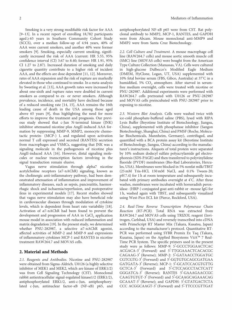
3.1. Nicotine Stimulated Phosphorylation of ERK1/2 and c-Jun in RAW264.7 Cells. Nicotine is the main ingredient ofcigarette smoke, and plasma concentration of nicotine israpidly increased after a single cigarette, ranging from10–50 ng/ml [20]. To investigate the underlying mecha-nisms of nicotine-induced AAA, RAW264.7 cells wereexposed to 10 ng/ml nicotine for 0, 10, 20, 30, 60, and120min. Western blot analysis (Figure 1(a)) revealed that10 ng/ml nicotine resulted in significant increase in thephosphorylation of ERK1/2 and c-Jun from 10min to60min; however, nicotine had no effect on p65 phosphor-ylation. Then, RAW264.7 cells were treated with nicotine for30min at the concentrations of 0, 1, 10, and 100ng/ml. Asseen in Figure 1(e), the phosphorylation of ERK1/2 andc-Jun was upregulated by nicotine, whereas the proteinlevels of p-p65 remained unchanged. These data suggestthat nicotine may exert biological effects via modulatingERK1/2 and c-Jun signaling pathway in RAW264.7 cells.
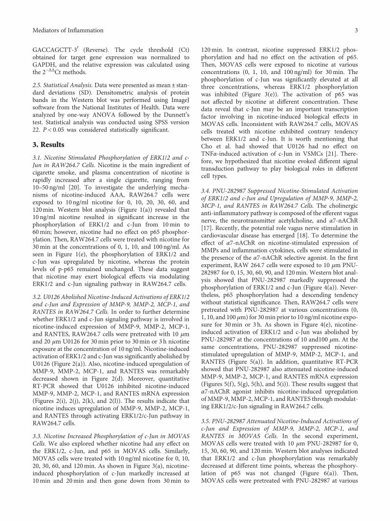
3.2. U0126 Abolished Nicotine-Induced Activations of ERK1/2and c-Jun and Expression of MMP-9, MMP-2, MCP-1, andRANTES in RAW264.7 Cells. In order to further determinewhether ERK1/2 and c-Jun signaling pathway is involved innicotine-induced expression of MMP-9, MMP-2, MCP-1,and RANTES, RAW264.7 cells were pretreated with 10 μmand 20 μm U0126 for 30min prior to 30min or 3 h nicotineexposure at the concentration of 10 ng/ml. Nicotine-inducedactivation of ERK1/2 and c-Jun was significantly abolished byU0126 (Figure 2(a)). Also, nicotine-induced upregulation ofMMP-9, MMP-2, MCP-1, and RANTES was remarkablydecreased shown in Figure 2(d). Moreover, quantitativeRT-PCR showed that U0126 inhibited nicotine-inducedMMP-9, MMP-2, MCP-1, and RANTES mRNA expression(Figures 2(i), 2(j), 2(k), and 2(l)). The results indicate thatnicotine induces upregulation of MMP-9, MMP-2, MCP-1,and RANTES through activating ERK1/2/c-Jun pathway inRAW264.7 cells.
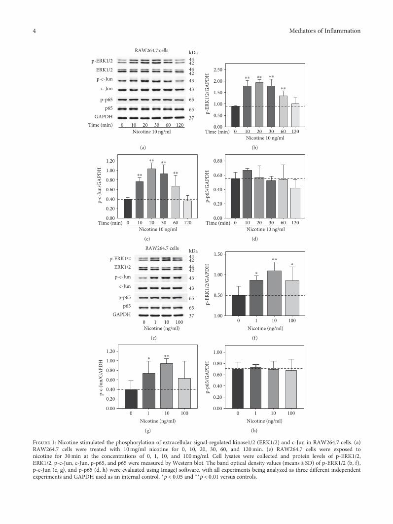
3.3. Nicotine Increased Phosphorylation of c-Jun in MOVASCells. We also explored whether nicotine had any effect onthe ERK1/2, c-Jun, and p65 in MOVAS cells. Similarly,MOVAS cells were treated with 10ng/ml nicotine for 0, 10,20, 30, 60, and 120min. As shown in Figure 3(a), nicotine-induced phosphorylation of c-Jun markedly increased at10min and 20min and then gone down from 30min to
120min. In contrast, nicotine suppressed ERK1/2 phos-phorylation and had no effect on the activation of p65.Then, MOVAS cells were exposed to nicotine at variousconcentrations (0, 1, 10, and 100ng/ml) for 30min. Thephosphorylation of c-Jun was significantly elevated at allthree concentrations, whereas ERK1/2 phosphorylationwas inhibited (Figure 3(e)). The activation of p65 wasnot affected by nicotine at different concentration. Thesedata reveal that c-Jun may be an important transcriptionfactor involving in nicotine-induced biological effects inMOVAS cells. Inconsistent with RAW264.7 cells, MOVAScells treated with nicotine exhibited contrary tendencybetween ERK1/2 and c-Jun. It is worth mentioning thatCho et al. had showed that U0126 had no effect onTNFα-induced activation of c-Jun in VSMCs [21]. There-fore, we hypothesized that nicotine evoked different signaltransduction pathway to play biological roles in differentcell types.
3.4. PNU-282987 Suppressed Nicotine-Stimulated Activationof ERK1/2 and c-Jun and Upregulation of MMP-9, MMP-2,MCP-1, and RANTES in RAW264.7 Cells. The cholinergicanti-inflammatory pathway is composed of the efferent vagusnerve, the neurotransmitter acetylcholine, and α7-nAChR[17]. Recently, the potential role vagus nerve stimulation incardiovascular disease has emerged [18]. To determine theeffect of α7-nAChR on nicotine-stimulated expression ofMMPs and inflammation cytokines, cells were stimulated inthe presence of the α7-nAChR selective agonist. In the firstexperiment, RAW 264.7 cells were exposed to 10 μm PNU-282987 for 0, 15, 30, 60, 90, and 120min. Western blot anal-ysis showed that PNU-282987 markedly suppressed thephosphorylation of ERK1/2 and c-Jun (Figure 4(a)). Never-theless, p65 phosphorylation had a descending tendencywithout statistical significance. Then, RAW264.7 cells werepretreated with PNU-282987 at various concentrations (0,1, 10, and 100 μm) for 30min prior to 10 ng/ml nicotine expo-sure for 30min or 3 h. As shown in Figure 4(e), nicotine-induced activation of ERK1/2 and c-Jun was abolished byPNU-282987 at the concentrations of 10 and100 μm. At thesame concentrations, PNU-282987 suppressed nicotine-stimulated upregulation of MMP-9, MMP-2, MCP-1, andRANTES (Figure 5(a)). In addition, quantitative RT-PCRshowed that PNU-282987 also attenuated nicotine-inducedMMP-9, MMP-2, MCP-1, and RANTES mRNA expression(Figures 5(f), 5(g), 5(h), and 5(i)). These results suggest thatα7-nAChR agonist inhibits nicotine-induced upregulationofMMP-9, MMP-2,MCP-1, and RANTES throughmodulat-ing ERK1/2/c-Jun signaling in RAW264.7 cells.
3.5. PNU-282987 Attenuated Nicotine-Induced Activations ofc-Jun and Expression of MMP-9, MMP-2, MCP-1, andRANTES in MOVAS Cells. In the second experiment,MOVAS cells were treated with 10 μm PNU-282987 for 0,15, 30, 60, 90, and 120min. Western blot analyses indicatedthat ERK1/2 and c-Jun phosphorylation was remarkablydecreased at different time points, whereas the phosphory-lation of p65 was not changed (Figure 6(a)). Then,MOVAS cells were pretreated with PNU-282987 at various
3Mediators of Inflammation
RAW264.7 cells
p-ERK1/2
ERK1/2
p-c-Jun
c-Jun
p-p65p65
GAPDHTime (min)
Nicotine 10 ng/ml
kDa444244424343
65
6537
0 10 20 30 60 120
(a)
p-ER
K1/2
/GA
PDH
Time (min)
2.50
2.00
1.50
⁎⁎ ⁎⁎ ⁎⁎
⁎⁎
0.50
0.00
1.00
Nicotine 10 ng/ml0 10 20 30 60 120
(b)
p-c-
Jun/
GA
PDH
1.20
⁎⁎
⁎⁎⁎⁎
⁎⁎1.00
0.80
0.60
0.40
0.20
0.00Time (min)
Nicotine 10 ng/ml0 10 20 30 60 120
(c)
p-p6
5/G
APD
H
0.80
0.60
0.40
0.20
0.00Time (min)
Nicotine 10 ng/ml0 10 20 30 60 120
(d)
RAW264.7 cells
p-ERK1/2ERK1/2
p-c-Jun
c-Jun
p-p65p65
GAPDH
kDa4442444243
43
65
6537
0 1 10 100Nicotine (ng/ml)
(e)
p-ER
K1/2
/GA
PDH
Nicotine (ng/ml)0 1 10 100
1.50
1.00
0.50
1.00
⁎⁎
⁎
⁎
(f)
p-c-
Jun/
GA
PDH
Nicotine (ng/ml)0 1 10 100
1.20
1.00
0.80
0.60
0.40
0.20
0.00
⁎⁎⁎
(g)
p-p6
5/G
APD
H
Nicotine (ng/ml)0 1 10 100
1.00
0.80
0.60
0.40
0.20
0.00
(h)
Figure 1: Nicotine stimulated the phosphorylation of extracellular signal-regulated kinase1/2 (ERK1/2) and c-Jun in RAW264.7 cells. (a)RAW264.7 cells were treated with 10mg/ml nicotine for 0, 10, 20, 30, 60, and 120min. (e) RAW264.7 cells were exposed tonicotine for 30min at the concentrations of 0, 1, 10, and 100mg/ml. Cell lysates were collected and protein levels of p-ERK1/2,ERK1/2, p-c-Jun, c-Jun, p-p65, and p65 were measured by Western blot. The band optical density values (means± SD) of p-ERK1/2 (b, f),p-c-Jun (c, g), and p-p65 (d, h) were evaluated using ImageJ software, with all experiments being analyzed as three different independentexperiments and GAPDH used as an internal control. ∗p < 0 05 and ∗∗p < 0 01 versus controls.
4 Mediators of Inflammation
RAW264.7 cells
p-ERK1/2
ERK1/2
p-c-Jun
c-Jun
GAPDHNicotine (ng/ml)
U0126 (�휇m)
kDa4244
44
4243
43
370 10 10 100 0 10 20
(a)
2.00⁎⁎
1.50
p-ER
K1/2
/GA
PDH
0.50
1.00
0.00Nicotine (ng/ml)
U0126 (�휇m)0 10 10 10
## ##
0 0 10 20
(b)
1.20
1.00
0.80
0.60
0.40
0.20
0.00
⁎⁎
p-c-
Jun/
GA
PDH
####
Nicotine (ng/ml)U0126 (�휇m)
0 10 10 100 0 10 20
(c)
RAW264.7 cells kDaMMP-9
MMP-2
MCP-1
RANTES
GAPDH
92
72
18
16
37Nicotine (ng/ml)
U0126 (�휇m)0 10 10 100 0 10 20
(d)
0.50
0.00
⁎⁎
MM
P-9/
GA
PDH
## #
Nicotine (ng/ml)U0126 (�휇m)
0 10 10 100 0 10 20
1.50
1.00
(e)
0.80
0.60
0.40
0.20
0.00
⁎⁎
MM
P-2/
GA
PDH
####
1.20
1.00
Nicotine (ng/ml)U0126 (�휇m)
0 10 10 100 0 10 20
(f)
0.80
0.60
0.40
0.20
0.00
⁎⁎
MCP
-1/G
APD
H
Nicotine (ng/ml)U0126 (�휇m)
0 10 10 100 0 10 20
##
##
(g)
⁎
RAN
TES/
GA
PDH
Nicotine (ng/ml)U0126 (�휇m)
0 10 10 100 0 10 20
###
0.50
0.00
1.50
1.00
(h)
⁎⁎
2.00
2.50
1.50M
MP-
9 m
RNA
0.50
1.00
0.00Nicotine (ng/ml)
U0126 (�휇m)0 10 10 100 0 10 20
## ##
(i)
3.00
2.00
1.00
0.00
⁎⁎
MM
P-2
mRN
A
Nicotine (ng/ml)U0126 (�휇m)
0 10 10 100 0 10 20
####
(j)
3.00
4.00
2.00
1.00
0.00
⁎⁎
MCP
-1 m
RNA
Nicotine (ng/ml)U0126 (�휇m)
0 10 10 100 0 10 20
####
(k)
3.00
2.00
1.00
0.00
⁎⁎
RAN
TES
mRN
A
Nicotine (ng/ml)U0126 (�휇m)
0 10 10 100 0 10 20
###
(l)
Figure 2: U0126 attenuated nicotine-induced activations of extracellular signal-regulated kinase1/2 (ERK1/2) and c-Jun and upregulation ofmatrix metalloproteinase- (MMP-) 9, MMP-2, monocyte chemotactic protein- (MCP-) 1, and regulated upon activation normal T cellexpressed and secreted (RANTES) in RAW264.7 cells. (a) RAW264.7 cells were pretreated with 10 μm and 20 μm U0126 for 30min priorto 10 ng/ml nicotine exposure for 30min. (d) RAW264.7 cells were pretreated with 10 μm and 20 μm U0126 for 30min prior to 10 ng/mlnicotine exposure for 3 h. (b, c, e–h) The intensity of protein bands (means± SD) in the Western blot were quantified by using ImageJsoftware and normalized to GAPDH. Representative results from three independent experiments are shown. (i) MMP-9, (j) MMP-2,(k) MCP-1, and (l) RANTES mRNA levels were determined by real-time reverse transcription polymerase chain reaction. All experimentswere analyzed as triplicate independent experiments. ∗p < 0 05 and ∗∗p < 0 01 versus controls; #p < 0 05 and ##p < 0 01 versus the grouptreated with nicotine.
5Mediators of Inflammation
MOVAS cells
p-ERK1/2
ERK1/2
p-c-Jun
c-Jun
GAPDH
p-p65
p65
Time (min)
kDa4244
44
42
43
4365
65
370 10 20 30
Nicotine 10 ng/ml60 120
(a)
p-ER
K1/2
/GA
PDH
1.50
⁎⁎
⁎⁎
1.00
0.50
0.00Time (min) 0 10 20 30
Nicotine 10 ng/ml60 120
⁎
(b)
p-c-
Jun/
GA
PDH
1.00
⁎⁎
⁎⁎
⁎⁎0.80
0.60
0.40
0.20
0.00Time (min) 0 10 20 30
Nicotine 10 ng/ml60 120
⁎
⁎
(c)
p-p6
5/G
APD
H 1.00
1.20
0.80
0.60
0.40
0.20
0.00Time (min) 0 10 20 30
Nicotine 10 ng/ml60 120
(d)
Nicotine (ng/ml)
MOVAS cells
p-ERK1/2
ERK1/2
p-c-Jun
c-Jun
GAPDH
p-p65
p65
kDa
4244
44
42
43
43
65
65
370 10 1001
(e)
p-ER
K1/2
/GA
PDH
⁎⁎ ⁎⁎ ⁎⁎
2.00
1.50
1.00
0.50
0.00
Nicotine (ng/ml)0 1 10 100
(f)
p-c-
Jun/
GA
PDH
⁎⁎ ⁎⁎1.50
1.00
0.50
0.00
Nicotine (ng/ml)0 1 10 100
⁎
(g)
p-p6
5/G
APD
H
1.50
1.00
0.50
0.00
Nicotine (ng/ml)0 1 10 100
(h)
Figure 3: Nicotine increased the phosphorylation of c-Jun in MOVAS cells. (a) MOVAS cells were exposed to 10 ng/ml nicotine for 0, 10, 20,30, 60, and 120min. (e) MOVAS cells were treated with nicotine for 30min at the concentrations of 0, 1, 10, and 100 ng/ml. The expression ofphosphorylated extracellular signal-regulated kinase1/2 (p-ERK1/2), ERK1/2, p-c-Jun, c-Jun, p-p65, and p65 were analyzed with Westernblot. (b–d, f–h) Densitometric analysis of protein bands was performed via ImageJ software. GAPDH was utilized as an internal control,and all experiments were analyzed as three different independent experiments. ∗p < 0 05 and ∗∗p < 0 01 versus controls.
6 Mediators of Inflammation
p-ERK1/2ERK1/2
p-c-Jun
c-Jun
p-p65
p65
GAPDH0 15 30 60 90 120Time (min)
PNU-282987 10 �휇m
kDa4244
44
4243
43
65
65
37
RAW264.7 cells
(a)
⁎⁎ ⁎⁎⁎⁎
⁎⁎
⁎⁎
1.20
1.00
p-ER
K1/2
/GA
PDH
0.80
0.60
0.40
0.20
0.000 15 30 60 90 120Time (min)
PNU-282987 10 �휇m
(b)
⁎⁎ ⁎⁎
⁎⁎
⁎
p-c-
Jun/
GA
PDH
0.80
0.60
0.40
0.20
0.000 15 30 60 90 120Time (min)
PNU-282987 10 �휇m
(c)
p-p6
5/G
APD
H 0.80
1.00
0.60
0.40
0.20
0.000 15 30 60 90 120Time (min)
PNU-282987 10 �휇m
(d)
p-ERK1/2
ERK1/2
p-c-Jun
c-Jun
GAPDH
PNU-282987 (�휇m)Nicotine (ng/ml)
kDa4244
44
4243
43
3700
10 10 10 100 101 100
RAW264.7 cells
(e)
PNU-282987 (�휇m)Nicotine (ng/ml) 0
010 10 10 100 101 100
⁎⁎1.50
##1.00
0.50
0.00
p-ER
K1/2
/GA
PDH
(f)
PNU-282987 (�휇m)Nicotine (ng/ml) 0
010 10 10 100 101 100
⁎⁎1.20#
#
1.00
0.80
0.60
0.40
0.20
0.00
p-c-
Jun/
GA
PDH
(g)
Figure 4: PNU-282987 suppressed nicotine-stimulated activation of extracellular signal-regulated kinase1/2 (ERK1/2) and c-Jun inRAW264.7 cells. (a) RAW264.7 cells were treated with 10 μm PNU-282987 for 0, 15, 30, 60, 90, and 120min. (e) RAW264.7 cells werepretreated with 10 μm PNU-282987 for 60min prior to 10 ng/ml nicotine exposure for 30min. Protein levels of p-ERK1/2, ERK1/2, p-c-Jun, c-Jun, p-p65, and p65 were measured by Western blot, and the intensity of protein bands (means± SD) (b–d, f, g) were assessedusing ImageJ software, with all experiments being analyzed as three different independent experiments and GAPDH used as an internalcontrol. ∗p < 0 05 and ∗∗p < 0 01 versus controls; #p < 0 05 and ##p < 0 01 versus the group treated with nicotine.
7Mediators of Inflammation
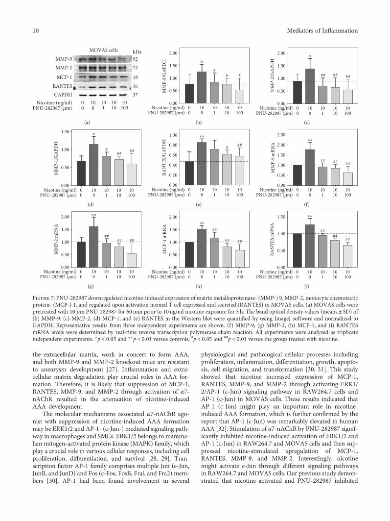
concentrations (0, 1, 10, and 100 μm) for 60min prior to10 ng/ml nicotine treatment for 30min or 3 h. As shownin Figure 6(e), nicotine-induced activation of c-Jun wasattenuated by PNU-282987 at the concentrations of 10and 100 μm. The inhibition of ERK phosphorylation bynicotine was further enhanced in MOVAS cells pretreatedwith PNU-282987. Moreover, PNU-282987 suppressednicotine-stimulated excretion of MMP-9, MMP-2, MCP-1, and RANTES from MOVAS cells (Figure 7(a)). Also,quantitative RT-PCR showed that PNU-282987 alsoreduced nicotine-induced mRNA expression of MMP-9,MMP-2, MCP-1, and RANTES shown in Figures 7(f),7(g), 7(h), and 7(i). These results suggest that α7-nAChRagonist inhibits nicotine-induced expression of MMP-9,MMP-2, MCP-1, and RANTES via c-Jun in MOVAS cells.
4. Discussion
In the present study, we demonstrated for the first time thatstimulation of α7-nAChR suppressed nicotine-inducedupregulation of inflammatory cytokines and MMP. MCP-1and RANTES, as representative of CC chemokine family,had also been demonstrated involvement in AAA develop-ment [7, 22] and thought to play greater roles than other che-mokines [23]. MCP-1 promoted macrophage infiltration,increased the MMP-9 expression in SMCs, and inducedapoptosis of SMCs within AAA, either through a directmechanism or via activation of macrophages [22, 24, 25].RANTES, similarly to MCP-1, acts on multiple immunecells and plays an important role in chronic and acuteinflammation [26]. MMP-9 and MMP-2, which degrade
MMP-9
RAW264.7 cells kDa92
72
18
16
370 10 10
10101000 0
MMP-2
MCP-1
RANTES
GAPDHNicotine (ng/ml)
PNU-282987 (�휇m)
(a)
1.50 ⁎⁎
1.00
MM
P-9/
GA
PDH
0.50
0.00
###
0 10 1010
101000 0
Nicotine (ng/ml)PNU-282987 (�휇m)
(b)
⁎
MM
P-2/
GA
PDH
##
0 10 1010
101000 0
Nicotine (ng/ml)PNU-282987 (�휇m)
2.00
1.50
1.00
0.50
0.00
(c)
1.00
0.80
0.60
0.40
0.20
0.00
⁎⁎
MCP
-1/G
APD
H
####
0 10 1010
101000 0
Nicotine (ng/ml)PNU-282987 (�휇m)
(d)
1.00
1.50
2.00
0.50
0.00
⁎⁎
RAN
TES/
GA
PDH
##
0 10 1010
101000 0
Nicotine (ng/ml)PNU-282987 (�휇m)
#
(e)
2.50
2.00
1.50
1.00
0.50
0.00
MM
P-9
mRN
A
⁎⁎
##
##
0 10 1010
101000 0
Nicotine (ng/ml)PNU-282987 (�휇m)
(f)
2.50
2.00
1.50
1.00
0.50
0.00
MM
P-2
mRN
A
⁎⁎
##
##
0 10 1010
101000 0
Nicotine (ng/ml)PNU-282987 (�휇m)
(g)
MCP
-1 m
RNA
⁎⁎
##
#
0 10 1010
101000 0
Nicotine (ng/ml)PNU-282987 (�휇m)
4.00
3.00
2.00
1.00
0.00
(h)
RAN
TES
mRN
A
⁎⁎
##
#
0 10 1010
101000 0
Nicotine (ng/ml)PNU-282987 (�휇m)
3.00
2.00
1.00
0.00
(i)
Figure 5: PNU-282987 abrogated nicotine-induced upregulation of matrix metalloproteinase- (MMP-) 9, MMP-2, monocyte chemotacticprotein- (MCP-) 1, and regulated upon activation normal T cell expressed and secreted (RANTES) in RAW264.7 cells. (a) RAW264.7 cellswere pretreated with 10 μm PNU-282987 for 60min prior to 10 ng/ml nicotine exposure for 3 h. The band optical density values (means± SD) of (b) MMP-9, (c) MMP-2, (d) MCP-1, and (e) RANTES in the Western blot were quantified by using ImageJ software andnormalized to GAPDH. Representative results from three independent experiments are shown. (f) MMP-9, (g) MMP-2, (h) MCP-1, and(i) RANTES mRNA levels were examined by real-time reverse transcription polymerase chain reaction. All experiments were analyzed astriplicate independent experiments. ∗p < 0 05 and ∗∗p < 0 01 versus controls; #p < 0 05 and ##p < 0 01 versus the group treated with nicotine.
8 Mediators of Inflammation
p-ERK1/2
ERK1/2
p-c-Jun
c-Jun
p-p65
p65
0 15 30 60 90 120GAPDH
Time (min)
kDa4244
44
4243
4365
65
37
MOVAS cells
PNU-282987 10 �휇m
(a)
p-ER
K1/2
/GA
PDH
1.50
1.00
0.50
0.000 15 30 60 90 120
⁎⁎
⁎⁎
⁎⁎⁎⁎
⁎⁎
Time (min)PNU-282987 10 �휇m
(b)
p-c-
Jun/
GA
PDH
⁎⁎
⁎
⁎⁎
⁎⁎
1.20
1.00
0.80
0.60
0.40
0.20
0.000 15 30 60 90 120Time (min)
PNU-282987 10 �휇m
(c)
p-p6
5/G
APD
H
1.00
0.80
0.60
0.40
0.20
0.000 15 30 60 90 120Time (min)
PNU-282987 10 �휇m
(d)
p-ERK1/2
ERK1/2
p-c-Jun
c-Jun
GAPDHNicotine (ng/ml)
PNU-282987 (�휇m)
kDaMOVAS cells
4244
44
4243
43
37
0 10 10 10 100 0 1 10 100
(e)
p-ER
K1/2
/GA
PDH
⁎
2.00
1.50
1.00
0.50
0.000 10 10
# #
1 1010 10
1000 0Nicotine (ng/ml)
PNU-282987 (�휇m)
(f)
p-c-
Jun/
GA
PDH
⁎⁎1.20
1.00
0.80
0.60
0.40
0.20
0.00
###
0 10 101 10
10 101000 0
Nicotine (ng/ml)PNU-282987 (�휇m)
(g)
Figure 6: PNU-282987 inhibited nicotine-stimulated activation of c-Jun in MOVAS cells. (a) MOVAS cells were exposed to 10 μm PNU-282987 for 0, 15, 30, 60, 90, and 120min. (e) MOVAS cells were pretreated with 10 μm PNU-282987 for 60min prior to 10 ng/mlnicotine exposure for 30min. Cell lysates were collected, and protein levels of phosphorylated extracellular signal-regulated kinase1/2 (p-ERK1/2), ERK1/2, p-c-Jun, c-Jun, p-p65, and p65 were measured by Western blot. (b–d, f, g) Densitometric analysis of protein bands wasperformed via ImageJ software, with all experiments being analyzed as three different independent experiments and GAPDH used as aninternal control. ∗p < 0 05 and ∗∗p < 0 01 versus controls; #p < 0 05 and ##p < 0 01 versus the group treated with nicotine.
9Mediators of Inflammation
the extracellular matrix, work in concert to form AAA,and both MMP-9 and MMP-2 knockout mice are resistantto aneurysm development [27]. Inflammation and extra-cellular matrix degradation play crucial roles in AAA for-mation. Therefore, it is likely that suppression of MCP-1,RANTES, MMP-9, and MMP-2 through activation of α7-nAChR resulted in the attenuation of nicotine-inducedAAA development.
The molecular mechanisms associated α7-nAChR ago-nist with suppression of nicotine-induced AAA formationmay be ERK1/2 and AP-1- (c-Jun-) mediated signaling path-way in macrophages and SMCs. ERK1/2 belongs to mamma-lian mitogen-activated protein kinase (MAPK) family, whichplay a crucial role in various cellular responses, including cellproliferation, differentiation, and survival [28, 29]. Tran-scription factor AP-1 family comprises multiple Jun (c-Jun,JunB, and JunD) and Fos (c-Fos, FosB, Fral, and Fra2) mem-bers [30]. AP-1 had been found involvement in several
physiological and pathological cellular processes includingproliferation, inflammation, differentiation, growth, apopto-sis, cell migration, and transformation [30, 31]. This studyshowed that nicotine increased expression of MCP-1,RANTES, MMP-9, and MMP-2 through activating ERK1/2/AP-1 (c-Jun) signaling pathway in RAW264.7 cells andAP-1 (c-Jun) in MOVAS cells. These results indicated thatAP-1 (c-Jun) might play an important role in nicotine-induced AAA formation, which is further confirmed by thereport that AP-1 (c-Jun) was remarkably elevated in humanAAA [32]. Stimulation of α7-nAChR by PNU-282987 signif-icantly inhibited nicotine-induced activation of ERK1/2 andAP-1 (c-Jun) in RAW264.7 and MOVAS cells and then sup-pressed nicotine-stimulated upregulation of MCP-1,RANTES, MMP-9, and MMP-2. Interestingly, nicotinemight activate c-Jun through different signaling pathwaysin RAW264.7 and MOVAS cells. Our previous study demon-strated that nicotine activated and PNU-282987 inhibited
MMP-9kDa92
MOVAS cells
72
18
16
37
MMP-2
MCP-1
RANTES
GAPDH00 0
10 101
1010
10100
Nicotine (ng/ml)PNU-282987 (�휇m)
(a)
2.00⁎
1.50
1.00
0.50
0.00
MM
P-9/
GA
PDH
00 0
10 101
1010
10
## #
100Nicotine (ng/ml)
PNU-282987 (�휇m)
(b)
⁎2.00
1.50
1.00
0.50
0.00
MM
P-2/
GA
PDH
## ## ##
00 0
10 101
1010
10100
Nicotine (ng/ml)PNU-282987 (�휇m)
(c)
⁎1.50
### ##1.00
0.50
0.00
MM
P-1/
GA
PDH
00 0
10 101
1010
10100
Nicotine (ng/ml)PNU-282987 (�휇m)
(d)
⁎⁎
# ##
1.00
0.80
0.60
0.40
0.20
0.00
RAN
TES/
GA
PDH
00 0
10 101
1010
10100
Nicotine (ng/ml)PNU-282987 (�휇m)
(e)
⁎⁎
## ## ##
2.00
2.50
1.50
1.00
0.50
0.00
MM
P-9
mRN
A
00 0
10 101
1010
10100
Nicotine (ng/ml)PNU-282987 (�휇m)
(f)
⁎⁎
#### ##
2.00
1.50
1.00
0.50
0.00
MM
P-2
mRN
A
00 0
10 101
1010
10100
Nicotine (ng/ml)PNU-282987 (�휇m)
(g)
⁎⁎
##
## ##
2.00
1.50
1.00
0.50
0.00
MCP
-1 m
RNA
00 0
10 101
1010
10100
Nicotine (ng/ml)PNU-282987 (�휇m)
(h)
⁎⁎
####
##
1.50
1.00
0.50
0.00RA
NTE
S m
RNA
00 0
10 101
1010
10100
Nicotine (ng/ml)PNU-282987 (�휇m)
(i)
Figure 7: PNU-282987 downregulated nicotine-induced expression of matrix metalloproteinase- (MMP-) 9, MMP-2, monocyte chemotacticprotein- (MCP-) 1, and regulated upon activation normal T cell expressed and secreted (RANTES) in MOVAS cells. (a) MOVAS cells werepretreated with 10 μm PNU-282987 for 60min prior to 10 ng/ml nicotine exposure for 3 h. The band optical density values (means± SD) of(b) MMP-9, (c) MMP-2, (d) MCP-1, and (e) RANTES in the Western blot were quantified by using ImageJ software and normalized toGAPDH. Representative results from three independent experiments are shown. (f) MMP-9, (g) MMP-2, (h) MCP-1, and (i) RANTESmRNA levels were determined by real-time reverse transcription polymerase chain reaction. All experiments were analyzed as triplicateindependent experiments. ∗p < 0 05 and ∗∗p < 0 01 versus controls; #p < 0 05 and ##p < 0 01 versus the group treated with nicotine.
10 Mediators of Inflammation
JNK signaling in MOVAS cells, which could be anotherupstream pathway of c-Jun [33]. The elucidation of such sig-naling pathways will reveal novel molecular targets that mayprovide a new therapeutic strategy for AAA. It needed tomention that nicotine could not evoke the activation of NF-κB p65 in this study. Although it is well known that NF-κBregulates expression of multiple inflammatory cytokines, itmay play little role in the process of nicotine-induced secre-tion of MCP-1 and RANTES.
The nAChRs are a family of ligand-gated ion channelreceptors composed of 17 subunits α1–α10, β1–β4, γ, δ, andε [34]. In addition to the central and peripheral nervous sys-tem, the nAChRs have been identified in vascular tissue andimmune cells [34, 35]. The different subunit combinationsresult in functionally diverse nAChR subtypes that havediffer-ent ligand affinity, cation permeability, and signaling [36, 37].The involvement of nAChR in nicotine-induced expressionof inflammatory cytokines and MMP and development ofAAA remains unknown. Nevertheless, accumulating evi-dence points towards a protective role for α7-nAChR ininflammation-based cardiovascular diseases. Cheng et al.showed that α7-nAChR activation reduced the expressionof TNFα and IL-6 and alleviated viral myocarditis [38].Treatment with PNU-282987 inhibitedADP-induced plateletaggregation inhumanplatelets, andhematopoieticα7-nAChRdeficiency increased number of peritoneal leukocytes andexpression of inflammatory mediators by both peritoneal leu-kocytes (TNFα and CRP) and the spleen (TNFα) [39], boththought to be important risk factors in atherosclerotic lesiondevelopment [40]. Stimulation of α7-nAChR by AR-R1779attenuates atherogenesis in apolipoprotein E-deficient micetreated with AngII possibly through an anti-inflammatoryeffect and reduction of blood pressure and lipid levels [41].
In contrast to nicotine that activates multiple nAChRssubtypes, PNU-282987 is a selected agonist for α7-nAChR.Thus, we inferred that distinct effects of nicotine and PNU-282987 might be mediated by differential nAChR subtypes.In accordance with the assumption, a recent study by deMoura and McMahon showed that PNU-282987 failed tosubstitute for the nicotine discriminative stimulus in maleC57BL/6J mice [42]. Another study showed that nicotine-induced catecholamine release from the adrenal glands wasmodulated by α3β4nAChR, but not by α7nAChR [43].
5. Conclusion
Taken together, α7-nAChR agonist inhibits nicotine-inducedupregulation of inflammatory cytokines and MMP throughmodulating ERK1/2/AP-1 signaling in RAW264.7 cells andAP-1 in MOVAS cells. α7-nAChR agonist is expected to bea new therapeutic strategy for AAA. However, more studies,experimental and clinical ones, are necessary to gain furtherinsights into the function and signaling of nAChRs and offerrational therapeutic strategies.
Conflicts of Interest
The authors have no specific funding in relation to thisresearch and no conflicts of interest to disclose.
Acknowledgments
This work was supported by grants from the NaturalScience Foundation of China (NSFC; no. 81370415 andno. 81670399).
References
[1] R. Erbel, V. Aboyans, C. Boileau et al., “2014 ESC guidelines onthe diagnosis and treatment of aortic diseases: document cov-ering acute and chronic aortic diseases of the thoracic andabdominal aorta of the adult. The task force for the diagnosisand treatment of aortic diseases of the European Society ofCardiology (ESC),” European Heart Journal, vol. 35, no. 41,pp. 2873–2926, 2014.
[2] P. S. Basnyat, A. H. Biffin, L. G. Moseley, A. R. Hedges, andM. H. Lewis, “Mortality from ruptured abdominal aortic aneu-rysm in Wales,” British Journal of Surgery, vol. 86, no. 6,pp. 765–770, 1999.
[3] I. M. Nordon, R. J. Hinchliffe, I. M. Loftus, and M. M. Thomp-son, “Pathophysiology and epidemiology of abdominal aorticaneurysms,” Nature Reviews Cardiology, vol. 8, no. 2, pp. 92–102, 2010.
[4] I. M. Nordon, R. J. Hinchliffe, P. J. Holt, I. M. Loftus, andM. M. Thompson, “Review of current theories for abdominalaortic aneurysm pathogenesis,” Vascular, vol. 17, no. 5,pp. 253–263, 2009.
[5] G. Ailawadi, J. L. Eliason, and G. R. Upchurch, “Current con-cepts in the pathogenesis of abdominal aortic aneurysm,” Jour-nal of Vascular Surgery, vol. 38, no. 3, pp. 584–588, 2003.
[6] T. Freestone, R. J. Turner, A. Coady, D. J. Higman, R. M.Greenhalgh, and J. T. Powell, “Inflammation and matrixmetalloproteinases in the enlarging abdominal aortic aneu-rysm,” Arteriosclerosis, Thrombosis, and Vascular Biology,vol. 15, pp. 1145–1151, 1995.
[7] A. E. Koch, G. K. Haines, R. J. Rizzo et al., “Human abdominalaortic aneurysms: immunophenotypic analysis suggesting animmune-mediated response,” The American Journal of Pathol-ogy, vol. 137, no. 5, pp. 1199–1213, 1990.
[8] R. K. Middleton, G. M. Lloyd, M. J. Bown, N. J. Cooper, N. J.London, and R. D. Sayers, “The pro-inflammatory and chemo-tactic cytokine microenvironment of the abdominal aorticaneurysm wall: a protein array study,” Journal of Vascular Sur-gery, vol. 45, no. 3, pp. 574–580, 2007.
[9] E. Jahangir, L. Lipworth, T. L. Edwards et al., “Smoking, sex,risk factors and abdominal aortic aneurysms: a prospectivestudy of 18 782 persons aged above 65 years in the SouthernCommunity Cohort Study,” Journal of Epidemiology and Com-munity Health, vol. 69, no. 5, pp. 481–488, 2015.
[10] T. B. Wilmink, C. R. Quick, and N. E. Day, “The associationbetween cigarette smoking and abdominal aortic aneurysms,”Journal of Vascular Surgery, vol. 30, no. 6, pp. 1099–1105,1999.
[11] K. C. Kent, R. M. Zwolak, N. N. Egorova et al., “Analysis of riskfactors for abdominal aortic aneurysm in a cohort of morethan 3 million individuals,” Journal of Vascular Surgery,vol. 52, no. 3, pp. 539–548, 2010.
[12] S. H. Forsdahl, K. Singh, S. Solberg, and B. K. Jacobsen, “Riskfactors for abdominal aortic aneurysms: a 7-year prospectivestudy: the Tromso study, 1994-2001,” Circulation, vol. 119,no. 16, pp. 2202–2208, 2009.
11Mediators of Inflammation
[13] M. J. Sweeting, S. G. Thompson, L. C. Brown, J. T. Powell, andcollaborators R, “Meta-analysis of individual patient data toexamine factors affecting growth and rupture of small abdom-inal aortic aneurysms,” British Journal of Surgery, vol. 99, no. 5,pp. 655–665, 2012.
[14] S. Svensjo, M. Bjorck, M. Gurtelschmid, K. Djavani Gidlund,A. Hellberg, and A. Wanhainen, “Low prevalence of abdomi-nal aortic aneurysm among 65-year-old Swedish men indicatesa change in the epidemiology of the disease,” Circulation,vol. 124, no. 10, pp. 1118–1123, 2011.
[15] F. A. Lederle, “The rise and fall of abdominal aortic aneurysm,”Circulation, vol. 124, no. 10, pp. 1097–1099, 2011.
[16] Z.-Z. Guo, Q.-A. Cao, Z.-Z. Li et al., “SP600125 attenuatesnicotine-related aortic aneurysm formation by inhibitingmatrix metalloproteinase production and CC chemokine-mediated macrophage migration,”Mediators of Inflammation,vol. 2016, Article ID 9142425, 11 pages, 2016.
[17] M. Rosas-Ballina and K. J. Tracey, “Cholinergic control ofinflammation,” Journal of Internal Medicine, vol. 265, no. 6,pp. 663–679, 2009.
[18] C. Leib, H. A. Katus, and Z. Kaya, “Cholinergic control ofinflammation in cardiovascular diseases,” Trends in Cardio-vascular Medicine, vol. 23, no. 2, pp. 46–51, 2013.
[19] A. Watanabe, T. Ichiki, H. Kojima et al., “Suppression ofabdominal aortic aneurysm formation by AR-R17779, anagonist for the α7 nicotinic acetylcholine receptor,” Atheroscle-rosis, vol. 244, pp. 113–120, 2016.
[20] P. F. Isaac andM. J. Rand, “Cigarette smoking and plasma levelsof nicotine,”Nature, vol. 236, no. 5345, pp. 308–310, 1972.
[21] A. Cho, J. Graves, andM. A. Reidy, “Mitogen-activated proteinkinases mediate matrix Metalloproteinase-9 expression invascular smooth muscle cells,” Arteriosclerosis, Thrombosis,and Vascular Biology, vol. 20, no. 12, pp. 2527–2532, 2000.
[22] C. W. Moehle, C. M. Bhamidipati, M. R. Alexander et al.,“Bone marrow–derived MCP1 required for experimentalaortic aneurysm formation and smooth muscle phenotypicmodulation,” The Journal of Thoracic and CardiovascularSurgery, vol. 142, no. 6, pp. 1567–1574, 2011.
[23] J. S. Colonnello, K. A. Hance, M. L. Shames et al., “Transientexposure to elastase induces mouse aortic wall smooth musclecell production of MCP-1 and RANTES during developmentof experimental aortic aneurysm,” Journal of Vascular Surgery,vol. 38, no. 1, pp. 138–146, 2003.
[24] Q. Wang, J. Ren, S. Morgan, Z. Liu, C. Dou, and B. Liu,“Monocyte chemoattractant protein-1 (MCP-1) regulatesmacrophage cytotoxicity in abdominal aortic aneurysm,” PLoSOne, vol. 9, no. 3, article e92053, 2014.
[25] C. Q. Yang, W. Li, S. Q. Li et al., “MCP-1 stimulates MMP-9expression via ERK 1/2 and p38 MAPK signaling pathwaysin human aortic smooth muscle cells,” Cellular Physiologyand Biochemistry, vol. 34, no. 2, pp. 266–276, 2014.
[26] P. Conti and M. DiGioacchino, “MCP-1 and RANTES aremediators of acute and chronic inflammation,” Allergy andAsthma Proceedings, vol. 22, no. 3, pp. 133–137, 2001.
[27] G. M. Longo, W. Xiong, T. C. Greiner, Y. Zhao, N. Fiotti, andB. T. Baxter, “Matrix metalloproteinases 2 and 9 work in con-cert to produce aortic aneurysms,” Journal of Clinical Investi-gation, vol. 110, no. 5, pp. 625–632, 2002.
[28] S. Torii, T. Yamamoto, Y. Tsuchiya, and E. Nishida, “ERKMAP kinase in G1 cell cycle progression and cancer,” CancerScience, vol. 97, no. 8, pp. 697–702, 2006.
[29] L. Chang and M. Karin, “Mammalian MAP kinase signallingcascades,” Nature, vol. 410, no. 6824, pp. 37–40, 2001.
[30] Y. Qiao, H. He, P. Jonsson, I. Sinha, C. Zhao, and K. Dahlman-Wright, “AP-1 is a key regulator of proinflammatory cytokineTNFα-mediated triple-negative breast cancer progression,”Journal of Biological Chemistry, vol. 291, no. 10, pp. 5068–5079, 2016.
[31] E. Shaulian, “AP-1—the Jun proteins: oncogenes or tumorsuppressors in disguise?,” Cellular Signalling, vol. 22, no. 6,pp. 894–899, 2010.
[32] H. Abdul-Hussien, R. Hanemaaijer, R. Kleemann, B. F.Verhaaren, J. H. van Bockel, and J. H. Lindeman, “The patho-physiology of abdominal aortic aneurysm growth: correspond-ing and discordant inflammatory and proteolytic processes inabdominal aortic and popliteal artery aneurysms,” Journal ofVascular Surgery, vol. 51, no. 6, pp. 1479–1487, 2010.
[33] Z.-Z. Li, Z.-Z. Guo, Z. Zhang et al., “Nicotine-induced upregu-lation of VCAM-1, MMP-2, and MMP-9 through the α7-nAChR-JNK pathway in RAW264.7 and MOVAS cells,”Molecular and Cellular Biochemistry, vol. 399, no. 1-2,pp. 49–58, 2014.
[34] J. Lee and J. P. Cooke, “The role of nicotine in the pathogenesisof atherosclerosis,” Atherosclerosis, vol. 215, no. 2, pp. 281–283, 2011.
[35] N. Santanam, B. A. Thornhill, J. K. Lau et al., “Nicotinic acetyl-choline receptor signaling in atherogenesis,” Atherosclerosis,vol. 225, no. 2, pp. 264–273, 2012.
[36] J. P. Changeux, “The acetylcholine receptor: a model for allo-steric membrane proteins,” Biochemical Society Transactions,vol. 23 of ThudichumMedal Lecture, no. 2, pp. 195–205, 1995.
[37] B. M. Conti-Tronconi, K. E. McLane, M. A. Raftery, S. A.Grando, and M. P. Protti, “The nicotinic acetylcholine recep-tor: structure and autoimmune pathology,” Critical Reviewsin Biochemistry and Molecular Biology, vol. 29, pp. 69–123,2008.
[38] Z. Cheng, G. Li-Sha, Z. Jing-Lin et al., “Protective role of thecholinergic anti-inflammatory pathway in a mouse model ofviral myocarditis,” PLoS One, vol. 9, no. 11, article e112719,2014.
[39] S. Kooijman, I. Meurs, M. van der Stoep et al., “Hematopoieticα7 nicotinic acetylcholine receptor deficiency increases inflam-mation and platelet activation status, but does not aggravateatherosclerosis,” Journal of Thrombosis and Haemostasis,vol. 13, no. 1, pp. 126–135, 2015.
[40] R. Ross, “Atherosclerosis – an inflammatory disease,” The NewEngland Journal of Medicine, vol. 340, no. 2, pp. 115–126,1999.
[41] T. Hashimoto, T. Ichiki, A. Watanabe et al., “Stimulation of α7nicotinic acetylcholine receptor by AR-R17779 suppresses ath-erosclerosis and aortic aneurysm formation in apolipoproteinE-deficient mice,” Vascular Pharmacology, vol. 61, no. 2-3,pp. 49–55, 2014.
[42] F. B. de Moura and L. R. McMahon, “The contribution of α4β2and non-α4β2 nicotinic acetylcholine receptors to the discrim-inative stimulus effects of nicotine and varenicline in mice,”Psychopharmacology, vol. 234, no. 5, pp. 781–792, 2016.
[43] A. J. Grottick, R. Wyler, and G. A. Higgins, “The α4β2 agonistSIB 1765F, but not the α7 agonist AR-R 17779, cross-sensitisesto the psychostimulant effects of nicotine,” Psychopharmacol-ogy, vol. 150, no. 2, pp. 233–236, 2000.