Braz J Med Biol Res 42(6) 2009 www.bjournal.com.br Brazilian Journal of Medical and Biological Research (2009) 42: 515-522 ISSN 0100-879X Successful scale-up of human embryonic stem cell production in a stirred microcarrier culture system A.M. Fernandes 1 *, P.A.N. Marinho 1,2 *, R.C. Sartore 1 , B.S. Paulsen 1 , R.M. Mariante 3 , L.R. Castilho 2 and S.K. Rehen 1 1 Instituto de Ciências Biomédicas, 2 COPPE - Programa de Engenharia Química, 3 Laboratório de Neurogênese, Instituto de Biofísica Carlos Chagas Filho, Universidade Federal do Rio de Janeiro, Rio de Janeiro, RJ, Brasil Correspondence to: S.K. Rehen, Instituto de Ciências Biomédicas, UFRJ, Av. Carlos Chagas Filho, s/n, Bloco F/F14, 21941-902 Rio de Janeiro, RJ, Brasil E-mail: [email protected]Future clinical applications of human embryonic stem (hES) cells will require high-yield culture protocols. Currently, hES cells are mainly cultured in static tissue plates, which offer a limited surface and require repeated sub-culturing. Here we describe a stirred system with commercial dextran-based microcarriers coated with denatured collagen to scale-up hES cell production. Maintenance of pluripotency in the microcarrier-based stirred system was shown by immunocytochemical and flow cytometry analyses for pluripotency-associated markers. The formation of cavitated embryoid bodies expressing markers of endoderm, ectoderm and mesoderm was further evidence of maintenance of differentiation capability. Cell yield per volume of medium spent was more than 2-fold higher than in static plates, resulting in a significant decrease in cultivation costs. A total of 10 8 karyotypically stable hES cells were obtained from a unitary small vessel that needed virtually no manipulation during cell proliferation, decreasing risks of contamination. Spinner flasks are available up to working volumes in the range of several liters. If desired, samples from the homogenous suspension can be withdrawn to allow process validation needed in the last expansion steps prior to transplantation. Especially when thinking about clinical trials involving from dozens to hundreds of patients, the use of a small number of larger spinners instead of hundreds of plates or flasks will be beneficial. To our knowledge, this is the first description of successful scale-up of feeder- and Matrigel™-free production of undifferentiated hES cells under continuous agitation, which makes this system a promising alternative for both therapy and research needs. Key words: Human embryonic stem cells; Scale-up; Microcarriers; Spinner bioreactors Research supported by FAPERJ, CNPq, Fundação Ary Frauzino para Pesquisa e Controle do Câncer (FAF), CAPES, as well as Ministério da Saúde, the Academy of Sciences of the Developing World (TWAS), and Pew Latin American Fellows Program in Biomedical Sciences. *These authors contributed equally to this study. Received February 15, 2009. Accepted April 13, 2009 Introduction Human embryonic stem (hES) cells have an unlimited capacity for self-renewal and the ability to differentiate into all cell types (1). These cells are of great interest for cell therapy and regenerative medicine (2,3) as well as for high throughput drug screening (4,5). Both applications, how- ever, still represent a challenge, mainly because of techni- cal limitations in scaling-up stem cell cultures (6,7). Stirred culture systems, such as the rotary RCCS™ bioreactor used by Chen et al. (8) and the spinner flasks employed by Fok and Zandstra (9), represent a significant improvement in culture techniques compared to static systems, as they offer several advantages in the scale-up process, includ-
Transcript
515
Braz J Med Biol Res 42(6) 2009
Stirred microcarrier culture of hES cells
www.bjournal.com.br
Brazilian Journal of Medical and Biological Research (2009) 42: 515-522ISSN 0100-879X
Successful scale-up of human embryonicstem cell production in a stirred microcarrierculture systemA.M. Fernandes1*, P.A.N. Marinho1,2*, R.C. Sartore1, B.S. Paulsen1, R.M. Mariante3,L.R. Castilho2 and S.K. Rehen1
1Instituto de Ciências Biomédicas, 2COPPE - Programa de Engenharia Química, 3Laboratório deNeurogênese, Instituto de Biofísica Carlos Chagas Filho, Universidade Federal do Rio de Janeiro,Rio de Janeiro, RJ, Brasil
Correspondence to: S.K. Rehen, Instituto de Ciências Biomédicas, UFRJ, Av. Carlos Chagas Filho, s/n,Bloco F/F14, 21941-902 Rio de Janeiro, RJ, BrasilE-mail: [email protected]
Future clinical applications of human embryonic stem (hES) cells will require high-yield culture protocols. Currently, hES cellsare mainly cultured in static tissue plates, which offer a limited surface and require repeated sub-culturing. Here we describe astirred system with commercial dextran-based microcarriers coated with denatured collagen to scale-up hES cell production.Maintenance of pluripotency in the microcarrier-based stirred system was shown by immunocytochemical and flow cytometryanalyses for pluripotency-associated markers. The formation of cavitated embryoid bodies expressing markers of endoderm,ectoderm and mesoderm was further evidence of maintenance of differentiation capability. Cell yield per volume of mediumspent was more than 2-fold higher than in static plates, resulting in a significant decrease in cultivation costs. A total of 108
karyotypically stable hES cells were obtained from a unitary small vessel that needed virtually no manipulation during cellproliferation, decreasing risks of contamination. Spinner flasks are available up to working volumes in the range of several liters.If desired, samples from the homogenous suspension can be withdrawn to allow process validation needed in the last expansionsteps prior to transplantation. Especially when thinking about clinical trials involving from dozens to hundreds of patients, the useof a small number of larger spinners instead of hundreds of plates or flasks will be beneficial. To our knowledge, this is the firstdescription of successful scale-up of feeder- and Matrigel™-free production of undifferentiated hES cells under continuousagitation, which makes this system a promising alternative for both therapy and research needs.
Key words: Human embryonic stem cells; Scale-up; Microcarriers; Spinner bioreactors
Research supported by FAPERJ, CNPq, Fundação Ary Frauzino para Pesquisa e Controle do Câncer (FAF), CAPES, as wellas Ministério da Saúde, the Academy of Sciences of the Developing World (TWAS), and Pew Latin American Fellows Programin Biomedical Sciences.
*These authors contributed equally to this study.
Received February 15, 2009. Accepted April 13, 2009
Introduction
Human embryonic stem (hES) cells have an unlimitedcapacity for self-renewal and the ability to differentiate intoall cell types (1). These cells are of great interest for celltherapy and regenerative medicine (2,3) as well as for highthroughput drug screening (4,5). Both applications, how-
ever, still represent a challenge, mainly because of techni-cal limitations in scaling-up stem cell cultures (6,7). Stirredculture systems, such as the rotary RCCS™ bioreactorused by Chen et al. (8) and the spinner flasks employed byFok and Zandstra (9), represent a significant improvementin culture techniques compared to static systems, as theyoffer several advantages in the scale-up process, includ-
516
Braz J Med Biol Res 42(6) 2009
A.M. Fernandes et al.
www.bjournal.com.br
ing: 1) a controlled environment leading to a homoge-neous culture, 2) simplicity of handling, and 3) less sus-ceptibility to contamination (10). Indeed, bioreactors havebeen used in a variety of bioprocesses, such as cellexpansion (11-14) and protein and virus production (15-17).
We investigated here the cultivation of hES cells in astirred system with commercial dextran-based microcarri-ers coated with denatured collagen (Cytodex™ 3), whichprovide an increased surface area for cell adhesion (18).Although microcarriers have been described for culturingstem cells from both animals (12,19) and humans (20-22),to our knowledge, this is the first description of the use ofmicrocarriers, without coating with mouse fibroblasts orMatrigel™, that successfully scaled-up human embryonicstem cell production in a stirred culture system. The culti-vation method presented here will facilitate expansion ofhES cells needed for cell therapy, research and industrialapplications.
Material and Methods
Undifferentiated H9 human embryonic stem cells(WiCell Research Institute, USA), which are so far the mostfrequently used hES cell line (23), were initially cultured in6-well plates on inactivated mouse embryonic fibroblasts(MEFs) (Hygeia Biotech, Brazil) in hES cell medium, whichconsisted of high-glucose DMEM/F12 medium supple-mented with 20% Knockout™ Serum Replacement (KSR),200 mM 2% glutamine, 55 mM 0.2% 2-mercaptoethanol,1% non-essential amino acids (all from Gibco InvitrogenCorporation, USA), and 8 ng/mL fibroblast growth factor-2(FGF-2, R&D Systems, UK).
For the stirred cultures, spinner flasks of 125-mL nomi-nal volume (Techne, USA) were used. Cytodex™ 3 micro-carriers (GE Healthcare, UK) were prepared according tomanufacturer instructions. In each spinner, 18 mL MEF-conditioned medium (MEF-CM) supplemented with 8 ng/mL FGF-2 and a total of 1.2-1.5 x 10⁷ cells dissociated fromplates with TrypLE™ (Gibco Invitrogen Corporation) wereinoculated onto 0.18 g of microcarriers suspended in 6 mLPBS remaining from the washing steps. hES cells adheredto the beads overnight under an intermittent agitationregime (cycles of 3 min/40 rpm followed by 27 min with noagitation). Then, 42 mL MEF-CM supplemented with 8 ng/mL FGF-2 was added, resulting in a final volume of 66 mL,a microcarrier concentration of 2.7 g/L and an inoculumdensity of 2.0-2.5 x 10⁵ cells/mL. MEF-CM was obtainedusing hES cell medium without FGF-2, conditioned for24 h by confluent MEFs in 75-cm2 T flasks. hES cells werethen cultured under continuous agitation (60 rpm) for 15
days, with daily exchange of 30 mL (45%) of medium. Toexchange the medium, agitation was stopped for a shortperiod to allow the microcarrier beads to settle and 30 mLof supernatant was easily withdrawn. Daily aliquots of1 mL were taken under agitation, centrifuged to removethe supernatant, incubated with crystal violet solution (1 h),and vortexed vigorously, followed by counting the releasednuclei using a hemocytometer, as described previously formicrocarrier cultures (24).
Control experiments were carried out in 6-well plates (9cm2 each well), which are the kind of static plates mostfrequently used to culture hES cells, using a feeder layer ofinactivated MEFs. Each well was initially filled with 5 mL ofmedium, which was completely exchanged for 4 mL offresh medium at 48 and 72 h. A sufficient number of wellswere inoculated to permit daily sampling of cells from 2wells by detaching them with TrypLE™. All culture experi-ments were carried out in duplicate.
In order to assess spontaneous differentiation throughembryoid body (EB) formation, cells growing in static plateswere dissociated with TrypLE™ and those from spinnercultures were detached from microcarriers also usingTrypLE™. Approximately 4 x 10⁶ cells were plated ontonon-adherent dishes (60 mm) in 7 mL hES cell mediumsupplemented with 15% KSR without FGF-2 for 7 days, inorder to obtain complete EB formation, with medium beingchanged every other day. Next, EBs were fixed in 4%paraformaldehyde (PF), embedded in Tissue Tek® OCTcompound and 10-μm sections were prepared with a cry-ostat.
Immunocytochemistry against the transcriptional fac-tor Oct-4 (Santa Cruz Biotechnology Inc., USA; Mouse-1:100), the specific cell surface marker SSEA-4 (Chemi-con-Temecula, USA; Mouse-1:100), and the proteoglycanrecognized by the antibody TRA 1-60 (Chemicon-Temec-ula; Mouse-1:100) was performed before, during and aftersubmitting cells to the stirred microcarrier culture. Thespontaneous EB differentiation assay was performed priorto and after microcarrier culture by re-plating cells de-tached from microcarriers with TrypLE™ on inactivatedMEFs. Immunocytochemistry against markers from thethree germ layers was performed in slices of these EBsusing antibodies against alphafetoprotein (AFP; Rabbit:1:100; Chemicon-Temecula), alpha smooth muscle actin(α-SMA; Mouse: 1:400; Sigma-Aldrich Sweden AB) andnestin (Mouse: 1:100; Chemicon-Temecula) for the endo-derm, mesoderm and ectoderm, respectively. Briefly, hEScells were fixed with 4% PF for 30 min and permeabilizedwith Triton X-100. Non-specific epitopes were blocked with5% bovine serum albumin (BSA) for 1 h. Primary anti-bodies were incubated for 1 h and the secondary anti-
517
Braz J Med Biol Res 42(6) 2009
Stirred microcarrier culture of hES cells
www.bjournal.com.br
bodies (Alexa Fluor 546 goat anti-mouse or anti-rabbit IgG,1:400; Molecular Probes, Gibco Invitrogen Corporation)for 30 min (all at room temperature). Nuclei were counter-stained with 4'-6-diamidino-2-phenylindole and imageswere obtained by fluorescence microscopy (Nikon, EclipseT300). Cytometric analyses were carried out using aFACScalibur flow cytometer (BD Biosciences). Undiffer-entiated colonies and EBs were manually dissociated andfixed with 4% PF, washed in FACS buffer (1% BSA and0.01% sodium azide in PBS), and blocked for 1 h withFACS buffer. Primary and secondary antibody stainingswere performed for 1 h. Data acquisition from at least10,000 events was performed using the CellQuest™ soft-ware (BD Biosciences, USA) and the Summit MoFlo soft-ware (Dako Cytomation, USA). To analyze specific pluri-potentiality markers, the same primary antibodies as usedin immunocytochemistry, directed against Oct-4, SSEA-4,and TRA 1-60, plus SOX-2 (Chemicon-Temecula; Rabbit:1: 200) were used. The same primary antibodies used inimmunocytochemistry against AFP, α-SMA and nestinwere used for the evaluation of spontaneous differentia-tion into three germ layers. Secondary antibodies wereAlexa Fluor 488 goat anti-rabbit IgG (Molecular Probes,Gibco Invitrogen Corporation; 1:400) and phycoerythrinF(ab’)2 fragment donkey anti-mouse IgG (Jackson Immuno-Research, USA; 1:200).
Cellular ploidy was determined before and after cultur-ing the hES cells in the spinner flasks as previously de-scribed (25). For this assay, hES cells were incubated with1 μg/mL Karyo MAX Colcemid (Gibco Invitrogen Corpora-tion) for 6 h to cause mitotic arrest at metaphase. Then,cells were detached using 0.05% trypsin/EDTA. After incu-bation in a hypotonic solution of 75 mM KCl for 15 min,swollen cells were fixed overnight with a methanol/aceticacid (3:1) solution and spread onto dry slides. The numberof chromosomes was determined in 20 metaphases afterG-banding.
Results and Discussion
Human embryonic stem cells were able to adhere toand to proliferate on commercial collagen-coated dextranmicrocarriers. For the sake of process simplification andelimination of additional animal-derived components, nospecial pre-treatment of the microcarrier beads was car-ried out, such as coating with Matrigel™ or MEF, as re-cently proposed by Nie et al. (22).
Although cells in the spinners underwent a long lagphase, cell growth between 120 and 340 h followed anexponential pattern (data fitting with R = 0.989). However,the doubling time in the exponential phase was 75 h, as
compared to 25 h in the static plates coated with MEFs andthis could be an indication that cells need to be furtheradapted to feeder-free culture conditions on microcarriersfor some passages. Nevertheless, 101 million cells wereobtained, on average, within 340 h in each spinner. Thespinner flasks used are relatively small (6.5 cm in diameterand 14.5 cm in height) and cells can be cultivated in themwith no manipulation (except for the daily partial mediumexchange), indicating that the proposed stirred culturetechnique is a promising method to scale-up hES cellproduction.
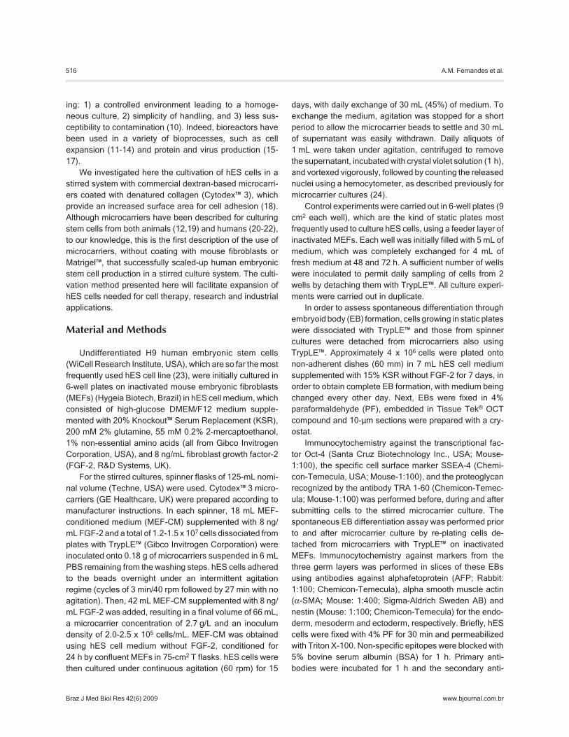
As shown in Figure 1, in terms of cells per growthsurface, cells in the spinners grew to a maximum concen-tration of 0.21 million cells/cm2 (Figure 1B), which is 30%more than the maximum concentration obtained in staticplates, 0.16 million cells/cm2 (Figure 1A). However, themost significant difference was observed in terms of cellsper volume of medium. In the spinner, a maximum concen-
Figure 1.Figure 1.Figure 1.Figure 1.Figure 1. Kinetics of human embryonic stem cell growth culturedin: A, 6-well plates; B, spinner flasks. Data are reported both interms of cells obtained per surface area available for cell attach-ment and in terms of cell concentration per volume of medium inthe culture vessel. Data are reported as means ± SE for experi-ments in duplicate.
518
Braz J Med Biol Res 42(6) 2009
A.M. Fernandes et al.
www.bjournal.com.br
tration of 1.5 x 10⁶ cells/mL was achieved compared to0.36 x 106 cells/mL in the static plates.
As stated earlier, daily medium exchanges (partialexchanges in spinners and complete exchanges in plates)were carried out. In view of this, the cell yield per totalvolume of medium spent was calculated and resulted in240,000 cells/mL in spinners versus 110,000 cells/mL instatic culture. This over 2-fold higher cell yield obtained inthe stirred system significantly decreased the costs asso-ciated with culture medium and with expensive supple-ments, such as FGF-2.
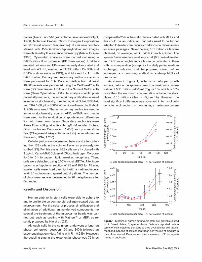
To characterize the pluripotency of hES cells grown instirred microcarrier cultures, we evaluated the presence ofthe pluripotency markers Oct-4, SSEA-4 and TRA 1-60 byimmunocytochemistry and confirmed their presence byflow cytometry of the same markers plus SOX-2 (Figure 2).The procedures were done before inoculating the hEScells into the spinner bioreactors during stirred microcarriercultivation, and after spinner culture in the cells that wererecovered from microcarriers and re-plated onto inacti-vated MEFs (Figure 2).
Cultures of hES cells grown on static plates have been
Figure 2. Figure 2. Figure 2. Figure 2. Figure 2. Pluripotency analysis:A-I, immunocytochemistry ofH9 human embryonic stem cells(P33) for Oct-4, SSEA-4 andTRA 1-60 before, during and af-ter cell growth in spinners. Cellssampled from spinners werereplated onto inactivated mouseembryonic fibroblasts and cul-tured for 2 weeks before the im-munocytochemical analyses(scale bar: 100 μm). J-K, FACSresults of human embryonicstem cells before (J) and after(K) cell culture in spinners, foreach of the four markers ana-lyzed (Oct-4, SSEA-4, SOX-2,and TRA 1-60).
Bef
ore
Dur
ing
Afte
r
A B
D E
C
F
G H
100 µm
Oct-4 SSEA-4 TRA 1-60
80.99%88.35%84.30%900
0
871
0
840
0
840
0
Eve
nts
Eve
nts
K
J
71.99%
76.92% 91.48% 88.93% 78.12%
104103102101100
832803832832
0000
Oct-4104103102101100
SSEA-4104103102101100
SOX-2104103102101100
TRA 1-60
I
519
Braz J Med Biol Res 42(6) 2009
Stirred microcarrier culture of hES cells
www.bjournal.com.br
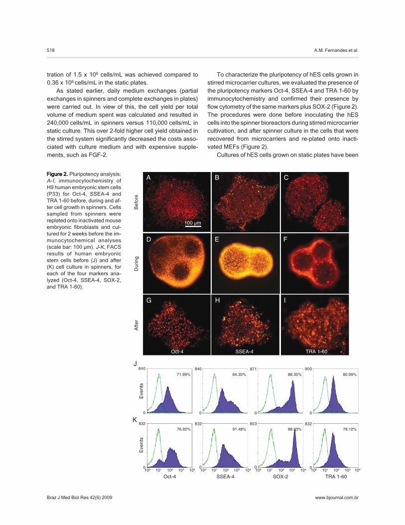
shown to maintain a stable karyotype even after long-termculturing (26). Likewise, in the present study, using TrypLE™to detach cells from the microcarriers (21,27), hES cellsmaintained a stable karyotype after 2 weeks of cultivationin the stirred microcarrier system (Figure 3).
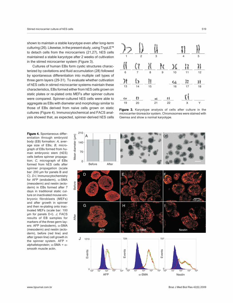
Cultures of human EBs form cystic structures charac-terized by cavitations and fluid accumulation (28) followedby spontaneous differentiation into multiple cell types ofthree germ layers (29-31). To evaluate whether cultivationof hES cells in stirred microcarrier systems maintain thesecharacteristics, EBs formed either from hES cells grown onstatic plates or re-plated onto MEFs after spinner culturewere compared. Spinner-cultured hES cells were able toaggregate as EBs with diameter and morphology similar tothose of EBs derived from naive cells grown on staticcultures (Figure 4). Immunocytochemical and FACS anal-ysis showed that, as expected, spinner-derived hES cells
Figure 4.Figure 4.Figure 4.Figure 4.Figure 4. Spontaneous differ-entiation through embryoidbody (EB) formation: A, aver-age size of EBs; B, micro-graph of EBs formed from hu-man embryonic stem (hES)cells before spinner propaga-tion; C, micrograph of EBsformed from hES cells afterspinner propagation (scalebar: 200 μm for panels B andC). D-I, Immunocytochemistryfor AFP (endoderm), α-SMA(mesoderm) and nestin (ecto-derm) in EBs formed after 7days in traditional static cul-ture on inactivated mouse em-bryonic fibroblasts (MEFs)and after growth in spinnerand then re-plating onto inac-tivated MEFs (scale bar: 100μm for panels D-I). J, FACSresults of EB samples formarkers of the three germ lay-ers: AFP (endoderm), α-SMA(mesoderm) and nestin (ecto-derm), before (red line) andafter (green line) cell growth inthe spinner system. AFP =alphafetoprotein; α-SMA = α-smooth muscle actin.
Eve
nts
J
104103102101100
1210
0
AFP
Mea
n di
amet
er (
µm)
Bef
ore
Afte
r
210
140
70
0AfterBefore
A B
D E
C
F
G H
100 µm
AFP α-SMA Nestin
I
Eve
nts
104103102101100
729
0
α-SMA
Eve
nts
104103102101100
727
Nestin
0
Figure 3.Figure 3.Figure 3.Figure 3.Figure 3. Karyotype analysis of cells after culture in themicrocarrier-bioreactor system. Chromosomes were stained withGiemsa and show a normal karyotype.
1 2 3 4 5
6 7 8 9 10 11 12
13 14 15 16 17 18
19 20 21 22 X Y
520
Braz J Med Biol Res 42(6) 2009
A.M. Fernandes et al.
www.bjournal.com.br
form EBs with the three germ layers (Figure 4). As previ-ously reported (28), endodermal markers were expressedin both the interior and exterior layers, with ectodermallabeling being peripheral and adjacent to the endodermlayer.
There is considerable interest in the use of agitatedsystems to culture embryonic stem cells. However, nostudy was able to successfully scale-up undifferentiatedhES cells using a defined microcarrier matrix. Althoughsome groups working with murine embryonic stem cellshave reached a higher final cell concentration in stirredcultures than that achieved with our microcarrier-basedculture system (12,32), two important facts should betaken into account: i) murine embryonic stem cells have afaster cell cycle, which ultimately results in a higher prolif-eration rate when compared to human cells (33), and ii) thewell-established culture conditions of murine embryonicstem cells provide a good basis for their adaptation to otherculture types. It is relatively easy to make murine embry-onic stem cells adhere to beads in suspension, whereashES cell culture conditions are not robust, and any changesin these conditions may result in them ultimately losingtheir pluripotentiality (9,20,34).
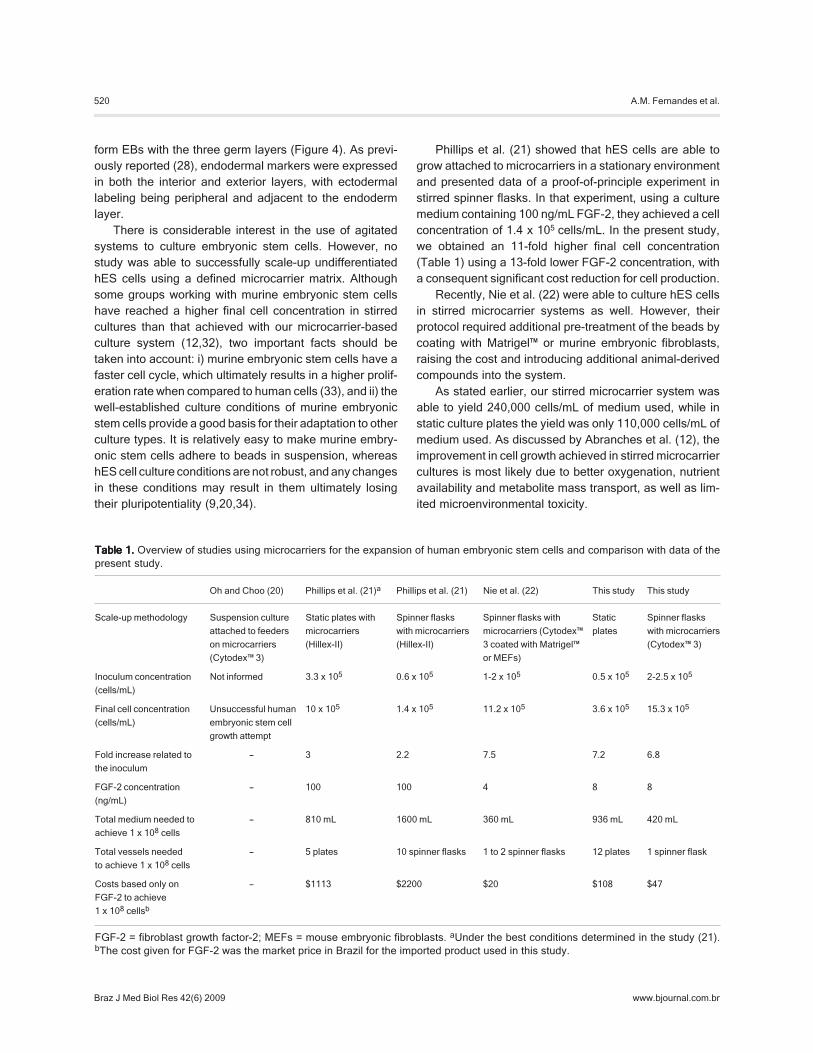
Phillips et al. (21) showed that hES cells are able togrow attached to microcarriers in a stationary environmentand presented data of a proof-of-principle experiment instirred spinner flasks. In that experiment, using a culturemedium containing 100 ng/mL FGF-2, they achieved a cellconcentration of 1.4 x 10⁵ cells/mL. In the present study,we obtained an 11-fold higher final cell concentration(Table 1) using a 13-fold lower FGF-2 concentration, witha consequent significant cost reduction for cell production.
Recently, Nie et al. (22) were able to culture hES cellsin stirred microcarrier systems as well. However, theirprotocol required additional pre-treatment of the beads bycoating with Matrigel™ or murine embryonic fibroblasts,raising the cost and introducing additional animal-derivedcompounds into the system.
As stated earlier, our stirred microcarrier system wasable to yield 240,000 cells/mL of medium used, while instatic culture plates the yield was only 110,000 cells/mL ofmedium used. As discussed by Abranches et al. (12), theimprovement in cell growth achieved in stirred microcarriercultures is most likely due to better oxygenation, nutrientavailability and metabolite mass transport, as well as lim-ited microenvironmental toxicity.
Table 1.Table 1.Table 1.Table 1.Table 1. Overview of studies using microcarriers for the expansion of human embryonic stem cells and comparison with data of thepresent study.
Oh and Choo (20) Phillips et al. (21)a Phillips et al. (21) Nie et al. (22) This study This study
Scale-up methodology Suspension culture Static plates with Spinner flasks Spinner flasks with Static Spinner flasks
attached to feeders microcarriers with microcarriers microcarriers (Cytodex™ plates with microcarriers
on microcarriers (Hillex-II) (Hillex-II) 3 coated with Matrigel™ (Cytodex™ 3)
(Cytodex™ 3) or MEFs)
Inoculum concentration Not informed 3.3 x 10⁵ 0.6 x 10⁵ 1-2 x 10⁵ 0.5 x 10⁵ 2-2.5 x 10⁵
(cells/mL)
Final cell concentration Unsuccessful human 10 x 105 1.4 x 10⁵ 11.2 x 10⁵ 3.6 x 10⁵ 15.3 x 10⁵
(cells/mL) embryonic stem cell
growth attempt
Fold increase related to - 3 2.2 7.5 7.2 6.8
the inoculum
FGF-2 concentration - 100 100 4 8 8
(ng/mL)
Total medium needed to - 810 mL 1600 mL 360 mL 936 mL 420 mL
achieve 1 x 10⁸ cells
Total vessels needed - 5 plates 10 spinner flasks 1 to 2 spinner flasks 12 plates 1 spinner flask
to achieve 1 x 10⁸ cells
Costs based only on - $1113 $2200 $20 $108 $47
FGF-2 to achieve
1 x 10⁸ cellsb
FGF-2 = fibroblast growth factor-2; MEFs = mouse embryonic fibroblasts. aUnder the best conditions determined in the study (21).bThe cost given for FGF-2 was the market price in Brazil for the imported product used in this study.
521
Braz J Med Biol Res 42(6) 2009
Stirred microcarrier culture of hES cells
www.bjournal.com.br
Stem cell therapies require large amounts of cells(35,36) and several protocols have been proposed inrecent years for the expansion of adult stem cell, (8,10,12,13,19,32,37-40). The use of hES cells in cell therapyimplies that a considerable number of cells are availablefor cell differentiation and subsequent transplantation. Thisposes the need for scaling-up the methods currently usedfor culturing these cells. The culture method proposedhere was not only able to yield a high cell concentrationand an advantageous yield of cells per volume of culture
medium, but, most importantly, it was able to support theproliferation of hES cells in a pluripotent state.
Acknowledgments
We thank Ismael Gomes and Maria R.L.S. Pinheirosfor technical assistance and Marcia Triunfol for manuscriptrevision and editing. We also thank Dr. Michael McConnell(Salk Institute) for helpful discussions.
References
1. Thomson JA, Itskovitz-Eldor J, Shapiro SS, Waknitz MA,Swiergiel JJ, Marshall VS, et al. Embryonic stem cell linesderived from human blastocysts. Science 1998; 282: 1145-1147.
3. Mountford JC. Human embryonic stem cells: origins, char-acteristics and potential for regenerative therapy. TransfusMed 2008; 18: 1-12.
4. Thomson H. Bioprocessing of embryonic stem cells for drugdiscovery. Trends Biotechnol 2007; 25: 224-230.
5. Pouton CW, Haynes JM. Embryonic stem cells as a sourceof models for drug discovery. Nat Rev Drug Discov 2007; 6:605-616.
6. Lemischka I. The power of stem cells reconsidered? ProcNatl Acad Sci U S A 1999; 96: 14193-14195.
7. Lemischka I. A few thoughts about the plasticity of stemcells. Exp Hematol 2002; 30: 848-852.
8. Chen X, Xu H, Wan C, McCaigue M, Li G. Bioreactor expan-sion of human adult bone marrow-derived mesenchymalstem cells. Stem Cells 2006; 24: 2052-2059.
9. Fok EY, Zandstra PW. Shear-controlled single-step mouseembryonic stem cell expansion and embryoid body-baseddifferentiation. Stem Cells 2005; 23: 1333-1342.
10. Yirme G, Amit M, Laevsky I, Osenberg S, Itskovitz-Eldor J.Establishing a dynamic process for the formation, propaga-tion, and differentiation of human embryoid bodies. StemCells Dev 2008; 17: 1227-1241.
11. Martin I, Wendt D, Heberer M. The role of bioreactors intissue engineering. Trends Biotechnol 2004; 22: 80-86.
12. Abranches E, Bekman E, Henrique D, Cabral JM. Expan-sion of mouse embryonic stem cells on microcarriers.Biotechnol Bioeng 2007; 96: 1211-1221.
13. Schop D, Janssen FW, Borgart E, de Bruijn JD, vanDijkhuizen-Radersma R. Expansion of mesenchymal stemcells using a microcarrier-based cultivation system: growthand metabolism. J Tissue Eng Regen Med 2008; 2: 126-135.
14. Niebruegge S, Nehring A, Bar H, Schroeder M, ZweigerdtR, Lehmann J. Cardiomyocyte production in mass suspen-sion culture: embryonic stem cells as a source for greatamounts of functional cardiomyocytes. Tissue Eng Part A2008; 14: 1591-1601.
15. Raffoul T, Swiech K, Arantes MB, Sousa APB, MendonçaRZ, Pereira CA, et al. Performance evaluation of CHO-K1cell in culture medium supplemented with hemolymph. BrazArch Biol Technol 2005; 48: 85-95.
16. Yokomizo AY, Antoniazzi MM, Galdino PL, Azambuja N Jr,Jorge SA, Pereira CA. Rabies virus production in high Verocell density cultures on macroporous microcarriers. Bio-technol Bioeng 2004; 85: 506-515.
17. Genzel Y, Behrendt I, Konig S, Sann H, Reichl U. Metabo-lism of MDCK cells during cell growth and influenza virusproduction in large-scale microcarrier culture. Vaccine 2004;22: 2202-2208.
18. van Wezel AL. Growth of cell-strains and primary cells onmicro-carriers in homogeneous culture. Nature 1967; 216:64-65.
19. Fernandes AM, Fernandes TG, Diogo MM, da Silva CL,Henrique D, Cabral JM. Mouse embryonic stem cell expan-sion in a microcarrier-based stirred culture system. JBiotechnol 2007; 132: 227-236.
20. Oh SK, Choo AB. Human embryonic stem cell technology:large scale cell amplification and differentiation. Cytotech-nology 2006; 50: 181-190.
21. Phillips BW, Horne R, Lay TS, Rust WL, Teck TT, Crook JM.Attachment and growth of human embryonic stem cells onmicrocarriers. J Biotechnol 2008; 138: 24-32.
22. Nie Y, Bergendahl V, Hei DJ, Jones JM, Palecek SP. Scal-able culture and cryopreservation of human embryonic stemcells on microcarriers. Biotechnol Prog 2009; 25: 20-31.
23. Guhr A, Kurtz A, Friedgen K, Loser P. Current state ofhuman embryonic stem cell research: an overview of celllines and their use in experimental work. Stem Cells 2006;24: 2187-2191.
24. Souza MCO, Freire MS, Schulze EA, Gaspar LP, CastilhoLR. Production of yellow fever virus in microcarrier-basedVero cell cultures. Vaccine 2009 (in press).
25. McConnell MJ, Kaushal D, Yang AH, Kingsbury MA, RehenSK, Treuner K, et al. Failed clearance of aneuploid embry-onic neural progenitor cells leads to excess aneuploidy inthe Atm-deficient but not the Trp53-deficient adult cerebralcortex. J Neurosci 2004; 24: 8090-8096.
26. Amit M, Carpenter MK, Inokuma MS, Chiu CP, Harris CP,Waknitz MA, et al. Clonally derived human embryonic stemcell lines maintain pluripotency and proliferative potential forprolonged periods of culture. Dev Biol 2000; 227: 271-278.
28. Itskovitz-Eldor J, Schuldiner M, Karsenti D, Eden A, YanukaO, Amit M, et al. Differentiation of human embryonic stemcells into embryoid bodies compromising the three embry-onic germ layers. Mol Med 2000; 6: 88-95.
29. Assady S, Maor G, Amit M, Itskovitz-Eldor J, Skorecki KL,Tzukerman M. Insulin production by human embryonic stemcells. Diabetes 2001; 50: 1691-1697.
30. Tian X, Woll PS, Morris JK, Linehan JL, Kaufman DS.Hematopoietic engraftment of human embryonic stem cell-derived cells is regulated by recipient innate immunity. StemCells 2006; 24: 1370-1380.
31. Erceg S, Lainez S, Ronaghi M, Stojkovic P, Perez-AragoMA, Moreno-Manzano V, et al. Differentiation of humanembryonic stem cells to regional specific neural precursorsin chemically defined medium conditions. PLoS ONE 2008;3: e2122.
32. Hwang YS, Cho J, Tay F, Heng JY, Ho R, Kazarian SG, etal. The use of murine embryonic stem cells, alginate encap-sulation, and rotary microgravity bioreactor in bone tissueengineering. Biomaterials 2009; 30: 499-507.
33. Gepstein L. Derivation and potential applications of humanembryonic stem cells. Circ Res 2002; 91: 866-876.
34. Phillips BW, Lim RY, Tan TT, Rust WL, Crook JM. Efficientexpansion of clinical-grade human fibroblasts on microcarri-
ers: cells suitable for ex vivo expansion of clinical-gradehESCs. J Biotechnol 2008; 134: 79-87.
35. Mukhida K, Baghbaderani BA, Hong M, Lewington M,Phillips T, McLeod M, et al. Survival, differentiation, andmigration of bioreactor-expanded human neural precursorcells in a model of Parkinson disease in rats. NeurosurgFocus 2008; 24: E8.
36. McCoy MK, Martinez TN, Ruhn KA, Wrage PC, Keefer EW,Botterman BR, et al. Autologous transplants of Adipose-Derived Adult Stromal (ADAS) cells afford dopaminergicneuroprotection in a model of Parkinson’s disease. ExpNeurol 2008; 210: 14-29.
37. Koller MR, Emerson SG, Palsson BO. Large-scale expan-sion of human stem and progenitor cells from bone marrowmononuclear cells in continuous perfusion cultures. Blood1993; 82: 378-384.
38. Frauenschuh S, Reichmann E, Ibold Y, Goetz PM, SittingerM, Ringe J. A microcarrier-based cultivation system forexpansion of primary mesenchymal stem cells. BiotechnolProg 2007; 23: 187-193.
39. Tielens S, Declercq H, Gorski T, Lippens E, Schacht E,Cornelissen M. Gelatin-based microcarriers as embryonicstem cell delivery system in bone tissue engineering: an in-vitro study. Biomacromolecules 2007; 8: 825-832.
40. Yin CH, Chen W, Hsiao CC, Kuo CY, Chen CL, Wu WT.Production of mouse embryoid bodies with hepatic differen-tiation potential by stirred tank bioreactor. Biosci BiotechnolBiochem 2007; 71: 728-734.