Institute of Medical Microbiology, Virology, and Hygiene, Rostock University Medical Centre, Rostock, Germany
Severe invasive infectious diseases remain a major and life-threatening health problem. In serious cases, a systemic activation ofthe coagulation cascade is a critical complication that is associated with high mortality rates. We report here that streptokinase, agroup A streptococcal plasminogen activator, triggers the activation of the human contact system. Activation of contact systemfactors at the surface of the Streptococcus pyogenes serotype M49 is dependent on streptokinase and plasminogen. Our resultsalso show that secreted streptokinase is an efficient contact system activator, independent from a contact surface. This results inthe processing of high-molecular-weight kininogen and the release of bradykinin, a potent vascular mediator. We further inves-tigated whether the ability of 50 different clinical S. pyogenes isolates to activate the contact system is associated with an invasivephenotype. The data reveal that isolates from invasive infections trigger an activation of the contact system more potently thanstrains isolated from noninvasive infections. The present study gives new insights into the mechanisms by which S. pyogenestriggers the human contact system and stresses the function of soluble and surface located plasmin exploited as a group A strep-tococcal virulence factor through the action of streptokinase.
Streptococcus pyogenes, referred to as group A streptococcus(GAS), is a Gram-positive major human pathogen responsible
for uncomplicated throat and skin infections such as pharyngitisand impetigo. Infections can occasionally develop into seriousand life-threatening conditions, of which streptococcal toxicshock syndrome and necrotizing fasciitis are associated with highmorbidity and mortality (1). Although GAS virulence factors havebeen studied intensively, the mechanisms by which local infec-tions progress to severe systemic infections are not yet fully un-derstood. The systemic activation of host immune responses hasbeen reported to account for several symptoms seen in septic pa-tients, i.e., hypotension, and coagulation abnormalities such asdisseminated intravascular coagulation, edema formation, andmultiorgan failure. Previous studies have shown that the systemicactivation of the human contact system plays an important role ininvasive GAS infections (for a review, see reference 2). The contactsystem comprises four plasma proteins, circulating as zymogensin the bloodstream or being assembled on various cell types: theserine proteases factor XII (FXII), factor XI (FXI), and prekalli-krein (PKK) and the nonenzymatic cofactor high-molecular-weight kininogen (HK). The latter forms equimolar complexeswith plasma kallikrein (PK) or FXI. The cascade is initiated uponcontact to a negatively charged surface and starts with the limitedautoactivation of FXII due to a conformational change. FXIIa thenactivates PKK to PK, which recruits further active FXII moleculesin a positive-feedback loop. Moreover, PK cleaves its complexpartner HK to release the vasoactive and proinflammatory peptidebradykinin. The nonapeptide has a very short half-life (a matter ofseconds) and exhibits its functions via the B1 and B2 receptors (3).Generating other mediators such as nitric oxide, prostaglandins,and leukotrienes, bradykinin is involved in the regulation of bloodpressure, the induction of fever and pain, vascular leakage, and thechemotaxis of immune cells (4). In addition, further processing ofHK may also lead to the generation of antimicrobial peptides (5),implicating a critical role of the human contact system in the earlydefense against microbial invaders (6).
Pathogens have evolved various mechanisms to manipulate
the host immune system and interference with contact factors hasbeen described for a number of bacterial species (for a review, seereference 7). In the case of S. pyogenes, the secreted streptococcalcysteine protease SpeB was found to cleave HK directly (8). Inaddition, the surface-bound M protein, one of the classical viru-lence determinants of GAS, has been demonstrated to bind HK,followed by bradykinin generation (9, 10). Binding and assemblyof contact system factors on the bacterial surface are thought to besufficient to activate this proteolytic system (6); however, addi-tional mechanisms must exist, since Sriskandan et al. documentedthat streptococcal culture supernatants were able to activate PK invitro (11).
Many pathogenic streptococci secrete streptokinase in order toaccelerate the conversion of human plasminogen to plasmin (12).Lacking enzymatic activity itself, streptokinase evolves its functionby forming a stoichiometric 1:1 complex with plasminogen or atrimolecular complex with plasminogen and fibrinogen. The ac-tivator complexes can bind at the bacterial surface via host factorreceptors and convert other plasminogen molecules to plasmin(13). Plasmin is a broad-spectrum serine protease and mainly dis-solves fibrin clots, but as early as 1995 Ewald and Eisenberg provedplasmin also to initiate the contact system cascade (40). This
Received 12 February 2015 Returned for modification 28 February 2015Accepted 7 May 2015
Accepted manuscript posted online 18 May 2015
Citation Nitzsche R, Rosenheinrich M, Kreikemeyer B, Oehmcke-Hecht S. 2015.Streptococcus pyogenes triggers activation of the human contact system bystreptokinase. Infect Immun 83:3035–3042. doi:10.1128/IAI.00180-15.
prompted us to investigate if the streptococcal plasminogen acti-vator streptokinase might contribute to contact system activationby S. pyogenes.
Here, we report that the human contact system is activated bythe action of streptokinase. The role of secreted and surface-bound streptokinase in this process was investigated by compar-ing an M49 S. pyogenes wild-type strain with its isogenic �skamutant, which is unable to trigger plasmin activity in humanplasma. Moreover, we analyzed contact activation by a set of clin-ical GAS strains isolated from invasive and noninvasive cases atthe Rostock Medical Center. Our results show a thus-far-unde-scribed mechanism by which S. pyogenes triggers the human con-tact system. This adds another important and prominent viru-lence mechanism and function to a growing list of streptokinaseactivities.
MATERIALS AND METHODSBacterial strains and culture conditions. GAS serotype M49 strain 591was obtained from R. Lütticken (Aachen, Germany). GAS M1 strain 90-226 was obtained from the World Health Organization Center for Refer-ence and Research on Streptococci at the University of Minnesota. It wasoriginally isolated from the blood of a patient with sepsis. The construc-tion of the M1-protein-lacking mutant strain from GAS M1 has beendescribed previously (14). The different clinical M serotype GAS strainsused in the present study have been described by Köller et al. (15). Astreptokinase-deficient mutant (�ska) in the GAS M49 591 chromosomalbackground was generated previously by insertional inactivation of theska gene (16). The construction of the epf, emm49, and prtF2 mutantstrains from GAS M49 591 has been described previously (17–20). TheGAS strains were cultured in Todd-Hewitt broth (THB; Invitrogen) at37°C under a 5% CO2 and 20% O2 atmosphere.
Materials. Pooled normal plasma and plasma deficient in FXII or PKKwere provided by George King Bio-Medical, Inc. (Overland Park, KS).Fresh frozen plasma samples from healthy individuals were obtained fromthe blood bank at Rostock University Hospital, Rostock, Germany, andkept frozen at 80°C until use. Plasminogen-deficient plasma was pur-chased from Haemochrom Diagnostica (Germany). Purified streptoki-nase (pSK) from beta-hemolytic streptococci was derived from Sigma-Aldrich.
Measurement of PK/FXIIa activity. PK/FXIIa activity on bacterial sur-faces exposed to normal or plasminogen-deficient plasma was measured us-ing chromogenic substrate S-2302 (H-D-Pro-Phe-Arg-pNA·2HCl; Chro-mogenix). Ten-milliliter overnight cultures (THB) of the S. pyogenes M49wild type and its isogenic knockout mutants were washed three times with50 mM Tris (pH 7.5) buffer and diluted (final concentration, 3 � 107
CFU/ml) in 50 mM Tris. Then, 100 �l of bacterial suspension was mixedwith 100 �l of plasma or buffer (control), followed by incubation at 37°Cfor 30 min. After centrifugation, the pellets were washed three times,centrifuged, and resuspended in 200 �l of buffer containing a 1 mMconcentration of the substrate. After 60 min, the samples were centri-fuged, and the absorbance of the supernatants was measured at 405 nm inan enzyme-linked immunosorbent assay (ELISA) reader. Control values(bacteria incubated in buffer) were used as a blank. No endogenous pro-teolytic activity was measured when S-2303 was incubated with bacteria inthe absence of plasma.
PK/FXIIa activity in normal and plasminogen-deficient plasma incu-bated with supernatants from overnight cultures was measured by adding100 �l of supernatant to 100 �l of plasma and 100 �l of 50 mM Tris (pH7.5) buffer containing a 1 mM concentration of the chromogenic sub-strate. The samples were incubated at 37°C for 60 min, and the absorbancewas determined at 405 nm. Plasma incubated with medium instead ofculture supernatant served as a control and was subtracted from the sam-ple values. When required, the plasmin inhibitor D-Val-Phe-Lys chloro-methyl ketone dihydrochloride (Merck) was added to a final concentra-
tion of 0.5 �g/ml after an initial incubation time of 15 min prior to sub-strate addition.
Measurement of plasmin activity. To measure the plasmin activity onbacterial surfaces exposed to plasma, bacteria were grown overnight in 10ml of THB, washed three times with phosphate-buffered saline (PBS), anddiluted (final, concentration 5 � 107 CFU/ml) in PBS. Next, 200-�l bac-terial suspensions were mixed with 200 �l of plasma or buffer, followed byincubation for 3 h in human plasma. After three further washing stepswith PBS, the pellet was suspended in Tris-NaCl buffer (19.2 mM/1.062M; pH 7.5) containing 20 �g/ml of the chromogenic substrate S-2251(H-D-Val-Leu-Lys-pNA·2HCl; Sigma), followed by an incubation for 60min at 37°C. Samples were centrifuged, and the absorbance of the super-natants was measured at 405 nm in an ELISA reader.
Plasmin activity in plasma treated with bacterial supernatants was de-termined as follows. Portions (50 �l) of plasma were mixed with 70 �l ofculture supernatant from a 10-ml THB overnight culture. The plasmin-specific chromogenic substrate was added, and the mixture was incubatedat 37°C. After 3 h, the absorbance at 405 nm was measured. Instead of thesupernatant, 70 �l of medium or 30 �l of pSK (10 U/�l) served as negativeand positive controls, respectively.
Electrophoresis and Western blot analysis. Sodium dodecyl sulfate-polyacrylamide gel electrophoresis (SDS-PAGE) was performed as de-scribed by Neville (21). Proteins from plasma samples were separated ongels of 10% total acrylamide with 3% bisacrylamide. In this way, untreatedplasma or plasma treated with kaolin, THB, or H2O for 240 min served ascontrols. Plasma was diluted 1/100 in sample buffer containing 2% (wt/vol) SDS and 5% (vol/vol) �-mercaptoethanol. Before loading, the sam-ples were boiled for 5 min. Western blot analyses were performed withsheep antibodies against HK (Affinity Biologicals) and its degradationproducts, as described previously (22).
Bradykinin measurement. Purified SK or plasmin was incubated withnormal human plasma for 15 min, and the bradykinin content was mea-sured as described earlier (22, 23). Kaolin-treated plasma samples servedas positive controls. Bradykinin release in H2O-treated plasma was used asa negative control.
Statistical analysis. Statistical analysis was performed using Graph-Pad Prism (v6.0). The P value was determined by using the unpaired t test(comparison of two groups). All samples were analyzed in triplicates, andall experiments were performed at least three times, if not otherwise de-clared. The bars in the figures indicate the standard deviations (SD).
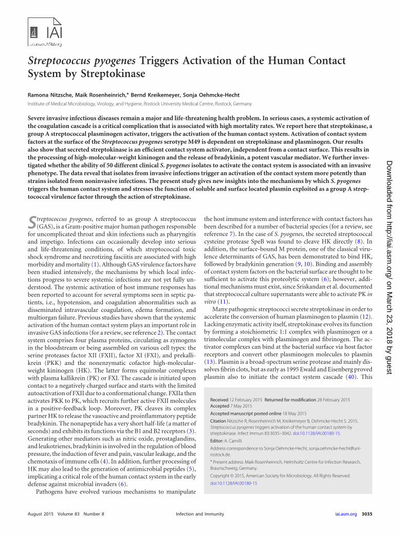
RESULTSActivation of PK and FXII at the surface of S. pyogenes M49 isdependent on streptokinase and plasminogen. The S. pyogenesM1 strain has previously been shown to bind and activate contactfactors on its surface (24, 25). To test whether S. pyogenes M49 andM1 strains trigger contact activation on their surfaces, the bacteriawere incubated with human plasma. After 30 min, unboundplasma proteins were removed by centrifugation and washingsteps. A chromogenic substrate (S-2302), specific for PK andFXIIa, was used to measure contact activation. Both strains exhib-ited PK/FXIIa activity on their surfaces (Fig. 1A). Interestingly,compared to the wild types, knockout of the M protein in thesestrains did not change PK/FXIIa activity significantly (Fig. 1A),suggesting that activation of contact system factors occurs inde-pendently of the M protein.
Several respective deletion mutants with M49 backgroundwere subsequently tested for their ability to activate the contactsystem at their surfaces. Strains deficient in adhesins, such as pro-tein F2 (�prtF2) (18) or Epf (�epf) (26), were not significantlyimpaired in PK/FXIIa activity (Fig. 1B). In contrast, the mutantstrain deficient in streptokinase (�ska) showed a significantly de-
Nitzsche et al.
3036 iai.asm.org August 2015 Volume 83 Number 8Infection and Immunity
creased PK/FXIIa activity on its surface compared to the wild-typestrain (Fig. 1B).
Streptokinase (SK), secreted by group A, C, and G streptococci,binds and activates human plasminogen (27) to plasmin, the keyserine protease in fibrinolysis. To test whether PK/FXIIa activity isdependent on plasminogen, the M49 wild type and its �ska mu-tant were incubated in normal or plasminogen-deficient plasmafor 30 min. After incubation, unbound plasma proteins were re-moved and PK/FXIIa activity determined. Kaolin, a potent con-tact system activator, was used as a positive control (Fig. 1C). Afterincubation of M49 wild-type bacteria in plasminogen-deficientplasma, no PK/FXIIa activity was detectable (A405 � 0.11) com-pared to normal plasma (A405 � 1.28). The PK/FXIIa activity ofthe �ska mutant in plasminogen-deficient plasma (A405 � 0.43)was as low as in normal plasma (A405 � 0.48). This indicates thatcontact system activation occurs at the surface of the �ska mutant,although to a significantly lower extent compared to the M49 wildtype (Fig. 1C). Since the �ska mutant shows no detectable surfaceplasmin activity (Fig. 1D), the surface contact activation of thismutant seems to occur independently from plasminogen. This isin contrast to the M49 wild type, as our data clearly show thatsurface-bound SK and plasminogen are important factors for con-tact system activation in this strain.
When the M49 strain was incubated in FXII- or PKK-deficientplasma, it retained surface PK/FXIIa activity (data not shown). Ina purified system, plasmin was shown to activate both FXII (28)and PK (29), and these findings are supported by our data.
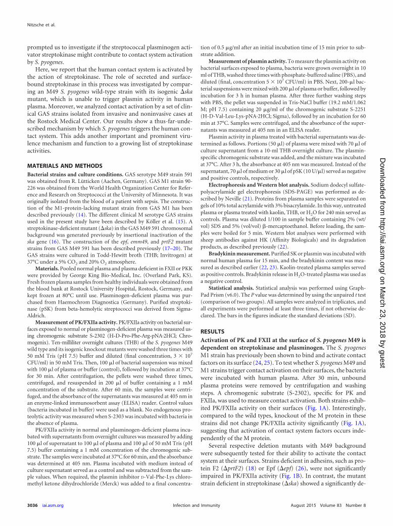
Surface-independent contact activation by secreted SK. SK issecreted by S. pyogenes into the surrounding area. We thereforetested bacterial culture supernatants from M49 wild-type and�ska mutant bacteria for contact system activation. Humanplasma was incubated with bacterial culture supernatants and PK/FXIIa activity determined, using the specific substrate. Purified SK(pSK) was used as a positive control, and THB medium served asa negative control, which was subtracted from the sample values.Culture supernatants from the M49 wild type, as well as pSK,induced strong PK/FXIIa activity in human plasma (Fig. 2A). Incontrast, no activity was observed when plasma was incubatedwith the supernatant from �ska mutant bacteria (Fig. 2A).
To test whether the observed PK/FXIIa activity is connected tothe presence of plasminogen, supernatants, THB (control), ka-olin, or pSK was incubated with plasminogen-deficient plasmaand the chromogenic substrate for PK/FXIIa. Neither the super-natants nor pSK induced an activation of PK/FXII in the absenceof plasminogen, which was, in contrast to kaolin, a potent activa-tor of the contact system via FXII (Fig. 2B).
FIG 1 PK/FXIIa and plasmin activity detected on bacterial surfaces. Overnight cultures were washed and incubated in plasma or buffer for 30 min. Unboundplasma proteins were removed by washing and the chromogenic substrate S-2302, specific for PK/FXIIa, was added. After incubation, bacteria were removed andhydrolysis of the substrate was measured at 405 nm. M1 and M49 wild type and corresponding mutant strains lacking the M protein (�emm) (A) and M49 mutantstrains lacking streptokinase (�ska), extracellular protein factor (�epf), or protein F2 (�prtF2) (B) were tested for surface PK/FXIIa activity after incubation withnormal human plasma. (C) The PK/FXIIa activity at the surfaces of M49 wild-type and �ska mutant strains was examined after exposure to normal orplasminogen-deficient plasma. Kaolin served as a positive control for plasmin-independent contact system activation. (D) Plasmin activity at the surfaces of M49wild-type and �ska mutant strains was examined after exposure to normal plasma. Overnight cultures were washed and incubated in plasma or in buffer for 3h. After washing, the chromogenic substrate S-2251, specific for plasmin, was added. After incubation, the bacteria were removed, and hydrolysis of the substratewas measured at 405 nm. As a positive control, purified SK (pSK) was incubated in plasma, and the substrate was added directly. The results are shown as themeans of at least three independent experiments with fresh frozen plasma from different donors � the SD. n.s., not significant; **, P � 0.01; ***, P � 0.001; ****,P � 0.0001.
S. pyogenes Streptokinase Triggers Contact Activation
August 2015 Volume 83 Number 8 iai.asm.org 3037Infection and Immunity
When measuring plasmin activity in normal or plasminogen-deficient plasma, the M49 supernatant as well as pSK inducedactivity in normal plasma but not in plasminogen-deficientplasma (Fig. 2C). The supernatant from the �ska mutant did notinduce plasmin activity (Fig. 2C). The results indicate that se-creted SK is a potent contact system activator, exhibiting its func-tion independently from a contact surface.
The chromogenic substrate S-2302 is specific for PK/FXIIa butis also sensitive to plasmin. We therefore measured the PK/FXIIaactivity after addition of chloromethyl-ketone D-Val-Phe-Lys,which is an efficient inhibitor of plasmin (30) and the SK-plas-minogen complex (data not shown). In the presence of the plas-min inhibitor, the PK/FXIIa activity in plasma induced by M49wild-type supernatant was reduced by ca. 50% (Fig. 2D). Thisshows that a substantial part of the measured activity is assigned toPK/FXIIa, but also that plasmin hydrolyzes the substrate.
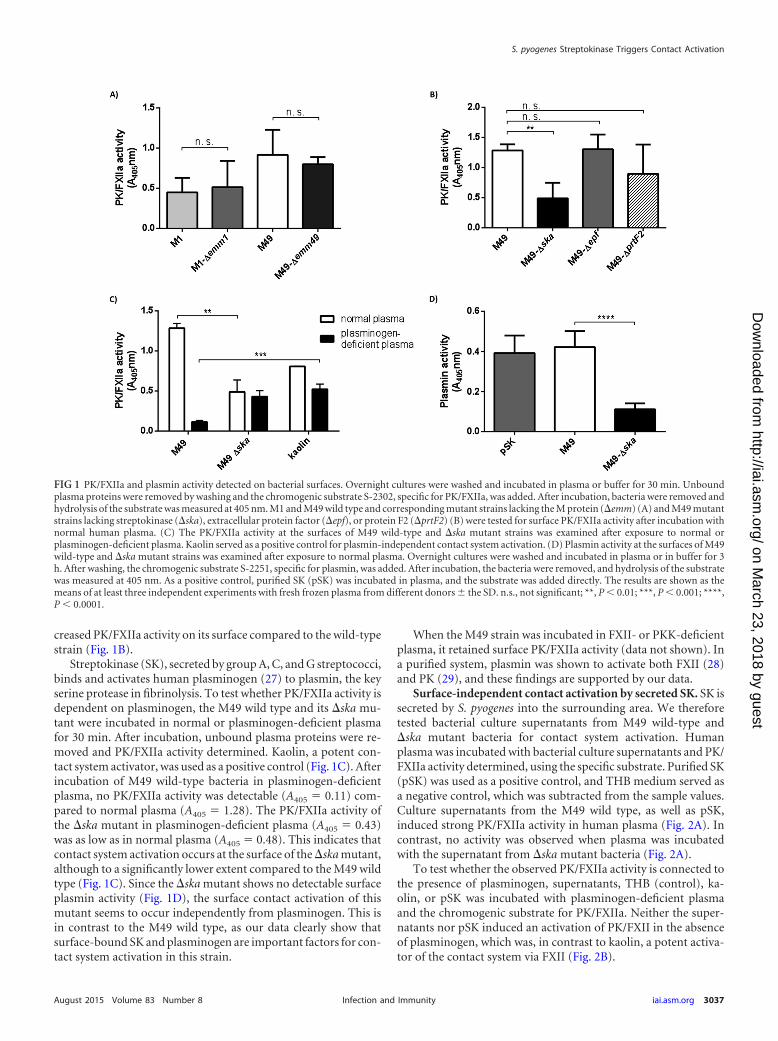
Degradation of HK and release of bradykinin by secreted SK.To investigate whether treatment of plasma with SK results in thedegradation of HK, pSK, plasmin, or kaolin was incubated withnormal plasma and investigated by Western blot analysis (Fig.3A). Samples were analyzed with an antibody directed against HK.Ponceau S staining of blotted plasma proteins was performed toconfirm equal plasma amounts per lane (data not shown). Figure3A depicts intact HK at 120 kDa in the control sample. Treatmentof plasma with pSK or kaolin initiated complete HK cleavagewithin 240 min of incubation (Fig. 3A). Interestingly, incubation
with plasmin had no obvious effect on HK cleavage (Fig. 3A),suggesting that free plasmin is rapidly inhibited by the plasmacomponent 2-antiplasmin (13). In addition, no cleavage of HKwas observed when plasminogen-deficient plasma was incubatedwith pSK for up to 240 min (Fig. 3B). We further incubatedplasma with bacterial supernatants. Purified SK and kaolin wereused as positive controls (Fig. 3C). Incubation of normal plasmawith M49 wild-type supernatant, pSK, or kaolin degraded HK,whereas intact HK was detected after incubation of plasma withmedium alone (THB-ctrl.) or �ska supernatant (Fig. 3C). Incu-bation of plasminogen-deficient plasma with bacterial superna-tants, pSK, or plasmin left HK intact, whereas treatment with ka-olin degraded HK (Fig. 3D), indicating that HK cleavage bybacterial supernatants is mainly triggered by the action of plasmin.This was supported when a PKK-deficient plasma was used (Fig.3E), revealing a cleavage of HK by M49 wild-type supernatant,pSK, and partially by kaolin, but not by the �ska supernatant.
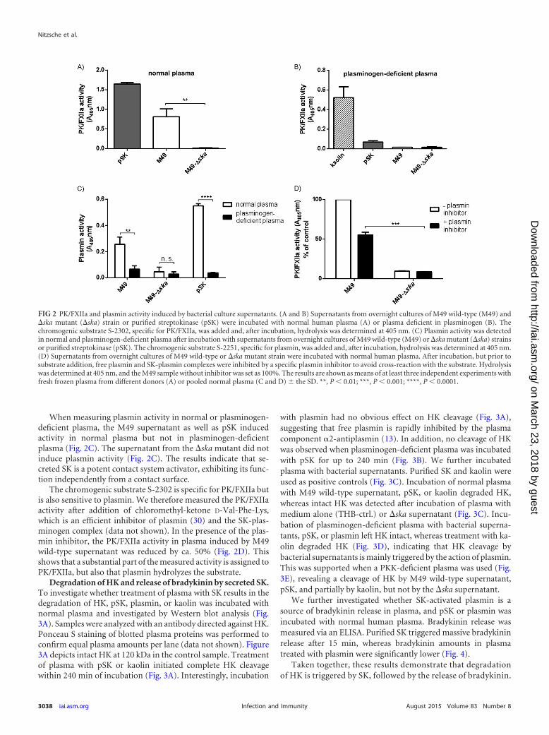
We further investigated whether SK-activated plasmin is asource of bradykinin release in plasma, and pSK or plasmin wasincubated with normal human plasma. Bradykinin release wasmeasured via an ELISA. Purified SK triggered massive bradykininrelease after 15 min, whereas bradykinin amounts in plasmatreated with plasmin were significantly lower (Fig. 4).
Taken together, these results demonstrate that degradationof HK is triggered by SK, followed by the release of bradykinin.
FIG 2 PK/FXIIa and plasmin activity induced by bacterial culture supernatants. (A and B) Supernatants from overnight cultures of M49 wild-type (M49) and�ska mutant (�ska) strain or purified streptokinase (pSK) were incubated with normal human plasma (A) or plasma deficient in plasminogen (B). Thechromogenic substrate S-2302, specific for PK/FXIIa, was added and, after incubation, hydrolysis was determined at 405 nm. (C) Plasmin activity was detectedin normal and plasminogen-deficient plasma after incubation with supernatants from overnight cultures of M49 wild-type (M49) or �ska mutant (�ska) strainsor purified streptokinase (pSK). The chromogenic substrate S-2251, specific for plasmin, was added and, after incubation, hydrolysis was determined at 405 nm.(D) Supernatants from overnight cultures of M49 wild-type or �ska mutant strain were incubated with normal human plasma. After incubation, but prior tosubstrate addition, free plasmin and SK-plasmin complexes were inhibited by a specific plasmin inhibitor to avoid cross-reaction with the substrate. Hydrolysiswas determined at 405 nm, and the M49 sample without inhibitor was set as 100%. The results are shown as means of at least three independent experiments withfresh frozen plasma from different donors (A) or pooled normal plasma (C and D) � the SD. **, P � 0.01; ***, P � 0.001; ****, P � 0.0001.
Nitzsche et al.
3038 iai.asm.org August 2015 Volume 83 Number 8Infection and Immunity
Moreover, HK degradation can be induced independentlyfrom PK.
Contact system activation by clinical isolates. To test whethercontact activation is associated with an invasive GAS phenotype,clinical isolates from invasive (n � 23) and uncomplicated (n �27) GAS infections, collected at a North-East German center fortertiary care (15), were compared for their ability to activate PK/FXIIa in normal pooled plasma. PK/FXIIa activity on bacterialsurfaces was measured for all isolates, but no significant differ-ences between isolates from invasive and noninvasive cases weredetermined (Fig. 5A). In contrast, the PK/FXIIa activity inducedby bacterial supernatants was significantly higher in isolates frominvasive cases (Fig. 5B).
Among all strains, 13 different emm genotypes were identifiedin the former study (15), and the data from at least three strains ofone serotype were plotted against the emm type (Fig. 6). The figureshows that M49 strains (n � 7) induced significantly more PK/FXIIa activity than did M1 (n � 10), M3 (n � 7), M6 (n � 4), orM89 strains (n � 3) (Fig. 6A). M12 (n � 7) and M28 (n � 10)
strains showed a high variability in PK/FXIIa activity. The M type-specific differences in contact activation were observed at bacterialsurfaces (Fig. 6A), as well as with bacterial supernatants (Fig. 6B)in all strains except for the M89 strains. All three M89 strainsshowed low PK/FXIIa surface activity and high PK/FXIIa super-natant activity (Fig. 6B).
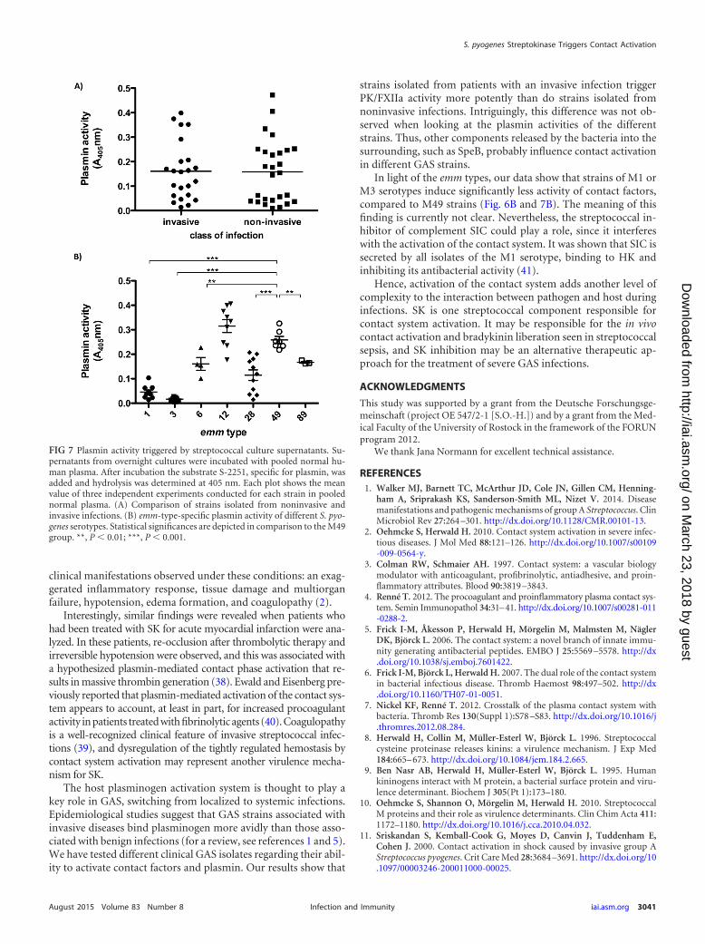
Plasmin activity in plasma triggered by bacterial supernatantswas also determined, but no difference was found between isolatesfrom invasive and noninvasive infections (Fig. 7A). Furthermore,there was no correlation between PK/FXIIa and plasmin activity(data not shown), suggesting that additional components in strep-tococcal supernatants influence contact activation in differentstrains. The plasmin activities from M1, M3, and M6 culture su-pernatants, as well as from M28 culture supernatants, were signif-icantly lower compared to the M49 strains (Fig. 7B).
Collectively, our data show that bacterial supernatants of clin-ical isolates from invasive diseases activate the contact systemmore potently compared to isolates from noninvasive diseases.
DISCUSSION
The contribution of the human contact system to the early innateimmune defense against bacteria is supported by a recent studydemonstrating that triggering the intrinsic pathway of coagula-tion promotes entrapment and killing of bacteria in a fibrin clot(31). The cleavage of HK results in the liberation of the nonapep-tide bradykinin, which is involved in the regulation of inflamma-tory processes, vascular permeability, and blood pressure. An-other consequence of contact system activation is the release ofantimicrobial peptides from HK (5, 32), disturbing the integrity ofthe bacterial membrane and recruiting further elements of theimmune system. Even so, bacteria use different mechanisms toactivate the contact system specifically and in turn manipulate thehost immune response (2, 7, 11, 33).
In the present study, we found that S. pyogenes M49 activatesthe contact system by the streptococcal plasminogen activator SK,leading to degradation of HK and the release of bradykinin. Con-tact activation occurs at the bacterial surface of S. pyogenes M49,and we showed for the first time that this activation is dependent
FIG 3 SK and SK-containing bacterial supernatants initiate plasmin-medi-ated HK degradation. Human pooled plasma was incubated for 240 min (if nototherwise indicated) at 37°C with pSK, plasmin, kaolin, or bacterial superna-tants from THB overnight cultures of M49 wild type (M49-WT) or the �skamutant strain (�ska) and analyzed for 120-kDa single-chain HK by immuno-blotting. (A) pSK, plasmin, or kaolin was incubated in normal plasma, andplasma treated with buffer served as a negative control. (B) Plasma deficient inplasminogen was incubated with pSK for up to 240 min and analyzed for HKcleavage. Bacterial supernatants from M49 wild type (M49-WT) and the �skamutant strain (�ska) were incubated in normal plasma (C), plasminogen-deficient plasma (D), or PKK-deficient plasma (E). SK, kaolin, and THB servedas positive and negative controls, respectively.
FIG 4 SK induces bradykinin release in plasma. Bradykinin release in dilutedplasma samples was measured after 15 min of incubation with either pSK (100U) or plasmin (10 �g) using an ELISA kit. The bradykinin concentration inplasma samples treated with equal volumes of H2O was set as background andsubtracted from the obtained values. The results are shown as means � SD ofthree technical replicates with fresh frozen plasma from different donors.*, P � 0.05.
S. pyogenes Streptokinase Triggers Contact Activation
August 2015 Volume 83 Number 8 iai.asm.org 3039Infection and Immunity
on SK and plasminogen. Deletion of the M protein in two differentserotypes (M49 and M1) did not influence PK/FXIIa activity at thebacterial surface, suggesting that the M protein is not importantfor the activation of contact factors. We further showed that thedeletion of the important streptococcal adhesins Epf and PrtF2from GAS M49 also had no effect on contact system activation;thus, the components responsible for surface binding of contactfactors in GAS M49 remain to be investigated.
The term “contact system” is related to its mode of action, sincebinding (contact) and assembling of the contact factors to a neg-atively charged surface trigger the activation of the system. Usingstreptococcal supernatant from an isogenic ska knockout strain,we found that SK is an important component activating the con-tact system independently from a bacterial surface through thegeneration of plasmin. Many invasive pathogens exploit plasminas a virulence factor to degrade fibrin clots, overcome tissue bar-riers, and evade peptide-derived host immune defenses (34, 35).Under normal conditions, soluble plasmin is immediately inhib-ited by 2-antiplasmin (6, 22, 32, 36); however, the SK-plasmincomplex is protected from this inhibitor and promotes uncon-
trolled plasmin activity. We found that the degradation of HK bySK generates high levels of bradykinin and can be induced inde-pendently of PK. SK accumulates as soluble component in theculture supernatants of S. pyogenes strains, and during bacteremiathe distribution of SK via the bloodstream may occur, as antibodytiters against streptokinase have been used in the serodiagnosis ofstreptococcal infections (37). Consequently, an interaction of theSK-plasmin complex with various plasma proteins such as contactfactors is more than likely. This mechanism could explain the invivo contact activation and bradykinin liberation seen duringstreptococcal invasive infections (2, 11, 33, 38). Local and con-trolled activation of the contact system has been discussed to pro-mote bacterial invasive spread via bradykinin-induced vascularleakage, since inflowing nutrient-rich plasma to the infected tissuesite might serve as a route for the disseminating pathogen (6, 22,36, 39). On the other hand, the systemic activation of the contactcascade is supposed to contribute to the pathophysiology of severeinvasive infections. The consumption of contact factors, as well ashigh bradykinin levels, was detected in patients with sepsis or toxicshock syndrome caused by S. pyogenes and correlated with the
FIG 5 PK/FXIIa activity detected on bacterial surfaces or triggered by culturesupernatants from clinical S. pyogenes isolates. (A) Bacterial overnight cultureswere washed and incubated in pooled normal plasma or buffer (negative con-trol) for 30 min. Unbound plasma proteins were removed by washing, and thePK/FXIIa-specific chromogenic substrate S-2302 was added. After incubation,the bacteria were removed and hydrolysis of the substrate was measured at 405nm. (B) Supernatants from overnight cultures were incubated with normalhuman plasma. After incubation but prior to substrate addition, free plasminand SK-plasmin complexes were inhibited by a specific plasmin inhibitor toavoid cross-reaction with the substrate. Hydrolysis was determined at 405 nm.Each plot shows the mean value of three independent experiments conductedfor each strain. n.s., not significant; **, P � 0.01.
FIG 6 emm-type-specific PK/FXIIa activity of different S. pyogenes serotypes.(A) Hydrolysis of the PK/FXIIa specific chromogenic substrate was measuredat the surface of bacteria, as described in Fig. 5A. (B) PK activation by bacterialsupernatants was examined as described above (see Fig. 5B). Prior to substrateaddition, free plasmin was inhibited by a specific plasmin inhibitor to avoidcross-reaction with the substrate. Each plot shows mean values of three inde-pendent experiments in normal pooled plasma conducted for each strain.Statistical significances are depicted compared to the M49 group. n.s., notsignificant; *, P � 0.05; ***, P � 0.001.
Nitzsche et al.
3040 iai.asm.org August 2015 Volume 83 Number 8Infection and Immunity
clinical manifestations observed under these conditions: an exag-gerated inflammatory response, tissue damage and multiorganfailure, hypotension, edema formation, and coagulopathy (2).
Interestingly, similar findings were revealed when patients whohad been treated with SK for acute myocardial infarction were ana-lyzed. In these patients, re-occlusion after thrombolytic therapy andirreversible hypotension were observed, and this was associated witha hypothesized plasmin-mediated contact phase activation that re-sults in massive thrombin generation (38). Ewald and Eisenberg pre-viously reported that plasmin-mediated activation of the contact sys-tem appears to account, at least in part, for increased procoagulantactivity in patients treated with fibrinolytic agents (40). Coagulopathyis a well-recognized clinical feature of invasive streptococcal infec-tions (39), and dysregulation of the tightly regulated hemostasis bycontact system activation may represent another virulence mecha-nism for SK.
The host plasminogen activation system is thought to play akey role in GAS, switching from localized to systemic infections.Epidemiological studies suggest that GAS strains associated withinvasive diseases bind plasminogen more avidly than those asso-ciated with benign infections (for a review, see references 1 and 5).We have tested different clinical GAS isolates regarding their abil-ity to activate contact factors and plasmin. Our results show that
strains isolated from patients with an invasive infection triggerPK/FXIIa activity more potently than do strains isolated fromnoninvasive infections. Intriguingly, this difference was not ob-served when looking at the plasmin activities of the differentstrains. Thus, other components released by the bacteria into thesurrounding, such as SpeB, probably influence contact activationin different GAS strains.
In light of the emm types, our data show that strains of M1 orM3 serotypes induce significantly less activity of contact factors,compared to M49 strains (Fig. 6B and 7B). The meaning of thisfinding is currently not clear. Nevertheless, the streptococcal in-hibitor of complement SIC could play a role, since it interfereswith the activation of the contact system. It was shown that SIC issecreted by all isolates of the M1 serotype, binding to HK andinhibiting its antibacterial activity (41).
Hence, activation of the contact system adds another level ofcomplexity to the interaction between pathogen and host duringinfections. SK is one streptococcal component responsible forcontact system activation. It may be responsible for the in vivocontact activation and bradykinin liberation seen in streptococcalsepsis, and SK inhibition may be an alternative therapeutic ap-proach for the treatment of severe GAS infections.
ACKNOWLEDGMENTS
This study was supported by a grant from the Deutsche Forschungsge-meinschaft (project OE 547/2-1 [S.O.-H.]) and by a grant from the Med-ical Faculty of the University of Rostock in the framework of the FORUNprogram 2012.
We thank Jana Normann for excellent technical assistance.
ham A, Sriprakash KS, Sanderson-Smith ML, Nizet V. 2014. Diseasemanifestations and pathogenic mechanisms of group A Streptococcus. ClinMicrobiol Rev 27:264 –301. http://dx.doi.org/10.1128/CMR.00101-13.
2. Oehmcke S, Herwald H. 2010. Contact system activation in severe infec-tious diseases. J Mol Med 88:121–126. http://dx.doi.org/10.1007/s00109-009-0564-y.
3. Colman RW, Schmaier AH. 1997. Contact system: a vascular biologymodulator with anticoagulant, profibrinolytic, antiadhesive, and proin-flammatory attributes. Blood 90:3819 –3843.
4. Renné T. 2012. The procoagulant and proinflammatory plasma contact sys-tem. Semin Immunopathol 34:31–41. http://dx.doi.org/10.1007/s00281-011-0288-2.
5. Frick I-M, Åkesson P, Herwald H, Mörgelin M, Malmsten M, NäglerDK, Björck L. 2006. The contact system: a novel branch of innate immu-nity generating antibacterial peptides. EMBO J 25:5569 –5578. http://dx.doi.org/10.1038/sj.emboj.7601422.
6. Frick I-M, Björck L, Herwald H. 2007. The dual role of the contact systemin bacterial infectious disease. Thromb Haemost 98:497–502. http://dx.doi.org/10.1160/TH07-01-0051.
7. Nickel KF, Renné T. 2012. Crosstalk of the plasma contact system withbacteria. Thromb Res 130(Suppl 1):S78 –S83. http://dx.doi.org/10.1016/j.thromres.2012.08.284.
8. Herwald H, Collin M, Müller-Esterl W, Björck L. 1996. Streptococcalcysteine proteinase releases kinins: a virulence mechanism. J Exp Med184:665– 673. http://dx.doi.org/10.1084/jem.184.2.665.
9. Ben Nasr AB, Herwald H, Müller-Esterl W, Björck L. 1995. Humankininogens interact with M protein, a bacterial surface protein and viru-lence determinant. Biochem J 305(Pt 1):173–180.
10. Oehmcke S, Shannon O, Mörgelin M, Herwald H. 2010. StreptococcalM proteins and their role as virulence determinants. Clin Chim Acta 411:1172–1180. http://dx.doi.org/10.1016/j.cca.2010.04.032.
11. Sriskandan S, Kemball-Cook G, Moyes D, Canvin J, Tuddenham E,Cohen J. 2000. Contact activation in shock caused by invasive group AStreptococcus pyogenes. Crit Care Med 28:3684 –3691. http://dx.doi.org/10.1097/00003246-200011000-00025.
FIG 7 Plasmin activity triggered by streptococcal culture supernatants. Su-pernatants from overnight cultures were incubated with pooled normal hu-man plasma. After incubation the substrate S-2251, specific for plasmin, wasadded and hydrolysis was determined at 405 nm. Each plot shows the meanvalue of three independent experiments conducted for each strain in poolednormal plasma. (A) Comparison of strains isolated from noninvasive andinvasive infections. (B) emm-type-specific plasmin activity of different S. pyo-genes serotypes. Statistical significances are depicted in comparison to the M49group. **, P � 0.01; ***, P � 0.001.
S. pyogenes Streptokinase Triggers Contact Activation
August 2015 Volume 83 Number 8 iai.asm.org 3041Infection and Immunity
12. Shannon O, Herwald H, Oehmcke S. 2013. Modulation of the coagula-tion system during severe streptococcal disease. Curr Top Microbiol Im-munol 368:189 –205. http://dx.doi.org/10.1007/82_2012_283.
13. Parry MA, Zhang XC, Bode I. 2000. Molecular mechanisms of plasmin-ogen activation: bacterial cofactors provide clues. Trends Biochem Sci25:53–59. http://dx.doi.org/10.1016/S0968-0004(99)01521-2.
14. Zimmerlein B, Park H-S, Li S, Podbielski A, Cleary PP. 2005. The Mprotein is dispensable for maturation of streptococcal cysteine proteaseSpeB. Infect Immun 73:859 – 864. http://dx.doi.org/10.1128/IAI.73.2.859-864.2005.
15. Köller T, Manetti AGO, Kreikemeyer B, Lembke C, Margarit I, GrandiG, Podbielski A. 2010. Typing of the pilus-protein-encoding FCT regionand biofilm formation as novel parameters in epidemiological investiga-tions of Streptococcus pyogenes isolates from various infection sites. J MedMicrobiol 59:442– 452. http://dx.doi.org/10.1099/jmm.0.013581-0.
16. Siemens N, Patenge N, Otto J, Fiedler T, Kreikemeyer B. 2011. Strep-tococcus pyogenes M49 plasminogen/plasmin binding facilitates keratino-cyte invasion via integrin-integrin-linked kinase (ILK) pathways and pro-tects from macrophage killing. J Biol Chem 286:21612–21622. http://dx.doi.org/10.1074/jbc.M110.202671.
17. Kreikemeyer B, Nakata M, Köller T, Hildisch H, Kourakos V, StandarK, Kawabata S, Glocker MO, Podbielski A. 2007. The Streptococcuspyogenes serotype M49 Nra-Ralp3 transcriptional regulatory network andits control of virulence factor expression from the novel eno ralp3 epf sagApathogenicity region. Infect Immun 75:5698 –5710. http://dx.doi.org/10.1128/IAI.00175-07.
18. Kreikemeyer B, Oehmcke S, Nakata M, Hoffrogge R, Podbielski A. 2004.Streptococcus pyogenes fibronectin-binding protein F2: expression profile,binding characteristics, and impact on eukaryotic cell interactions. J BiolChem 279:15850 –15859. http://dx.doi.org/10.1074/jbc.M313613200.
19. Woischnik M, Buttaro BA, Podbielski A. 2000. Inactivation of the cysteineprotease SpeB affects hyaluronic acid capsule expression in group A streptococci.Microb Pathog 28:221–226. http://dx.doi.org/10.1006/mpat.1999.0341.
20. Fiedler T, Kreikemeyer B, Sugareva V, Redanz S, Arlt R, Standar K,Podbielski A. 2010. Impact of the Streptococcus pyogenes Mga regulator onhuman matrix protein binding and interaction with eukaryotic cells. Int JMed Microbiol 300:248 –258. http://dx.doi.org/10.1016/j.ijmm.2009.07.004.
21. Neville DM. 1971. Molecular weight determination of protein-dodecylsulfate complexes by gel electrophoresis in a discontinuous buffer system.J Biol Chem 246:6328 – 6334.
22. Mattsson E, Herwald H, Cramer H, Persson K, Sjöbring U, Björck L.2001. Staphylococcus aureus induces release of bradykinin in humanplasma. Infect Immun 69:3877–3882. http://dx.doi.org/10.1128/IAI.69.6.3877-3882.2001.
23. Oehmcke S, Mörgelin M, Malmström J, Linder A, Chew M, ThorlaciusH, Herwald H. 2012. Stimulation of blood mononuclear cells with bac-terial virulence factors leads to the release of pro-coagulant and proin-flammatory microparticles. Cell Microbiol 14:107–119. http://dx.doi.org/10.1111/j.1462-5822.2011.01705.x.
24. Oehmcke S, Shannon O, von Koeckritz-Blickwede M, Morgelin M,Linder A, Olin AI, Björck L, Herwald H. 2009. Treatment of invasivestreptococcal infection with a peptide derived from human high-molecular-weight kininogen. Blood 114:444 – 451. http://dx.doi.org/10.1182/blood-2008-10-182527.
25. Ben Nasr A, Herwald H, Sjöbring U, Renné T, Müller-Esterl W, BjörckL. 1997. Absorption of kininogen from human plasma by Streptococcuspyogenes is followed by the release of bradykinin. Biochem J 326(Pt 3):657– 660.
26. Linke C, Siemens N, Oehmcke S, Radjainia M, Law RHP, Whisstock JC,
Baker EN, Kreikemeyer B. 2012. The extracellular protein factor Epf fromStreptococcus pyogenes is a cell surface adhesin that binds to cells throughan N-terminal domain containing a carbohydrate-binding module. J BiolChem 287:38178 –38189. http://dx.doi.org/10.1074/jbc.M112.376434.
27. Christensen LR, Macleod CM. 1945. A proteolytic enzyme of serum:characterization, activation, and reaction with inhibitors. J Gen Physiol28:559 –583. http://dx.doi.org/10.1085/jgp.28.6.559.
28. Kaplan AP, Austen KF. 1971. A prealbumin activator of prekallikrein. II.Derivation of activators of prekallikrein from active Hageman factor bydigestion with plasmin. J Exp Med 133:696 –712.
29. Vogt W. 1964. Kinin formation by plasmin, an indirect process, mediatedby activation of kallikrein. J Physiol (Lond) 170:153–166. http://dx.doi.org/10.1113/jphysiol.1964.sp007320.
30. Collen D, Lijnen HR, De Cock F, Durieux JP, Loffet A. 1980. Kineticproperties of tripeptide lysyl chloromethyl ketone and lysyl p-nitroanilidederivatives toward trypsin-like serine proteinases. Biochim Biophys Acta615:158 –166. http://dx.doi.org/10.1016/0005-2744(80)90019-4.
31. Loof TG, Mörgelin M, Johansson L, Oehmcke S, Olin AI, Dickneite G,Norrby-Teglund A, Theopold U, Herwald H. 2011. Coagulation, anancestral serine protease cascade, exerts a novel function in early immunedefense. Blood 118:2589 –2598. http://dx.doi.org/10.1182/blood-2011-02-337568.
32. Cederholm-Williams SA, De Cock F, Lijnen HR, Collen D. 1979.Kinetics of the reactions between streptokinase, plasmin, and 2-antiplasmin. Eur J Biochem 100:125–132. http://dx.doi.org/10.1111/j.1432-1033.1979.tb02040.x.
33. Linder A, Johansson L, Thulin P, Hertzen E, Mörgelin M, ChristenssonB, Björck L, Norrby-Teglund A, Åkesson P. 2010. Erysipelas caused bygroup A streptococcus activates the contact system and induces the releaseof heparin-binding protein. J Investig Dermatol 130:1365–1372. http://dx.doi.org/10.1038/jid.2009.437.
34. Law RHP, Abu-Ssaydeh D, Whisstock JC. 2013. New insights into thestructure and function of the plasminogen/plasmin system. Curr OpinStruct Biol. 23:836 – 841. http://dx.doi.org/10.1016/j.sbi.2013.10.006.
35. Sun H, Ringdahl U, Homeister JW, Fay WP, Engleberg NC, Yang AY,Rozek LS, Wang X, Sjobring U, Ginsburg D. 2004. Plasminogen is acritical host pathogenicity factor for group A streptococcal infection. Sci-ence 305:1283–1286. http://dx.doi.org/10.1126/science.1101245.
36. Bengtson SH, Phagoo SB, Norrby-Teglund A, Påhlman L, Mörgelin M,Zuraw BL, Leeb-Lundberg LMF, Herwald H. 2006. Kinin receptor ex-pression during Staphylococcus aureus infection. Blood 108:2055–2063.http://dx.doi.org/10.1182/blood-2006-04-016444.
37. Bisno A, Brito M, Collins C. 2003. Molecular basis of group A strepto-coccal virulence. Lancet Infect Dis 3:191–200. http://dx.doi.org/10.1016/S1473-3099(03)00576-0..
38. Hoffmeister HM, Ruf M, Wendel HP, Heller W, Seipel L. 1998. Strepto-kinase-induced activation of the kallikrein-kinin system and of the contactphase in patients with acute myocardial infarction. J Cardiovasc Pharm 31:764–772. http://dx.doi.org/10.1097/00005344-199805000-00016.
39. Olsen RJ, Shelburne SA, Musser JM. 2009. Molecular mechanisms un-derlying group A streptococcal pathogenesis. Cell Microbiol 11:1–12.http://dx.doi.org/10.1111/j.1462-5822.2008.01225.x.
40. Ewald GA, Eisenberg PR. 1995. Plasmin-mediated activation of contactsystem in response to pharmacological thrombolysis. Circulation 91:28 –36. http://dx.doi.org/10.1161/01.CIR.91.1.28.
41. Frick I-M, Shannon O, PÅkesson Mörgelin M, Collin M, SchmidtchenA, Björck L. 2011. Antibacterial activity of the contact and complementsystems is blocked by SIC, a protein secreted by Streptococcus pyogenes. JBiol Chem 286:1331–1340. http://dx.doi.org/10.1074/jbc.M110.178350.
Nitzsche et al.
3042 iai.asm.org August 2015 Volume 83 Number 8Infection and Immunity