20
Stroke-associated pneumonia: aetiology and diagnostic challenges Craig J Smith Greater Manchester Comprehensive Stroke Centre, Salford Royal NHS Foundation Trust University of Manchester
| Date post: | 26-Apr-2019 |
| Category: |
Documents |
| Upload: | nguyenduong |
| View: | 221 times |
| Download: | 0 times |

Stroke-associated pneumonia: aetiology and diagnostic
challenges
Craig J Smith
Greater Manchester Comprehensive Stroke Centre, Salford Royal NHS Foundation Trust
University of Manchester

Scope of the problem
Stroke-associated pneumonia (SAP):
• Common: around 10% but >30% in those at greatest risk
• Adversely impacts on outcome: mortality increase 2-6 fold
• Expensive: almost doubles length of stay
• Does everyone with suspected SAP need antibiotics?
• Urgent need for improved diagnosis and antibiotic stewardship
Smith and Tyrrell, 2010; Westendorp et al, 2011; Ingeman et al, 2011A; Hoffman et al, 2012

Pathophysiology of SAP
INFECTIOUS SUBSTRATE
ORO-PHARYNGEAL ASPIRATION
TRANSIENT IMMUNE-SUPPRESSION
‒ Innate‒ Adaptive
PULMONARY CLEARANCE
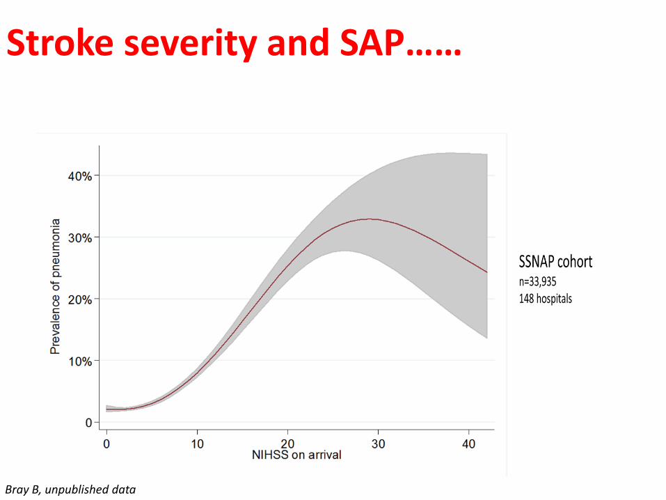
Stroke severity and SAP……
Bray B, unpublished data
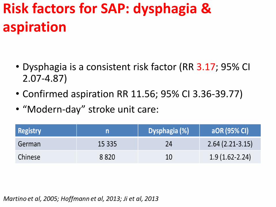
Risk factors for SAP: dysphagia & aspiration
• Dysphagia is a consistent risk factor (RR 3.17; 95% CI 2.07-4.87)
• Confirmed aspiration RR 11.56; 95% CI 3.36-39.77)
• “Modern-day” stroke unit care:

Cellular immune suppression
Emsley, Smith et al, 2007; McCulloch, Smith et al, 2017
MONOCYTE FUNCTION
P<0.01B-CELL FUNCTION
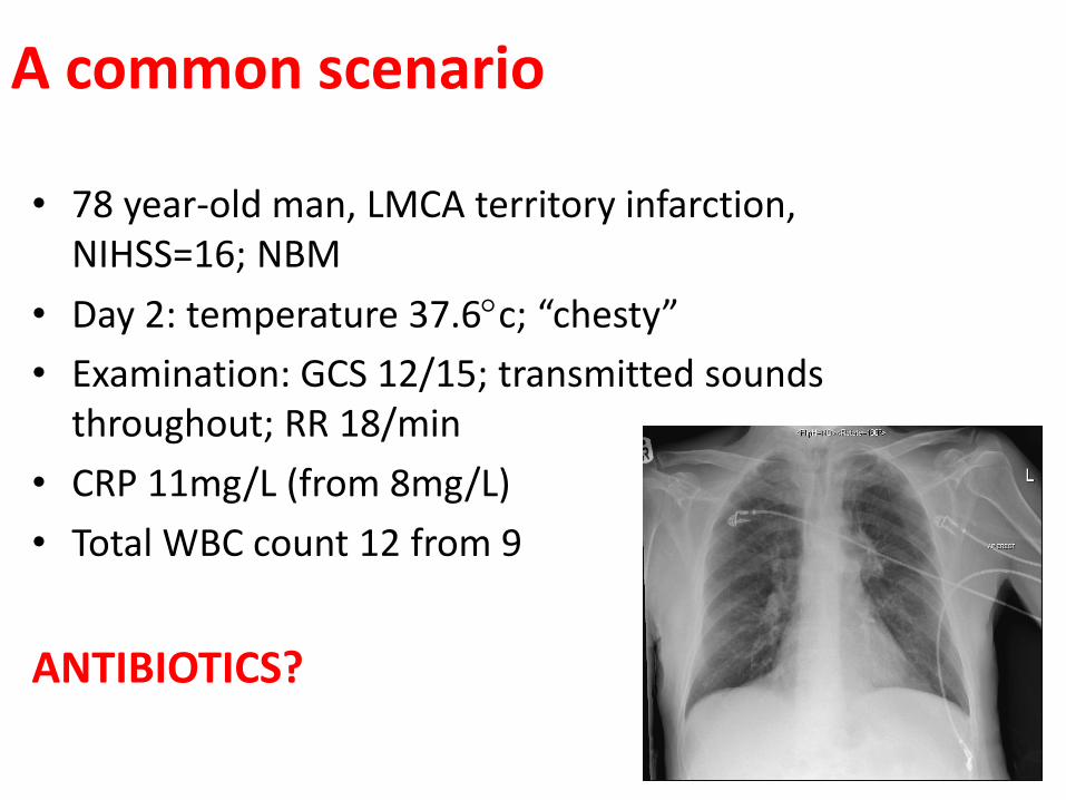
A common scenario
• 78 year-old man, LMCA territory infarction, NIHSS=16; NBM
• Day 2: temperature 37.6c; “chesty”
• Examination: GCS 12/15; transmitted sounds throughout; RR 18/min
• CRP 11mg/L (from 8mg/L)
• Total WBC count 12 from 9
ANTIBIOTICS?
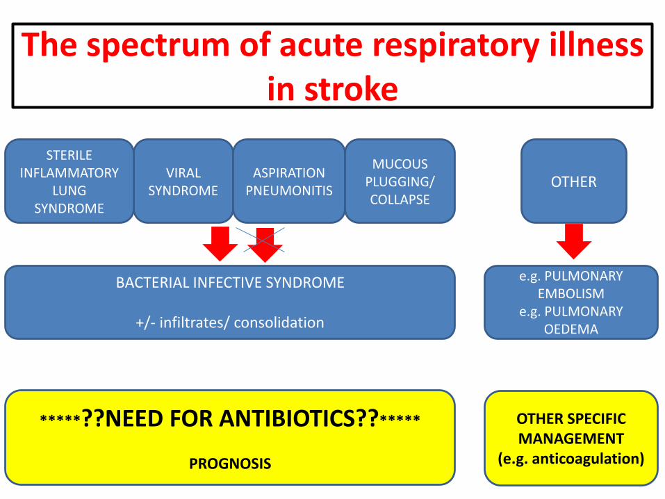
The spectrum of acute respiratory illness in stroke
BACTERIAL INFECTIVE SYNDROME
+/- infiltrates/ consolidation
VIRAL SYNDROME
ASPIRATION PNEUMONITIS
STERILE INFLAMMATORY
LUNG SYNDROME
MUCOUS PLUGGING/ COLLAPSE
OTHER
e.g. PULMONARY EMBOLISM
e.g. PULMONARY OEDEMA
*****??NEED FOR ANTIBIOTICS??*****
PROGNOSIS
OTHER SPECIFIC MANAGEMENT
(e.g. anticoagulation)
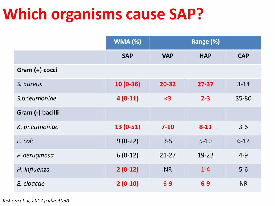
Which organisms cause SAP?
WMA (%) Range (%)
SAP VAP HAP CAP
Gram (+) cocci
S. aureus 10 (0-36) 20-32 27-37 3-14
S.pneumoniae 4 (0-11) <3 2-3 35-80
Gram (-) bacilli
K. pneumoniae 13 (0-51) 7-10 8-11 3-6
E. coli 9 (0-22) 3-5 5-10 6-12
P. aeruginosa 6 (0-12) 21-27 19-22 4-9
H. influenza 2 (0-12) NR 1-4 5-6
E. cloacae 2 (0-10) 6-9 6-9 NR
Kishore et al, 2017 (submitted)
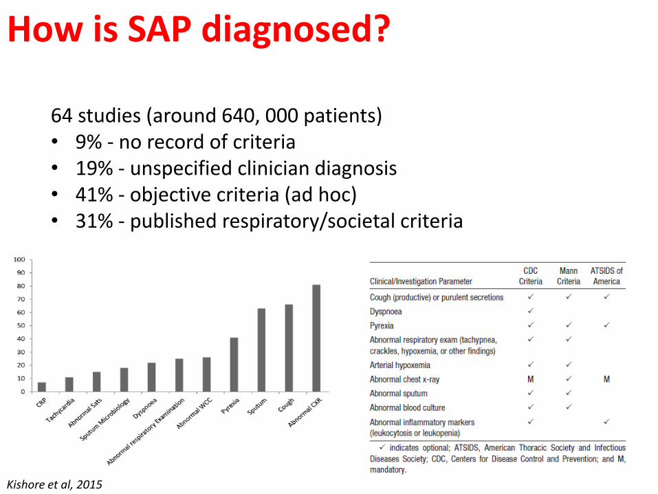
How is SAP diagnosed?
Kishore et al, 2015
64 studies (around 640, 000 patients)• 9% - no record of criteria• 19% - unspecified clinician diagnosis• 41% - objective criteria (ad hoc)• 31% - published respiratory/societal criteria
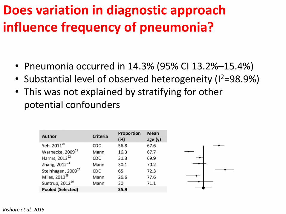
Does variation in diagnostic approach influence frequency of pneumonia?
Kishore et al, 2015
• Pneumonia occurred in 14.3% (95% CI 13.2%–15.4%)• Substantial level of observed heterogeneity (I2=98.9%)• This was not explained by stratifying for other
potential confounders

Observed SAP prevalence is highly variable in SSNAP
• n=230, 838; 186 hospitals in SSNAP (2013-16)• Predicted prevalence estimated using multifactorial model
and compared with observed prevalence
• Lowest 20 units, obs 2.3% (95% CI 1.7-2.9 ), • Highest 20 units, obs 18.8% (95% CI 17.2-20.4%)
Bray B, et al (unpublished data)
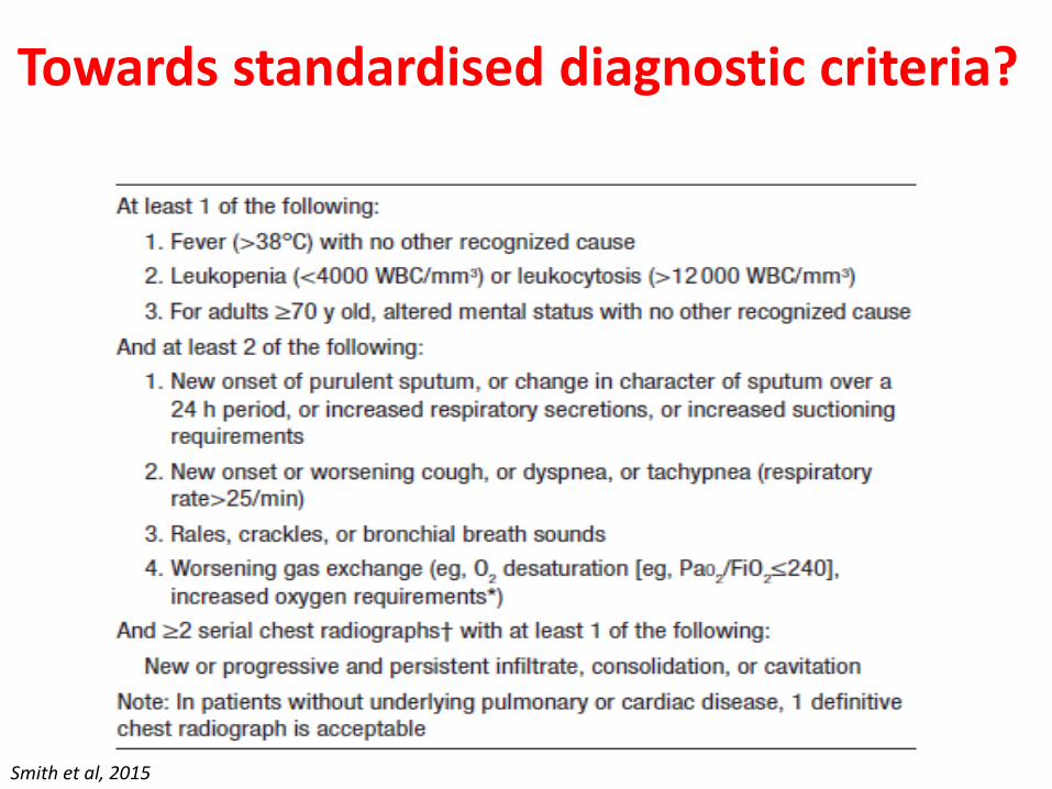
Towards standardised diagnostic criteria?
Smith et al, 2015
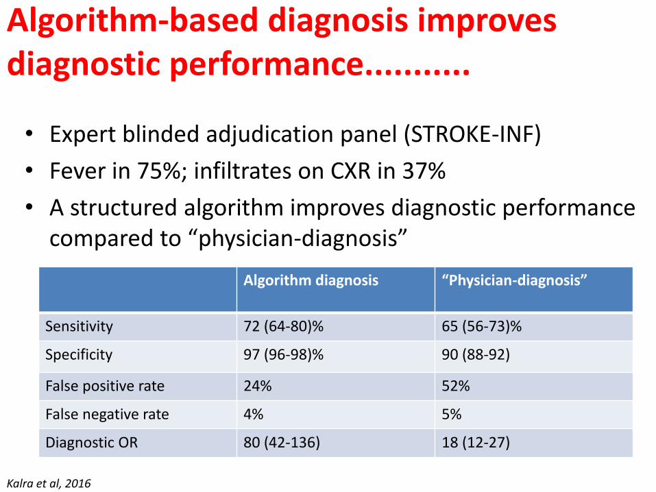
Algorithm-based diagnosis improves diagnostic performance...........
• Expert blinded adjudication panel (STROKE-INF)
• Fever in 75%; infiltrates on CXR in 37%
• A structured algorithm improves diagnostic performance compared to “physician-diagnosis”
Kalra et al, 2016
Algorithm diagnosis “Physician-diagnosis”
Sensitivity 72 (64-80)% 65 (56-73)%
Specificity 97 (96-98)% 90 (88-92)
False positive rate 24% 52%
False negative rate 4% 5%
Diagnostic OR 80 (42-136) 18 (12-27)

Can blood biomarkers detect early SAP?
Warusevitane et al, 2016; Bustamante et al, 2016
Plasma CRP; AUC 0.83 (95% CI 0.72-0.93)Plasma CRP; AUC 0.87 (95% CI 0.80-0.95)
ANY INFECTION STROKE-ASSOCIATED PNEUMONIA

........Elevated CRP increases diagnostic accuracy of algorithm
Algorithm Algorithm + (Fever or CRP)*
Sensitivity 72 (64-80)% 95 (89-98)%
Specificity 97 (96-98)% 98 (97-99)%
False positive rate 24% 15%
False negative rate 4% 1%
*Algorithm + (Fever or CRP):
(1) Temp ≥ 38·0°C or ≥ 37·5°C on two consecutive measurements OR CRP >30 mg/dLAND(2) Respiratory rate ≥ 20 / min OR cough and breathlessness OR purulent sputumAND(3) White cell count >11·0 x 109/L OR new chest infiltrates on X-ray OR positive sputum culture/microbiology OR positive blood culture
Kalra et al, submitted

Exhaled breath biomarkers for discriminating SAP?
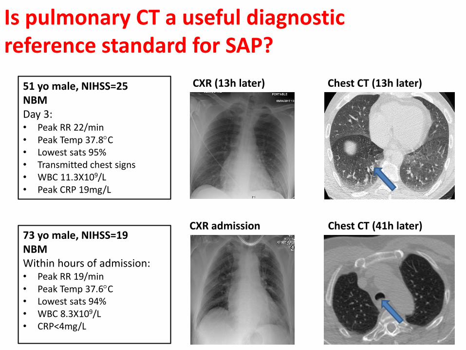
51 yo male, NIHSS=25NBMDay 3:• Peak RR 22/min • Peak Temp 37.8C• Lowest sats 95%• Transmitted chest signs• WBC 11.3X10⁹/L• Peak CRP 19mg/L
CXR (13h later) Chest CT (13h later)
73 yo male, NIHSS=19NBMWithin hours of admission:• Peak RR 19/min• Peak Temp 37.6C• Lowest sats 94%• WBC 8.3X10⁹/L• CRP<4mg/L
CXR admission Chest CT (41h later)
Is pulmonary CT a useful diagnostic reference standard for SAP?
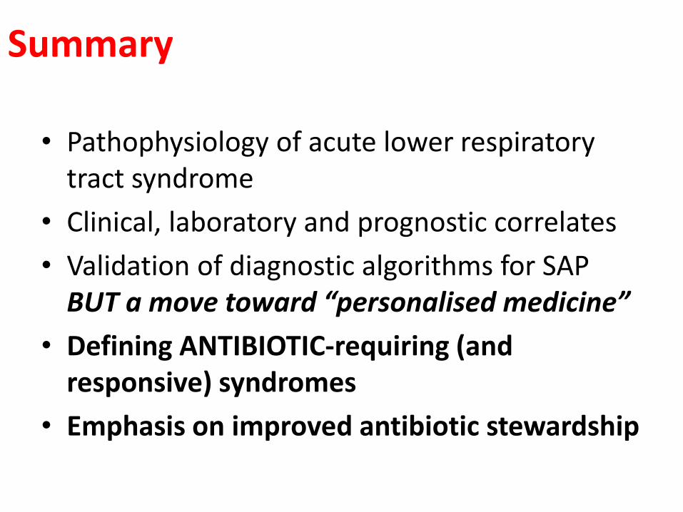
Summary
• Pathophysiology of acute lower respiratory tract syndrome
• Clinical, laboratory and prognostic correlates
• Validation of diagnostic algorithms for SAP BUT a move toward “personalised medicine”
• Defining ANTIBIOTIC-requiring (and responsive) syndromes
• Emphasis on improved antibiotic stewardship

Acknowledgements• Andreas Meisel, Joan Montaner, Lalit Kalra, Amit Kishore and the PISCES
Collaboration
• Ben Bray and SSNAP team
• Natasha James, Anand Devaraj, Anna Walsham, James Carruth and the SRFT radiography team
• Christine Roffe, Holly Maguire and the Royal Stoke radiography team
• Steve Fowler, Roy Goodacre, Iain White and Manchester Institute of Biotechnology team
Moulton CharitableTrust


![Ventilator Associated Pneumonia Treatment[1]](https://static.documents.pub/doc/80x56/577d23921a28ab4e1e9a2bfc/ventilator-associated-pneumonia-treatment1.jpg)


