JOURNAL OF VIROLOGY, Apr. 1987, p. 962-971 Vol. 61, No. 4 0022-538X/87/040962-10$02.00/0 Copyright C) 1987, American Society for Microbiology Structural and Transcriptional Analysis of Human Papillomavirus Type 16 Sequences in Cervical Carcinoma Cell Lines CARL C. BAKER,'* WILLIAM C. PHELPS,1 VALERIE LINDGREN,1 MICHAEL J. BRAUN,2 MATTHEW A. GONDA,2 AND PETER M. HOWLEY' Laboratory of Tumor Virus Biology, National Cancer Institute, Bethesda, Maryland 20892,1 and Laboratory of Cell and Molecular Structuire, Program Resources, Inc., Frederick Cancer Research Facility, National Cancer Institute, Frederick, Maryland 217012 Received 23 September 1986/Accepted 18 November 1986 We cloned and analyzed the integrated human papillomavirus type 16 (HPV-16) genomes that are present in the human cervical carcinoma cell lines SiHa and CaSki. The single HPV-16 genome in the SiHa line was cloned as a 10-kilobase (kb) Hindlll fragment. Integration of the HPV-16 genome occurred at bases 3132 and 3384 with disruption of the E2 and E4 open reading frames (ORFs). An additional 52-base-pair deletion of HPV-16 sequences fused the E2 and E4 ORFs. The 5' portion of the disrupted E2 ORF terminated immediately in the contiguous human right-flanking sequences. Heteroduplex analysis of this cloned integrated viral genome with the prototype HPV-16 DNA revealed no other deletions, insertions, or rearrangements. DNA sequence analysis of the El ORF, however, revealed the presence of an additional guanine at nucleotide 1138, resulting in the fusion of the Ela and Elb ORFs into a single El ORF. Sequence analysis of the human flanking sequences revealed one-half of an Alu sequence at the left junction and a sequence highly homologous to the human 0 repeat in the right-flanking region. Analysis of the three most abundant BamHI clones from the CaSki line showed that these consisted of (i) full-length, 7.9-kb HPV-16 DNA; (ii) a 6.5-kb genome resulting from a 1.4-kb deletion of the long control region; and (iii) a 10.5-kb clone generated by a 2.6-kb tandem repeat of the 3' early region. These HPV-16 genomes were arranged in the host chromosomes as head-to-tail, tandemly repeated arrays. Transcription analysis revealed expression of the HPV-16 genome in each of these two cervical carcinoma cell lines, albeit at significantly different levels. Preliminary mapping of the viral RNA with subgenomic strand-specific probes indicated that viral transcription appeared to be derived primarily from the E6 and E7 ORFs. The papillomaviruses are a group of small DNA viruses that are associated with benign squamous epithelial tumors in higher vertebrates. Several animal models have shown that benign papillomavirus lesions can progress to malignant lesions in the presence of a cocarcinogen (14-16, 29). There are over 40 types of human papillomaviruses (HPVs), each of which is usually associated with specific pathologic enti- ties. HPV types 6 (HPV-6), 11, 16, 18, 31, and 33 have been associated with a variety of benign and malignant anogenital lesions (2, 5, 6, 8, 10, 11, 24, 25, 41). HPV-6, -11, and -31 have generally been found in condyloma acuminata and mild cervical dysplasias. Malignant progression of these lesions is apparently rare. The DNAs of HPV-16, -18, and -33 have been detected predominantly in moderate to severe cervical dysplasias, as well as in invasive cervical carcinomas, al- though mature virus is usually not found in the more malignant lesions. HPV DNA has also been found in metas- tases of cervical carcinomas (36). Analysis of the HPV-16 or HPV-18 DNA in malignant cervical lesions has demonstrated that the DNA is usually integrated into the human genome, although in some cases there is also abundant extrachromosomal viral DNA (2, 8, 34, 36). Integration of the viral genome generally results in the disruption of the E2 open reading frame (ORF) (22, 33, 34), which in the bovine papillomavirus has been shown to encode a trans-activator of viral transcription (38). Several cell lines derived from cervical carcinomas have been shown to contain transcriptionally active HPV-16 or HPV-18 DNA in an integrated state (2, 27, 33, 34, 44). * Corresponding author. We present here the cloning and analysis of the integrated HPV-16 DNA in the SiHa and CaSki cervical carcinoma cell lines and an analysis of HPV-16 transcription in these cell lines. The CaSki and SiHa cells contain greater than 600 and 1 to 2 copies of the HPV-16 genome, respectively (44). In the SiHa cell line, the HPV-16 genome is integrated into the cellular DNA with disruption and partial deletion of the viral E2 ORF. In both of the cell lines, transcription of the HPV-16 genome is limited predominantly to the E6 and E7 ORFs. MATERIALS AND METHODS Cell culture. The human cervical carcinoma cell lines SiHa and CaSki were obtained from the American Type Culture Collection (Rockville, Md.) and grown in Dulbecco modified Eagle medium supplemented with 10% fetal calf serum (GIBCO Laboratories, Grand Island, N.Y.). DNA isolation and Southern blot analysis. DNA was ex- tracted from monolayer cell cultures, digested with restric- tion enzymes, separated by electrophoresis through 0.5% agarose gels, and transferred to nitrocellulose filters as described previously (17). DNA probes were prepared by labeling gel-purified DNA fragments with 32P by nick trans- lation (28) or random primer labeling (9) to specific activities of 108 or 109 cpm/,ug of DNA, respectively. HPV-16 DNA probes were made from the prototypic clone described by Durst et al. (8). Hybridization was performed with 107 to 108 cpm of probe in 3x SSC (1x SSC is 0.15 M NaCl plus 0.015 M sodium citrate)-lOx Denhardt solution-0.1% sodium do- decyl sulfate (SDS)-50 p.g of sheared salmon sperm DNA per ml at 60°C for 12 to 18 h. Subsequent washing was in 2x 962 Downloaded from https://journals.asm.org/journal/jvi on 17 January 2022 by 31.208.148.36.
Transcript
JOURNAL OF VIROLOGY, Apr. 1987, p. 962-971 Vol. 61, No. 40022-538X/87/040962-10$02.00/0Copyright C) 1987, American Society for Microbiology
Structural and Transcriptional Analysis of Human PapillomavirusType 16 Sequences in Cervical Carcinoma Cell Lines
CARL C. BAKER,'* WILLIAM C. PHELPS,1 VALERIE LINDGREN,1 MICHAEL J. BRAUN,2MATTHEW A. GONDA,2 AND PETER M. HOWLEY'
Laboratory of Tumor Virus Biology, National Cancer Institute, Bethesda, Maryland 20892,1 and Laboratory of Cell andMolecular Structuire, Program Resources, Inc., Frederick Cancer Research Facility, National Cancer Institute,
Frederick, Maryland 217012
Received 23 September 1986/Accepted 18 November 1986
We cloned and analyzed the integrated human papillomavirus type 16 (HPV-16) genomes that are present inthe human cervical carcinoma cell lines SiHa and CaSki. The single HPV-16 genome in the SiHa line was clonedas a 10-kilobase (kb) Hindlll fragment. Integration of the HPV-16 genome occurred at bases 3132 and 3384with disruption of the E2 and E4 open reading frames (ORFs). An additional 52-base-pair deletion of HPV-16sequences fused the E2 and E4 ORFs. The 5' portion of the disrupted E2 ORF terminated immediately in thecontiguous human right-flanking sequences. Heteroduplex analysis of this cloned integrated viral genome withthe prototype HPV-16 DNA revealed no other deletions, insertions, or rearrangements. DNA sequence analysisof the El ORF, however, revealed the presence of an additional guanine at nucleotide 1138, resulting in thefusion of the Ela and Elb ORFs into a single El ORF. Sequence analysis of the human flanking sequencesrevealed one-half of an Alu sequence at the left junction and a sequence highly homologous to the human 0repeat in the right-flanking region. Analysis of the three most abundant BamHI clones from the CaSki lineshowed that these consisted of (i) full-length, 7.9-kb HPV-16 DNA; (ii) a 6.5-kb genome resulting from a 1.4-kbdeletion of the long control region; and (iii) a 10.5-kb clone generated by a 2.6-kb tandem repeat of the 3' earlyregion. These HPV-16 genomes were arranged in the host chromosomes as head-to-tail, tandemly repeatedarrays. Transcription analysis revealed expression of the HPV-16 genome in each of these two cervicalcarcinoma cell lines, albeit at significantly different levels. Preliminary mapping of the viral RNA withsubgenomic strand-specific probes indicated that viral transcription appeared to be derived primarily from theE6 and E7 ORFs.
The papillomaviruses are a group of small DNA virusesthat are associated with benign squamous epithelial tumorsin higher vertebrates. Several animal models have shownthat benign papillomavirus lesions can progress to malignantlesions in the presence of a cocarcinogen (14-16, 29). Thereare over 40 types of human papillomaviruses (HPVs), eachof which is usually associated with specific pathologic enti-ties. HPV types 6 (HPV-6), 11, 16, 18, 31, and 33 have beenassociated with a variety of benign and malignant anogenitallesions (2, 5, 6, 8, 10, 11, 24, 25, 41). HPV-6, -11, and -31have generally been found in condyloma acuminata and mildcervical dysplasias. Malignant progression of these lesions isapparently rare. The DNAs of HPV-16, -18, and -33 havebeen detected predominantly in moderate to severe cervicaldysplasias, as well as in invasive cervical carcinomas, al-though mature virus is usually not found in the moremalignant lesions. HPV DNA has also been found in metas-tases of cervical carcinomas (36).
Analysis of the HPV-16 or HPV-18 DNA in malignantcervical lesions has demonstrated that the DNA is usuallyintegrated into the human genome, although in some casesthere is also abundant extrachromosomal viral DNA (2, 8,34, 36). Integration of the viral genome generally results inthe disruption of the E2 open reading frame (ORF) (22, 33,34), which in the bovine papillomavirus has been shown toencode a trans-activator of viral transcription (38). Severalcell lines derived from cervical carcinomas have been shownto contain transcriptionally active HPV-16 or HPV-18 DNAin an integrated state (2, 27, 33, 34, 44).
* Corresponding author.
We present here the cloning and analysis of the integratedHPV-16 DNA in the SiHa and CaSki cervical carcinoma celllines and an analysis of HPV-16 transcription in these celllines. The CaSki and SiHa cells contain greater than 600 and1 to 2 copies of the HPV-16 genome, respectively (44). In theSiHa cell line, the HPV-16 genome is integrated into thecellular DNA with disruption and partial deletion of the viralE2 ORF. In both of the cell lines, transcription of theHPV-16 genome is limited predominantly to the E6 and E7ORFs.
MATERIALS AND METHODSCell culture. The human cervical carcinoma cell lines SiHa
and CaSki were obtained from the American Type CultureCollection (Rockville, Md.) and grown in Dulbecco modifiedEagle medium supplemented with 10% fetal calf serum(GIBCO Laboratories, Grand Island, N.Y.).DNA isolation and Southern blot analysis. DNA was ex-
tracted from monolayer cell cultures, digested with restric-tion enzymes, separated by electrophoresis through 0.5%agarose gels, and transferred to nitrocellulose filters asdescribed previously (17). DNA probes were prepared bylabeling gel-purified DNA fragments with 32P by nick trans-lation (28) or random primer labeling (9) to specific activitiesof 108 or 109 cpm/,ug of DNA, respectively. HPV-16 DNAprobes were made from the prototypic clone described byDurst et al. (8). Hybridization was performed with 107 to 108cpm of probe in 3x SSC (1x SSC is 0.15 M NaCl plus 0.015M sodium citrate)-lOx Denhardt solution-0.1% sodium do-decyl sulfate (SDS)-50 p.g of sheared salmon sperm DNAper ml at 60°C for 12 to 18 h. Subsequent washing was in 2x
962
Dow
nloa
ded
from
http
s://j
ourn
als.
asm
.org
/jour
nal/j
vi o
n 17
Jan
uary
202
2 by
31.
208.
148.
36.
HPV-16 IN HUMAN CERVICAL CARCINOMA CELL LINES 963
SSC and 0.1% SDS at 60°C followed by three washes in 0.1 xSSC and 0.1% SDS at 60°C. Blots were exposed at -70°Cwith Kodak Lanex medium screens for 1 to 21 days.Genomic cloning. CaSki or SiHa cellular DNA cut with
BamHI or HindIII, respectively, was cloned into the BamHIor HindIII sites of lambda L47.1 following the proceduresdescribed by Maniatis et al. (21). Recombinants wereplaqued on Escherichia coli K802. Plaques were transferredto nitrocellulose membranes and hybridized with 32P-labeledHPV-16 DNA, as described above. The HPV-16-containingcellular inserts were cleaved from lambda L47.1 recombi-nants with BamHI or HindIII and cloned into the plasmidvectors pML2 or pML2d for further analysis. Characteriza-tion of the clones was carried out by comparison of restric-tion endonuclease cleavage patterns of the genomic clonesand the HPV-16 prototypic clone described by Durst et al.(8) with the published sequence of HPV-16 (35).
Heteroduplex analysis. Insert DNA was cleaved fromplasmid sequences with BamHI for the prototypic HPV-16plasmid or with IIindIII for the SiHa genomic clone andpurified by agarose gel electrophoresis (40). Heteroduplexeswere prepared by the method described by Davis et al. (7).The stringency of hybridization and spreads was calculatedfrom the equations described by McConaughy et al. (26),assuming that the G+C content of DNA was 54%. Theconditions of hybridization and spreading were approxi-mately -25 and -4°C, respectively.Sequence analysis. DNA sequencing was done by the
method described by Maxam and Gilbert (23). DNA wascleaved with the restriction endonucleases shown in Fig. 7and either 5' end labeled with polynucleotide kinase or 3'end labeled with the Klenow fragment ofDNA polymerase I.Most sequence analyses were performed on a computer (PCAT; International Business Machines). Protein data basesearches were done with the programs described by Lipmanand Pearson (19). DNA sequence comparisons againstGenBank Release 44.0 (Genetic Sequence Data Bank) weredone on a computer (VAX) with the programs described byWilbur and Lipman (42).
Preparation of RNA. RNA was extracted from subconflu-ent flasks (150 mm2) of SiHa or CaSki cells by the proceduredescribed by Chirgwin et al. (3). Cells were washed oncewith phosphate-buffered saline (GIBCO) and lysed immedi-ately in 4 M guanidinium thiocyanate-0.5% sodium N-lauroylsarcosine-25 mM sodium citrate (pH 7.0)-0.1 M2-mercaptoethanol. High-molecular-weight DNA wassheared by several passages through an 18-gauge needle toreduce the viscosity. The lysate was then layered over acushion of 5.7 M CsCl and 0.1 M EDTA (pH 7.0) in an SW41polyallomer tube. RNA was pelleted by centrifugation at30,000 rpm (Beckman SW41 rotor) for 20 h at 17°C. TheRNA pellets were suspended in distilled water and precipi-tated at -20°C by the addition of one-tenth volume of 3 Msodium acetate (pH 5.2) and two volumes of ethanol.Poly(A)+ RNA was selected by two cycles of oligo(dT)-cellulose affinity chromatography (1).
Northern blot analysis. Poly(A)+ RNAs were fractionatedin 1.2 to 1.4% agarose gels in 2.2 M formamide (18). TheRNA was transferred to filters (Gene Screen) by capillaryblotting in 25 mM sodium phosphate (pH 6.5) for 12 h. Thenylon filter was than wrapped in Saran Wrap while wet andirradiated with UV light (4). Prehybridization and hybridiza-tion buffers were composed of 1% crystalline grade bovineserum albumin-1 mM EDTA-0.5 M sodium phosphate (pH7.2)-7% SDS. Prehybridization (30 min) and hybridization(12 to 20 h) were carried out at 65°C in a hot air shaker with
gentle agitation. The filters were then washed twice in 0.5%bovine serum albumin-1 mM EDTA-40 mM sodiumphosphate-5% SDS for 5 min with agitation and eight timesin 1 mM EDTA-40 mM sodium phosphate-1% SDS for 5 mineach by the method described by Church and Gilbert (4).Autoradiography was for 1 to 7 days at -80°C with aLightning-Plus (Du Pont Cronex; Du Pont Co., Wilmington,Del.) intensifying screen.
Generation of hybridization probes. Strand-specific hybrid-ization probes were generated by the method described byHu and Messing (13). Single-stranded M13 DNAs withinserts were prepared by precipitation with polyethyleneglycol by the method described by Heidecker et al. (12).Briefly, 0.2 to 1 ,g of single-stranded DNA was annealed to1 ,ul (0.03 A260 U/ml) of 17-base hybridization probe primer(P-L Biochemicals, Inc., Milwaukee, Wis.) in ix HaeIIIbuffer (6 mM Tris hydrochloride [pH 7.5], 6 mM NaCl, 6 mMMgCl2, and 1 mM dithiothreitol). The mixture was heated for10 min at 55°C and cooled slowly to room temperature.[ot-32P]dATP (20 ,uCi; 3,000 Ci/mmol) and 1 ,ul of a 500 ,uMmixture of dGTP, dCTP, and dTTP were added. Elongationwas accomplished by the addition of 1 p.1 of DNA polymer-ase Klenow fragment (0.5 U/,u) and incubation at 15°C for 1h. Free label was removed by purification through a Sepha-dex G-50 spin column (21).
RESULTS
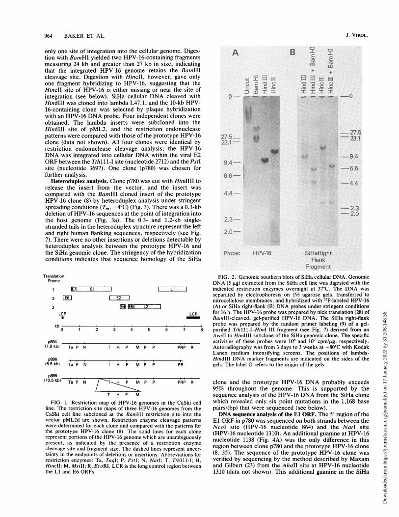
Analysis of HPV-16 DNA integrated into the CaSki cellgenome. Southern blot analysis of CaSki cellular DNA withHPV-16 probes has shown the presence of approximately600 copies of integrated HPV-16 DNA per genome (44).Digestion of CaSki DNA with BamHI yielded two majorHPV-16-containing fragments of 7.9 and 6.5 kilobases (kb)and several minor fragments including 7.25- and 10.5-kbfragments (data not shown). The production of only a fewmajor HPV-16-containing fragments by digestion withBamHI, a single-cut restriction endonuclease for HPV-16,indicates that most of the HPV-16 genomes are tandemlyarranged in a head-to-tail fashion. Representative clones ofHPV-16 containing BamHI fragments were obtained bycloning the BamHI-cut CaSki DNA into lambda L47.1 andselecting by hybridization to a HPV-16 probe. Representa-tive clones with insert sizes of 10.5, 7.9, and 6.5 kb wereanalyzed by detailed restriction analysis. The comparison ofthese clones with the restriction endonuclease site map ofthe prototypic HPV-16 DNA (8, 35) is shown in Fig. 1. Clonep894 (7.9-kb insert) was indistinguishable from the prototypeHPV-16 DNA clone and represented tandemly repeatedfull-length copies of the HPV-16 genome with no largedeletions, insertions, or rearrangements. Clone p896 had adeletion of 1.4 kb centered on the long control region (LCR).This deletion did not extend to the EcoRI site at nucleotide6818 or the TaqI site at nucleotide 505. The deletion includedthe 3' end of the L1 ORF, the LCR, and most of the E6 ORF.The third clone, p895, had a tandem repeat of approximately2.6 kb which contained the E2, E4, and E5 ORFs andportions of the El and L2 ORFs. The Narl site at nucleotide1310 and the PstI site at nucleotide 4759 lie outside theregion of duplication.
Cloning of HPV-16 DNA integrated into the SiHa genome.SiHa genomic DNA Was digested with no cut (HindIII) or
single-cut (BamHI and HincII) restriction endonucleases forHPV-16 DNA and hybridized with a HPV-16 DNA probe(Fig. 2A). Digestion with HindIII revealed only a singleHPV-16-containing fragment of 10 kb, indicating that there is
VOL. 61, 1987
Dow
nloa
ded
from
http
s://j
ourn
als.
asm
.org
/jour
nal/j
vi o
n 17
Jan
uary
202
2 by
31.
208.
148.
36.
964 BAKER ET AL.
only one site of integration into the cellular genome. Diges-tion with BamHI yielded two HPV-16-containing fragmentsmeasuring 24 kb and greater than 27 kb in size, indicatingthat the integrated HPV-16 genome retains the BamHIcleavage site. Digestion with HincII, however, gave onlyone fragment hybridizing to HPV-16, suggesting that theHinclI site of HPV-16 is either missing or near the site ofintegration (see below). SiHa cellular DNA cleaved withHindIII was cloned into lambda L47.1, and the 10-kb HPV-16-containing clone was selected by plaque hybridizationwith an HPV-16 DNA probe. Four independent clones wereobtained. The lambda inserts were subcloned into theHindIll site of pML2, and the restriction endonucleasepatterns were compared with those of the prototype HPV-16clone (data not shown). All four clones were identical byrestriction endonuclease cleavage analysis; the HPV-16DNA was integrated into cellular DNA within the viral E2ORF between the Tthlll-I site (nucleotide 2712) and the PstIsite (nucleotide 3697). One clone (p780) was chosen forfurther analysis.
Heteroduplex analysis. Clone p780 was cut with HindlIl torelease the insert from the vector, and the insert wascompared with the BamHI cloned insert of the prototypeHPV-16 clone (8) by heteroduplex analysis under stringentspreading conditions (Tm, -4°C) (Fig. 3). There was a 0.3-kbdeletion of HPV-16 sequences at the point of integration intothe host genome (Fig. 3a). The 0.3- and 1.2-kb single-stranded tails in the heteroduplex structure represent the leftand right human flanking sequences, respectively (see Fig.7). There were no other insertions or deletions detectable byheteroduplex analysis between the prototype HPV-16 andthe SiHa genomic clone. The stringency of the hybridizationconditions indicates that sequence homology of the SiHa
TranslatioFrame
2
3L(
kb
p894(7.9 kb)
p896(6.5 kb)
A
c_ _
D cn I I
O-__27.5-~~~~~~~ t.
27.5 --23.1-
9.4-
B mEm
_occI
.fr':
-
Em
P_-4 P
Q 0c c:I I
".P-W. -0
-27.523.1
-9.4
- -6.66.6-
-4.4
4.4-
-2.3- 2.0
2.3-
2.0-
Probe: HPV-16 SiHaRightFlank
Fragment
FIG. 2. Genomic southern blots of SiHa cellular DNA. GenomicDNA (5 ,ug) extracted from the SiHa cell line was digested with the
IE71 El L 1 indicated restriction enzymes overnight at 37°C. The DNA wasFE-6 1 E2___1 separated by electrophoresis on 1% agarose gels, transferred to[EIiE2iII nitrocellulose membranes, and hybridized with 32P-labeled HPV-16
1E5 Li i2 I (A) or SiHa right-flank (B) DNA probes under stringent conditionsCR LCR for 16 h. The HPV-16 probe was prepared by nick translation (28) of
BamHI-cleaved, gel-purified HPV-16 DNA. The SiHa right-flank______,____,_____,____,_____,____ ,_____ ,____ ,___ probe was prepared by the random primer labeling (9) of a gel-
1 2 3 4 5 6 7 8 purified Tthlll-I-Hind III fragment (see Fig. 7) derived from anAvall to HindlIl subclone of the SiHa genomic clone. The specific
_____,___,____ ,___ ,____,___,____,___,____ ,,,______,__ activities of these probes were 108 and 109 cpm/,ug, respectively.Ta P N T H P M P P PRP R Autoradiography was from 3 days to 3 weeks at -80°C with Kodak
Lanex medium intensifying screens. The positions of lambda--t____I___I ___I___I___I___I___I___I ______ Hindlll DNA marker fragments are indicated on the sides of theTa P N T H P M P P PR gels. The label 0 refers to the origin of the gels.
p895(10.5 kb) Ta P N T P M P P PRP R
T H P M
FIG. 1. Restriction map of HPV-16 genomes in the CaSki cellline. The restriction site maps of three HPV-16 genomes from theCaSki cell line subcloned at the BamHI restriction site into thevector pML2d are shown. Restriction enzyme cleavage patternswere determined for each clone and compared with the patterns forthe prototype HPV-16 clone (8). The solid lines for each clonerepresent portions of the HPV-16 genome which are unambiguouslypresent, as indicated by the presence of a restriction enzymecleavage site and fragment size. The dashed lines represent uncer-tainty in the endpoints of deletions or insertions. Abbreviations forrestriction enzymes: Ta, TaqI; P, PstI; N, Narl; T, Tthlll-I; H,HincII; M, MstII; R, EcoRI. LCR is the long control region betweenthe Li and E6 ORFs.
clone and the prototype HPV-16 DNA probably exceeds95% throughout the genome. This is supported by thesequence analysis of the HPV-16 DNA from the SiHa clonewhich revealed only six point mutations in the 1,168 basepairs*(bp) that were sequenced (see below).DNA sequence analysis of the El ORF. The 5' region of the
El ORF in p780 was sequenced on both strands between theNcoI site (HPV-16 nucleotide 864) and the NarI site(HPV-16 nucleotide 1310). An additional guanine at HPV-16nucleotide 1138 (Fig. 4A) was the only difference in thisregion between clone p780 and the prototype HPV-16 clone(8, 35). The sequence of the prototype HPV-16 clone wasverified by sequencing by the method described by Maxamand Gilbert (23) from the AhaII site at HPV-16 nucleotide1310 (data not shown). This additional guanine in the SiHa
J. VIROL.
Dow
nloa
ded
from
http
s://j
ourn
als.
asm
.org
/jour
nal/j
vi o
n 17
Jan
uary
202
2 by
31.
208.
148.
36.
HPV-16 IN HUMAN CERVICAL CARCINOMA CELL LINES 965
f14~~~~~~~~~~~~~~~~~;A.%9 ~ *~74~., '. A
4'"~~~~~~~.4..4?~~~~~~~~.4~~~;4
" left flank
I 0.3 kb deletion
1.2 kb human right flank
a b
clone resulted in a frame shift which fused the Ela and ElbORFs into a single long El ORF (Fig. 4B). This same changehas also been noted in the El ORF of another HPV-16 cloneisolated from a human cervical carcinoma (22). Thus, theorganization of the HPV-16 genome is similar to that of allother papillomaviruses that have been sequenced in that itcontains an intact El ORF.DNA sequence analysis of the junctional and flanking re-
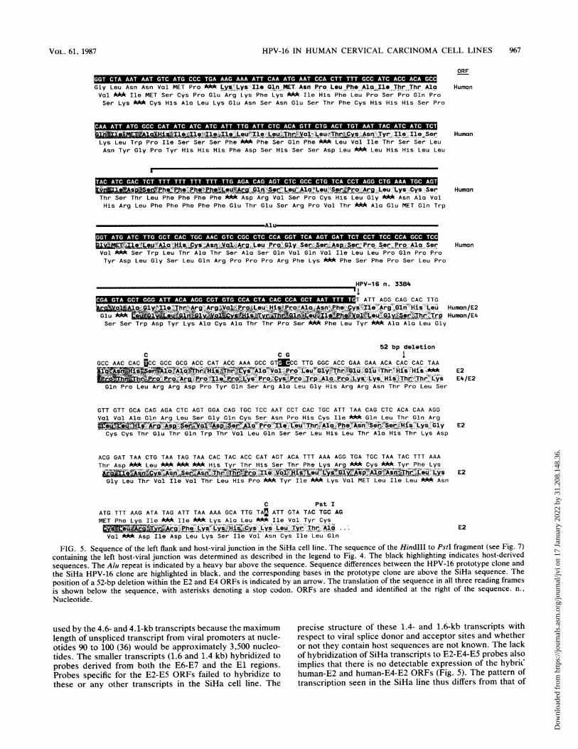
gions. The entire 315 bp of left human-flanking region and thehost-viral junction of the SiHa clone was sequenced from theHindIll site to the PstI site (HPV-16 nucleotide 3696). Asearch of GenBank for sequences homologous to the left-flanking region showed that integration occurred within thecenter of an Alu repeat (32) (Fig. 5). Only half of the Alurepeat remained. The left viral junction was at HPV-16nucleotide 3384. The 5' nucleotides of the E2 and E4 ORFsof HPV-16 were 2725 and 3332, respectively; integrationdisrupted both the E2 and E4 ORFs. A 52-bp deletion ofHPV-16 nucleotides 3459 to 3511 caused a frameshift termi-nation of the E2 ORF and fused the E4 ORF to the 3' portionof the E2 ORF. Two human-viral hybrid ORFs were gener-
tt.> ', b,* V. - .. 4'r - ,,v. ,
1' ;t t ' t'4 -1 4.i
%a .,. p. O'
S wr Uclb X *D '* ' ' t'14~~~~~~~~~~~
To a 4 .
FIG. 3. Heteroduplex comparison of HPV-16 genomic clones.The inserts of the SiHa genomic clone p780 cleaved with HindIIIand the prototype HPV-16 clone (8) cleaved with BamHI were gelpurified by the method described by Vogelstein and Gillespie (40).Hybridization was carried out at Tm -25°C and electron microscopyspreads were performed at T,,, -4°C in 0.01 M Tris (pH 8.5)-0.01 MEDTA-80% deionized formamide. (A and B) Two representativeelectron micrographs of heteroduplexes between cloned inserts.Interpretive drawings of panels A and B with dimensions of substi-tutions and deletions are shown in panels a and b, respectively.
ated across the left junction, encoding potential human-E2(labeled H2) and human-E4-E2 (labeled H4/2) fusion pro-teins of 123 and 152 amino acids, respectively (Fig. 5). Asearch for homology with proteins in the National Biomed-ical Research Foundation protein data base yielded nosignificant homologies other than with the E2 and E4 pro-teins of other papillomaviruses.A 1.2-kb AvaII fragment was sequenced which contained
the 5' half of the E2 ORF and the right viral-host junction.The right viral-host junction was at HPV-16 nucleotide 3132.There was a deletion of 251 nucleotides of the HPV-16 E2ORF between the two viral-host junctions. This deletionincluded the HincII site at HPV-16 nucleotide 3210. The 5'E2 ORF terminated after three codons in the human flankingregion (Fig. 6A). No splice consensus sequence was found inthis region, and therefore, there was no evidence that the E2ORF might be fused at the level of transcriptional processingto a host ORF. In addition, no significantly large ORFs werefound in the greater than 750 bp of right flanking DNA thatwas sequenced. The sequence immediately to the right of thejunction did not contain any Alu repeat sequences, suggest-
VOL. 61, 1987
Dow
nloa
ded
from
http
s://j
ourn
als.
asm
.org
/jour
nal/j
vi o
n 17
Jan
uary
202
2 by
31.
208.
148.
36.
966 BAKER ET AL.
AG+A G C+T C A+C
T SA . ,TTT _ *,
* G f _ _G wiT
AG1-T
114.1-C_%k _
B 859 116/
Ela1104 2810
Elbb
100 1110 1120 1130 1140 115
ATAGAG TGCA G T A 5AG 3 - C T AAAA C 0A A P.GA `TATT GG TAGTCCACTAGr GAT aTTAGT G G ATG TG -AGAC AATAATAT
G
859 2811
L . _ ElFIG. 4. Sequence analysis of the El region of the SiHa genomic clone. (A) The 5' portion of the El ORF between the NcoI and Narl sites
in the SiHa genomic clone p780 was sequenced on both strands by the method described by Maxam and Gilbert (23). Fragments were labeledwith 32P at the 3' ends with the Klenow fragment of DNA polymerase 1 (21). The sequencing gel of the region between HPV-16 nucleotides1131 and 1144 is presented along with the DNA sequence at the left of the figure, and the cleavage specificities are indicated above each lane.The arrow indicates the additional guanine. (B) The sequence of the prototype HPV-16 clone (35) from nucleotides 1100 to 1180 is shown inthe center, along with the location of the additional guanine (arrow) found in the SiHa HPV-16 genomic clone p780 at HPV-16 nucleotide 1138.Above the sequence is a schematic representation of the El ORFs of the prototype HPV-16 clone. The schematic representation of the ElORF of the SiHa genomic clone is shown below the sequence.
ing that cellular sequences were deleted along with the 251nucleotides of HPV-16 (see below). The sequence of theSpeI to AvaIl portion of the AvaIl fragment is shown in Fig.6B. A search of GenBank for homologous sequences re-vealed a close homology with the human 0 repeat (39).
Determination of the HPV-16 DNA copy number in the SiHacell line. Hybridization of Southern blots of SiHa cellularDNA with a unique probe from the human-flanking DNAwas used to determine the number of copies of the HPV-16genome that are integrated into the SiHa genome. Such aprobe should give bands of equal intensity for both thenormal allele and the allele with the integrated HPV-16 DNAif there is only a single integrated copy. The 850-nucleotideTthlll-I to HindlIl fragment from the right human flank(Fig. 7) was chosen to avoid the highly repeated Alu and0-repeat elements. The intensity of hybridization to the10-kb HindIll and 13.4-kb HincIl HPV-16-containing frag-ments was, at most, twice the intensity of the hybridizationto the 4.6- and 2.7-kb fragments from the normal allele (Fig.2B). This indicates that there are at most two copies of theHPV-16 genome integrated in the SiHa cell line. Becausethere was only a single site of integration, it is most likelythat there is only one copy of the HPV-16 genome per cell.We cannot rule out that there was a duplication of the regionof the human genome containing the integrated HPV-16genome, however.Only the 10-kb HindlIl and 13.4-kb HincII fragments were
cut by BamHI, as expected for a single-cut enzyme forHPV-16 DNA. BamHI did not cleave within the vicinity(>18 kb) of the integrated HPV-16 DNA (Fig. 2A). Assum-ing that there is no restriction site polymorphism, we canconclude from the length of the HindlIl fragment (4.6 kb) ofthe normal allele that there must be at least a 2.6-kb deletionof human DNA associated with the integration of HPV-16into the SiHa genome. This is consistent with the presence ofonly half of an Alu repeat at the left host-virus junction andno Alu repeat at the right junction. There was insufficienthomology between cellular sequences at the host-viral junc-tions and viral sequences in the E2 ORF to suggest thatintegration occurred by homologous recombination. Be-
cause of the large deletions that occurred, however, homol-ogous recombination cannot be ruled out without sequencingthe normal cellular allele.
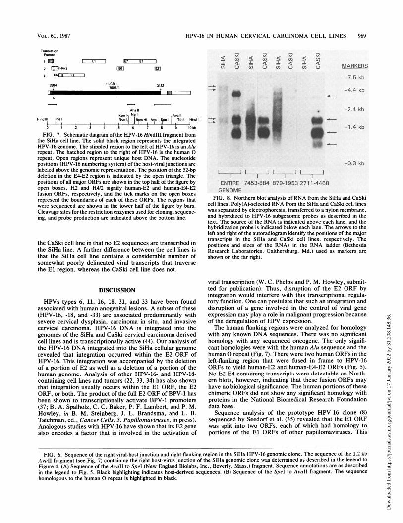
Transcription analysis of SiHa and CaSki. HPV-16 tran-scription in the SiHa and CaSki cell lines was examined byNorthern blot analysis by using a variety of strand-specificand double-stranded subgenomic probes. Strand-specificsubgenomic probes were generated that covered nearly all ofboth strands of the HPV-16 genome. In each cell line, nohybridization was detected to fragments representing thelate ORFs or the LCR. The viral transcripts observed in thecell lines were all transcribed from one strand. The majorviral transcripts in both cell lines were composed principallyof sequences derived from the E6, E7, and El ORFs.The CaSki cell line contained, at relatively high abun-
dance, four major transcripts: 3.6, 1.5, 1.0, and 0.6 kb (Fig.8). Minor transcripts of 3.0 and 2.2 kb were detected withlonger exposures. Viral transcription in the CaSki cell lineincluded one long transcript (3.6 kb) that originated in the E6region and that continued through the El region into the E2ORF. It could not be determined from the Northern blot datawhether this transcript was spliced. The 1.5- and 1.0-kbtranscripts appear to splice the E6-E7 region to the E2region. The smaller 650-base transcript hybridized only toE2 ORF-specific probes, suggesting that a downstream pro-moter, possibly analogous to the P2443 or P3080 promoter ofbovine papillomavirus type 1 (BPV-1) (38, 43), might beactive in these cells.The SiHa cell line also contained four major transcripts:
4.6, 4.1, 1.6, and 1.4 kb (Fig. 8). In addition, longer autora-diographic exposures revealed a number of low-abundanceviral transcripts, particularly when probes that spanned theEl ORF were used. The two large viral transcripts (4.6 and4.1 kb) hybridized only with E6-E7-specific probes, suggest-ing that they are largely comprised of host cellular se-quences. All transcripts must use cellular polyadenylationsignals because integration placed the normal viralpolyadenylation signal upstream of the viral promoter(s). Apolyadenylation signal (AATAAA) was found in the rightflank sequence (Fig. 6B, nucleotide 320) but could not be
J. VIROL.
Dow
nloa
ded
from
http
s://j
ourn
als.
asm
.org
/jour
nal/j
vi o
n 17
Jan
uary
202
2 by
31.
208.
148.
36.
HPV-16 IN HUMAN CERVICAL CARCINOMA CELL LINES 967
Gly Leu Asn Asn Val MET Pro ***Lys'-L, Ile Gln MET Asn Pro Lou Phe AlIle ;Thr Thr AlaVal A** Ile MET Ser Cys Pro Glu Arg Lys Phe Lys #b* Ile His Phe Leu Pro Ser Pro Gln ProSer Lys *A* Cys His Ala Leu Lys Glu Asn Ser Asn Glu Ser Thr Phe Cys His His His Ser Pro
Lys Leu Trp Pro Ile Ser Ser Ser Phe *** Phe Ser Gln Phe b** Leu Val Ile Thr Ser Ser LeuAsn Tyr Gly Pro Tyr His His His Phe Asp Ser His Ser Ser Asp Leu *A* Leu His His Leu Leu
S ___.h3stu ,ysGy Ser-Thr Ser Thr Leu Phe Phe Phe Phe *** Asp Arg Val Ser Pro Cys His Leu Gly * Asn Ala ValHis Arg Leu Phe Phe Phe Phe Phe Glu Thr Glu Ser Arg Pro Val Thr *** Ala Glu MET Gln Trp
Val *M* Ser Trp Leu Thr Ala Thr Ser Ala Ser Gln Val Gln Val Ile Leu Leu Pro Gln Pro Pro
Tyr Asp Leu Gly Ser Leu Gln Arg Pro Pro Pro Arg Phe Lys *M* Phe Ser Phe Pro Ser Leu Pro
ORF
Human
Human
Human
Human
_HPV-16 n. 3384
T ATT AGG CAG CAC TTGKo~Gl~~HstrgGl& His L Human/E2
Glu AQh,l _,
Human/E4Ser Ser Trp Asp Tyr Lys Ala Cys Ala Thr Thr Pro Ser *** Phe Leu Tyr *** Ala Ala Leu Gly
Gln Pro Leu Arg Arg Asp Pro Tyr Gln Ser Arg Ala Leu Gly His Arg Arg Asn Thr Pro Leu Ser
GTT GTT GCA CAG AGA CTC AGT GGA CAG TGC TCC AAT CCT CAC TGC ATT TAA CAG CTC ACA CAA AGGVal Val Ala Gln Arg Leu Ser Gly Gln Cys Ser Asn Pro His Cys Ile **A Gln Leu Thr Gln Arg
< y E2Cys Cys Thr Glu Thr Gln Trp Thr Val Leu Gln Ser Ser Leu His Leu Thr Ala His Thr Lys Asp
ACG GAT TAA CTG TAA TAG TAA CAC TAC ACC CAT AGT ACA TTT AAA AGG TGA TGC TAA TAC TTT AAAThr Asp * Leu * His Tyr Thr His Ser Thr Phe Lys Arg *** Cys * Tyr Phe Lys
~~~~~T]~~~~~~~~~~~E2Gly Leu Thr Val Ile Val Thr Leu His Pro *** Tyr Ile *kM Lys Val MET Leu Ile Leu ** Asn
C Pst IATG TTT AAG ATA TAG ATT TAA AAA GCA TTG TAn ATT GTA TAC TGC AGMET Phe Lys Ile *** Ile MA Lys Ala Leu MA* Ile Val Tyr Cys~ TyySThr E2
Val MA* Asp Ile Asp Leu Lys Ser Ile Val Asn Cys Ile Leu Gln
FIG. 5. Sequence of the left flank and host-viral junction in the SiHa cell line. The sequence of the HindIII to PstI fragment (see Fig. 7)containing the left host-viral junction was determined as described in the legend to Fig. 4. The black highlighting indicates host-derivedsequences. The Alu repeat is indicated by a heavy bar above the sequence. Sequence differences between the HPV-16 prototype clone andthe SiHa HPV-16 clone are highlighted in black, and the corresponding bases in the prototype clone are above the SiHa sequence. Theposition of a 52-bp deletion within the E2 and E4 ORFs is indicated by an arrow. The translation of the sequence in all three reading framesis shown below the sequence, with asterisks denoting a stop codon. ORFs are shaded and identified at the right of the sequence. n.,
Nucleotide.
used by the 4.6- and 4.1-kb transcripts because the maximum precise structure of these 1.4- and 1.6-kb transcripts withlength of unspliced transcript from viral promoters at nucle- respect to viral splice donor and acceptor sites and whetherotides 90 to 100 (36) would be approximately 3,500 nucleo- or not they contain host sequences are not known. The lacktides. The smaller transcripts (1.6 and 1.4 kb) hybridized to of hybridization of SiHa transcripts to E2-E4-E5 probes alsoprobes derived from both the E6-E7 and the El regions. implies that there is no detectable expression of the hybricProbes specific for the E2-E5 ORFs failed to hybridize to human-E2 and human-E4-E2 ORFs (Fig. 5). The pattern ofthese or any other transcripts in the SiHa cell line. The transcription seen in the SiHa line thus differs from that of
AspIle Lys Phe Ala Arg Gly Arg Gly Gln Gly Lys Arg Trp Arg Leu Phe Ala Asn Val A MET
Leu Ser Leu His Glu Asp Glu Asp Lys Glu Asn Asp Gly Asp Ser Leu Pro Thr Phe Lys Cys
GTG TGT CAG GAC AAA ATA CTA ACA CAT TAT GAA AAT GAT AGT ACA GAC CTA CGT GAC CAT ATA
VYlC ~ p~ys I1~ LOU ~j ~i~i1ePCys Val Arg Thr Lys Tyr ** His Ile MET Lys MET Ile Val Gln Thr Tyr Val Thr Ile A*Val Ser Gly Gln Asn Thr Asn Thr Leu AcA Lys M*A * Tyr Arg Pro Thr MAc Pro Tyr Arg
GAC TAT TGG AAA CAC ATG CGC CTA GAA TGT GCT ATT TAT TAC AAG GCC AGA GAA ATG GGA TTT
Thr Ile Gly Asn Thr Cys Ala c Asn Val Leu Phe Ile Thr Arg Pro Glu Lys Trp Asp Leu
Leu Leu Glu Thr His Ala Pro Arg MET Cys Tyr Leu Leu Gln Gly Gln Arg Asn Gly Ile ***
Asn Ile Leu Thr Thr Lys Trp Cys Gln His Trp Leu Tyr Gln Arg Ile Lys His Tyr Lys Gln
Thr Tyr MA* Pro Pro Ser Gly Ala Asn Thr Gly Cys Ile Lys Glu MA Ser Ile Thr Ser Asn
ATT GAA CTG CAA CTA ACG TTA GAA ACA ATA TAT AAC TCA CAA TAT AGT AAT GAA AAG TGG ACA
Leu Asn Cys Asn o*l Arg *M Lys Gln Tyr Ile Thr His Asn Ile Val MET Lys Ser Gly HisMA Thr Ala Thr Asn Val Arg Asn Asn Ile MA Leu Thr Ile MA* A M*A Lys Val Asp Ile
GTTA CAA GAC GTT AGC CTT GAA GTG TAT TTA ACT ECA CCA ACA GGA TGT ATA AAA AAA CAT GGA
Tyr Lys Thr Leu Ala Leu Lys Cys Ile A Leu His Gln Gln Asp Val A Lys Asn MET AspThr Arg Arg M* Pro A* Ser Val Phe Asn Tyr Thr Asn Arg MET Tyr Lys Lys Thr Trp Ile
HPV-16 n. 3132
TAT ACA GTG GAA GTG CAG TTT GAT GGA GAC ATA TGC |_yr~iU~. :.V_a xWCycS,,o ~Al~ MA Leu Thr Asn Leu AspIle Gln Trp Lys Cys Ser Leu MET Glu Thr Tyr Ala Ala Gln Leu Ser MA Leu Thr Trp Thr
Tyr Ser Gly Ser Ala Val MAoA Trp Arg His MET Leu Leu Ser Leu Ala Asp *MA Pro Gly Gln
Arg A* Asp Trp Ser Leu Leu Gly Ile Val Thr MET Ser Lys Asp Asp Cys MA Glu Pro Ala
Asp Lys Thr Gly Ala Tyr Trp Ala Leu Ser Gln Cys Leu Lys MET Thr Val Arg Asn Leu Leu
Ile Arg Leu Glu Pro Ile Gly His Cys His Asn Val *MA Arg MAd Leu Leu Gly Thr Cys Phe
ATAAATTTACCACTTCTCCAATTGGACTTAAATCAACCAGCCTGG 1IIf1f1lot te e T
330 340 350 360 370 380 390 400
410 420 430 440 450 460 470 480
490 500 510 520 530 540 550 560
.atteleu,IF$X ilte. _ ATTT
570 580 590 600 610 620 630 640
J. VIROL.
Dow
nloa
ded
from
http
s://j
ourn
als.
asm
.org
/jour
nal/j
vi o
n 17
Jan
uary
202
2 by
31.
208.
148.
36.
HPV-16 IN HUMAN CERVICAL CARCINOMA CELL LINES 969
TranslationFrames
1 Et C2 [E3H4/23 E5t IF
3384
Ll I IE7 El
E [m
-LCR-47905/1
CI
Y c ) E< C < II)I C: UI C)I 6 I Cuz C) en V (n u en U
3132
_
Aha IIKpnlI NanI Ava Il
Hind II PI Nco Bam Hl Ava Il Spe Tth Hind ilI
0 1 2 3 4 5 6 7 8 9 10kb
FIG. 7. Schematic diagram of the HPV-16 HindIII fragment fromthe SiHa cell line. The solid black region represents the integratedHPV-16 genome. The stippled region to the left of HPV-16 is an Alurepeat. The hatched region to the right of HPV-16 is the human 0repeat. Open regions represent unique host DNA. The nucleotidepositions (HPV-16 numbering system) of the host-viral junctions arelabeled above the genomic representation. The position of the 52-bpdeletion in the E4-E2 region is indicated by the open triangle. Thepositions of all major ORFs are shown in the top half of the figure byopen boxes. H2 and H4/2 signify human-E2 and human-E4-E2fusion ORFs, respectively, and the tick marks on the open boxesrepresent the boundaries of each of these ORFs. The regions thatwere sequenced are shown in the lower half of the figure by bars.Cleavage sites for the restriction enzymes used for cloning, sequenc-ing, and probe production are indicated above the bottom line.
the CaSki cell line in that no E2 sequences are transcribed inthe SiHa line. A further difference between the cell lines isthat the SiHa cell line contains a considerable number ofsomewhat poorly delineated viral transcripts that traversethe El region, whereas the CaSki cell line does not.
DISCUSSION
HPVs types 6, 11, 16, 18, 31, and 33 have been foundassociated with human anogenital lesions. A subset of these(HPV-16, -18, and -33) are associated predominantly withsevere cervical dysplasia, carcinoma in situ, and invasivecervical carcinoma. HPV-16 DNA is integrated into thegenomes of the SiHa and CaSki cervical carcinoma derivedcell lines and is transcriptionally active (44). Our analysis ofthe HPV-16 DNA integrated into the SiHa cellular genomerevealed that integration occurred within the E2 ORF ofHPV-16. This integration was accompanied by the deletionof a portion of E2 as well as a deletion of a portion of thehuman genome. Analysis of other HPV-16- and HPV-18-containing cell lines and tumors (22, 33, 34) has also shownthat integration usually occurs within the El ORF, the E2ORF, or both. The product of the full E2 ORF of BPV-1 hasbeen shown to transcriptionally activate BPV-1 promoters(37; B. A. Spalholz, C. C. Baker, P. F. Lambert, and P. M.Howley, in B. M. Steiriberg, J. L. Brandsma, and L. B.Taichman, ed., Cancer Cells. 5. Papillomaviruses, in press).Analogous studies with HPV-16 have shown that its E2 genealso encodes a factor that is involved in the activation of
iii
-2.4 kb
-1.4 kb
-0.3 kb
ENTIRE 7453-884 879-1953 2711-4468GENOME
FIG. 8. Northern blot analysis ofRNA from the SiHa and CaSkicell lines. Poly(A)-selected RNA from the SiHa and CaSki cell lineswas separated by electrophoresis, transferred to a nylon membrane,and hybridized to HPV-16 subgenomic probes as described in thetext. The source of the RNA is indicated above each lane, and thehybridization probe is indicated below each lane. The arrows to theleft and right of the autoradiogram identify the positions of the majortranscripts in the SiHa and CaSki cell lines, respectively. Thepositions and sizes of the RNAs in the RNA ladder (BethesdaResearch Laboratories, Gaithersburg, Md.) used as markers areshown on the far right.
viral transcription (W. C. Phelps and P. M. Howley, submit-ted for publication). Thus, disruption of the E2 ORF byintegration would interfere with this transcriptional regula-tory function. One can postulate that such an integration anddisruption of a gene involved in the control of viral geneexpression may play a role in malignant progression becauseof the deregulation of HPV expression.The human flanking regions were analyzed for homology
with any known DNA sequences. There was no significanthomology with any sequenced oncogene. The only signifi-cant homologies were with the human Alu sequence and thehuman 0 repeat (Fig. 7). There were two human ORFs in theleft-flanking region that were fused in frame to HPV-16ORFs to yield human-E2 and human-E4-E2 ORFs (Fig. 5).No E2-E4-containing transcripts were detectable on North-ern blots, however, indicating that these fusion ORFs mayhave no biological significance. The human portions of thesechimeric ORFs did not show any significant homology withproteins in the National Biomedical Research Foundationdata base.Sequence analysis of the prototype HPV-16 clone (8)
sequenced by Seedorf et al. (35) revealed that the El ORFwas split into two ORFs, each of which had homology toportions of the El ORFs of other papillomaviruses. This
MARKERS
-7.5 kb
-4.4 kb
FIG. 6. Sequence of the right viral-host junction and right-flanking region in the SiHa HPV-16 genomic clone. The sequence of the 1.2 kbAvall fragment (see Fig. 7) containing the right host-virus junction of the SiHa genomic clone was determined as described in the legend toFigure 4. (A) Sequence of the Avall to Spel (New England Biolabs, Inc., Beverly, Mass.) fragment. Sequence annotations are as describedin the legend to Fig. 5. Black highlighting indicates host-derived sequences. (B) Sequence of the Spel to Avall fragment. The sequencehomologous to the human 0 repeat is highlighted in black.
VOL. 61, 1987
-.W.-
All--
*14.
Dow
nloa
ded
from
http
s://j
ourn
als.
asm
.org
/jour
nal/j
vi o
n 17
Jan
uary
202
2 by
31.
208.
148.
36.
970 BAKER ET AL.
prototype clone of HPV-16 was isolated from a tumor andmay have been an integrated genome. Because the El ORFof BPV-1 is known to be involved with DNA replication andplasmid maintenance (20, 30), this mutation might be respon-sible for integration of E4PV-16 DNA in the tumor fromwhich it was cloned. Analysis of the corresponding region ofthe El ORF in the SiHa genomic clone, however, revealedan extra guanine at nucleotide 1138, with the result that theEl ORF was intact. This has also been found for HPV-16DNA that was integrated into the genome of another cervicalcarcinoma (22). It is unlikely, therefore, that a mutation inEl is a prerequisite for integration of HPV DNA andsubsequent malignant progression.
Analysis of HPV-16 transcription in the SiHa cell lineindicated that there is no detectable expression of theE2-E4-E5 region, the LCR, or the late region. This impliesthat there is no significant readthrough from upstream cellu-lar promoters into viral sequences. The predominant tran-scripts come from the E6-E7 region. The E6 gene product ofBPV-1 has been shown to have a transforming function (31,43). In addition, the E6 ORFs of HPV-16 and HPV-18contain splice donor and acceptor sites which are not presentin HPV-6 or HPV-11 (33, 36). Thus, the HPVs associatedwith malignant cervical lesions may make E6 proteins thatdiffer from those of the other HPVs. It is tempting tospeculate that expression of the E6 gene or an E6-E7 fusiongene product is important in malignant progression. TheE6-E7 region is presumably transcribed from viral promot-ers. Because the E2 trans-activating protein is not expressedin the SiHa cells, we presume that the viral promoters areactivated directly by cellular transcriptional factors or areenhanced by adjacent cellular cis-acting regulatory ele-ments. In addition, the normal early region polyadenylationsignals are no longer downstream of the promoters. TheHPV-16 sequences must be spliced to RNA from adjacenthuman DNA, and transcription must use cellular polyade-nylation sites. The control of the expression of a potentialviral oncogene by cellular regulatory elements may becrucial for the progression of a benign to a malignant lesion.The correlation of transcription patterns with the genomic
structure is more difficult for the CaSki cell line than for theSiHa cell line because of the many integrated copies ofHPV-16 DNA in the CaSki cell line. The three most abun-dant HPV-16 genomic structures (Fig. 1) are present in largetandem arrays. The junctional copies are present in very lowabundance and have not yet been isolated or analyzed.Similar to HPV-16 transcription in the SiHa cell line, how-ever, there is no evidence of transcription of the late regionor of transcription of the LCR in CaSki cells. Three of thefour transcripts contain the E6-E7 region and are presum-ably transcribed from viral LCR promoters. Unlike the SiHacells, only the largest transcript contains the El region, andall transcripts contain the E2 region. The smallest transcriptcontains only the E2 region and is probably transcribed fromthe equivalent of the P2443 or P3080 promoter in BPV-1 (38,43). Results of the analyses of Smotkin and Wettstein (36)have suggested that the CaSki transcripts do not utilize theviral polyadenylation site, implying that viral sequences areeither spliced to cellular sequences or are fused to cellularsequences at the viral-host junctions. This suggests that onlythe terminal HPV-16 copies in the large tandem repeats maybe transcriptionally active and supports the hypothesis thatHPV-16 promoters are under altered regulation, perhapsbecause of adjacent cis-acting regulatory elements. It ispossible that the key event in malignant progression is theintegration of HPV-16 near such a cellular regulatory ele-
ment and that it is necessary to destroy the HPV-16 trans-acting transcriptional regulatory element(s) by integrationwithin the E2 region of HPV-16.
ACKNOWLEDGMENTS
We thank Jean Noe for technical assistance, Warren Jelinek forsupplying the lambda L47.1, Susan Haynes for advice and help withthe genomic cloning, and George Michaels for running the GenBanksearches.W.C.P. was supported by National Research Service Award
postdoctoral fellowship CA 07713. This study was supported in partby grant 1760 from the Council for Tobacco Research to V.L.M.J.B. and M.A.G. were supported by Public Health Servicecontract NOI-CO-23910 with Program Resources, Inc., from theNational Cancer Institute.
LITERATURE CITED1. Aviv, H., and P. Leder. 1972. Purification of biologically active
globin messenger RNA by chromatography on oligothymidylicacid-cellulose. Proc. Natl. Acad. Sci. USA 69:1408-1412.
2. Boshart, M., L. Gissmann, H. Ikenberg, A. Kleinheinz, W.Scheurlen, and H. zur Hausen. 1984. A new type of papil-lomavirus DNA and its presence in genital cancer biopsies andin cell lines derived from cervical cancer. EMBO J. 3:1151-1157.
3. Chirgwin, J. M., A. E. Przybyla, R. J. MacDonald, and W. J.Rutter. 1979. Isolation of biologically active ribonucleic acidfrom sources enriched in ribonuclease. Biochemistry 18:5294-5299.
4. Church, G. M., and W. Gilbert. 1984. Genomic sequencing.Proc. Natl. Acad. Sci. USA 81:1991-1995.
5. Crum, C. P., H. Ikenberg, R. M. Richart, and L. Gissmann.1984. Human papillomavirus type 16 and early cervical neopla-sia. N. Engl. J. Med. 310:880-883.
6. Crum, C. P., M. Mitao, R. U. Levine, and S. Silverstein. 1985.Cervical papillomaviruses segregate within morphologically dis-tinct precancerous lesions. J. Virol. 54:675-681.
7. Davis, R. W., M. Simon, and N. Davidson. 1971. Electronmicroscopic heteroduplex methods for mapping regions of basehomology in nucleic acids. Methods Enzymol. 21:413-428.
8. Durst, M., L. Gissmann, H. Ikenberg, and H. zur Hausen. 1983.A papillomavirus DNA from a cervical carcinoma and itsprevalence in cancer biopsy samples from different geographicregions. Proc. Natl. Acad. Sci. USA 80:3812-3815.
9. Feinberg, A. P., and B. Vogelstein. 1984. A technique forradiolabeling DNA restriction endonuclease fragments to highspecific activity. Anal. Biochem. 137!266-267.
10. Gissmann, L. 1984. Papillomaviruses and their association withcancer in animals and in man. Cancer Surv. 3:161-181.
11. Gissmann, L., and E. Schwarz. 1986. Persistence and expressionof human papillomavirus DNA in genital cancer. CIBA Found.Symp. 120:190-207.
12. Heidecker, G., J. Messing, and B. Gronenborn. 1980. A versatileprimer for DNA sequencing in the M13mp2 cloning system.Gene 10:69-73.
13. Hu, N. T., and J. Messing. 1982. The making of strand-specificM13 probes. Gene 17:271-277.
14. Jarrett, W. F. H., P. E. McNeil, W. T. R. Grimshaw, I. E.Selman, and W. I. M. McIntyre. 1978. High incidence area ofcattle cancer with a possible interaction between an environ-mental carcinogen and a papilloma virus. Nature (London)274:215-217.
15. Jarrett, W. F. H., J. Murphy, B. W. O'Neil, and H. M. Laird.1978. Virus-induced papillomas of the alimentary tract of cattle.Int. J. Cancer 22:323-328.
16. Kidd, J. G., and P. Rous. 1937. Effect of the papillomavirus(Shope) upon tar warts of rabbits. Proc. Soc. Exp. Biol. Med.37:518-520.
17. Law, M. F., D. R. Lowy, I. Dvoretzky, and P. M. Howley. 1981.Mouse cells transformed by bovine papillomavirus contain onlyextrachromosomal viral DNA sequences. Proc. Natl. Acad. Sci.USA 78:2727-2731.
J. VIROL.
Dow
nloa
ded
from
http
s://j
ourn
als.
asm
.org
/jour
nal/j
vi o
n 17
Jan
uary
202
2 by
31.
208.
148.
36.
HPV-16 IN HUMAN CERVICAL CARCINOMA CELL LINES 971
18. Lehrach, H., D. Diamond, J. M. Wozney, and H. Boedtker. 1977.RNA molecular weight determination by gel electrophoresisunder denaturing conditions, a critical re-examination. Bio-chemistry 16:4743-4751.
19. Lipman, D., and W. Pearson. 1985. Rapid and sensitive proteinsimilarity searches. Science 227:1435-1441.
20. Lusky, M., and M. R. Botchan. 1985. Genetic analysis of bovinepapillomavirus type 1 trans-acting replication factors. J. Virol.53:955-965.
21. Maniatis, T., E. F. Fritsch, and J. Sambrook. 1982. Molecularcloning: a laboratory manual. Cold Spring Harbor Laboratory,Cold Spring Harbor, N.Y.
22. Matsukura, T., T. Kanda, A. Furuno, H. Yoshikawa, T.Kawana, and K. Yoshiike. 1986. Cloning of monomeric humanpapillomavirus type 16 DNA integrated within cell DNA from acervical carcinoma. J. Virol. 58z979-982.
23. Maxam, A., and W. Gilbert. 1977. A new method for sequencingDNA. Proc. Natl. Acad. Sci. USA 74:560-564.
24. McCance, D. J., M. J, Campion, P. K. Clarkson, P. M. Chesters,D. Jenkins, and A. Singer. 1985. Prevalence of human papil-lomavirus type 16 DNA sequences in cervical intraepithelialneoplasia and invasive carcinoma of the cervix. Br. J. Obstet.Gynaecol. 92:1101-1105.
25. McCance, D. J., P. K. Clarkson, J. L. Dyson, and A. Singer.1985. Human papillomavirus types 6 and 16 in multifocalintraepithelial neoplasias of the female lower genital tract. Br. J.Obstet. Gynaecol. 92:1093-1100.
26. McConaughy, B. L., C. D. Laird, and B. J. McCarthy. 1969.Nucleic acid reassociation in formamide. Biochemistry 8:3289-3295.
27. Pater, M. M., and A. Pater. 1985. Human papillomavirus types16 and 18 sequences in carcinoma cell lines of the cervix.Virology 145:3313-318.
28. Rigby, P. D., M. D. Rhodes, and P. Berg. 1977. Labelingdeoxyribonucleic acid to high specific activity in vitro by nicktranslation with DNA polymerase I. J. Mol. Biol. 113:237-251.
29. Rous, P., and J. G. Kidd. 1936. The carcinogenic effect of avirus upon tarred skin. Science 83:468-469.
30. Sarver, N., M. S. Rabson, Y. C. Yang, J. C. Byrne, and P. M.Howley. 1984. Localization and analysis of bovine papil-lomavirus type 1 transforming functions. J. Virol. 52:377-388.
31. Schiller, J. T., W. C. Vass, and D. R. Lowy. 1984. Identificationof a second transforming region in bovine papillomavirus DNA.Proc. Natl. Acad. Sci. USA 81:7880-7884.
32. Schmid, C. W., and W. R. Jelinek. 1982. The Alu family ofdispersed repetitive sequences. Science 216:1065-1070.
33. Schneider-Gadicke, A., and E. Schwarz. 1986. Different humancervical carcinoma cell lines show similar transcription patternsof human papillomavirus type 18 early genes. EMBO J.5:2285-2292.
34. Schwarz, E., U. K. Freese, L. Gissmann, W. Mayer, B.Roggenbuck, A. Stremlau, and H. zur Hausen. 1985. Structureand transcription of human papillomavirus sequences in cervicalcarcinoma cells. Nature (London) 314:111-114.
35. Seedorf, K., G. Krammer, M. Durst, S. Suhai, and W.Rowekamp. 1985. Human papillomavirus type 16 DNA se-quence. Virology 145:181-185.
36. Smotkin, D., and F. 0. Wettstein. 1986. Transcription of humanpapillomavirus type 16 early genes in a cervical cancer and acancer-deri-ved cell line and identification of the E7 protein.Proc. Natl. Acad. Sci. USA 83:4680-4684.
37. Spalholz, B. A., Y. C. Yang, and P. M. Howley. 1985.Transactivation of a bovine papilloma virus transcriptionalregulatory element by the E2 gene product. Cell 42:183-191.
38. Stenlund, A., J. Zabielski, H. Ahola, J. Moreno-Lopez, and U.Pettersson. 1985. Messenger RNAs from the transformingregion of bovine papilloma virus type 1. J. Mol. Biol. 182:541-554.
39. Sun, L., K. E. Paulson, C. W. Schmid, L. Kadyk, and L.Leinwand. 1984. Non-Alu family interspersed repeats in humanDNA and their transcriptional activity. Nucleic Acids Res.12:2669-2690.
40. Vogelstein, B., and D. Gillespie. 1979. Preparative and analyticalpurification of DNA from agarose. Proc. Natl. Acad. Sci. USA76:615-619.
41. Wagner, D., H. Ikenberg, N. Boehm, and L. Gissmann. 1984.Identification of human papillomavirus in cervical swabs bydeoxyribonucleic acid in situ hybridization. Obstet. Gynecol.64:767-772.
42. Wilbur, W., and D. Lipman. 1983. Rapid similarity searches ofnucleic acid and protein databases. Proc. Natl. Acad. Sci. USA80:726-730.
43. Yang, Y.-C., H. Okayama, and P. M. Howley. 1985. Bovinepapillomavirus contains multiple transforming genes. Proc.Natl. Acad. Sci. USA 82:1030-1034.
44. Yee, C., I. Krishnan-Hewlett, C. C. Baker, R. Schlegel, andP. M. Howley. 1985. Presence and expression of human papil-lomavirus sequences in human cervical carcinoma cell lines.Am. J. Pathol. 119:361-366.