Molecular basis of Borrelia immune evasion 1 Structural basis for complement evasion by Lyme disease pathogen Borrelia burgdorferi* Arnab Bhattacharjee 1,2 , Jesper S. Oeemig 2,# , Robert Kolodziejczyk 2,3# , Taru Meri 1,3 , Tommi Kajander 2 , Markus J. Lehtinen 1 , Hideo Iwaï 2 , T. Sakari Jokiranta 1,§ and Adrian Goldman 2,3§ . 1 Haartman Institute, Department of Bacteriology & Immunology and Immunobiology Research Program, University of Helsinki, Finland. 2 Institute of Biotechnology, University of Helsinki, Finland. 3 Department of Biosciences, Division of Biochemistry and Biotechnology, University of Helsinki, Helsinki, Finland. *Running title: Molecular basis of Borrelia immune evasion Correspondence should be addressed to: Dr. T. Sakari Jokiranta, Haartman Institute, Haartmaninkatu 3 University of Helsinki, FIN-00014 Helsinki, FINLAND, Tel.: +358 9 1911; Fax.: +358 9 191 26382 email: T.S.J. ([email protected]) or A.G. ([email protected]) #,§ These authors contributed equally to this work Keywords: Immune evasion; X-ray crystallography; NMR spectroscopy; protein complex structure _____________________________________________________________________________________ Background: Borrelia burgdorferi OspE protein recruits complement regulator FH onto the bacteria for immune evasion. Results: We solved the structure of OspE and the OspE:FH complex by NMR and X-ray crystallography. Conclusion: The OspE:FH structure shows how Borrelia evade complement attack by mimicking how host cells protect themselves. Significance: This explains how the bacteria survive in the host and facilitates vaccine design against borreliosis. SUMMARY Borrelia burgdorferi spirochetes that cause Lyme borreliosis survive for a long time in human serum because they successfully evade the complement system, an important arm of innate immunity. The outer surface protein E (OspE) of B. burgdorferi is needed for this since it recruits complement regulator factor H (FH) onto the bacterial surface to evade complement-mediated cell lysis. To understand this process at the molecular level we used a structural approach. First, we solved the solution structure of OspE by NMR, revealing a fold that has not been seen before in proteins involved in complement regulation. Next, we solved the X-ray structure of the complex between OspE and the factor H (FH) C-terminal domains 19 and 20 (FH19-20) at 2.83 Å resolution. The structure shows that OspE binds FH19-20 in a way similar to, but not identical with, that used by endothelial cells to bind FH via glycosaminoglycans. The observed interaction of OspE with FH19-20 allows the full function of FH in down- regulation of complement activation on the bacteria. This reveals the molecular basis for how Borrelia burgdorferi evades innate immunity, and suggests how OspE could be used as a potential vaccine antigen. ________________________________________ The number of patients with Lyme disease, also known as Lyme borreliosis, is increasing in the United States and Europe (1) making this disease one of the major emerging arthropod- borne infections in the world (2,3). The disease is caused by the three main genospecies from the Borrelia burgdorferi sensu lato (s.l.) group: Borrelia burgdorferi sensu stricto (s.s.), Borrelia afzelii, and Borrelia garinii (4). These spirochetes are transmitted to human skin from the mid-gut of Ixodes sp. ticks if the feeding time is long enough (5). The earliest manifestation of the infection is http://www.jbc.org/cgi/doi/10.1074/jbc.M113.459040 The latest version is at JBC Papers in Press. Published on May 8, 2013 as Manuscript M113.459040 Copyright 2013 by The American Society for Biochemistry and Molecular Biology, Inc. by guest on June 28, 2018 http://www.jbc.org/ Downloaded from
Transcript
Molecular basis of Borrelia immune evasion
1
Structural basis for complement evasion by Lyme disease pathogen Borrelia burgdorferi*
Arnab Bhattacharjee1,2, Jesper S. Oeemig2,#, Robert Kolodziejczyk2,3#, Taru Meri1,3, Tommi Kajander2, Markus J. Lehtinen1, Hideo Iwaï2, T. Sakari Jokiranta1,§ and Adrian Goldman2,3§.
1 Haartman Institute, Department of Bacteriology & Immunology and Immunobiology Research Program, University of Helsinki, Finland.
2 Institute of Biotechnology, University of Helsinki, Finland.
3 Department of Biosciences, Division of Biochemistry and Biotechnology, University of Helsinki,
Helsinki, Finland.
*Running title: Molecular basis of Borrelia immune evasion Correspondence should be addressed to: Dr. T. Sakari Jokiranta, Haartman Institute, Haartmaninkatu 3 University of Helsinki, FIN-00014 Helsinki, FINLAND, Tel.: +358 9 1911; Fax.: +358 9 191 26382 email: T.S.J. ([email protected]) or A.G. ([email protected]) #,§ These authors contributed equally to this work Keywords: Immune evasion; X-ray crystallography; NMR spectroscopy; protein complex structure _____________________________________________________________________________________
Background: Borrelia burgdorferi OspE protein recruits complement regulator FH onto the bacteria for immune evasion.
Results: We solved the structure of OspE and the OspE:FH complex by NMR and X-ray crystallography.
Conclusion: The OspE:FH structure shows how Borrelia evade complement attack by mimicking how host cells protect themselves.
Significance: This explains how the bacteria survive in the host and facilitates vaccine design against borreliosis.
SUMMARY Borrelia burgdorferi spirochetes that cause Lyme borreliosis survive for a long time in human serum because they successfully evade the complement system, an important arm of innate immunity. The outer surface protein E (OspE) of B. burgdorferi is needed for this since it recruits complement regulator factor H (FH) onto the bacterial surface to evade complement-mediated cell lysis. To understand this process at the molecular level we used a structural approach. First, we solved the solution structure of OspE by NMR,
revealing a fold that has not been seen before in proteins involved in complement regulation. Next, we solved the X-ray structure of the complex between OspE and the factor H (FH) C-terminal domains 19 and 20 (FH19-20) at 2.83 Å resolution. The structure shows that OspE binds FH19-20 in a way similar to, but not identical with, that used by endothelial cells to bind FH via glycosaminoglycans. The observed interaction of OspE with FH19-20 allows the full function of FH in down-regulation of complement activation on the bacteria. This reveals the molecular basis for how Borrelia burgdorferi evades innate immunity, and suggests how OspE could be used as a potential vaccine antigen. ________________________________________ The number of patients with Lyme disease, also known as Lyme borreliosis, is increasing in the United States and Europe (1) making this disease one of the major emerging arthropod-borne infections in the world (2,3). The disease is caused by the three main genospecies from the Borrelia burgdorferi sensu lato (s.l.) group: Borrelia burgdorferi sensu stricto (s.s.), Borrelia afzelii, and Borrelia garinii (4). These spirochetes are transmitted to human skin from the mid-gut of Ixodes sp. ticks if the feeding time is long enough (5). The earliest manifestation of the infection is
http://www.jbc.org/cgi/doi/10.1074/jbc.M113.459040The latest version is at JBC Papers in Press. Published on May 8, 2013 as Manuscript M113.459040
Copyright 2013 by The American Society for Biochemistry and Molecular Biology, Inc.
erythema chronicum migrans, a slowly expanding skin rash around the tick bite, while the variable later stage manifestations are from organs where the bacteria have spread from skin mainly via blood circulation (6). These manifestations include arthritis and central nervous system disorders, depending on the Borrelia genospecies involved in the infection: B. burgdorferi s.s. is relatively arthrotropic while B. garinii is generally neurotropic (7). The Lyme borrelia can survive for years inside the human body without evoking an efficient immune attack by the host. This is widely attributed to the powerful tools that these spirochetes use to evade host innate and acquired immune responses. Efficient down regulation of complement, a major innate immune system of the host, on the surface of borrelia is essential for survival of these pathogens in human serum and therefore central in their immune evasion (8,9). Complement is an ancient arm of innate immunity composed of a group of plasma proteins activated by three initiation pathways. The alternative pathway initiates a non-selective attack against all surfaces in contact with host plasma, and so is responsible for the wide spectrum attack mounted by innate immunity. If a surface is unprotected, the initial attack leads to amplified activation, which results in enhanced phagocytosis, formation of lytic membrane pores, and release of chemotactic peptides (10). Down regulation of complement on borrelia is mediated by recruitment of host complement regulator factor H (FH) onto the cell surface by two outer surface proteins, OspE (also known as the B. burgdorferi complement regulator-acquiring surface protein 3, BbCRASP-3), and BbCRASP-1 (11-13). These proteins, unlike some other borrelial FH-binding proteins, are expressed on borrelia in vitro at human body temperature and in vivo in a mammalian host (14). Practically all patient-derived B. burgdorferi sensu stricto strains have genes encoding OspE and/or one or more of the highly homologous FH-binding Erp proteins (the Erp paralogs) (15). OspE has been shown to be important for survival of B. burgdorferi s.s. and B. garinii since addition of fluid phase recombinant OspE to serum (blocking binding of FH onto the bacterial surface) results in impaired bacterial survival (13).
FH is a 150 kDa plasma glycoprotein made up of twenty globular complement control protein modules (CCP modules), each consisting of approximately 60 amino acids. It is essential for keeping complement activation under control both in plasma and on host cells. The central role of FH-mediated regulation on host cells is clearly demonstrated by impaired regulation due to mutations in domains 19-20 leading to a severe systemic disease, atypical hemolytic uremic syndrome (16,17). While the FH domains 1-4 mediate the regulatory activity (18), we and others have recently shown that the recognition of host cells results from joint binding of domain 20 to cell surface glycosaminoglycans (or heparin) and domain 19 to the main complement opsonin C3b (19,20).
Borrelia are not the only pathogenic microbes able to bind human FH to protect themselves against the host complement attack (21-23). Two regions on FH mediate binding to practically all FH-binding pathogens, one in domains 6-7 and another in domains 19-20 (FH19-20) (24-29). Both these regions also contain glycosylaminoglycan-binding sites (30,31). B. burgdorferi acquires FH via FH19-20 using the surface lipoprotein OspE (27), but it is not known which part of OspE is responsible for FH binding since both N- and C-terminal OspE truncations abolish FH binding (15). To date, there are no bacterial surface protein structures solved in complex with FH19-20, and only one structure of a bacterial protein bound to FH domains 6-7 (32).
Currently there is no vaccine available for Lyme borreliosis since the earlier used outer surface lipoprotein A (OspA) based vaccine was withdrawn from the market a decade ago due to serious side effects (33). A vaccine would, however, be highly valuable in clinical practice. Since the recent advancement in developing a vaccine against another difficult and important bacterial target, group B meningococcus, is based on a Neisseria meningitidis protein that binds host FH, we thought that a similar strategy might work with Borrelia. We therefore decided to determine the structural basis of host FH binding by Borrelia burgdorferi. We used both NMR and X-ray crystallography together with further biophysical analyses and mutagenesis data described elsewhere (34) to characterize the interaction between OspE and domains 19-20 of FH. This is
the first structure of a microbial protein that binds FH20. The OspE structure in solution and in complex with FH19-20 reveals how B. burgdorferi evades complement attack and is therefore able to cause human infection, paving the way to develop OspE as a vaccine candidate.
EXPERIMENTAL PROCEDURES
Expression of proteins and OspE:FH19-20 complex formation – Cloning and purification have been previously described for wild type (35) and mutant FH19-20 proteins (36,37). For crystallography, OspE was cloned, expressed and purified as described earlier (13). For NMR, OspE (residues 21-171) was cloned into the pHYRSF53-36 vector between the BamHI and HindIII sites with an N-terminal hexahistidine-tag and a yeast Smt3 domain (38). The construct was expressed in E. coli ER2566 strain grown in M9-medium containing 15NH4Cl and 13C6-D-glucose as sole nitrogen and carbon sources, respectively. Cells were grown at 310 K, and at OD600 ~0.5 protein expression was induced by addition of 0.5 mM IPTG. After 4 hours of protein expression cells were harvested by centrifugation at 9,400!g at 277 K for 10 minutes. The cell pellet was resuspended in 50 mM sodium phosphate buffer (pH 8.0) with 300 mM NaCl and flash frozen in liquid nitrogen until protein purification. After being thawed the cells were sonicated for 15 min (60% of the total power) using a rod type sonicator on an ice bath for protein release. After centrifugation (45 min, 42,500!g) the supernatant was loaded onto a 5 mL HisTrap FF column (GE Healthcare) and the bound protein was eluted with imidazole gradient (25 mL gradient of 30-250 mM imidazole at 2 mL/min). Fractions containing His6-Smt3-OspE were digested with yeast Ubiquitin-like-protein protease (Ulp1) in PBS containing 1 mM DTT at 298 K for 4 hours (20 µl of Ulp1 for each 20 mL of the OspE containing solution). The digested sample was then loaded on a 5 mL HisTrap FF column to remove His6-Smt3 and His6 Ulp1 and OspE was collected in the flow through. After dialysis (overnight against 2 L of 20 mM sodium phosphate, pH 6.0), the purity of OspE in SDS-PAGE was >95% (Supplemental Fig 1a).
Light scattering and size exclusion chromatography analysis – We used size exclusion chromatography coupled with multi angle light scattering (SEC-MALS) to find the
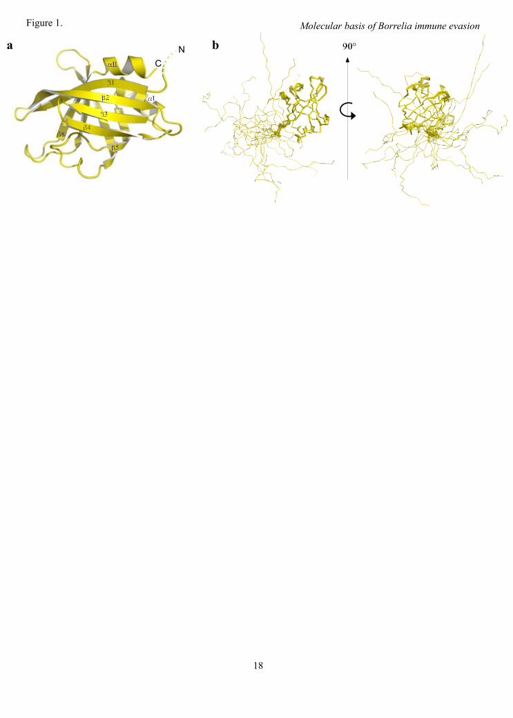
optimal proportions of FH19-20 and OspE to form the complex in solution (Supplemental Fig 1b). The proteins alone and their mixtures at two different apparent molar ratios (1:1 and 1:1.25) were run over a Superdex200 column attached to HPLC equipment (Shimadzu). MiniDAWN TREOS light-scattering detector and Optilab rEX refractive index detector together with ASTRA 5.3.4.20 software (Wyatt Technology Corporation) were used to calculate masses of proteins and complexes eluted from the column. A size exclusion analysis of OspE and FH19-20 alone and the OspE:FH19-20 complex was done by running the preparates through a Superdex200 column attached to an ÄKTA purifier HPLC system (GE Healthcare) (Fig. 2a). All samples were eluted in PBS at 0.5 mL/min flow rate at 22˚C.
NMR spectroscopy and resonance assignment of OspE - All NMR spectroscopy measurements were performed on Varian Inova 600 MHz or Varian Inova 800 MHz spectrometers, both equipped with a triple resonance cold-probe. For structure determination of OspE, measurements were performed at 298 K on a 1 mM 15N and 13C labelled protein sample in 20 mM phosphate buffer (pH 6.0). Specific resonance assignment was based on a series of standard spectra: 15N-heteronuclear single quantum correlation (HSQC), aliphatic constant time (CT)-13C-HSQC, HNCA, HNcoCA, HNCACB, CBCAcoNH, HNcaCO, HNCO, HBHAcoNH, 15N-edited total correlation spectroscopy (TOCSY) with 50 ms mixing time, CCCcoNH, HcCH- correlated spectroscopy (COSY), and HcCH-TOCSY with 50 ms mixing time. Aromatic rings were assigned based on (HB)CB(CD)HD, (HB)CB(CDCE)HE, and aromatic CT-13C-HSQC. For structure calculation, 15N-NOE spectroscopy (NOESY)-HSQC and 13C-NOESY-HSQC were recorded with 80 ms mixing time measured at 1H frequency of 800 MHz (Table 1). All spectra were processed using NMRPipe (39) and specific resonance assignment was performed using the CCPNMR analysis suite software v.2.2.1 (40). The chemical shifts and the unassigned NOE peak lists were used as inputs for NMR structure calculation with the program CYANA 3.0 (41,42). Twenty structures with the lowest CYANA target functions were selected for energy refinement in a 5 Å water shell using the
AMBER force field. Ramachandran plot was generated using PROCHECK-NMR (43) and it indicated that 81.9% of the residues were in the most-favored and 18.1% in the additional allowed regions.
All relaxation measurements were performed at 1H frequency of 600 MHz. T1 relaxation rates were determined using the T1 relaxation times of 10, 20, 30, 40, 50, 60, 70, 90, 110, 130, 150, and 210 ms. T2 relaxation rates were determined using a CPMG based pulse sequence with 1.3 ms refocusing interval and the used T2 relaxation times were 10, 30, 50, 70, 90, 110, 130, and 150 ms (44). Heteronuclear 15N-NOEs were determined by recording [1H,15N]-HSQC spectra with and without 3.5 s of 1H saturation (Supplemental Fig 2). Peak intensities from the recorded spectra were used for data analysis.
Monitoring OspE and FH19-20 interaction by NMR spectroscopy - The 15N-labelled OspE was titrated with unlabelled FH19-20, and monitoring the [1H,15N]-transverse relaxation optimized spectroscopy (TROSY) spectra indicated that OspE and FH19-20 formed a complex. Backbone resonance assignments of the OspE in complex with FH19-20 were performed on 0.3 mM 15N- and 13C-labelled OspE saturated with FH19-20, using TROSY variants of HNCA, HN(CO)CA, HNCO, and HN(CA)CO spectra recorded at 308 K. The chemical shift differences between free OspE and OspE:FH19-20 complex were calculated for each residue using the equation:
!"ave[ppm]=((!"HN[ppm])2+0.17#(!"N[ppm])2)0.5,
where !"HN and !"N are the chemical shift differences for backbone HN and N atoms (Supplemental Fig 3).
Crystallizing the OspE:FH19-20 complex and solving its structure - FH19-20 tends to crystallize as a homotetramer (35) and therefore we used FH19-20 with mutations D1119G and Q1139A to prevent this happening, as we have done before (20). TheFH19-20D1119G,Q1139A protein has similar binding affinity for OspE as the wild type FH19-20 (mean IC50 of 0.55 vs. 0.42 "M) (Supplemental Fig 4). The FH19-20D1119G,Q1139A:OspE complex was crystallized at 293 K from sitting drops in the presence of 2 M ammonium sulfate, 0.1 M citric acid, and 0.2 M
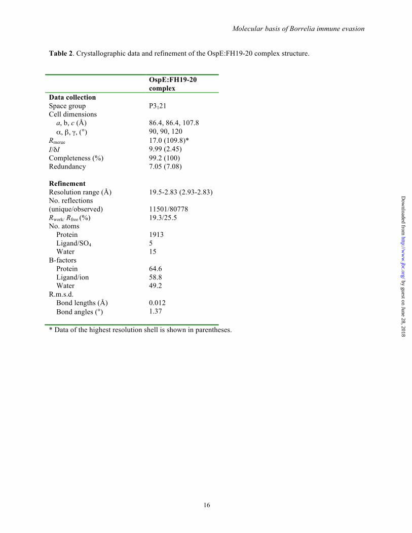
sodium chloride at pH 5.5. Crystals appeared within 4 days and were cryo-protected in the mother liquor supplemented with 25% glycerol. The diffraction data (to 2.83 Å) were collected at ESRF ID14-4 beam line at 100 K on a Q315r ADSC CCD detector at 0.97372 Å. The data were indexed and scaled using XDS (45). The structure of the OspE:FH19-20 complex was initially solved by structure-optimized molecular replacement with Rosetta (46). A molecular replacement solution was obtained using the wildtype FH19-20 structure (PDB code 2G7I (35) and a truncated NMR structure of OspE (without the loops) with an R-factor of 0.3. After successive rounds of building with Coot (47) and refinement with REFMAC (48) or PHENIX (49), we could identify one OspE molecule bound to a single FH19-20. The loops were modeled during real-space refinement using “phenix.refine” software without any applied restraints. The final R-factors (Rwork/ Rfree) of the refined complex structure are 19.3/25.5 (%) (shown in Table 2). The last refinement cycles were done using TLS parameters (9 TLS groups). In the Ramachandran plot 94.0% of the residues in the structure are in the most favored regions. The structure illustrations have been prepared using PyMol software (The PyMOL Molecular Graphics System, Version 1.3, Schrödinger, Portland). The interface between FH19-20 and OspE was analyzed using the PISA (50) server (http://www.ebi.ac.uk/pdbe/prot_int/pistart.html).
Sequence alignments - Sequence alignments were done using the CLUSTALW software (51) for Fig. 4 and DALI software (52) for Supplemental Fig. 5.
Statistical analyses - Values are expressed as means ± SD. All statistical analyses were performed using GraphPad Prism version 5.0. RESULTS
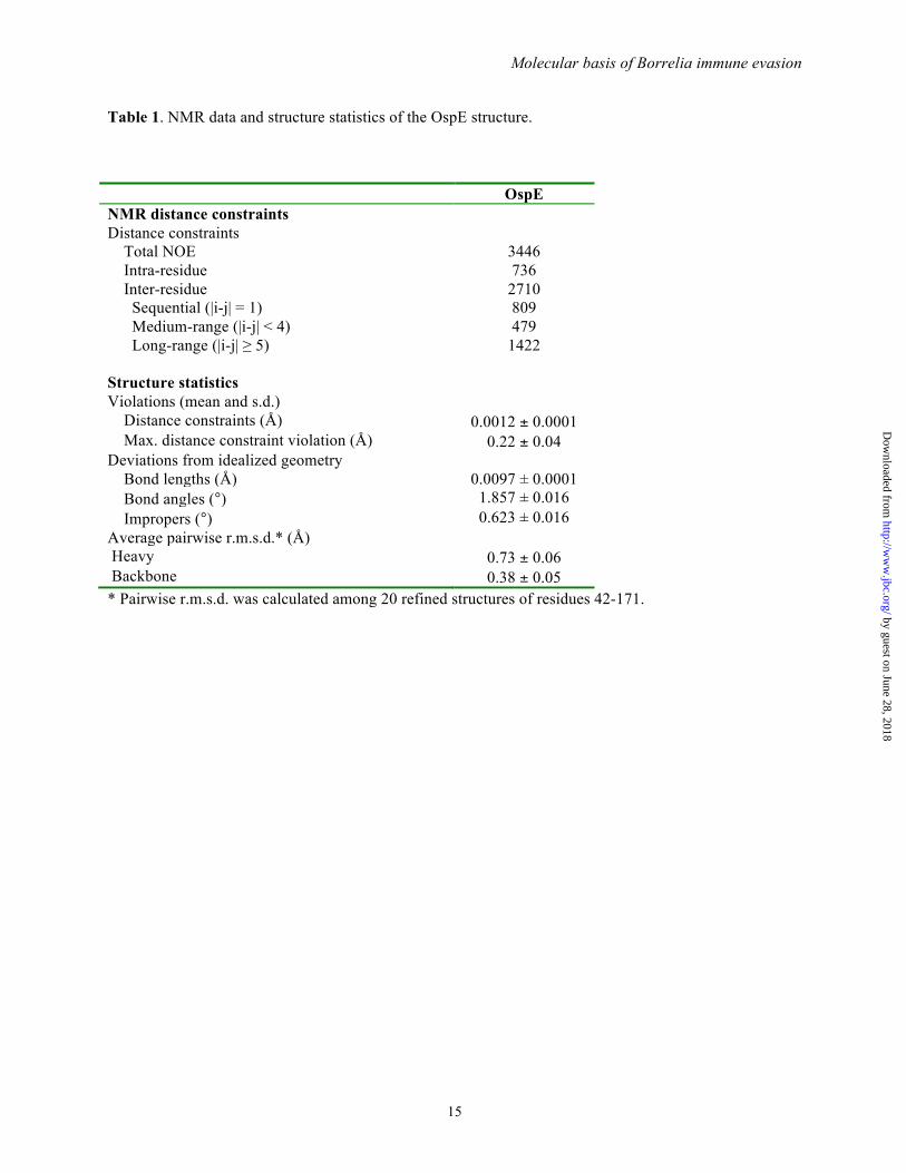
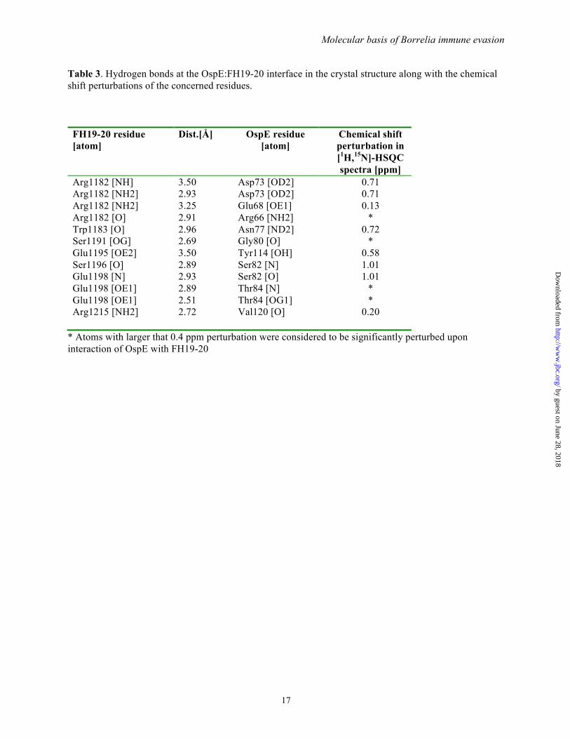
OspE consists of an unexpected globular domain with a flexible N-terminus - The solution structure of OspE was solved by NMR spectroscopy (Table 1, Fig 1a). Residues 42-171 of OspE form a globular single-domain protein with a backbone r.m.s.d. of 0.38±0.05 Å indicating a well defined structure. OspE has a rigid a+b fold of eight $-strands and two short %-helices arranged in a repeating topology of four $-strands
followed by an %-helix ($1-$2-$3-$4-%I-$5-$6-$7-$8-%II). The $-strands form antiparallel $-sheets where $1-$4 and $5-$8 are orientated almost perpendicularly. $1 and $8 are held together by hydrogen bonds thus forming an up-down squashed asymmetric $-barrel. It can be viewed as a $-barrel with 2 distinctive sides, one uniform and another non-uniform. The $2 and $6 strands are highly twisted and deviate from the formation of classic $-barrel strands. $1, $2, and $6 are very long strands (10, 12 and 11 residues respectively) and $5 along with $8 are the smallest $-strands (6 residues each).
The N-terminal region (residues 20-41) of OspE is structurally flexible as shown by superimposition of residues 42-171 from 20 NMR structures (Fig 1b). The 15N relaxation data of OspE (Supplemental Fig. 2) also shows that these residues are moving freely, because of the very low T1/T2 ratio. OspE is attached to the outer membrane via an N-terminal lipid anchor putatively bound to the N-terminal cysteine at position 20 (53). The anchoring topology of OspE is not known, but we can safely assume the flexible N-terminus allows the protein to move freely about its lipid anchor. To assess the tightness of OspE binding to the membrane we incubated killed Borrelia in a buffer for up to 24 h and after centrifugation analyzed the detached OspE from the supernatant. The amount of OspE in the supernatant was low and did not increase as a function of time (Supplemental Fig. 6) indicating tight anchoring of OspE to the bacterial surface.
A DALI (52) search showed that the globular domain of OspE resembles the SsgA like proteins (SALP) family of proteins (54), certain DNA/RNA binding proteins (55-58), some fatty acid binding proteins (55,59,60) and the ‘Homer’ family of proteins (61,62) (Supplemental Fig. 5). The fold has never, to our knowledge, been reported in any extracellular or immune evasion related molecule from any microbe. OspE has less than 15% sequence identity to any member of the structural family (Supplemental Fig. 5). The mammalian PUR-% protein, which binds nucleic acids, has been reported to be involved in immune evasion of viruses like John Cunningham Virus (JCV) and Human Immunodeficiency Virus (HIV) (58). The nucleic acid binding proteins that were found to be structural relatives of OspE were
mainly from the ‘Whirly’ protein group, so named because of the resemblance of their quaternary structure to a whirligig.
The OspE:FH19-20 complex reveals the interacting surfaces - Optimization of heterodimer formation by gel filtration analysis of OspE and FH19-20 (Fig. 2a) enabled us to crystallize OspE in complex with FH19-20 (Supplemental Fig 1b). Using the optimized 1:1 molar ratio of the proteins we obtained rod shaped co-crystals of OspE and FH19-20 in the presence of 2 M ammonium sulfate, 0.1 M citric acid and 0.2 M sodium chloride (pH 5.5) at 20 ºC. The crystal structure of the OspE:FH19-20 complex was solved at 2.83 Å resolution (Table 2, Fig. 2b) using our previously solved FH19-20 structure (35) and the NMR structure of OspE (described above) as models in molecular replacement (Fig. 2b). In the complex, OspE binds to domain 20 of FH with a contact surface area of 691 Å. The complex is held together by an E68OspE-R1182FH-D73OspE ion triple, which is also buttressed by a sulfate group forming an R1182FH-SO4
2--R66OspE ion triple, while R1182FH also forms an ion-pair with E1198FH (Fig. 2c and 2d). These interactions form the top part of a pocket for W1183FH, which makes hydrophobic contacts with the aliphatic side chain of R1182FH and with the OspE backbone around G75 and A83 (Table 3).
The OspE structure in solution (NMR) and in complex with FH19-20 (X-ray) were very similar (backbone r.m.s.d. of 1.43 Å/C%) (Supplemental Fig. 7a). The structure of FH19-20 in complex with OspE was also practically identical to the published structures of FH19-20 in homotetramers (35) (backbone r.m.s.d. 1.43 Å/C%) or in the FH19-20:C3d complex (20) (backbone r.m.s.d. 1.15 Å/C%) (Supplemental Fig. 7b). The sulfate ion found in the OspE:FH19-20 interface did not affect the orientation of the side chain of FH R1182 (Supplemental Fig. 8a,b). When the region of OspE close to the sulfate ion was compared between the NMR ensemble and the crystal complex structure it was observed that orientation of E68 was similar while that of R66 was slightly different, most probably due to a hydrogen bond between the NH2 of R66 and O of R1182 (shown in red in Supplemental Fig. 8a).
The interaction site on OspE confirmed by NMR analysis - To verify the observed interface
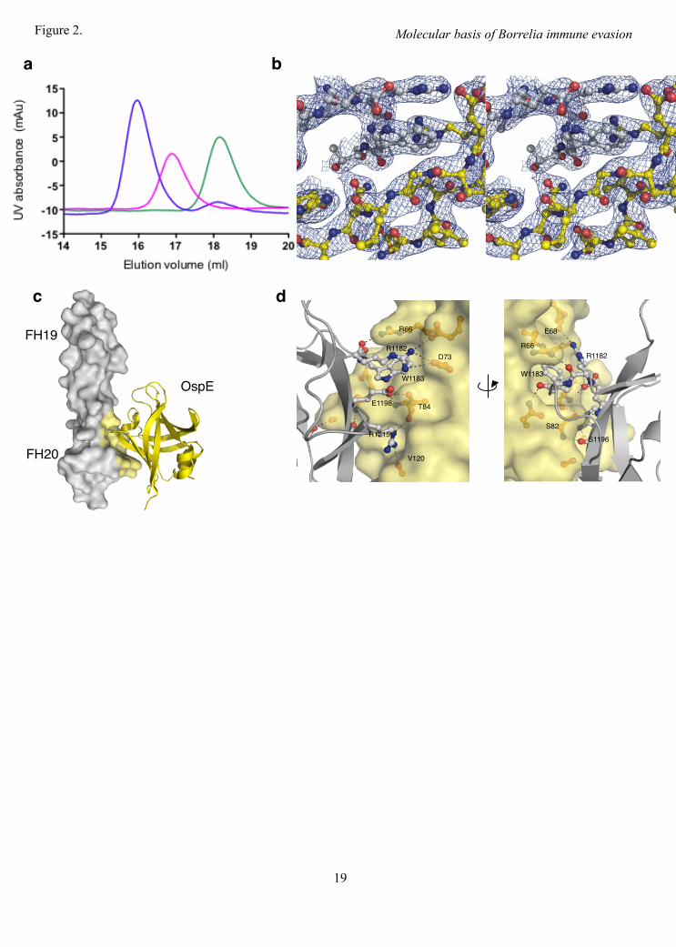
between OspE and FH19-20, we analyzed chemical shift perturbations in the NMR spectrum of OspE upon addition of saturating concentrations of wild type FH19-20 (Fig. 3a, Supplemental Fig. 3). The residues of OspE that shifted most (>0.4 ppm) are clustered on $-strands 1-4 that form the core of the OspE:FH19-20 interface in the crystal structure (Fig. 3b). Of the nine OspE residues involved in hydrogen bonding, six could be assigned in the assay and five of those were among the residues that shifted most (>0.4 ppm) (Table 3). The other three residues involved in binding (R66, G80 and T84) could not be assigned in the spectra of the FH19-20 bound OspE, probably due to large changes in the microenvironment caused by formation of the complex.
We confirmed the interface of OspE from the FH19-20 side using mutants of FH19-20 that we have reported elsewhere (34).
The OspE binding site overlaps with the heparin and endothelial cell binding sites on FH - The OspE binding site was next compared with the previously mapped binding sites for heparin or endothelial cells on FH domain 20 (Fig. 3c). Mutations in residues R1182, K1186, K1188, R1203, R1206, R1210, R1215, and K1230 impair binding of FH19-20 to heparin (37,63,64). Of these eight residues five are on the same side of FH19-20 as the binding site for OspE and two (R1182 and R1215) directly interact with OspE. This indicates that the heparin/endothelial cell binding site and the OspE binding site on FH domain 20 overlap.
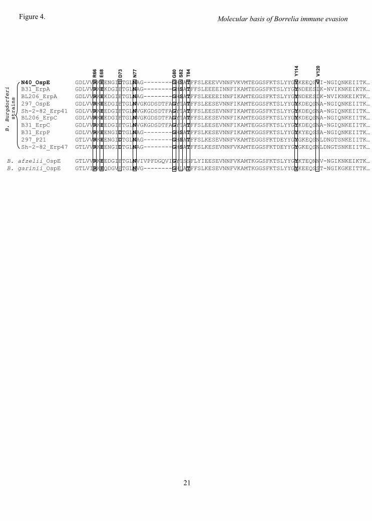
The OspE residues mediating binding to FH19-20 are conserved - The sequence identity between OspE and the other FH binding Erp paralog proteins (i.e. the OspE or BbCRASP-3 protein family) is more than 85% (Fig. 4). The sequence alignment shows that, considering only the subset of Erp paralog proteins from B. burgdorferi s.s that bind FH, eight of the nine OspE residues that form hydrogen bonds with FH19-20 in the crystal structure are conserved. R66, E68, N77, G80, T84, and Y114 are identical in all the FH binding Erp paralog proteins (Fig. 4), and D73 and S82 were conserved, being replaced by the conservative D73E and S82T mutations in some of the proteins. Seven of the nine residues were also conserved in the OspEs from B. afzelii and B. garinii (all except S82 and V120) (Fig. 4).
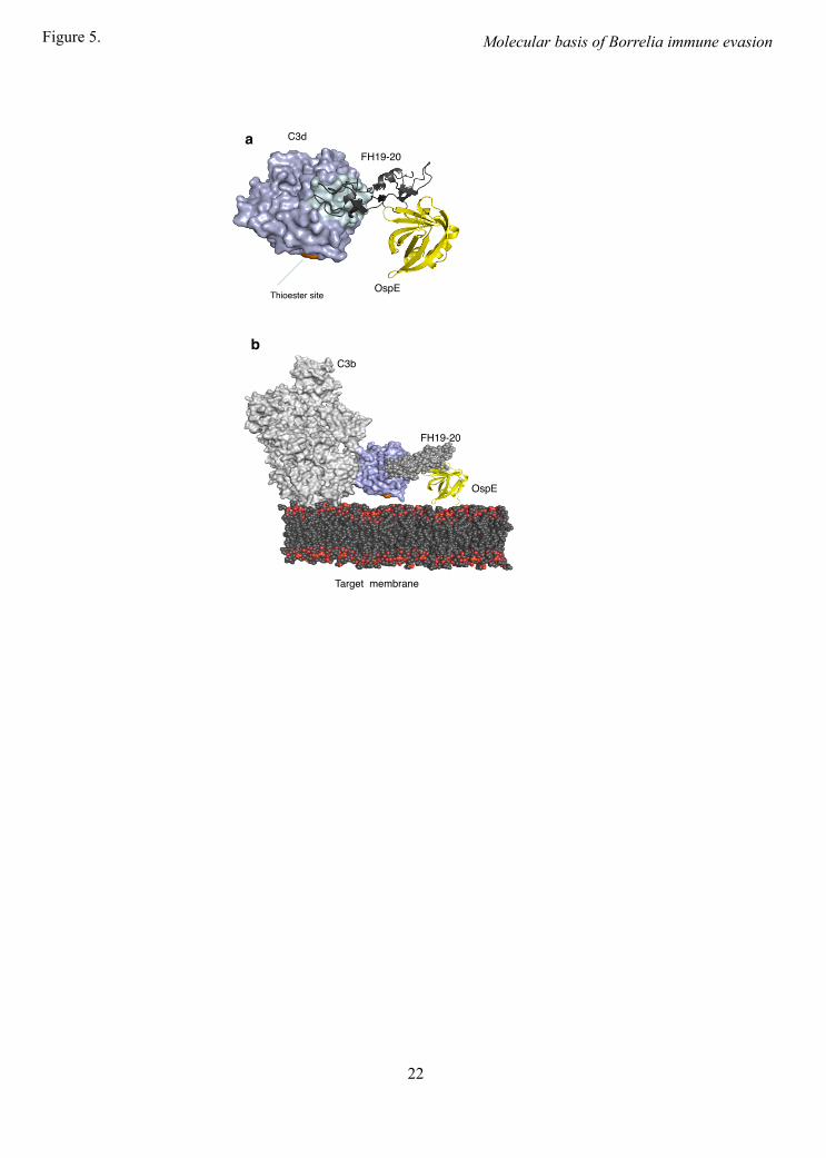
The OspE binding site is available in FH19-20 bound to C3b - To analyze if the OspE binding site is available on FH19-20 bound to its physiological ligand C3b, we superimposed our OspE:FH19-20 complex structure on the previously solved structure of FH19-20 in complex with C3d (20) by superimposing FH19-20 structures. The r.m.s.d. of C% for the superimposed FH19-20 structures was only 0.9Å and there were no steric clashes between OspE and C3d. Next we generated an OspE:FH19-20:C3b complex by superimposing the C3d from our OspE:FH19-20:C3d superimposition model on the structure of C3b (containing the C3d domain, also called the thioester domain, TED) (65). In this model, OspE had no steric clashes with any part of C3b and, most importantly, OspE and the thioester site of C3b faced the same direction without blocking each other (Fig. 5b). This indicates that FH bound to OspE on the borrelial surface is sterically able to bind a C3b molecule deposited onto the same surface. This is consistent with the role of OspE in mediating complement evasion of Borrelia burgdorferi.
DISCUSSION This study resolves the underlying
molecular mechanism of complement evasion by Borrelia burgdorferi. It reveals the structural fold of borrelial OspE, identifies the binding surfaces and residues used for the interaction between OspE and host FH, shows the similarity in binding of FH19-20 to host endothelial cell surface via the heparin binding site and to the microbial OspE protein, and finally explains how FH recruited by OspE onto the borrelial surface is sterically able to eliminate the central complement component C3b on the same surface.
A previous computational analysis suggested that the structure of OspE was comprised of coiled coil motifs (66). Our structure, however, clearly shows that this is wrong: OspE has an 8-stranded up-down $-barrel globular structure (residues 42-171) linked to the membrane by a very flexible 21 amino acid tail (Fig. 1b). A DALI (52) search indicated that the structural fold has been found in a trimeric protein from the SsgA like protein (SALP) family, several proteins from the ‘Nucleic acid binding proteins’ group, some proteins from the ‘Fatty acid binding
proteins’ family and a few proteins from the ‘Homer’ family (Supplemental Fig. 5). The sequence homology between OspE and its structural relatives proteins is less than 15% explaining why the structural similarity has not been identified earlier. As far as we know, this is the first time this fold has been identified in any extracellular or immune evasion related protein of microbial origin. Of the OspE distant homologues, the mammalian transcriptional activator PUR-% appears to be involved in viral immune evasion (58) by acting as the host factor for viral replication. Distant structural homologues of OspE are also found amongst the ‘Homer’ family of proteins. Certain members of this family are involved in neuronal plasticity by binding to and regulating metabotropic glutamate receptors (67) while others function by binding to members of the nuclear factor of activated T cells (NFAT) family of proteins, and down-regulating T-cell activation (62). There is, however, no similarity in the ligand binding residues between PUR-% or Homer proteins and OspE, which is hardly surprising given the difference in the charge properties of the two ligands. The fatty acid binding proteins that have a $-sheet structure similar to OspE mainly interact with the fatty acids via the residues inside the hydrophobic core of the protein (55,59,60).
The fold of the globular domain seems to be stable and fairly rigid since the root mean square deviation (r.m.s.d.) of the globular domain of the 20 NMR structures was only 0.38±0.05 Å for the backbone atoms and the structures of the liganded (X-ray) and unliganded (NMR) OspE were very similar (r.m.s.d. of 1.43 Å for C% atoms)(Supplemental Fig. 7a). The length of the extended, structurally flexible tail is more than 50 Å. Although the anchor topology of OspE is not known, trypsinolysis studies on the anchor topologies of borrelial OspA and Vsp1 proteins indicate that in both OspA (N-terminal tethering domain of just 12 residues) and Vsp1 (tethering domain of 21 residues), the tail is on the outside. In OspA, residues in positions 6-11 can be cleaved by trypsin, while in Vsp1, residues in positions 7-14 can be cleaved (68,69): i.e. most of the N-terminal tail in both proteins is surface exposed, and so can not be part of a protein membrane anchor but is instead presumably part of a flexible
linker. Consequently, the most plausible anchoring topology for OspE is that it binds through a triacyl Cysteine anchor, and the N-terminal region provides flexibility. Our results on incubation of killed Borrelia (Supplemental Fig. 6) indicate that majority of OspE is tightly bound to the borrelial surface and suggests that it is bound very firmly to the OM, indeed. Such a lipid anchor would provide OspE with good lateral mobility in the membrane, while the longer tethering domain would allows OspE the freedom to rotate and tilt. OspE can obviously bind to host FH on the cell suface since the binding site is far from the tethering domains.
In our OspE:FH19-20 structure, four residues (R1182, W1183, E1198, and R1215) in the FH C-terminus play a key role in binding to OspE. The central role of these residues is supported by the chemical shift perturbations in the NMR spectrum of OspE upon addition of saturating concentrations of wild type FH19-20 (Fig 3a and Supplemental Fig 3) and by a competition-binding assay with 14 mutant FH19-20 proteins (34). We have previously proposed, on the basis of alanine scanning mutations to OspE peptides, that multiple lysines in OspE might be required to bind FH19-20 (27). None of the peptides used in that work, however, could inhibit OspE binding to FH19-20 (27) indicating that binding of the linear OspE peptides to FH19-20 was weak. In our OspE:FH19-20 structure, there are no OspE lysines in the interface and so our previous results with peptides did not reflect the true OspE:FH19-20 interaction but an artificial binding of positively charged short linear synthetic peptides to a negatively charged patch on FH19-20.
A sulfate ion was found in the OspE:FH19-20 interface (Supplemental Fig. 8a). In principle a sulfate ion could potentially influence orientation of side chains in its vicinity either by steric hindrance or by forming hydrogen bonds. Superposition of the OspE:FH19-20 complex structure with the OspE NMR structure and the WT FH19-20 structure separately (Supplemental Fig 8b and 8c) showed hardly any difference in the side chains close to the sulfate ion. Therefore the sulfate ion itself has obviously not disturbed the arrangement of the residues within the interface.
Is it possible that the OspE:FH19-20 structure we see is a crystallographic artifact? The NMR based perturbation assay clearly excludes that possibility because the chemical shift of the OspE residues that interact with FH19-20 in the crystal structure changed when wild type FH19-20 was bound to OspE in solution (Fig. 3a). Radio-ligand binding assays using 14 point mutated FH19-20 proteins also support our results. Single point mutations of five (R1182A, W1183L, L1189R, E1198A and R1215Q) of seven key residues in the OspE:FH19-20 interface caused clear impairment in binding of FH19-20 to OspE (34) (the other two were not in the mutation panel). Deletion of the C-terminal 15 residues of the highly homologous OspE paralog p21 impairs, but does not abolish, binding to FH, while deletion of the N-terminal 35 residues has no effect (27). On the basis of the OspE:FH19-20 structure it is possible that the effect of the C-terminal OspE deletion is either due to disturbance of the globular OspE fold, since the last 15 residues in OspE form %-helix 2 and part of $-sheet 8, or due to a local effect of the disruption of the $-sheet 8 on the loop between the $-sheets 3 and 4 and thereby on the FH-binding residues on those sheets. On the basis of the structure (only part of the $-sheet 8 needed for the barrel was missing) and since the deletion did not abolish binding to FH19-20, it is likely that the deletion did not disrupt the fold completely. Our results thus explain well the previous data with deletion mutants of an OspE homolog.
We and others have earlier shown that binding of FH is essential for B. burgdorferi to survive in human serum, and that OspE (i.e. BbCRASP-3) and BbCRASP-1 (70) are needed for this survival (8,9,13,15,27). It is obvious that the microbe benefits from the recruitment of host FH onto the microbial surface since FH acts as a cofactor for factor I in the degradation process of the central complement component C3b needed for both opsonization and propagation of the complement cascade to form membrane attack complexes (71). Therefore it is easy to understand that acquisition of host FH onto Borrelia correlates strictly with survival of the spirochetes in non-immune serum or blood.
The C-terminal domains 19-20 of FH are known to mediate physiologically important binding of FH to both the C3d part of C3b (19,20)
and heparin/endothelial cells (37,63,64). Comparing the binding site of OspE to the previously reported binding sites for these physiological ligands showed that the OspE site overlaps with the heparin/endothelial cell binding site (residues R1182 and R1215 and the surrounding region) (37,63,64) while the C3d/C3b binding site on domain 19 is clearly distinct. This is also clear in the superimposition of the OspE:FH19-20 structure with the previously published FH19-20:C3d structure (20) (Fig. 5a). The overlap of the OspE and heparin binding sites indicates that B. burgdorferi mimics host cells in acquiring FH with OspE by binding to the same site as host cells bind with heparin. This resembles acquisition of FH by Neisseria meningitidis via heparin binding site on domain 7 of FH (32).
Two requirements have been suggested for physiologically relevant interaction between a microbe and host FH: the microbial binding site on FH needs to be easily accessible and the cofactor site on FH domains 1-4 must not be disturbed (72). Our work shows that the interaction between FH and OspE fulfills both these requirements at the structural level, since binding of OspE to FH19-20 does not block binding of FH (neither domains 1-4 nor 19-20) to C3b (20,73), and the OspE site is fully accessible on FH as seen in our OspE:FH19-20:C3b model (Fig. 5). Finally, this superimposition shows that the borrelial binding site on FH is directed towards the surface to which C3b is bound to via the thioester bond. This suggests that FH bound to OspE on the borrelial surface can recruit factor I not only to fluid phase C3b but also to opsonising C3b bound to the borrelial surface, thus preventing both opsonophagocytosis and propagation of the complement cascade to formation of lytic membrane attack complexes.
Humoral immune response against an outer surface lipoprotein of B. burgdorferi, OspA, has been shown to protect humans from Lyme borreliosis (74). Although the OspA based vaccine against the disease failed owing to side-effects (75), it indicated that a humoral immune response can protect from Lyme borreliosis. The FH-binding protein (fHbp) from Neisseria meningitidis serotype B has recently been successfully used as a candidate for vaccine development against meningococcal disease, and is now in phase III clinical trials (76). Therefore
studies aimed at identifying a new borrelial surface antigen, preferably one binding FH, are warranted. A suitable vaccine candidate must: (1) be surface exposed, (2) be conserved among different strains and genospecies of the pathogen, (3) be produced during human infection, (4) be necessary for development of a clinical infection, and (5) raise an immune response in humans in vivo. There is an increasing body of evidence that the FH binding protein OspE meets all these criteria (13). Our current study increases the acceptability of OspE as a vaccine candidate since it shows the residues needed for the OspE:FH19-20 interaction are highly conserved among the different Borrelia genospecies and strains (Fig. 4).
Our OspE:FH19-20 structure is the first structure of a microbial protein binding to the
domains 19-20 of FH. It is known that more than ten other pathogenic microbes than Borrelia acquire host FH using the same domains. We have studied the binding of several other microbes to FH19-20, and the data indicate a conserved pattern of microbial acquisition of FH via these domains (34). The domains 19-20 of FH are evidently essential for recognition of host cells and surfaces by FH since mutations in these domains lead to a life-threatening disease, aHUS. Therefore it is logical that microbes use these domains as a key way of avoiding complement attack, and in this report we have showed an example how an important and emerging human pathogen, Lyme disease causing Borrelia burgdorferi, utilizes these domains.
REFERENCES 1. Barbour, A. G., Maupin, G. O., Teltow, G. J., Carter, C. J., and Piesman, J. (1996) J Infect Dis 173, 403-
409 2. Control, C. f. D. (1996) MMWR Morb Mortal Wkly Rep 45, 481-484 3. Barbour, A. G., and Fish, D. (1993) Science 260, 1610-1616 4. Steere, A. C., Grodzicki, R. L., Kornblatt, A. N., Craft, J. E., Barbour, A. G., Burgdorfer, W., Schmid, G.
P., Johnson, E., and Malawista, S. E. (1983) N Engl J Med 308, 733-740 5. Ohnishi, J., Piesman, J., and de Silva, A. M. (2001) Proc Natl Acad Sci U S A 98, 670-675 6. Steere, A. C., Malawista, S. E., Hardin, J. A., Ruddy, S., Askenase, W., and Andiman, W. A. (1977) Ann
Intern Med 86, 685-698 7. van Dam, A. P., Kuiper, H., Vos, K., Widjojokusumo, A., de Jongh, B. M., Spanjaard, L., Ramselaar, A.
C., Kramer, M. D., and Dankert, J. (1993) Clin Infect Dis 17, 708-717 8. Alitalo, A., Meri, T., Comstedt, P., Jeffery, L., Tornberg, J., Strandin, T., Lankinen, H., Bergström, S.,
Cinco, M., Vuppala, S. R., Akins, D. R., and Meri, S. (2005) Eur J Immunol 35, 3043-3053 9. Alitalo, A., Meri, T., Rämö, L., Jokiranta, T. S., Heikkilä, T., Seppälä, I. J., Oksi, J., Viljanen, M., and
Meri, S. (2001) Infect Immun 69, 3685-3691 10. Walport, M. J. (2001) N Engl J Med 344, 1058-1066 11. Hartmann, K., Corvey, C., Skerka, C., Kirschfink, M., Karas, M., Brade, V., Miller, J. C., Stevenson, B.,
Wallich, R., Zipfel, P. F., and Kraiczy, P. (2006) Mol Microbiol 61, 1220-1236 12. Kraiczy, P., Hellwage, J., Skerka, C., Becker, H., Kirschfink, M., Simon, M. M., Brade, V., Zipfel, P. F.,
and Wallich, R. (2004) J Biol Chem 279, 2421-2429 13. Hellwage, J., Meri, T., Heikkilä, T., Alitalo, A., Panelius, J., Lahdenne, P., Seppälä, I. J., and Meri, S.
(2001) J Biol Chem 276, 8427-8435 14. Hefty, P. S., Jolliff, S. E., Caimano, M. J., Wikel, S. K., Radolf, J. D., and Akins, D. R. (2001) Infect
Immun 69, 3618-3627 15. Alitalo, A., Meri, T., Lankinen, H., Seppälä, I., Lahdenne, P., Hefty, P. S., Akins, D., and Meri, S. (2002) J
Immunol 169, 3847-3853 16. de Cordoba, S. R., and de Jorge, E. G. (2008) Clin Exp Immunol 151, 1-13 17. Atkinson, J. P., and Goodship, T. H. (2007) J Exp Med 204, 1245-1248 18. Gordon, D. L., Kaufman, R. M., Blackmore, T. K., Kwong, J., and Lublin, D. M. (1995) J Immunol 155,
348-356 19. Morgan, H. P., Schmidt, C. Q., Guariento, M., Blaum, B. S., Gillespie, D., Herbert, A. P., Kavanagh, D.,
Mertens, H. D., Svergun, D. I., Johansson, C. M., Uhrin, D., Barlow, P. N., and Hannan, J. P. (2011) Nat Struct Mol Biol 18, 463-470
20. Kajander, T., Lehtinen, M. J., Hyvärinen, S., Bhattacharjee, A., Leung, E., Isenman, D. E., Meri, S., Goldman, A., and Jokiranta, T. S. (2011) Proc Natl Acad Sci U S A 108, 2897-2902
21. Haapasalo, K., Vuopio, J., Syrjänen, J., Suvilehto, J., Massinen, S., Karppelin, M., Jarvela, I., Meri, S., Kere, J., and Jokiranta, T. S. (2012) J Immunol 188, 426-435
22. Zipfel, P. F., Hallström, T., Hammerschmidt, S., and Skerka, C. (2008) Vaccine 26 Suppl 8, I67-74 23. Lambris, J. D., Ricklin, D., and Geisbrecht, B. V. (2008) Nat Rev Microbiol 6, 132-142 24. Meri, T., Hartmann, A., Lenk, D., Eck, R., Würzner, R., Hellwage, J., Meri, S., and Zipfel, P. F. (2002)
Infect Immun 70, 5185-5192 25. Ho, D. K., Jarva, H., and Meri, S. (2011) J Immunol 185, 1763-1769 26. Kraiczy, P., Skerka, C., Kirschfink, M., Brade, V., and Zipfel, P. F. (2001) Eur J Immunol 31, 1674-1684 27. Alitalo, A., Meri, T., Chen, T., Lankinen, H., Cheng, Z. Z., Jokiranta, T. S., Seppälä, I. J., Lahdenne, P.,
Hefty, P. S., Akins, D. R., and Meri, S. (2004) J Immunol 172, 6195-6201 28. Amdahl, H., Jarva, H., Haanperä, M., Mertsola, J., He, Q., Jokiranta, T. S., and Meri, S. (2011) Mol
Immunol 48, 697-705 29. McDowell, J. V., Harlin, M. E., Rogers, E. A., and Marconi, R. T. (2005) J Bacteriol 187, 1317-1323 30. Blackmore, T. K., Hellwage, J., Sadlon, T. A., Higgs, N., Zipfel, P. F., Ward, H. M., and Gordon, D. L.
(1998) J Immunol 160, 3342-3348 31. Blackmore, T. K., Sadlon, T. A., Ward, H. M., Lublin, D. M., and Gordon, D. L. (1996) J Immunol 157,
5422-5427 32. Schneider, M. C., Prosser, B. E., Caesar, J. J., Kugelberg, E., Li, S., Zhang, Q., Quoraishi, S., Lovett, J. E.,
Deane, J. E., Sim, R. B., Roversi, P., Johnson, S., Tang, C. M., and Lea, S. M. (2009) Nature 458, 890-893
33. Batsford, S., Rust, C., and Neubert, U. (1998) J Infect Dis 178, 1676-1683 34. Meri, T., Amdahl, H., Lehtinen, M. J., Hyvärinen, S., McDowell, J. V., Bhattacharjee, A., Meri, S.,
Marconi, R., Goldman, A., and Jokiranta, T. S. (2013) PLoS Pathog 35. Jokiranta, T. S., Jaakola, V. P., Lehtinen, M. J., Pärepalo, M., Meri, S., and Goldman, A. (2006) EMBO J
25, 1784-1794 36. Bhattacharjee, A., Lehtinen, M. J., Kajander, T., Goldman, A., and Jokiranta, T. S. (2010) Mol Immunol 47,
1686-1691 37. Lehtinen, M. J., Rops, A. L., Isenman, D. E., van der Vlag, J., and Jokiranta, T. S. (2009) J Biol Chem 284,
15650-15658 38. Muona, M., Aranko, A. S., and Iwai, H. (2008) Chembiochem 9, 2958-2961 39. Delaglio, F., Grzesiek, S., Vuister, G. W., Zhu, G., Pfeifer, J., and Bax, A. (1995) J Biomol NMR 6, 277-
293 40. Vranken, W. F., Boucher, W., Stevens, T. J., Fogh, R. H., Pajon, A., Llinas, M., Ulrich, E. L., Markley, J.
L., Ionides, J., and Laue, E. D. (2005) Proteins 59, 687-696 41. Guntert, P., Mumenthaler, C., and Wuthrich, K. (1997) J Mol Biol 273, 283-298 42. Herrmann, T., Guntert, P., and Wuthrich, K. (2002) J Mol Biol 319, 209-227 43. Laskowski, R. A., Rullmannn, J. A., MacArthur, M. W., Kaptein, R., and Thornton, J. M. (1996) J Biomol
NMR 8, 477-486 44. Farrow, N. A., Muhandiram, R., Singer, A. U., Pascal, S. M., Kay, C. M., Gish, G., Shoelson, S. E.,
Pawson, T., Forman-Kay, J. D., and Kay, L. E. (1994) Biochemistry 33, 5984-6003 45. Kabsch, W. (2010) Acta Crystallogr D Biol Crystallogr 66, 125-132 46. DiMaio, F., Tyka, M. D., Baker, M. L., Chiu, W., and Baker, D. (2009) J Mol Biol 392, 181-190 47. Emsley, P., and Cowtan, K. (2004) Acta Crystallogr D Biol Crystallogr 60, 2126-2132 48. Murshudov, G. N., Vagin, A. A., and Dodson, E. J. (1997) Acta Crystallogr D Biol Crystallogr 53, 240-255 49. Adams, P. D., Grosse-Kunstleve, R. W., Hung, L. W., Ioerger, T. R., McCoy, A. J., Moriarty, N. W., Read,
R. J., Sacchettini, J. C., Sauter, N. K., and Terwilliger, T. C. (2002) Acta Crystallogr D Biol Crystallogr 58, 1948-1954
50. Krissinel, E., and Henrick, K. (2007) J Mol Biol 372, 774-797 51. Larkin, M. A., Blackshields, G., Brown, N. P., Chenna, R., McGettigan, P. A., McWilliam, H., Valentin, F.,
Wallace, I. M., Wilm, A., Lopez, R., Thompson, J. D., Gibson, T. J., and Higgins, D. G. (2007) Bioinformatics 23, 2947-2948
52. Holm, L., and Rosenstrom, P. (2010) Nucleic Acids Res 38, W545-549 53. Lam, T. T., Nguyen, T. P., Montgomery, R. R., Kantor, F. S., Fikrig, E., and Flavell, R. A. (1994) Infect
Immun 62, 290-298 54. Xu, Q., Traag, B. A., Willemse, J., McMullan, D., Miller, M. D., Elsliger, M. A., Abdubek, P., Astakhova,
T., Axelrod, H. L., Bakolitsa, C., Carlton, D., Chen, C., Chiu, H. J., Chruszcz, M., Clayton, T., Das, D., Deller, M. C., Duan, L., Ellrott, K., Ernst, D., Farr, C. L., Feuerhelm, J., Grant, J. C., Grzechnik, A., Grzechnik, S. K., Han, G. W., Jaroszewski, L., Jin, K. K., Klock, H. E., Knuth, M. W., Kozbial, P., Krishna, S. S., Kumar, A., Marciano, D., Minor, W., Mommaas, A. M., Morse, A. T., Nigoghossian, E., Nopakun, A., Okach, L., Oommachen, S., Paulsen, J., Puckett, C., Reyes, R., Rife, C. L., Sefcovic, N., Tien, H. J., Trame, C. B., van den Bedem, H., Wang, S., Weekes, D., Hodgson, K. O., Wooley, J., Deacon, A. M., Godzik, A., Lesley, S. A., Wilson, I. A., and van Wezel, G. P. (2009) J Biol Chem 284, 25268-25279
55. Sharma, A. (2011) J Biol Chem 286, 31924-31928 56. Schumacher, M. A., Karamooz, E., Zikova, A., Trantirek, L., and Lukes, J. (2006) Cell 126, 701-711 57. Desveaux, D., Allard, J., Brisson, N., and Sygusch, J. (2002) Nat Struct Biol 9, 512-517 58. Graebsch, A., Roche, S., and Niessing, D. (2009) Proc Natl Acad Sci U S A 106, 18521-18526 59. Capaldi, S., Guariento, M., Saccomani, G., Fessas, D., Perduca, M., and Monaco, H. L. (2007) J Biol Chem
282, 31008-31018 60. Nichesola, D., Perduca, M., Capaldi, S., Carrizo, M. E., Righetti, P. G., and Monaco, H. L. (2004)
Biochemistry 43, 14072-14079 61. Beneken, J., Tu, J. C., Xiao, B., Nuriya, M., Yuan, J. P., Worley, P. F., and Leahy, D. J. (2000) Neuron 26,
143-154 62. Huang, G. N., Huso, D. L., Bouyain, S., Tu, J., McCorkell, K. A., May, M. J., Zhu, Y., Lutz, M., Collins,
S., Dehoff, M., Kang, S., Whartenby, K., Powell, J., Leahy, D., and Worley, P. F. (2008) Science 319, 476-481
63. Herbert, A. P., Deakin, J. A., Schmidt, C. Q., Blaum, B. S., Egan, C., Ferreira, V. P., Pangburn, M. K., Lyon, M., Uhrin, D., and Barlow, P. N. (2007) J Biol Chem 282, 18960-18968
64. Ferreira, V. P., Herbert, A. P., Cortes, C., McKee, K. A., Blaum, B. S., Esswein, S. T., Uhrin, D., Barlow, P. N., Pangburn, M. K., and Kavanagh, D. (2009) J Immunol 182, 7009-7018
65. Janssen, B. J., Christodoulidou, A., McCarthy, A., Lambris, J. D., and Gros, P. (2006) Nature 444, 213-216 66. McDowell, J. V., Wolfgang, J., Senty, L., Sundy, C. M., Noto, M. J., and Marconi, R. T. (2004) J Immunol
173, 7471-7480 67. Brakeman, P. R., Lanahan, A. A., O'Brien, R., Roche, K., Barnes, C. A., Huganir, R. L., and Worley, P. F.
(1997) Nature 386, 284-288 68. Chen, S., and Zuckert, W. R. (2011) J Bacteriol 193, 6724-6732 69. Chen, S., Kumru, O. S., and Zuckert, W. R. (2011) J Bacteriol 193, 6379-6383 70. Cordes, F. S., Roversi, P., Kraiczy, P., Simon, M. M., Brade, V., Jahraus, O., Wallis, R., Skerka, C., Zipfel,
P. F., Wallich, R., and Lea, S. M. (2005) Nat Struct Mol Biol 12, 276-277 71. Pangburn, M. K., and Müller-Eberhard, H. J. (1978) Proc Natl Acad Sci U S A 75, 2416-2420 72. Ferreira, V. P., Pangburn, M. K., and Cortes, C. (2010) Mol Immunol 47, 2187-2197 73. Wu, J., Wu, Y. Q., Ricklin, D., Janssen, B. J., Lambris, J. D., and Gros, P. (2009) Nat Immunol 10, 728-733 74. Fikrig, E., Barthold, S. W., Kantor, F. S., and Flavell, R. A. (1991) J Infect Dis 164, 1224-1227 75. Rose, C. D., Fawcett, P. T., and Gibney, K. M. (2001) J Rheumatol 28, 2555-2557 76. Giuliani, M. M., Adu-Bobie, J., Comanducci, M., Arico, B., Savino, S., Santini, L., Brunelli, B., Bambini,
S., Biolchi, A., Capecchi, B., Cartocci, E., Ciucchi, L., Di Marcello, F., Ferlicca, F., Galli, B., Luzzi, E., Masignani, V., Serruto, D., Veggi, D., Contorni, M., Morandi, M., Bartalesi, A., Cinotti, V., Mannucci, D., Titta, F., Ovidi, E., Welsch, J. A., Granoff, D., Rappuoli, R., and Pizza, M. (2006) Proc Natl Acad Sci U S A 103, 10834-10839
Acknowledgments -- We thank Miia Eholuoto and Ilkka J. T. Seppälä (Univ. of Helsinki and Huslab, Helsinki Univ. Central Hospital Laboratory, Finland) for providing the OspE clone. We acknowledge Marjatta Ahonen, Pirkko Kokkonen, and Kirsti Widing for excellent technical assistance. We also thank ESRF for beamtime on ID14-4, Seija Mäki, Serranda Gashi and the Biocenter Finland crystallization facility. J.S.O acknowledges the National Graduate School in Informational and Structural Biology graduate school for financial support, AG, HI, TSJ and TK acknowledge research grants from the Sigrid Jusélius foundation and the Academy of Finland (grants 1252206 (AG), 131413, 137995 (HI) 128646, 255922, 259793 (TSJ) and 251700 (TK)) FOOTNOTES Coordinates and structure factors are available from the RCSB Protein Data Bank (http://www.pdb.org). The accession number is 2M4F for the 20 NMR structure ensemble of OspE and 4J38 for the crystal structure of the OspE:FH19-20 complex. OspE resonance assignment has been deposited to BMRB with accession number 19001. Correspondence and requests for materials should be addressed to A.G. ([email protected]) or T.S.J. ([email protected]). The abbreviations used are: CFH, Complement Factor H; OspE, Outer surface protein E; IPTG, Isopropyl $-D-1-thiogalactopyranoside FIGURE LEGENDS
Figure 1. The NMR structure of OspE. (a), Cartoon representation of the mean NMR structure of OspE (residues 21 to 171, dashed line indicating the flexible N-terminus). (b), Overlay of 20 energy minimized NMR structures of OspE with the backbone of residues 42-171 being superimposed. Two orientations are shown rotated 90° from each other. Figure 2. OspE:FH19-20 complex formation and the X-ray crystal structure. (a) Gel filtration analysis of OspE (magenta), FH19-20 (green) and OspE:FH19-20 complex (blue). (b) A detail of the electron density map (2FO-FC) of the OspE:FH19-20 complex in stereo. Carbon atoms of FH19-20 are shown in grey and those of OspE in yellow. (c) Crystal structure of OspE (yellow cartoon) in complex with FH19-20 (grey surface model, interacting atoms in yellow). (d) Interface between FH domain 20 (grey cartoon) and OspE (yellow surface model) from two directions about 180 ° apart with the interacting residues shown in ball-and-stick and the main interacting residues annotated. Figure 3. The interaction site between OspE and FH19-20. (a) Chemical shift perturbation of OspE residues in an HSQC titration NMR experiment upon addition of FH19-20 (residues not assigned indicated with asterisks). (b) Residues with larger that 0.4 ppm (dotted line) perturbation considered to interact with FH19-20 and colored red in the cartoon representation of OspE, while residues not observed in the OspE-FH19-20 complex are colored black. The $-strands involved are indicated ($1-$4). (c) Surface representation of FH domain 20 with the OspE binding residues found in the crystal structure highlighted in green (left), the heparin binding residues highlighted in blue (middle) and the endothelial cell binding residues highlighted in orange (right). The key heparin and endothelial cell binding residues have been annotated. Figure 4. Sequence alignment of the FH binding region of OspE with the homologous parts of the Erps. Sequence alignment between OspE from the N40 strain of B. burgdorferi (used in this study) and Erp paralog proteins encoded by other FH binding B. burgdorferi strains (B31, BL206, 297, and Sh-2-82) and single strains of B. afzelii and B. garinii is shown. Residues of OspE forming hydrogen bonds with FH19-20 are annotated, their alignments shown with boxes, and identical residues highlighted in bold. Figure 5. Superimposition based models of OspE in complex with FH19-20 and C3d or C3b. (a) Superimposition of the OspE:FH19-20 structure with the FH19-20:C3d structure (20) indicating no steric
clashes in the OspE:FH19-20:C3d complex. (b) Model of the spatial organization of the OspE:FH19-20 complex bound to C3b (65) on the target surface as based on superimposition of the OspE:FH19-20:C3d complex model with the structure of C3b (containing the C3d domain). The location of the thioester site is indicated in orange.
Rwork/ Rfree (%) 19.3/25.5 No. atoms Protein 1913 Ligand/SO4 5 Water 15 B-factors Protein 64.6 Ligand/ion 58.8 Water 49.2 R.m.s.d. Bond lengths (Å) 0.012 Bond angles (°) 1.37 * Data of the highest resolution shell is shown in parentheses.
Table 3. Hydrogen bonds at the OspE:FH19-20 interface in the crystal structure along with the chemical shift perturbations of the concerned residues. FH19-20 residue [atom]
Dist.[Å]
OspE residue [atom]
Chemical shift perturbation in [1H,15N]-HSQC spectra [ppm]
* Atoms with larger that 0.4 ppm perturbation were considered to be significantly perturbed upon interaction of OspE with FH19-20 by guest on June 28, 2018
Kajander, Markus J. Lehtinen, Hideo Iwaï, T. Sakari Jokiranta and Adrian GoldmanArnab Bhattacharjee, Jesper S. Oeemig, Robert Kolodziejczyk, Taru Meri, Tommi
burgdorferiStructural basis for complement evasion by Lyme disease pathogen Borrelia
published online May 8, 2013J. Biol. Chem.
10.1074/jbc.M113.459040Access the most updated version of this article at doi:
Alerts:
When a correction for this article is posted•
When this article is cited•
to choose from all of JBC's e-mail alertsClick here


![Complement evasion by Bordetella pertussis: implications for ......LP, or AP [14]. Complement is not only present in the blood, but also on healthy human mucosal surfaces of the upper](https://static.documents.pub/doc/80x56/60801ef01e98eb3e7b37da28/complement-evasion-by-bordetella-pertussis-implications-for-lp-or-ap-14.jpg)


