Page 1
www.sciencemag.org/content/343/6175/1133/suppl/DC1
Supplementary Materials for
Structural Basis for Heavy Metal Detoxification by an Atm1-Type ABC
Exporter
Jonas Y. Lee, Janet G. Yang, Daniel Zhitnitsky, Oded Lewinson, Douglas C. Rees*
*Corresponding author. E-mail: [email protected]
Published 7 March 2014, Science 343, 1133 (2014)
DOI: 10.1126/science.1246489
This PDF file includes:
Materials and Methods
Figs. S1 to S10
Tables S1 to S3
References
Page 2
2
Materials and Methods Summary: Finding the Best Target and Maximizing the Resolution of Data Collection
The crystal structure of Novosphingobium aromaticivorans Atm1 (NaAtm1) was
solved following an initial screen of 40 different Atm1 genes. Of these 40, we were able
to successfully clone 34, express 18, and crystallize 4 of these gene products in 7
different crystal forms. Of the 7 different crystal forms, 4 did not diffract, 2 diffracted to
7-10 Å resolution, and 1 (NaAtm1) diffracted anisotropically to 3.5-4 Å resolution in one
direction. The best diffracting crystals were improved by growing larger crystals and
optimizing the cryocooling conditions to yield a resolution of either 2.9 Å ((I/σI) > 2) or
3.2 Å (R-merge > 66%) (Table S1). To further improve the usable resolution and quality
of the resulting electron density maps, we utilized Karplus' CC* (Pearson's correlation
coefficient) based data cutoff approach (40) (Table S2), with Liu's method of maximizing
the anomalous CC1/2 signal through high redundancy data collection (41), and found that
the mean I/σI), CC1/2, and usable resolution were optimized with a data multiplicity of
greater than 15. For the initial model building and refinement, the initial resolution limit
was set as CC1/2 ~ 20% based on the data merging statistics from Aimless (Table S3).
For the final refinement and publication, the final resolution limit was reduced by CC*
analysis against unmerged intensities in Phenix package (Table S2) satisfying Karplus’
CC* against CC-work and CC-free criteria, as well as, R-free of the highest resolution
shell against the refined structure being less than or equal to ~50%.
Cloning, Purification, and Crystallization
Forty genes from various organisms including Novosphingobium aromaticivorans
DSM 12444 were selected by searching for proteins containing the conserved domain
ATM1 (NCBI CDD:34862) in the NCBI GenPept database (42). The full length gene
(Accession ABD27067) was PCR amplified from purified genomic DNAs and cloned
into a pJL-H6 ligation independent cloning vector (43) which adds a 6-His tag on the
carboxy terminus.
Page 3
3
The target was expressed in Escherichia coli BL21(DE3) cells grown in ZYM-5052
autoinduction media (44). The expressed cell paste was resuspended in lysis buffer
containing 20 mM Tris pH 7.5, 0.1 M NaCl, 0.1 mg/mL hen egg lysozyme, 0.01 mg/mL
DNaseI, and 1 mM PMSF. The resuspended cells were lysed using a M-110L pneumatic
microfluidizer (Microfluidics). Unlysed cells were removed by centrifugation at 10,000
x g, and membranes containing NaAtm1p were separated by ultracentrifugation at
100,000 x g. The membrane pellets were resuspended in lysis buffer and stored at -80oC
until use.
The protein was extracted from the resuspended membrane pellet by directly adding
0.25% w/v (final concentration) each of n-dodecyl-N,N-dimethylamine-N-oxide
(LDAO), 3-[(3-cholamidopropyl)dimethylammonio]-2-hydroxy-1-propanesulfonate
(CHAPSO), n-octyl-β-D-glucopyranoside (OG), and undecanoyl-N-
hydroxyethylglucamide (HEGA-11). Although these four detergents were individually
poor in extracting protein from membranes and in maintaining homogeneity, their
mixture was unexpectedly found to be quite effective in both aspects. Unextracted
membranes were removed by ultracentrifugation at 100,000 x g. NaAtm1p was purified
by immobilized nickel ion-affinity chromatography using a HisTrap HP 5 mL column
(GE Healthcare) followed by size-exclusion chromatography using a HiLoad 16/60
Superdex-200 prep grade column (GE Healthcare) equilibrated with buffer containing 20
mM Tris pH 7.5, 0.1 M NaCl, 0.025% w/v LDAO, 0.025% w/v CHAPSO, 0.025% w/v
OG, and 0.025% w/v HEGA-11. The eluted sample was pooled and concentrated to a
final concentration of 10 mg/mL using a 100 kDa molecular-weight cutoff spin
concentrator. 20 mM (final conc.) of sodium citrate at pH 7.5 was added to the final
protein solution as a crystallization aid.
The initial crystallization trials were performed using the sparse-matrix method (45)
using Index (Hampton Research), and MemGold (Molecular Dimensions) screens. All
drops were set up in 96-well MRC sitting-drop crystallization plates (Hampton Research)
with 0.7 uL of protein mixed with 0.7 uL of precipitant equilibrated with 50 uL of
precipitant and incubated at 4 oC. Six initial hits were found. The best diffracting
Page 4
4
crystals were found from 100 mM sodium citrate pH 5.4, 200 mM magnesium acetate,
and 12% w/v polyethylene glycol 5,000 monomethyl ether after optimizing MemGold
condition A12. Crystals appeared within a week and grew to a typical size of 100 μm x
50 μm, with some crystals as large as 250 μm x 150 μm (~20% of crystallizing drops)
observed within two to three weeks. Selenomethionine substituted NaAtm1 crystals were
prepared the same way as the native crystals except that the expression utilized
B834(DEC) cells grown in PASM-5052 autoinduction media (44).
X-Ray Data Collection and Structure Determination
Crystals were harvested from the crystallization plate using a nylon loop and soaked
in cryoprotectant solution containing 100 mM sodium citrate pH 5.4, 200 mM
magnesium acetate, and polyethylene glycol 5,000 monomethyl ether in three steps of
10, 18, and 25% before flash-freezing in liquid nitrogen. The crystals were soaked with
substrates by adding 1 to 5 mM of substrate into the cryoprotectant solution. The
diffraction data was collected at the Stanford Synchrotron Radiation Laboratory beamline
12-2 equipped with a PILATUS 6M PAD detector. The data was collected to maximize
the signal (mean(I/σI)) in the highest resolution outer shell while maintaining an overall
data completeness of greater than 95% by collecting 900̊ or 1080˚ data sets with 0.15˚ or
0.2˚ oscillation steps at the maximum allowable attenuation (97%) before predicted
radiation damage occurred (Table S1).
The diffraction data were indexed and integrated using XDS (46), and scaled using
Aimless with the CCP4i interface (47) with default options. The experimental phases
were determined by AutoSol in the Phenix package (48) with default options except
Kramers-Kronig constants were calculated by the SSRL BluIce software from the Se-
edge MAD scan, and input manually. For the apo model, all residues in the
transmembrane domains (residue number < 350) were built manually using Coot by
placing helices and assigning the sequence manually (49). The nucleotide binding
domain structure was generated using Sav1866 (PDB ID 2HYD) NBD structure as a
template using Sculptor in Phenix, and manually placed using Coot.
Page 5
5
The sequence registry was initially checked using the heavy atoms sites in the Pt
derivative, and subsequently from the positions of the SeMet sites, which independently
established the positions of methionine residues. Additional validation was obtained
from the locations of mercury sites observed in anomalous difference Fourier maps for
HgCl2 soaked crystals of NaAtm1 variants with Cys incorporated at either residues 143,
200 and 293, in addition to a composite omit map calculated with AutoBuild in Phenix
(48) at 2.35 Å resolution.
The apo structure was refined using Phenix.Refine in Phenix (48) with the following
refinement strategies: “XYZ coordinates,” “real-space,” “TLS parameters,”
“occupancies,” and “individual B-factors.” The following restraints were used: “NCS
restraints,” “secondary structure restraints,” and “experimental phase restraints” along
with “update waters” function in the later refinement stages. The TLS groups were
determined by Find_tls_groups in Phenix. All restraints were used with the default
option, which included torsion-angle NCS restraints with a 15 degree limit and a 2.5
degree sigma. We verified that the use of two-fold NCS restraint between subunits in the
homodimer did not significantly bias R-free by removing the NCS restraints for the
GSSG bound structure. Removing the NCS restraints marginally lowered R-work (by
0.11%) and raised R-free (by 0.13%) relative to the NCS restrained refinement.
Nevertheless, implementing the NCS restraints gave a better geometry in terms of
Ramachandran (1.4% more in the favored region) and rotamer (0.53% fewer outliers)
distributions, which minimized the need to manually place geometry restraints on
disordered loop regions for all the refined structures.
Phases for the substrate bound structures were calculated after rigid-body refinement
of the protein chains in the apo structure using Refmac5 (47). All cif libraries for the
ligands were generated using eLBOW in Phenix (48). The ligands were manually placed
using Coot. All ligand bound structures were refined using Phenix.Refine in Phenix (48)
with the same option as the apo structure described above except experimental phase
restraints were removed. CC*, CC-work, and CC-free were calculated using Phenix
Page 6
6
using the unmerged intensity outputs generated from Aimless in CCP4 to determine the
final resolution cutoff. The final refinements for all the structures were done using
Phenix.Refine with the same option as the above without the real-space refinement
strategy and experimental phase restraints.
Structural Superpositions
The Secondary Structure Matching (SSM) program (50) implemented in CCP4i (47)
was used to superimpose TM1-2, TM4-5, and TM3&6 of structurally characterized ABC
exporters onto the corresponding elements of the NaAtm1 structure (Fig. 4A). The rmsd
in Cα positions for the nine independent chains relative to the NaAtm1 structure were
found to be: TM1-2 (ave 2.06 Å, min/max = 1.60 - 2.61 Å, with an average of 99/118
residues used in the superimposition), TM4-5 (ave 1.89 Å, min/max = 1.41 - 2.48 Å, with
an average of 106/114 residues used in the superimposition), and TM3&6 (ave 2.65 Å,
min/max = 2.20 - 3.11 Å, with an average of 82/94 residues used in the superimposition).
Measuring Michaelis-Menten Constants of Various Substrates
Reduced glutathione (GSH), oxidized glutathione (GSSG), S-methyl glutathione, S-
hexyl glutathione, S-lactoyl glutathione, glutathione S-sulfonic acid, γ-Glu-Cys peptide,
Cys-Gly peptide, sodium acetate, sodium propionate, and all amino acids used in the
study were purchased from Sigma-Aldrich. Ophthalmic acid was purchased from VWR.
Bimane conjugated GSH was synthesized by mixing monobromobimane (Sigma-Aldrich)
with reduced GSH in a 1 to 1 molar ratio respectively and incubating overnight at room
temperature. The completion of the reaction and the depletion of free monobromobimane
were confirmed by adding additional reduced GSH and measuring the change in
fluorescence at 478 nm using a Tecan Infinite 200 microplate reader. Dinitrobenzene
conjugated GSH was synthesized by reacting 2,4-dinitrochlorobenzene (Sigma-Aldrich)
with reduced GSH in a 1 to 1 molar ratio respectively with glutathione S-transferase from
equine liver (Sigma-Aldrich). The completion of the reaction and the depletion of free
2,4-dinitrochlorobenzene were confirmed by adding additional reduced GSH and
Page 7
7
measuring the change in absorbance at 340 nm. The glutathione-mercury complex was
synthesized by mixing reduced GSH and mercury(II) chloride (Sigma-Aldrich) in 2 to 1
molar ratio respectively and incubating at room temperature overnight. The glutathione-
silver complex was synthesized by mixing reduced GSH and silver(I) nitrate (Sigma-
Aldrich) in 2 to 1 molar ratio respectively, adjusting the pH to 7.0, incubating at room
temperature overnight, then boiling at 100oC for 5 minutes. All substrates were adjusted
to pH 7-8 with NaOH or HCl before use.
The ATPase activity of NaAtm1 was determined from the rate of appearance of free
phosphate generated by the hydrolysis of ATP using the Invitrogen EnzCheck Phosphate
Assay kit implemented in a 96-well plate format with a Tecan Infinite 200 microplate
reader. Protein concentrations of 100 to 250 nM, quantitated using the Bradford reagent,
were used for each assay. All reactions were performed on a 100 uL scale in 20 mM Tris
pH 7.5, 100 mM NaCl, 5 mM EDTA, 0.32% w/v CHAPSO, 1 mM ATP, and 0.015% w/v
n-dodecyl-β-D-maltopyranoside (DDM). The reaction was started by adding 10 mM
(final conc.) of MgCl2. S-hexyl GSH measurements were done in duplicates, and all
other measurements were done in triplicate. The kinetic parameters were determined by a
nonlinear least squares fit using the program using the program R (51), fitting the ATPase
data to a Michaelis-Menten model with a constant basal ATPase activity:
vET
=kcat S[ ]
Km + S[ ]+ kbasal
where kbasal is measured to be 8.8 + 0.8 min-1 in 1 mM ATP.
Proteoliposome GSSG Transport Assay
Membrane vesicles were prepared from the fractioned E. coli cell membranes used
for protein purification. Membranes were washed with 20 mM Tris pH 7.5, 500 mM
NaCl, pelleted, and resuspended at 20 mg/ml in transport buffer (20 mM Tris pH 7.5, 100
mM NaCl). Membrane suspensions were then sonicated on ice using a Misonix S-4000
ultrasonic processor, power 60, for three cycles at 15s on/45 s off. Vesicles were
subjected to 5 cycles of freeze-thaw to scramble the orientation of NaAtm1 in the
membrane (52). Vesicles were extruded 11 times through a 400-nm polycarbonate filter
Page 8
8
and centrifuged at 70k rpm for 25 min in a TLA 100.3 rotor. Pellets were washed 3x and
resuspended to 20 mg/ml in transport buffer. Transport reactions contained 17 mM Tris,
pH 7.5, 85 mM NaCl, 15 mg/ml membranes, 0.5 mM GSSG, and 10 mM MgATP.
Reactions were incubated at 30 ˚C and initiated by the addition of ATP. 150 ul samples
were removed at various time points and added to 1 ml ice-cold 0.85x transport buffer.
Samples were immediately pelleted and washed three times, and solubilized to 22.5
mg/mL membranes in 0.9x transport buffer with 1% w/v DDM. The amount of GSSG
was quantified using the glutathione assay kit from Sigma-Aldrich based on the 5,5'-
dithiobis-(2-nitrobenzoic acid)-based glutathione reductase recycling assay (53). The
concentration of NaAtm1 was estimated from the intensity of the relevant band on a
Coomassie stained SDS-PAGE gel.
Metal Sensitivity Assay
Metal sensitive E. coli strains GG44 (Cu+/Ag+ sensitive) and GG48 (Zn+2/Cd2+/Hg2+
sensitive) were used (30, 54). Cells transformed with the indicated plasmids were grown
at 37 ̊ C in LB medium containing ampicillin (100 µg/ml) and kanamycin (30 µg/ml).
The cells were then diluted to an OD600 of 0.05 in 150 µl of the same media, containing
L-arabinose (0.02 %) and in the presence or absence of metal salts (CuSO4, ZnSO4 or
CdCl2). No protection was conferred by NaAtm1 against Cu+2, Zn+2 or Cd+2 toxicity (data
not shown). Metal sensitivities with AgNO3 and HgCl2 were performed in a similar
manner, but in Davis minimal medium. Growth was monitored continuously for 12 hours
in a Tecan Infinite 200 microplate reader.
References
30. G. Grass et al., J. Bacteriol. 183, 4664 (2001).
40. P. A. Karplus, K. Diederichs, Science 336, 1030 (2012).
41. Q. Liu, Z. Zhang, W. A. Hendrickson, Acta Crystallogr. D67, 45 (2011).
Page 9
References
1. R. Lill, G. Kispal, Mitochondrial ABC transporters. Res. Microbiol. 152, 331–340 (2001).
doi:10.1016/S0923-2508(01)01204-9 Medline
2. I. B. Holland, S. P. C. Cole, K. Kuchler, C. F. Higgins, ABC Proteins: From Bacteria to Man
(Academic Press, London, 2003).
3. A. Zutz, S. Gompf, H. Schägger, R. Tampé, Mitochondrial ABC proteins in health and
disease. Biochim. Biophys. Acta 1787, 681–690 (2009).
doi:10.1016/j.bbabio.2009.02.009 Medline
4. H. Ye, T. A. Rouault, Human iron-sulfur cluster assembly, cellular iron homeostasis, and
disease. Biochemistry 49, 4945–4956 (2010). doi:10.1021/bi1004798 Medline
5. R. Lill, B. Hoffmann, S. Molik, A. J. Pierik, N. Rietzschel, O. Stehling, M. A. Uzarska, H.
Webert, C. Wilbrecht, U. Mühlenhoff, The role of mitochondria in cellular iron-sulfur
protein biogenesis and iron metabolism. Biochim. Biophys. Acta 1823, 1491–1508
(2012). doi:10.1016/j.bbamcr.2012.05.009 Medline
6. G. Kispal, P. Csere, B. Guiard, R. Lill, The ABC transporter Atm1p is required for
mitochondrial iron homeostasis. FEBS Lett. 418, 346–350 (1997). doi:10.1016/S0014-
5793(97)01414-2 Medline
7. S. Bekri, G. Kispal, H. Lange, E. Fitzsimons, J. Tolmie, R. Lill, D. F. Bishop, Human ABC7
transporter: Gene structure and mutation causing X-linked sideroblastic anemia with
ataxia with disruption of cytosolic iron-sulfur protein maturation. Blood 96, 3256–3264
(2000). Medline
8. P. C. Krishnamurthy, G. Du, Y. Fukuda, D. Sun, J. Sampath, K. E. Mercer, J. Wang, B. Sosa-
Pineda, K. G. Murti, J. D. Schuetz, Identification of a mammalian mitochondrial
porphyrin transporter. Nature 443, 586–589 (2006). Medline
9. H. Chavan, M. Oruganti, P. Krishnamurthy, The ATP-binding cassette transporter ABCB6 is
induced by arsenic and protects against arsenic cytotoxicity. Toxicol. Sci. 120, 519–528
(2011). doi:10.1093/toxsci/kfr008 Medline
Page 10
10. M. Tsuchida, Y. Emi, Y. Kida, M. Sakaguchi, Human ABC transporter isoform B6 (ABCB6)
localizes primarily in the Golgi apparatus. Biochem. Biophys. Res. Commun. 369, 369–
375 (2008). doi:10.1016/j.bbrc.2008.02.027 Medline
11. K. Kiss, A. Brozik, N. Kucsma, A. Toth, M. Gera, L. Berry, A. Vallentin, H. Vial, M. Vidal,
G. Szakacs, Shifting the paradigm: The putative mitochondrial protein ABCB6 resides in
the lysosomes of cells and in the plasma membrane of erythrocytes. PLOS ONE 7,
e37378 (2012). doi:10.1371/journal.pone.0037378 Medline
12. J. K. Paterson, S. Shukla, C. M. Black, T. Tachiwada, S. Garfield, S. Wincovitch, D. N.
Ernst, A. Agadir, X. Li, S. V. Ambudkar, G. Szakacs, S. Akiyama, M. M. Gottesman,
Human ABCB6 localizes to both the outer mitochondrial membrane and the plasma
membrane. Biochemistry 46, 9443–9452 (2007). doi:10.1021/bi700015m Medline
13. O. K. Vatamaniuk, E. A. Bucher, M. V. Sundaram, P. A. Rea, CeHMT-1, a putative
phytochelatin transporter, is required for cadmium tolerance in Caenorhabditis elegans.
J. Biol. Chem. 280, 23684–23690 (2005). doi:10.1074/jbc.M503362200 Medline
14. S. Prévéral, L. Gayet, C. Moldes, J. Hoffmann, S. Mounicou, A. Gruet, F. Reynaud, R.
Lobinski, J. M. Verbavatz, A. Vavasseur, C. Forestier, A common highly conserved
cadmium detoxification mechanism from bacteria to humans: Heavy metal tolerance
conferred by the ATP-binding cassette (ABC) transporter SpHMT1 requires glutathione
but not metal-chelating phytochelatin peptides. J. Biol. Chem. 284, 4936–4943 (2009).
doi:10.1074/jbc.M808130200 Medline
15. T. Sooksa-Nguan, B. Yakubov, V. I. Kozlovskyy, C. M. Barkume, K. J. Howe, T. W.
Thannhauser, M. A. Rutzke, J. J. Hart, L. V. Kochian, P. A. Rea, O. K. Vatamaniuk,
Drosophila ABC transporter, DmHMT-1, confers tolerance to cadmium. DmHMT-1 and
its yeast homolog, SpHMT-1, are not essential for vacuolar phytochelatin sequestration.
J. Biol. Chem. 284, 354–362 (2009). doi:10.1074/jbc.M806501200 Medline
16. G. Kuhnke, K. Neumann, U. Mühlenhoff, R. Lill, Stimulation of the ATPase activity of the
yeast mitochondrial ABC transporter Atm1p by thiol compounds. Mol. Membr. Biol. 23,
173–184 (2006). doi:10.1080/09687860500473630 Medline
Page 11
17. A. Meister, Glutathione metabolism and its selective modification. J. Biol. Chem. 263,
17205–17208 (1988). Medline
18. R. C. Fahey, A. R. Sundquist, Evolution of glutathione metabolism. Adv. Enzymol. 64, 1–53
(1991). Medline
19. L. Masip, K. Veeravalli, G. Georgiou, The many faces of glutathione in bacteria. Antioxid.
Redox Signal. 8, 753–762 (2006). doi:10.1089/ars.2006.8.753 Medline
20. See supplementary materials on Science Online.
21. V. Srinivasan, A. J. Pierik, R. Lill, Science 343, 1137 (2014).
22. R. J. Dawson, K. P. Locher, Structure of a bacterial multidrug ABC transporter. Nature 443,
180–185 (2006). doi:10.1038/nature05155 Medline
23. S. G. Aller, J. Yu, A. Ward, Y. Weng, S. Chittaboina, R. Zhuo, P. M. Harrell, Y. T. Trinh, Q.
Zhang, I. L. Urbatsch, G. Chang, Structure of P-glycoprotein reveals a molecular basis
for poly-specific drug binding. Science 323, 1718–1722 (2009).
doi:10.1126/science.1168750 Medline
24. M. Hohl, C. Briand, M. G. Grütter, M. A. Seeger, Crystal structure of a heterodimeric ABC
transporter in its inward-facing conformation. Nat. Struct. Mol. Biol. 19, 395–402 (2012).
doi:10.1038/nsmb.2267 Medline
25. M. S. Jin, M. L. Oldham, Q. Zhang, J. Chen, Crystal structure of the multidrug transporter P-
glycoprotein from Caenorhabditis elegans. Nature 490, 566–569 (2012).
doi:10.1038/nature11448 Medline
26. C. A. Shintre, A. C. Pike, Q. Li, J. I. Kim, A. J. Barr, S. Goubin, L. Shrestha, J. Yang, G.
Berridge, J. Ross, P. J. Stansfeld, M. S. Sansom, A. M. Edwards, C. Bountra, B. D.
Marsden, F. von Delft, A. N. Bullock, O. Gileadi, N. A. Burgess-Brown, E. P. Carpenter,
Structures of ABCB10, a human ATP-binding cassette transporter in apo- and nucleotide-
bound states. Proc. Natl. Acad. Sci. U.S.A. 110, 9710–9715 (2013).
doi:10.1073/pnas.1217042110 Medline
Page 12
27. A. Ward, C. L. Reyes, J. Yu, C. B. Roth, G. Chang, Flexibility in the ABC transporter MsbA:
Alternating access with a twist. Proc. Natl. Acad. Sci. U.S.A. 104, 19005–19010 (2007).
doi:10.1073/pnas.0709388104 Medline
28. Z. E. Sauna, K. Nandigama, S. V. Ambudkar, Multidrug resistance protein 4 (ABCC4)-
mediated ATP hydrolysis: Effect of transport substrates and characterization of the post-
hydrolysis transition state. J. Biol. Chem. 279, 48855–48864 (2004).
doi:10.1074/jbc.M408849200 Medline
29. M. Herget, N. Kreissig, C. Kolbe, C. Schölz, R. Tampé, R. Abele, Purification and
reconstitution of the antigen transport complex TAP: A prerequisite for determination of
peptide stoichiometry and ATP hydrolysis. J. Biol. Chem. 284, 33740–33749 (2009).
doi:10.1074/jbc.M109.047779 Medline
30. G. Grass, B. Fan, B. P. Rosen, S. Franke, D. H. Nies, C. Rensing, ZitB (YbgR), a member of
the cation diffusion facilitator family, is an additional zinc transporter in Escherichia coli.
J. Bacteriol. 183, 4664–4667 (2001). doi:10.1128/JB.183.15.4664-4667.2001 Medline
31. O. Lewinson, A. T. Lee, D. C. Rees, A P-type ATPase importer that discriminates between
essential and toxic transition metals. Proc. Natl. Acad. Sci. U.S.A. 106, 4677–4682
(2009). doi:10.1073/pnas.0900666106 Medline
32. M. S. Pittman, H. C. Robinson, R. K. Poole, A bacterial glutathione transporter (Escherichia
coli CydDC) exports reductant to the periplasm. J. Biol. Chem. 280, 32254–32261
(2005). doi:10.1074/jbc.M503075200 Medline
33. J. Li, K. F. Jaimes, S. G. Aller, Refined structures of mouse P-glycoprotein. Protein Sci. 23,
34–46 (2014). doi:10.1002/pro.2387 Medline
34. E. Screpanti, C. Hunte, Discontinuous membrane helices in transport proteins and their
correlation with function. J. Struct. Biol. 159, 261–267 (2007).
doi:10.1016/j.jsb.2007.01.011 Medline
35. W. Qi et al., J. Am. Chem. Soc. 134, 10754 (2012).
36. K. Hollenstein, R. J. P. Dawson, K. P. Locher, Structure and mechanism of ABC transporter
proteins. Curr. Opin. Struct. Biol. 17, 412–418 (2007). doi:10.1016/j.sbi.2007.07.003
Medline
Page 13
37. J. F. Cotten, M. J. Welsh, Cystic fibrosis-associated mutations at arginine 347 alter the pore
architecture of CFTR. Evidence for disruption of a salt bridge. J. Biol. Chem. 274, 5429–
5435 (1999). doi:10.1074/jbc.274.9.5429 Medline
38. G. Cui, C. S. Freeman, T. Knotts, C. Z. Prince, C. Kuang, N. A. McCarty, Two salt bridges
differentially contribute to the maintenance of cystic fibrosis transmembrane conductance
regulator (CFTR) channel function. J. Biol. Chem. 288, 20758–20767 (2013).
doi:10.1074/jbc.M113.476226 Medline
39. A. W. R. Serohijos, T. Hegedus, A. A. Aleksandrov, L. He, L. Cui, N. V. Dokholyan, J. R.
Riordan, Phenylalanine-508 mediates a cytoplasmic-membrane domain contact in the
CFTR 3D structure crucial to assembly and channel function. Proc. Natl. Acad. Sci.
U.S.A. 105, 3256–3261 (2008). doi:10.1073/pnas.0800254105 Medline
40. P. A. Karplus, K. Diederichs, Linking crystallographic model and data quality. Science 336,
1030–1033 (2012). doi:10.1126/science.1218231 Medline
41. Q. Liu, Z. Zhang, W. A. Hendrickson, Multi-crystal anomalous diffraction for low-resolution
macromolecular phasing. Acta Crystallogr. D 67, 45–59 (2011).
doi:10.1107/S0907444910046573 Medline
42. R. L. Tatusov, N. D. Fedorova, J. D. Jackson, A. R. Jacobs, B. Kiryutin, E. V. Koonin, D. M.
Krylov, R. Mazumder, S. L. Mekhedov, A. N. Nikolskaya, B. S. Rao, S. Smirnov, A. V.
Sverdlov, S. Vasudevan, Y. I. Wolf, J. J. Yin, D. A. Natale, The COG database: An
updated version includes eukaryotes. BMC Bioinformatics 4, 41 (2003).
doi:10.1186/1471-2105-4-41 Medline
43. J. Lee, S. H. Kim, High-throughput T7 LIC vector for introducing C-terminal poly-histidine
tags with variable lengths without extra sequences. Protein Expr. Purif. 63, 58–61
(2009). doi:10.1016/j.pep.2008.09.005 Medline
44. F. W. Studier, Protein production by auto-induction in high density shaking cultures. Protein
Expr. Purif. 41, 207–234 (2005). doi:10.1016/j.pep.2005.01.016 Medline
45. J. Jancarik, S. H. Kim, Sparse matrix sampling: A screening method for crystallization of
proteins. J. Appl. Crystallogr. 24, 409–411 (1991). doi:10.1107/S0021889891004430
Page 14
46. W. Kabsch, XDS. Acta Crystallogr. D 66, 125–132 (2010).
doi:10.1107/S0907444909047337 Medline
47. M. D. Winn, C. C. Ballard, K. D. Cowtan, E. J. Dodson, P. Emsley, P. R. Evans, R. M.
Keegan, E. B. Krissinel, A. G. Leslie, A. McCoy, S. J. McNicholas, G. N. Murshudov, N.
S. Pannu, E. A. Potterton, H. R. Powell, R. J. Read, A. Vagin, K. S. Wilson, Overview of
the CCP4 suite and current developments. Acta Crystallogr. D 67, 235–242 (2011).
doi:10.1107/S0907444910045749 Medline
48. P. D. Adams, P. V. Afonine, G. Bunkóczi, V. B. Chen, N. Echols, J. J. Headd, L. W. Hung,
S. Jain, G. J. Kapral, R. W. Grosse Kunstleve, A. J. McCoy, N. W. Moriarty, R. D.
Oeffner, R. J. Read, D. C. Richardson, J. S. Richardson, T. C. Terwilliger, P. H. Zwart,
The Phenix software for automated determination of macromolecular structures. Methods
55, 94–106 (2011). doi:10.1016/j.ymeth.2011.07.005 Medline
49. P. Emsley, B. Lohkamp, W. G. Scott, K. Cowtan, Features and development of Coot. Acta
Crystallogr. D 66, 486–501 (2010). doi:10.1107/S0907444910007493 Medline
50. E. Krissinel, K. Henrick, Secondary-structure matching (SSM), a new tool for fast protein
structure alignment in three dimensions. Acta Crystallogr. D 60, 2256–2268 (2004).
doi:10.1107/S0907444904026460 Medline
51. www.R-project.org (R Foundation for Statistical Computing, Vienna, 2012).
52. E. R. Geertsma, N. A. Nik Mahmood, G. K. Schuurman-Wolters, B. Poolman, Membrane
reconstitution of ABC transporters and assays of translocator function. Nat. Protoc. 3,
256–266 (2008). doi:10.1038/nprot.2007.519 Medline
53. T. P. Akerboom, H. Sies, Assay of glutathione, glutathione disulfide, and glutathione mixed
disulfides in biological samples. Methods Enzymol. 77, 373–382 (1981).
doi:10.1016/S0076-6879(81)77050-2 Medline
54. J. Scherer, D. H. Nies, CzcP is a novel efflux system contributing to transition metal
resistance in Cupriavidus metallidurans CH34. Mol. Microbiol. 73, 601–621 (2009).
doi:10.1111/j.1365-2958.2009.06792.x Medline
Page 16
11
Fig. S1 CLUSTALW sequence alignment of Atm1/ABCB7/HMT1/ABCB6 from representative organisms from different kingdoms of life. Positions of the TMD helices and conserved ABC sequence motifs are indicated above the alignment. The key residues interacting with the primary GSSG binding sites are marked with green circles, while the residues interacting with the secondary GSSG binding sites are marked with red circles.
Page 17
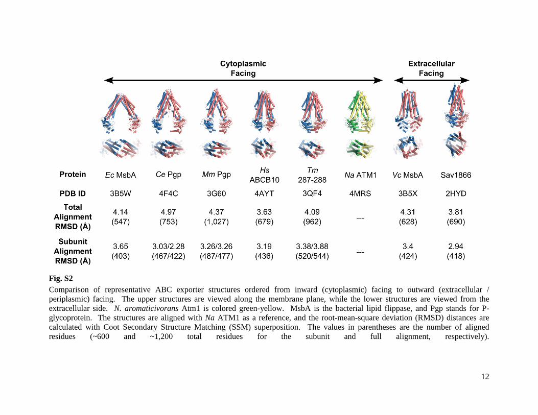
12
Fig. S2 Comparison of representative ABC exporter structures ordered from inward (cytoplasmic) facing to outward (extracellular / periplasmic) facing. The upper structures are viewed along the membrane plane, while the lower structures are viewed from the extracellular side. N. aromaticivorans Atm1 is colored green-yellow. MsbA is the bacterial lipid flippase, and Pgp stands for P-glycoprotein. The structures are aligned with Na ATM1 as a reference, and the root-mean-square deviation (RMSD) distances are calculated with Coot Secondary Structure Matching (SSM) superposition. The values in parentheses are the number of aligned residues (~600 and ~1,200 total residues for the subunit and full alignment, respectively).
Page 18
13
0
8
16
24
32
0 1 2 3 4 5
[LDAO] (mM)
k cat
(min
-1)
in 1
0 m
M G
SH
Fig. S3 Binding sites for LDAO, one of the critical crystallization detergents, in the TM2-ICL1-TM3 pocket and LDAO concentration dependent inhibition of the NaAtm1 ATPase activity. LDAO molecules (yellow carbon sphere models) are bound in the hydrophobic pocket (contoured in gray surfaces) formed between TM2-ICL1-TM3 and may stabilize ICL1 in an extended disordered conformation which to our knowledge has not been observed in other ABC exporter structures. This extension of ICL1 may prevent rearrangements of TM2 and TM3 during the transition to the ATPase competent outward facing conformation, thereby inhibiting the ATPase activity. The data points in the kinetic plot represent a mean value of N=3 with the error bars representing ± 1 S.D.
Page 19
14
Fig. S4 Stimulation of the ATPase activity of NaAtm1 by glutathione derivatives. Each data point represents the average of N=3 independent measures (except S-Hexyl GSH : N=2) with the error bars representing ± 1 S.D.
Page 20
15
0
8
16
24
32
Non
e
Se-M
et Gly
Met
Phe
Cys
Ala
Gln
Asn
Ser
Leu
Glu
Asp
Lys
Val
kcat
(min
-1)
in 1
0 m
M A
min
o A
cids
Fig. S5 Crystal structure of the binding of SeMet to NaAtm1, and stimulation of ATPase activity by 10 mM SeMet and other amino acids. The SeMet bound crystal structure indicates that the general binding model of amino acids follows the same motif as γ-Glu of GSH where primary interactions are made with the base functional groups of the amino acid. The blue mesh is 2Fo-Fc map contoured at 1.2σ, and the red mesh is Se-edge anomalous density map contoured at 3.5σ. The significant ATPase activities observed for SeMet and other amino acids indicates that the interactions of the α-amino and carboxyl groups with Asn 269 and Gln 272 are sufficient to stimulate ATPase activity. Each data point represents the average of N=3 independent measurements, with the error bars representing ± 1 S.D.
Page 21
16
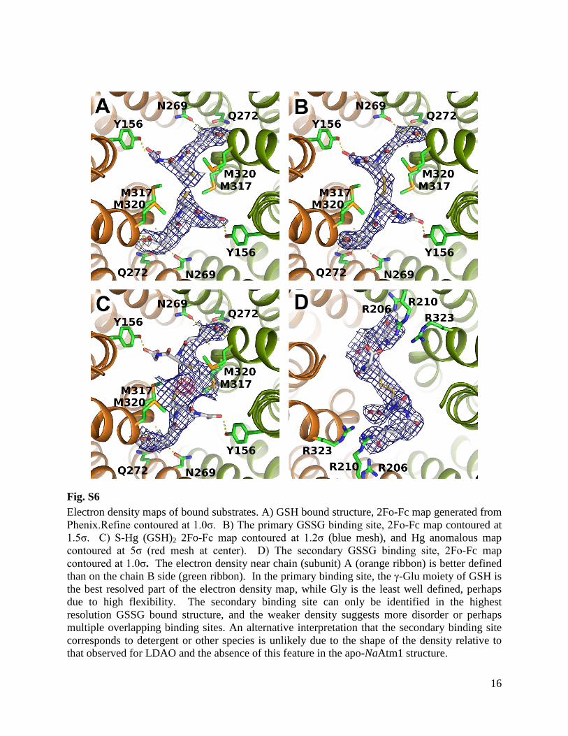
Fig. S6 Electron density maps of bound substrates. A) GSH bound structure, 2Fo-Fc map generated from Phenix.Refine contoured at 1.0σ. B) The primary GSSG binding site, 2Fo-Fc map contoured at 1.5σ. C) S-Hg (GSH)2 2Fo-Fc map contoured at 1.2σ (blue mesh), and Hg anomalous map contoured at 5σ (red mesh at center). D) The secondary GSSG binding site, 2Fo-Fc map contoured at 1.0σ. The electron density near chain (subunit) A (orange ribbon) is better defined than on the chain B side (green ribbon). In the primary binding site, the γ-Glu moiety of GSH is the best resolved part of the electron density map, while Gly is the least well defined, perhaps due to high flexibility. The secondary binding site can only be identified in the highest resolution GSSG bound structure, and the weaker density suggests more disorder or perhaps multiple overlapping binding sites. An alternative interpretation that the secondary binding site corresponds to detergent or other species is unlikely due to the shape of the density relative to that observed for LDAO and the absence of this feature in the apo-NaAtm1 structure.
Page 22
17
Fig. S7 Hydrogen bonding interaction map of GSSG. GSSG is modeled with light gray carbon atoms. The interacting backbone residue atoms are modeled as orange sticks. The interacting side chains are modeled as green carbon sticks. Hydrogen bonding distances are measured in Ångstroms, and averaged between homodimeric pairs.
Page 23
18
Fig. S8 Surface and sphere model of the primary GSSG binding site. Non-polar interactions are made by Leu 265 and Leu 268 (not shown for clarity) covering the γ-Glu moiety of GSH which contacts Asn 268 and Gln 272. Met 317/Met 320 from both NaAtm1 subunits make contacts along the GSH peptide backbone.
Page 24
19
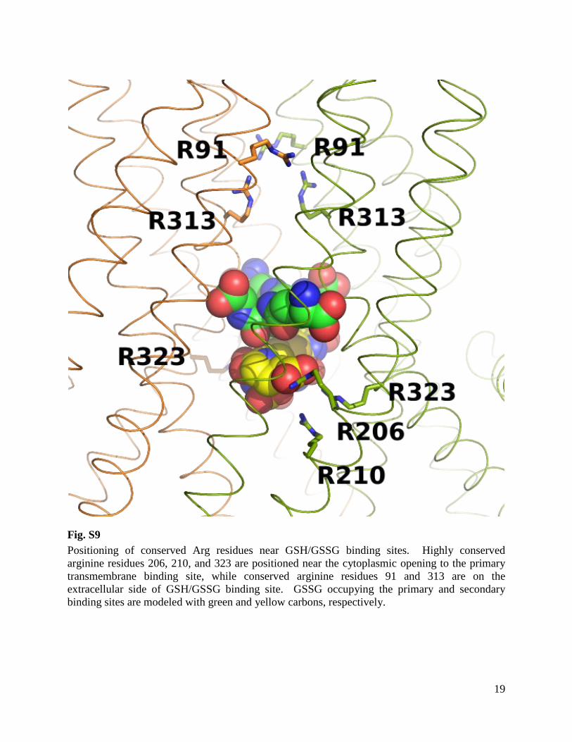
Fig. S9 Positioning of conserved Arg residues near GSH/GSSG binding sites. Highly conserved arginine residues 206, 210, and 323 are positioned near the cytoplasmic opening to the primary transmembrane binding site, while conserved arginine residues 91 and 313 are on the extracellular side of GSH/GSSG binding site. GSSG occupying the primary and secondary binding sites are modeled with green and yellow carbons, respectively.
Page 25
20
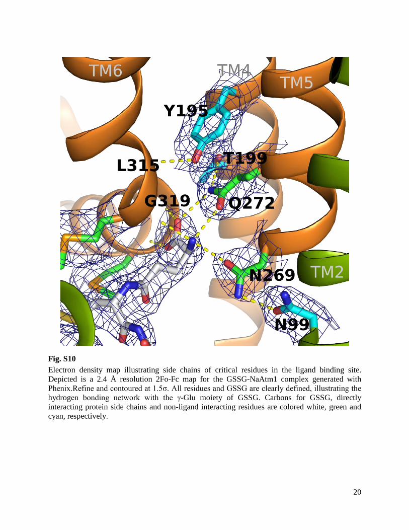
Fig. S10 Electron density map illustrating side chains of critical residues in the ligand binding site. Depicted is a 2.4 Å resolution 2Fo-Fc map for the GSSG-NaAtm1 complex generated with Phenix.Refine and contoured at 1.5σ. All residues and GSSG are clearly defined, illustrating the hydrogen bonding network with the γ-Glu moiety of GSSG. Carbons for GSSG, directly interacting protein side chains and non-ligand interacting residues are colored white, green and cyan, respectively.
Page 26
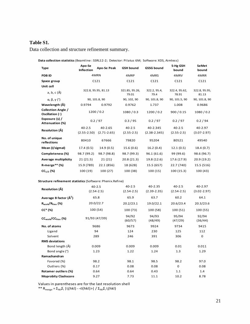
21
Table S1. Data collection and structure refinement summary.
Data collection statistics (Beamline : SSRL12-2; Detector: Pilatus 6M; Software: XDS, Aimless)
TypeApo-Se
InflectionApo-Se Peak GSH bound GSSG bound
S-Hg GSH bound
SeMet bound
PDB ID 4MRP 4MRS 4MRV 4MRR
Space group C121 C121 C121 C121
Unit cell
a, b, c (Å)321.85, 95.26,
79.01322.2, 95.4,
79.4322.4, 95.62,
78.91322.8, 95.95,
81.13
α, β, γ (°) 90, 102, 90 90, 101.8, 90 90, 101.5, 90 90, 101.8, 90
Wavelength (Å) 0.9794 0.9792 0.9762 1.737 1.008 0.9686
Collection Angle / Oscillation (◦) 1080 / 0.3 1200 / 0.2 900 / 0.15 1080 / 0.2
Exposure (s) / Attenuation (%)
0.3 / 95 0.2 / 97 0.2 / 97 0.2 / 94
40-2.5 40-2.65 40-2.5 40-2.345 40-2.5 40-2.97
(2.55-2.50) (2.71-2.65) (2.55-2.5) (2.38-2.345) (2.55-2.5) (3.07-2.97)
No. of unique reflections
80410 67666 79820 95204 80521 49540
Mean (I/sigmaI) 17.4 (0.5) 14.9 (0.5) 15.6 (0.6) 16.2 (0.4) 12.1 (0.5) 18.4 (0.7)
Completeness (%) 98.7 (99.2) 98.7 (98.8) 98.7 (99.3) 96.1 (61.6) 99 (99.6) 98.6 (96.7)
Average multiplicity 21 (21.5) 21 (21) 20.8 (21.3) 19.8 (12.6) 17.6 (17.9) 20.9 (19.2)
R-merge** (%) 15.9 (789) 22.1 (856) 18 (628) 15.5 (657) 22.7 (740) 15.5 (516)
CC1/2 (%) 100 (19) 100 (27) 100 (38) 100 (15) 100 (15.3) 100 (43)
Structure refinement statistics (Software: Phenix.Refine)
40-2.5 40-2.35 40-2.5 40-2.97
(2.54-2.5) (2.39-2.35) (2.54-2.5) (3.02-2.97)
Average B factor (Å2) 65.9 63.7 60.2 64.1
Rwork/Rfree (%) 20.2/23.1 19.0/22.1 20.6/23.4 20.3/23.6
CC* (%) 100 (73) 100 (58) 100 (51) 100 (55)
CCwork/CCfree (%)94/92
(60/57)94/93
(48/49)95/94
(47/29)92/94
(36/44)
No. of atoms 9673 9924 9734 9415
Ligand 124 230 125 112
Solvent 246 391 306 0
RMS deviations
Bond length (Å) 0.009 0.009 0.01 0.011
Bond angle (°) 1.22 1.24 1.3 1.29
Ramachandran
Favored (%) 98.1 98.5 98.2 97.0
Outliers (%) 0.08 0.08 0 0.08
Rotamer outliers (%) 0.64 0.43 1.1 1.4
Moprobity Clashscore 7.73 11.1 10.2 8.78
Values in parentheses are for the last resolution shell** Rmerge = ΣhklΣi |Ii(hkl) - <I(hkl)>| / ΣhklΣi Ii(hkl)
0.64
9.27
4MRN
289
0.009
1.23
20.0/22.7
100 (54)
91/93 (47/39)
9686
94
C121
65.8
1200 / 0.2
0.2 / 97
98.2
0.17
Resolution (Å)
(2.54-2.5)Resolution (Å)
322.8, 95.95, 81.13
90, 101.8, 90
40-2.5
Page 27
22
Table S2. CC* analysis of the refined structure against the unmerged intensities.
Type ApoResolution Shell
(Å)CC* (%) CC-work (%) CC-free (%) R-work (%) R-free (%)
39.8 - 6.78 100 86.8 88.6 18.7 20.26.77 - 5.38 100 92.3 91 19.1 20.25.38 - 4.7 100 95.7 92.1 15.3 16.84.7 - 4.27 100 96.2 96.5 14.6 16.2
4.27 - 3.97 100 95.1 89.5 16.9 22.33.97 - 3.73 99.9 94.1 94.5 17.7 20.33.73 - 3.55 99.9 94.3 89.9 18.1 23.13.55 - 3.39 99.6 92.6 89.9 19.7 24.73.39 - 3.26 99.2 92.4 83.7 21 25.63.26 - 3.15 98.8 91 86.4 22.9 27.63.15 - 3.05 97.8 90.2 86.3 23.6 293.05 - 2.96 96.2 87.8 84.8 24.9 27.82.96 - 2.89 94.6 86.1 81.6 27 302.89 - 2.82 90.1 81.1 62.9 29.7 35.62.82 - 2.75 87.4 79.3 57.7 30.8 38.42.75 - 2.69 83.3 74.7 68.7 32.7 32.12.69 - 2.64 81.1 70.5 56.3 35.1 362.64 - 2.59 74.2 60.4 69.9 38.9 39.52.59 - 2.54 67.6 58.8 44 39.6 44.42.54 - 2.5 53.6 46.6 38.7 41.7 42.5Overall 100 91.2 92.6 20 22.7
Type GSH bound
Resolution Shell (Å)
CC* (%) CC-work (%) CC-free (%) R-work (%) R-free (%)
39.74 - 6.78 100 90.7 88.1 18.2 19.76.78 - 5.38 100 92.3 87.9 19.3 21.65.38 - 4.7 100 95.9 93.2 15.7 16.44.7 - 4.27 100 96.1 94.7 15.3 18.1
4.27 - 3.97 100 94.8 89.4 16.8 22.73.97 - 3.73 99.9 94.9 96.7 16.6 19.33.73 - 3.55 99.9 94.7 90.9 18.1 233.55 - 3.39 99.7 92.6 89.6 20.3 23.63.39 - 3.26 99.5 91.6 79.3 21.4 26.73.26 - 3.15 99.1 89 84.1 23.9 25.63.15 - 3.05 98.6 91 84.1 25.2 31.53.05 - 2.96 97.7 89.3 80.3 25.6 29.72.96 - 2.89 96.7 88.6 82.6 26.1 28.42.89 - 2.82 94.8 85.4 69.7 28.4 35.42.82 - 2.75 92.4 81.9 70.4 29.9 34.62.75 - 2.69 90 81.9 78.8 30.3 32.92.69 - 2.64 88.7 78.4 70.1 33.6 36.42.64 - 2.59 85.2 75.2 60.5 36.6 38.52.59 - 2.54 80.5 70.7 60.8 40 44.92.54 - 2.5 72.5 59.8 57.2 45.4 44.7Overall 100 93.9 91.8 20.2 23.1
Type GSSG bound
Resolution Shell (Å)
CC* (%) CC-work (%) CC-free (%) R-work (%) R-free (%)
39.42 - 6.37 100 91.4 89.8 17.8 18.3
6.37 - 5.06 100 94 92.3 18 20.4
5.06 - 4.42 100 95.3 95 15 16.5
4.42 - 4.02 100 94.9 92.2 15.5 20
4.02 - 3.73 100 94.5 94 16.3 19.5
3.73 - 3.51 99.9 94.5 91.6 17.2 21.5
3.51 - 3.33 99.8 92.5 87.2 19 22.7
3.33 - 3.19 99.6 91.7 85 19.4 23.6
3.19 - 3.07 99.4 92.6 86 20.4 25.2
3.07 - 2.96 98.8 91.1 84.9 21.4 25.9
2.96 - 2.87 98.2 90.6 88.7 22 27.5
2.87 - 2.79 97.1 89.8 77.8 22.6 28.2
2.79 - 2.71 96.1 88.9 81.7 24 28.5
2.71 - 2.65 93.4 85.4 68.6 25.7 30.9
2.65 - 2.59 92 83.9 78.5 27.2 31.5
2.59 - 2.53 88.8 80.5 71.7 28.2 33.3
2.53 - 2.48 82.7 73.6 66.7 30.4 34.3
2.48 - 2.43 79.2 69.2 61.1 32.3 36
2.43 - 2.39 68.7 59 55 34.7 38.7
2.39 - 2.35 58.3 48.2 48.7 37.9 37.7
Overall 100 94.1 93.4 19 22
Type S-Hg GSH boundResolution
Shell (Å)CC* (%)
CC-work (%)
CC-free (%) R-work (%) R-free (%)
39.49 - 6.78 100 93 91.4 18 19.16.77 - 5.38 100 92.1 88.9 19.4 21.15.38 - 4.7 100 94.4 91.1 16.9 19.44.7 - 4.27 100 95.4 94.9 15.8 18.9
4.27 - 3.97 99.9 94.3 92.7 16.7 203.97 - 3.73 99.9 94.2 92 17 193.73 - 3.55 99.8 94.1 90.7 18.7 23.83.55 - 3.39 99.4 92 89.3 20.9 22.63.39 - 3.26 99 90.9 80.4 21.3 27.93.26 - 3.15 98.2 89.7 86.3 23 25.43.15 - 3.05 96.9 88.9 87.1 24.1 29.93.05 - 2.96 94.9 85.6 80.1 26.9 30.52.96 - 2.89 92.4 82.1 77.7 29.5 33.92.89 - 2.82 88.1 78 68.1 29.5 34.92.82 - 2.75 86.1 77.4 60 30.9 36.52.75 - 2.69 80.4 71.7 67.9 31.8 33.22.69 - 2.64 77.4 68.1 56.5 32.8 36.52.64 - 2.59 72.2 61.2 64.8 35.8 40.92.59 - 2.54 63.2 54.5 51.3 37 36.92.54 - 2.5 51.4 46.9 28.6 42 42.8
Overall 100 95.2 93.9 20.6 23.4
Type Se-Met boundResolution Shell
(Å)CC* (%) CC-work (%) CC-free (%) R-work (%) R-free (%)
39.5 - 8.04 100 87.8 92.6 18.8 19.68.04 - 6.39 100 94.1 92.9 18.3 20.76.39 - 5.59 100 91.5 87.2 20.5 22.85.59 - 5.08 100 95.3 94.6 17.5 21.95.08 - 4.71 100 95.9 90.7 15.2 20.24.71 - 4.44 100 96.3 96.4 15.5 204.43 - 4.21 100 96 90.9 17.1 18.74.21 - 4.03 99.9 95.1 94.6 18 21.64.03 - 3.88 99.8 94.9 89.5 18.9 243.88 - 3.74 99.7 94.2 88.7 19.5 26.33.74 - 3.62 99.5 94.1 93.9 20.3 27.63.62 - 3.52 99.1 93.3 85.7 22.1 29.13.52 - 3.43 97.8 87.5 65.8 27 33.73.43 - 3.34 97.2 89.9 86.2 27.7 29.83.34 - 3.27 95.8 85.7 74.8 28.5 35.63.27 - 3.2 93.7 83.1 69.3 31.1 36.13.2 - 3.14 90.4 80.4 62 32 40.9
3.14 - 3.08 87.1 74.5 69.9 36.2 39.53.08 - 3.02 80.3 61.8 51.1 43.5 43.43.02 - 2.97 74.3 59.7 37.5 52 54.4
Overall 100 91.9 94.7 20.3 23.6
Page 28
23
Table S3. Detailed data processing statistics. Type
Resolution Shell (Å)
Total Reflection
Unique Reflection
Multiplicity
Mean (I/sigI)
Completeness (%)
Rmerge (%)
CC1/2 (%)
CC1/2 pairs
40 - 12.75 12292 621 19.8 130.9 94.1 2.1 100 621
12.75 - 9.02 22672 1140 19.9 120.7 98.8 2.2 100 1140
9.02 - 7.36 31509 1484 21.2 94.2 99.8 2.8 100 1484
7.36 - 6.38 34036 1711 19.9 60.2 97.3 4.4 99.9 1709
6.38 - 5.7 40489 1940 20.9 48.4 98.6 5.8 99.9 1940
5.7 - 5.21 46410 2176 21.3 45.5 99.7 6.3 99.9 2176
5.21 - 4.82 51329 2360 21.7 50.5 99.5 5.5 100 2360
4.82 - 4.51 52559 2499 21 46.2 98.5 6.1 100 2498
4.51 - 4.25 52417 2616 20 38.6 97.1 7.4 99.9 2613
4.25 - 4.03 59805 2816 21.2 29.8 99.2 10.4 99.9 2815
4.03 - 3.84 63905 2978 21.5 24.5 99.1 13 99.8 2978
3.84 - 3.68 66766 3099 21.5 19.2 99.5 17.6 99.6 3099
3.68 - 3.54 70234 3253 21.6 13.9 99.5 25.4 99.4 3253
3.54 - 3.41 65789 3290 20 9.3 97.4 37.1 98 3287
3.41 - 3.29 69420 3404 20.4 7.6 98.2 47.4 97.4 3401
3.29 - 3.19 74797 3558 21 5.8 98.6 63.7 95.9 3557
3.19 - 3.09 78483 3710 21.2 4.2 98.9 89.7 92.5 3708
3.09 - 3 82664 3855 21.4 3.2 99.3 116.7 88.1 3855
3 - 2.92 84509 3922 21.5 2.6 99 144.8 83.4 3921
2.92 - 2.85 87165 4022 21.7 1.9 99.6 195.6 76.9 4022
2.85 - 2.78 80815 4037 20 1.3 97.2 270.9 61.1 4032
2.78 - 2.72 84985 4179 20.3 1.2 98.1 301.5 61.6 4177
2.72 - 2.66 88144 4238 20.8 0.9 98.8 394.2 50.5 4237
2.66 - 2.6 94416 4459 21.2 0.7 98.9 504.5 39.1 4457
2.6 - 2.55 95172 4469 21.3 0.6 98.8 622.7 33.6 4468
2.55 - 2.5 98135 4574 21.5 0.5 99.2 788.5 19 4570
40 -2.5 1688917 80410 21 17.4 98.7 15.9 100 80378
Apo – SeMet Inflection
Type
Resolution Shell (Å)
Total Reflection
Unique Reflection
Multiplicity
Mean (I/sigI)
Completeness (%)
Rmerge (%)
CC1/2 (%)
CC1/2 pairs
40 - 12.75 11718 617 19 100 94.5 2.6 100 617
12.75 - 9.02 21555 1130 19.1 94 98.4 2.7 100 1129
9.02 - 7.36 30066 1464 20.5 73.2 99.5 3.5 100 1464
7.36 - 6.38 33117 1697 19.5 47.1 97.5 5.4 99.9 1695
6.38 - 5.7 39170 1927 20.3 38.5 98.6 7.1 99.9 1926
5.7 - 5.21 45395 2168 20.9 38.4 99.8 7.3 99.9 2167
5.21 - 4.82 49893 2339 21.3 44.4 99.2 6.3 99.9 2339
4.82 - 4.51 50743 2470 20.5 41.4 98.5 6.8 99.9 2470
4.51 - 4.25 51344 2590 19.8 35.6 96.9 8.1 99.9 2589
4.25 - 4.03 58307 2794 20.9 29.4 99.2 10.5 99.9 2792
4.03 - 3.84 62290 2951 21.1 24.2 99.4 13.3 99.8 2950
3.84 - 3.68 65375 3077 21.2 20.1 99.6 16.8 99.7 3075
3.68 - 3.54 69088 3211 21.5 14.7 99.5 23.9 99.5 3210
3.54 - 3.41 65056 3246 20 10.4 97.2 33.9 98.7 3245
3.41 - 3.29 68771 3419 20.1 8.6 98.3 43.7 98.1 3410
3.29 - 3.19 74115 3545 20.9 6.7 98.5 58.4 97.1 3541
3.19 - 3.09 77528 3689 21 5 99 86.3 95.2 3682
3.09 - 3 80755 3785 21.3 3.9 99.2 98.2 92.3 3784
3 - 2.92 83501 3882 21.5 3.2 99.2 119.1 90.2 3880
2.92 - 2.85 86981 4032 21.6 2.4 99.5 159.8 84.6 4032
2.85 - 2.78 79796 4035 19.8 1.7 97.6 211.8 76.3 4027
2.78 - 2.72 81918 4068 20.1 1.5 98 248.9 72.4 4058
2.72 - 2.66 88905 4295 20.7 1.2 99 309.1 68 4292
2.66 - 2.6 92619 4377 21.2 1 98.6 372.1 60.3 4375
2.6 - 2.55 94174 4438 21.2 0.8 99.1 456.9 51.1 4435
2.55 - 2.5 97212 4574 21.3 0.6 99.3 628.4 38.2 4571
40 - 2.5 1659392 79820 20.8 15.6 98.7 18 99.9 79755
GSH bound
Type
Resolution Shell (Å)
Total Reflection
Unique Reflection
Multiplicity
Mean (I/sigI)
Completeness (%)
Rmerge (%)
CC1/2 (%)
CC1/2 pairs
40 - 12.82 12225 629 19.4 96.9 96.6 2.9 100 629
12.82 - 9.07 20167 1093 18.5 90.7 96.2 2.7 100 1093
9.07 - 7.4 28779 1443 19.9 75.5 99.1 3.3 100 1443
7.4 - 6.41 35220 1729 20.4 53.6 99.4 4.8 100 1729
6.41 - 5.74 36489 1864 19.6 44.4 96.9 5.8 99.9 1864
5.74 - 5.24 40745 2111 19.3 42.9 98.5 6.2 99.9 2111
5.24 - 4.85 47267 2314 20.4 51.6 99.2 5.1 100 2310
4.85 - 4.53 51506 2476 20.8 50.6 99.6 5.4 100 2476
4.53 - 4.27 55477 2643 21 46.5 99.7 6 99.9 2643
4.27 - 4.06 55711 2762 20.2 38.5 98.3 7.4 99.9 2761
4.06 - 3.87 55344 2836 19.5 32.2 97 9 99.9 2831
3.87 - 3.7 62581 3046 20.5 27.6 99.2 10.9 99.8 3043
3.7 - 3.56 66696 3182 21 21.5 99.2 14.8 99.7 3180
3.56 - 3.43 68946 3295 20.9 16.3 99.2 20.5 99.4 3292
3.43 - 3.31 72202 3403 21.2 13.6 98.7 25 99.3 3399
3.31 - 3.21 71310 3505 20.3 10.7 98.1 31.7 98.6 3504
3.21 - 3.11 68832 3560 19.3 7.7 97.4 44.3 97.7 3551
3.11 - 3.02 74387 3690 20.2 6.2 98.2 56.5 96.5 3679
3.02 - 2.94 77811 3805 20.4 5 98.4 72.8 95.1 3797
2.94 - 2.86 81648 3966 20.6 4.1 98.7 88.3 93.3 3964
2.86 - 2.8 83521 4035 20.7 3.2 99.1 112.7 89 4032
2.8 - 2.73 85051 4109 20.7 2.8 98.7 126.6 88.3 4099
2.73 - 2.67 77197 4180 18.5 1.9 97.1 173.3 78.3 4169
2.67 - 2.62 84582 4270 19.8 1.7 97.9 206.9 75.6 4257
2.62 - 2.56 87292 4345 20.1 1.5 98 233.6 71.7 4335
2.56 - 2.51 90145 4465 20.2 1.1 98.1 303.2 58.1 4461
2.51 - 2.47 91769 4528 20.3 1 97.1 349.3 50.1 4526
2.47 - 2.42 90975 4552 20 0.8 96.3 400.3 44.5 4539
2.42 - 2.38 74207 4373 17 0.6 91.7 501.2 29.4 4348
2.38 - 2.34 37675 2995 12.6 0.4 61.6 657.1 15 2892
40-2.34 1885757 95204 19.8 16.2 96.1 15.5 99.9 94957
GSSG Bound
Type
Resolution Shell (Å)
Total Reflection
Unique Reflection
Multiplicity
Mean (I/sigI)
Completeness (%)
Rmerge (%)
CC1/2 (%)
CC1/2 pairs
40 - 12.75 10841 641 16.9 87.1 96.7 2.9 99.9 641
12.75 - 9.02 17789 1124 15.8 79.4 97.8 3 100 1124
9.02 - 7.36 25433 1466 17.3 58.4 99.1 4.1 100 1465
7.36 - 6.38 31591 1763 17.9 37.4 99.9 6.9 99.9 1763
6.38 - 5.7 35767 1962 18.2 29.6 99.9 9.1 99.9 1962
5.7 - 5.21 33224 2095 15.9 27.4 96.5 9 99.8 2092
5.21 - 4.82 40752 2331 17.5 32.6 98.8 7.9 99.9 2331
4.82 - 4.51 44714 2513 17.8 31.2 99.4 8.6 99.9 2512
4.51 - 4.25 48553 2690 18 27.7 99.5 10 99.8 2689
4.25 - 4.03 51938 2828 18.4 22.2 99.5 13.2 99.7 2827
4.03 - 3.84 54158 2952 18.3 18.9 99.8 15.7 99.7 2952
3.84 - 3.68 48733 3009 16.2 14.7 95.9 19 99.4 3004
3.68 - 3.54 55023 3204 17.2 11 98.9 27.8 98.9 3201
3.54 - 3.41 59214 3360 17.6 8.3 99.3 38.8 97.8 3358
3.41 - 3.29 61809 3448 17.9 6.6 99.4 50.8 96.4 3447
3.29 - 3.19 64587 3615 17.9 4.9 99.4 68 94.1 3611
3.19 - 3.09 67234 3732 18 3.7 99.7 94 90.2 3728
3.09 - 3 68187 3793 18 2.9 99.7 119.1 84.1 3792
3 - 2.92 71857 3965 18.1 2.4 99.8 147.6 80.7 3964
2.92 - 2.85 63370 3900 16.2 1.7 95.7 196.6 65.6 3882
2.85 - 2.78 69640 4099 17 1.4 98.3 234.6 62.2 4095
2.78 - 2.72 71722 4141 17.3 1.2 99.1 285.3 52.4 4135
2.72 - 2.66 75739 4332 17.5 0.9 99.3 372.4 44.1 4326
2.66 - 2.6 78231 4414 17.7 0.7 99.5 480.4 38.2 4411
2.6 - 2.55 80339 4505 17.8 0.6 99.7 566.6 26.5 4504
2.55 - 2.5 82809 4639 17.9 0.5 99.6 740.3 15.3 4636
40 - 2.5 1413254 80521 17.6 12.1 99 22.7 99.9 80452
GS-Hg Bound
Page 29
24
Type
Resolution Shell (Å)
Total Reflection
Unique Reflection
Multiplicity
Mean (I/sigI)
Completeness (%)
Rmerge (%)
CC1/2 (%)
CC1/2 pairs
40 - 11.91 16585 806 20.6 120.6 96.4 2.3 100 806
11.91 - 8.41 27689 1424 19.4 98.4 96.5 2.6 100 1424
8.41 - 6.86 39683 1868 21.2 62.1 99.5 4.2 100 1868
6.86 - 5.94 47387 2200 21.5 37.2 99.5 7.5 99.9 2200
5.94 - 5.31 47388 2444 19.4 28.5 96.4 9.6 99.8 2442
5.31 - 4.85 57779 2743 21.1 32.6 99.4 8.7 99.9 2743
4.85 - 4.49 64051 2984 21.5 29.1 99.4 10.3 99.9 2983
4.49 - 4.2 69979 3209 21.8 22.3 99.6 14.1 99.8 3209
4.2 - 3.96 71212 3393 21 15.6 98.3 20.2 99.6 3389
3.96 - 3.76 68930 3519 19.6 10.3 97.7 31.7 99 3514
3.76 - 3.58 78533 3750 20.9 7.1 99.1 50 98.1 3750
3.58 - 3.43 83666 3953 21.2 4.2 99.4 94.9 93.7 3950
3.43 - 3.29 88052 4112 21.4 2.8 99.2 140.6 88.2 4108
3.29 - 3.18 92819 4275 21.7 1.9 99.2 190.9 79.3 4272
3.18 - 3.07 95851 4404 21.8 1.2 99.1 306.7 63.9 4404
3.07 - 2.97 85448 4456 19.2 0.7 96.7 515.8 43 4430
40 - 2.97 1035052 49540 20.9 18.4 98.6 15.5 100 49492
SeMet Bound


![General Principles of Detoxification - Image Awareness [Compatibility Mode].pdf · General Principles of Detoxification The Problem with Detoxification ... commercial fertilizers,](https://static.documents.pub/doc/80x56/5aaad8427f8b9a81188e8673/general-principles-of-detoxification-image-compatibility-modepdfgeneral-principles.jpg)


![Welcome [naturalhopeherbals.com]...Heavy Metal Detoxification 7. Systemic Detoxification 5. Cleanse the Lymphatic System Two of the best lymphatic exercises are daily dry skin brushing](https://static.documents.pub/doc/80x56/5f345078f835511b8e6bf89c/welcome-heavy-metal-detoxification-7-systemic-detoxification-5-cleanse.jpg)


