Structure Article Structural Basis for Molecular Interactions Involving MRG Domains: Implications in Chromatin Biology Tao Xie, 1 Richard Graveline, 2 Ganesan Senthil Kumar, 1 Yongbo Zhang, 1 Arvind Krishnan, 1 Gregory David, 2, * and Ishwar Radhakrishnan 1, * 1 Department of Molecular Biosciences, Northwestern University, 2205 Tech Drive, Evanston, IL 60208, USA 2 Department of Pharmacology, New York University School of Medicine, 500 First Avenue, New York City, NY 10016, USA *Correspondence: [email protected](I.R.), [email protected](G.D.) DOI 10.1016/j.str.2011.10.019 SUMMARY MRG15 is a member of the mortality family of transcription factors that targets a wide variety of multiprotein complexes involved in transcription regulation, DNA repair, and alternative splicing to chromatin. The structure of the apo-MRG15 MRG domain implicated in interactions with diverse pro- teins has been described, but not in complex with any of its targets. Here, we structurally and function- ally characterize the interaction between MRG15 and Pf1, two constitutively associated subunits of the histone deacetylase-associated Rpd3S/Sin3S core- pressor complex. The MRG domain adopts a struc- ture reminiscent of the apo state, whereas the Pf1 MRG-binding domain engages two discrete hydro- phobic surfaces on the MRG domain via a bipartite motif comprising an a-helix and a segment in an extended conformation, both of which are critical for high-affinity interactions. Multiple MRG15 inter- actors share an FxLP motif in the extended segment, but equivalent sequence/helical motifs are not readily evident, implying potential diversity in MRG- recognition mechanisms. INTRODUCTION The chromatin promotes a variety of processes critical for cell growth and survival, including transcription, replication, recom- binational repair, and splicing. Emerging evidence suggests that besides serving as a substrate for many of these processes, chromatin takes an active role in their regulation by harboring signals in the form of diverse posttranslational modifications of histones. These signals are better characterized in transcription biology, providing the basis for the histone code/language hypothesis (Gardner et al., 2011; Jenuwein and Allis, 2001), and are interpreted at the molecular level by specific chro- matin-binding modules embedded within multiprotein core- gulator complexes to yield specific transcriptional outcomes. Although a broad range of transcription factors have been impli- cated in the recognition of these signals, the MRG15 protein, a chromodomain-containing protein and a member of the so-called mortality family of transcription factors (Chen et al., 2010), appears to transcend its role in transcription regulation by also playing crucial roles in recombinational repair and alter- native splicing. The genes encoding MRG15 and two other members of the family, MORF4 and MRGX, were originally identified in a screen for genes involved in cellular senescence (Bertram et al., 1999). Subsequent studies revealed that only MORF4 is involved in replicative senescence, whereas MRG15 and MRGX promote cell-cycle progression and cell proliferation (Chen et al., 2009, 2011; Tominaga et al., 2005). MRG15 is a subunit of a number of multiprotein coregulator complexes involved in both transcrip- tional activation and repression, including the Rb-associated MAF1 complex and at least three disparate complexes con- taining chromatin-modifying activities, including the histone- acetyltransferase (HAT)-associated MAF2 and NuA4/Tip60 complexes and the histone-deacetylase (HDAC)-associated Rpd3S/Sin3S complex (Carrozza et al., 2005b; Doyon et al., 2004; Jelinic et al., 2011; Pardo et al., 2002; Yochum and Ayer, 2002). More recent studies have revealed that the MRG15 also associates with the BRCA complex involved in DNA damage repair by homologous recombination and a cotranscriptional splicing complex involved in alternative splicing (Hayakawa et al., 2010; Luco et al., 2010; Sy et al., 2009). Consistent with its varied roles, MRG15 knockouts result in embryonically lethal phenotype with significant defects in cell proliferation and differ- entiation and organ development, as well as defects in DNA repair (Garcia et al., 2007; Tominaga et al., 2005). MRG15 is unique in that it possesses an N-terminal chromo- domain that is absent in its paralogs. This atypical chromo- domain appears to bind specifically, albeit with low affinity, to histones enriched in H3 K36(me 2/3 ) found in the intragenic re- gions of actively transcribed genes (Carrozza et al., 2005b; Joshi and Struhl, 2005; Keogh et al., 2005; Sun et al., 2008; Xu et al., 2008). The C-terminal MRG domain on the other hand is directly involved in protein-protein interactions with diverse proteins in the aforementioned complexes. Crystal structures of the MRG15 MRG domain have been described, but only in the apo state and not in complex with any of its targets (Bowman et al., 2006; Zhang et al., 2006). The sequence and structural require- ments for effective interactions with MRG domains were not defined, although a hydrophobic groove on the surface of the domain was implicated by genetic and biochemical studies as Structure 20, 151–160, January 11, 2012 ª2012 Elsevier Ltd All rights reserved 151
Transcript
Structure
Article
Structural Basis for Molecular InteractionsInvolving MRG Domains: Implicationsin Chromatin BiologyTao Xie,1 Richard Graveline,2 Ganesan Senthil Kumar,1 Yongbo Zhang,1 Arvind Krishnan,1 Gregory David,2,*and Ishwar Radhakrishnan1,*1Department of Molecular Biosciences, Northwestern University, 2205 Tech Drive, Evanston, IL 60208, USA2Department of Pharmacology, New York University School of Medicine, 500 First Avenue, New York City, NY 10016, USA*Correspondence: [email protected] (I.R.), [email protected] (G.D.)
DOI 10.1016/j.str.2011.10.019
SUMMARY
MRG15 is a member of the mortality family oftranscription factors that targets a wide variety ofmultiprotein complexes involved in transcriptionregulation, DNA repair, and alternative splicing tochromatin. The structure of the apo-MRG15 MRGdomain implicated in interactions with diverse pro-teins has been described, but not in complex withany of its targets. Here, we structurally and function-ally characterize the interaction betweenMRG15 andPf1, two constitutively associated subunits of thehistone deacetylase-associated Rpd3S/Sin3S core-pressor complex. The MRG domain adopts a struc-ture reminiscent of the apo state, whereas the Pf1MRG-binding domain engages two discrete hydro-phobic surfaces on the MRG domain via a bipartitemotif comprising an a-helix and a segment in anextended conformation, both of which are criticalfor high-affinity interactions. Multiple MRG15 inter-actors share an FxLP motif in the extended segment,but equivalent sequence/helical motifs are notreadily evident, implying potential diversity in MRG-recognition mechanisms.
INTRODUCTION
The chromatin promotes a variety of processes critical for cell
growth and survival, including transcription, replication, recom-
binational repair, and splicing. Emerging evidence suggests
that besides serving as a substrate for many of these processes,
chromatin takes an active role in their regulation by harboring
signals in the form of diverse posttranslational modifications of
histones. These signals are better characterized in transcription
biology, providing the basis for the histone code/language
hypothesis (Gardner et al., 2011; Jenuwein and Allis, 2001),
and are interpreted at the molecular level by specific chro-
matin-binding modules embedded within multiprotein core-
gulator complexes to yield specific transcriptional outcomes.
Although a broad range of transcription factors have been impli-
cated in the recognition of these signals, the MRG15 protein,
Structure 20, 151
a chromodomain-containing protein and a member of the
so-called mortality family of transcription factors (Chen et al.,
2010), appears to transcend its role in transcription regulation
by also playing crucial roles in recombinational repair and alter-
native splicing.
The genes encoding MRG15 and two other members of the
family, MORF4 and MRGX, were originally identified in a screen
for genes involved in cellular senescence (Bertram et al., 1999).
Subsequent studies revealed that only MORF4 is involved in
replicative senescence, whereas MRG15 and MRGX promote
cell-cycle progression and cell proliferation (Chen et al., 2009,
2011; Tominaga et al., 2005). MRG15 is a subunit of a number
ofmultiprotein coregulator complexes involved in both transcrip-
tional activation and repression, including the Rb-associated
MAF1 complex and at least three disparate complexes con-
taining chromatin-modifying activities, including the histone-
acetyltransferase (HAT)-associated MAF2 and NuA4/Tip60
complexes and the histone-deacetylase (HDAC)-associated
Rpd3S/Sin3S complex (Carrozza et al., 2005b; Doyon et al.,
2004; Jelinic et al., 2011; Pardo et al., 2002; Yochum and Ayer,
2002). More recent studies have revealed that the MRG15 also
associates with the BRCA complex involved in DNA damage
repair by homologous recombination and a cotranscriptional
splicing complex involved in alternative splicing (Hayakawa
et al., 2010; Luco et al., 2010; Sy et al., 2009). Consistent with
its varied roles, MRG15 knockouts result in embryonically lethal
phenotype with significant defects in cell proliferation and differ-
entiation and organ development, as well as defects in DNA
repair (Garcia et al., 2007; Tominaga et al., 2005).
MRG15 is unique in that it possesses an N-terminal chromo-
domain that is absent in its paralogs. This atypical chromo-
domain appears to bind specifically, albeit with low affinity, to
histones enriched in H3 K36(me2/3) found in the intragenic re-
gions of actively transcribed genes (Carrozza et al., 2005b; Joshi
and Struhl, 2005; Keogh et al., 2005; Sun et al., 2008; Xu et al.,
2008). The C-terminal MRG domain on the other hand is directly
involved in protein-protein interactions with diverse proteins
in the aforementioned complexes. Crystal structures of the
MRG15 MRG domain have been described, but only in the apo
state and not in complex with any of its targets (Bowman et al.,
2006; Zhang et al., 2006). The sequence and structural require-
ments for effective interactions with MRG domains were not
defined, although a hydrophobic groove on the surface of the
domain was implicated by genetic and biochemical studies as
–160, January 11, 2012 ª2012 Elsevier Ltd All rights reserved 151
drophobic interactions are plausible but are not consistently
detected in a majority of the NMR conformers.
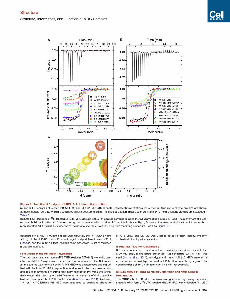
Functional Analysis of Pf1-MRG15 InteractionsTo evaluate the contributions of various residues toward the
stability of the MRG15-Pf1 complex, we engineered single-site
alanine mutations (with two exceptions) of selected residues at
the protein-protein interface of each protein and measured the
affinities of the resulting proteins in vitro using ITC; the effects
of the mutations were also evaluated in cells in the context of
the full-length proteins via coimmunoprecipitation (coIP) assays.
The wild-type Pf1 MBD and MRG15 MRG polypeptides inter-
acted with 12 nM affinity (Figures 4A and 4B; Table 2). Alanine
mutations of Pf1 F210 and F225 had the most debilitating effect
on the interaction, as these mutations caused over 100- and
1000-fold reductions, respectively, in affinity relative to the
wild-type protein. Mutations of L213 and L227 also caused
between 20- and 30-fold reductions in affinity, although the
mutation involving P228 produced a relatively modest effect
(an �7-fold reduction). Pf1 N221E and L212A bound to the
MRG15 MRG polypeptide almost as well as the wild-type Pf1
MBD, consistent with their noninvolvement or peripheral involve-
ment, respectively, in MRG interactions in the NMR structure.
Residues exhibiting large effects on binding are well conserved
(Figure 3B). However, not all conserved residues were important
for the interaction, as shown by the mutational analyses (e.g.,
N221E) and as implied by the lack of interactions requiring
specific groups on these side chains in the NMR structure.
Among the MRG domain mutants, the largest effects
(>15-fold) were witnessed for three mutants including V227A
and L287A in site 1 and V268 in site 2. In each case, the residue
is an integral part of the binding site in the NMR structure. Signif-
icant, albeit comparably smaller, effects on binding were noted
for M228A and M273A in site 1 and W172 in site 2 (>5-fold but
154 Structure 20, 151–160, January 11, 2012 ª2012 Elsevier Ltd All rights reserved
<10-fold reductions in affinity), consis-
tent with their slightly peripheral location
in the binding site. The effects of L168A
and Y235A, which lines the pocket for
the side chain of Pf1 F225, could not be
evaluated because of poor protein expression or a lack of
detectable signal in the ITC experiment, respectively.
We then asked whether the MRG15-Pf1 association was
similarly affected by mutations within the respective proteins
inside the cells. To this end, we conducted coIP analyses using
HA-tagged and FLAG-tagged constructs of the full-length Pf1
and MRG15 proteins harboring various single-site mutations.
Strongly reduced MRG15 binding was seen for Pf1 mutants
F210A, L213A, F225A, L227A, and P228A, whereas L212A was
only slightly affected, thus mirroring the activity trends noted
in vitro (Figure 5A); two additional mutants that could not be eval-
uated by in vitro assays, A216V and C233A (a noninteracting
residue), showed binding comparable to that observed for the
wild-type protein. The activity of the A216V mutant suggests
that a slightly bulkier residue could be readily accommodated
at this position in the complex. MRG15 mutants L168A, V227A,
and L287A showed strongly reduced Pf1-binding activity,
whereas W172A, V268A, and M273A were comparatively
modestly perturbed (Figure 5B). These trendswere again in qual-
itative agreement with those observed from in vitro analyses. We
further tested the transcriptional repression profiles of a subset
of the MRG mutants (as GAL4 fusions) including L168A,
V227A, and L287A and compared their activity relative to that
of the wild-type protein and the empty vector. In accordance
with the results from coIP and/or in vitro binding experiments
(cf. above), and consistent with the notion thatMRG15 represses
transcription at least partly through its ability to recruit Pf1/Sin3-
associated HDAC activity, each of the mutants showed reduced
repression activity when compared with the wild-type protein
(Figure 5C).
An FxLP Motif Is Found in Other MRG-Interactorsbut Is Not Sufficient for High-Affinity BindingSince MRG15 was previously shown to interact via its MRG
domain with a variety of proteins, we asked whether these
Table 2. Equilibrium Dissociation Constants for Various MRG15
MRG-Pf1 Complexes
Reactants KD (mM)
MRG15 MRG + Pf1 (aa 114-258) 0.016 ± 0.004a
MRG15 MRG + Pf1 MBD 0.012 ± 0.002
MRG15 MRG + Pf1 MBD F210A 2.6 ± 0.7
MRG15 MRG + Pf1 MBD L212A 0.035 ± 0.017a
MRG15 MRG + Pf1 MBD L213A 0.280 ± 0.063
MRG15 MRG + Pf1 MBD N221E 0.015 ± 0.001a
MRG15 MRG + Pf1 MBD F225A 19 ± 14
MRG15 MRG + Pf1 MBD L227A 0.370 ± 0.087
MRG15 MRG + Pf1 MBD P228A 0.085 ± 0.017
MRG15 MRG + Pf1 (aa 219-232) 119 ± 26b
Pf1 MBD + MRG15 MRG W172A 0.067 ± 0.013a
Pf1 MBD + MRG15 MRG R201Kc 0.023 ± 0.006
Pf1 MBD + MRG15 MRG V227A 0.194 ± 0.073
Pf1 MBD + MRG15 MRG M228A 0.075 ± 0.013
Pf1 MBD + MRG15 MRG Y235A —d
Pf1 MBD + MRG15 MRG V268A 0.184 ± 0.062
Pf1 MBD + MRG15 MRG M273A 0.096 ± 0.019
Pf1 MBD + MRG15 MRG L287A 0.407 ± 0.102aAverage values from two independent measurements; all others are
from at least three independent measurements.bMeasured using NMR; all others were measured using ITC.cNote that all studies with MRG15 MRG proteins were inadvertently
conducted in a K201R mutant background.d This reactant showed no detectable binding and hence could not be
quantified.
Structure
Structure, Informatics, and Function of MRG Domains
proteins shared any conserved sequence feature(s) with
Pf1. Analysis of the PAM14, MRGBP, and PALB2 sequences—
all of which have been biochemically shown to interact with
MRG15—revealed that each of the proteins contained an FxLP
sequence motif analogous to Pf1 (Figure 3C; Bowman et al.,
2006; Sy et al., 2009; Zhang et al., 2006). The polypyrimidine-
tract-binding proteins PTB1 and PTB2 have not been shown to
bind to MRG15, but specific isoforms of PTB1 contain an FxIP
motif, potentially implicating this protein in direct interactions
(Luco et al., 2010).
It is interesting that none of the aforementioned interactors
share obvious sequence similarity in the Pf1 helical region. Given
the higher degree of sequence conservation for the Pf1 tail
segment relative to the helix, we asked whether the tail could
bind efficiently to the MRG domain. A Pf1 peptide spanning resi-
dues E219–T232 when titrated with 15N-labeled MRG15 MRG
domain produced small but significant changes in the NMR
spectrum that were characteristic of a rapidly associating and
dissociating complex (Figure 4C). Analysis of the chemical-shift
deviations yielded a KD indicative of a low-affinity interaction
(�120 mM; Table 2), implying that this segment alone was not
sufficient for efficient interactions with MRG15.
We then sought to test the involvement of the FxLP motif
of one of the MRG-interactors. A previous study had coarsely
mapped the MBD of PALB2 to a segment spanning residues
562–814 (Sy et al., 2009). The same study also showed that resi-
dues 610–765 were necessary, as deletion of this segment
Structure 20, 151
abrogated the interaction. Since the PALB2 FxLPmotif straddles
residues 612–615 (Figure 3C), we generated two constructs
spanning residues 562–814 and 562–629, both encompassing
the FxLP motif, and evaluated the interaction with MRG15
MRG in vitro via size-exclusion chromatography. Both proteins
coeluted with the MRG domain (Figure S6), implying that the
shorter construct harbored the necessary affinity determinants
to form a stable complex.
DISCUSSION
Previous studies of MRG domains have aimed to clarify the
structure of the domain and deduce the location of the PAM14
and MRGBP interaction surface(s) (Bowman et al., 2006; Zhang
et al., 2006). These studies suggest the involvement of a shallow
hydrophobic surface (i.e., site 2) in these interactions. Our
studies of the interaction between MRG15 and Pf1—two evolu-
tionarily conserved proteins with orthologs from yeast to
human—reveal the involvement of two discrete surfaces on the
MRG domain in these interactions, with one site overlapping
the surface previously identified for efficient interactions with
PAM14 and the other site overlapping a surface that harbors
a weak dimerization activity for the apo protein (Bowman et al.,
2006; Zhang et al., 2006). Both surfaces are enriched in hydro-
phobic residues and, as expected, the MRG-Pf1 interaction is
dominated by hydrophobic interactions. Each of two phenylala-
nine residues, F210 and F225 of Pf1, targets a specific surface
(sites 1 and 2, respectively) and serves to anchor the respective
polypeptide segment, giving rise to a bipartite structural motif,
both parts of which are key to high-affinity interactions with the
MRG domain.
Our studies provide insight into the sequence and structure
requirements for efficient interactions with MRG domains, high-
lighting in particular the importance of hydrophobic residues
within an amphipathic helix and an FxLP motif within a largely
extended conformation. The FxLP motif is also present in other
MRG interactors, including the PAM14, MRGBP, and PALB2
proteins found in the MAF1, NuA4/Tip60, and BRCA complexes,
respectively; a variation of this motif (FxIP) is present in the PTB1
subunit of a cotranscriptional alternative splicing complex (inter-
estingly, the FxIP motif is itself located within an alternatively
spliced segment). The absence of a readily apparent Pf1-like
helical structure immediately N- or C-terminal to the FxLP motif
in these MRG interactors is intriguing, since our studies with Pf1
revealed that an FxLP motif is necessary but not sufficient for
high-affinity interactions. However, our observation that PALB2
engages in high-affinity interactions with MRG15 MRG suggests
that a surface other than site 2 (targeted by the FxLP motif) must
also be contacted by PALB2. Additional studies are required to
clarify whether PALB2 and the other interactors also employ
a helical motif to target site 1 and, indeed, whether this site is
utilized at all or whether the interactors prefer instead to interact
with an alternative MRG surface. A broader question is whether
MRG15, by virtue of its chromodomain, serves as a general-
purpose ‘‘reader’’ of the H3 K36(me2/3) mark on histones for all
of these different complexes. Indeed, besides serving as
a marker for transcriptionally active chromatin, H3 K36(me2/3)
appears to be also involved in signaling DNA repair and cotran-
scriptional splicing (de Almeida et al., 2011; Fnu et al., 2011).
–160, January 11, 2012 ª2012 Elsevier Ltd All rights reserved 155
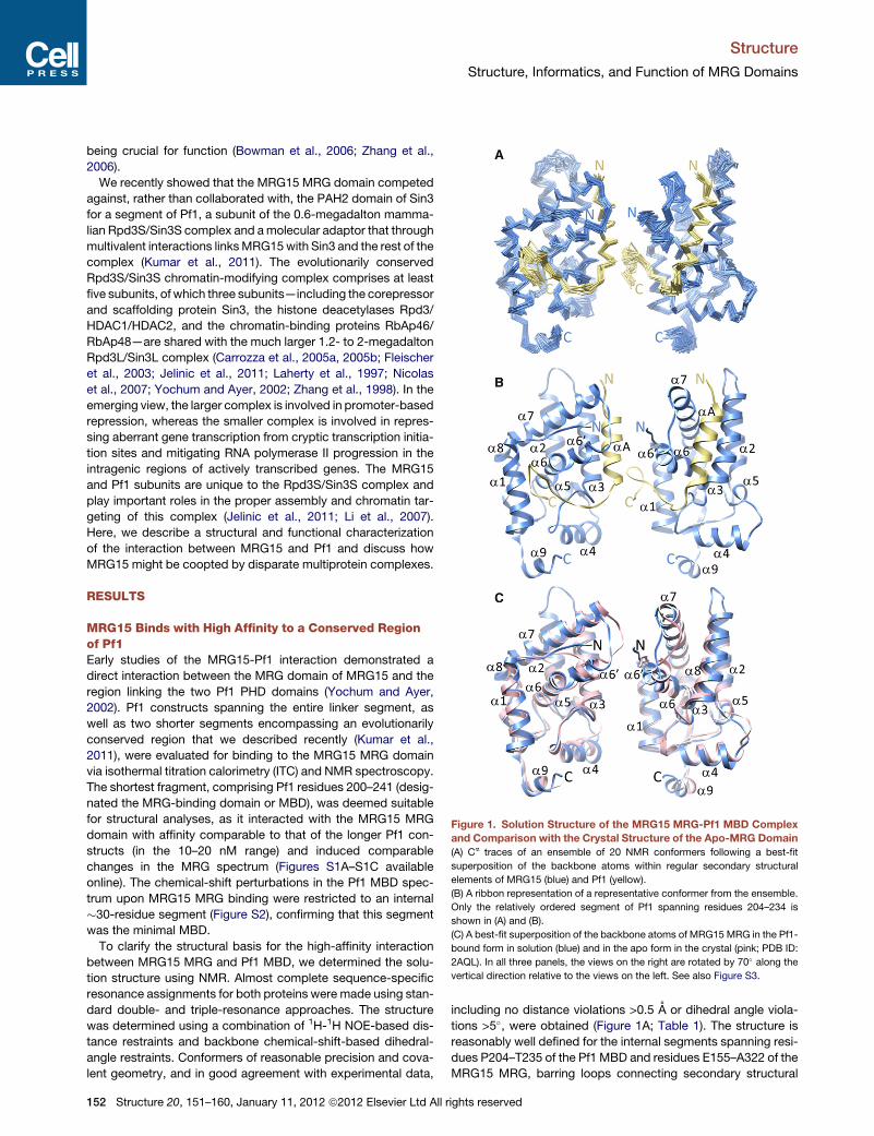
Figure 3. The MRG15-Pf1 Intermolecular Interface and Conservation of a Sequence Motif in MRG Binders
(A) Noncovalent interactions at theMRG15MRG-Pf1MBD interface. TheMRGdomain is rendered as amolecular surface with residuesmaking contacts with Pf1
shown in light blue with the side-chain oxygen, nitrogen, and sulfur atoms colored in red, blue and yellow, respectively.
(B) A MEME-guided multiple sequence alignment of the MRG-binding domain of Pf1 orthologs. Species abbreviations: Hs, Homo sapiens; Mm, Mus musculus;
Rn, Rattus norvegicus. Conserved and invariant residues are highlighted in yellow and blue, respectively. Filled circles denote intermolecular hydrophobic
(magenta) and hydrogen bonding (green) interactions in the MRG15-Pf1 complex.
(C) A multiple sequence alignment of various MRG interactors that harbor FxLP motifs in their MRG-binding domains. See also Figure S5.
Structure
Structure, Informatics, and Function of MRG Domains
We previously described competition between Sin3 and
MRG15, two subunits of the Rpd3S/Sin3S complex, for the Pf1
subunit (Kumar et al., 2011). The structure of the MRG15
MRG-Pf1 MBD complex provides the molecular basis for this
competition, with the MRG domain engaging an overlapping
surface of the Pf1MBD involving the nonpolar surface of the helix
that is also targeted by Sin3. However, unlike the Sin3 PAH2
domain, the MRG15 MRG domain engages a much larger Pf1
MBD surface, accounting for its affinity being more than two
orders of magnitude higher compared to the corresponding
Sin3 PAH2-Pf1 MBD complex. Unlike Pf1, which engages in
multivalent interactions with Sin3, the Pf1 MBD appears to
be the sole point of contact for MRG15 with the rest of the
Rpd3S/Sin3S complex, justifying the need for a high-affinity
interaction.
The dual specificity (i.e., engaging two structurally dissimilar
targets) of the Pf1 MBD suggests that the domain might func-
tion as a molecular switch cycling between an ‘‘off’’ state when
bound to the Sin3 PAH2 domain in the absence of MRG15, signi-
fying an inactive complex, and an ‘‘on’’ state in the presence of
MRG15, signifying an active, fully matured Rpd3S/Sin3S com-
plex available for engaging chromatin targets. In so doing, the
MBD might preclude misrecruitment of an incompletely assem-
bled complex by sequence-specific DNA-binding repressors
156 Structure 20, 151–160, January 11, 2012 ª2012 Elsevier Ltd All r
that often target the Sin3A PAH2 domain (Swanson et al.,
2004). Also, given the widespread involvement of f-x-x-f-f
(where f indicates hydrophobic and x any nonproline residues)
helical motifs in transcription coregulator interactions (Plevin
et al., 2005) and the occurrence of this motif in Pf1 MBD, the
MBD, by interacting with Sin3A PAH2, might also be protected
from misrecruitment by other coregulators.
The conformational plasticity demonstrated by the Pf1 MBD
in binding to diverse targets, including Sin3 PAH2 and MRG15
MRG domains, brings into sharp focus the role of intrinsically
unstructured regions in regulating basic cellular processes.
Indeed, the ability to bind diverse targets was one of the early
predictions of theWright-Dyson intrinsically unstructured protein
hypothesis (Wright and Dyson, 1999).
EXPERIMENTAL PROCEDURES
Production of the MRG15 MRG Polypeptide
The human MRG15 MRG polypeptide (residues 155–323) was expressed and
purified as described previously (Kumar et al., 2011). Uniformly 15N- and/or13C-labeled proteins were produced using this procedure, except that cells
were grown in M9 minimal medium containing 15N-ammonium sulfate and/or13C-D-glucose. Protein identity, integrity, and the extent of 15N/13C isotope
incorporation were assessed by electrospray ionization-mass spectrometry
(ESI-MS). NMR and ITC studies with MRG15 MRG were inadvertently
ights reserved
Figure 4. Functional Analysis of MRG15-Pf1 Interactions In Vitro
(A and B) ITC analysis of various Pf1 MBD (A) and MRG15 MRG (B) mutants. Representative titrations for various mutant and wild-type proteins are shown.
Symbols denote raw data while the continuous lines correspond to fits. The fitted equilibrium dissociation constants (KDs) for the various proteins are cataloged in
Table 2.
(C) Left, NMR titrations of 15N-labeled MRG15 MRG domain with a Pf1 peptide corresponding to the tail segment (residues 219–232). The movement of a well-
resolved MRG peak in the 1H-15N correlated spectrum as a function of added Pf1 peptide is shown. Right, Graphs of the raw chemical-shift deviations for three
representative MRG peaks as a function of molar ratio and the curves resulting from the fitting procedure. See also Figure S6.
Structure
Structure, Informatics, and Function of MRG Domains
conducted in a K201R mutant background; however, the Pf1 MBD-binding
affinity of the R201K ‘‘mutant’’ is not significantly different from K201R
(Table 2), and the mutation itself, besides being conserved, is not at the inter-
molecular interface.
Production of the Pf1 MBD Polypeptide
The coding sequence for human Pf1 MBD (residues 200–241) was subcloned
into the pMCSG7 expression vector, but the sequence for the N-terminal
24-residue tag was removed by PCR. Pf1 MBD was coexpressed and copuri-
fied with the MRG15 MRG polypeptide analogous to the coexpression and
copurification protocol described previously except the Pf1 MBD was selec-
tively eluted after binding to the Ni2+-resin in the presence of 6 M guanidine
hydrochloride prior to HPLC purification (Kumar et al., 2011). Uniformly15N- or 15N,13C-labeled Pf1 MBD were produced as described above for
Structure 20, 151
MRG15 MRG, and ESI-MS was used to assess protein identity, integrity,
and extent of isotope incorporation.
Isothermal Titration Calorimetry
ITC experiments were performed as previously described, except that
a 20 mM sodium phosphate buffer (pH 7.8) containing 0.15 M NaCl was
used (Kumar et al., 2011). Wild-type and mutant MRG15 MRG were in the
cell, whereas the wild-type and mutant Pf1 MBD were in the syringe at initial
concentrations of 10–20 mM and 0.15–0.55 mM, respectively.
MRG15 MRG-Pf1 MBD-Complex Generation and NMR Sample
Preparation
The MRG15 MRG-Pf1 MBD complex was generated by mixing equimolar
amounts of uniformly 15N,13C-labeled MRG15 MRG with unlabeled Pf1 MBD
–160, January 11, 2012 ª2012 Elsevier Ltd All rights reserved 157
Figure 5. Functional Analysis of MRG15-Pf1 Interactions in Cells
(A and B) CoIP analysis of various FLAG-tagged Pf1 mutants with wild-type
HA-tagged MRG15 (A) and various HA-tagged MRG15 mutants with wild-type
FLAG-tagged Pf1 conducted in HEK293T cells (B).
(C) Transcriptional repression assays conducted in HEK293T cells with GAL4
DBD fusions of wild-type and mutant MRG15 using a luciferase reporter and
a thymidine kinase promoter harboring four tandem GAL4 DNA-binding sites.
The error bars represent standard deviations from three independent
measurements.
Structure
Structure, Informatics, and Function of MRG Domains
or vice versa at a low concentration (�20 mM) in NMR buffer (50 mM sodium
phosphate (pH 6.8) containing 5 mM dithiothreitol-d10, 10% D2O, and 0.2%
NaN3). The samples were concentrated via ultrafiltration to �0.9 mM for
NMR studies. Protein concentrationswere determined spectrophotometrically
(Gill and von Hippel, 1989). Samples of the complex were lyophilized and dis-
solved in 99.996% D2O for experiments in D2O.
NMR Spectroscopy and Structure Determination
NMR data were acquired on a Varian Inova 600 MHz spectrometer at 25�C.NMR data processing and analysis were performed using Felix 98.0 and
Sparky (Goddard and Kneller, 2004). Backbone and side-chain 1H, 15N, and13C resonances for each protein in the MRG15 MRG-Pf1 MBD complex
were assigned by analyzing 3D HNCACB, CBCA(CO)NH, HNCA, HN(CO)CA,
HNCO, HCACO, HCCH-COSY, and HCCH-TOCSY spectra (Bax and Grze-
siek, 1993; Ferentz and Wagner, 2000). Aromatic resonances were assigned
from 2D 1H-13C aromatic HSQC, 3D 13C-edited NOESY, and 15N,13C-double
half-filtered NOESY spectra (Otting and Wuthrich, 1990).
Backbone4 andc torsion angle restraints were derived from analysis of 1Ha,13Ca, 13Cb, 13C0, and amide 15N chemical shifts using TALOS+ (Shen et al.,
158 Structure 20, 151–160, January 11, 2012 ª2012 Elsevier Ltd All r
2009); only those residues with TALOS+ reliability scores of 10 were
restrained. NOE restraints for each protein in the complex were derived
from 3D 15N-edited NOESY (mixing time, tm = 75 ms) and 3D 15N,13C-filtered,15N,13C-edited NOESY (tm = 120 ms) spectra recorded in H2O, 3D 13C-edited