Structural Analyses of a Purine Biosynthetic Enzyme fromMycobacterium tuberculosis Reveal a Novel BoundNucleotide*□S
Received for publication, August 8, 2011 Published, JBC Papers in Press, September 28, 2011, DOI 10.1074/jbc.M111.291138
Jérôme Le Nours1, Esther M. M. Bulloch, Zhening Zhang2, David R. Greenwood, Martin J. Middleditch,James M. J. Dickson, and Edward N. Baker3
From the Maurice Wilkins Centre for Molecular Biodiscovery and School of Biological Sciences, University of Auckland,Private Bag 92019, Auckland, New Zealand
Background: The purine biosynthetic enzyme ATIC is a recognized candidate for the development of therapeutic drugs.Results: Structure of mycobacterial ATIC was determined and a novel bound nucleotide characterized.Conclusion: ATIC structure shows basis for half-sites reactivity and evidence for tight binding of the novel nucleotide CFAIR.Significance: CFAIR could be used as the basis for new inhibitor design against ATIC.
Enzymes of the de novo purine biosynthetic pathway havebeen identified as essential for the growth and survival ofMyco-bacterium tuberculosis and thus have potential for the develop-ment of anti-tuberculosis drugs. The final two steps of thispathway are carried out by the bifunctional enzyme 5-amino-imidazole-4-carboxamide ribonucleotide transformylase/inos-ine monophosphate cyclohydrolase (ATIC), also known asPurH. This enzyme has already been the target of anti-cancerdrugdevelopment.Wehavedetermined the crystal structures ofthe M. tuberculosis ATIC (Rv0957) both with and without thesubstrate 5-aminoimidazole-4-carboxamide ribonucleotide, atresolutions of 2.5 and 2.2 Å, respectively. As for other ATICenzymes, the protein is folded into two domains, theN-terminaldomain (residues 1–212) containing the cyclohydrolase activesite and the C-terminal domain (residues 222–523) containingthe formyltransferase active site. An adventitiously boundnucleotide was found in the cyclohydrolase active site in bothstructures andwas identified byNMRandmass spectral analysisas a novel 5-formyl derivative of an earlier intermediate in thebiosynthetic pathway 4-carboxy-5-aminoimidazole ribonucle-otide. This result andother studies suggest that this novel nucle-otide is a cyclohydrolase inhibitor. The dimer formed by M.tuberculosis ATIC is different from those seen for human andavian ATICs, but it has a similar �50-Å separation of the two
active sites of the bifunctional enzyme. Evidence inM. tubercu-losis ATIC for reactivity of half-the-sites in the cyclohydrolasedomains can be attributed to ligand-induced movements thatpropagate across the dimer interface andmay be a common fea-ture of ATIC enzymes.
Purine nucleotides are required by all organisms to providethe building blocks for DNA and RNA. Two types of pathwaysexist inmost organisms for the production of these compoundsas follows: the de novo biosynthetic pathway, in which nucleo-tides are synthesized from 5�-phosphoribosylpyrophosphate(PRPP)4 in a multistep series of reactions (1); and salvage path-ways, in which nucleotides are retrieved after the breakdown ofnucleic acids or coenzymes (2, 3). These two alternatives maybe relatively more or less important in a given organism or inresponse to different cellular requirements. In humans, forexample, the demands of rapidly dividing tumor cells require anabundant source of nucleotides, which is provided by de novobiosynthesis (4), whereas normal cellular growthmay be largelymaintained by salvage. Enzymes of the de novo pathway havetherefore been targeted for anti-cancer drug development(5–9). Similar efforts are being made toward the developmentof new anti-infectives (10), and inMycobacterium tuberculosis,the cause of tuberculosis, the de novo purine biosynthetic path-way appears to be critical for growth and survival, with 10 of 13identified purine biosynthetic enzymes proving to be essentialin a genome-wide transposon mutagenesis study (11).The classical de novo purine biosynthetic pathway includes
10 enzymatic reactions that successively convert PRPP to 5�-in-osine monophosphate (IMP) (1). Variations exist betweenorganisms, however, with some single step reactions in
* This work was supported in part by the Foundation for Research, Science,and Technology of New Zealand, the Health Research Council of New Zea-land, and the Tertiary Education Commission through funding of the Mau-rice Wilkins Centre of Molecular Biodiscovery.
□S The on-line version of this article (available at http://www.jbc.org) containssupplemental Table S1 and Figs. S1–S4.
The atomic coordinates and structure factors (codes 3zzm and 4a1o) have beendeposited in the Protein Data Bank, Research Collaboratory for StructuralBioinformatics, Rutgers University, New Brunswick, NJ (http://www.rcsb.org/).
1 Present address: Dept. of Biochemistry and Molecular Biology, School ofBiomedical Sciences, Monash University-Clayton, Victoria 3800, Australia.
2 Supported by a doctorate scholarship from the Maurice Wilkins Centre forMolecular Biodiscovery. Present address: Biology Dept., BrookhavenNational Laboratory, Upton, NY 11973.
3 To whom correspondence should be addressed: School of Biological Sci-ences, University of Auckland, Private Bag 92019, Auckland Mail Centre,Auckland 1142, New Zealand. Tel.: 64-9-373-7599; E-mail: [email protected].
4 The abbreviations used are: PRPP, 5�-phosphoribosyl pyrophosphate; ATIC,5-aminoimidazole-4-carboxamide ribonucleotide transformylase/IMPcyclohydrolase; MtbATIC, ATIC enzyme from M. tuberculosis; AICAR, 5-ami-noimidazole-4-carboxamide ribonucleotide; FAICAR, 5-formylaminoimi-dazole-4-carboxamide ribonucleotide; CFAIR, 4-carboxy-5-formylamino-imidazole ribonucleotide; IMPCHase, IMP cyclohydrolase; CAIR, 4-carboxy-5-aminoimidazole; FAIR, 5-formylaminoimidazole ribonucleoside; ESI,electrospray ionization.
eukaryotes being carried out as two separate steps in pro-karyotes and with somemultifunctional enzymes in eukaryotesbeing substituted by monofunctional enzymes in prokaryotes.In most microorganisms, 12 biosynthetic steps are required, 6of which utilize ATP, 2 utilize glutamine, and 2 utilize N10-formyl tetrahydrofolate (THF). All of the substrates in the path-way share a common ribose 5-phosphate moiety on which thepurine base is built sequentially (1). The final two steps areusually catalyzed by a single-chain bifunctional enzyme 5-ami-noimidazole-4-carboxamide ribonucleotide transformylase/IMP cyclohydrolase (ATIC), also known as PurH (12). The firststep in the reaction catalyzed by ATIC involves the transfer of aformyl group from the cofactor N10-formyltetrahydrofolate tothe substrate 5-aminoimidazole-4-carboxamide ribonucle-otide (AICAR) to form the intermediate 5-formylaminoimida-zole-4-carboxamide ribonucleotide (FAICAR), which is thencyclized with the loss of water to produce the final product ofthe pathway, IMP (Fig. 1A). In archaea, however, these two
steps are carried out by quite distinct enzymes, unrelated toPurH, and without the use of tetrahydrofolate (THF) (13).The eukaryotic ATIC enzymes have been extensively char-
acterized through structural analyses of the chicken (14, 15)and human (16) enzymes, coupled with kinetic, mutagenesis,and inhibition studies (17, 18). These studies have establisheddetailed catalytic mechanisms for both reactions (15, 18). Bothenzymes are dimeric, with two distinct functional domains permonomer. The THF-dependent formyl transfer reaction takesplace in the C-terminal domain of the protein, residues 200–593 in the chicken enzyme, whereas the product of this reac-tion, FAICAR, undergoes cyclization in the active site of theN-terminal domain, some 50 Å away (14). Intriguingly, there isno evidence of any tunnel connecting the two active sites. Afeature of both the chicken and human ATIC structures wasthat a nucleotidewas found bound in each case to just one of thetwo cyclohydrolase domains of the respective dimers. This wasevidently carried over from expression in the Escherichia coli
FIGURE 1. A, reaction scheme for ATIC, showing the two steps. B, overall architecture of the MtbATIC dimer, with the C-terminal formyltransferase domainsabove and the N-terminal cyclohydrolase domains below. The two monomers A and B are colored in light blue and gold, respectively. Potassium ions bound ineach monomer are shown as spheres. The bound AICAR and CFAIR molecules are shown in magenta and green stick mode, respectively. The �-ribbon formedby the connecting peptides (green for molecule A and red for molecule B) is also shown. C, comparison of the dimers formed by MtbATIC (left, monomers A andB colored in gold and light gold, respectively) and human ATIC (right, monomers A and B colored in blue and sky blue, respectively). For comparison, the AICARformyltransferase domains of the Mtb and human ATIC structures were superimposed, and the two molecular surface representations are shown in the sameorientation. Figures were generated using PyMOL (42).
Crystal Structure and Nucleotide Binding by MtbATIC
NOVEMBER 25, 2011 • VOLUME 286 • NUMBER 47 JOURNAL OF BIOLOGICAL CHEMISTRY 40707
host used, remaining bound through all purification and crys-tallization steps. This nucleotide was identified as xanthosinemonophosphate (XMP) on the basis of HPLC and mass spec-trometry (18).Here, we describe crystal structures of ATIC fromM. tuber-
culosis (MtbATIC) at 2.2 Å resolution in the absence of boundsubstrate and at 2.48 Å resolution with the substrate AICARbound. Interestingly, a nucleotide is again found in one of thetwo cyclohydrolase active sites of the dimer, evidently carriedover fromE. coli expression. This addsweight to the conclusionthat ATIC exhibits half-of-the-sites reactivity. In contrast tothe conclusions drawn for the human and chicken enzymes,however, we use NMR and mass spectrometry to identify theadventitiously bound nucleotide as a novel formylated nucleo-tide potentially derived from an intermediate formed in an ear-lier step of the purine biosynthetic pathway.
EXPERIMENTAL PROCEDURES
Protein Expression and Purification—The open readingframe encoding Rv0957 (ATIC) was amplified by nested PCRfrom the genomic DNA of M. tuberculosis H37Rv. Gateway�cloning was used as described by Moreland et al. (19) to gener-ate the expression vector pDEST17 harboring an N-terminalHis6 tag. The Rv0957 Gateway� plasmid was transformed intoE. coli BL21 (DE3) cells that were plated on Luria-Bertani agarplates containing 50 �g/ml ampicillin. A 5-ml pre-culture ofE. coli BL21 (DE3) cells was grown in noninducingMDGmediaovernight at 37 °C, and a 1:200 dilution of this pre-culture wasused to inoculate the expression culture. Expression of theN-terminally His6-tagged MtbATIC was carried out in theautoinductionmediumZYM-5052 (20). The culturewas grownat 37 °C until the A600 reached 0.6 and was then transferred to10 °C for 2 days. The cells were harvested by centrifugation for15min (4000� g) at 4 °C, resuspended in 50mMNaH2PO4, pH7.5, 1 mM EDTA, 14 mM �-mercaptoethanol, and lysed with acell disruptor (Constant Systems Ltd.) at 18,000 p.s.i. Cellulardebris was separated by centrifugation (14,000 � g, 20 min,4 °C). His-taggedMtbATIC was purified by immobilized metalaffinity chromatography using a 5-ml Ni2�-charged Hi-trapcolumn (Amersham Biosciences). The ATIC-containing frac-tions were dialyzed overnight (4 °C) against 50 mM NaH2PO4,pH 7.5, 1mMEDTA, 14mM �-mercaptoethanol in the presenceof recombinant tobacco etch virus protease to cleave the His6tag. A second immobilized metal affinity chromatography col-umn to remove any remaining His6-tagged material was fol-lowed by size exclusion chromatography using an S200 10/30column (Amersham Biosciences). The fractions containing
MtbATIC were concentrated to 20 mg/ml using 10,000 molec-ular weight cutoff concentrators (Vivaspin).Crystallization and Data Collection—Initial crystallization
conditions were found using a Cartesian nanoliter dispensingrobot (HONEYBEETM) with a 480-component screen (19).These experiments were performed at 18 °C by sitting-dropvapor diffusion, using 100-nl drops of protein (20 mg/ml)mixedwith 100nl of precipitant. Themost promising condition(30% PEG 8000, 0.2 M sodium acetate, 0.1 M sodium cacodylate,pH 6.5) led to the formation of one small single crystal after 2days. After further optimizations, crystals suitable for data col-lection were obtained in hanging drops consisting of 1 �l ofprotein (20mg/ml) and 3�l of reservoir (24–34%PEG8000, 0.2M sodium acetate, 0.1 M sodium cacodylate, pH 6.5). For datacollection, a native crystal was soaked for a few seconds in asolution composed of mother liquor and 20% glycerol and thenflash-cooled. X-ray diffraction data to 2.2 Å resolution weremeasured at 100 K using CuK� radiation from a rotating anodegenerator (Rigaku Micromax-007HF) equipped with a MAR345 imaging plate detector. For the AICAR complex, a nativecrystal was soaked for 2 min in a solution containing 100 mM
AICAR (Sigma), the reservoir solution, and 20% glycerol (cryo-protectant). X-ray data to 2.48 Å resolution were collected at100 K at the Stanford Synchrotron Radiation Laboratory. Bothdata sets (see Table 1) were processed using HKL2000 (21).Structure Determination and Refinement—The native
MtbATIC three-dimensional structure was solved by molecu-lar replacement using PHASER (22). A composite searchmodelwas used comprising residues 3–195 from the human ATICcrystal structure (Protein Data Bank code 1PKX) (16) and resi-dues 167–452 from the Thermotoga maritima ATIC(TmATIC) crystal structure (Protein Data Bank code 1ZCZ)(23). The solution contained two molecules in the asymmetricunit. An atomicmodel was built and adjusted using COOT (24)and refined by restrained least squares with REFMAC 5.3 (25).BUSTER 2.8 (26) was used for the latter stages of refinement.No interpretable electron density was found for the N-terminalresidues of either molecule; the final model consisted of resi-dues 5–523 in molecule A and residues 7–523 in molecule B.Residues 65–67 could also not bemodeled inmoleculeAdue tolack of interpretable density. The twomolecules are very similarwith a root mean square difference of 1.2 Å for 517 C� atoms.After a few cycles of refinement, clear difference electron den-sity was visible for an unidentified nucleotide bound to mole-cule A and for a potassium ion in both molecules. The nucleo-tide was initially modeled as XMP but was later determined to
TABLE 1Data collection and processing statistics
Parameters MtbATIC MtbATIC/AICAR
Space group P212121 P212121a, b, c (Å) 76.6, 108.2, 130.6 75.2, 107.9, 130.9No. of measured reflections 539,267 403,256No. of unique reflections 54,907 38,276Resolution rangea 30 to 2.2 (2.28 to 2.2 Å) 50 to 2.48 (2.58 to 2.48 Å)Completenessa 99.8% (87.0%) 98.8% (88.9%)Mean I/�(I)a 30.7 (3.7) 20.8 (4.7)Rmerge
a,b 7.2% (40.3%) 11.6% (38.6%)a Values shown in parentheses correspond to the high resolution shell.b Rmerge � �hkli �i�I(hkl)i � �I(hkl)��/�hkl�i I(hkl)i.
Crystal Structure and Nucleotide Binding by MtbATIC
40708 JOURNAL OF BIOLOGICAL CHEMISTRY VOLUME 286 • NUMBER 47 • NOVEMBER 25, 2011
be 4-carboxy-5-formylaminoimidazole ribonucleotide(CFAIR) and was modeled as such, using the Dundee PRO-DRG2 server (27). The final values for Rwork and Rfree were 19.9and 23.7%, respectively. For the AICAR complex, the refinednative MtbATIC structure was used as the initial model andrefined to 2.48Å resolutionwith REFMAC5.3 and BUSTER 2.8using the same refinement procedure as for the native struc-ture. Difference electron density maps showed clear density forthe substrate AICAR in both monomers. A CFAIR molecule asthe E-amide conformer and two potassium ionswere alsomod-eled in the same locations as for the native structure. Residualelectron density was also found in the active site of the cyclo-hydrolase domain ofmolecule B. Thiswas suggestive of a nucle-otide, possibly an additional AICAR molecule, but only thephosphate moiety could be modeled with certainty. The finalvalues of Rwork and Rfree for the AICAR complex were 17.9 and22.3%, respectively. Full details of the refinement statistics andthe final model for each structure are in Table 2.Mass Spectrometry of Nucleotide Ligand—The ligand bound
to MtbATIC was directly analyzed using mass spectrometrywith electrospray ionization (ESI) on quadrupole-time of flight(QTOF MS) and Fourier transform ion cyclotron resonanceinstruments. Purified MtbATIC was exchanged into 50 mM
NH4HCO3 and infused into the mass spectrometer ion sourcein 50% v/v acetonitrile, and the bound ligand was released in-source. ESI-QTOFMS was carried out on a QSTAR XL hybridmass spectrometer (Applied Biosystems) in both positive andnegative ion mode. Species of interest were further analyzed byMS2 fragmentation, and amajor negative ion species ofm/z 366was identified as the nucleotide ligand. Precise mass measure-ments were then carried out on this parental species and asso-ciatedMSn fragments using a LTQFourier transform ion cyclo-tron resonance mass spectrometer (Thermo Finnigan).Infusion into the ion source was at 3 �l/min with a source volt-age of 2.5 kV, a tube lens setting of �100 V, and a capillarytemperature of 225 °C. Fragmentation of the ligand was carriedout to MS5 with accurate mass determination on ions up toMS4.Isolation of Nucleotide Ligand—The nucleotide ligand was
isolated from MtbATIC by denaturation of the enzyme and
purification of the released nucleotide by ion exchange chro-matography. His6-tagged ATIC purified using immobilizedmetal affinity chromatography, as detailed above, was dialyzedinto 20 mM NH4HCO3, pH 8. Solid urea was added to the dia-lyzed His6-tagged ATIC with stirring to give a final concentra-tion of 6 M. In initial experiments, this preparation was alsoheated to 100 °C for 10min to ensure complete denaturation ofthe enzyme. However, this heating step was later found to beunnecessary, and a 15-min incubation at room temperaturewas sufficient for release of the nucleotide. The urea-containingMtbATIC solution was loaded onto a Mono Q HR 5/5 anionexchange column (GE Healthcare), and a gradient of 20–400mM NH4HCO3 was run over 40 ml at 2 ml/min. Eluate absorb-ance was monitored at 220, 260, and 280 nm. One major peakwas detected at a concentration of 130–150 mM NH4HCO3.Analysis of this peak by ESI-QTOF MS by direct infusion con-firmed that it consisted of them/z 366 species detected by MSanalysis of MtbATIC. Peak fractions were combined and driedby lyophilization or on a centrifugal vacuum concentrator, andthe nucleotide was stored at �20 °C.NMR Characterization of Nucleotide Ligand—The purified
nucleotide was dissolved in D2O for analysis by NMR spectros-copy. Preliminary one-dimensional 1HNMR and temperature-dependent one-dimensional 1H NMR experiments wereacquired on a DRX 400 spectrometer (Bruker). Detailed analy-sis of the 1H and 13C chemical shifts was carried out using one-and two-dimensional spectra acquired on an AV600 spectrom-eter (Bruker) fitted with a cryoprobe and at 300 K. Thisincluded 1H-1H COSY, 1H-13C HSQC, and 1H-13C HMBCexperiments. Two separate chemical species were observed inall NMR spectra at a 74:26 ratio at 300 K. Temperature-depen-dent 1H NMR spectra were collected at 25, 40, 50, and 60 °C toexamine whether these two species were slowly interchangingisomers of the nucleotide. Chemical shifts are reported in parts/million and were relative to an external TMS reference (� 0.0ppm).Computational Analysis of Nucleotide Ligand—Calculations
on the predicted relative stability of theZ- andE-amide isomersof CFAIR were carried out using the Gaussian 09 software suite(28). For simplicity, the phosphate groupwas removed from the
TABLE 2Refinement statistics
Parameter MtbATIC MtbATIC/AICAR
Resolution rangea 28.8 to 2.20 (2.26 to 2.20) 35.5 to 2.48 (2.55 to 2.48)R/Rfree
Root mean square deviations from ideal geometryBonds (Å) 0.010 0.010Angles (°) 1.0 1.1
Ramachandran plot (30)Residues in favored regions 98.7% 98.2%Outliers 0.2% 0.1%
a Values in parentheses are for the outermost resolution shell.b R and Rfree � ��Fobs� � �Fcalc�/��Fobs�, where Rfree was calculated over 5% of amplitudes that were chosen at random and not used in refinement.
Crystal Structure and Nucleotide Binding by MtbATIC
NOVEMBER 25, 2011 • VOLUME 286 • NUMBER 47 JOURNAL OF BIOLOGICAL CHEMISTRY 40709
structures. Global energy minima were identified for both iso-mers by comparison of relaxed potential energy surface scans,at theRHF/3–21G* level, of the dihedral angles emanating fromthe imidazole ring. Gas phase calculations were performed atthe RB3LYP/6–31G(d)//RB3LYP/6–31G(d) level. By compar-ison of the unscaled ZPE corrected electronic energies, the low-est energy Z-conformer was found to be 15 kJ/mol more stablethan the lowest energy E-conformer. In repeating the con-former search and optimization using the polarizable contin-uum model solvent model (29) at the same level of theory, theZ-conformerwas again found to bemore stable than theE-con-former by 4.5 kJ/mol. Assuming that the lowest energy confor-mations would have the greatest influence on the Boltzmanpopulation ratio, the latter calculated energy difference corre-sponds to a population ratio of �86:14 in favor of the Z-isomerof CFAIR.
RESULTS
Crystallographic Analyses—The MtbATIC enzyme wasexpressed in E. coli and crystallized in its presumed apo-form,without the addition of any added ligand.We refer to this as theapoenzyme, although one of the twomolecules in the asymmet-ric unit was found to contain a ligand derived from the E. coliexpression host. The characterization of this ligand is describedlater. The complex of MtbATIC with the substrate AICAR wasprepared by soaking crystals of the apoenzyme with AICAR.Both structures were determined by x-ray crystallography andrefined at resolutions of 2.2 and 2.48 Å resolution, respectively.The asymmetric unit of the crystal in each case contained two
independent ATIC molecules, organized as tightly associateddimers. For both structures, the 523-residue polypeptide chainwas fully defined except for a few residues at the N terminus(3–4 formolecule A and 6 formolecule B) and in a flexible loopof the cyclohydrolase domain, for which no interpretable den-sity for residues 65–67 inmolecule A of the apo structure couldbe found. Analysis with MOLPROBITY (30) showed that 99%of residues in each structure are in the most favored regions ofthe Ramachandran plot and that the apo structure and AICARstructures each have an overall MolProbity score in the 99thpercentile.Structural Organization ofMtbATIC—As has been found for
the human, chicken, and T. maritima enzymes, MtbATICforms a tightly associated dimer (Fig. 2B) that is the presumedfunctional unit. Eachmonomer includes twodistinct functionaldomains as follows: the N-terminal cyclohydrolase domain,which carries out the IMP cyclohydrolase reaction, and theC-terminal transformylase domain, which mediates the trans-fer of the formyl group fromN10-formyltetrahydrofolate to thesubstrate AICAR. The two domains are joined by a linker pep-tide, residues 203–219, which forms a long �-ribbon that iscritical to the mode of dimerization, described later.The twomajor domains are very similar to their counterparts
in the human, chicken, and T. maritima enzymes. The cyclo-hydrolase domain, residues 1–202, has a modified Rossmannfold topology, including a central 5-stranded �-sheet, withstrand order 3–2-1–4-5, against which are packed three heliceson each face, three more around the periphery, and a long final
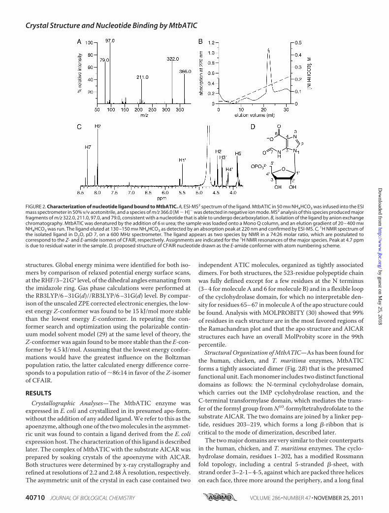
FIGURE 2. Characterization of nucleotide ligand bound to MtbATIC. A, ESI-MS2 spectrum of the ligand. MtbATIC in 50 mM NH4HCO3 was infused into the ESImass spectrometer in 50% v/v acetonitrile, and a species of m/z 366.0 [M � H]� was detected in negative ion mode. MS2 analysis of this species produced majorfragments of m/z 322.0, 211.0, 97.0, and 79.0, consistent with a nucleotide that is able to undergo decarboxylation. B, isolation of the ligand by anion exchangechromatography. MtbATIC was denatured by the addition of 6 M urea; the sample was loaded onto a Mono Q column, and an elution gradient of 20 – 400 mM
NH4HCO3 was run. The ligand eluted at 130 –150 mM NH4HCO3 as detected by an absorption peak at 220 nm and confirmed by ESI-MS. C, 1H NMR spectrum ofthe isolated ligand in D2O, pD 7, on a 600 MHz spectrometer. The ligand appears as two species by NMR in a 74:26 molar ratio, which are postulated tocorrespond to the Z- and E-amide isomers of CFAIR, respectively. Assignments are indicated for the 1H NMR resonances of the major species. Peak at 4.7 ppmis due to residual water in the sample. D, proposed structure of CFAIR nucleotide drawn as the E-amide conformer with atom numbering scheme.
Crystal Structure and Nucleotide Binding by MtbATIC
40710 JOURNAL OF BIOLOGICAL CHEMISTRY VOLUME 286 • NUMBER 47 • NOVEMBER 25, 2011
helix�10 that leads into the interdomain linker. The active site,in which the cyclization of FAICAR to the purine nucleotideIMP takes place, extends from the�2-�2 loop across strands�1and �4 to a site between helix �8 and the loop joining residues71–74.The C-terminal transformylase domain, residues 220–523,
contains two tandem subdomains, residues 242–367 and 388–523, each of them with an � � � fold comprising a mixed6-stranded �-sheet and four �-helices arranged as in cytidinedeaminase (31). Each of these subdomains is also preceded byan extended �-hairpin, residues 220–242 and 368–387. Apotassium ion is present in this domain, bound to the mainchain carbonyl oxygens of residues 421, 422, 424, and 520, anda carboxyl oxygen of Asp-470, with metal-ligand distances of2.6–2.8Å.Adjacent to this is a cis-peptide between Ser-426 andAsn-427, which is conserved in all ATIC structures. Both resi-dues may be functionally important; Asn-427 is part of theAICAR-binding site, and Ser-426 helps orient Asp-470, one ofthe potassium ion ligands. The formyl transfer active site lies atthe interface between the twomonomers of the dimer; there arethus two such active sites per dimer. Although only the bindingof theAICAR substrate has been defined in the present analysis,binding studies on the chicken ATIC enzyme, using folate ana-logs and a bisubstrate inhibitor, indicate that both monomerscontribute equally to each active site, one predominantly bind-ing AICAR and the other predominantly binding folate.Dimerization must thus be essential for the formyl transferreaction, as shown previously for the human enzyme (32).The MtbATIC dimer is classified as a parallel, nontwisted
dimer according to the web server class PPI (33). It matchesclosely with the dimer formed by TmATIC (23), but it is funda-mentally different from those formed by the human andchicken ATIC enzymes (Fig. 1C). The difference is that if theC-terminal transformylase domains of the four proteins aresuperimposed, the cyclohydrolase domains of the two bacterialenzymes must be rotated about 90° about the long axis of thedimer to overlay on their eukaryotic counterparts; the latter areclassified as twisted dimers. The origins of this difference lie inthe structure formed by the connecting peptide, because thereare otherwise no contacts between the N- and C-terminaldomains.In both MtbATIC and TmATIC, the connecting peptides of
the two monomers form a long antiparallel �-ribbon that fol-lows the last helix (�10) of the cyclohydrolase domain and isoriented orthogonally to the long axis of the molecule (Fig. 1B).In MtbATIC, this �-ribbon includes residues 211–219 fromeach monomer, two residues more than in TmATIC, and hasregular hydrogen bonds over its whole length. In contrast, aquite different substructure is found between the majordomains in the two eukaryotic enzymes. In the MtbATICdimer, the �-ribbon linker is followed immediately by the firstof the two �-hairpins of the C-terminal domains and is inti-mately associated with it. Overall, the total buried surfacewithin the dimer interface is about 5800Å2, as calculated by theprogram PISA (34), representing about 23% of the average sur-face area of each monomer. Of 523 amino acid residues, about150 contribute to this interface. Much of the buried surface isattributed to the two�-hairpins of eachmonomer, which inter-
digitate like four fingers, but the buried surface extends foralmost the whole length of the molecule.Identification of the Adventitiously Bound Ligand—Clear
electron density resembling that of a purine nucleotide wasfound in the active site of the cyclohydrolase domain of mole-cule A (but not molecule B) of the apo-MtbATIC structure.Because no ligand had been added, we assume that this is anadventitiously bound ligand derived from the E. coli expressionhost. A similar phenomenon was observed in the crystal struc-tures of the apo-forms of the human and chicken ATICenzymes (14–16), in which it was modeled as XMP. This iden-tification was not unequivocal, however, and we thereforesought to characterize the ligand bound to MtbATIC by massspectrometry and NMR.Initial characterization of the ligandwas based onmass spec-
trometry of the purified MtbATIC directly. The ligand wasreleased from the enzyme in-source during ESI-MS anddetected as a negative [M � H]� ion ofm/z 366.0. Fragmenta-tion of this species byMS2 resulted inmajor species ofm/z 322,211, 97, and 79 (Fig. 2A). The three lower masses match thoseexpected for phosphoribosyl, phosphate, and phosphite frag-ments, respectively, consistent with identification of the ligandas a nucleotide. However, the overall mass of the ligand doesnot correspond to any known nucleotide. This includes theAICAR substrate and IMP product of the MtbATIC reaction,which would give [M � H]� ions of m/z 337.0 and 347.0,respectively. The ligand mass is close to that for the reactionintermediate FAICAR but does not match its expected m/z of365.0. It is also inconsistent with XMP (m/z 363.0) as modeledin the human and chicken ATIC structures.Detailed structural analysis of the nucleotide ligand was car-
ried out by accurate mass determination and further MSn frag-mentation with Fourier transform ion cyclotron resonanceMS(supplemental Fig. S1 and supplemental Table S1). An exactmass ofm/z 366.034 was measured for this ion, correspondingto an elemental composition of C10H13O10N3P1� within 0.58ppm (theoretical m/z 366.0344). This is close to the elementalcomposition of FAICAR (C10H14O9N4P1�), except that theligand contains one less nitrogen and hydrogen atombut has anadditional oxygen, accounting for the 1 atomic mass unit dif-ference in mass between the compounds. Additional fragmen-tation of an MS2 species corresponding to the base moiety ofthe nucleotide (m/z 154.1) produced ions of m/z 126.0 and110.1. The latter is consistent with a 5-formylaminoimidazolefragment as would be expected from a FAICAR-like structure.However, inconsistent with FAICAR, the base of the nucleotideundergoes decarboxylation producing a m/z 322.04 speciesfrom the parental nucleotide ion. This decarboxylated specieswas also observed in ESI-MS without MSn fragmentation, sug-gesting that the base of the nucleotide contains a carboxylategroup that can be readily eliminated.Combining the elemental composition, MSn fragment anal-
ysis, and the nature of the electron density at the cyclohydrolaseactive site of MtbATIC, it was speculated that the unknownnucleotide ligand may be a FAICAR analog in which the car-boxyamide group is exchanged with a carboxylate (Fig. 2D). Toour knowledge, this compound, CFAIR, is not a known metab-olite and has not been previously prepared synthetically. To
Crystal Structure and Nucleotide Binding by MtbATIC
NOVEMBER 25, 2011 • VOLUME 286 • NUMBER 47 JOURNAL OF BIOLOGICAL CHEMISTRY 40711
provide further evidence for this proposed structure, we soughtto isolate the ligand from MtbATIC and analyze it by NMRspectroscopy. The nucleotide ligand was released fromMtbATIC by adding 6 M urea to the enzyme and was purifiedusing anion exchange chromatography (Fig. 2B). ESI-MS con-firmed that the isolated ligand was unaltered from the nucleo-tide detected in the experiments described above.A range of one- and two-dimensional 1H and 13C NMR
experiments was carried out on the ligand in D2O (supplemen-tal Figs. S2 and S3). Two separate species were observed in allNMR spectra at 25 °C in a 74:26 molar ratio. Initially, we spec-ulated that this may be due to decarboxylation of the nucleo-tide, as a related compound, and intermediate in purine biosyn-thesis, 4-carboxy-5-aminoimidazole (CAIR) readily undergoesnonenzymatic decarboxylation at pH 8 (35, 36). However,unsuccessful attempts to separate the two species and theobservation that their relative molar ratio was constant in dif-ferent preparations of the nucleotide suggested that these cor-responded to different isomers of the nucleotide. FAICAR andthe related compound 5-formylaminoimidazole ribonucleoside(FAIRs) exist as two slowly interconverting Z and E rotationalisomers around the formamide bond (37, 38).Consistent with the existence of Z- and E-rotational isomers
of the nucleotide ligand, temperature-dependent 1H NMRexperiments from 25 to 60 °C showed considerable peak broad-eningwith heating and some signals from the two species beganto coalesce at 60 °C (supplemental Fig. S4). Computationalanalysis of the lowest energy conformations of theZ- andE-am-ide isomers of CFAIR using a polarizable continuum modelsolvent model (29) estimates that the Z-isomer is 4.5 kJ/molmore stable that the E-isomer. This correlates to a Boltzmanpopulation of 86:14 in favor of the Z-isomer of CFAIR, suggest-ing that the Z-isomer is the major species observed in solutionby NMR spectroscopy. In agreement with this, the analogousnucleoside FAIRs was previously shown to exist in a 78:22 ratioof Z- and E-isomers in D2O (38).Complete assignment of 1H NMR chemical shifts was possi-
ble for the major isomer of the nucleotide (Fig. 2C). Two of theminor isomer 1H peaks were obscured by overlap with those ofthe major isomer. H2 of the imidazole ring was observed toundergo slow exchange with deuterium from the solvent.Through a combination of 1H-13C HSQC and 1H-13C HMBCexperiments, a nearly complete assignment was also obtainedfor 13C NMR resonances of the major isomer. However, therewas not sufficient signal to unequivocally assign the 13C peakfor the significant C6 carbonyl. Overall, the 1H and 13C chem-ical shifts for the nucleotide (Tables 3 and 4) are similar to those
previously reported for FAICAR (37). The 13C resonances forC2 and the formamide C7 of 134.1 (major), 164.8 (major), and168.2 (minor) ppm, respectively, are particularly close to the134.9, 164.5, and 167.6 ppm for FAICAR. Notably, there is noequivalent 13C resonance evident in the nucleotide spectra atthe 165.4 ppm reported for the amide C6 of FAICAR, with theremaining unassigned peaks in this region occurring at 162.8and 168.7 ppm.LigandBinding in the Cyclohydrolase Active Site—In the sub-
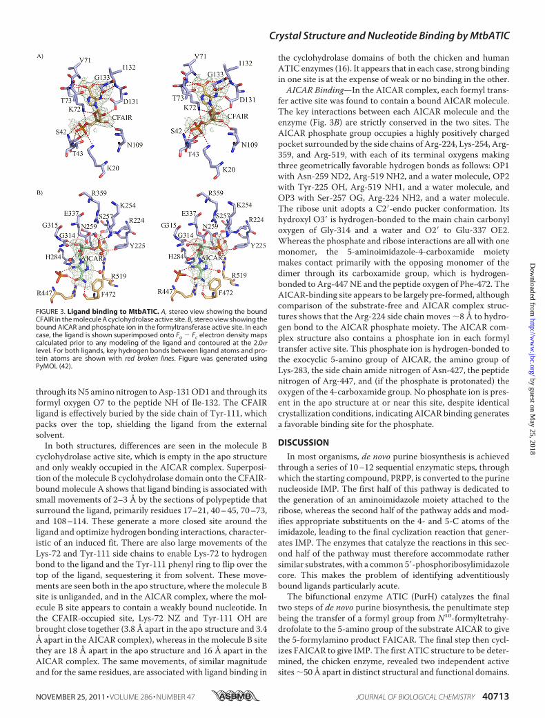
strate-free structure, a bound nucleotide is present in the activesite of molecule A, whereas the molecule B active site is notoccupied by any ligand. The nucleotide wasmodeled as CFAIR,in the E-isomer conformation, given the strong mass spectraland NMR support for this assignment. In the AICAR complex,molecule A again contains a bound CFAIR ligand, which is evi-dently not displaced by substrate. In contrast to the apo struc-ture, however, molecule B of the AICAR complex does containelectron density for a bound ligand. The density is weak anddiscontinuous, making it difficult to unequivocally identify thisligand or to model it. Because the complex was obtained bysoaking AICAR into the substrate-free crystals, in which thecyclohydrolase active site of molecule B was empty, we assumethe bound ligand to be AICAR, although only the phosphatecan be modeled with confidence.The CFAIR ligand in molecule A is represented by excellent
electron density in both structures and has the same conforma-tion and makes the same interactions in each case (Fig. 3A);when the two structures are superimposed based only on theprotein atoms, the root mean square difference in atomic posi-tions for the ligand is only 0.33 Å (24 atoms). The CFAIR phos-phate is bound at the N terminus of helix �2, with its O2Poxygen hydrogen-bonded to the peptide NH of Ser-42, O3P tothe peptide NH of Thr-43 and the Thr-43 hydroxyl group, andO1P to the Ser-42 hydroxyl group and the amino group of Lys-20. A second lysine amino group, of Lys-72, hydrogen bonds tothe phosphate ester oxygen O4P and the ribose ring oxygenO4�. Hydrogen bonds linking the ribose O2� andO3� hydroxylsto Asp-131 OD1 and the peptide oxygen of Asn-109, respec-tively, complete very extensive recognition of the phosphori-bose moiety. The substituted imidazole group of CFAIR hastwo substituents. The 4-carboxyl group sits between the 71–74loop and the N terminus of helix �8 (residues 131–145); onecarboxyl oxygen accepts hydrogen bonds from the peptide NHgroups of Lys-72 and Thr-73 and the other a hydrogen bondfrom the peptide NH of Gly-133. The other imidazole substit-uent is the C5-formylamino group, which hydrogen bonds
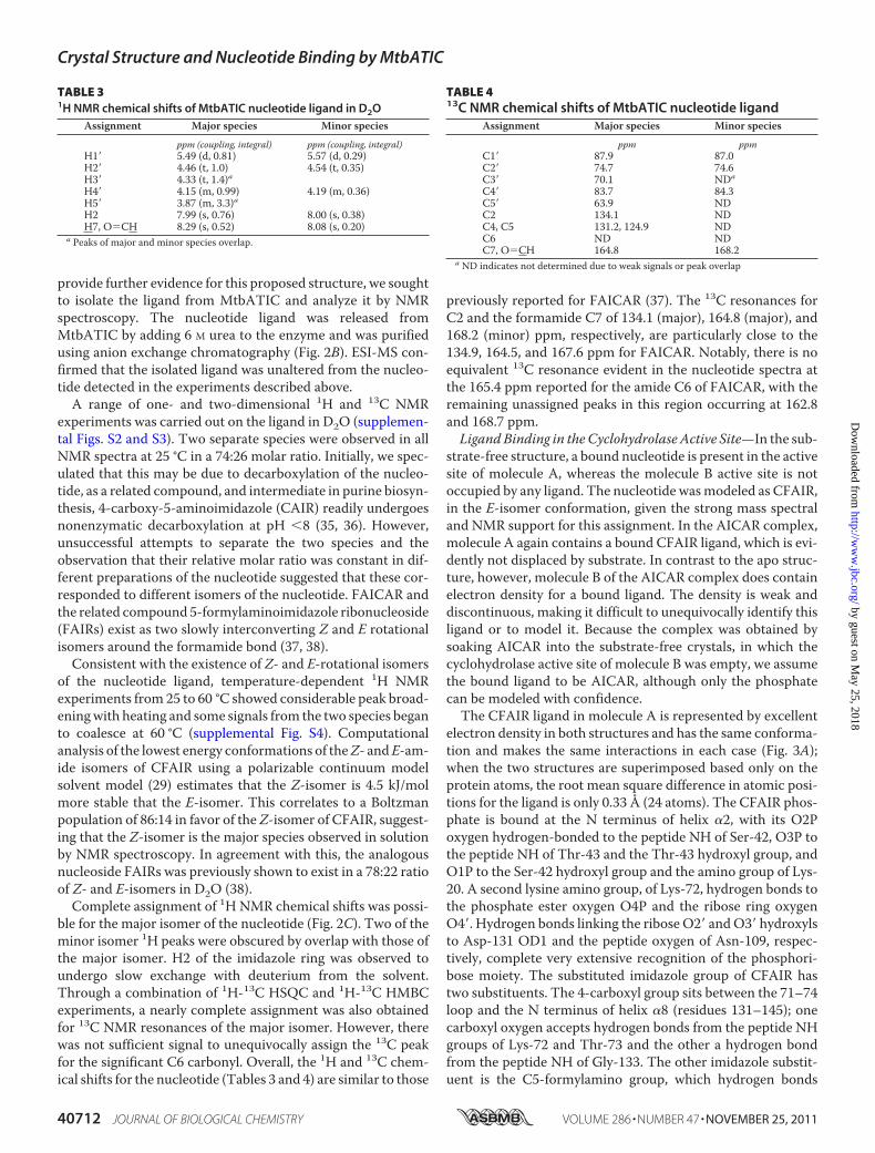
TABLE 31H NMR chemical shifts of MtbATIC nucleotide ligand in D2O
through its N5 amino nitrogen toAsp-131OD1 and through itsformyl oxygen O7 to the peptide NH of Ile-132. The CFAIRligand is effectively buried by the side chain of Tyr-111, whichpacks over the top, shielding the ligand from the externalsolvent.In both structures, differences are seen in the molecule B
cyclohydrolase active site, which is empty in the apo structureand only weakly occupied in the AICAR complex. Superposi-tion of themolecule B cyclohydrolase domain onto the CFAIR-bound molecule A shows that ligand binding is associated withsmall movements of 2–3 Å by the sections of polypeptide thatsurround the ligand, primarily residues 17–21, 40–45, 70–73,and 108–114. These generate a more closed site around theligand and optimize hydrogen bonding interactions, character-istic of an induced fit. There are also large movements of theLys-72 and Tyr-111 side chains to enable Lys-72 to hydrogenbond to the ligand and the Tyr-111 phenyl ring to flip over thetop of the ligand, sequestering it from solvent. These move-ments are seen both in the apo structure, where the molecule Bsite is unliganded, and in the AICAR complex, where the mol-ecule B site appears to contain a weakly bound nucleotide. Inthe CFAIR-occupied site, Lys-72 NZ and Tyr-111 OH arebrought close together (3.8 Å apart in the apo structure and 3.4Å apart in the AICAR complex), whereas in the molecule B sitethey are 18 Å apart in the apo structure and 16 Å apart in theAICAR complex. The same movements, of similar magnitudeand for the same residues, are associated with ligand binding in
the cyclohydrolase domains of both the chicken and humanATIC enzymes (16). It appears that in each case, strong bindingin one site is at the expense of weak or no binding in the other.AICAR Binding—In the AICAR complex, each formyl trans-
fer active site was found to contain a bound AICAR molecule.The key interactions between each AICAR molecule and theenzyme (Fig. 3B) are strictly conserved in the two sites. TheAICAR phosphate group occupies a highly positively chargedpocket surrounded by the side chains of Arg-224, Lys-254, Arg-359, and Arg-519, with each of its terminal oxygens makingthree geometrically favorable hydrogen bonds as follows: OP1with Asn-259 ND2, Arg-519 NH2, and a water molecule, OP2with Tyr-225 OH, Arg-519 NH1, and a water molecule, andOP3 with Ser-257 OG, Arg-224 NH2, and a water molecule.The ribose unit adopts a C2�-endo pucker conformation. Itshydroxyl O3� is hydrogen-bonded to the main chain carbonyloxygen of Gly-314 and a water and O2� to Glu-337 OE2.Whereas the phosphate and ribose interactions are all with onemonomer, the 5-aminoimidazole-4-carboxamide moietymakes contact primarily with the opposing monomer of thedimer through its carboxamide group, which is hydrogen-bonded to Arg-447 NE and the peptide oxygen of Phe-472. TheAICAR-binding site appears to be largely pre-formed, althoughcomparison of the substrate-free and AICAR complex struc-tures shows that the Arg-224 side chain moves �8 Å to hydro-gen bond to the AICAR phosphate moiety. The AICAR com-plex structure also contains a phosphate ion in each formyltransfer active site. This phosphate ion is hydrogen-bonded tothe exocyclic 5-amino group of AICAR, the amino group ofLys-283, the side chain amide nitrogen of Asn-427, the peptidenitrogen of Arg-447, and (if the phosphate is protonated) theoxygen of the 4-carboxamide group. No phosphate ion is pres-ent in the apo structure at or near this site, despite identicalcrystallization conditions, indicating AICAR binding generatesa favorable binding site for the phosphate.
DISCUSSION
In most organisms, de novo purine biosynthesis is achievedthrough a series of 10–12 sequential enzymatic steps, throughwhich the starting compound, PRPP, is converted to the purinenucleoside IMP. The first half of this pathway is dedicated tothe generation of an aminoimidazole moiety attached to theribose, whereas the second half of the pathway adds and mod-ifies appropriate substituents on the 4- and 5-C atoms of theimidazole, leading to the final cyclization reaction that gener-ates IMP. The enzymes that catalyze the reactions in this sec-ond half of the pathway must therefore accommodate rathersimilar substrates, with a common 5�-phosphoribosylimidazolecore. This makes the problem of identifying adventitiouslybound ligands particularly acute.The bifunctional enzyme ATIC (PurH) catalyzes the final
two steps of de novo purine biosynthesis, the penultimate stepbeing the transfer of a formyl group from N10-formyltetrahy-drofolate to the 5-amino group of the substrate AICAR to givethe 5-formylamino product FAICAR. The final step then cycl-izes FAICAR to give IMP. The first ATIC structure to be deter-mined, the chicken enzyme, revealed two independent activesites�50 Å apart in distinct structural and functional domains.
FIGURE 3. Ligand binding to MtbATIC. A, stereo view showing the boundCFAIR in the molecule A cyclohydrolase active site. B, stereo view showing thebound AICAR and phosphate ion in the formyltransferase active site. In eachcase, the ligand is shown superimposed onto Fo � Fc electron density mapscalculated prior to any modeling of the ligand and contoured at the 2.0�level. For both ligands, key hydrogen bonds between ligand atoms and pro-tein atoms are shown with red broken lines. Figure was generated usingPyMOL (42).
Crystal Structure and Nucleotide Binding by MtbATIC
NOVEMBER 25, 2011 • VOLUME 286 • NUMBER 47 JOURNAL OF BIOLOGICAL CHEMISTRY 40713
Surprisingly, an adventitiously bound nucleotide was found inone, but not both, of the two cyclohydrolase domains of thedimeric enzyme (14). This nucleotide was modeled first asGMP and later, after analysis by HPLC andmass spectrometry,as XMP (18). Curiously, however, when the crystals weresoaked with XMP, a nucleotide was found in each cyclohydro-lase domain but with one molecule (but not the other) having adistinctly nonplanar exocyclic oxygen on one ring (16).The three-dimensional structure of the ATIC enzyme from
MtbATIC, presented here, reveals a similar phenomenon asfollows: a nucleotide bound to one, but not both, of the cyclo-hydrolase domains of this dimeric enzyme. After mass spectro-metric analysis, followed by isolation of the nucleotide andcharacterization by NMR, we have identified it as CFAIR. Thishas a mass of 366.03 atomic mass units (theoretical andobserved) as a singly charged negative ion, which is 3 atomicmass units greater than that of XMP (363.03 atomic massunits). We infer that CFAIR is produced by formylation of anearlier intermediate of the pathway, CAIR, probably by ATICunder the conditions of overexpression in E. coli. The naturalsubstrate for the formyl transfer step catalyzed by ATIC(AICAR) differs from CAIR only in having a 4-carboxamidegroup in place of a 4-carboxyl group and could certainly beaccommodated in the formyltransferase active site.The CFAIR ligand inmolecule A of the apo-MtbATIC struc-
ture is represented by excellent continuous electron density(Fig. 3A), with no additional electron density present betweenthe 4-carboxyl and 5-formylamino substituents to suggest thatthey should be joined into a ring structure as in XMP. Analysisof the related nucleoside FAIRs shows that some rotation aboutthe C5–N5 bond can occur (38). This relieves the intramolec-ular contact (2.9 Å) between the formyl carbon C7 and theoxygen atoms of the 4-carboxyl group. Significantly, it meansthat the formyl oxygen,O7, lies 0.3–0.5Åbelow the plane of theimidazole ring, displaced toward the peptide nitrogen of Ile-132, from which it accepts a hydrogen bond of 2.8 Å. In theAICAR complex, the electron density for the ligand in the cyc-lohydrolase domain of molecule A is again consistent withCFAIR, with the same conformation, in which O7 is again 0.3–0.5 Å below the imidazole ring plane and hydrogen-bonded toIle-132 NH (2.9 Å). Evidently, CFAIR is not displaced from thissite by AICAR. Indeed, the bound CFAIR molecule is fullyenclosed by the protein structure, with the side chain of Tyr-111 flipped over the top to shield it from solvent and every polaratom involved in at least one hydrogen bond with surroundingprotein atoms.Comparison with the avian ATIC structure is instructive.
Although the adventitious ligand found in the cyclohydrolasedomain of molecule A of that structure was modeled as XMP,the authors noted that the C2 carbonyl groupwas unexpectedlybent out of the ring plane (16). They suggested that either theligandwas not XMP or it was distorted in the avian structure byinduced fit. The carbonyl oxygen O2 of XMP correspondsstructurally to the formylamino oxygen O7 of CFAIR, andsuperposition of these ligands in the avian andMtbATIC struc-tures shows that in both cases the oxygen is out-of-plane to asimilar degree, displaced toward the Ile-132 peptideNH (Ile127in the avian ATIC) at the N terminus of helix �8 (residues 131–
145 inMtbATIC). If this ligand in the avian apo-ATIC structurewas indeed CFAIR, as we suggest, the conformational differ-ence between the two “XMP” ligands in the avian ATIC/XMP/AICAR structure (Protein Data Bank code 1m9n) would beexplained by the “bent” ligand being CFAIR and the planar onebeing XMP.Assuming that the adventitious ligand in both the Mtb and
avian ATIC structures is CFAIR and is carried through expres-sion, purification, and crystallization, it evidently has high affin-ity for the cyclohydrolase active site. In the case of the avianenzyme it is not displaced by the 10-fold molar excess of XMPused to produce the avianATIC/XMP/AICAR crystal structure(15), and in MtbATIC it is not displaced by AICAR. XMP isknown to be a micromolar IMP cyclohydrolase (IMPCHase)inhibitor, with an inhibition constant Ki � 0.12 �M (39), andalthough CFAIR has not to our knowledge been synthesized ortested, its nonformylated precursorCAIR is a 10�M inhibitor ofATIC (40). These observations suggest that CFAIR is an excel-lent IMPCHase inhibitor, with a higher affinity than XMP. Theaffinity for CFAIR presumably arises because it is isostructuralwith FAICAR, the true IMPCHase substrate, differing only inthe substitution of the carboxyamide-NH2 of FAICAR by thecarboxylate oxygen of CFAIR. CFAIR cannot cyclize, however,because cyclization proceeds via nucleophilic attack by the–NH2 nitrogen on the formyl carbon (40). The interaction ofthe formyl oxygen of CFAIR with the N terminus of helix �8 intheM. tuberculosis and avianATIC structures also supports theFAICAR-binding mode proposed byWolan et al. (16) from theavian and human ATIC-XMP complexes.A persistent feature of the crystal structures of theM. tuber-
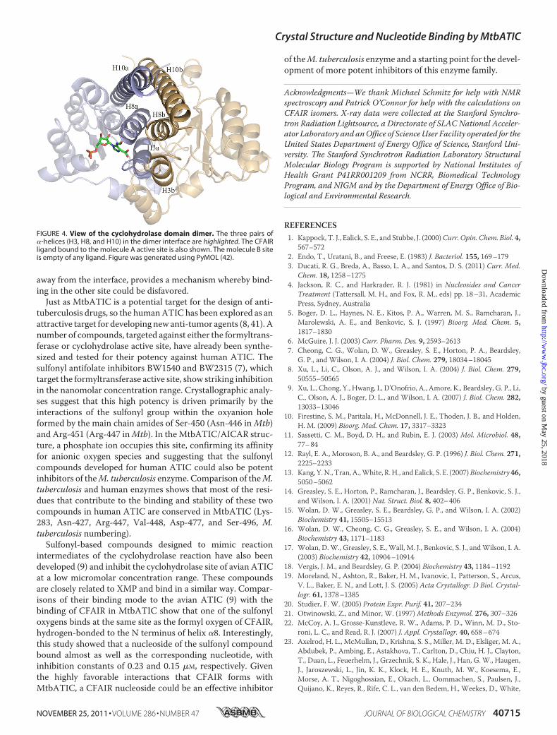
culosis, avian, and human ATIC structures is the preference forligand binding in only one of the two IMPCHase active sites ofthe ATIC dimer, an apparent half-the-sites reactivity. This iscoupled with active site plasticity, in which sections of the poly-peptide surrounding the bound ligand move 2–3 Å inward toenclose it, a tyrosine side chain (Tyr-111 in MtbATIC) flipsover the top, and a lysine (Lys-72 in MtbATIC) moves in tointeract with both the ligand and theTyr side chain. In contrast,in those structures where a second ligand occupies the otheractive site (AICAR for MtbATIC and XMP for avian ATIC),binding in that site appears to bemuchweaker, as judged by theelectron density. The site remains more open, and the Lys andTyr side chains remain 15–20 Å apart. Consideration of thedimer interface suggests an explanation. The interface betweenthe two IMPCHase domains involves three helices from eachdomain (�3 (residues 75–83) and the preceding loop 71–74;�8(residue 131–145); and �10 (residue 170–201); residue num-bering as in MtbATIC). These pack as three antiparallel pairs(Fig. 4). The loop 71–74 plays an important role in ligand bind-ing; Lys-72 from this loop moves to engage with the ligandphosphate and ribose moieties and with Tyr-111, and Thr-73hydrogen bonds to the ligand imidazole nitrogen N3 (throughOG1) and the 4-carboxyl (or 4-carboxamide) group. TheN ter-minus of helix�8 also contacts the ligand, hydrogen bonding tothe 5-formylamino substituent. Thus, although themovementsinduced by ligand binding are relatively small, they involve fourof the six principal components of the dimer interface. Move-ment of the helices of one monomer, toward the ligand and
Crystal Structure and Nucleotide Binding by MtbATIC
40714 JOURNAL OF BIOLOGICAL CHEMISTRY VOLUME 286 • NUMBER 47 • NOVEMBER 25, 2011
away from the interface, provides a mechanism whereby bind-ing in the other site could be disfavored.Just as MtbATIC is a potential target for the design of anti-
tuberculosis drugs, so the humanATIC has been explored as anattractive target for developing new anti-tumor agents (8, 41). Anumber of compounds, targeted against either the formyltrans-ferase or cyclohydrolase active site, have already been synthe-sized and tested for their potency against human ATIC. Thesulfonyl antifolate inhibitors BW1540 and BW2315 (7), whichtarget the formyltransferase active site, show striking inhibitionin the nanomolar concentration range. Crystallographic analy-ses suggest that this high potency is driven primarily by theinteractions of the sulfonyl group within the oxyanion holeformed by the main chain amides of Ser-450 (Asn-446 inMtb)and Arg-451 (Arg-447 inMtb). In theMtbATIC/AICAR struc-ture, a phosphate ion occupies this site, confirming its affinityfor anionic oxygen species and suggesting that the sulfonylcompounds developed for human ATIC could also be potentinhibitors of theM. tuberculosis enzyme. Comparison of theM.tuberculosis and human enzymes shows that most of the resi-dues that contribute to the binding and stability of these twocompounds in human ATIC are conserved in MtbATIC (Lys-283, Asn-427, Arg-447, Val-448, Asp-477, and Ser-496, M.tuberculosis numbering).Sulfonyl-based compounds designed to mimic reaction
intermediates of the cyclohydrolase reaction have also beendeveloped (9) and inhibit the cyclohydrolase site of avian ATICat a low micromolar concentration range. These compoundsare closely related to XMP and bind in a similar way. Compar-isons of their binding mode to the avian ATIC (9) with thebinding of CFAIR in MtbATIC show that one of the sulfonyloxygens binds at the same site as the formyl oxygen of CFAIR,hydrogen-bonded to the N terminus of helix �8. Interestingly,this study showed that a nucleoside of the sulfonyl compoundbound almost as well as the corresponding nucleotide, withinhibition constants of 0.23 and 0.15 �M, respectively. Giventhe highly favorable interactions that CFAIR forms withMtbATIC, a CFAIR nucleoside could be an effective inhibitor
of theM. tuberculosis enzyme and a starting point for the devel-opment of more potent inhibitors of this enzyme family.
Acknowledgments—We thank Michael Schmitz for help with NMRspectroscopy and Patrick O’Connor for help with the calculations onCFAIR isomers. X-ray data were collected at the Stanford Synchro-tron Radiation Lightsource, a Directorate of SLAC National Acceler-ator Laboratory and anOffice of ScienceUser Facility operated for theUnited States Department of Energy Office of Science, Stanford Uni-versity. The Stanford Synchrotron Radiation Laboratory StructuralMolecular Biology Program is supported by National Institutes ofHealth Grant P41RR001209 from NCRR, Biomedical TechnologyProgram, and NIGM and by the Department of Energy Office of Bio-logical and Environmental Research.
REFERENCES1. Kappock, T. J., Ealick, S. E., and Stubbe, J. (2000)Curr. Opin. Chem. Biol. 4,
567–5722. Endo, T., Uratani, B., and Freese, E. (1983) J. Bacteriol. 155, 169–1793. Ducati, R. G., Breda, A., Basso, L. A., and Santos, D. S. (2011) Curr. Med.
Chem. 18, 1258–12754. Jackson, R. C., and Harkrader, R. J. (1981) in Nucleosides and Cancer
Treatment (Tattersall, M. H., and Fox, R. M., eds) pp. 18–31, AcademicPress, Sydney, Australia
5. Boger, D. L., Haynes, N. E., Kitos, P. A., Warren, M. S., Ramcharan, J.,Marolewski, A. E., and Benkovic, S. J. (1997) Bioorg. Med. Chem. 5,1817–1830
6. McGuire, J. J. (2003) Curr. Pharm. Des. 9, 2593–26137. Cheong, C. G., Wolan, D. W., Greasley, S. E., Horton, P. A., Beardsley,
G. P., and Wilson, I. A. (2004) J. Biol. Chem. 279, 18034–180458. Xu, L., Li, C., Olson, A. J., and Wilson, I. A. (2004) J. Biol. Chem. 279,
50555–505659. Xu, L., Chong, Y., Hwang, I., D’Onofrio, A., Amore, K., Beardsley, G. P., Li,
C., Olson, A. J., Boger, D. L., and Wilson, I. A. (2007) J. Biol. Chem. 282,13033–13046
10. Firestine, S. M., Paritala, H., McDonnell, J. E., Thoden, J. B., and Holden,H. M. (2009) Bioorg. Med. Chem. 17, 3317–3323
11. Sassetti, C. M., Boyd, D. H., and Rubin, E. J. (2003) Mol. Microbiol. 48,77–84
12. Rayl, E. A., Moroson, B. A., and Beardsley, G. P. (1996) J. Biol. Chem. 271,2225–2233
13. Kang, Y.N., Tran,A.,White, R.H., andEalick, S. E. (2007)Biochemistry46,5050–5062
14. Greasley, S. E., Horton, P., Ramcharan, J., Beardsley, G. P., Benkovic, S. J.,and Wilson, I. A. (2001) Nat. Struct. Biol. 8, 402–406
15. Wolan, D. W., Greasley, S. E., Beardsley, G. P., and Wilson, I. A. (2002)Biochemistry 41, 15505–15513
16. Wolan, D. W., Cheong, C. G., Greasley, S. E., and Wilson, I. A. (2004)Biochemistry 43, 1171–1183
17. Wolan, D.W., Greasley, S. E., Wall, M. J., Benkovic, S. J., andWilson, I. A.(2003) Biochemistry 42, 10904–10914
18. Vergis, J. M., and Beardsley, G. P. (2004) Biochemistry 43, 1184–119219. Moreland, N., Ashton, R., Baker, H. M., Ivanovic, I., Patterson, S., Arcus,
V. L., Baker, E. N., and Lott, J. S. (2005) Acta Crystallogr. D Biol. Crystal-logr. 61, 1378–1385
20. Studier, F. W. (2005) Protein Expr. Purif. 41, 207–23421. Otwinowski, Z., and Minor, W. (1997)Methods Enzymol. 276, 307–32622. McCoy, A. J., Grosse-Kunstleve, R. W., Adams, P. D., Winn, M. D., Sto-
roni, L. C., and Read, R. J. (2007) J. Appl. Crystallogr. 40, 658–67423. Axelrod, H. L., McMullan, D., Krishna, S. S., Miller, M. D., Elsliger, M. A.,
Abdubek, P., Ambing, E., Astakhova, T., Carlton, D., Chiu, H. J., Clayton,T., Duan, L., Feuerhelm, J., Grzechnik, S. K., Hale, J., Han, G.W., Haugen,J., Jaroszewski, L., Jin, K. K., Klock, H. E., Knuth, M. W., Koesema, E.,Morse, A. T., Nigoghossian, E., Okach, L., Oommachen, S., Paulsen, J.,Quijano, K., Reyes, R., Rife, C. L., van den Bedem, H., Weekes, D., White,
FIGURE 4. View of the cyclohydrolase domain dimer. The three pairs of�-helices (H3, H8, and H10) in the dimer interface are highlighted. The CFAIRligand bound to the molecule A active site is also shown. The molecule B siteis empty of any ligand. Figure was generated using PyMOL (42).
Crystal Structure and Nucleotide Binding by MtbATIC
NOVEMBER 25, 2011 • VOLUME 286 • NUMBER 47 JOURNAL OF BIOLOGICAL CHEMISTRY 40715
A.,Wolf, G., Xu,Q.,Hodgson, K.O.,Wooley, J., Deacon,A.M.,Godzik, A.,Lesley, S. A., and Wilson, I. A. (2008) Proteins 71, 1042–1049
24. Emsley, P., and Cowtan, K. (2004)Acta Crystallogr. D Biol. Crystallogr. 60,2126–2132
25. Murshudov, G. N., Vagin, A. A., and Dodson, E. J. (1997)Acta Crystallogr.D Biol. Crystallogr. 53, 240–255
26. Bricogne, G., Blanc, E., Brandl, M., Flensburg, C., Keller, P., Paciorek, W.,Roversi, P., Sharff, A., Smart, O. S., Vonrhein, C., and Womack, T. O.(2011) autoBUSTER, Version 1.6.0, Global Phasing Ltd., Cambridge, UK
27. Schüttelkopf, A.W., and vanAalten, D.M. (2004)Acta Crystallogr. D Biol.Crystallogr. 60, 1355–1363
28. Frisch, M. J., Trucks, G. W., Schlegel, H. B., Scuseria, G. E., Robb, M. A.,Cheesman, J. r., Scalmani, G., Barone, V., Mennucci, B., Petersson, G. A.,Nakatsuji, H., Caricato,M., Li, X., Hratchian, H. P., Izmaylov, A. F., Bloino,J., Zheng, G., Sonnenberg, J. L., Hada, M., Ehara, M., Toyota, K., Fukuda,R., Hasegawa, J., Ishida, M., Nakajima, T., Honda, Y., Kitao, O., Nakai, H.,Vreven, T., Montgomery, J. A., Peralta, J. E., Ogliaro, F., Bearpark, M.,Heyd, J. J., Brothers, E., Kudin, K. N., Staroverov, V. N., Kobayashi, R.,Normand, J., Raghavachari, K., Rendell, A., Burant, J. C., Iyengar, S. S.,Tomasi, J., Cossi, M., Rega, N., Millam, N. J., Klene, M., Knox, J. E., Cross,J. B., Bakken, V., Adamo, C., Jaramillo, J., Gomperts, R., Stratmann, R. E.,Yazyev, O., Austin, A. J., Cammi, R., Pomelli, C., Ochterski, J. W., Martin,R. L.,Morokuma, K., Zakrzewski, V. G., Voth, G. A., Salvador, P., Dannen-berg, J. J., Dapprich, S., Daniels, A. D., Farkas, Ö., Foresman, J. B., Ortiz,J. V., Cioslowski, J., and Fox, D. J. (2009) Gaussian 09, Revision A.02,Gaussian Inc., Wallingford, CT
29. Scalmani, G., and Frisch, M. J. (2010) J. Chem. Phys. 132, 114110–11411530. Davis, I. W., Leaver-Fay, A., Chen, V. B., Block, J. N., Kapral, G. J., Wang,
X., Murray, L. W., Arendall, W. B., 3rd, Snoeyink, J., Richardson, J. S., andRichardson, D. C. (2007) Nucleic Acids Res. 35,W375–W383
31. Betts, L., Xiang, S., Short, S. A.,Wolfenden, R., and Carter, C.W., Jr (1994)J. Mol. Biol. 235, 635–656
32. Vergis, J. M., Bulock, K. G., Fleming, K. G., and Beardsley, G. P. (2001)J. Biol. Chem. 276, 7727–7733
33. Tsuchiya, Y., Kinoshita, K., andNakamura, H. (2006) Protein Eng. Des. Sel.19, 421–429
34. Krissinel, E., and Henrick, K. (2007) J. Mol. Biol. 372, 774–79735. Litchfield, G. J., and Shaw, G. (1971) J. Chem. Soc. (C) 817–82036. Groziak, M. P., Bhat, B., and Leonard, N. J. (1988) Proc. Natl. Acad. Sci.
U.S.A. 85, 7174–717637. Mueller, W. T., and Benkovic, S. J. (1981) Biochemistry 20, 337–34438. Groziak, M. P., Huan, Z. W., Ding, H., Meng, Z., Stevens, W. C., and
Robinson, P. D. (1997) J. Med. Chem. 40, 3336–334539. Szabados, E., Hindmarsh, E. J., Phillips, L., Duggleby, R. G., and Chris-
topherson, R. I. (1994) Biochemistry 33, 14237–1424540. Wall, M., Shim, J. H., and Benkovic, S. J. (2000) Biochemistry 39,
11303–1131141. Li, C., Xu, L., Wolan, D. W., Wilson, I. A., and Olson, A. J. (2004) J. Med.
Chem. 47, 6681–669042. DeLano, W. L. (2002) The PyMOL Molecular Graphics System, DeLano
Scientific LLC, San Carlos, CA
Crystal Structure and Nucleotide Binding by MtbATIC
40716 JOURNAL OF BIOLOGICAL CHEMISTRY VOLUME 286 • NUMBER 47 • NOVEMBER 25, 2011