Structure−Function Studies of Naphthalene, Phenanthrene,Biphenyl, and Their Derivatives in Interaction with and Oxidation byCytochromes P450 2A13 and 2A6Tsutomu Shimada,*,† Shigeo Takenaka,*,† Kensaku Kakimoto,‡ Norie Murayama,§ Young-Ran Lim,∥
Donghak Kim,∥ Maryam K. Foroozesh,⊥ Hiroshi Yamazaki,*,§ F. Peter Guengerich,*,#
and Masayuki Komori*,†
†Laboratory of Cellular and Molecular Biology, Graduate School of Life and Environmental Sciences, Osaka Prefecture University,1-58 Rinku-Orai-Kita, Izumisano, Osaka 598-8531, Japan‡Osaka Prefectural Institute of Public Health, 1-3-69 Nakamichi, Higashinari-ku, Osaka 537-0025, Japan§Laboratory of Drug Metabolism and Pharmacokinetics, Showa Pharmaceutical University, Machida, Tokyo 194-8543, Japan∥Department of Biological Sciences, Konkuk University, Seoul 143-701, Republic of Korea⊥Department of Chemistry, Xavier University of Louisiana, New Orleans, Louisiana 70125, United States#Department of Biochemistry, Vanderbilt University School of Medicine, Nashville, Tennessee 37232-0146, United States
*S Supporting Information
ABSTRACT: Naphthalene, phenanthrene, biphenyl, and their derivatives having differentethynyl, propynyl, butynyl, and propargyl ether substitutions were examined for their interactionwith and oxidation by cytochromes P450 (P450) 2A13 and 2A6. Spectral interaction studiessuggested that most of these chemicals interacted with P450 2A13 to induce Type I bindingspectra more readily than with P450 2A6. Among the various substituted derivatives examined,2-ethynylnaphthalene, 2-naphthalene propargyl ether, 3-ethynylphenanthrene, and 4-biphenylpropargyl ether had larger ΔAmax/Ks values in inducing Type I binding spectra with P450 2A13than their parent compounds. P450 2A13 was found to oxidize naphthalene, phenanthrene, andbiphenyl to 1-naphthol, 9-hydroxyphenanthrene, and 2- and/or 4-hydroxybiphenyl, respectively,at much higher rates than P450 2A6. Other human P450 enzymes including P450s 1A1, 1A2,1B1, 2C9, and 3A4 had lower rates of oxidation of naphthalene, phenanthrene, and biphenylthan P450s 2A13 and 2A6. Those alkynylated derivatives that strongly induced Type I bindingspectra with P450s 2A13 and 2A6 were extensively oxidized by these enzymes upon analysiswith HPLC. Molecular docking studies supported the hypothesis that ligand-interactionenergies (U values) obtained with reported crystal structures of P450 2A13 and 2A6 bound to 4-(methylnitrosamino)-1-(3-pyridyl)-1-butanone, indole, pilocarpine, nicotine, and coumarin are of use in understanding the basis of possible molecularinteractions of these xenobiotic chemicals with the active sites of P450 2A13 and 2A6 enzymes. In fact, the ligand-interactionenergies with P450 2A13 4EJG bound to these chemicals were found to relate to their induction of Type I binding spectra.
■ INTRODUCTION
Naphthalene, phenanthrene, and biphenyl are aromatic hydro-carbon contaminants found in the environment, and the formertwo chemicals belong to the class of polycyclic aromatichydrocarbons (PAHs) that includes toxic and carcinogeniccompounds such as benzo[a]pyrene, 7,12-dimethylbenz[a]-anthracene, and benzo[c]phenanthrene.1−5 These carcinogenicPAHs have been shown to require metabolic activation by so-called xenobiotic-metabolizing enzymes, such as cytochromesP450 (P450), to evoke their toxic and carcinogenic responses inlaboratory animals as well as in humans.6−8
Naphthalene has been shown to cause toxic responses inseveral animal species and is known to require bioactivation byP450 2F2 in mice to elicit toxicity.9,10 Hu et al.11 also reportedthat P450 2A5 plays an important role in olfactory mucosaltoxicity in mice. Metabolism of naphthalene by different P450
enzymes has been reported in humans12,13 and also in Rhesusmonkeys.14 Metabolism of phenanthrene and biphenyl has alsobeen reported in various laboratory animals;15−22 however,detailed studies on the roles of human P450s 2A13 and 2A6 inthe metabolism of naphthalene, phenanthrene, and biphenylhave not been reported, except that Fukami et al. showed thatP450 2A13 can catalyze the oxidation of naphthalene inhumans.13
Our previous studies have shown that naphthalene,phenanthrene, biphenyl, and their substituted derivativesinduce Type I binding spectra with P450s 2A13 and 2A6,suggesting that these chemicals may be oxidized by these P450enzymes.23 In this study, we studied how these chemicals are
oxidized by P450s 2A13 and 2A6 and whether there arestructure−function relationships for interacting with andbiotransformation by these enzymes, based on analysis withHPLC. We used naphthalene and six substituted derivatives,phenanthrene and six substituted derivatives, and biphenyl andnine substituted derivatives for spectral interaction with andmetabolism by P450s 2A13 and 2A6. Other P450 enzymeshuman 1A1, 1A2, 1B1, 2C9, and 3A4were also used to studythe oxidation of parent compounds naphthalene, phenanthrene,and biphenyl. Docking simulations of the interactions of P450s2A13 and 2A6 and these chemicals were also examined tovisualize how these chemicals interact with these P450enzymes.
■ EXPERIMENTAL PROCEDURESChemicals. Naphthalene, phenanthrene, and biphenyl were
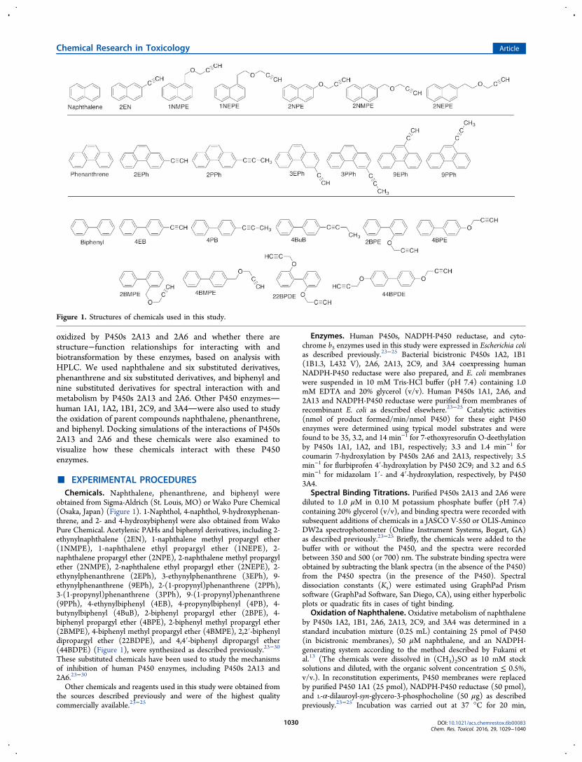
obtained from Sigma-Aldrich (St. Louis, MO) or Wako Pure Chemical(Osaka, Japan) (Figure 1). 1-Naphthol, 4-naphthol, 9-hydroxyphenan-threne, and 2- and 4-hydroxybiphenyl were also obtained from WakoPure Chemical. Acetylenic PAHs and biphenyl derivatives, including 2-ethynylnaphthalene (2EN), 1-naphthalene methyl propargyl ether(1NMPE), 1-naphthalene ethyl propargyl ether (1NEPE), 2-naphthalene propargyl ether (2NPE), 2-naphthalene methyl propargylether (2NMPE), 2-naphthalene ethyl propargyl ether (2NEPE), 2-ethynylphenanthrene (2EPh), 3-ethynylphenanthrene (3EPh), 9-ethynylphenanthrene (9EPh), 2-(1-propynyl)phenanthrene (2PPh),3-(1-propynyl)phenanthrene (3PPh), 9-(1-propynyl)phenanthrene(9PPh), 4-ethynylbiphenyl (4EB), 4-propynylbiphenyl (4PB), 4-butynylbiphenyl (4BuB), 2-biphenyl propargyl ether (2BPE), 4-biphenyl propargyl ether (4BPE), 2-biphenyl methyl propargyl ether(2BMPE), 4-biphenyl methyl propargyl ether (4BMPE), 2,2′-biphenyldipropargyl ether (22BDPE), and 4,4′-biphenyl dipropargyl ether(44BDPE) (Figure 1), were synthesized as described previously.23−30
These substituted chemicals have been used to study the mechanismsof inhibition of human P450 enzymes, including P450s 2A13 and2A6.23−30
Other chemicals and reagents used in this study were obtained fromthe sources described previously and were of the highest qualitycommercially available.23−25
Enzymes. Human P450s, NADPH-P450 reductase, and cyto-chrome b5 enzymes used in this study were expressed in Escherichia colias described previously.23−25 Bacterial bicistronic P450s 1A2, 1B1(1B1.3, L432 V), 2A6, 2A13, 2C9, and 3A4 coexpressing humanNADPH-P450 reductase were also prepared, and E. coli membraneswere suspended in 10 mM Tris-HCl buffer (pH 7.4) containing 1.0mM EDTA and 20% glycerol (v/v). Human P450s 1A1, 2A6, and2A13 and NADPH-P450 reductase were purified from membranes ofrecombinant E. coli as described elsewhere.23−25 Catalytic activities(nmol of product formed/min/nmol P450) for these eight P450enzymes were determined using typical model substrates and werefound to be 35, 3.2, and 14 min−1 for 7-ethoxyresorufin O-deethylationby P450s 1A1, 1A2, and 1B1, respectively; 3.3 and 1.4 min−1 forcoumarin 7-hydroxylation by P450s 2A6 and 2A13, respectively; 3.5min−1 for flurbiprofen 4′-hydroxylation by P450 2C9; and 3.2 and 6.5min−1 for midazolam 1′- and 4′-hydroxylation, respectively, by P4503A4.
Spectral Binding Titrations. Purified P450s 2A13 and 2A6 werediluted to 1.0 μM in 0.10 M potassium phosphate buffer (pH 7.4)containing 20% glycerol (v/v), and binding spectra were recorded withsubsequent additions of chemicals in a JASCO V-550 or OLIS-AmincoDW2a spectrophotometer (Online Instrument Systems, Bogart, GA)as described previously.23−25 Briefly, the chemicals were added to thebuffer with or without the P450, and the spectra were recordedbetween 350 and 500 (or 700) nm. The substrate binding spectra wereobtained by subtracting the blank spectra (in the absence of the P450)from the P450 spectra (in the presence of the P450). Spectraldissociation constants (Ks) were estimated using GraphPad Prismsoftware (GraphPad Software, San Diego, CA), using either hyperbolicplots or quadratic fits in cases of tight binding.
Oxidation of Naphthalene. Oxidative metabolism of naphthaleneby P450s 1A2, 1B1, 2A6, 2A13, 2C9, and 3A4 was determined in astandard incubation mixture (0.25 mL) containing 25 pmol of P450(in bicistronic membranes), 50 μM naphthalene, and an NADPH-generating system according to the method described by Fukami etal.13 (The chemicals were dissolved in (CH3)2SO as 10 mM stocksolutions and diluted, with the organic solvent concentration ≤ 0.5%,v/v.). In reconstitution experiments, P450 membranes were replacedby purified P450 1A1 (25 pmol), NADPH-P450 reductase (50 pmol),and L-α-dilauroyl-syn-glycero-3-phosphocholine (50 μg) as describedpreviously.23−25 Incubation was carried out at 37 °C for 20 min,
Figure 1. Structures of chemicals used in this study.
following a preincubation time of 1 min. Reactions were terminated byadding 100 μL of CH3CN, and following centrifugation, the upperlayer was subjected to HPLC using a Mightsil RP-18 C18 GP column.(Note that naphthalene and its metabolites are volatile under anitrogen stream after these chemicals are extracted with organicsolvents.) The mobile phase used was 30% CH3CN (v/v, in H2O)containing 0.01% H3PO4 (w/v).Oxidation of Phenanthrene and Biphenyl and Their
Derivatives and Naphthalene Derivatives. Oxidative metabolismof phenanthrene and biphenyl and their derivatives (and alsonaphthalene derivatives) was determined in a standard reactionmixture (final volume of 0.25 mL) as described above. After incubationat 37 °C for 20 min, extraction was done by adding 0.25 mL of coldCH3OH and then 0.5 mL of a mixture of CHCl3 and ethyl acetate(1:1, v/v). After centrifugation, the lower organic layer was recoveredand the extraction was repeated. The extracts were combined andconcentrated under a nitrogen stream. HPLC separation was donewith a JASCO system (Tokyo, Japan) equipped with a Wakopack Navi
C18-5 octadecylsilane column (2.0 mm × 150 mm) (Wako PureChemical) with UV detection at 254 nm and fluorescence detectionwith an excitation wavelength of 242 nm and emission wavelength of380 nm. Elution of chemicals and their metabolites utilized a lineargradient from 20 to 100% CH3OH (v/v, in H2O) for 25 min, followedby holding at 100% CH3OH for 5 min, with a flow rate of 0.2 mL/min.
Other Enzyme Assays. Coumarin 7-hydroxylation, 7-ethoxyr-esorufin O-deethylation, flurbiprofen 4-hydroxylation, and midazolam1′- and 4′-hydroxylation activities were determined using bicistronicbacterial membrane or reconstituted monooxygenase systems asdescribed previously.23−25,31−33
Docking Simulations into Human P450 Enzymes. Crystalstructures of P450 2A13 bound to NNK (PDB 4EJH),34 indole (PDB2P85),35 pilocarpine (PDB 3T3S),36 and nicotine (PDB 4EJG)34 havebeen reported and were used for these studies. Structures of P450 2A6bound to coumarin (PDB 1Z10),37 pilocarpine (PDB 3T3R),36 andnicotine (PDB 4EJJ)34 have also been reported. Simulations werecarried out after removing each ligand from these P450 structures
Figure 2. Spectral changes produced by the interaction of naphthalene (A), phenanthrene (B), and biphenyl (C) with P450 2A13. The lowerportion of each figure indicates the difference spectra obtained.
Figure 3. Comparison of spectral changes (spectral binding efficiency, ΔAmax/Ks ratio) from the interaction of various chemicals with P450s 2A6 and2A13.
using the MMFF94x force field described in MOE software (ver.2015.10, Chemical Computing Group, Montreal, Canada).23−25
Ligand-interaction energies (U values) were obtained by use of theASEdock program in MOE. Lower U values indicate a strongerinteraction between a chemical and the enzyme.Kinetic Analysis. Kinetic parameters were estimated by nonlinear
regression analysis using KaleidaGraph (Synergy Software, Reading,PA) or GraphPad Prism (GraphPad Software, La Jolla, CA).
■ RESULTS
Spectral Interactions of Naphthalene, Phenanthrene,Biphenyl, and Their Derivatives with P450s 2A13 and2A6. As has been reported previously,23 naphthalene,phenanthrene, and biphenyl interact with P450 2A13, inducingType I binding spectral changes (Figure 2). The Ks values
obtained with these three chemicals were determined to be0.35, 2.3, and 0.88 μM, respectively, and the ΔAmax valuesobtained were 0.043, 0.063, and 0.045, respectively (Figure 2).Naphthalene and its six derivatives, phenanthrene and its six
derivatives, and biphenyl and its nine derivatives were alsocompared for their abilities (ΔAmax/Ks ratio) to induce spectralchanges with P450 2A6 as well as P450 2A13 (Figure 3). Itshould be pointed out that the scale for the ΔAmax/Ks ratio onthe x axis is different for P450s 2A13 and 2A6, indicating thatthe former enzyme showed higher spectral changes than thelatter. 2EN induced Type I binding spectra with P450 2A13 toa much greater extent than the parent naphthalene, and theΔAmax/Ks ratio for the interaction of 2EN with P450 2A13 wasabout 10-fold greater than that with P450 2A6 (Figure 3).2NPE, 3EPh, and 4BPE were also found to show higher
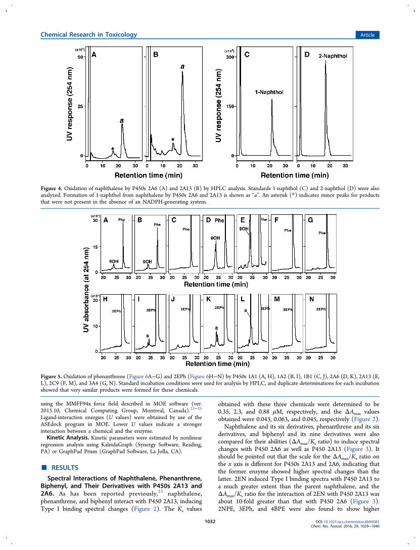
Figure 4. Oxidation of naphthalene by P450s 2A6 (A) and 2A13 (B) by HPLC analysis. Standards 1-naphthol (C) and 2-naphthol (D) were alsoanalyzed. Formation of 1-naphthol from naphthalene by P450s 2A6 and 2A13 is shown as “a”. An asterisk (*) indicates minor peaks for productsthat were not present in the absence of an NADPH-generating system.
Figure 5. Oxidation of phenanthrene (Figure 6A−G) and 2EPh (Figure 6H−N) by P450s 1A1 (A, H), 1A2 (B, I), 1B1 (C, J), 2A6 (D, K), 2A13 (E,L), 2C9 (F, M), and 3A4 (G, N). Standard incubation conditions were used for analysis by HPLC, and duplicate determinations for each incubationshowed that very similar products were formed for these chemicals.
intensities of interaction with P450 2A13 than parentcompounds naphthalene, phenanthrene, and biphenyl. 4BPEwas also found to induce spectral changes with P450 2A6 at ahigher level than biphenyl.Oxidation of Naphthalene, Phenanthrene, and Bi-
phenyl by Human P450 Enzymes. Oxidation of naph-thalene was determined with P450s 2A13 and 2A6 according tothe method of Fukami et al.13 P450s 2A13 and 2A6 producedone major peak that corresponded to the 1-naphthol standard,but not 2-naphthol, in the retention time on HPLCchromatograms, with P450 2A13 showing more activity thanP450 2A6 (Figure 4). Since we did not extract naphthalene andits metabolites and concentrate them for HPLC analysis due totheir volatile nature, it is not known whether the small peaksseen in the chromatograms are due to 2-naphthol and othermetabolites.P450 2A13 was also active in oxidizing phenanthrene to 9-
hydroxyphenanthrene (Supporting Information Figure S1).The 9-hydroxyphenanthrene standard and a metaboliteproduced by P450 2A13 had similar, profiles showing twopeaks on HPLC chromatograms (it is not known at presentwhy two peaks appear for 9-hydroxyphenanthrene or if it arisesby an experimental artifact possible to detector saturation).When zooming in on the x-axis, several minor peaks appearedonly in the presence of an NADPH-generating system withP450 2A13 (Supporting Information Figure S1b). We alsofound very similar, but different, chromatographic profiles ofproduct formation for the metabolism of 2EPh andphenanthrene by P450 2A13 (Supporting Information FigureS1C,D,c,d). We do not have a standard 2EPh metabolite, but
the major peak (*) seems to be similar in nature to 9-hydroxyphenanthrene.In order to compare the products formed for the metabolism
of phenanthrene and 2EPh, we determined their activities withseven human P450 enzymes, P450s 1A1, 1A2, 1B1, 2A6, 2A13,2C9, and 3A4 (Figure 5). The results show that P450 2A13 isthe most active at oxidizing phenanthrene (Figure 5E) and2EPh (Figure 5L) to 9-hydroxyphenanthrene and metabolite a,respectively, followed by P450 2A6 (Figure 5D,K). Other P450enzymes examinedP450s 1A1, 1A2, and 1B1.3were activeat producing 9-hydroxyphenanthrene from phenanthrene, andP450s 1A2 and 1B1 oxidized 2EPh at lower levels than P450s2A13 and 2A6.P450 2A13 oxidized biphenyl to 2- and/or 4-hydroxybi-
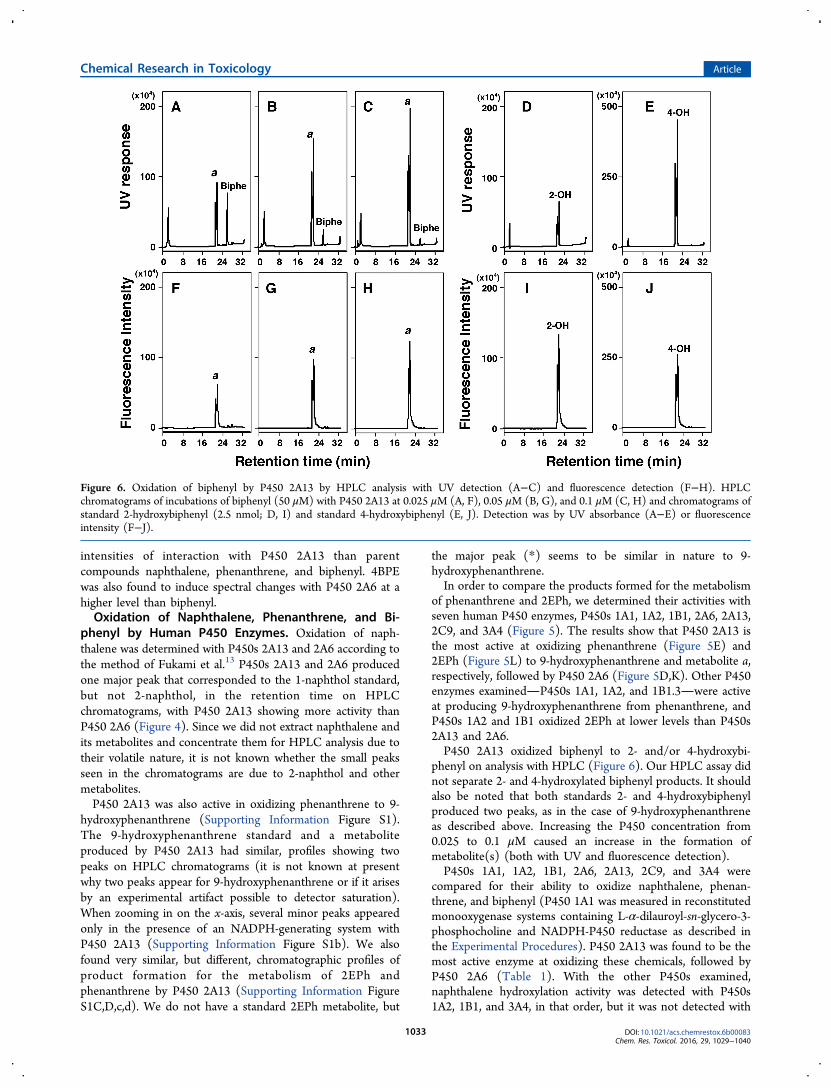
phenyl on analysis with HPLC (Figure 6). Our HPLC assay didnot separate 2- and 4-hydroxylated biphenyl products. It shouldalso be noted that both standards 2- and 4-hydroxybiphenylproduced two peaks, as in the case of 9-hydroxyphenanthreneas described above. Increasing the P450 concentration from0.025 to 0.1 μM caused an increase in the formation ofmetabolite(s) (both with UV and fluorescence detection).P450s 1A1, 1A2, 1B1, 2A6, 2A13, 2C9, and 3A4 were
compared for their ability to oxidize naphthalene, phenan-threne, and biphenyl (P450 1A1 was measured in reconstitutedmonooxygenase systems containing L-α-dilauroyl-sn-glycero-3-phosphocholine and NADPH-P450 reductase as described inthe Experimental Procedures). P450 2A13 was found to be themost active enzyme at oxidizing these chemicals, followed byP450 2A6 (Table 1). With the other P450s examined,naphthalene hydroxylation activity was detected with P450s1A2, 1B1, and 3A4, in that order, but it was not detected with
Figure 6. Oxidation of biphenyl by P450 2A13 by HPLC analysis with UV detection (A−C) and fluorescence detection (F−H). HPLCchromatograms of incubations of biphenyl (50 μM) with P450 2A13 at 0.025 μM (A, F), 0.05 μM (B, G), and 0.1 μM (C, H) and chromatograms ofstandard 2-hydroxybiphenyl (2.5 nmol; D, I) and standard 4-hydroxybiphenyl (E, J). Detection was by UV absorbance (A−E) or fluorescenceintensity (F−J).
P450s 1A1 and 2C9. Phenanthrene 9-hydroxylation activity wasdetected with P450s 1A1, 1A2, 3A4, and 1B1.3 but not P4502C9. Oxidation of biphenyl was detected with P450s 1A1 and1A2 but not with P450s 1B1, 2C9, and 3A4.Oxidation of Various Substituted Derivatives of
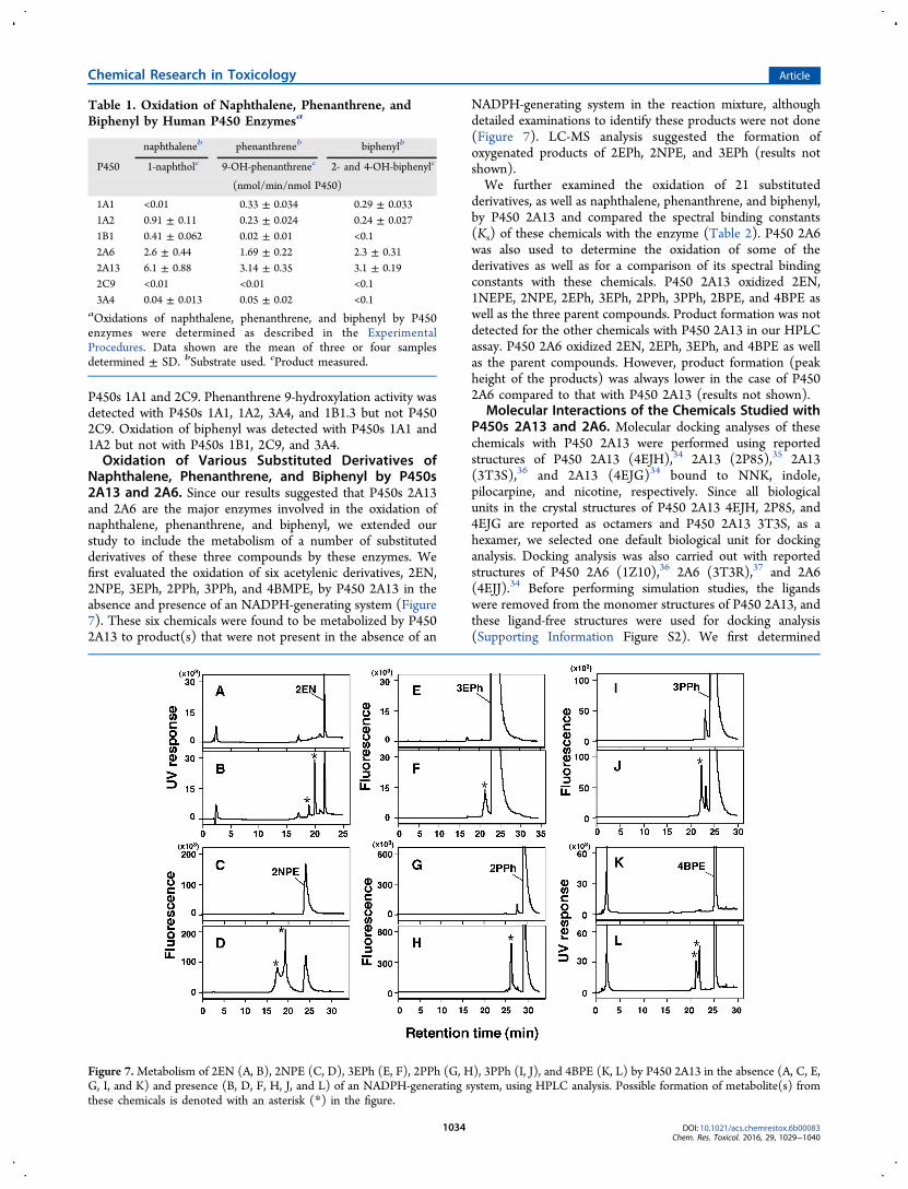
Naphthalene, Phenanthrene, and Biphenyl by P450s2A13 and 2A6. Since our results suggested that P450s 2A13and 2A6 are the major enzymes involved in the oxidation ofnaphthalene, phenanthrene, and biphenyl, we extended ourstudy to include the metabolism of a number of substitutedderivatives of these three compounds by these enzymes. Wefirst evaluated the oxidation of six acetylenic derivatives, 2EN,2NPE, 3EPh, 2PPh, 3PPh, and 4BMPE, by P450 2A13 in theabsence and presence of an NADPH-generating system (Figure7). These six chemicals were found to be metabolized by P4502A13 to product(s) that were not present in the absence of an
NADPH-generating system in the reaction mixture, althoughdetailed examinations to identify these products were not done(Figure 7). LC-MS analysis suggested the formation ofoxygenated products of 2EPh, 2NPE, and 3EPh (results notshown).We further examined the oxidation of 21 substituted
derivatives, as well as naphthalene, phenanthrene, and biphenyl,by P450 2A13 and compared the spectral binding constants(Ks) of these chemicals with the enzyme (Table 2). P450 2A6was also used to determine the oxidation of some of thederivatives as well as for a comparison of its spectral bindingconstants with these chemicals. P450 2A13 oxidized 2EN,1NEPE, 2NPE, 2EPh, 3EPh, 2PPh, 3PPh, 2BPE, and 4BPE aswell as the three parent compounds. Product formation was notdetected for the other chemicals with P450 2A13 in our HPLCassay. P450 2A6 oxidized 2EN, 2EPh, 3EPh, and 4BPE as wellas the parent compounds. However, product formation (peakheight of the products) was always lower in the case of P4502A6 compared to that with P450 2A13 (results not shown).
Molecular Interactions of the Chemicals Studied withP450s 2A13 and 2A6. Molecular docking analyses of thesechemicals with P450 2A13 were performed using reportedstructures of P450 2A13 (4EJH),34 2A13 (2P85),35 2A13(3T3S),36 and 2A13 (4EJG)34 bound to NNK, indole,pilocarpine, and nicotine, respectively. Since all biologicalunits in the crystal structures of P450 2A13 4EJH, 2P85, and4EJG are reported as octamers and P450 2A13 3T3S, as ahexamer, we selected one default biological unit for dockinganalysis. Docking analysis was also carried out with reportedstructures of P450 2A6 (1Z10),36 2A6 (3T3R),37 and 2A6(4EJJ).34 Before performing simulation studies, the ligandswere removed from the monomer structures of P450 2A13, andthese ligand-free structures were used for docking analysis(Supporting Information Figure S2). We first determined
Table 1. Oxidation of Naphthalene, Phenanthrene, andBiphenyl by Human P450 Enzymesa
naphthaleneb phenanthreneb biphenylb
P450 1-naphtholc 9-OH-phenanthrenec 2- and 4-OH-biphenylc
aOxidations of naphthalene, phenanthrene, and biphenyl by P450enzymes were determined as described in the ExperimentalProcedures. Data shown are the mean of three or four samplesdetermined ± SD. bSubstrate used. cProduct measured.
Figure 7.Metabolism of 2EN (A, B), 2NPE (C, D), 3EPh (E, F), 2PPh (G, H), 3PPh (I, J), and 4BPE (K, L) by P450 2A13 in the absence (A, C, E,G, I, and K) and presence (B, D, F, H, J, and L) of an NADPH-generating system, using HPLC analysis. Possible formation of metabolite(s) fromthese chemicals is denoted with an asterisk (*) in the figure.
optimal U values (ligand-interaction energy) (on analysis with
MMFF94x force field) using P450 4EJG and found that U
values for nicotine, naphthalene, phenanthrene, and biphenyl
were −35.2, −24.3, −29.2, and −25.7, respectively (Supporting
Information Figure S2), and that of coumarin was −34.4(results not shown).There was a good relationship between the U values of P450
2A13 4EJG (nicotine-type) and 4EJH (NNK-type) with thesechemicals (r = 0.79, p < 0.01), and we found that the parent
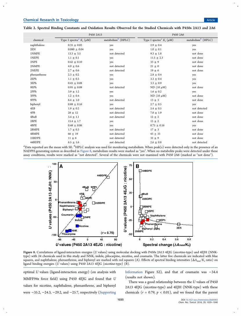
Table 2. Spectral Binding Constants and Oxidation Results Observed for the Studied Chemicals with P450s 2A13 and 2A6
P450 2A13 P450 2A6
chemical Type I spectraa Ks (μM) metabolismb (HPLC) Type I spectraa Ks (μM) metabolismb (HPLC)
naphthalene 0.35 ± 0.02 yes 2.9 ± 0.4 yes2EN 0.080 ± 0.04 yes 1.0 ± 0.1 yes1NMPE 13.3 ± 3.5 not detected 9.3 ± 1.8 not done1NEPE 1.1 ± 0.1 yes 11.5 ± 2.3 not done2NPE 0.42 ± 0.10 yes 33 ± 9 not done2NMPE 4.9 ± 0.6 not detected 21 ± 0 not done2NEPE 2.7 ± 0.6 not detected 19 ± 6 not donephenanthrene 2.3 ± 0.2 yes 2.8 ± 0.4 yes2EPh 1.1 ± 0.3 yes 3.3 ± 0.4 yes3EPh 0.45 ± 0.08 yes 3.3 ± 0.9 yes9EPh 0.95 ± 0.08 not detected ND (10 μM) not done2PPh 3.9 ± 1.2 yes 1.6 ± 0.2 yes3PPh 1.2 ± 0.4 yes ND (10 μM) not done9PPh 6.4 ± 1.0 not detected 13 ± 3 not donebiphenyl 0.88 ± 0.16 yes 2.7 ± 0.5 yes4EB 1.9 ± 0.2 not detected 5.4 ± 0.5 not detected4PB 28 ± 12 not detected 7.0 ± 1.9 not done4BuB 3.4 ± 1.1 not detected 12 ± 2 not done2BPE 15.4 ± 1.7 yes 15 ± 2 not done4BPE 0.48 ± 0.06 yes 0.75 ± 0.16 yes2BMPE 1.7 ± 0.3 not detected 17 ± 3 not done4BMPE 66 ± 19 not detected 43 ± 33 not done22BDPE 11 ± 6 not detected 32 ± 9 not done44BDPE 6.5 ± 1.6 not detected 2.6 ± 0.8 not detected
aData reported are the mean with SE. bHPLC analysis was used for monitoring metabolism. When peak(s) were detected only in the presence of anNADPH-generating system as described in Figure 8, metabolism results were marked as “yes”. When no metabolite peaks were detected under theseassay conditions, results were marked as “not detected”. Several of the chemicals were not examined with P450 2A6 (marked as “not done”).
Figure 8. Correlations of ligand-interaction energies (U values) using molecular docking with P450s 2A13 4EJG (nicotine-type) and 4EJH (NNK-type) with 24 chemicals used in this study and NNK, indole, pilocarpine, nicotine, and coumarin. The latter five chemicals are indicated with bluesquares, and naphthalene, phenanthrene, and biphenyl are marked with red squares (A). Effects of spectral binding intensities (ΔAmax/Ks ratio) onligand binding energies (U values) using P450 2A13 4EJG (nicotine-type) (B).
compounds naphthalene, phenanthrene, biphenyl, and coumar-in had U values comparable to those of NNK, indole,pilocarpine, nicotine, and coumarin (Figure 8). Compoundsthat were oxidized by P450 2A13 (indicated by HPLC analysis)had small U values under these assay conditions (Figure 8A).When spectral changes (ΔAmax/Ks values) and U values (usingP450 2A13 4EJG for docking) were compared, somerelationship was found between these values when they wereanalyzed with logarithmic curve fitting (Cricket Graph) (Figure8B). However, we did not find any positive correlation when Uvalues (with P450 2A13 4EJG) and spectral binding constants(Ks values) were compared.A correlation of the U values using P450 2A13 4EJH, 2P85,
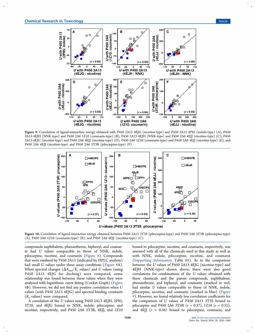
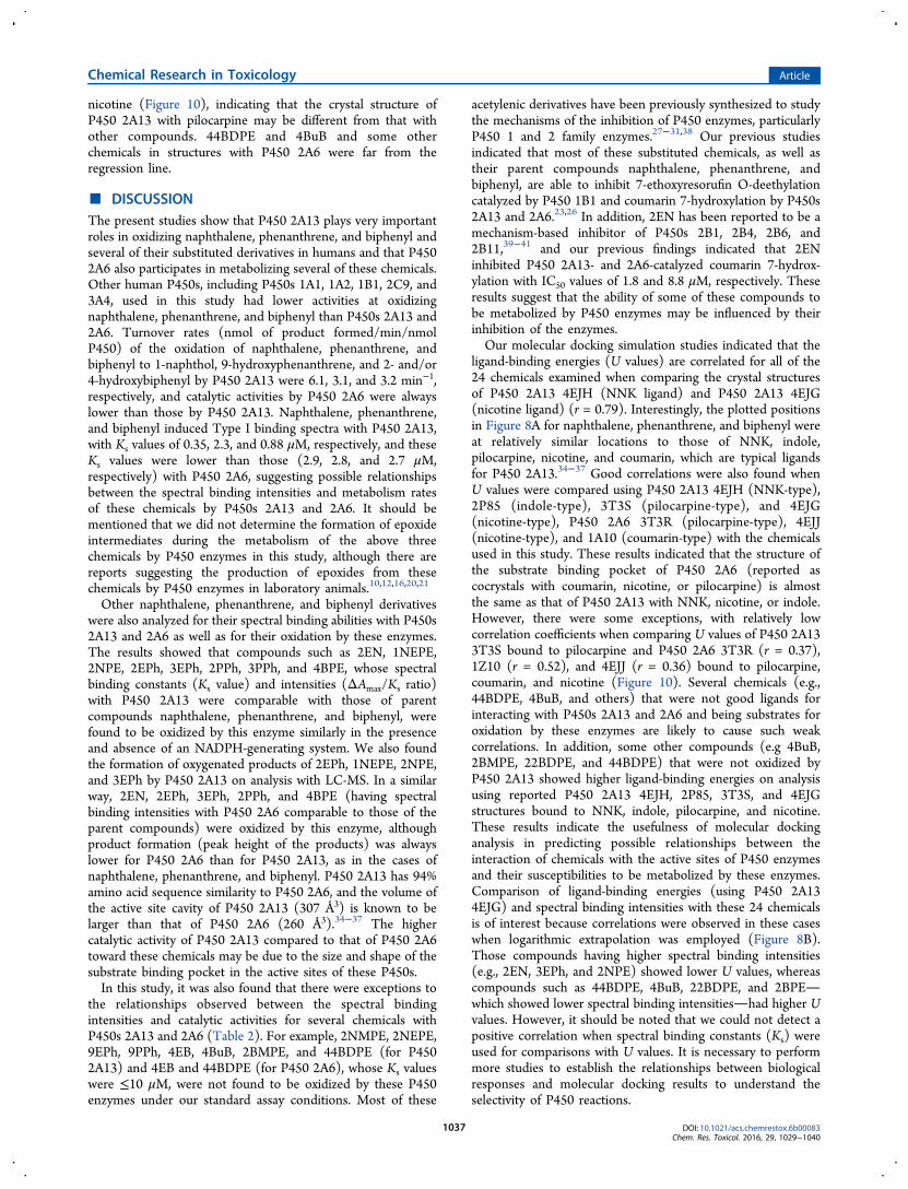
3T3S, and 4EJG bound to NNK, indole, pilocarpine, andnicotine, respectively, and P450 2A6 3T3R, 4EJJ, and 1Z10
bound to pilocarpine, nicotine, and coumarin, respectively, wasassessed with all of the chemicals used in this study as well aswith NNK, indole, pilocarpine, nicotine, and coumarin(Supporting Information Table S1). As in the comparisonbetween the U values of P450 2A13 4EJG (nicotine-type) and4EJH (NNK-type) shown above, there were also goodcorralations for combinations of the U values obtained withthese chemicals and the parent compounds, naphthalene,phenanthrene, and biphenyl, and coumarin (marked in red)had similar U values comparable to those of NNK, indole,pilocarpine, nicotine, and coumarin (marked in blue) (Figure9). However, we found relatively low correlation coefficients forthe comparison of U values of P450 2A13 3T3S bound topilocarpine and P450 2A6 3T3R (r = 0.37), 1Z10 (r = 0.52),and 4EJJ (r = 0.36) bound to pilocarpine, coumarin, and
Figure 9. Correlation of ligand-interaction energy obtained with P450 2A13 4EJG (nicotine-type) and P450 2A13 2P85 (indole-type) (A), P4502A13 4EJH (NNK-type) and P450 2A6 1Z10 (coumarin-type) (B), P450 2A13 4EJH (NNK-type) and P450 2A6 4EJJ (nicotine-type) (C), P4502A13 4EJG (nicotine-type) and P450 2A6 4EJJ (nicotine-type) (D), P450 2A6 1Z10 (coumarin-type) and P450 2A6 4EJJ (nicotine-type) (E), andP450 2A6 4EJJ (nicotine-type) and P450 2A6 3T3R (pilocarpine-type) (F).
Figure 10. Correlation of ligand-interaction energy obtained between P450 2A13 3T3S (pilocarpine-type) and P450 2A6 3T3R (pilocarpine-type)(A), P450 2A6 1Z10 (coumarin-type) (B), and P450 2A6 4EJJ (nicotine-type) (C).
nicotine (Figure 10), indicating that the crystal structure ofP450 2A13 with pilocarpine may be different from that withother compounds. 44BDPE and 4BuB and some otherchemicals in structures with P450 2A6 were far from theregression line.
■ DISCUSSIONThe present studies show that P450 2A13 plays very importantroles in oxidizing naphthalene, phenanthrene, and biphenyl andseveral of their substituted derivatives in humans and that P4502A6 also participates in metabolizing several of these chemicals.Other human P450s, including P450s 1A1, 1A2, 1B1, 2C9, and3A4, used in this study had lower activities at oxidizingnaphthalene, phenanthrene, and biphenyl than P450s 2A13 and2A6. Turnover rates (nmol of product formed/min/nmolP450) of the oxidation of naphthalene, phenanthrene, andbiphenyl to 1-naphthol, 9-hydroxyphenanthrene, and 2- and/or4-hydroxybiphenyl by P450 2A13 were 6.1, 3.1, and 3.2 min−1,respectively, and catalytic activities by P450 2A6 were alwayslower than those by P450 2A13. Naphthalene, phenanthrene,and biphenyl induced Type I binding spectra with P450 2A13,with Ks values of 0.35, 2.3, and 0.88 μM, respectively, and theseKs values were lower than those (2.9, 2.8, and 2.7 μM,respectively) with P450 2A6, suggesting possible relationshipsbetween the spectral binding intensities and metabolism ratesof these chemicals by P450s 2A13 and 2A6. It should bementioned that we did not determine the formation of epoxideintermediates during the metabolism of the above threechemicals by P450 enzymes in this study, although there arereports suggesting the production of epoxides from thesechemicals by P450 enzymes in laboratory animals.10,12,16,20,21
Other naphthalene, phenanthrene, and biphenyl derivativeswere also analyzed for their spectral binding abilities with P450s2A13 and 2A6 as well as for their oxidation by these enzymes.The results showed that compounds such as 2EN, 1NEPE,2NPE, 2EPh, 3EPh, 2PPh, 3PPh, and 4BPE, whose spectralbinding constants (Ks value) and intensities (ΔAmax/Ks ratio)with P450 2A13 were comparable with those of parentcompounds naphthalene, phenanthrene, and biphenyl, werefound to be oxidized by this enzyme similarly in the presenceand absence of an NADPH-generating system. We also foundthe formation of oxygenated products of 2EPh, 1NEPE, 2NPE,and 3EPh by P450 2A13 on analysis with LC-MS. In a similarway, 2EN, 2EPh, 3EPh, 2PPh, and 4BPE (having spectralbinding intensities with P450 2A6 comparable to those of theparent compounds) were oxidized by this enzyme, althoughproduct formation (peak height of the products) was alwayslower for P450 2A6 than for P450 2A13, as in the cases ofnaphthalene, phenanthrene, and biphenyl. P450 2A13 has 94%amino acid sequence similarity to P450 2A6, and the volume ofthe active site cavity of P450 2A13 (307 Å3) is known to belarger than that of P450 2A6 (260 Å3).34−37 The highercatalytic activity of P450 2A13 compared to that of P450 2A6toward these chemicals may be due to the size and shape of thesubstrate binding pocket in the active sites of these P450s.In this study, it was also found that there were exceptions to
the relationships observed between the spectral bindingintensities and catalytic activities for several chemicals withP450s 2A13 and 2A6 (Table 2). For example, 2NMPE, 2NEPE,9EPh, 9PPh, 4EB, 4BuB, 2BMPE, and 44BDPE (for P4502A13) and 4EB and 44BDPE (for P450 2A6), whose Ks valueswere ≤10 μM, were not found to be oxidized by these P450enzymes under our standard assay conditions. Most of these
acetylenic derivatives have been previously synthesized to studythe mechanisms of the inhibition of P450 enzymes, particularlyP450 1 and 2 family enzymes.27−31,38 Our previous studiesindicated that most of these substituted chemicals, as well astheir parent compounds naphthalene, phenanthrene, andbiphenyl, are able to inhibit 7-ethoxyresorufin O-deethylationcatalyzed by P450 1B1 and coumarin 7-hydroxylation by P450s2A13 and 2A6.23,26 In addition, 2EN has been reported to be amechanism-based inhibitor of P450s 2B1, 2B4, 2B6, and2B11,39−41 and our previous findings indicated that 2ENinhibited P450 2A13- and 2A6-catalyzed coumarin 7-hydrox-ylation with IC50 values of 1.8 and 8.8 μM, respectively. Theseresults suggest that the ability of some of these compounds tobe metabolized by P450 enzymes may be influenced by theirinhibition of the enzymes.Our molecular docking simulation studies indicated that the
ligand-binding energies (U values) are correlated for all of the24 chemicals examined when comparing the crystal structuresof P450 2A13 4EJH (NNK ligand) and P450 2A13 4EJG(nicotine ligand) (r = 0.79). Interestingly, the plotted positionsin Figure 8A for naphthalene, phenanthrene, and biphenyl wereat relatively similar locations to those of NNK, indole,pilocarpine, nicotine, and coumarin, which are typical ligandsfor P450 2A13.34−37 Good correlations were also found whenU values were compared using P450 2A13 4EJH (NNK-type),2P85 (indole-type), 3T3S (pilocarpine-type), and 4EJG(nicotine-type), P450 2A6 3T3R (pilocarpine-type), 4EJJ(nicotine-type), and 1A10 (coumarin-type) with the chemicalsused in this study. These results indicated that the structure ofthe substrate binding pocket of P450 2A6 (reported ascocrystals with coumarin, nicotine, or pilocarpine) is almostthe same as that of P450 2A13 with NNK, nicotine, or indole.However, there were some exceptions, with relatively lowcorrelation coefficients when comparing U values of P450 2A133T3S bound to pilocarpine and P450 2A6 3T3R (r = 0.37),1Z10 (r = 0.52), and 4EJJ (r = 0.36) bound to pilocarpine,coumarin, and nicotine (Figure 10). Several chemicals (e.g.,44BDPE, 4BuB, and others) that were not good ligands forinteracting with P450s 2A13 and 2A6 and being substrates foroxidation by these enzymes are likely to cause such weakcorrelations. In addition, some other compounds (e.g 4BuB,2BMPE, 22BDPE, and 44BDPE) that were not oxidized byP450 2A13 showed higher ligand-binding energies on analysisusing reported P450 2A13 4EJH, 2P85, 3T3S, and 4EJGstructures bound to NNK, indole, pilocarpine, and nicotine.These results indicate the usefulness of molecular dockinganalysis in predicting possible relationships between theinteraction of chemicals with the active sites of P450 enzymesand their susceptibilities to be metabolized by these enzymes.Comparison of ligand-binding energies (using P450 2A134EJG) and spectral binding intensities with these 24 chemicalsis of interest because correlations were observed in these caseswhen logarithmic extrapolation was employed (Figure 8B).Those compounds having higher spectral binding intensities(e.g., 2EN, 3EPh, and 2NPE) showed lower U values, whereascompounds such as 44BDPE, 4BuB, 22BDPE, and 2BPEwhich showed lower spectral binding intensitieshad higher Uvalues. However, it should be noted that we could not detect apositive correlation when spectral binding constants (Ks) wereused for comparisons with U values. It is necessary to performmore studies to establish the relationships between biologicalresponses and molecular docking results to understand theselectivity of P450 reactions.
Human P450s 2A13 and 2A6 are known to catalyze theactivation and detoxication of environmental carcinogens suchas tobacco-related nitrosamines (e.g., NNK and N-nitroso-nornicotine) and to metabolize different kinds of chemicalsincluding coumarin, nicotine, phenacetin, naphthalene, 4-aminobiphenyl, and styrene.13,35,42−46 P450 2A13 is expressedmainly in the respiratory tract, whereas P450 2A6 is foundprimarily in the liver.47−49 We have previously shown that P4502A13 plays a more significant role than P450 2A6 in interactingwith and metabolizing diverse environmental chemicalsincluding PAHs.23,25 Our past and present studies also supportthe importance of P450s 2A13 and 2A6 in metabolizingacenaphthene and acenaphthylene40 as well as pyrene, 1-hydroxypyrene, 1-nitropyrene, and 1-acethylpyrene,50,51 and inthe present study, their importance was also shown formetabolizing naphthalene, phenanthrene, biphenyl, and theiralkynyl derivatives.A role for P450 2A13 in the hydroxylation of naphthalene
was reported previously by Fukami et al.,13 who showed thatnaphthalene is oxidized by this enzyme to form a majormetabolite, 1-naphthol, and a minor metabolite, 2-naphthol,through the suggested formation of naphthalene 1,2-epoxide.They also showed that these metabolic activities of P450 2A13were higher than those observed with P450s 1A1 and 1A2. Choet al.12 also reported the in vitro metabolism of naphthalene byhuman liver microsomal P450 enzymes and showed that severalP450 enzymesincluding P450s 1A2, 2A6, and 2B6catalyzethe formation of 1-naphthol and 2-naphthol on analysis withHPLC, but they did not examine metabolism by P450 2A13.Naphthalene has been shown to be bioactivated by mouse P4502F2, goat P450 2F3, and rat P450 2F4 to lung toxicants in thesespecies;9,11 however, the roles of human P450 2F1 (which isreported to be present in the lung and other tissues) in themetabolic activation of naphthalene and other chemicalsincluding styrene, 3-methylindole, benzene, and trichloro-ethylene are not known at present due to difficulties withexpressing P450 2F1 in heterogeneous systems such as E. coli.52
Mouse P450 2A5 has been reported to play important roles inolfactory mucosal toxicity but not in lung toxicity.11
Urinary hydroxylated metabolites of phenanthrene have beenused as biomarkers to determine exposure of humans toenvironmental PAHs in gasoline, diesel fuel, tobacco smoke,and diet.53−55 Several metabolitesincluding 1-, 4-, and 9-hydroxyphenanthrenehave been identified in humanurine,53−55 and in vitro studies have suggested that a numberof P450 enzymes are involved in the formation of severalhydroxylated and dihydrodiol metabolites of phenanthrene inhumans.18,56 Our present results support the conclusion that 9-hydroxyphenanthrene is a major metabolite resulting from theincubation of phenanthrene with P450s 2A13 and 2A6.Little is known about the metabolism of biphenyl by human
P450 enzymes. Creaven et al.19 reported that liver microsomesfrom several animal species including rats, mice, rabbits, andother species produce more 4-hydroxybiphenyl than 2-hydroxybiphenyl from biphenyl. Other studies have suggestedthat 3-hydroxybiphenyl (as well as 2- and 4-hydroxybiphenyl) isformed with liver microsomes from rats, hamsters, mice, andrabbits and that the major metabolite is 4-hydroxybiphenyl inall animal species examined.20,21 Our present studies identified2- and/or 4-hydroxybiphenyl in incubations of human P450s2A13 and 2A6 with biphenyl, but the two products were notseparated in this study. Human P450s 1A1 and 1A2 produced
biphenyl metabolite(s), but P450s 1B1.1, 1B1.3, 2C9, and 3A4did not.In conclusion, our present study shows that P450s 2A13 and
2A6 are important enzymes at oxidizing naphthalene,phenanthrene, biphenyl, and several of their alkynyl derivatives.Other human P450 enzymesincluding P450s 1A1, 1A2, 1B1,2C9, and 3A4had some role in several oxidation pathways ofthese chemicals. Molecular docking analysis showed correla-tions between ligand-interaction energies (U values) for these24 chemicals with the active sites of P450s 2A13 and 2A6 andthe susceptibilities of these chemicals to be oxidized by theenzymes. The results also support the usefulness of moleculardocking analysis in understanding the basis of molecularinteraction of xenobiotic chemicals with active sites of P450proteins and possibly other enzymes.
■ ASSOCIATED CONTENT*S Supporting InformationThe Supporting Information is available free of charge on theACS Publications website at DOI: 10.1021/acs.chemres-tox.6b00083.
Correlation coefficients (r values) in ligand-interactionenergies (U values) using reported structures of P4502A13 and 2A6; oxidation of phenanthrene and 2EPh byP450 2A13 in the absence and presence of an NADPH-generating system (HPLC with UV detection); anddocking simulation of the interactions of nicotine,naphthalene, phenanthrene, and biphenyl with P4502A13 (4EJG) (PDF)
■ AUTHOR INFORMATIONCorresponding Authors*(T.S.) Telephone: 72-463-5326. Fax: 72-463-5326. E-mail: [email protected].*(S.T.) Telephone: 72-463-5326. Fax: 72-463-5326. E-mail:[email protected].*(H.Y.) Telephone: 42-721-1406. Fax: 42-721-1406. E-mail:[email protected].*(F.P.G.) Telephone: 615-322-2261. Fax: 615-322-4349. E-mail: [email protected].*(M.K.) Telephone: 72-463-5326. Fax: 72-463-5326. E-mail:[email protected] work was supported in part by grants from the Ministry ofEducation, Science, and Culture of Japan, the Ministry ofHealth and Welfare of Japan (T.S., S.T., K.K., N.M., H.Y.,M.K.), NIH grant S06 GM08008 (M.K.F.), DOE grant DE-FC26-00NT40843 (M.K.F.), and NIH grants R37 CA090426and P30 ES000267 (F.P.G.).NotesThe authors declare no competing financial interest.
■ REFERENCES(1) Anttila, S., Raunio, H., and Hakkola, J. (2011) Cytochrome P450-mediated pulmonary metabolism of carcinogens: regulation and cross-talk in lung carcinogenesis. Am. J. Respir. Cell Mol. Biol. 44, 583−590.(2) Nebert, D. W., Dalton, T. P., Okey, A. B., and Gonzalez, F. J.(2004) Role of aryl hydrocarbon receptor-mediated induction of theCYP1 enzymes in environmental toxicity and cancer. J. Biol. Chem.279, 23847−23850.(3) Conney, A. H. (1982) Induction of microsomal enzymes byforeign chemicals and carcinogenesis by polycyclic aromatic hydro-carbons: G. H. A. Clowes Memorial Lecture. Cancer Res. 42, 4875−4917.(4) Baum, M., Amin, S., Guengerich, F. P., Hecht, S. S., Kohl, W., andEisenbrand, G. (2001) Metabolic activation of benzo[c]phenanthreneby cytochrome P450 enzymes in human liver and lung. Chem. Res.Toxicol. 14, 686−693.(5) Wood, A. W., Chang, R. L., Levin, W., Ryan, D. E., Thomas, P. E.,Croisy-Delcey, M., Ittah, Y., Yagi, H., Jerina, D. M., and Conney, A. H.(1980) Mutagenicity of the dihydrodiols and bay-region diol-epoxidesof benzo[c]phenanthrene in bacterial and mammalian cells. Cancer Res.40, 2876−2883.(6) Rendic, S., and Guengerich, F. P. (2012) Contributions of humanenzymes in carcinogen metabolism. Chem. Res. Toxicol. 25, 1316−1383.(7) Shimada, T., and Fujii-Kuriyama, Y. (2004) Metabolic activationof polycyclic aromatic hydrocarbons to carcinogens by cytochromesP450 1A1 and 1B1. Cancer Sci. 95, 1−6.(8) Shimada, T. (2006) Xenobiotic-metabolizing enzymes involvedin activation and detoxification of carcinogenic polycyclic aromatichydrocarbons. Drug Metab. Pharmacokinet. 21, 257−276.(9) Li, L., Wei, Y., Van Winkle, L., Zhang, Q. Y., Zhou, X., Hu, J., Xie,F., Kluetzman, K., and Ding, X. (2011) Generation and character-ization of a Cyp2f2-null mouse and studies on the role of CYP2F2 innaphthalene-induced toxicity in the lung and nasal olfactory mucosa. J.Pharmacol. Exp. Ther. 339, 62−71.(10) Buckpitt, A., Chang, A. M., Weir, A., Van Winkle, L., Duan, X.,Philpot, R., and Plopper, C. (1995) Relationship of cytochrome P450activity to Clara cell cytotoxicity. IV. Metabolism of naphthalene andnaphthalene oxide in microdissected airways from mice, rats, andhamsters. Mol. Pharmacol. 47, 74−81.(11) Hu, J., Sheng, L., Li, L., Zhou, X., Xie, F., D’Agostino, J., Li, Y.,and Ding, X. (2014) Essential role of the cytochrome P450 enzymeCYP2A5 in olfactory mucosal toxicity of naphthalene. Drug Metab.Dispos. 42, 23−27.(12) Cho, T. M., Rose, R. L., and Hodgson, E. (2005) In vitrometabolism of naphthalene by human liver microsomal cytochromeP450 enzymes. Drug Metab. Dispos. 34, 176−183.(13) Fukami, T., Katoh, M., Yamazaki, H., Yokoi, T., and Nakajima,M. (2008) Human cytochrome P450 2A13 efficiently metabolizeschemicals in air pollutants: naphthalene, styrene, and toluene. Chem.Res. Toxicol. 21, 720−725.(14) Buckpitt, A., Morin, D., Murphy, S., Edwards, P., and VanWinkle, L. (2013) Kinetics of naphthalene metabolism in target andnon-target tissues of rodents and in nasal and airway microsomes fromthe Rhesus monkey. Toxicol. Appl. Pharmacol. 270, 97−105.(15) Boyland, E., and Sims, P. (1962) Metabolism of polycycliccompounds. 20. The metabolism of phenanthrene in rabbits and rats:mercapturic acids and related compounds. Biochem. J. 84, 564−570.(16) Boyland, E., and Sims, P. (1962) Metabolism of polycycliccompounds. 21. The metabolism of phenanthrene in rabbits and rats:
dihydrodihydroxy compounds and related glucosiduronic acids.Biochem. J. 84, 571−582.(17) Chaturapit, S., and Holder, G. M. (1978) Studies on the hepaticmicrosomal metabolism of (14C) phenanthrene. Biochem. Pharmacol.27, 1865−1871.(18) Shou, M., Korzekwa, K. R., Krausz, K. W., Crespi, C. L.,Gonzalez, F. J., and Gelboin, H. V. (1994) Regio- and stereo-selectivemetabolism of phenanthrene by twelve cDNA-expressed human,rodent, and rabbit cytochromes P-450. Cancer Lett. 83, 305−313.(19) Creaven, P. J., Parke, D. V., and Williams, R. T. (1965) Afluorimetric study of the hydroxylation of biphenyl in vitro by liverpreparations of various species. Biochem. J. 96, 879−885.(20) Haugen, D. A. (1981) Biphenyl metabolism by rat livermicrosomes: regioselective effects of inducers, inhibitors, and solvents.Drug Metab. Dispos. 9, 212−218.(21) Billings, R. E., and McMahon, R. E. (1978) Microsomalbiphenyl hydroxylation: the formation of 3- hydroxybiphenyl andbiphenyl catechol. Mol. Pharmacol. 14, 145−154.(22) Burke, M. D., and Mayer, R. T. (1975) Inherent specificities ofpurified cytochromes P-450 and P-448 toward biphenyl hydroxylationand ethoxyresorufin deethylation. Drug Metab. Dispos. 3, 245−253.(23) Shimada, T., Kim, D., Murayama, N., Tanaka, K., Takenaka, S.,Nagy, L. D., Folkman, L. M., Foroozesh, M. K., Komori, M., Yamazaki,H., and Guengerich, F. P. (2013) Binding of diverse environmentalchemicals with human cytochromes P450 2A13, 2A6, and 1B1 andenzyme inhibition. Chem. Res. Toxicol. 26, 517−528.(24) Shimada, T., Tanaka, K., Takenaka, S., Murayama, N., Martin,M. V., Foroozesh, M. K., Yamazaki, H., Guengerich, F. P., and Komori,M. (2010) Structure-function relationships of inhibition of humancytochromes P450 1A1, 1A2, 1B1, 2C9, and 3A4 by 33 flavonoidderivatives. Chem. Res. Toxicol. 23, 1921−1935.(25) Shimada, T., Murayama, N., Tanaka, K., Takenaka, S.,Guengerich, F. P., Yamazaki, H., and Komori, M. (2011) Spectralmodification and catalytic inhibition of human cytochromes P450 1A1,1A2, 1B1, 2A6, and 2A13 by four chemopreventive organoseleniumcompounds. Chem. Res. Toxicol. 24, 1327−1337.(26) Shimada, T., Tanaka, K., Takenaka, S., Foroozesh, M. K.,Murayama, N., Yamazaki, H., Guengerich, F. P., and Komori, M.(2009) Reverse type I binding spectra of human cytochrome P4501B1 induced by flavonoid, stilbene, pyrene, naphthalene, phenan-threne, and biphenyl derivatives that inhibit catalytic activity: astructure-function relationship study. Chem. Res. Toxicol. 22, 1325−1333.(27) Foroozesh, M., Primrose, G., Guo, Z., Bell, L. C., Guengerich, F.P., and Alworth, W. L. (1997) Propynylaryl acetylenes as mechanism-based inhibitors of cytochrome P450 1A1, 1A2, and 2B1 enzymes.Chem. Res. Toxicol. 10, 91−102.(28) Kelley, A. T., Mesbah, J. Y., McKendall, M. E., Smith, T. P., andForoozesh, M. (2004) Synthesis of a family of naphthyl propargylethers as potential cytochrome P450 inhibitors. J. Undergrad. Chem.Res. 3, 103−106.(29) Zhu, N., Lightsey, K., Foroozesh, M., Alworth, W., Chaudhary,A., Willett, K. L., and Klein Stevens, C. L. K. (2006) Naphthoflavonepropargyl ether inhibitors of cytochrome P450. J. Chem. Crystallogr. 36,289−296.(30) Bowman, B., Lightsey, D., McKendall, M., Smith, T., Zhu, N.,Stevens, C. L. K., and Foroozesh, M. (2005) Synthesis of a family ofbiphenylpropagyl ethers as potential inhibitors of P450 enzymes. X-raycrystal structure of 2,2′-biphenyldipropargyl ether. J. Undergrad. Chem.Res. 2, 57−61.(31) Shimada, T., Wunsch, R. M., Hanna, I. H., Sutter, T. R.,Guengerich, F. P., and Gillam, E. M. (1998) Recombinant humancytochrome P450 1B1 expression in Escherichia coli. Arch. Biochem.Biophys. 357, 111−120.(32) Yamazaki, H., Nakajima, M., Nakamura, M., Asahi, S., Shimada,N., Gillam, E. M., Guengerich, F. P., Shimada, T., and Yokoi, T. (1999)Enhancement of cytochrome P450 3A4 catalytic activities bycytochrome b5 in bacterial membranes. Drug Metab. Dispos. 27,999−1004.
(33) Yamazaki, H., Nakamura, M., Komatsu, T., Ohyama, K.,Hatanaka, N., Asahi, S., Shimada, N., Guengerich, F. P., Shimada, T.,Nakajima, M., and Yokoi, T. (2002) Roles of NADPH-P450 reductaseand apo- and holo-cytochrome cytochrome b5 on xenobioticoxidations catalyzed by 12 recombinant human cytochrome P450sexpressed in membranes of Escherichia coli. Protein Expression Purif. 24,329−337.(34) DeVore, N. M., and Scott, E. E. (2012) Nicotine and 4-(methylnitrosamino)-1-(3-pyridyl)-1-butanone binding and accesschannel in human cytochrome P450 2A6 and 2A13 enzymes. J. Biol.Chem. 287, 26576−26585.(35) Smith, B. D., Sanders, J. L., Porubsky, P. R., Lushington, G. H.,Stout, C. D., and Scott, E. E. (2007) Structure of the human lungcytochrome P450 2A13. J. Biol. Chem. 282, 17306−17313.(36) DeVore, N. M., Meneely, K. M., Bart, A. G., Stephens, E. S.,Battaile, K. P., and Scott, E. E. (2012) Structural comparison ofcytochromes P450 2A6, 2A13, and 2E1 with pilocarpine. FEBS J. 279,1621−1631.(37) Yano, J. K., Hsu, M. H., Griffin, K. J., Stout, C. D., and Johnson,E. F. (2005) Structures of human microsomal cytochrome P450 2A6complexed with coumarin and methoxsalen. Nat. Struct. Mol. Biol. 12,822−823.(38) Shimada, T., Yamazaki, H., Foroozesh, M., Hopkins, N. E.,Alworth, W. L., and Guengerich, F. P. (1998) Selectivity of polycyclicinhibitors for human cytochromes P450 1A1, 1A2, and 1B1. Chem. Res.Toxicol. 11, 1048−1056.(39) Roberts, E. S., Pernecky, S. J., Alworth, W. L., and Hollenberg, P.F. (1996) A role for threonine 302 in the mechanism-basedinactivation of P450 2B4 by 2-ethynylnaphthalene. Arch. Biochem.Biophys. 331, 170−176.(40) Roberts, E. S., Hopkins, N. E., Foroozesh, M., Alworth, W. L.,Halpert, J. R., and Hollenberg, P. F. (1997) Inactivation of cytochromeP450s 2B1, 2B4, 2B6, and 2B11 by arylalkynes. Drug Metab. Dispos. 25,1242−1248.(41) Cheng, D., Harris, D., Reed, J. R., and Backes, W. L. (2007)Inhibition of CYP2B4 by 2-ethynylnaphthalene: evidence for the co-binding of substrate and inhibitor within the active site. Arch. Biochem.Biophys. 468, 174−182.(42) Chiang, H. C., Wang, C. Y., Lee, H. L., and Tsou, T. C. (2011) )Metabolic effects of CYP2A6 and CYP2A13 on 4-(methylnitrosami-no)-1-(3-pyridyl)-1-butanone (NNK)-induced gene mutation-a mam-malian cell-based mutagenesis approach. Toxicol. Appl. Pharmacol. 253,145−152.(43) Wong, H. L., Murphy, S. E., and Hecht, S. S. (2005)Cytochrome P450 2A-catalyzed metabolic activation of structurallysimilar carcinogenic nitrosamines: N-nitrosonornicotine enantiomers,N-nitrosopiperidine, and N-nitrosopyrrolidine. Chem. Res. Toxicol. 18,61−69.(44) Wong, H. L., Zhang, X., Zhang, Q. Y., Gu, J., Ding, X., Hecht, S.S., and Murphy, S. E. (2005) Metabolic activation of the tobaccocarcinogen 4-(methylnitrosamino)-(3-pyridyl)-1-butanone by cyto-chrome P450 2A13 in human fetal nasal microsomes. Chem. Res.Toxicol. 18, 913−918.(45) DeVore, N. M., Smith, B. D., Wang, J. L., Lushington, G. H., andScott, E. E. (2009) Key residues controlling binding of diverse ligandsto human cytochrome P450 2A enzymes. Drug Metab. Dispos. 37,1319−1327.(46) DeVore, N. M., Smith, B. D., Urban, M. J., and Scott, E. E.(2008) Key residues controlling phenacetin metabolism by humancytochrome P450 2A enzymes. Drug Metab. Dispos. 36, 2582−2590.(47) Su, T., Bao, Z., Zhang, Q.-Y., Smith, T. J., Hong, J.-Y., and Ding,X. (2000) Human cytochrome P450 CYP2A13: Predominantexpression in the respiratory tract and its high efficiency metabolicactivation of a tobacco-specific carcinogen, 4-(methylnitrosamino)-1-(3-pyridyl)-1-butanone. Cancer Res. 60, 5074−5079.(48) Chiang, H.-C., Wang, C.-K., and Tsou, T.-C. (2012) Differentialdistribution of CYP2A6 and CYP2A13 in the human respiratory tract.Respiration 84, 319−326.
(49) Zhu, L. R., Thomas, P. E., Lu, G., Reuhl, K. R., Yang, G. Y.,Wang, L. D., Wang, S. L., Yang, C. S., He, X. Y., and Hong, J. Y. (2006)CYP2A13 in human respiratory tissues and lung cancers: animmunohistochemical study with a new peptide-specific antibody.Drug Metab. Dispos. 34, 1672−1676.(50) Shimada, T., Takenaka, S., Murayama, N., Kramlinger, V. M.,Kim, J. H., Kim, D., Liu, J., Foroozesh, M. K., Yamazaki, H.,Guengerich, F. P., and Komori, M. (2016) Oxidation of pyrene, 1-hydroxypyrene, 1-nitropyrene and 1-acetylpyrene by human cyto-chrome P450 2A13. Xenobiotica 46, 211−224.(51) Shimada, T., Takenaka, S., Murayama, N., Yamazaki, H., Kim, J.H., Kim, D., Yoshimoto, F. K., Guengerich, F. P., and Komori, M.(2015) Oxidation of acenaphthene and acenaphthylene by humancytochrome P450 enzymes. Chem. Res. Toxicol. 28, 268−278.(52) Behrendorff, J. B., Moore, C. D., Kim, K. H., Kim, D. H., Smith,C. A., Johnston, W. A., Yun, C. H., Yost, G. S., and Gillam, E. M.(2012) Directed evolution reveals requisite sequence elements in thefunctional expression of P450 2F1 in Escherichia coli. Chem. Res.Toxicol. 25, 1964−74.(53) Lintelmann, J., Hellemann, C., and Kettrup, A. (1994) Coupled-column high-performance liquid chromatographic method for thedetermination of four metabolites of polycyclic aromatic hydrocarbons,1-, 4- and 9-hydroxyphenanthrene and 1-hydroxypyrene, in urine. J.Chromatogr., Biomed. Appl. 660, 67−73.(54) Kuusimaki, L., Peltonen, Y., Mutanen, P., Peltonen, K., andSavela, K. (2004) Urinary hydroxy-metabolites of naphthalene,phenanthrene and pyrene as markers of exposure to diesel exhaust.Int. Arch. Occup. Environ. Health 77, 23−30.(55) Wang, J., Zhong, Y., Carmella, S. G., Hochalter, J. B., Rauch, D.,Oliver, A., Jensen, J., Hatsukami, D. K., Upadhyaya, P., Hecht, S. S.,and Zimmerman, C. L. (2012) Phenanthrene metabolism in smokers:use of a two-step diagnostic plot approach to identify subjects withextensive metabolic activation. J. Pharmacol. Exp. Ther. 342, 750−760.(56) Schober, W., Pusch, G., Oeder, S., Reindl, H., Behrendt, H., andButers, J. T. (2010) Metabolic activation of phenanthrene by humanand mouse cytochromes P450 and pharmacokinetics in CYP1A2knockout mice. Chem.-Biol. Interact. 183, 57−66.