ARTICLE Received 29 Dec 2015 | Accepted 2 Sep 2016 | Published 13 Oct 2016 Structure-mechanism-based engineering of chemical regulators targeting distinct pathological factors in Alzheimer’s disease Michael W. Beck 1,2, *, Jeffrey S. Derrick 1, *, Richard A. Kerr 2 , Shin Bi Oh 3 , Woo Jong Cho 4 , Shin Jung C. Lee 1 , Yonghwan Ji 1 , Jiyeon Han 1 , Zahra Aliakbar Tehrani 1 , Nayoung Suh 3 , Sujeong Kim 3 , Scott D. Larsen 5 , Kwang S. Kim 1 , Joo-Yong Lee 3,6 , Brandon T. Ruotolo 2 & Mi Hee Lim 1 The absence of effective therapeutics against Alzheimer’s disease (AD) is a result of the limited understanding of its multifaceted aetiology. Because of the lack of chemical tools to identify pathological factors, investigations into AD pathogenesis have also been insubstantial. Here we report chemical regulators that demonstrate distinct specificity towards targets linked to AD pathology, including metals, amyloid-b (Ab), metal–Ab, reactive oxygen species, and free organic radicals. We obtained these chemical regulators through a rational structure-mechanism-based design strategy. We performed structural variations of small molecules for fine-tuning their electronic properties, such as ionization potentials and mechanistic pathways for reactivity towards different targets. We established in vitro and/or in vivo efficacies of the regulators for modulating their targets’ reactivities, ameliorating toxicity, reducing amyloid pathology, and improving cognitive deficits. Our chemical tools show promise for deciphering AD pathogenesis and discovering effective drugs. DOI: 10.1038/ncomms13115 OPEN 1 Department of Chemistry, Ulsan National Institute of Science and Technology (UNIST), Ulsan 44919, Republic of Korea. 2 Department of Chemistry, University of Michigan, Ann Arbor, Michigan 48109, USA. 3 Asan Institute for Life Sciences, Asan Medical Center, Seoul 05505, Republic of Korea. 4 School of Life Sciences, Ulsan National Institute of Science and Technology (UNIST), Ulsan 44919, Republic of Korea. 5 Department of Medicinal Chemistry, University of Michigan, Ann Arbor, Michigan 48109, USA. 6 Department of Convergence Medicine, University of Ulsan College of Medicine, Seoul 05505, Republic of Korea. * These authors contributed equally to this work. Correspondence and requests for materials should be addressed to M.H.L. (email: [email protected]) or to B.T.R. (email: [email protected]) or to J.-Y.L. (email: [email protected]) or to K.S.K. (email: [email protected]) NATURE COMMUNICATIONS | 7:13115 | DOI: 10.1038/ncomms13115 | www.nature.com/naturecommunications 1
Transcript
ARTICLE
Received 29 Dec 2015 | Accepted 2 Sep 2016 | Published 13 Oct 2016
Structure-mechanism-based engineering ofchemical regulators targeting distinct pathologicalfactors in Alzheimer’s diseaseMichael W. Beck1,2,*, Jeffrey S. Derrick1,*, Richard A. Kerr2, Shin Bi Oh3, Woo Jong Cho4, Shin Jung C. Lee1,
Yonghwan Ji1, Jiyeon Han1, Zahra Aliakbar Tehrani1, Nayoung Suh3, Sujeong Kim3, Scott D. Larsen5,
Kwang S. Kim1, Joo-Yong Lee3,6, Brandon T. Ruotolo2 & Mi Hee Lim1
The absence of effective therapeutics against Alzheimer’s disease (AD) is a result of the
limited understanding of its multifaceted aetiology. Because of the lack of chemical tools
to identify pathological factors, investigations into AD pathogenesis have also been
insubstantial. Here we report chemical regulators that demonstrate distinct specificity
towards targets linked to AD pathology, including metals, amyloid-b (Ab), metal–Ab, reactive
oxygen species, and free organic radicals. We obtained these chemical regulators through a
rational structure-mechanism-based design strategy. We performed structural variations of
small molecules for fine-tuning their electronic properties, such as ionization potentials and
mechanistic pathways for reactivity towards different targets. We established in vitro and/or
in vivo efficacies of the regulators for modulating their targets’ reactivities, ameliorating
toxicity, reducing amyloid pathology, and improving cognitive deficits. Our chemical tools
show promise for deciphering AD pathogenesis and discovering effective drugs.
DOI: 10.1038/ncomms13115 OPEN
1 Department of Chemistry, Ulsan National Institute of Science and Technology (UNIST), Ulsan 44919, Republic of Korea. 2 Department of Chemistry,University of Michigan, Ann Arbor, Michigan 48109, USA. 3 Asan Institute for Life Sciences, Asan Medical Center, Seoul 05505, Republic of Korea. 4 School ofLife Sciences, Ulsan National Institute of Science and Technology (UNIST), Ulsan 44919, Republic of Korea. 5 Department of Medicinal Chemistry, Universityof Michigan, Ann Arbor, Michigan 48109, USA. 6 Department of Convergence Medicine, University of Ulsan College of Medicine, Seoul 05505, Republic ofKorea. * These authors contributed equally to this work. Correspondence and requests for materials should be addressed to M.H.L. (email: [email protected])or to B.T.R. (email: [email protected]) or to J.-Y.L. (email: [email protected]) or to K.S.K. (email: [email protected])
Multifactorial disease pathology is a unifying themeof Alzheimer’s disease (AD), the most common ofall neurodegenerative diseases1–3. Misfolded protein
aggregate formation, metal ion dyshomeostasis and oxidativestress are some of the many factors that have been implicatedin AD onset and progression1–13. The inter-relationshipsbetween these individual facets further impede our abilityto fully comprehend the disease mechanisms and thus identifythe most upstream causative elements. For example, theproduction of metal–protein complexes can subsequentlypromote the misfolding and stabilization of abnormal and toxicprotein conformations, along with the generation of reactiveoxygen species (ROS) through Fenton-like chemistry (in the caseof redox-active metals)1–3,6–11,13–19. Thus, to address theinherent complexities of AD, novel strategies must be availablefor determination of pathological factors (for example, misfoldedproteins, metal ions and ROS) and elucidation of their individualor inter-related roles in the disease.
The rational design of chemical tools to specifically probeindividual pathological facets of interest and modulate theiractivities in vitro is valuable for providing a molecular-levelunderstanding of AD pathogenesis. Such molecular-level findingscannot be easily achieved from other commonly used in vivoor genetic approaches that are further limited by the absence ofmodel systems that completely mimic human AD2,3,20–25.Particularly, chemical tools with the ability to specificallyinteract with different targets of interest must be devised. Todate, the development of tools to investigate the involvement ofmetal-free Ab, metal–Ab and ROS in AD pathology has beenimpaired due to a few key reasons. First, there have been a limitednumber of suitable molecular frameworks that can be used as astarting point for rational structure-based design and possess thebiological properties required (for example, blood–brain barrier(BBB) permeability, water solubility)2,20–26. Furthermore, there islittle understanding of how slight structural alterations to existingAb-imaging frameworks vary tools’ reactivity and targetspecificity. Without such knowledge that could eventually beapplied to establish the criteria for newly designed tools,researchers must rely on costly, time-consuming high-throughput screening methods to discover effective molecules.Finally, there are limited reports that describe the modes of actionbetween chemical tools and targets of interest at the molecularlevel22,24,25,27–29. Correct applications of chemical tools cannotbe pursued without detailed information on how the moleculeand protein interact.
Herein, we report chemical tools (particularly, chemicalregulators) designed based on a structure-mechanism-basedconcept. This design principle exemplifies that differentproperties (for example, metal binding, Ab interaction andionization potentials (IPs)) of small molecules afford chemicaltools that have specific reactivity with distinct pathological targetsassociated with AD (that is, metals, metal-free and metal-boundAb, ROS and free organic radicals; Fig. 1a) throughdisparate mechanistic pathways. Such chemical regulators werereadily obtained through slight structural variations to a parentframework (Fig. 1a). On the basis of biochemical, biophysical andcomputational approaches, our chemical regulators are indicatedto modulate metal-free or metal-bound Ab aggregation in vitro todifferent degrees through multiple structure-dependentmechanisms (for example, complex or covalent adduct formationwith peptides and peptide modifications). In addition, structuralmodifications to the framework are presented to tune regulatoryactivities towards ROS and free organic radicals, as predicted bytheir IPs. Furthermore, the in vivo efficacy for our chemicalregulator (1, Fig. 1a) was confirmed in an AD mouse model.Overall, our studies demonstrate the structure-mechanism-based
development of chemical tools capable of targeting and control-ling individual or inter-related AD pathological factors via minorstructural modifications to a parent entity.
ResultsRational design and characterization of small molecules. Foursmall molecules (1–4; Fig. 1a) with similar chemical structureswere rationally designed to interact with and regulatedistinct targets (that is, metals, metal-free Ab, metal (Cu(II) orZn(II))–Ab, ROS, free organic radicals; Fig. 1a) by incorporatingstructural moieties for metal binding and Ab interaction into aframework, along with potential antioxidant activity and BBBpermeability. These compounds were obtained and used afterpurification (Supplementary Methods). For metal chelation, themolecules have two nitrogen (N) donor atoms provided bystructural portions of 2-picolylamine (for 1 and 2), quinolin-2-ylmethanamine (for 3), or (1H-pyrrol-2-yl)methylamine (for 4)(Fig. 1a). Moreover, for different Ab interacting properties, thestructures were varied by installing amino (for 1), 3,5-dimethoxy(for 2), or dimethylamino (for 3 and 4) functionalities (Fig. 1a).
Furthermore, tuning the electronic properties, such as IPs, wasconsidered in our molecule design for their antioxidant activity.The first and second adiabatic IPs (IP1 and IP2) for 1–4 werecalculated in both the gas and aqueous phases (Fig. 1b-d).These studies present that 2, relative to 1, 3 and 4, has higher IPs.This is because the unpaired electron in the cation radical speciesis computed to be mainly stabilized via p-delocalization on thebenzene ring, s-conjugation on the para-substituted position, andcombinations of p-delocalization and s-conjugation features.As shown in Fig. 1c, the singly occupied molecular orbitals(SOMOs) of the cationic radicals of 1, 3 and 4 are located onthe benzene ring and the para-diamino substitution(via p-delocalization and s-conjugation), whereas the radical on2 is mainly located at the benzene ring. In addition, thepara-substituted groups of 1, 3 and 4 can be mixed into theSOMOs, but meta-substitutions in 2 cannot. As a result, theamino group in 1, 3 or 4 raises the level of SOMOs by itsmesomeric effect, in contrast to the methoxy groups in 2 loweringthe SOMOs by the inductive effect. Thus, 2 is least likely toundergo oxidation. Lastly, the BBB permeability of 1–4 wasexamined. First, adherence to Lipinski’s rules and calculatedlogBB values were confirmed. All calculated values suggest that 1–4 could penetrate the BBB, along with the experimental results(permeability values, –logPe) from an in vitro parallel artificialmembrane permeability assay adapted for the BBB(Supplementary Table 1).
Modulation of metal-free and metal-induced Ab aggregation.The ability of 1–4 to control the aggregation of both metal-freeAb and metal-Ab in inhibition (Fig. 2) and disaggregationexperiments (Supplementary Fig. 1) was evaluated using the twomajor Ab isoforms (Ab40/Ab42)1–3,6,16 found in the AD-affectedbrain. Molecular weight (MW) distributions and morphologies ofthe resulting Ab species were determined by gel electrophoresisfollowed by western blotting (gel/western blot) and transmissionelectron microscopy (TEM). Generally, under the experimentalconditions employed herein, compound-free Ab sampleswith and without metal ions assemble into a distribution oflarge aggregates that are too big to penetrate into the gel matrix,which yields very little smearing in the gel/western blots, but theycan be visualized by TEM. The administration of compounds,able to interact with Ab, inhibit the formation of high MWaggregates, and/or disassemble preformed aggregates, typicallygenerates smaller-sized Ab species that can enter into the gel and
produce a substantial amount of streaking compared with thesamples containing Ab only.
The alteration of metal-free Ab aggregation by 1–4 was firststudied. Noticeable influence of 1–3 on metal-free Ab aggregationwas not observed (Fig. 2b–d, Supplementary Figs 1 and 2).Changing the pyridine (from 1 and 2) and quinoline (from 3)moieties to a pyrrole (from 4), however, had a pronouncedinfluence on metal-free Ab aggregation. Treatment of Ab40 with 4produced aggregates that were 450 kDa in both inhibition anddisaggregation samples, while increased species (MWr50 kDa)were detected in the case of Ab42 by gel/western blots, which wasmore evident in experiments of inhibition over disaggregation(Fig. 2c and Supplementary Fig. 1b,d). Additionally, in theinhibition and disaggregation experiments using Ab40, smallerand more amorphous species were indicated by TEM (Fig. 2dand Supplementary Fig. 1c); however, these changes were lessnoticeable in the samples containing Ab42 (Supplementary Figs 1eand 2).
Next, Cu(II)- and Zn(II)-induced aggregation was detectedupon treatment of 1–4. 1 demonstrated a capacity to only redirectCu(II)-promoted Ab40 and Ab42 aggregation (Fig. 2c andSupplementary Fig. 1b,d). TEM revealed the presence of lessstructured forms of aggregates (Fig. 2d and SupplementaryFigs 1c,e and 2). 1 did not show any noticeable activity towardsZn(II)-mediated Ab40/Ab42 aggregation even at higher Zn(II)concentrations (Fig. 2f). Compound 2 was found to only have
a modulating activity at higher Cu(II) concentrations, indicatedby gel/western blot and TEM (Fig. 2c–e and SupplementaryFigs 1b–e and 2).
The Cu(II)-specific activity of 1 and 2 is contrasted to 3 whichmodulates Ab aggregation involved by both Cu(II) and Zn(II)(Fig. 2c,d and Supplementary Fig. 1b–e). 4 was also found inthe gel/western blots to redirect both Cu(II)- and Zn(II)-inducedAb aggregation, indicating the production of smaller lessstructured fibrils, which was visualized by TEM (Fig. 2c,d andSupplementary Figs 1b–e and 2). Overall, the results fromgel/western blots and TEM demonstrate that by varying thestructures of compounds we are able to change their potential tointeract with metal-free Ab and/or metal–Ab (both Cu(II)–Aband Zn(II)–Ab; only Cu(II)–Ab) and divert the aggregationpathways to form potentially nontoxic off-pathway species27–29.
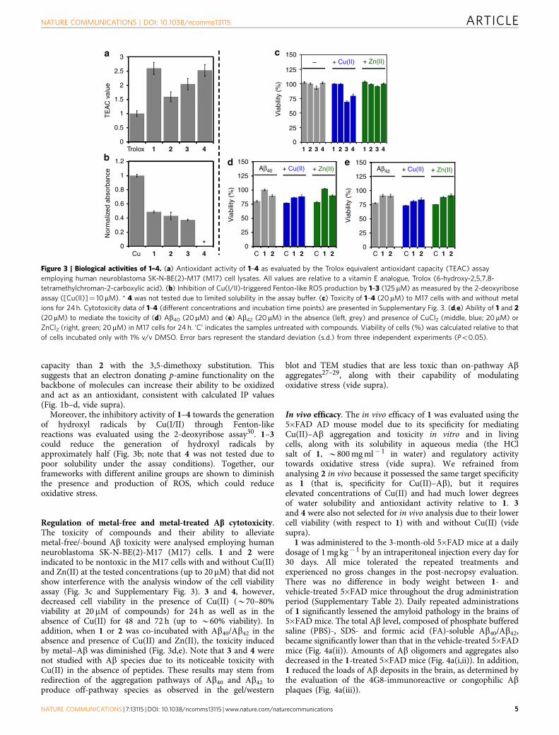
Mediation of oxidative stress. The capability of 1–4 to scavengefree organic radicals was explored in cell lysates using theTrolox equivalence antioxidant capacity (TEAC) assay usedfor testing their aptness to quench the cationic organic radicalof 2,20-azino-bis(3-ethylbenzothiazoline-6-sulfonic acid (ABTS)compared with the known antioxidant, Trolox, a water-solubleanalogue of vitamin E25,28,29. All compounds showed a greaterfree radical scavenging ability than Trolox (Fig. 3a). Specifically,1, 3 and 4 containing the p-amine substitutions had a greater
R2
R1
N
3
H
N
N
N N
N
OCH3
H
2
N
H
NH2
1
N
H
4
NH
NH
N
Cmpd
R2
R2
N
Chemical tools
Cu(II)−AβZn(II)−AβMetal-free Aβ
>
1
3
4
Structural similarity but different targets, interactions, and reactivities
•OOH
O2•– H
2O
2
•OH
ROS
bHN NR
– 1 e–
HN NR– 1 e–
N NRXXX
– 1 H+
1
c
3
2
4
First ionizationpotential
(eV)
Second ionizationpotential
(eV)
CompoundGas phase /
aqueous phaseGas phase /
aqueous phase
1
2
3
4
6.23 / 4.63 6.02 / 4.65
6.76 / 5.44 7.16 / 5.83
5.90 / 4.53 6.13 / 4.57
6.07 / 4.55 6.01 / 4.59
d
a
HN
HN
HN
HN HN
N
N
NH2
OCH3
OCH32
R1
Oxidative stress
++
Figure 1 | Targets associated with AD and ionization potentials of chemical tools (1–4). (a) Structures of 1–4 [1, N1-(pyridin-2-ylmethyl)benzene-
capacity than 2 with the 3,5-dimethoxy substitution. Thissuggests that an electron donating p-amine functionality on thebackbone of molecules can increase their ability to be oxidizedand act as an antioxidant, consistent with calculated IP values(Fig. 1b–d, vide supra).
Moreover, the inhibitory activity of 1–4 towards the generationof hydroxyl radicals by Cu(I/II) through Fenton-likereactions was evaluated using the 2-deoxyribose assay30. 1–3could reduce the generation of hydroxyl radicals byapproximately half (Fig. 3b; note that 4 was not tested due topoor solubility under the assay conditions). Together, ourframeworks with different aniline groups are shown to diminishthe presence and production of ROS, which could reduceoxidative stress.
Regulation of metal-free and metal-treated Ab cytotoxicity.The toxicity of compounds and their ability to alleviatemetal-free/-bound Ab toxicity were analysed employing humanneuroblastoma SK-N-BE(2)-M17 (M17) cells. 1 and 2 wereindicated to be nontoxic in the M17 cells with and without Cu(II)and Zn(II) at the tested concentrations (up to 20mM) that did notshow interference with the analysis window of the cell viabilityassay (Fig. 3c and Supplementary Fig. 3). 3 and 4, however,decreased cell viability in the presence of Cu(II) (B70–80%viability at 20 mM of compounds) for 24 h as well as in theabsence of Cu(II) for 48 and 72 h (up to B60% viability). Inaddition, when 1 or 2 was co-incubated with Ab40/Ab42 in theabsence and presence of Cu(II) and Zn(II), the toxicity inducedby metal–Ab was diminished (Fig. 3d,e). Note that 3 and 4 werenot studied with Ab species due to its noticeable toxicity withCu(II) in the absence of peptides. These results may stem fromredirection of the aggregation pathways of Ab40 and Ab42 toproduce off-pathway species as observed in the gel/western
blot and TEM studies that are less toxic than on-pathway Abaggregates27–29, along with their capability of modulatingoxidative stress (vide supra).
In vivo efficacy. The in vivo efficacy of 1 was evaluated using the5�FAD AD mouse model due to its specificity for mediatingCu(II)–Ab aggregation and toxicity in vitro and in livingcells, along with its solubility in aqueous media (the HClsalt of 1, B800 mg ml� 1 in water) and regulatory activitytowards oxidative stress (vide supra). We refrained fromanalysing 2 in vivo because it possessed the same target specificityas 1 (that is, specificity for Cu(II)–Ab), but it requireselevated concentrations of Cu(II) and had much lower degreesof water solubility and antioxidant activity relative to 1. 3and 4 were also not selected for in vivo analysis due to their lowercell viability (with respect to 1) with and without Cu(II) (videsupra).
1 was administered to the 3-month-old 5�FAD mice at a dailydosage of 1 mg kg� 1 by an intraperitoneal injection every day for30 days. All mice tolerated the repeated treatments andexperienced no gross changes in the post-necropsy evaluation.There was no difference in body weight between 1- andvehicle-treated 5�FAD mice throughout the drug administrationperiod (Supplementary Table 2). Daily repeated administrationsof 1 significantly lessened the amyloid pathology in the brains of5�FAD mice. The total Ab level, composed of phosphate bufferedsaline (PBS)-, SDS- and formic acid (FA)-soluble Ab40/Ab42,became significantly lower than that in the vehicle-treated 5�FADmice (Fig. 4a(ii)). Amounts of Ab oligomers and aggregates alsodecreased in the 1-treated 5�FAD mice (Fig. 4a(i,ii)). In addition,1 reduced the loads of Ab deposits in the brain, as determined bythe evaluation of the 4G8-immunoreactive or congophilic Abplaques (Fig. 4a(iii)).
b d
ca
e43
2
2
1
1
1
1.2
0.8
0.6
0.4
0.2
0Cu 1 1C2 2
1 2
1C 2 1C 2 1C 2 1C 2 1C 23 4
3 4 1 2 3 4 1 2 3 4
0
25
50
100
125
150Aβ40
75
0
25
50
100
125
150
75
0
25
50
100
125
150
75
*
Trolox
Nor
mal
ized
abs
orba
nce
TE
AC
val
ue
Via
bilit
y (%
)
Via
bilit
y (%
)
Via
bilit
y (%
)
0
2.5
3
1.5
0.5
–
Aβ42 + Zn(II)
+ Zn(II)
+ Zn(II) + Cu(II)
+ Cu(II)
+ Cu(II)
Figure 3 | Biological activities of 1–4. (a) Antioxidant activity of 1–4 as evaluated by the Trolox equivalent antioxidant capacity (TEAC) assay
employing human neuroblastoma SK-N-BE(2)-M17 (M17) cell lysates. All values are relative to a vitamin E analogue, Trolox (6-hydroxy-2,5,7,8-
tetramethylchroman-2-carboxylic acid). (b) Inhibition of Cu(I/II)-triggered Fenton-like ROS production by 1–3 (125 mM) as measured by the 2-deoxyribose
assay ([Cu(II)]¼ 10mM). * 4 was not tested due to limited solubility in the assay buffer. (c) Toxicity of 1–4 (20mM) to M17 cells with and without metal
ions for 24 h. Cytotoxicity data of 1–4 (different concentrations and incubation time points) are presented in Supplementary Fig. 3. (d,e) Ability of 1 and 2
(20mM) to mediate the toxicity of (d) Ab40 (20mM) and (e) Ab42 (20mM) in the absence (left, grey) and presence of CuCl2 (middle, blue; 20mM) or
ZnCl2 (right, green; 20mM) in M17 cells for 24 h. ‘C’ indicates the samples untreated with compounds. Viability of cells (%) was calculated relative to that
of cells incubated only with 1% v/v DMSO. Error bars represent the standard deviation (s.d.) from three independent experiments (Po0.05).
Moreover, the Morris water maze test was conductedto evaluate the cognitive functions of 5�FAD mice in responseto the persistent treatment with 1. During the trial trainings,the 1-treated 5�FAD mice could find the hidden escape platformfaster than the vehicle-treated 5�FAD mice, which was compar-able to the performance of wild-type mice at the same age(Fig. 4b(i)). The probe trials also showed significant improvementof the long-term spatial memory in the 1-treated animals(Fig. 4b(ii,iii)). Therefore, 1 could produce beneficial effects toprevent or reverse cognitive deficits as well as amyloid pathologyin 5�FAD mice. The alleviation of AD symptoms and pathology,which arises from mediating Cu(II)–Ab aggregation and toxicity
by treatment with 1, is suggested to be effectiveas indicated by the previously reported studies usingcopper ionophores (for example, clioquinol and PBT2) intransgenic AD mice31,32.
Characterization of solution species. Studies of the speciespresent in solution indicated that 1 and 2 were stable over 5 h(Supplementary Fig. 4a–h). 3 showed changes in itsUltraviolet-Visible (UV-vis) spectra after 5 h of incubationoccurring at a very slow rate (Supplementary Fig. 4i andSupplementary Table 3). Electrospray ionization mass
**
***
Wild-type
Wild-type
Wild-type
Wild-type
Wild-type
NW
Quadrant NW (target) NE SW
SW
SE
SE
NE
5×FAD
5×FAD-1
5×FAD-1
5×FAD-1
5×FAD-Vehicle
5×FAD-Vehicle
5×FAD–Vehicle
5×FAD-1
5×FAD-Vehicle
1Aggregates
Oligomers
monomer
β-actin
Aβ4
0 (n
mol
/g o
f tis
sue)
Aβ
olig
omer
s(μ
g/g
of ti
ssue
)A
β ol
igom
ers
(μg/
g of
tiss
ue)
Aβ4
2 (n
mol
/g o
f tis
sue)
Con
go r
ed
Con
goph
ilic
plaq
ues
(tot
al n
umbe
r)
4G8
Late
ncy
(s)
Late
ncy
to p
latfo
rm (
s)
% ti
me
in q
uadr
ant4G
8-po
sitiv
e pl
aque
s
(%
area
)
Aβs
Dimer
M Vehicle755037
25
15
52
SDS-soluble FA-soluble Total PBS-/SDS-soluble
Trial session (day)
10
40
30
20
100
1 2 3 4 51.0
0.1
0
6.0
4.0
0.3 1.5
1.0
0.5
0
10.0
10.0
12.0
8.0
8.0
6.0
6.0
4.0
4.0
2.0
2.0
0
0
7.06.05.04.03.02.01.0
10
25
40
30
20
10
0
20
15
10
5
0
8
6
4
2
0
600
500
400
300
200
100
0
0
0.2
2.0
0
0.5
0
4.0
****
**
b
a i
i
ii
ii
iii
iv
iii
*****
***
***###
###
## ####
*
*
* **
*
3.0
2.0
1.0
0Vehicle
Vehicle
Vehicle 1
ctx
ctxctx
ctx
cccc
hip hipsub
sub sub
sub
20
1
Vehicle 1
1 Vehicle 1 Vehicle Vehicle1 1
Figure 4 | In vivo efficacy of 1 against amyloid pathology and cognitive defects. (a) Determination of Ab levels (i,ii) and loads of 4G8-immunoreactive
(iii, top, brown) and Congo red-positive (iii, bottom, red) amyloid plaques in the brains of 5�FAD mice after 30-day treatment with 1. ELISA analyses were
performed in triplicate per sample to quantify Ab oligomers or aggregates as well as SDS-soluble, FA-soluble, and total Ab40 and Ab42 (ii) (n¼ 7 and 10 for
vehicle- and 1-treated 5�FAD mice, respectively). Ab-immunohistochemistry (brown) or Congo red staining (red, inset) was conducted in the brains of
vehicle- or 1-treated 5�FAD mice (iii). The area of 4G8-immunoreactive amyloid deposits or the total number of congophilic amyloid plaques in the same
cortical region of interest was measured in five brain sections taken from each animal. Representative microscopic images of cortical or subiculum area in
the Congo red-stained (red) brain sections of 5�FAD mice. ctx, cortex; hip, hippocampus, cc; corpus callosum, sub; subiculum. Scale bars¼ 100 mm. All
bars denote mean±s.e.m. (n¼ 14 and 17 for vehicle- and 1-treated 5�FAD mice, respectively). *Po0.05, **Po0.01, or ***Po0.001 by unpaired two-tail t-
test. (b) Enhancement of cognitive performance by 1 in the 5�FAD mice. Using the Morris water maze (MWM) test, spatial learning and memory
performance was compared between 5�FAD and their littermate wild-type mice after 30-day treatment with vehicle or 1. (i) The escape latency time was
measured every day for 5 days from the day of the 30th drug treatment. (ii–iv) The probe trials were conducted at 3 h after the final trial of the MWM test.
ii, The images depict the representative traces of mice to search for the escape platform in the water maze for 60 s. (iii,iv) Bars denote the time when they
reached the platform area (iii) and stay in the target quadrant (NW, grey area in ii; iv). The statistical comparisons were performed between 5�FAD and
their wild-type littermate mice with vehicle (#), or between vehicle and 1 treatment in 5�FAD mice (*). All values denote mean±s.e.m. (n¼ 14 and 17 for
vehicle- and 1-treated 5�FAD mice, respectively; n¼ 17 for vehicle-treated wild-type mice). *Po0.05, **,##Po0.01 or ***,###Po0.01.
spectrometry (ESI–MS) studies, however, exhibited no change inthe major peak at 278 m/z corresponding to the [MþH]þ ionafter 5 h of incubation supporting the stability of 3(Supplementary Fig. 4j–l). Moreover, 4 was unstable with a half-life of B40 min (Supplementary Table 3). After 5 h of incubation,ESI–MS identified a peak at 137 m/z corresponding to [M þH]þ of N,N-dimethyl-p-phenylenediamine (DMPD)29 but otherpossible degradation products of 4 could not be identified(Supplementary Fig. 4m–p). This could be due to the reportedpropensity of the pyrrole moiety to polymerize as possiblyevidenced by unidentified peaks at 288 and 297 m/z(Supplementary Fig. 4o)33,34. DMPD was recently reported tointeract with both metal-free Ab and metal–Ab and redirect theirself-assembly routes to form off-pathway aggregates, suggested tobe less toxic29.
The stability in the presence of Cu(II) was also determined. UV-vis and ESI–MS studies indicated that 1 initially formed CuL2
complexes (L¼ ligand; [M þ H]þ ¼ 460 m/z), with the half-lifebeing B5 min, followed by the generation of one-electron ([M]þ )or two-electron oxidation ([M þ H]þ ) products at 198 m/z(Supplementary Fig. 5a–d and Supplementary Table 3). Thesetypes of oxidation products are well defined in the literature forunsubstituted and substituted p-phenylenediamine derivatives withan oxidant35–38. 3 also presented UV-vis spectral changes similarto 1 with the decay of the peak attributed to Cu(II) binding atB450 nm (half-life B50 min) corresponding to the growth of anew peak at B550 nm comparable to the reported one-electronoxidation products of p-phenylenediamine derivatives(Supplementary Fig. 5i). ESI–MS studies confirmed the initialformation of a CuL2 complex of 3 which was observed to degradeinto the metal-free two-electron oxidized form (SupplementaryFig. 5j–l). Note that other species were detected by ESI–MS after5 h of incubation, possibly from further degradation of 3. Theidentity of these species will be the subject of future studies.
In the case of 2, the addition of Cu(II) created a new peak atB375 nm that did not dissipate over 5 h (Supplementary Fig. 5e).ESI–MS identified an ion at 588 m/z corresponding to the CuL2
complex ([M þ K]þ ; Supplementary Fig. 5f-h). Differentfrom 1 and 3, 2 generated a complex with Cu(II) without anyfurther transformations over the course of 5 h; thus, the bindingaffinity of 2 for Cu(II) was estimated using UV–vis variable-pHtitrations. The presence of a 1:1 complex was observed with anapproximate disassociation constant in the micromolar range(Supplementary Fig. 6). This low binding affinity is likely to betoo weak to interact with the low micromolar to high picomolar(B10� 7–10� 11 M)1,16,39 binding affinity for the first Cu(II)binding site of Ab. Interaction is possible, however, with thesecond metal binding site in Ab which has a weaker affinity ofB10� 5 M for Cu(II)1,16,39. This is consistent with the resultsfrom the gel/western blots where at least two equivalents of Cu(II)are required for activity (Fig. 2e), suggesting that 2 can onlyinteract with Ab when both metal binding sites are metalated(vide supra).
Lastly, the addition of Cu(II) to 4 resulted in intense double peaksat B510 and 550 nm within 1 min, similar to the previouslyreported spectra of the cationic radical of DMPD (SupplementaryFig. 5m)29. ESI–MS displayed the peaks at 137 and 216 m/zcorresponding to [DMPDþH]þ and [4þH]þ , respectively(Supplementary Fig. 5n,o; note that longer incubations led toprecipitation thus limiting analysis). This suggests that DMPD maybe responsible for the activity of 4 towards Ab (ref. 29).
Mass spectrometric studies for Ab and metal–Ab interactions.To investigate the interactions between Ab and 1–4, nano-elec-trospray ionization MS (nESI-MS) combined with ion mobility–
mass spectrometry (IM–MS), optimized for the detection of non-covalent protein complexes40–42, was employed. Data obtainedfrom the samples incubated for 30 min indicated that 1–3exhibited a metal-dependent interaction with both Ab40 and Ab42
(Fig. 5 and Supplementary Fig. 7, respectively). Both 1 and 3 werecapable of producing Cu–ligand-dependent signals correspondingto a mass 89 Da lighter than the apo Ab40/Ab42, albeit with cleardifferences in the abundance of this product. Tandem massspectrometry (MS2) sequencing of the Ab peak supported that itcorresponded to a N-terminally truncated form of the peptide(Supplementary Fig. 8 and Supplementary Table 5), consistentwith data previously reported for L2-b (ref. 25). No data acquiredpresented a stable interaction between 1 and 3 with either Ab40 orAb42 under our conditions, indicative of the formation of atransient Cu-containing ternary complex.
2 produced stable ternary complexes comprising singleequivalents of both Ab40/Ab42 and the ligand, with one or twoCu(II) bound. IM–MS data support that binding of 2 to Ab40 shiftsthe arrival time distributions when compared with the ligand-freecomplexes (Fig. 5b,c and Supplementary Fig. 7). Because of theincreased chemical noise, we were unable to produce IM–MSobservations for these compounds in the presence of Ab42. Usingthe Ab40 as a model system which exhibited reduced chemicalnoise and kinetics of aggregation, these results demonstrate thatthe formation of a ternary complex between Cu(II), Ab and 2results in an altered downstream Ab aggregation pathway. This isconsistent with previous observations for other small moleculesthat can modulate metal–Ab reactivity25,27. In the absence ofCu(II), the above small molecules were not observed in complexwith the peptide (Supplementary Fig. 9).
In addition, the Cu(II)–Ab40/Ab42 samples prepared under theconditions of the inhibition experiments shown in Fig. 2(for example, buffer, 24 h incubation) were analysed bymatrix-assisted laser desorption ionization mass spectrometry(MALDI–MS). As depicted in Supplementary Fig. 10, ionssmaller than full-length Ab40 (loss of 89 Da) appeared from thesamples treated with 1 and 3, consistent with the nESI-MS results(vide supra; Fig. 5a). Along with truncation of the peptide, uponincubation of Cu(II)–Ab with 1 or 3 for 24 h, oxidation of Ab40
was also observed. As indicated by nESI-MS and IM–MS data, Abadded with 2 was not indicated to be modified even after 24 h(Supplementary Fig. 10). Thus, both events (that is, truncationand oxidation of Ab) by 1 and 3 with Cu(II) being present mightbe responsible for redirecting Ab aggregation pathways.
To determine if intact or transformed 4 would be the activespecies which could interact with Ab, both copper-free andcopper-present samples incubated with the molecule were alsomonitored. When 4 was reacted with metal-free Ab40 for 6 h([Ab40]:[4]¼ 1:5, [Ab40]¼ 100mM), the compound, observed byESI–MS, was indicated to be degraded showing its multiplefragments and their nonspecific adducts of monomeric andoligomeric Ab species (Supplementary Fig. 11). Note that inconditions comparable to the nESI-MS studies of 1–3 (Fig. 5), 4was not shown to bind to Ab40 in 30 min and 24 h longincubations at 37 �C (Fig. 5a and Supplementary Fig. 12). In thecase of the Ab samples incubated with 4 and Cu(II) for 1 h,oxidized Ab was shown, along with formation of covalentcomplexes of BQ29, Ab and/or Cu(II) (Fig. 5d). The generation ofoxidized Ab was also indicated in the sample incubated with Ab,Cu(II) and 4 for 24 h under the same condition of the studies(Fig. 2), analysed by MALDI–MS (Supplementary Fig. 10).
To identify the oxidation sites in Ab, MS2 analysis wascarried out on the non-oxidized and singly oxidized Ab40
(Supplementary Fig. 13). In the MS2 of the singly oxidized Ab,ions larger than b35 fragments, including the first 35 amino acidresidues from the N-terminus, existed as the oxidized form;
however, both non-oxidized and oxidized forms were found infragments smaller than b34. These results suggest that oxidationcan occur at several sites, including the methionine residue (M35;
Ab sequence shown in Fig. 2a). As presented in SupplementaryFig. 13, the smallest oxidized ion is b13 and no oxidation wasobserved in the b9 fragment. Thus, along with M35 (ref. 43), H13
c
Drift time (ms)
Drif
t tim
e (m
s)
86 10 12
1
2
1
3
2
1
2
2
4
5
4
5
Aβ40 †: Aβ40 + Cu
Aβ40 + 2Cu •: Aβ40 – 89 Da
*
4
6
10
12
14
16
8
8501,050
1,2501,250
1,4501,450
1,6501,650
8501,050
4
6
10
12
14
16
8
m/z
+
+
+
+
+
+
†
†
†
†
‡
‡
‡
‡
b
m /z
a
+
+
+
+ †
†
†
‡‡
‡
‡†
†
Aβ40 + 2Cu(II) + 6Cmpd
Cmpd = 1
1,050 1,100 1,150 1,200 1,250
%
*
*
Cmpd = 4
Cmpd = 3
Cmpd = 2
No Cmpd
Aβ40 + 2Cu + 6Cmpd Aβ40 + 2Cu + 6Cmpd
Cmpd = 1 Cmpd = 2
Aβ40 + 256 DaAβ40 + Cu + 2
: Aβ40 + 2Cu + 2
+[Aβ40]4+
†[Aβ40 + 2Cu]4+
‡
[Aβ40 + 2Cu]4+
[Aβ40 + Cu + 2]4+
[Aβ40 + 2Cu + 2]4+
Nor
mal
ized
inte
nsity
(%
)
Nor
mal
ized
inte
nsity
(%
)
d
1,4251,525
1,5001,475
1,450
m /z
Aβ40 + O
Aβ40 + Cu
Aβ40 + DMPDtransformed
Aβ40 + DMPDtransformed + Cu
Aβ40
4+
4+
4+
4+
4+4+ 3+3+
4+ 3+ 4+ 3+
::
:
:
‡
+
Aβ40 Aβ40 + 2Cu
Figure 5 | ESI–MS and IM–MS analyses of 1–4 in the presence or absence of Cu(II). (a) Metal-dependent interactions of 1–4 with monomeric Ab40. Both
1 and 3 promote the formation of Ab mass loss product 89 Da lighter than the metal-free peptide (K), consistent with previously published data25.
Incubations of Ab with 1 were additionally shown to produce an adduct consistent with a mass gain of 256 Da compared with the intact, unmodified,
peptide (*). 2 forms stable ternary complexes with Ab40 and Cu(II), existing in two different stoichiometries that contain either one or two equivalents of
the metal (. and ~, respectively). Conditions for (a): [Ab40]¼ 18mM; [copper(II) acetate]¼40mM; [1–4]¼ 120mM; 100 mM ammonium acetate (pH
7.5) with 1% v/v DMSO; 37 �C; 30 min incubation. (b) IM drift time versus m/z plots comparing the data acquired for 1 (bottom left) and 2 (bottom right)
against Cu(II)-bound Ab40 (top right) and Cu-free Ab40 (top left). (c) Arrival time distribution data extracted from the plots which provide that these
stable binding interactions support altered distributions upon binding to 2 when compared with the metal-free and Cu-bound Ab40 complexes (collision
cross section (CCS) data presented in Supplementary Table 4). (d) ESI–MS spectra of Ab40 with 4 in the presence of Cu(II) under different conditions from
(a). Ab–DMPDtransformed complexes and oxidized Ab40 were found when Cu(II) was present. The different positive charge states (with Hþ and Naþ ) are
used to best represent the complexes observed. Conditions for d (actual injected [Ab]¼ 10 mM): [Ab40]¼ 100mM; [Cu(II)]¼ 100 mM; [4]¼ 500mM;
100 mM ammonium acetate (pH 7.5) with 1% v/v DMSO; 37 �C; 1 h incubation.
and H14 could be additional plausible oxidation sites in Ab, aspreviously reported44,45.
Together, our overall MS studies suggest that 1–4 can interactwith metal-free Ab and/or Cu(II)-bound Ab in differentmanners. The distinct redirecting activity of 1–4 towards metal-free and metal-treated Ab aggregation pathways, indicated inboth inhibition and disaggregation experiments (vide supra),could be directed by multiple mechanisms, including Abmodifications (that is, degradation and oxidation), non-covalentcomplex formation, and covalently linked adduct generation. Theoverall proposed mechanisms of 1–4 towards individual targetsare described in detail in the following sections (vide infra).
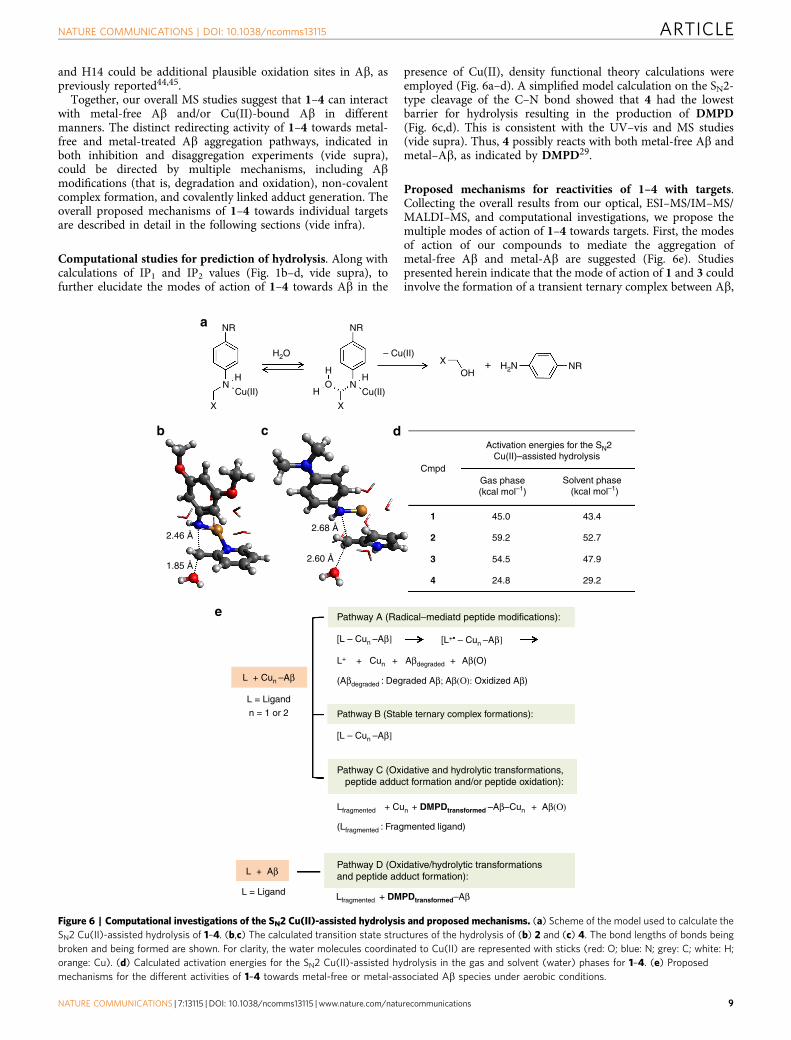
Computational studies for prediction of hydrolysis. Along withcalculations of IP1 and IP2 values (Fig. 1b–d, vide supra), tofurther elucidate the modes of action of 1–4 towards Ab in the
presence of Cu(II), density functional theory calculations wereemployed (Fig. 6a–d). A simplified model calculation on the SN2-type cleavage of the C–N bond showed that 4 had the lowestbarrier for hydrolysis resulting in the production of DMPD(Fig. 6c,d). This is consistent with the UV–vis and MS studies(vide supra). Thus, 4 possibly reacts with both metal-free Ab andmetal–Ab, as indicated by DMPD29.
Proposed mechanisms for reactivities of 1–4 with targets.Collecting the overall results from our optical, ESI–MS/IM–MS/MALDI–MS, and computational investigations, we propose themultiple modes of action of 1–4 towards targets. First, the modesof action of our compounds to mediate the aggregation ofmetal-free Ab and metal-Ab are suggested (Fig. 6e). Studiespresented herein indicate that the mode of action of 1 and 3 couldinvolve the formation of a transient ternary complex between Ab,
HH
H OH+ H2N
OH
N N
XX
X
Cu(II) Cu(II)
– Cu(II)
NRa
L = Ligand
L = Ligand
L + Cun –Aβ
L + Aβ
L+ + Cun + Aβdegraded + Aβ(O)
[L – Cun –Aβ]
[L – Cun –Aβ]
Lfragmented + Cun + DMPDtransformed –Aβ–Cun + Aβ(Ο)
(Lfragmented : Fragmented ligand)
Lfragmented + DMPDtransformed–Aβ
(Aβdegraded : Degraded Aβ; Aβ(Ο): Oxidized Aβ)
[L+• – Cun –Aβ]
n = 1 or 2
Pathway A (Radical–mediatd peptide modifications):
Pathway B (Stable ternary complex formations):
Pathway C (Oxidative and hydrolytic transformations, peptide adduct formation and/or peptide oxidation):
Pathway D (Oxidative/hydrolytic transformationsand peptide adduct formation):
e
1
2
45.02.68 Å
2.60 Å
2.46 Å
1.85 Å
43.4
52.7
47.9
29.2
59.2
54.5
24.8
3
4
CmpdGas phase(kcal mol–1)
Solvent phase (kcal mol–1)
Activation energies for the SN2 Cu(II)–assisted hydrolysis
b c d
NR
NRH2O
Figure 6 | Computational investigations of the SN2 Cu(II)-assisted hydrolysis and proposed mechanisms. (a) Scheme of the model used to calculate the
SN2 Cu(II)-assisted hydrolysis of 1–4. (b,c) The calculated transition state structures of the hydrolysis of (b) 2 and (c) 4. The bond lengths of bonds being
broken and being formed are shown. For clarity, the water molecules coordinated to Cu(II) are represented with sticks (red: O; blue: N; grey: C; white: H;
orange: Cu). (d) Calculated activation energies for the SN2 Cu(II)-assisted hydrolysis in the gas and solvent (water) phases for 1–4. (e) Proposed
mechanisms for the different activities of 1–4 towards metal-free or metal-associated Ab species under aerobic conditions.
Cu(II) and compound. This could be subsequently followed bythe oxidation of 1 and 3 as well as the oxidation and/ordegradation of the peptide by well-documented radical pathways(proposed pathway A: radical-mediated peptide modifications;Fig. 6e)43–48. 2 is most likely to undergo a different pathway ofAb aggregation modulation due to its higher IPs (Fig. 1d).Thus, it is proposed that the interaction of 2 with Cu(II)–Abresults in the formation of a stable ternary complex consistingof Ab, Cu(II) and 2 which subsequently diverts Cu(II)–Abaggregation pathways producing off-pathway species withdifferent conformations (proposed pathway B; stable ternarycomplex formation; Fig. 6e). In the case of 4, based on the densityfunctional theory calculations, along with the experimentallyobserved Ab–DMPDtransformed adducts, this molecule might betransformed to DMPD via oxidative and hydrolytic pathways29,which could be responsible for interaction and reactivity withboth metal-free and metal-associated Ab through the proposedmodes of action C and D (oxidative/hydrolytic transformations,peptide adduct formation and/or peptide oxidation; Fig. 6e).
In addition, 1–4 are able to mediate oxidative stress caused bythe presence of free organic radicals by acting as antioxidants todonate an electron to quench the radials49. This is evidenced bytheir activity towards the radicals roughly correlating with thecalculated IPs (Fig. 1d) which measure the ease of releasing anelectron. Furthermore, in the presence of Cu(I/II), 1–3 canalso inhibit ROS generation. This activity is possibly originatedby 1–3 binding to copper and preventing it being reduced toform ROS (for example, hydroxyl radicals)49, along withcompounds’ antioxidant activity. The previously discussedelectronic properties (for example, IPs, Fig. 1d) of thecompounds could also compliment this activity. More detailedinvestigations of the modes of action of 1–4 towards targets willbe the subject of future studies.
DiscussionTo date, effective drugs against AD are not available since thepathological factors and pathways of AD are still unclear. Toidentify such pathological features associated with AD, chemicaltools with distinct specificity towards various individual orassociated targets are needed. Herein, we designed four smallmolecules based on a structure-mechanism-based design strategyfor targeting and regulating distinct pathological factors(for example, metal ions, metal-free Ab, metal–Ab, ROS, freeorganic radicals) linked to AD pathology as chemical tools usefulfor AD research. Our studies indicate that the desired chemicalproperties of small molecules can be achieved for reactions withpathological features simply through minor structural variationsto a parent framework. In addition, such property tuning of smallmolecules is observed to successfully afford different modes ofaction towards the targets. Furthermore, along with in vitrocharacterizations at the molecular level, the validation of ourchemical tool in the 5�FAD AD mouse model confirms its utilityin investigating AD aetiology. Therefore, our findings of the smallmolecules, able to probe distinct pathological facets via disparatemechanisms, demonstrate the feasibility of applying a structure-mechanism-based design concept to rationally construct chemicaltools capable of illuminating the roles of multiple individualtargets and their inter-relationships in AD pathogenesis. More-over, such chemical tools will provide an unconventional avenuefor discovering effective diagnostics and therapeutics for AD.
MethodsParallel artificial membrane permeability assay adapted for BBB (PAMPA-BBB).PAMPA-BBB experiments were conducted using the PAMPA Explorer kit(pION Inc., Billerica, MA, USA) using previously reported protocols24,28,29.Compounds (25mM, 200ml) in pH 7.4 Prisma HT buffer (pION) were added to the
wells of a donor plate (number of replicates¼ 12). The polyvinylidene fluoride(PVDF, 0.45mM) filter membrane on the acceptor plate was coated with BBB-1 lipidformulation (5ml, pION). The acceptor plate was placed on top of the donor plate.Brain sink buffer (BSB, 200ml, pION) was added to each well of the acceptor plateand was incubated for 4 h at ambient temperature without stirring. UV–vis spectra ofthe solutions in the reference, acceptor, and donor plates were measured using amicroplate reader. The PAMPA Explorer software v. 3.5 (pION) was used tocalculate the �logPe values for compounds. CNS±designations were assigned bycomparison with compounds that were identified in previous reports50–52.
Ab aggregation experiments. Before experiments, Ab40 or Ab42 was dissolved inammonium hydroxide (NH4OH; 1% v/v, aq). The resulting solution wasaliquoted, lyophilized overnight and stored at �80 �C. A stock solution ofAb was then prepared by dissolving lyophilized peptide in 1% NH4OH (10 ml) anddiluting with ddH2O. The concentration of the solution was determined bymeasuring the absorbance of the solution at 280 nm (e¼ 1,450 M–1 cm–1 for Ab40;e¼ 1,490 M–1 cm–1 for Ab42). The peptide stock solution was diluted to a finalconcentration of 25mM in Chelex-treated buffered solution containing HEPES(20 mM; pH 6.6 for Cu(II) samples; pH 7.4 for metal-free and Zn(II) samples) andNaCl (150 mM). For the inhibition studies, compounds (final concentration 50 mM,1% v/v dimethyl sulfoxide (DMSO)) were added to the sample of Ab (25 mM) inthe absence and presence of a metal chloride salt (CuCl2 or ZnCl2; 25, 50, 100 or125 mM) followed by incubation at 37 �C with constant agitation for 24 h. For thedisaggregation studies, Ab with and without a metal chloride salt was incubated for24 h at 37 �C with constant agitation to generate preformed Ab aggregates. Theresulting samples were then treated with compounds (50 mM) and incubated withconstant agitation for additional 24 h.
Gel electrophoresis and western blotting. The Ab samples from in vivo orin vitro experiments were analysed by gel electrophoresis followed by westernblotting using an anti-Ab antibody (6E10)24,25,27–29. Samples (10 ml) wereseparated on a 10–20% Tris-tricine gel (Invitrogen, Grand Island, NY, USA).Following separation, the proteins were transferred onto nitrocellulose membranesand blocked with bovine serum albumin (BSA, 3% w/v, Sigma-Aldrich, St Louis,MO, USA) in Tris-buffered saline (TBS) containing 0.1% Tween-20 (TBS-T) for2 h at room temperature or overnight at 4 �C. The membranes were incubated withan anti-Ab antibody (6E10, 1:2,000, Covance, Princeton, NJ, USA) in a solution of2% BSA (w/v in TBS-T) for 4 h at room temperature or overnight at 4 �C.After washing with TBS-T (3� , 10 min), a horseradish peroxidase-conjugated goatanti-mouse secondary antibody (1:5,000 in 2% BSA w/v in TBS-T; CaymanChemical Company, Ann Arbor, MI, USA) was added for 1 h at room temperature.The Thermo Scientific SuperSignal West Pico Chemiluminescent Substrate(Thermo Scientific, Rockford, IL, USA), Biosesang ECL Plus kit (Biosesang,Gyeonggi-do, Republic of Korea), or a homemade ECL kit53 was used to visualizethe results on a ChemiDoc MP Imaging System (Bio-Rad, Hercules, CA, USA).
Transmission electron microscopy (TEM). Samples for TEM were preparedaccording to a previously reported method using glow-discharged grids(Formar/Carbon 300-mesh, Electron Microscopy Sciences, Hatfield, PA,USA)24,25,27–29. Images for each sample were taken on a JEOL JEM-2100transmission electron microscope (UNIST Central Research Facilities, UlsanNational Institute of Science and Technology, Ulsan, Republic of Korea).
Cell viability studies. The M17 cell line was purchased from the American TypeCulture Collection (ATCC, Manassas, VA, USA). The cell line was maintained inmedia containing 50% minimum essential medium and 50% F12 (GIBCO, GrandIsland, NY, USA), supplemented with 10% fetal bovine serum (Sigma), 100 U ml� 1
penicillin, and 100 mg ml� 1 streptomycin (GIBCO). The cells were grown andmaintained at 37 �C in a humidified atmosphere with 5% CO2. The cell cultureused in this work did not indicate mycoplasma contamination. Cell viability upontreatment of compounds was determined using the MTT assay (Sigma). M17 cellswere seeded in a 96-well plate (15,000 cells in 100ml per well). The cells weretreated with Ab (20 mM) with or without CuCl2 or ZnCl2 (20 mM), followed by theaddition of compounds (20 mM, 1% v/v final DMSO concentration), and incubatedfor 24 h. After incubation, 25 ml MTT (5 mg ml� 1 in phosphate buffered saline(PBS, pH 7.4, GIBCO) was added to each well and the plate was incubated for 4 hat 37 �C. Formazan produced by the cells was solubilized using an acidic solution ofN,N-dimethylformamide (DMF, 50%, v/v aq) and SDS (20%, w/v) overnight atroom temperature in the dark. The absorbance was measured at 600 nm using amicroplate reader. Cell viability was calculated relative to cells containing anequivalent amount of DMSO.
TEAC assay. The assay employing M17 cell lysates was conducted following theprotocol of the antioxidant assay kit purchased from Cayman Chemical Company(Ann Arbor, MI, USA) with minor modifications25,28,29. For the antioxidant assayusing cell lysates, cells were seeded in a six-well plate and grown to B80–90%confluence. Cell lysates were prepared following a previously reported method withmodifications54. M17 cells were washed once with cold PBS (pH 7.4, GIBCO) and
harvested by gently pipetting off adherent cells with cold PBS. The cell pellet wasgenerated by centrifugation (2,000g for 10 min at 4 �C). This cell pellet wassonicated on ice (5 s pulses, three times with 20 s intervals between each pulse) in2 ml of cold Assay Buffer (5 mM potassium phosphate, pH 7.4, containing 0.9%NaCl and 0.1% glucose). The cell lysates were centrifuged at 5,000g for 10 min at4 �C. The supernatant was removed and stored on ice until use. For standard andsamples in 96-well plates, 10 ml of the supernatant of cell lysates was deliveredfollowed by addition of compound, metmyoglobin, ABTS and H2O2 in order.After 5 min incubation at room temperature on a shaker, absorbance values at750 nm were recorded. The antioxidant concentration was calculated according tothe measured absorbance (% inhibition¼ 100� (A0 – A)/A0, where A0 isabsorbance of the supernatant of cell lysates). The measurements were conductedin triplicate.
2-Deoxyribose assay. The ability of 1–3 to decrease free radical formation fromFenton-like chemistry by Cu(I/II) was determined using previously reportedprocedures28,30. To summarize, solutions of phosphate buffer (50 mM NaH2PO4,pH 7.2) treated with Chelex overnight, compound (125 mM in water),CuCl2 (10 mM), 2-deoxy-D-ribose (15 mM), H2O2 (200 mM), and sodiumascorbate (2 mM) were mixed in the listed order and incubated at 37 �Cwith constant agitation. These conditions were chosen to optimize theformation of the chromogen produced during the course of the assay.After 1 h, the samples were quenched with trichloroacetic acid (2.8% w/v, 200 ml)and 2-thiobarbituric acid (1% w/v, 200 ml) and heated at 100 �C for 20 min.The samples were allowed to cool for 5 min before measuring the absorbancevalues at 532 nm on a microplate reader. Samples without compounds werealso tested as a control. Normalized absorbance values were calculated aspreviously reported28.
Determination of solution speciation for 2 and Cu(II)–2 complex. ThepKa value for 2 was determined by UV–vis variable-pH titrations aspreviously reported24,55. To establish the pKa value, a solution (10 mM NaOH, pH12, 100 mM NaCl) of 2 (100 mM) was titrated with small amounts of HCl. At least30 spectra were recorded in the range of pH 2–10. Similarly, a solution containingCuCl2 and 2 (50 mM) in a metal to ligand ratio of 1:2 was titrated with smalladditions of HCl and at least 30 spectra were recorded over the range pH 2–7.The acidity and stability constants were calculated by using the HypSpec program(Protonic Software, UK)56. Speciation diagrams were modelled in the HySS2009program (Protonic Software)57.
Stability studies. The stability of 1–4 (50mM) in the absence and presenceof CuCl2 (25 mM) was monitored every 10 min using UV–vis for 5 h in buffer(20 mM HEPES, pH 7.4, 150 mM NaCl; 1% DMSO) at 37 �C. The resulting spectrawere corrected for baseline shifts at 800 nm and the half-life and rate of decay ofthe absorbance at 250, 385 and 400 nm for 4, [Cu(II)þ 1], and [Cu(II)þ 3],respectively, was calculated using the first-order exponential decay function asimplemented in Origin 9.1 (OrginLab Corp., Northampton, MA, USA). Addi-tionally, the species present were identified using ESI–MS. Samples containing1–4 (50 mM) with or without CuCl2 (25 mM) were incubated in ddH2O (1%DMSO) at 37 �C for the selected time points before being freshly frozen usingliquid nitrogen and stored at �80 �C until they were thawed immediatelybefore measurement.
Ion mobility–mass spectrometry (IM–MS). All IM–MS experiments were carriedout on a Synapt G2 (Waters, Milford, MA)27–29,58. Samples were ionized using anano-electrospray source operated in positive ion mode. MS instrumentation wasoperated at a backing pressure of 2.7 mbar and sample cone voltage of 40 V.The m/z scale was calibrated using 20 mg ml� 1 aqueous cesium iodide. Forpeptide-metal ligation studies, the aliquots of Ab peptides (final concentration18mM) were sonicated for 5 s before pre-incubation with or without a source ofCu(II) (copper(II) acetate) at 37 �C for 10 min. After pre-incubation, the sampleswere treated with or without 1–4 (final concentration 160mM) and incubated at37 �C for 30 min before analysis. Solution conditions were 100 mM ammoniumacetate (pH 7.5) with 1% v/v DMSO. Covalent binding studies with 4 wereperformed by incubating aliquots of Ab with the ligand at a ratio of 1:0, 1:5 and1:25 in water for either 24 h or a week at 25 �C. After incubation the samples werelyophilized for storage until analysis. Immediately before analysis samples werereconstituted to 50 mM peptide concentration in 1,1,1,3,3,3-hexafluoro-2-propanol,sonicated and diluted further to 25 mM for data acquisition. Accurate mass valuesfor ligand-bound complexes were calculated using the monoisotopic peakdifference between apo and ligated states with errors reported as a function of twotimes the s.d. collision cross section (CCS) measurements were externally calibratedusing a database of known values in helium, using values for proteins that bracketthe likely CCS and ion mobility values of the unknown ions58,59. CCS values are theaverage of six replicates with errors reported as the least square product. This leastsquare analysis combines inherent calibrant error from drift tube measurements(3%)59. calibration curve error, and two times the replicate s.d. error. All otherconditions are consistent with previously published methods25.
Electrospray ionization mass spectrometry (ESI–MS). Ab (100 mM) wasincubated with 4 (500 mM) in 100 mM ammonium acetate, pH 7.5, without andwith the addition of CuCl2 (100 mM) for 1 h at 37 �C without agitation. Beforeinjection into the mass spectrometer, the resulting Ab was diluted by 10-fold.ESI–MS analysis was performed using a Synapt G2-Si quadrupole time-of-flightmass spectrometer (Waters, Manchester, UK) equipped with electrosprayionization source. The capillary voltage, sampling cone voltage, and sourcetemperature were set to 2.8 kV, 70 V, and 60 �C, respectively. The backing pressurewas adjusted to 3.2 mbar.
Matrix-assisted laser desorption ionization mass spectrometry (MALDI–MS).Ab samples (prepared in the same procedure with Ab aggregation experiments)were mixed with the equivalent volume of the matrix solution and loaded on thetarget plate. The matrix solution was prepared with a-cyano-4-hydroxycinnamicacid (Sigma-Aldrich) by dissolving in 40% CH3CN and 2% trifluoroacetic acid andadjusting the concentration to 5 mg ml� 1. MALDI–MS analysis was conductedusing an Ultraflex III time-of-flight mass spectrometer (Bruker Daltonics, Bremen,Germany). Mass spectra were acquired over the range of 1,000–6,000 m/z.
Calculation of transition state energies and ionization potentials. First-principles calculations using Gaussian09 (ref. 60) were carried out for 1–4 in orderto study the SN2 hydrolysis and their one and two-electron oxidation. For the directC–N bond hydrolysis mechanism assisted by Cu(II), five additional water mole-cules were added to the first hydration sphere of Cu(II) in addition to the watermolecule acting as a nucleophile. The structure optimizations were performed inthe vacuum at M06/6-31G(d) level. The Los Alamos effective-core potential (ECP)LanL2DZ basis set was applied for Cu(II). The hydration effect was taken intoaccount by additional single point calculations with polarizable continuum modelat M06/6-31G(d) level. To find transition states as the first-order saddle points onthe PES, Berny algorithm was used. The validity of the transition states wereconfirmed by frequency calculations (one imaginary) corresponding to the trans-lational motion of the carbon from C–N bond. For the two-electron oxidation of 1–4, we calculated two successive adiabatic ionization potentials accounting for thesequential loss of electrons. The radical cation from the first ionization is assumedto undergo immediate deprotonation before the second ionization. All the relevantchemical species were optimized at M06/6-31G(d) level, and their thermodynamicparameters were calculated at M06/6-311þG(2df,2p) level. Inclusion of the sol-vation effect of water using the polarizable continuum model reduces the ionizationpotential, but the overall trend is conserved.
Animal studies. The amyloidogenic characteristics of 5�FAD mice have beendescribed previously29. In brief, 5�FAD mice express the Swedish/London/Floridamutations of the human amyloid precursor protein (hAPP) and the M146L/L286Vmutations of the human presenilin-1 (PSEN1). They develop the early and robustpathology of AD with the cognitive and behavioural impairments. The 5�FAD micehave been widely used to test the possible efficacy of a compound targeted for AD25,29.We maintained the mice on a C57BL/6� SJL hybrid background. All animalexperiments were performed in accordance with the guidelines of the Asan Institutefor Life Science for Laboratory Animal Care and Use (Seoul, Korea), where the animalshad free access to water and food, and were housed on 12 h light/12 h dark cycle.
Compound treatment to animals. Chemicals used were freshly preparedshortly before administration to the animals. We treated 5�FAD mice with 1(1 mg kg� 1 of body weight) or the vehicle (1% v/v DMSO in 20 mM HEPES, pH7.4, 150 mM NaCl) starting at 3 months of age, which is the same method used inour previous studies25,29. 1 was daily injected into the lower abdomen of the5�FAD mice for 30 days.
Assessment of cognitive function by the Morris water maze test. Mice weresubjected to the Morris water maze test to assess their performance of spatiallearning and memory25,61. The water maze was a circular plastic pool (120 cmdiameter) and filled with murky water (21.0±1.0 �C). A cylindrical escape platform(15 cm diameter) stood 0.5 cm under the water surface. Three hours after the 30thcompound treatment, the mice were allowed the first training to swim and find thehidden escape platform in the water with three repeats per training. Thereafter,they daily tried the task on the next 4 consecutive days. Three hours after the finalescape test, the platform was removed, and the mice exercised a probe trial for 60 s.The animal performance was traced with SMART Video Tracking System(Harvard Apparatus, Holliston, MA, USA).
Tissue preparation. Immediately after the behavioural test, the mice were sacri-ficed under deep anaesthesia. The brain was divided into left and right cerebralhemispheres for biochemical and histological analysis, respectively and quicklyfrozen in liquid nitrogen.
ELISA for quantification of the cerebral Ab. We measured the levels of varioustypes of Ab in the brain as described previously25,29. The protein fractions were
serially prepared from the left hemispheres in PBS (pH 7.4), in 2% SDS and in 70%FA, and then subjected to the human Ab40 and Ab42 ELISA quantificationaccording to the manufacturer’s methods (Invitrogen, Carlsbad, CA, USA). Theaggregated (Invitorgen) and oligomeric Ab (82E1-specific; IBL International,Hamburg, Germany) ELISA quantifications were also performed using PBSfractions. The cerebral levels of Ab40 and Ab42 were represented as moles per gramof wet brain tissue, whereas the aggregated or oligomeric Ab amounts wereexpressed as grams per gram of wet brain tissue.
Histological quantification of cerebral amyloid pathology. 12-mm sagittalsections of the brain were prepared from the right hemispheres on a cryostat(HM550; Microm, Walldorf, Germany), and mounted onto 1% poly-L-lysine-coated glass slides. Immunohistochemistry was performed on the tissue sectionusing the human Ab(17–24)-specific antibody 4G8 (Covance, Princeton, NJ, USA).They were immunologically reacted with 4G8 (1:1,000 dilution) and biotinylatedanti-mouse IgG secondary antibody (Vector Laboratories, Burlingame, CA, USA),and then developed with 0.015% diaminobenzidine/0.001% H2O2 (in PBS; VectorLaboratories). The immuno-reacted sections were examined or photographedunder a light microscope (Eclipse 80i; Nikon, Tokyo, Japan). The congophillicamyloid plaques were examined after staining the sections with Accustain CongoRed amyloid staining solution (Sigma). The loads of amyloid deposits in the brainwere expressed as the per cent area of 4G8-immunoreactive deposits or the numberof congophilic plaques per mm2 of a cortical region of interest.
Statistics. Data are expressed as mean±s.e.m. Statistical differences betweengroups were determined with the unpaired t-test. Statistical significance wasconsidered at Po0.05. The current animal study was performed in parallel with thestudy previously reported29 using the same control groups (vehicle-treatedwild-type and 5�FAD mice); thus, the same control data were used in both studies.
Data availability. All relevant data are available from the authors upon request.
References1. Beck, M. W., Pithadia, A. S., DeToma, A. S., Korshavn, K. J. & Lim, M. H. in
Ligand Design in Medicinal Inorganic Chemistry. Ch. 10 (Wiley, Chichester,2014).
2. Derrick, J. S. & Lim, M. H. Tools of the trade: Investigations into designstrategies of small molecules to target componets in Alzheimer’s disease.ChemBioChem 16, 887–898 (2015).
3. Jakob-Roetne, R. & Jacobsen, H. Alzheimer’s disease: from pathology totherapeutic approaches. Angew Chem. Int. Ed. Engl. 48, 3030–3059 (2009).
4. Bush, A. I. et al. Rapid induction of Alzheimer Ab amyloid formation by zinc.Science 265, 1464–1467 (1994).
5. Soto, C. Unfolding the role of protein misfolding in neurodegenerative diseases.Nat. Rev. Neurosci. 4, 49–60 (2003).
6. Savelieff, M. G., Lee, S., Liu, Y. & Lim, M. H. Untangling amyloid-b, tau, andmetals in Alzheimer’s disease. ACS Chem. Biol. 8, 856–865 (2013).
7. Greenough, M. A., Camakaris, J. & Bush, A. I. Metal dyshomeostasis andoxidative stress in Alzheimer’s disease. Neurochem. Int. 62, 540–555 (2013).
8. Ayton, S., Lei, P. & Bush, A. I. Biometals and their therapeutic implications inAlzheimer’s disease. Neurotherapeutics 12, 109–120 (2015).
9. Barnham, K. J. & Bush, A. I. Biological metals and metal-targeting compoundsin major neurodegenerative diseases. Chem. Soc. Rev. 43, 6727–6749 (2014).
10. Barnham, K. J., Masters, C. L. & Bush, A. I. Neurodegenerative diseases andoxidative stress. Nat. Rev. Drug Discov. 3, 205–214 (2004).
11. Ayton, S., Lei, P. & Bush, A. I. Metallostasis in Alzheimer’s disease. Free. Radic.Biol. Med. 62, 76–89 (2013).
12. Rowinska-Zyrek, M., Salerno, M. & Kozlowski, H. Neurodegenerativediseases—Understanding their molecular bases and progress in thedevelopment of potential treatments. Coord. Chem. Rev. 284, 298–312 (2015).
13. DeToma, A. S., Salamekh, S., Ramamoorthy, A. & Lim, M. H. Misfoldedproteins in Alzheimer’s disease and type II diabetes. Chem. Soc. Rev. 41,608–621 (2012).
14. Leal, S. S., Botelho, H. M. & Gomes, C. M. Metal ions as modulators of proteinconformation and misfolding in neurodegeneration. Coord. Chem. Rev. 256,2253–2270 (2012).
15. Viles, J. H. Metal ions and amyloid fiber formation in neurodegenerativediseases. Copper, zinc and iron in Alzheimer’s, Parkinson’s and prion diseases.Coord. Chem. Rev. 256, 2271–2284 (2012).
16. Kepp, K. P. Bioinorganic chemistry of Alzheimer’s disease. Chem. Rev. 112,5193–5239 (2012).
17. Faller, P. & Hureau, C. A bioinorganic view of Alzheimer’s disease: whenmisplaced metal ions (re)direct the electrons to the wrong target. Eur. J. Chem18, 15910–15920 (2012).
18. Faller, P., Hureau, C. & La Penna, G. Metal ions and intrinsically disorderedproteins and peptides: from Cu/Zn amyloid-b to general principles. Acc. Chem.Res. 47, 2252–2259 (2014).
19. Que, E. L., Domaille, D. W. & Chang, C. J. Metals in neurobiology: probingtheir chemistry and biology with molecular imaging. Chem. Rev. 108,1517–1549 (2008).
20. Rodrıguez-Rodrıguez, C., Telpoukhovskaia, M. & Orvig, C. The art of buildingmultifunctional metal-binding agents from basic molecular scaffolds for thepotential application in neurodegenerative diseases. Coord. Chem. Rev. 256,2308–2332 (2012).
21. Perez, L. R. & Franz, K. J. Minding metals: tailoring multifunctionalchelating agents for neurodegenerative disease. Dalton Trans. 39, 2177–2187(2010).
22. Savelieff, M. G., DeToma, A. S., Derrick, J. S. & Lim, M. H. The ongoing searchfor small molecules to study metal-associated amyloid-b species in Alzheimer’sdisease. Acc. Chem. Res. 47, 2475–2482 (2014).
23. Telpoukhovskaia, M. A. & Orvig, C. Werner coordination chemistry andneurodegeneration. Chem. Soc. Rev. 42, 1836–1846 (2013).
24. Choi, J.-S., Braymer, J. J., Nanga, R. P. R., Ramamoorthy, A. & Lim, M. H.Design of small molecules that target metal–Ab species and regulatemetal-induced Ab aggregation and neurotoxicity. Proc. Natl Acad. Sci. USA107, 21990–21995 (2010).
25. Beck, M. W. et al. A rationally designed small molecule for identifying anin vivo link between metal-amyloid-b complexes and the pathogenesis ofAlzheimer’s disease. Chem. Sci 6, 1879–1886 (2015).
26. Kerns, E. H. & Di, L. in Drug-like Properties: Concepts, Structure Design andMethods. Ch. 8 (Academic Press, 2008).
27. Hyung, S. J. et al. Insights into antiamyloidogenic properties of the green teaextract (� )-epigallocatechin-3-gallate toward metal-associated amyloid-bspecies. Proc. Natl Acad. Sci. USA 110, 3743–3748 (2013).
28. Lee, S. et al. Rational design of a structural framework with potential use todevelop chemical reagents that target and modulate multiple facets ofAlzheimer’s disease. J. Am. Chem. Soc. 136, 299–310 (2014).
29. Derrick, J. S. et al. A redox-active, compact tool for crosslinking amyloidogenicpeptides into off-pathway aggregates: in vitro and in vivo efficacy and molecularmechanisms. J. Am. Chem. Soc. 137, 14785–14797 (2015).
30. Charkoudian, L. K., Pham, D. M. & Franz, K. J. A pro-chelator triggered byhydrogen peroxide inhibits iron-promoted hydroxyl radical formation. J. Am.Chem. Soc. 128, 12424–12425 (2006).
31. Cherny, R. A. et al. Treatment with a copper-zinc chelator markedly andrapidly inhibits b-amyloid accumulation in Alzheimer’s disease transgenicmice. Neuron 30, 665–676 (2001).
32. Adlard, P. A. et al. Rapid restoration of cognition in Alzheimer’s transgenicmice with 8-hydroxy quinoline analogs is associated with decreased interstitialAb. Neuron 59, 43–55 (2008).
33. Potts, H. A. & Smith, G. F. The structure of pyrrole trimer. J. Chem. Soc.4018–4022 (1957).
34. Tan, Y. & Ghandi, K. Kinetics and mechanism of pyrrole chemicalpolymerization. Synth. Met. 175, 183–191 (2013).
35. Michaelis, L., Schubert, M. P. & Granick, S. The free radicals of the type ofWurster’s salts. J. Am. Chem. Soc. 61, 1981–1992 (1939).
36. Maleki, A. & Nematollahi, D. Mechanism diversity in anodic oxidation ofN,N-dimethyl-p-phenylenediamine by varying pH. J. Electroanal. Chem. 704,75–79 (2013).
37. Modestov, A. D., Gun, J., Savotine, I. & Lev, O. On-line electrochemical–massspectrometry study of the mechanism of oxidation of N,N-dimethyl-p-phenylenediamine in aqueous electrolytes. J. Electroanal. Chem. 565, 7–19(2004).
38. Nickel, U., Peris, C. V. & Ramminger, U. A radical chain mechanism coupled toautocatalysis. The oxidation of N,N-dimethyl-p-phenylenediamine byperoxodisulfate. J. Phys. Chem. A 106, 3773–3786 (2002).
39. Hureau, C. & Dorlet, P. Coordination of redox active metal ions to the amyloidprecursor protein and to amyloid-b peptides involved in Alzheimer disease.Part 2: dependence of Cu(II) binding sites with Ab sequences. Coord. Chem.Rev. 256, 2175–2187 (2012).
40. Hernandez, H. & Robinson, C. V. Determining the stoichiometry andinteractions of macromolecular assemblies from mass spectrometry. Nat.Protoc. 2, 715–726 (2007).
41. Hilton, G. R. & Benesch, J. L. Two decades of studying non-covalentbiomolecular assemblies by means of electrospray ionization massspectrometry. J. R. Soc. Interface 9, 801–816 (2012).
42. Lanucara, F., Holman, S. W., Gray, C. J. & Eyers, C. E. The power of ionmobility-mass spectrometry for structural characterization and the study ofconformational dynamics. Nat. Chem. 6, 281–294 (2014).
43. Stadtman, E. R. & Levine, R. L. Protein oxidation. Ann. N. Y. Acad. Sci. 899,191–208 (2000).
44. Schoneich, C. & Williams, T. D. Cu(II)-catalyzed oxidation of b-amyloidpeptide targets His13 and His14 over His6: detection of 2-oxo-histidine byHPLC-MS/MS. Chem. Res. Toxicol. 15, 717–722 (2002).
45. Uchida, K. Histidine and lysine as targets of oxidative modification. AminoAcids 25, 249–257 (2003).
46. Hawkins, C. L. & Davies, M. J. Generation and propagation of radical reactionson proteins. Biochim. Biophys. Acta 1504, 196–219 (2001).
47. Garrison, W. M. Reaction mechanisms in the radiolysis of peptides,polypeptides, and proteins. Chem. Rev. 87, 381–398 (1987).
48. Porter, M. R., Kochi, A., Karty, J. A., Lim, M. H. & Zaleski, J. M. Chelation-induced diradical formation as an approach to modulation of the amyloid-baggregation pathway. Chem. Sci. 6, 1018–1026 (2015).
49. Halliwell, B. Antioxidants: the basics-what they are and how to evaluate them.Adv. Pharmacol. 38, 3–20 (1996).
50. Di, L., Kerns, E. H., Fan, K., McConnell, O. J. & Carter, G. T. High throughputartificial membrane permeability assay for blood-brain barrier. Eur. J. Med.Chem. 38, 223–232 (2003).
51. Avdeef, A. et al. PAMPA-critical factors for better predictions of absorption.J. Pharm. Sci. 96, 2893–2909 (2007).
52. BBB protocol and test compounds; pION, Inc. Woburn, MA (2009).53. Mruk, D. D. & Cheng, C. Y. Enhanced chemiluminescence (ECL) for routine
immunoblotting: an inexpensive alternative to commercially available kits.Spermatogenesis 1, 121–122 (2011).
54. Spencer, V. A., Sun, J. M., Li, L. & Davie, J. R. Chromatin immunoprecipitation:a tool for studying histone acetylation and transcription factor binding.Methods 31, 67–75 (2003).
55. Braymer, J. J. et al. Development of bifunctional stilbene derivatives for targetingand modulating metal-amyloid-b species. Inorg. Chem. 50, 10724–10734 (2011).
56. Gans, P., Sabatini, A. & Vacca, A. Determination of equilibrium constants fromspectrophotometric data obtained from solutions of known pH: the programpHab. Ann. Chim. 89, 45–49 (1999).
57. Alderighi, L. et al. Hyperquad simulation and speciation (HySS): a utilityprogram for the investigation of equilibria involving soluble and partiallysoluble species. Coord. Chem. Rev. 184, 311–318 (1999).
58. Ruotolo, B. T., Benesch, J. L. P., Sandercock, A. M., Hyung, S.-J. & Robinson, C. V.Ion mobility–mass spectrometry analysis of large protein complexes. Nat. Protoc.3, 1139–1152 (2008).
59. Bush, M. F. et al. Collision cross sections of proteins and their complexes: acalibration framework and database for gas-phase structural biology. Anal.Chem. 82, 9557–9565 (2010).
60. Frisch, M. J. et al. Gaussian, Inc., Gaussian 09, Revision A.02 (Gaussian, Inc.,Wallingford CT, USA, 2009.
61. Oakley, H. et al. Intraneuronal b-amyloid aggregates, neurodegeneration, andneuron loss in transgenic mice with five familial Alzheimer’s disease mutations:potential factors in amyloid plaque formation. J. Neurosci. 26, 10129–10140 (2006).
AcknowledgementsThis work was supported by the 2016 Research Fund (Project Number 1.160001.01)of Ulsan National Institute of Science and Technology (UNIST) and the NationalResearch Foundation of Korea (NRF) grant funded by the Korean government
(NRF-2014R1A2A2A01004877 and NRF-2014S1A2A2028270) (to M.H.L.); the Uni-versity of Michigan Protein Folding Disease Initiative (to B.T.R. and M.H.L.); the AsanInstitute for Life Sciences, Asan Medical Center (2015-7012) and the National ResearchFoundation of Korea (NRF) grant funded by the Korean government (NRF-2015R1A2A1A15052049) (to J.-Y.L.); National Honor Scientist Program (2010-0020414)of National Research Foundation of Korea (to K.S.K.). We thank Akiko Kochi, HanMyung Lee, Younwoo Nam and Milim Jang for their assistance with PAMPA anddeoxyribose assays, calculation studies, TEM measurements and TEAC/MTT assays,respectively.
Author contributionsM.H.L., M.W.B. and J.S.D. designed research. M.W.B., J.S.D., Y.J., J.H., S.D.L. and M.H.L.contributed new reagents. M.W.B., J.S.D., Y.J. and J.H. were fully or partially involved inacquiring and analysing the data from gel/western, TEM, solution studies (speciation,metal binding and stability; UV–vis and ESI–MS), in vitro assays of PAMPA-BBB, TEACand deoxyribose and cell studies (MTT assay). R.A.K. and B.T.R. carried out the studies ofcompounds’ interactions with metal-free and Cu(II)-treated Ab by mass spectrometry(nESI-MS and IM–MS) and analysed the data. S.B.O., N.S., S.K. and J.-Y.L. conductedin vivo studies with the data analysis. W.J.C., Z.A.T. and K.S.K. performed computationalstudies with the data analysis. S.J.C.L. obtained and analysed MS data for compounds’reactivities with metal-free Ab and Cu(II)–Ab by MALDI–MS and ESI–MS. M.H.L., J.S.D.and M.W.B. wrote the paper with input from all authors.
Additional informationSupplementary Information accompanies this paper at http://www.nature.com/naturecommunications
Competing financial interests: The authors declare no competing financial interests.
Reprints and permission information is available online at http://npg.nature.com/reprintsandpermissions/
How to cite this article: Beck, M. W. et al. Structure-mechanism-based engineeringof chemical regulators targeting distinct pathological factors in Alzheimer’s disease.Nat. Commun. 7, 13115 doi: 10.1038/ncomms13115 (2016).
This work is licensed under a Creative Commons Attribution 4.0International License. The images or other third party material in this
article are included in the article’s Creative Commons license, unless indicated otherwisein the credit line; if the material is not included under the Creative Commons license,users will need to obtain permission from the license holder to reproduce the material.To view a copy of this license, visit http://creativecommons.org/licenses/by/4.0/


![arXiv:1703.09528v3 [stat.ML] 4 Jul 2018Anh Tong and Jaesik Choi Ulsan National Institute of Science and Technology 50 UNIST-gil, Ulsan, South Korea, 44919 Abstract Analyzing time series](https://static.documents.pub/doc/80x56/5f1f3fd4a6f308609546ca02/arxiv170309528v3-statml-4-jul-2018-anh-tong-and-jaesik-choi-ulsan-national.jpg)


