Structure of the Arabidopsis Glucan Phosphatase LIKE SEX FOUR2 Reveals a Unique Mechanism for Starch Dephosphorylation W David A. Meekins, a,1 Hou-Fu Guo, a,1 Satrio Husodo, a Bradley C. Paasch, a Travis M. Bridges, a Diana Santelia, b Oliver Kötting, c Craig W. Vander Kooi, a,2 and Matthew S. Gentry a,2,3 a Department of Molecular and Cellular Biochemistry and Center for Structural Biology, University of Kentucky, Lexington, Kentucky 40535-0509 b Institute of Plant Biology, University of Zürich, 8092 Zurich, Switzerland. c Institute for Agricultural Sciences, ETH Zürich, 8092 Zurich, Switzerland Starch is a water-insoluble, Glc-based biopolymer that is used for energy storage and is synthesized and degraded in a diurnal manner in plant leaves. Reversible phosphorylation is the only known natural starch modi fication and is required for starch degradation in planta. Critical to starch energy release is the activity of glucan phosphatases; however, the structural basis of dephosphorylation by glucan phosphatases is unknown. Here, we describe the structure of the Arabidopsis thaliana starch glucan phosphatase LIKE SEX FOUR2 (LSF2) both with and without phospho-glucan product bound at 2.3Å and 1.65Å, respectively. LSF2 binds maltohexaose-phosphate using an aromatic channel within an extended phosphatase active site and positions maltohexaose in a C3-specific orientation, which we show is critical for the specific glucan phosphatase activity of LSF2 toward native Arabidopsis starch. However, unlike other starch binding enzymes, LSF2 does not possess a carbohydrate binding module domain. Instead we identify two additional glucan binding sites located within the core LSF2 phosphatase domain. This structure is the first of a glucan-bound glucan phosphatase and provides new insights into the molecular basis of this agriculturally and industrially relevant enzyme family as well as the unique mechanism of LSF2 catalysis, substrate specificity, and interaction with starch granules. INTRODUCTION As the major energy cache for plants and algae, starch is a central component of human and animal diets and a key constituent in many manufacturing processes. Starch is composed of two Glc polymers: amylopectin (75 to 90%) and amylose (10 to 25%) (Tester et al., 2004; Streb and Zeeman, 2012). Structurally, amylose is a linear molecule composed of a-1,4-glycosidic–linked chains with few branches, while amylopectin is formed from a-1,4-glycosidic– linked chains with a-1,6-glycosidic branches, similar to glycogen in animal tissues. However, compared with glycogen, the glucan chains composing amylopectin are longer with fewer branches that are organized into clusters at regular intervals (Gallant et al., 1997; Buléon et al., 1998; Roach, 2002). The biophysical prop- erties of amylopectin result in the formation of double helices by adjacent glucan chains that interact to form crystalline lamellae, significantly contributing to the water insolubility of starch (Buléon et al., 1998; Streb and Zeeman, 2012). Starch is synthesized and degraded in a diurnal manner in plant leaves via the concerted activity of starch synthases, branching enzymes, isoamylases, and amylases (Keeling and Myers, 2010; Streb and Zeeman, 2012). Water insolubility is an es- sential feature of starch that underlies the ability of starch granules to function in energy storage by regulating the access of starch hydro- lyzing enzymes (e.g., amylases) (Caspar et al., 1991; Yu et al., 2001; Blennow and Engelsen, 2010). Reversible starch phosphorylation, via glucan dikinases and phosphatases, is necessary to solubilize glucans in the outer layer of the starch granule and permit their catabolism during nonphotosynthetic periods (Fettke et al., 2009; Blennow and Engelsen, 2010; Kötting et al., 2010; Streb and Zeeman, 2012). Thus, starch degradation is based on a cyclical enzymatic process involving glucan dikinases, amylases, and glucan phosphatases. In plants, there are two glucan dikinases that phosphorylate starch. a-Glucan water dikinase (GWD) phosphorylates the C6- position of Glc moieties on the starch granule surface, and this event triggers C3-phosphorylation by phosphoglucan water di- kinase (PWD) (Ritte et al., 2002; Baunsgaard et al., 2005; Kötting et al., 2005; Ritte et al., 2006). Biophysical studies suggest that C3-phosphorylation imposes steric effects that result in amylo- pectin helix unwinding and local solubilization of surface glucans, thus permitting amylolytic degradation by b-amylases (Edner et al., 2007; Hansen et al., 2009; Blennow and Engelsen, 2010). However, b-amylase activity is inhibited when a phosphate group is reached; therefore, glucan phosphatases must release phos- phate from starch to allow processive starch breakdown (Takeda, 1981; Kötting et al., 2009). Plants contain two glucan phospha- tases that dephosphorylate starch and allow further degradation by b-amylase (Niittylä et al., 2006; Gentry et al., 2007; Kötting et al., 2009; Santelia et al., 2011). 1 These authors contributed equally to this work. 2 These authors contributed equally to this work. 3 Address correspondence to [email protected]. The authors responsible for distribution of materials integral to the findings presented in this article in accordance with the policy described in the Instructions for Authors (www.plantcell.org) are Matthew S. Gentry ([email protected]) and Craig W. Vander Kooi (craig.vanderkooi@ uky.edu). W Online version contains Web-only data. www.plantcell.org/cgi/doi/10.1105/tpc.113.112706 The Plant Cell, Vol. 25: 2302–2314, June 2013, www.plantcell.org ã 2013 American Society of Plant Biologists. All rights reserved.
Transcript
Structure of the Arabidopsis Glucan PhosphataseLIKE SEX FOUR2 Reveals a Unique Mechanismfor Starch DephosphorylationW
David A. Meekins,a,1 Hou-Fu Guo,a,1 Satrio Husodo,a Bradley C. Paasch,a Travis M. Bridges,a Diana Santelia,b
Oliver Kötting,c Craig W. Vander Kooi,a,2 and Matthew S. Gentrya,2,3
a Department of Molecular and Cellular Biochemistry and Center for Structural Biology, University of Kentucky, Lexington,Kentucky 40535-0509b Institute of Plant Biology, University of Zürich, 8092 Zurich, Switzerland.c Institute for Agricultural Sciences, ETH Zürich, 8092 Zurich, Switzerland
Starch is a water-insoluble, Glc-based biopolymer that is used for energy storage and is synthesized and degraded in a diurnalmanner in plant leaves. Reversible phosphorylation is the only known natural starch modification and is required for starchdegradation in planta. Critical to starch energy release is the activity of glucan phosphatases; however, the structural basis ofdephosphorylation by glucan phosphatases is unknown. Here, we describe the structure of the Arabidopsis thaliana starchglucan phosphatase LIKE SEX FOUR2 (LSF2) both with and without phospho-glucan product bound at 2.3Å and 1.65Å,respectively. LSF2 binds maltohexaose-phosphate using an aromatic channel within an extended phosphatase active site andpositions maltohexaose in a C3-specific orientation, which we show is critical for the specific glucan phosphatase activity ofLSF2 toward native Arabidopsis starch. However, unlike other starch binding enzymes, LSF2 does not possess a carbohydratebinding module domain. Instead we identify two additional glucan binding sites located within the core LSF2 phosphatasedomain. This structure is the first of a glucan-bound glucan phosphatase and provides new insights into the molecular basisof this agriculturally and industrially relevant enzyme family as well as the unique mechanism of LSF2 catalysis, substratespecificity, and interaction with starch granules.
INTRODUCTION
As the major energy cache for plants and algae, starch is a centralcomponent of human and animal diets and a key constituent inmany manufacturing processes. Starch is composed of two Glcpolymers: amylopectin (75 to 90%) and amylose (10 to 25%) (Testeret al., 2004; Streb and Zeeman, 2012). Structurally, amylose isa linear molecule composed of a-1,4-glycosidic–linked chains withfew branches, while amylopectin is formed from a-1,4-glycosidic–linked chains with a-1,6-glycosidic branches, similar to glycogenin animal tissues. However, compared with glycogen, the glucanchains composing amylopectin are longer with fewer branchesthat are organized into clusters at regular intervals (Gallant et al.,1997; Buléon et al., 1998; Roach, 2002). The biophysical prop-erties of amylopectin result in the formation of double helices byadjacent glucan chains that interact to form crystalline lamellae,significantly contributing to the water insolubility of starch (Buléonet al., 1998; Streb and Zeeman, 2012).
Starch is synthesized and degraded in a diurnal manner inplant leaves via the concerted activity of starch synthases,
branching enzymes, isoamylases, and amylases (Keeling andMyers, 2010; Streb and Zeeman, 2012). Water insolubility is an es-sential feature of starch that underlies the ability of starch granules tofunction in energy storage by regulating the access of starch hydro-lyzing enzymes (e.g., amylases) (Caspar et al., 1991; Yu et al., 2001;Blennow and Engelsen, 2010). Reversible starch phosphorylation, viaglucan dikinases and phosphatases, is necessary to solubilize glucansin the outer layer of the starch granule and permit their catabolismduring nonphotosynthetic periods (Fettke et al., 2009; Blennow andEngelsen, 2010; Kötting et al., 2010; Streb and Zeeman, 2012). Thus,starch degradation is based on a cyclical enzymatic process involvingglucan dikinases, amylases, and glucan phosphatases.In plants, there are two glucan dikinases that phosphorylate
starch. a-Glucan water dikinase (GWD) phosphorylates the C6-position of Glc moieties on the starch granule surface, and thisevent triggers C3-phosphorylation by phosphoglucan water di-kinase (PWD) (Ritte et al., 2002; Baunsgaard et al., 2005; Köttinget al., 2005; Ritte et al., 2006). Biophysical studies suggest thatC3-phosphorylation imposes steric effects that result in amylo-pectin helix unwinding and local solubilization of surface glucans,thus permitting amylolytic degradation by b-amylases (Edneret al., 2007; Hansen et al., 2009; Blennow and Engelsen, 2010).However, b-amylase activity is inhibited when a phosphate groupis reached; therefore, glucan phosphatases must release phos-phate from starch to allow processive starch breakdown (Takeda,1981; Kötting et al., 2009). Plants contain two glucan phospha-tases that dephosphorylate starch and allow further degradationby b-amylase (Niittylä et al., 2006; Gentry et al., 2007; Köttinget al., 2009; Santelia et al., 2011).
1 These authors contributed equally to this work.2 These authors contributed equally to this work.3 Address correspondence to [email protected] authors responsible for distribution of materials integral to thefindings presented in this article in accordance with the policy describedin the Instructions for Authors (www.plantcell.org) are Matthew S. Gentry([email protected]) and Craig W. Vander Kooi ([email protected]).W Online version contains Web-only data.www.plantcell.org/cgi/doi/10.1105/tpc.113.112706
The Plant Cell, Vol. 25: 2302–2314, June 2013, www.plantcell.org ã 2013 American Society of Plant Biologists. All rights reserved.
Glucan phosphatases are members of the protein tyrosinephosphatase (PTP) superfamily characterized by a conserved Cx5Rcatalytic motif (Yuvaniyama et al., 1996; Gentry et al., 2007; Tonks,2013). The PTPs include a heterogeneous group of phosphatasescalled the dual specificity phosphatases (DSPs) that dephos-phorylate phospho-Ser, -Thr, and -Tyr of proteinaceous substratesas well as more diverse substrates such as lipids, nucleic acids,and glucans (Alonso et al., 2003; Tonks, 2006; Moorhead et al.,2009). The glucan phosphatase STARCH EXCESS4 (SEX4) hasbeen shown to preferentially dephosphorylate the C6-position, andLIKE SEX FOUR2 (LSF2) specifically dephosphorylates the C3-position of Glc moieties (Hejazi et al., 2010; Santelia et al., 2011).However, the structural basis for specific glucan phosphatase ac-tivity and position specificity has not been determined. LSF2 andSEX4 are DSPs that are conserved in Archaeplastida/Plantae ge-nomes from land plants to single-cell green algae (Gentry et al.,2007; Gentry and Pace, 2009; Santelia et al., 2011). Arabidopsisthaliana lacking SEX4 activity have larger starch granules, more leafstarch, and altered patterns of starch phosphorylation, a phenotypefurther exacerbated upon the simultaneous loss of LSF2 activity(Zeeman et al., 2002; Niittylä et al., 2006; Kötting et al., 2009;Santelia et al., 2011). Furthermore, LSF2 and SEX4 are functionallyrelated to the glycogen phosphatase laforin that is found in allvertebrates and a subset of unicellular protozoa (Worby et al., 2006;Gentry et al., 2007, 2009; Tagliabracci et al., 2007). Mutations in thegene that encodes laforin in humans cause the accumulation ofinsoluble carbohydrates leading to the fatal epileptic disorder La-fora’s disease (Minassian et al., 1998; Serratosa et al., 1999; Gentryet al., 2009). These findings demonstrate that glucan phosphataseactivity is highly conserved in nature and essential for both starchand glycogen metabolism.
SEX4 and LSF2 proteins both contain a chloroplast targetingpeptide, a DSP domain, and a unique C-terminal (CT) motif (Kerket al., 2006; Niittylä et al., 2006; Sokolov et al., 2006; Gentry et al.,2007; Kötting et al., 2009; Vander Kooi et al., 2010; Santelia et al.,2011). Chloroplast targeting peptides localize proteins to thechloroplast, the site of starch metabolism. The CT motif wasoriginally identified in the SEX4 structure as a motif that folds intothe DSP core and is essential for protein stability and function(Vander Kooi et al., 2010). Additionally, SEX4 contains a carbohy-drate binding module (CBM) that is common in starch-interactingenzymes (Niittylä et al., 2006; Sokolov et al., 2006; Gentry et al.,2007; Glaring et al., 2011). CBMs are nonenzymatic domains thattypically bind a specific carbohydrate and allow the catalyticportion of the enzyme to modify the substrate (Coutinho andHenrissat, 1999; Boraston et al., 2004; Machovic and Janecek,2006). The previously determined glucan-free SEX4 structuredemonstrated that its CBM and DSP domains interact to forma continuous binding pocket that coordinates the dual functionsof glucan binding and dephosphorylation (Vander Kooi et al.,2010). An additional plant protein called LSF1 also containsa CBM and DSP domain (Comparot-Moss et al., 2010). WhileLSF1 is required for starch degradation, it lacks phosphataseactivity and is not considered a glucan phosphatase (Comparot-Moss et al., 2010). Conversely, LSF2 binds and dephosphor-ylates glucans, but it is notable that this glucan phosphataselacks a CBM (Santelia et al., 2011). Indeed, the glucan phos-phatase family was first defined as any protein containing both
a DSP and CBM (Gentry et al., 2007). Thus, the physical basisfor LSF2–starch interaction and dephosphorylation is unclear,although it has been previously suggested that LSF2 may usea scaffold protein or an unidentified glucan binding interface tomaintain interactions with starch (Comparot-Moss et al., 2010;Santelia et al., 2011).Here, we elucidate the functional basis for LSF2 as a specific
glucan phosphatase by determining the x-ray crystal structureof LSF2 with and without the phospho-glucan products malto-hexaose and phosphate. LSF2 possesses a unique DSP activesite that incorporates both a glucan binding platform andphosphatase catalytic residues. Moreover, we identify two ad-ditional secondary binding sites (SBSs) located >20 Å from theactive site that intimately involve the CT motif and are essentialfor LSF2 glucan binding and dephosphorylation, providing thedistinct mechanism necessary for LSF2 to function withouta CBM.
RESULTS
Crystal Structure of LSF2 Bound to Maltohexaoseand Phosphate
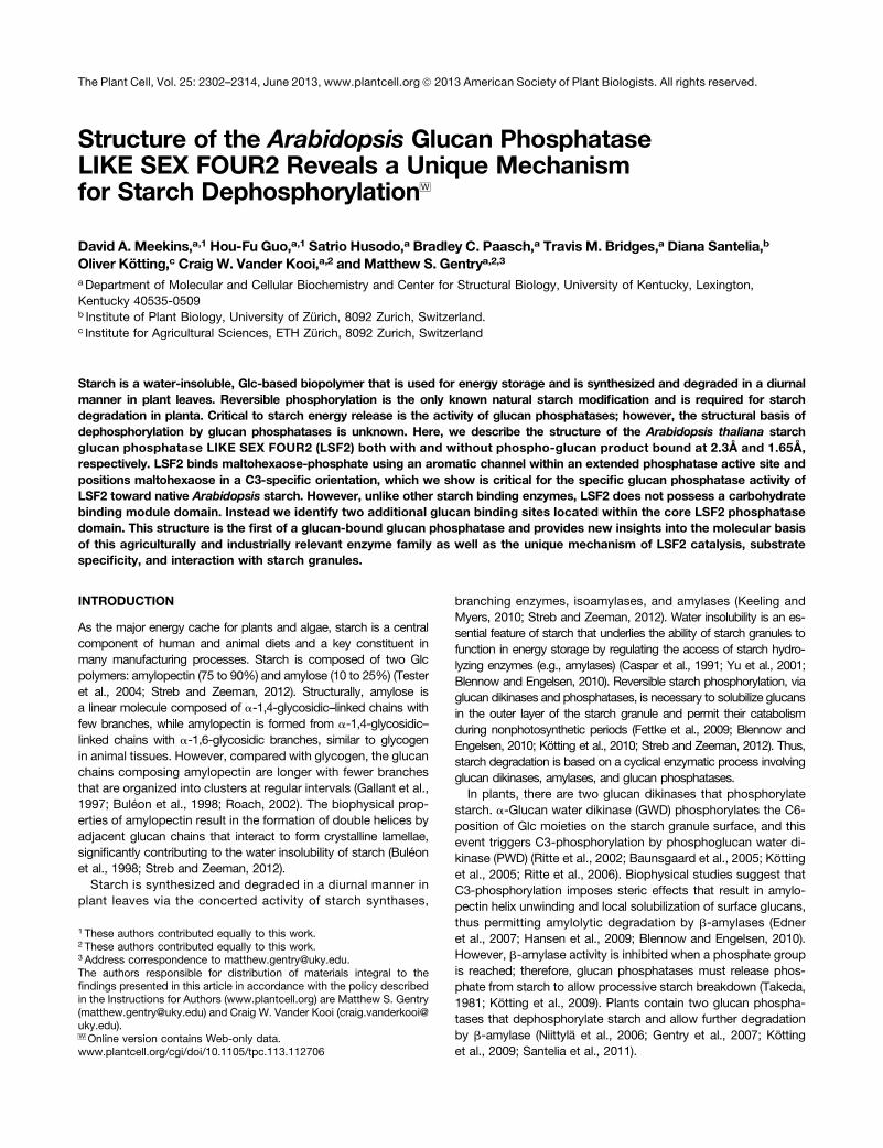
The structure of the Arabidopsis LSF2 glucan phosphatase (res-idues 79 to 282, C193S [catalytically inactive]) bound to malto-hexaose and phosphate was determined to a resolution of 2.30 Åusing molecular replacement with one molecule in the asymmetricunit (Figure 1A, Table 1). The LSF2 DSP domain (residues 79 to244) possesses a characteristic core PTP fold consisting of acentral five-stranded b-sheet region flanked by eight a-helices(see Supplemental Figure 1 online). The CT motif (residues 245 to282) consists of a loop region culminating in an a-helix that in-tegrally folds into the DSP domain, a characteristic also found inthe glucan phosphatase SEX4 (Vander Kooi et al., 2010). A searchfor structural homologs of the LSF2 DSP domain (residues 79 to244) identified the DSP domain of Arabidopsis SEX4 (residues 90to 250, root mean square deviation [RMSD] of 1.1 Å, PDB code3NME; Vander Kooi et al., 2010) and mouse PTPMT1 (residues105 to 256, RMSD of 2.2 Å, PDB code 3RGQ; Xiao et al., 2011) asthe structures most similar to LSF2 despite the fact that the LSF2DSP is only 48 and 17% identical at the amino acid level to theDSP domain of SEX4 and PTPMT1, respectively.Maltohexaose is composed of six Glc moieties with a-1,4-
glycosidic linkages; thus, it is similar to the unwound helices onthe starch granular surface. In the structure, maltohexaose isbound to the LSF2 active site and two distal sites. The LSF2active-site region contains a single maltohexaose chain andphosphate molecule within the catalytic pocket. Multiple con-served DSP active-site motifs converge to form an extendedactive-site binding pocket within LSF2 that is ;19Å long and;9Å deep with 511 Å2 contact area (Figure 1B). These motifsinclude a recognition domain from a1 through b1 (83 to 92),a variable (V-) loop from a3 through a4 (132 to 150), a WPD (D-)loop between b4 and a5 (158 to 163), a PTP-loop between b5and a6 (192 to 199) that contains the LSF2 catalytic signature(Cx5R) motif, and an R-motif between a7 and a8 (225 to 244)(see Supplemental Figures 1 and 2 online).
LSF2 possesses robust activity against starch and displays highspecificity for the C3 position, as measured by a 33P-radio-labeled starch dephosphorylation assay (Figure 1C) (Santeliaet al., 2011). While both glucan dikinases and glucan phos-phatases display strong positional specificity, the basis of thisspecificity is unclear. The electron density of the maltohexaosein the active site allows clear assignment of glucan chain posi-tion and orientation, labeled Glc1-Glc6 from the nonreducingend to the reducing (Figure 1D; see Supplemental Figures 3Aand 3B online). Strikingly, the O3 group of Glc3 directly interactswith the phosphate at the LSF2 catalytic site at a distance of2.4 Å compared with 7.0 Å for the O6 group. Furthermore, theorientation of the PTP catalytic triad (DX30-35CX5R) within LSF2is proximal to the O3 and phosphate and poised for catalysis ofa C3-phosphorylated Glc (Figure 1E). C193S is located at thebase of the active-site cleft, 2.5 Å from the nearest phosphateoxygen, and represents the key nucleophilic catalytic residuethat covalently attacks the phosphate group during catalysis.Arg-199 is positioned 2.8 Å from the phosphate and orients thephosphate of the substrate toward the catalytic Cys. At thetop of the active-site cleft is the D-loop Asp-161 that partic-ipates in catalysis as a general acid/base, forming the reaction
intermediate and then assisting in hydrolysis and product ex-pulsion. Asp-161 is located 2.5 Å from the O3 of Glc3 and 3.8 Åfrom the nearest phosphate oxygen.
Maltohexaose-Phosphate Product Bound at the LSF2Active Site
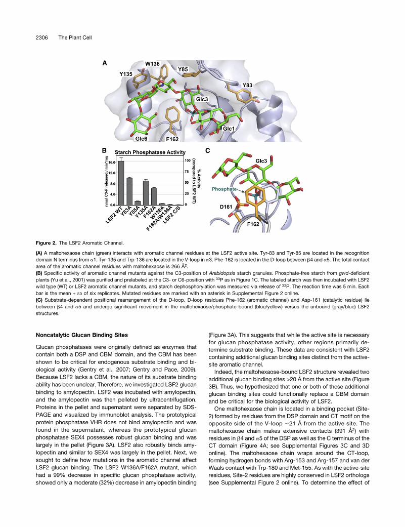
Five highly conserved aromatic residues delineate the bound-aries of the extended active-site channel, forming extensiveinteractions with the Glc rings of the maltohexaose chain(Figure 2A). These five aromatic residues provide the majorityof the interface between the LSF2 active site and the sub-strate. Tyr-83, Tyr-85, Tyr-135, and Trp-136 form one side ofthe channel and interact with Glc moieties Glc1-5. Tyr-83 andTyr-85 are located within the recognition domain, directlyadjacent to the R-motif and PTP-loop, respectively. Tyr-135and Trp-136 are both located in the V-loop and form a con-tinuous interaction surface with Tyr-85. Phe-162, located inthe D-loop, forms the opposite side of the aromatic channeland interacts with Glc2-6. These Glc moieties form a helicalstructure around Phe-162 and interact with both faces ofthe Phe ring. The residues that form the aromatic channelare strictly conserved in all land plants as well as in mostsingle-celled members of Kingdom Plantae, with only one
Figure 1. Structure of LSF2 Bound to Maltohexaose and Phosphate.
(A) Ribbon diagram of LSF2 (residues 79 to 282, C193S). Maltohexaose chains (green, cyan, orange, and pink) and phosphate (teal) are shown.Elements of secondary structure are numbered consecutively from N to C termini.(B)Maltohexaose chain (green) and phosphate (teal) at the active-site (red) channel. Image correlates with the red box in (A). Glc moieties are numberedfrom the nonreducing to the reducing end. The total contact area of the active-site channel with phosphate and maltohexaose is 511 Å2.(C) Specific activity of LSF2 wild type (WT) and inactive mutant LSF2 C193S (C/S) against the C6- and C3-position of Arabidopsis starch. Phosphate-free starch was purified from gwd-deficient Arabidopsis (Yu et al., 2001), and the starch was prelabeled with 33P at either the C6- or C3-position andthen incubated with recombinant protein. Starch dephosphorylation over time was linear and was measured via release of 33P. The reaction time was5 min. Each bar is the mean + SD of six replicates.(D) 2Fo-Fc electron density map of maltohexaose chain (green) and phosphate (teal) at the active site (1.35 s). The O3 group of Glc3 is highlighted.(E) LSF2 active-site catalytic triad (yellow) residues interact with maltohexaose and phosphate. S(C)193 (catalytically inactive mutant, C193S) and R199are located within the PTP-loop between b5 and a6, and Asp-161 is located within the D-loop between b4 and a5.
nonconservative substitution (Volvox carteri L83; see SupplementalFigure 2 online).
To investigate the functionality of these aromatic residues, wegenerated Ala mutants of each channel residue and tested theirability to dephosphorylate starch granules isolated from Arabi-dopsis. Single Ala point mutations of Tyr-83, Tyr-85, Tyr-135, Trp-136, and Phe-162 resulted in a decrease of C3-dephosphorylationby 38 to 96% (Figure 2B). Mutation of both sides of the channel(W136A/F162A) resulted in a 99% loss of glucan phosphatase
activity. Importantly, the observed decreases in specific glucanphosphatase activity were not due to destabilization of the activesite or misfolding of the protein as evidenced by near-wild-typephosphatase activity for all mutant proteins toward the genericsubstrate para-nitrophenyl phosphate (pNPP) (see SupplementalFigure 4A online). Thus, our findings indicate that LSF2 possessesan aromatic channel that forms an extended active site uniquelysuited to bind to polyglucan substrates and necessary for glucanphosphatase activity.
Conformational Changes in the LSF2 Active Site
Intriguingly, one of these key aromatic residues, Phe-162, isfound in the D-loop directly after Asp-161, which serves as thegeneral acid/base as discussed above. In fact, Phe-162 makesthe most contact of any active-site residue with the substrateglucan (136 Å2, 27% of the total contact area). To compare thecatalytic site of glucan-bound and unbound LSF2, we crystal-lized LSF2 without maltohexaose and determined the structureto a resolution of 1.65 Å using molecular replacement (seeSupplemental Figure 5 online; Table 1). The glucan-free LSF2produced a different crystal form and contained one molecule inthe asymmetric unit and bound citrate from the crystallizationbuffer (see Supplemental Figure 5 online). The RMSD of theglucan and citrate-bound DSP domain (residues 79 to 244)structures was 1.0 Å. A comparison of product-bound and citrate-bound LSF2 structures revealed a substrate-dependent re-arrangement of the D-loop architecture upon glucan binding(Figure 2C). In the product-bound structure, the orientations ofAsp-161 and Phe-162 are significantly different. The D-loop aro-matic residue Phe-162, important for the specific activity of LSF2,interacts with multiple Glc moieties of the glucan chain and shiftstoward the V-loop. This movement is associated with a significantreorientation of the critical general acid/base, residue Asp-161.Comparison of the two structures reveals that the terminal car-boxylate of Asp-161 is 3.1 Å closer to the catalytic Cys and di-rectly in contact with the O3 group of Glc3. To further analyze theposition of Asp-161, we compared the LSF2 DSP with structuresof the glucan phosphatase SEX4 and the prototypical proteinphosphatases VHR (PDB 1VHR; Yuvaniyama et al., 1996) andSSH-2 (PDB 2NT2; Jung et al., 2007). Analysis of the catalytictriad from each of these structures reveals that the substrate-bound orientation of LSF2 Asp-161 is in a catalytically competentorientation only in the product-bound structure (see SupplementalFigure 6 online). Thus, LSF2 undergoes a substrate-induced con-formational change with the LSF2 product-bound form ideally po-sitioned for catalysis of an O3 phosphorylated glucan substrate.Most DSPs possess a short-chain hydrophilic reside, S/T/N/
H, at the +1 residue from the general acid/base Asp (VanderKooi et al., 2010). However, Phe-162 is invariant in LSF2, andthe corresponding residue is also strictly conserved in SEX4,Phe-167. We previously demonstrated that mutating SEX4Phe-167 to a short-chain hydrophilic residue (F167S) resultedin a 50% decrease in the glucan/pNPP phosphatase activity ofSEX4 F167S (Vander Kooi et al., 2010). Cumulatively, these datademonstrate an important role for D-loop movement in orderto correctly position the catalytic triad and maximize glucanphosphatase activity.
Table 1. Crystallographic Statistics
Crystal
D78-LSF2 C193S:Maltohexaose,Phosphate
D78-LSF2 WildType: Citrate
PDB code 4KYR 4KYQData collectionSpace group P6522 P21212Cell dimensionsa, b, c (Å) 92,78, 92.78, 144.94 51.82, 99.58, 37.76a, b, g (°) 90, 90, 120 90, 90, 90Wavelength (Å) 1.00 1.00Resolution range
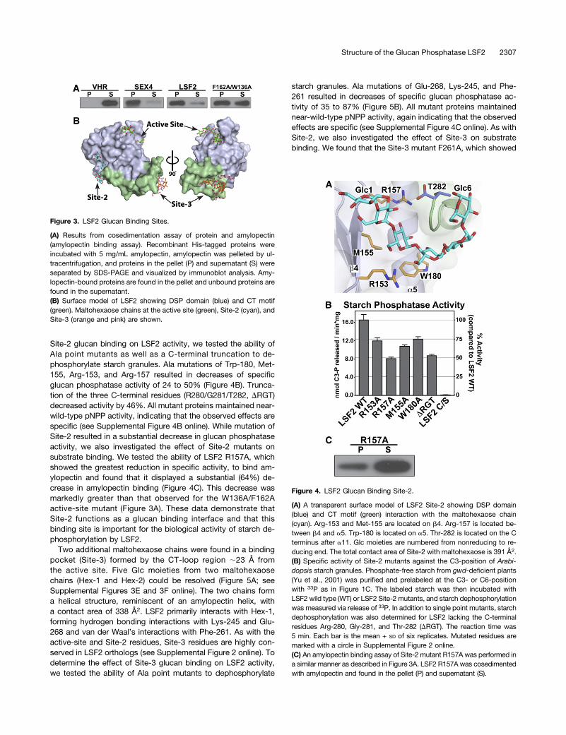
Glucan phosphatases were originally defined as enzymes thatcontain both a DSP and CBM domain, and the CBM has beenshown to be critical for endogenous substrate binding and bi-ological activity (Gentry et al., 2007; Gentry and Pace, 2009).Because LSF2 lacks a CBM, the nature of its substrate bindingability has been unclear. Therefore, we investigated LSF2 glucanbinding to amylopectin. LSF2 was incubated with amylopectin,and the amylopectin was then pelleted by ultracentrifugation.Proteins in the pellet and supernatant were separated by SDS-PAGE and visualized by immunoblot analysis. The prototypicalprotein phosphatase VHR does not bind amylopectin and wasfound in the supernatant, whereas the prototypical glucanphosphatase SEX4 possesses robust glucan binding and waslargely in the pellet (Figure 3A). LSF2 also robustly binds amy-lopectin and similar to SEX4 was largely in the pellet. Next, wesought to define how mutations in the aromatic channel affectLSF2 glucan binding. The LSF2 W136A/F162A mutant, whichhad a 99% decrease in specific glucan phosphatase activity,showed only a moderate (32%) decrease in amylopectin binding
(Figure 3A). This suggests that while the active site is necessaryfor glucan phosphatase activity, other regions primarily de-termine substrate binding. These data are consistent with LSF2containing additional glucan binding sites distinct from the active-site aromatic channel.Indeed, the maltohexaose-bound LSF2 structure revealed two
additional glucan binding sites >20 Å from the active site (Figure3B). Thus, we hypothesized that one or both of these additionalglucan binding sites could functionally replace a CBM domainand be critical for the biological activity of LSF2.One maltohexaose chain is located in a binding pocket (Site-
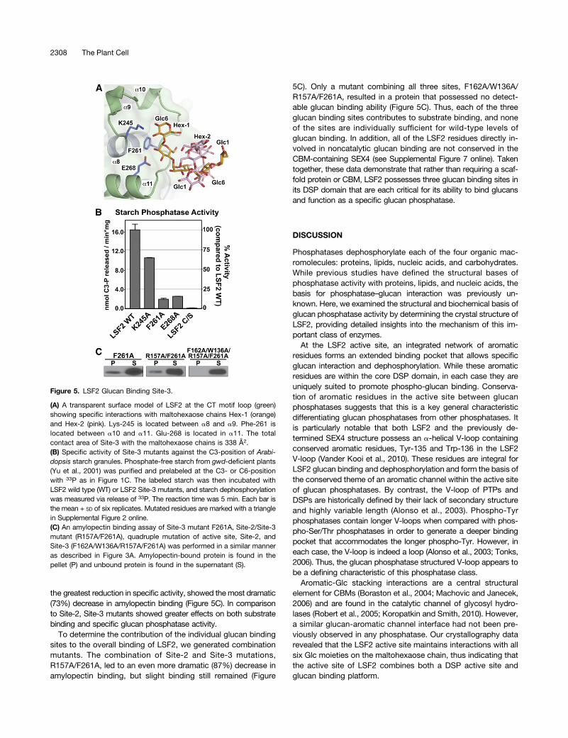
2) formed by residues from the DSP domain and CT motif on theopposite side of the V-loop ;21 Å from the active site. Themaltohexaose chain makes extensive contacts (391 Å2) withresidues in b4 and a5 of the DSP as well as the C terminus of theCT domain (Figure 4A; see Supplemental Figures 3C and 3Donline). The maltohexaose chain wraps around the CT-loop,forming hydrogen bonds with Arg-153 and Arg-157 and van derWaals contact with Trp-180 and Met-155. As with the active-siteresidues, Site-2 residues are highly conserved in LSF2 orthologs(see Supplemental Figure 2 online). To determine the effect of
Figure 2. The LSF2 Aromatic Channel.
(A) A maltohexaose chain (green) interacts with aromatic channel residues at the LSF2 active site. Tyr-83 and Tyr-85 are located in the recognitiondomain N terminus from a1. Tyr-135 and Trp-136 are located in the V-loop in a3. Phe-162 is located in the D-loop between b4 and a5. The total contactarea of the aromatic channel residues with maltohexaose is 266 Å2.(B) Specific activity of aromatic channel mutants against the C3-position of Arabidopsis starch granules. Phosphate-free starch from gwd-deficientplants (Yu et al., 2001) was purified and prelabeled at the C3- or C6-position with 33P as in Figure 1C. The labeled starch was then incubated with LSF2wild type (WT) or LSF2 aromatic channel mutants, and starch dephosphorylation was measured via release of 33P. The reaction time was 5 min. Eachbar is the mean + SD of six replicates. Mutated residues are marked with an asterisk in Supplemental Figure 2 online.(C) Substrate-dependent positional rearrangement of the D-loop. D-loop residues Phe-162 (aromatic channel) and Asp-161 (catalytic residue) liebetween b4 and a5 and undergo significant movement in the maltohexaose/phosphate bound (blue/yellow) versus the unbound (gray/blue) LSF2structures.
Site-2 glucan binding on LSF2 activity, we tested the ability ofAla point mutants as well as a C-terminal truncation to de-phosphorylate starch granules. Ala mutations of Trp-180, Met-155, Arg-153, and Arg-157 resulted in decreases of specificglucan phosphatase activity of 24 to 50% (Figure 4B). Trunca-tion of the three C-terminal residues (R280/G281/T282, DRGT)decreased activity by 46%. All mutant proteins maintained near-wild-type pNPP activity, indicating that the observed effects arespecific (see Supplemental Figure 4B online). While mutation ofSite-2 resulted in a substantial decrease in glucan phosphataseactivity, we also investigated the effect of Site-2 mutants onsubstrate binding. We tested the ability of LSF2 R157A, whichshowed the greatest reduction in specific activity, to bind am-ylopectin and found that it displayed a substantial (64%) de-crease in amylopectin binding (Figure 4C). This decrease wasmarkedly greater than that observed for the W136A/F162Aactive-site mutant (Figure 3A). These data demonstrate thatSite-2 functions as a glucan binding interface and that thisbinding site is important for the biological activity of starch de-phosphorylation by LSF2.
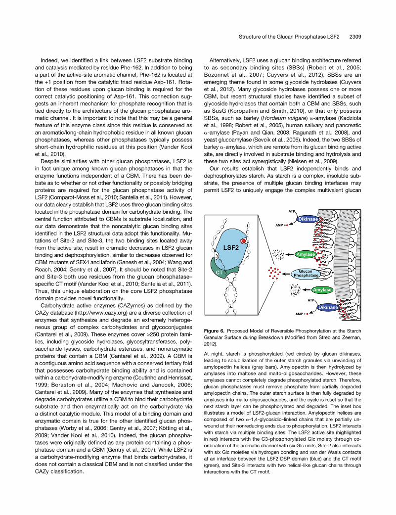
Two additional maltohexaose chains were found in a bindingpocket (Site-3) formed by the CT-loop region ;23 Å fromthe active site. Five Glc moieties from two maltohexaosechains (Hex-1 and Hex-2) could be resolved (Figure 5A; seeSupplemental Figures 3E and 3F online). The two chains forma helical structure, reminiscent of an amylopectin helix, witha contact area of 338 Å2. LSF2 primarily interacts with Hex-1,forming hydrogen bonding interactions with Lys-245 and Glu-268 and van der Waal’s interactions with Phe-261. As with theactive-site and Site-2 residues, Site-3 residues are highly con-served in LSF2 orthologs (see Supplemental Figure 2 online). Todetermine the effect of Site-3 glucan binding on LSF2 activity,we tested the ability of Ala point mutants to dephosphorylate
starch granules. Ala mutations of Glu-268, Lys-245, and Phe-261 resulted in decreases of specific glucan phosphatase ac-tivity of 35 to 87% (Figure 5B). All mutant proteins maintainednear-wild-type pNPP activity, again indicating that the observedeffects are specific (see Supplemental Figure 4C online). As withSite-2, we also investigated the effect of Site-3 on substratebinding. We found that the Site-3 mutant F261A, which showed
Figure 3. LSF2 Glucan Binding Sites.
(A) Results from cosedimentation assay of protein and amylopectin(amylopectin binding assay). Recombinant His-tagged proteins wereincubated with 5 mg/mL amylopectin, amylopectin was pelleted by ul-tracentrifugation, and proteins in the pellet (P) and supernatant (S) wereseparated by SDS-PAGE and visualized by immunoblot analysis. Amy-lopectin-bound proteins are found in the pellet and unbound proteins arefound in the supernatant.(B) Surface model of LSF2 showing DSP domain (blue) and CT motif(green). Maltohexaose chains at the active site (green), Site-2 (cyan), andSite-3 (orange and pink) are shown.
Figure 4. LSF2 Glucan Binding Site-2.
(A) A transparent surface model of LSF2 Site-2 showing DSP domain(blue) and CT motif (green) interaction with the maltohexaose chain(cyan). Arg-153 and Met-155 are located on b4. Arg-157 is located be-tween b4 and a5. Trp-180 is located on a5. Thr-282 is located on the Cterminus after a11. Glc moieties are numbered from nonreducing to re-ducing end. The total contact area of Site-2 with maltohexaose is 391 Å2.(B) Specific activity of Site-2 mutants against the C3-position of Arabi-dopsis starch granules. Phosphate-free starch from gwd-deficient plants(Yu et al., 2001) was purified and prelabeled at the C3- or C6-positionwith 33P as in Figure 1C. The labeled starch was then incubated withLSF2 wild type (WT) or LSF2 Site-2 mutants, and starch dephosphorylationwas measured via release of 33P. In addition to single point mutants, starchdephosphorylation was also determined for LSF2 lacking the C-terminalresidues Arg-280, Gly-281, and Thr-282 (DRGT). The reaction time was5 min. Each bar is the mean + SD of six replicates. Mutated residues aremarked with a circle in Supplemental Figure 2 online.(C) An amylopectin binding assay of Site-2 mutant R157A was performed ina similar manner as described in Figure 3A. LSF2 R157A was cosedimentedwith amylopectin and found in the pellet (P) and supernatant (S).
the greatest reduction in specific activity, showed themost dramatic(73%) decrease in amylopectin binding (Figure 5C). In comparisonto Site-2, Site-3 mutants showed greater effects on both substratebinding and specific glucan phosphatase activity.
To determine the contribution of the individual glucan bindingsites to the overall binding of LSF2, we generated combinationmutants. The combination of Site-2 and Site-3 mutations,R157A/F261A, led to an even more dramatic (87%) decrease inamylopectin binding, but slight binding still remained (Figure
5C). Only a mutant combining all three sites, F162A/W136A/R157A/F261A, resulted in a protein that possessed no detect-able glucan binding ability (Figure 5C). Thus, each of the threeglucan binding sites contributes to substrate binding, and noneof the sites are individually sufficient for wild-type levels ofglucan binding. In addition, all of the LSF2 residues directly in-volved in noncatalytic glucan binding are not conserved in theCBM-containing SEX4 (see Supplemental Figure 7 online). Takentogether, these data demonstrate that rather than requiring a scaf-fold protein or CBM, LSF2 possesses three glucan binding sites inits DSP domain that are each critical for its ability to bind glucansand function as a specific glucan phosphatase.
DISCUSSION
Phosphatases dephosphorylate each of the four organic mac-romolecules: proteins, lipids, nucleic acids, and carbohydrates.While previous studies have defined the structural bases ofphosphatase activity with proteins, lipids, and nucleic acids, thebasis for phosphatase–glucan interaction was previously un-known. Here, we examined the structural and biochemical basis ofglucan phosphatase activity by determining the crystal structure ofLSF2, providing detailed insights into the mechanism of this im-portant class of enzymes.At the LSF2 active site, an integrated network of aromatic
residues forms an extended binding pocket that allows specificglucan interaction and dephosphorylation. While these aromaticresidues are within the core DSP domain, in each case they areuniquely suited to promote phospho-glucan binding. Conserva-tion of aromatic residues in the active site between glucanphosphatases suggests that this is a key general characteristicdifferentiating glucan phosphatases from other phosphatases. Itis particularly notable that both LSF2 and the previously de-termined SEX4 structure possess an a-helical V-loop containingconserved aromatic residues, Tyr-135 and Trp-136 in the LSF2V-loop (Vander Kooi et al., 2010). These residues are integral forLSF2 glucan binding and dephosphorylation and form the basis ofthe conserved theme of an aromatic channel within the active siteof glucan phosphatases. By contrast, the V-loop of PTPs andDSPs are historically defined by their lack of secondary structureand highly variable length (Alonso et al., 2003). Phospho-Tyrphosphatases contain longer V-loops when compared with phos-pho-Ser/Thr phosphatases in order to generate a deeper bindingpocket that accommodates the longer phospho-Tyr. However, ineach case, the V-loop is indeed a loop (Alonso et al., 2003; Tonks,2006). Thus, the glucan phosphatase structured V-loop appears tobe a defining characteristic of this phosphatase class.Aromatic-Glc stacking interactions are a central structural
element for CBMs (Boraston et al., 2004; Machovic and Janecek,2006) and are found in the catalytic channel of glycosyl hydro-lases (Robert et al., 2005; Koropatkin and Smith, 2010). However,a similar glucan-aromatic channel interface had not been pre-viously observed in any phosphatase. Our crystallography datarevealed that the LSF2 active site maintains interactions with allsix Glc moieties on the maltohexaose chain, thus indicating thatthe active site of LSF2 combines both a DSP active site andglucan binding platform.
Figure 5. LSF2 Glucan Binding Site-3.
(A) A transparent surface model of LSF2 at the CT motif loop (green)showing specific interactions with maltohexaose chains Hex-1 (orange)and Hex-2 (pink). Lys-245 is located between a8 and a9. Phe-261 islocated between a10 and a11. Glu-268 is located in a11. The totalcontact area of Site-3 with the maltohexaose chains is 338 Å2.(B) Specific activity of Site-3 mutants against the C3-position of Arabi-dopsis starch granules. Phosphate-free starch from gwd-deficient plants(Yu et al., 2001) was purified and prelabeled at the C3- or C6-positionwith 33P as in Figure 1C. The labeled starch was then incubated withLSF2 wild type (WT) or LSF2 Site-3 mutants, and starch dephosphorylationwas measured via release of 33P. The reaction time was 5 min. Each bar isthe mean + SD of six replicates. Mutated residues are marked with a trianglein Supplemental Figure 2 online.(C) An amylopectin binding assay of Site-3 mutant F261A, Site-2/Site-3mutant (R157A/F261A), quadruple mutation of active site, Site-2, andSite-3 (F162A/W136A/R157A/F261A) was performed in a similar manneras described in Figure 3A. Amylopectin-bound protein is found in thepellet (P) and unbound protein is found in the supernatant (S).
Indeed, we identified a link between LSF2 substrate bindingand catalysis mediated by residue Phe-162. In addition to beinga part of the active-site aromatic channel, Phe-162 is located atthe +1 position from the catalytic triad residue Asp-161. Rota-tion of these residues upon glucan binding is required for thecorrect catalytic positioning of Asp-161. This connection sug-gests an inherent mechanism for phosphate recognition that istied directly to the architecture of the glucan phosphatase aro-matic channel. It is important to note that this may be a generalfeature of this enzyme class since this residue is conserved asan aromatic/long-chain hydrophobic residue in all known glucanphosphatases, whereas other phosphatases typically possessshort-chain hydrophilic residues at this position (Vander Kooiet al., 2010).
Despite similarities with other glucan phosphatases, LSF2 isin fact unique among known glucan phosphatases in that theenzyme functions independent of a CBM. There has been de-bate as to whether or not other functionality or possibly bridgingproteins are required for the glucan phosphatase activity ofLSF2 (Comparot-Moss et al., 2010; Santelia et al., 2011). However,our data clearly establish that LSF2 uses three glucan binding siteslocated in the phosphatase domain for carbohydrate binding. Thecentral function attributed to CBMs is substrate localization, andour data demonstrate that the noncatalytic glucan binding sitesidentified in the LSF2 structural data adopt this functionality. Mu-tations of Site-2 and Site-3, the two binding sites located awayfrom the active site, result in dramatic decreases in LSF2 glucanbinding and dephosphorylation, similar to decreases observed forCBM mutants of SEX4 and laforin (Ganesh et al., 2004; Wang andRoach, 2004; Gentry et al., 2007). It should be noted that Site-2and Site-3 both use residues from the glucan phosphatase–specific CT motif (Vander Kooi et al., 2010; Santelia et al., 2011).Thus, this unique elaboration on the core LSF2 phosphatasedomain provides novel functionality.
Carbohydrate active enzymes (CAZymes) as defined by theCAZy database (http://www.cazy.org) are a diverse collection ofenzymes that synthesize and degrade an extremely heteroge-neous group of complex carbohydrates and glycoconjugates(Cantarel et al., 2009). These enzymes cover >250 protein fami-lies, including glycoside hydrolases, glycosyltransferases, poly-saccharide lyases, carbohydrate esterases, and nonenzymaticproteins that contain a CBM (Cantarel et al., 2009). A CBM isa contiguous amino acid sequence with a conserved tertiary foldthat possesses carbohydrate binding ability and is containedwithin a carbohydrate-modifying enzyme (Coutinho and Henrissat,1999; Boraston et al., 2004; Machovic and Janecek, 2006;Cantarel et al., 2009). Many of the enzymes that synthesize anddegrade carbohydrates utilize a CBM to bind their carbohydratesubstrate and then enzymatically act on the carbohydrate viaa distinct catalytic module. This model of a binding domain andenzymatic domain is true for the other identified glucan phos-phatases (Worby et al., 2006; Gentry et al., 2007; Kötting et al.,2009; Vander Kooi et al., 2010). Indeed, the glucan phospha-tases were originally defined as any protein containing a phos-phatase domain and a CBM (Gentry et al., 2007). While LSF2 isa carbohydrate-modifying enzyme that binds carbohydrates, itdoes not contain a classical CBM and is not classified under theCAZy classification.
Alternatively, LSF2 uses a glucan binding architecture referredto as secondary binding sites (SBSs) (Robert et al., 2005;Bozonnet et al., 2007; Cuyvers et al., 2012). SBSs are anemerging theme found in some glycoside hydrolases (Cuyverset al., 2012). Many glycoside hydrolases possess one or moreCBM, but recent structural studies have identified a subset ofglycoside hydrolases that contain both a CBM and SBSs, suchas SusG (Koropatkin and Smith, 2010), or that only possessSBSs, such as barley (Hordeum vulgare) a-amylase (Kadziolaet al., 1998; Robert et al., 2005), human salivary and pancreatica-amylase (Payan and Qian, 2003; Ragunath et al., 2008), andyeast glucoamylase (Sevcík et al., 2006). Indeed, the two SBSs ofbarley a-amylase, which are remote from its glucan binding activesite, are directly involved in substrate binding and hydrolysis andthese two sites act synergistically (Nielsen et al., 2009).Our results establish that LSF2 independently binds and
dephosphorylates starch. As starch is a complex, insoluble sub-strate, the presence of multiple glucan binding interfaces maypermit LSF2 to uniquely engage the complex multivalent glucan
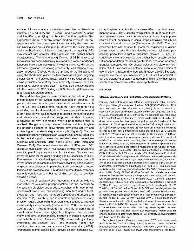
Figure 6. Proposed Model of Reversible Phosphorylation at the StarchGranular Surface during Breakdown (Modified from Streb and Zeeman,2012).
At night, starch is phosphorylated (red circles) by glucan dikinases,leading to solubilization of the outer starch granules via unwinding ofamylopectin helices (gray bars). Amylopectin is then hydrolyzed byamylases into maltose and malto-oligosaccharides. However, theseamylases cannot completely degrade phosphorylated starch. Therefore,glucan phosphatases must remove phosphate from partially degradedamylopectin chains. The outer starch surface is then fully degraded byamylases into malto-oligosaccharides, and the cycle is reset so that thenext starch layer can be phosphorylated and degraded. The inset boxillustrates a model of LSF2-glucan interaction. Amylopectin helices arecomposed of two a-1,4-glycosidic–linked chains that are partially un-wound at their nonreducing ends due to phosphorylation. LSF2 interactswith starch via multiple binding sites: The LSF2 active site (highlightedin red) interacts with the C3-phosphorylated Glc moiety through co-ordination of the aromatic channel with six Glc units, Site-2 also interactswith six Glc moieties via hydrogen bonding and van der Waals contactsat an interface between the LSF2 DSP domain (blue) and the CT motif(green), and Site-3 interacts with two helical-like glucan chains throughinteractions with the CT motif.
surface of its endogenous substrate. Indeed, the combined-sitemutants (R157A/F261A and F162A/W136A/R157A/F261A) showadditive effects, implying that the sites function together. Thissuggests a model whereby starch binding involves the en-gagement of longer or multiple glucan chains by the three glu-can binding sites on LSF2 (Figure 6). Moreover, the helical glucanchains at Site-3 are reminiscent of amylopectin, suggesting LSF2may interact with complex starch granules with distinct helicalcharacteristics. The functional significance of SBSs in glycosylhydrolases has been extensively reviewed and various additionalfunctions have been postulated, including substrate disruption,allosteric regulation, enhancing processivity, and relaying of re-action products (Cuyvers et al., 2012). Due to the limitations ofusing the short-chain glucan maltohexaose as a ligand, ongoingstudies using more diverse glucan chains will be required to ex-amine possible cooperativity or connectivity between the addi-tional LSF2 glucan binding sites. This may also provide insightsinto the position of LSF2 binding and C3-phosphorylation relativeto amylopectin branch points.
These data also give a clearer picture of the role of glucanphosphatases in the cyclical starch degradation process. Theglucan dikinases phosphorylate the outer Glc moieties of starchat the C6- and C3-positions, resulting in amylopectin helixunwinding and local solubilization. These events allow a- andb-amylases as well as isoamylases to access the outer glucansand release maltose and malto-oligosaccharides. However,b-amylase activity is inhibited when a phosphate group isreached. The glucan phosphatases bind and dephosphorylatethe phospho-glucans to allow further amylolytic activity anda resetting of the starch degradation cycle (Figure 6). The co-ordinated phosphorylation of starch Glc at the C6- and C3-positionsis the central signaling event orchestrating starch breakdown(Blennow and Engelsen, 2010; Kötting et al., 2010; Streb andZeeman, 2012). The recent characterization of SEX4 and LSF2illustrates that plants use a two-enzyme system for phosphateremoval, permitting complete starch catabolism. Our structuresreveal the basis for the glucan binding and C3-specificity of LSF2.Determination of additional glucan phosphatase structures willreveal further insights into the mechanism of activity and specificityof glucan phosphatases. In particular, it will be interesting to de-termine if the presence of a CBM domain in other family membersnot only contributes to substrate binding but also to position-specific function.
As the central regulatory event governing starch breakdown,modulation of reversible phosphorylation has the potential toincrease starch yields and produce starches with novel physi-cochemical properties, thus enhancing manufacturing of feed-stock for both food and nonfood applications (Santelia andZeeman, 2011). Starch is used in countless applications, manyof which require chemical and physical modifications to improveand diversify its functionality (Blennow et al., 2002; Santelia andZeeman, 2011). Phosphorylation is the only known naturalmodification of starch, and highly phosphorylated starches havemany attractive characteristics, including increased hydrationstatus (Muhrbeck and Eliasson, 1991), decreased crystallinity(Muhrbeck and Eliasson, 1991), and improved freeze-thawstability, viscosity, and transparency (Blennow et al., 2001).Arabidopsis plants lacking LSF2 activity display increased C3-
phosphorylated starch without adverse effects on plant growth(Santelia et al., 2011). Genetic manipulation of LSF2 could there-fore represent a new means to produce starch with higher phos-phate content, particularly in cereal crops containing virtually nocovalently bound phosphate (Blennow et al., 2000). The datapresented here can be used to inform the engineering of glucanphosphatases to alter their functionality for industrial starch pro-cessing, particularly in light of disparities between C6- and C3-contributions to starch superstructure. It has been established thatC3-phosphorylation results in greater local hydration of starchgranules compared with C6-phosphorylation; therefore, manipu-lation of LSF2 represents a potential avenue for manipulation ofoverall starch structure (Blennow and Engelsen, 2010). Structuralinsights into the unique mechanism of LSF2 are fundamental toour understanding of starch catabolism and ultimately harnessingstarch as a biomolecule with diverse applications.
METHODS
Cloning, Expression, and Purification of Recombinant Proteins
Primers used in this work are listed in Supplemental Table 1 online.Cloning of full-length Arabidopsis thaliana LSF2 (AT3G10940) from cDNAwas previously described (Santelia et al., 2011). Based on data fromsecondary structure predictions, disorder predictions, sequence similarityto SEX4, and analysis of LSF2 orthologs, we generated an ArabidopsisLSF2 construct lacking the first 78 amino acids (D78-LSF2). D78 LSF2does not contain the chloroplast targeting peptide (predicted to beresidues 1 to 65) along with residues up to the DSP recognition domain.D78-LSF2was subcloned into pET28 (Novagen) usingNdeI and XhoI sitesto encode a His6 tag, a thrombin cleavage site, and Δ78-LSF2 (Santeliaet al., 2011).We generated an active-site Cys-to-Ser mutation (C193S), anestablished technique that generates a catalytically inactive constructused to trap the substrate and as a negative control in enzymatic assays(Zhou et al., 1994; Jia et al., 1995; Begley et al., 2006). All point mutantswere generated using a site-directed mutagenesis kit (Agilent) or muta-genesis services (GenScript). Cloning and purification of ArabidopsisSEX4 lacking the first 89 amino acids (D89-SEX4) (Vander Kooi et al.,2010) and Hs-VHR (Yuvaniyama et al., 1996) was performed as previouslydescribed. All DNA sequencing (ACGT) was confirmed using MacVector.Amino acid sequences of LSF2 orthologs were aligned with ClustalW inMacVector. Expression and purification of D78-LSF2 was performedsimilarly to the previously described method for D89-SEX4 (Vander Kooiet al., 2010). Briefly, BL21-CodonPlus Escherichia coli cells were trans-formed with expression vectors for the production of native LSF2 protein.Cells were grown at 37°C in 23 YT media to OD600 = 0.6 to 0.8, placed onice for 20 min, induced with 1 mM isopropyl b-D-thiogalactoside, grown at16°C for 16 h, and harvested by centrifugation. Cells were lysed in 20 mMTris-HCl, pH 7.5, 100 mM NaCl, and 2mM DTT and centrifuged, and theproteins were purified via a Profinia IMAC Ni2+ column (Bio-Rad) witha Profinia protein purification system (Bio-Rad). Protein was dialyzed in20 mM Tris-HCl, pH 7.5, 100 mM NaCl, and 2mM DTT overnight at 4°C inthe presence of thrombin. Affinity purified protein was then reverse purifiedover the Profinia IMAC Ni2+ column, and the flow-through fraction wascollected. Protein was further purified to homogeneity using a HiLoad 26/60Superdex 200 size exclusion column (GE Healthcare). Protein used forenzyme and binding assays was stored in 10%glycerol as a cryoprotectantand flash frozen for later use.
Recombinant potato (Solanum tuberosum) GWD and recombinantArabidopsis PWD for 33P labeling of Arabidopsis starch were purified aspreviously described with the following modifications (Ritte et al., 2002;Kötting et al., 2005). GWD and PWD were transformed into BL21-
CodonPlus E. coli cells and expressed similarly to D78-LSF2 as statedabove. GWD was lysed in buffer (50 mM Tris/HCl, pH 7.5, 2.5 mM EDTA,2.5 mM DTT, and 0.5 mM PMSF), and proteins were purified using ananion exchange column (Q-sepharose-FF; GE-Healthcare) with a saltgradient (50 mM Tris/HCl, pH 7.5, 2.5 mM EDTA, 2.5 mM DTT, 0.5 mMPMSF, and 0.5 NaCl) to elute the protein. Fractions were collected andprotein was further purified using a HiLoad 26/60 Superdex 200 sizeexclusion column (GE Healthcare) in new buffer (100 mMMOPS/KOH, pH7.6, 1 mMEDTA, 2mMDTT, 0.5mMPMSF, and 150mMNaCl). GWDwasthen put over a desalting column using a Bio-ScaleMini Bio-Gel P6 desaltingcolumn (Bio-Rad) usingaProfinia proteinpurification system (Bio-Rad). PWDwas lysed in buffer (50 mM HEPES/NaOH, pH 8.0, 300 mM NaCl, 10 mMimidazole, and 0.5 mM PMSF) and centrifuged, and proteins were purifiedusing a Profinia IMAC Ni2+ column (Bio-Rad) with a Profinia protein purifi-cation system (Bio-Rad). PWD was further purified using a HiLoad 26/60Superdex 200 size exclusion column (GE Healthcare).
Crystal Structure Determination and Refinement
For glucan-bound crystals, single, high-quality crystals with one moleculein the asymmetric unit were obtained via hanging drop vapor diffusionusing a Mosquito liquid handling robot (TTPLabtech) using a 200-nL dropwith a 1:3 ratio of D78-LSF2 C193S (4.8 mg/mL) preincubated with 25mMmaltohexaose (Sigma-Aldrich): 0.1 M diammonium hydrogen phosphate,pH 5.7, 17% 2-propoanol, and 31% polyethylene glycol 4000 at 18°C.Single, high-quality D78-LSF2 wild-type crystals, with one molecule in theasymmetric unit, were obtained with a 200-nL drop using a 1:1 ratio ofLSF2 (4.8 mg/mL): 0.1 M trisodium citrate, pH 5.8, 16% 2-propanol, 31%polyethylene glycol 4000, and 2% glycerol at 18°C. A single crystal wasused for data collection and structural determination for both LSF2structures. Both D78-LSF2 C193S (phosphate/maltohexaose) and D78-LSF2wild-type (citrate) datawere collected on the 22-ID beamline of SER-CAT at the Advanced Photon Source, Argonne National Laboratory (Table1) at 110K at a wavelength of 1.0 Å. Data were processed using HKL2000(Otwinowski and Minor, 1997). PHENIX (Adams et al., 2010) was used formolecular replacement using the SEX4 DSP and a10 helix of the SEX4 CTdomain as search models (Vander Kooi et al., 2010). The structures werethen fully built and refined via iterative model building and refinementusing Coot (Emsley et al., 2010) and Refmac5 (Murshudov et al., 1997),respectively. Stereochemistry of the model was analyzed using Mol-Probity (Davis et al., 2007). Analysis and molecular graphics were pre-pared using Pymol (Schrodinger, 2010). Density maps were producedusing the FTT program in CCP4 (Winn et al., 2011). Comparative structureanalyses were performed using the DaliServer (Holm and Rosenström,2010) and DaliLite (Hasegawa and Holm, 2009). Protein-ligand contactanalyses were performed with Areaimol (Lee and Richards, 1971).
Phosphatase Assays
Phosphatase assays using pNPP have been previously described andwere performed with the following modifications (Worby et al., 2006;Gentry et al., 2007; Sherwood et al., 2013). Hydrolysis of pNPP wasperformed in 50 mL reactions, containing 13 phosphatase buffer (0.1 Msodium acetate, 0.05 M bis-Tris, 0.05 M Tris-HCl, pH 7.0, and 2 mMDTT),50 mM pNPP, and 1 µg of enzyme at 37°C for 15 min. The reaction wasterminated by the addition of 200 mL of 0.25 NaOH, and absorbance wasmeasured at 410 nm. The assay was performed with each protein sixtimes or more to determine specific activity.
Phosphate release from 33P-Labeled granules was performed aspreviously described with the following variations (Hejazi et al., 2010;Santelia et al., 2011). C6-33P-labeled starch was generated by isolatingphosphate-free starch granules from the Arabidopsis sex1-3 mutant (Yuet al., 2001), phosphorylating the starch with 33P at the C6-position byGWD followed by washing until all unincorporated 33P had been removed.
Phosphorylation with unlabeled ATP at the C3-position by PWD wasperformed as previously described (Hejazi et al., 2010). C3-33P-labeledstarch was generated by isolating phosphate-free starch granules fromthe Arabidopsis sex1-3 mutant (Yu et al., 2001), phosphorylating thestarch with unlabeled ATP at the C6-position by GWD followed byphosphorylation with 33P at the C3-position by PWD and washing until allunincorporated 33P had been removed, as previously described (Hejaziet al., 2010). In both cases, the starch granules were phosphorylated atboth positions; however, the 33P-label was located at only one or the otherposition. [b-33P]ATP was obtained from Hartmann Analytic. Recombinantproteins (150 ng) were incubated in dephosphorylation buffer (100 mMsodium acetate, 50mMbis-Tris, 50mM Tris-HCl, pH 6.5, 0.05% [v/v] TritonX-100, 1 µg/mL [w/v] BSA, and 2mMDTT) with C6- or C3-prelabeled starch(4 mg/mL) in a final volume of 150 mL on a rotating wheel for 5 min at 25°C.The reaction was terminated by the addition of 50 mL of 10% SDS. Thereaction tubes were then centrifuged at 13,000 rpm for 5 min to pellet thestarch. 33P release into 150mL of supernatant was determined using a 1900TR liquid scintillation counter (Packard). The assay was performed with eachprotein six times or more to determine specific activity.
Glucan Binding Assay
Glucan binding assays were performed as previously described with thefollowing modifications (Gentry et al., 2007; Dukhande et al., 2011). Allproteins used in the glucan binding assays were purified as described abovewithout cleavage of the His6 tag to maintain the epitope for immunoblotanalysis. Amylopectin, from potato starch (Sigma-Aldrich) was solubilized bythe Roach method (Wang and Roach, 2004) at a concentration of 5 mg/mL.Five milligrams of amylopectin was then prepelleted via centrifugation at110,000g for 1.5 h at 4°C to collect only pelletable amylopectin and thenresuspended in 0.5mLbindingbuffer (50mMTris, 150mMNaCl, pH7.5, and2 mM DTT). One microgram of recombinant protein was incubated in am-ylopectin solution for an hour at 4°C with rocking. The solution was thencentrifuged at 110,000g for 1.5 h. Cosedimentation with amylopectin wasmeasured by centrifuging the samples at 110,000g for 1.5 h. All supernatantwas removed and protein was precipitated with 4 volumes of acetone storedat -20°C. Precipitated protein was then pelleted via centrifugation at 21,130gfor 30 min, and excess acetone was removed using a SpeedVac concen-trator (Savant) at 65°C for 1.25 h. Both soluble and pellet fractions were thenresuspended in 30 mL RIPA buffer before the addition of 30 mL SDS-PAGEbuffer (60 mL total volume for both the soluble and pellet fractions). Fifteenmicroliters of the pellet and soluble fraction were then resolved via SDS-PAGE, and relative concentration of pellet and soluble protein was analyzedby immunoblotting with a-His6 antibody. Quantification of the signal wasdetermined using ImageJ (Abramoff et al., 2004). The assay was performedthree times or more with each protein to determine binding capacity.
Accession Numbers
Sequence data from this article can be found in the Arabidopsis GenomeInitiative or GenBank/EMBL databases under the following accessionnumbers: LSF2, At3g10940; SEX4, At3g52180; GWD, At1g10760; PWD,At5g26570; andVHR,Hs03174. The atomic coordinates and structure factorshave been deposited in the Protein Data Bank (PDB code 4KYR and 4KYQ,for maltohexaose/phosphate and citrate bound structures, respectively).
Supplemental Data
The following materials are available in the online version of this article.
Supplemental Figure 1. DSP Subdomains in LSF2.
Supplemental Figure 2. Conservation of LSF2 Orthologs.
Supplemental Figure 3. Electron Density Maps of Ligands in LSF2Structure.
Supplemental Figure 4. Specific Activity of LSF2 and Mutants againstpara-Nitrophenyl Phosphate.
Supplemental Figure 5. Stereo View of Citrate at the LSF2 ActiveSite.
Supplemental Figure 6. Structural Alignment of the DSP CatalyticTriad of LSF2.
Supplemental Figure 7. Sequence Conservation of LSF2 and SEX4.
Supplemental Table 1. Sequences of Primers Used in This Work.
ACKNOWLEDGMENTS
We thank Samuel Zeeman and members of the Gentry and Vander Kooilabs for fruitful discussions as well as Carol Beach and Martin Chow inthe Molecular Basis of Human Disease COBRE Proteomics Core andProtein Analytical Core, respectively, for technical assistance. This studywas supported in part by an National Science Foundation CAREER GrantMCB-1252345 (M.S.G.), National Institutes of Health Grants R01NS070899(M.S.G.) and P20GM103486 (M.S.G. and C.W.V.K.), Kentucky Science andEnergy Foundation Grant KSEF-2268-RDE-014 (M.S.G.), a University ofKentucky Summer Research and Creativity Fellowship (T.M.B.), Universityof Kentucky College of Medicine startup funds (M.S.G.), the ETH-Zürich(O.K.), and Swiss-South African Joint Research Program Grant IZ LSX3122916 (D.S. and O.K.). The contents are solely the responsibility of theauthors and do not necessarily represent the official views of the NationalScience Foundation, National Institutes of Health, or other fundingagencies.
AUTHOR CONTRIBUTIONS
M.S.G., C.W.V.K., and D.A.M. designed the research. All authors performedresearch. C.W.V.K., D.A.M., H-.F.G., M.S.G., and S.H. analyzed data.D.A.M., C.W.V.K., and M.S.G. wrote the article, and D.A.M. generated thefigures C.W.V.K. and M.S.G. contributed equally to the publication of thisarticle.
Received April 16, 2013; revised May 31, 2013; accepted June 12, 2013;published July 5, 2013.
REFERENCES
Abramoff, M.D., Magalhaes, P.J., and Ram, S.J. (2004). Imageprocessing with ImageJ. Biophotonics International 11: 36–42.
Adams, P.D., et al. (2010). PHENIX: A comprehensive Python-basedsystem for macromolecular structure solution. Acta Crystallogr. D Biol.Crystallogr. 66: 213–221.
Alonso, A., Rojas, A., Godzik, A., and Mustelin, T. (2003). The Dual-Specific Protein Tyrosine Phosphatase Family. (Berlin: Springer).
Baunsgaard, L., Lütken, H., Mikkelsen, R., Glaring, M.A., Pham,T.T., and Blennow, A. (2005). A novel isoform of glucan, waterdikinase phosphorylates pre-phosphorylated alpha-glucans and isinvolved in starch degradation in Arabidopsis. Plant J. 41: 595–605.
Begley, M.J., Taylor, G.S., Brock, M.A., Ghosh, P., Woods, V.L., andDixon, J.E. (2006). Molecular basis for substrate recognition byMTMR2, a myotubularin family phosphoinositide phosphatase. Proc.Natl. Acad. Sci. USA 103: 927–932.
Blennow, A., Bay-Smidt, A.M., Olsen, C.E., and Møller, B.L. (2000).The distribution of covalently bound phosphate in the starch
granule in relation to starch crystallinity. Int. J. Biol. Macromol. 27:211–218.
Blennow, A., and Engelsen, S.B. (2010). Helix-breaking news:Fighting crystalline starch energy deposits in the cell. Trends PlantSci. 15: 236–240.
Blennow, A., Mette Bay-Smidt, A., and Bauer, R. (2001).Amylopectin aggregation as a function of starch phosphate contentstudied by size exclusion chromatography and on-line refractiveindex and light scattering. Int. J. Biol. Macromol. 28: 409–420.
Blennow, A., Nielsen, T.H., Baunsgaard, L., Mikkelsen, R., andEngelsen, S.B. (2002). Starch phosphorylation: A new front line instarch research. Trends Plant Sci. 7: 445–450.
Boraston, A.B., Bolam, D.N., Gilbert, H.J., and Davies, G.J. (2004).Carbohydrate-binding modules: Fine-tuning polysaccharide recognition.Biochem. J. 382: 769–781.
Bozonnet, S., Jensen, M.T., Nielsen, M.M., Aghajari, N., Jensen,M.H., Kramhøft, B., Willemoës, M., Tranier, S., Haser, R., andSvensson, B. (2007). The ‘pair of sugar tongs’ site on the non-catalytic domain C of barley alpha-amylase participates in substratebinding and activity. FEBS J. 274: 5055–5067.
Buléon, A., Colonna, P., Planchot, V., and Ball, S. (1998). Starchgranules: Structure and biosynthesis. Int. J. Biol. Macromol. 23:85–112.
Cantarel, B.L., Coutinho, P.M., Rancurel, C., Bernard, T., Lombard,V., and Henrissat, B. (2009). The Carbohydrate-Active EnZymesdatabase (CAZy): An expert resource for glycogenomics. NucleicAcids Res. 37 (Database issue): D233–D238.
Caspar, T., Lin, T.P., Kakefuda, G., Benbow, L., Preiss, J., andSomerville, C. (1991). Mutants of Arabidopsis with altered regulationof starch degradation. Plant Physiol. 95: 1181–1188.
Comparot-Moss, S., et al. (2010). A putative phosphatase, LSF1, isrequired for normal starch turnover in Arabidopsis leaves. PlantPhysiol. 152: 685–697.
Coutinho, P.M., and Henrissat, B. (1999). Carbohydrate-activeenzymes: An integrated database approach. Recent Advances inCarbohydrate Bioengineering, 246: 3–12.
Cuyvers, S., Dornez, E., Delcour, J.A., and Courtin, C.M. (2012).Occurrence and functional significance of secondary carbohydratebinding sites in glycoside hydrolases. Crit. Rev. Biotechnol. 32:93–107.
Davis, I.W., Leaver-Fay, A., Chen, V.B., Block, J.N., Kapral, G.J.,Wang, X., Murray, L.W., Arendall, W.B., III., Snoeyink, J.,Richardson, J.S., and Richardson, D.C. (2007). MolProbity: all-atom contacts and structure validation for proteins and nucleicacids. Nucleic Acids Res. 35 (Web Server issue): W375–W383.
Dukhande, V.V., Rogers, D.M., Romá-Mateo, C., Donderis, J.,Marina, A., Taylor, A.O., Sanz, P., and Gentry, M.S. (2011).Laforin, a dual specificity phosphatase involved in Lafora disease, ispresent mainly as monomeric form with full phosphatase activity.PLoS ONE 6: e24040.
Edner, C., Li, J., Albrecht, T., Mahlow, S., Hejazi, M., Hussain, H.,Kaplan, F., Guy, C., Smith, S.M., Steup, M., and Ritte, G. (2007).Glucan, water dikinase activity stimulates breakdown of starchgranules by plastidial beta-amylases. Plant Physiol. 145: 17–28.
Emsley, P., Lohkamp, B., Scott, W.G., and Cowtan, K. (2010).Features and development of Coot. Acta Crystallogr. D Biol. Crystallogr.66: 486–501.
Fettke, J., Hejazi, M., Smirnova, J., Höchel, E., Stage, M., andSteup, M. (2009). Eukaryotic starch degradation: Integration of plastidialand cytosolic pathways. J. Exp. Bot. 60: 2907–2922.
Gallant, D.J., Bouchet, B., and Baldwin, P.M. (1997). Microscopy ofstarch: Evidence of a new level of granule organization. Carbohydr.Polym. 32: 177–191.
Ganesh, S., Tsurutani, N., Suzuki, T., Hoshii, Y., Ishihara, T., Delgado-Escueta, A.V., and Yamakawa, K. (2004). The carbohydrate-bindingdomain of Lafora disease protein targets Lafora polyglucosan bodies.Biochem. Biophys. Res. Commun. 313: 1101–1109.
Gentry, M.S., Dixon, J.E., and Worby, C.A. (2009). Lafora disease:Insights into neurodegeneration from plant metabolism. TrendsBiochem. Sci. 34: 628–639.
Gentry, M.S., Dowen, R.H., III., Worby, C.A., Mattoo, S., Ecker,J.R., and Dixon, J.E. (2007). The phosphatase laforin crossesevolutionary boundaries and links carbohydrate metabolism to neuronaldisease. J. Cell Biol. 178: 477–488.
Gentry, M.S., and Pace, R.M. (2009). Conservation of the glucanphosphatase laforin is linked to rates of molecular evolution and theglucan metabolism of the organism. BMC Evol. Biol. 9: 138.
Glaring, M.A., Baumann, M.J., Abou Hachem, M., Nakai, H., Nakai,N., Santelia, D., Sigurskjold, B.W., Zeeman, S.C., Blennow, A.,and Svensson, B. (2011). Starch-binding domains in the CBM45family—Low-affinity domains from glucan, water dikinase and a-amylaseinvolved in plastidial starch metabolism. FEBS J. 278: 1175–1185.
Hansen, P.I., Spraul, M., Dvortsak, P., Larsen, F.H., Blennow, A.,Motawia, M.S., and Engelsen, S.B. (2009). Starch phosphorylation—Maltosidic restrains upon 39- and 69-phosphorylation investigated bychemical synthesis, molecular dynamics and NMR spectroscopy.Biopolymers 91: 179–193.
Hasegawa, H., and Holm, L. (2009). Advances and pitfalls of proteinstructural alignment. Curr. Opin. Struct. Biol. 19: 341–348.
Hejazi, M., Fettke, J., Kötting, O., Zeeman, S.C., and Steup, M.(2010). The Laforin-like dual-specificity phosphatase SEX4 fromArabidopsis hydrolyzes both C6- and C3-phosphate esters introduced bystarch-related dikinases and thereby affects phase transition of alpha-glucans. Plant Physiol. 152: 711–722.
Holm, L., and Rosenström, P. (2010). Dali server: conservationmapping in 3D. Nucleic Acids Res. 38 (Web Server issue): W545–W549.
Jia, Z., Barford, D., Flint, A.J., and Tonks, N.K. (1995). Structuralbasis for phosphotyrosine peptide recognition by protein tyrosinephosphatase 1B. Science 268: 1754–1758.
Jung, S.K., Jeong, D.G., Yoon, T.S., Kim, J.H., Ryu, S.E., and Kim,S.J. (2007). Crystal structure of human slingshot phosphatase 2.Proteins 68: 408–412.
Kadziola, A., Søgaard, M., Svensson, B., and Haser, R. (1998). Molecularstructure of a barley alpha-amylase-inhibitor complex: Implications forstarch binding and catalysis. J. Mol. Biol. 278: 205–217.
Keeling, P.L., and Myers, A.M. (2010). Biochemistry and genetics ofstarch synthesis. Annu. Rev. Food Sci. Technol. 1: 271–303.
Kerk, D., Conley, T.R., Rodriguez, F.A., Tran, H.T., Nimick, M.,Muench, D.G., and Moorhead, G.B. (2006). A chloroplast-localizeddual-specificity protein phosphatase in Arabidopsis contains aphylogenetically dispersed and ancient carbohydrate-binding domain,which binds the polysaccharide starch. Plant J. 46: 400–413.
Koropatkin, N.M., and Smith, T.J. (2010). SusG: A unique cell-membrane-associated alpha-amylase from a prominent human gutsymbiont targets complex starch molecules. Structure 18: 200–215.
Kötting, O., Kossmann, J., Zeeman, S.C., and Lloyd, J.R. (2010).Regulation of starch metabolism: The age of enlightenment? Curr.Opin. Plant Biol. 13: 321–329.
Kötting, O., Pusch, K., Tiessen, A., Geigenberger, P., Steup, M.,and Ritte, G. (2005). Identification of a novel enzyme requiredfor starch metabolism in Arabidopsis leaves. The phosphoglucan, waterdikinase. Plant Physiol. 137: 242–252.
Kötting, O., Santelia, D., Edner, C., Eicke, S., Marthaler, T., Gentry,M.S., Comparot-Moss, S., Chen, J., Smith, A.M., Steup, M.,Ritte, G., and Zeeman, S.C. (2009). STARCH-EXCESS4 is a laforin-
like Phosphoglucan phosphatase required for starch degradation inArabidopsis thaliana. Plant Cell 21: 334–346.
Lee, B., and Richards, F.M. (1971). The interpretation of proteinstructures: Estimation of static accessibility. J. Mol. Biol. 55:379–400.
Machovic, M., and Janecek, S. (2006). Starch-binding domains in thepost-genome era. Cell. Mol. Life Sci. 63: 2710–2724.
Minassian, B.A., et al. (1998). Mutations in a gene encoding a novelprotein tyrosine phosphatase cause progressive myoclonus epilepsy.Nat. Genet. 20: 171–174.
Moorhead, G.B., De Wever, V., Templeton, G., and Kerk, D. (2009).Evolution of protein phosphatases in plants and animals. Biochem.J. 417: 401–409.
Muhrbeck, P., and Eliasson, A. (1991). Influence on the naturallyoccuring phosphate esters on the crystallinity of potato starch. J.Sci. Food Agric. 55: 13–18.
Murshudov, G.N., Vagin, A.A., and Dodson, E.J. (1997). Refinementof macromolecular structures by the maximum-likelihood method.Acta Crystallogr. D Biol. Crystallogr. 53: 240–255.
Nielsen, M.M., Bozonnet, S., Seo, E.S., Mótyán, J.A., Andersen,J.M., Dilokpimol, A., Abou Hachem, M., Gyémánt, G., Naested,H., Kandra, L., Sigurskjold, B.W., and Svensson, B. (2009). Twosecondary carbohydrate binding sites on the surface of barleyalpha-amylase 1 have distinct functions and display synergy in hydrolysisof starch granules. Biochemistry 48: 7686–7697.
Niittylä, T., Comparot-Moss, S., Lue, W.L., Messerli, G., Trevisan,M., Seymour, M.D., Gatehouse, J.A., Villadsen, D., Smith, S.M.,Chen, J., Zeeman, S.C., and Smith, A.M. (2006). Similar proteinphosphatases control starch metabolism in plants and glycogenmetabolism in mammals. J. Biol. Chem. 281: 11815–11818.
Otwinowski, Z., and Minor, W. (1997). Processing of X-ray diffractiondata collected in oscillation mode. Method. Enzymol. 276: 307–326.
Payan, F., and Qian, M. (2003). Crystal structure of the pig pancreaticalpha-amylase complexed with malto-oligosaccharides. J. ProteinChem. 22: 275–284.
Ragunath, C., Manuel, S.G., Venkataraman, V., Sait, H.B.,Kasinathan, C., and Ramasubbu, N. (2008). Probing the role ofaromatic residues at the secondary saccharide-binding sites of humansalivary alpha-amylase in substrate hydrolysis and bacterial binding.J. Mol. Biol. 384: 1232–1248.
Ritte, G., Heydenreich, M., Mahlow, S., Haebel, S., Kötting, O., andSteup, M. (2006). Phosphorylation of C6- and C3-positions of glucosylresidues in starch is catalysed by distinct dikinases. FEBS Lett. 580:4872–4876.
Ritte, G., Lloyd, J.R., Eckermann, N., Rottmann, A., Kossmann, J.,and Steup, M. (2002). The starch-related R1 protein is an alpha-glucan, water dikinase. Proc. Natl. Acad. Sci. USA 99: 7166–7171.
Roach, P.J. (2002). Glycogen and its metabolism. Curr. Mol. Med. 2:101–120.
Robert, X., Haser, R., Mori, H., Svensson, B., and Aghajari, N.(2005). Oligosaccharide binding to barley alpha-amylase 1. J. Biol.Chem. 280: 32968–32978.
Santelia, D., Kötting, O., Seung, D., Schubert, M., Thalmann, M.,Bischof, S., Meekins, D.A., Lutz, A., Patron, N., Gentry, M.S., Allain,F.H., and Zeeman, S.C. (2011). The phosphoglucan phosphatase likesex Four2 dephosphorylates starch at the C3-position in Arabidopsis.Plant Cell 23: 4096–4111.
Santelia, D., and Zeeman, S.C. (2011). Progress in Arabidopsis starchresearch and potential biotechnological applications. Curr. Opin. Biotechnol.22: 271–280.
Schrodinger, LLC. (2010). The PyMOL Molecular Graphics System,Version 1.3.
Structure of the Glucan Phosphatase LSF2 2313
Serratosa, J.M., et al. (1999). A novel protein tyrosine phosphatasegene is mutated in progressive myoclonus epilepsy of the Laforatype (EPM2). Hum. Mol. Genet. 8: 345–352.
Sevcík, J., Hostinová, E., Solovicová, A., Gasperík, J., Dauter, Z.,and Wilson, K.S. (2006). Structure of the complex of a yeastglucoamylase with acarbose reveals the presence of a raw starchbinding site on the catalytic domain. FEBS J. 273: 2161–2171.
Sherwood, A.R., Paasch, B.C., Worby, C.A., and Gentry, M.S. (2013). Amalachite green-based assay to assess glucan phosphatase activity.Anal. Biochem. 435: 54–56.
Sokolov, L.N., Dominguez-Solis, J.R., Allary, A.L., Buchanan, B.B.,and Luan, S. (2006). A redox-regulated chloroplast protein phosphatasebinds to starch diurnally and functions in its accumulation. Proc. Natl.Acad. Sci. USA 103: 9732–9737.
Streb, S., and Zeeman, S.C. (2012). Starch metabolism in Arabidopsis.In The Arabidopsis Book 10: e0160, doi/10.1199/tab.0160.
Tagliabracci, V.S., Turnbull, J., Wang, W., Girard, J.M., Zhao, X.,Skurat, A.V., Delgado-Escueta, A.V., Minassian, B.A., Depaoli-Roach, A.A., and Roach, P.J. (2007). Laforin is a glycogenphosphatase, deficiency of which leads to elevated phosphorylationof glycogen in vivo. Proc. Natl. Acad. Sci. USA 104: 19262–19266.
Takeda, Y, and Hizukuri, S. (1981). Studies on starch phosphate.Part 5. Reexamination of the action of sweet-potato bet-amylase onphosphorylated (1->4)-a-D-glucan. Carbohydr. Res. 89: 174–178.
Tester, R.F., Karkalas, J., and Qi, X. (2004). Starch - Composition,fine structure and architechture. J. Cereal Sci. 39: 151–165.
Tonks, N.K. (2006). Protein tyrosine phosphatases: From genes, tofunction, to disease. Nat. Rev. Mol. Cell Biol. 7: 833–846.
Tonks, N.K. (2013). Special issue: Protein phosphatases: From moleculesto networks: Introduction. FEBS J. 280: 323.
Vander Kooi, C.W., Taylor, A.O., Pace, R.M., Meekins, D.A., Guo,H.F., Kim, Y., and Gentry, M.S. (2010). Structural basis for theglucan phosphatase activity of Starch Excess4. Proc. Natl. Acad.Sci. USA 107: 15379–15384.
Wang, W., and Roach, P.J. (2004). Glycogen and related polysaccharidesinhibit the laforin dual-specificity protein phosphatase. Biochem. Biophys.Res. Commun. 325: 726–730.
Winn, M.D., et al. (2011). Overview of the CCP4 suite and currentdevelopments. Acta Crystallogr. D Biol. Crystallogr. 67: 235–242.
Worby, C.A., Gentry, M.S., and Dixon, J.E. (2006). Laforin, a dualspecificity phosphatase that dephosphorylates complex carbohydrates.J. Biol. Chem. 281: 30412–30418.
Xiao, J., Engel, J.L., Zhang, J., Chen, M.J., Manning, G., and Dixon,J.E. (2011). Structural and functional analysis of PTPMT1, a phosphataserequired for cardiolipin synthesis. Proc. Natl. Acad. Sci. USA 108: 11860–11865.
Yu, T.S., et al. (2001). The Arabidopsis sex1 mutant is defective inthe R1 protein, a general regulator of starch degradation in plants,and not in the chloroplast hexose transporter. Plant Cell 13:1907–1918.
Yuvaniyama, J., Denu, J.M., Dixon, J.E., and Saper, M.A. (1996).Crystal structure of the dual specificity protein phosphatase VHR.Science 272: 1328–1331.
Zeeman, S.C., Tiessen, A., Pilling, E., Kato, K.L., Donald, A.M.,and Smith, A.M. (2002). Starch synthesis in Arabidopsis. Granulesynthesis, composition, and structure. Plant Physiol. 129: 516–529.
Zhou, G., Denu, J.M., Wu, L., and Dixon, J.E. (1994). The catalyticrole of Cys124 in the dual specificity phosphatase VHR. J. Biol.Chem. 269: 28084–28090.
2314 The Plant Cell
DOI 10.1105/tpc.113.112706; originally published online June 28, 2013; 2013;25;2302-2314Plant Cell
Oliver Kötting, Craig W. Vander Kooi and Matthew S. GentryDavid A. Meekins, Hou-Fu Guo, Satrio Husodo, Bradley C. Paasch, Travis M. Bridges, Diana Santelia,
Mechanism for Starch Dephosphorylation Glucan Phosphatase LIKE SEX FOUR2 Reveals a UniqueArabidopsisStructure of the
This information is current as of July 10, 2018
Supplemental Data /content/suppl/2013/06/17/tpc.113.112706.DC1.html /content/suppl/2013/06/24/tpc.113.112706.DC2.html