Date post: | 21-Oct-2016 |
Category: |
Documents |
Upload: | michael-koch |
View: | 216 times |
Download: | 1 times |
Immunity
Article
Structures of an MHC Class I Moleculefrom B21 Chickens IllustratePromiscuous Peptide BindingMichael Koch,1,7,16 Simon Camp,2,7 Trevor Collen,2,7,17 David Avila,3,9 Jan Salomonsen,3,10
Hans-Joachim Wallny,3,11 Andrew van Hateren,2 Lawrence Hunt,2 Jansen P. Jacob,2 Fiona Johnston,2
Denise A. Marston,2,12 Iain Shaw,2,13 P. Rod Dunbar,4,14 Vincenzo Cerundolo,4 E. Yvonne Jones,1,8,*and Jim Kaufman2,3,5,6,8,15,*1Cancer Research UK Receptor Structure Research Group, The Henry Wellcome Building for Genomic
Medicine, Roosevelt Drive, Headington, Oxford OX3 7BN, UK2Institute for Animal Health, Compton, Berks RG20 7NN, UK3Basel Institute for Immunology, Grenzacherstrasse 487, CH-4005 Basel, Switzerland4Tumour Immunology Unit, Weatherall Institute of Molecular Medicine, Oxford University, Oxford OX3 9DS, UK5University of Cambridge, Department of Pathology, Tennis Court Road, Cambridge CB2 1QP, UK6Department of Veterinary Medicine, Madingley Road, Cambridge CB3 0ES, UK7These authors contributed equally to this work.8These authors contributed equally to this work.9Present address: Hoffman-La Roche, CH-4005 Basel, Switzerland.10Present address: Copenhagen University, DK-1870 Frederiksberg C, Denmark.11Present address: Novartis Pharma AG, CH-4002 Basel, Switzerland.12Present address: Veterinary Laboratories Agency, New Haw, Addlestone, Surrey KT15 3NB, UK.13Present address: National Diagnostics Centre, National University of Ireland, Galway, Ireland.14Present address: University of Auckland, Auckland 1142, New Zealand.15Present address: University of Cambridge, Department of Pathology, Tennis Court Road, Cambridge CB2 1QP, UK.16Present address: Centre Europeen de Biologie et de Genomique Structurales, Parc d’Innovation, 1 rue Laurent Fries,
F-67404 Illkirch Cedex, France.17We dedicate this paper to Trevor Collen, who died during the course of the project—a tragic loss many years too soon.*Correspondence: [email protected] (E.Y.J.), [email protected] (J.K.)
DOI 10.1016/j.immuni.2007.11.007
SUMMARY
Little is known about the structure of major his-tocompatibility complex (MHC) molecules out-side of mammals. Only one class I molecule inthe chicken MHC is highly expressed, leadingto strong genetic associations with infectiouspathogens. Here, we report two structures ofthe MHC class I molecule BF2*2101 from theB21 haplotype, which is known to confer resis-tance to Marek’s disease caused by an onco-genic herpesvirus. The binding groove has anunusually large central cavity, which conferssubstantial conformational flexibility to the cru-cial residue Arg9, allowing remodeling of keypeptide-binding sites. The coupled variation ofanchor residues from the peptide, utilizinga charge-transfer system unprecedented inMHC molecules, allows peptides with conspic-uously different sequences to be bound. Thispromiscuous binding extends our understand-ing of ways in which MHC class I moleculescan present peptides to the immune systemand might explain the resistance of the B21haplotype to Marek’s disease.
Im
INTRODUCTION
Classical class I molecules encoded in the major histo-
compatibility complex (MHC) are crucial to protection
from numerous pathogens, and the genes for such mole-
cules have been identified in many jawed vertebrates,
down to bony and cartilaginous fish (Flajnik and Kasahara,
2001; Kelley et al., 2005). However, beyond the gene se-
quences, little is known about the structure and function
of MHC class I molecules outside of mammals.
Among the nonmammalian vertebrates, the chicken is
the best characterized in terms of the immune response,
the genetics of disease resistance and vaccine response,
and the genomic structure and function of the MHC.
Chickens are assailed by a large variety of pathogens,
including important zoonotic organisms such as avian in-
fluenza H5N1. Among the potential immune response loci,
the chicken MHC can determine striking resistance and
susceptibility to infectious pathogens, as well as response
to vaccines (Bacon, 1987; Bacon et al., 1987; Plachy et al.,
1992). The first such association to be described, and still
among the strongest known, is the association of the MHC
haplotype B21 with resistance to Marek’s disease, caused
by an alphaherpesvirus (Hutt and Cole, 1947; Hansen
et al., 1967; Briles et al., 1977).
Compared to the MHC of typical mammals, the chicken
MHC is much smaller and simpler, with a different
munity 27, 885–899, December 2007 ª2007 Elsevier Inc. 885
Immunity
Promiscuous Peptide Binding to a Class I Molecule
genomic organization (Guillemot et al., 1988; Kaufman
et al., 1995; Kaufman et al., 1999a). Recombination is
rare within the chicken MHC, and this property has been
proposed to allow coevolution between interacting genes,
such as the class I, TAP1, TAP2, and tapasin genes. A
consequence of this coevolution is the expression of a sin-
gle dominantly expressed class I molecule, explaining the
striking associations with resistance to certain infectious
pathogens (Kaufman et al., 1995; Kaufman et al., 1999a,
1999b; Wallny et al., 2006). Indeed, the features of geno-
mic organization leading to the high expression of a single
class I molecule are found in many if not most nonmam-
malian vertebrates (Kaufman, 1999).
The structural basis for peptide binding to mammalian
MHC class I molecules is now fairly well understood (Stern
and Wiley, 1994; Wilson and Fremont, 1993; Madden,
1995; Matsumura et al., 1992). In general, the peptides
bound to classical MHC class I molecules have N and C
termini that interact with invariant MHC class I residues
in pockets A and F at either end of the peptide-binding
groove. The peptides are usually octamers or nonamers
that make invariant and polymorphic contacts all along
the groove, although longer peptides that bulge out in
the middle of the groove are described. Comparisons of
structures show that the main-chain conformation of the
bound peptide is usually conserved over the N- and
C-terminal portions but can show substantial differences
in the central portion (even between peptides of similar
length, e.g., nonamers).
In mammals, the peptide-binding motif for a particular
MHC classical class I molecule generally specifies two
or more positions (anchor positions) at which all suitable
peptides have one or a very limited set of residues
(Rammensee, 1995; Rammensee et al., 1995). Structural
analyses have revealed that the side chains of these an-
chor residues (along with secondary anchors) interact
with polymorphic residues that define the binding speci-
ficities of a series of pockets (B through F) along the pep-
tide-binding groove. Anchor residues are found usually at
peptide position 2 (P2) interacting with pocket B, some-
times at position 5 or 6 (P5 or P6) interacting with pocket
C or E, and usually at the C-terminal residue (PC) binding
in pocket F (Madden, 1995). High-resolution crystal
structures have demonstrated that tightly bound water
molecules are frequently vital in the optimization of the
hydrogen bonding and shape complementarity of the
peptide-binding groove interface, e.g., HLA-B53 (Smith
et al., 1996).
Only in the chicken, among all nonmammalian verte-
brates, are there sequences of individual peptides and
peptide pools leading to peptide motifs for the dominantly
expressed classical MHC class I molecule. For some MHC
haplotypes, such as B4, B12, and B15, it was trivial to
define simple peptide motifs for octamer and nonamer
peptides on the basis of sequences of peptide pools
and individual peptides eluted from MHC class I mole-
cules. Moreover, the location and chemical nature of the
polymorphic residues in the groove of the corresponding
MHC class I molecules made it easy to rationalize the
886 Immunity 27, 885–899, December 2007 ª2007 Elsevier Inc
specificity of binding (Kaufman et al., 1995; Wallny et al.,
2006).
In contrast, it was very difficult to discern a clear MHC
class I peptide motif for some chicken MHC haplotypes,
such as B21. Here, we report first the sequences of pep-
tide pools from cells of the B21 haplotype. We then deter-
mined the structure of the dominantly expressed MHC
class I molecule of the B21 haplotype, the BF2*2101
molecule, bound to two peptides with conspicuously dif-
ferent sequences. Finally, we used substituted peptides
to define a preliminary peptide motif and used the motif
to illustrate how the BF2*2101 molecule could explain
the resistance to Marek’s disease. The results provide
the first structures of MHC molecules outside of mam-
mals, point to a striking mode of binding that explains
the initially confusing data for the peptides, and extend
our understanding of the ways in which MHC class I mol-
ecules can present peptides to the immune system.
RESULTS
Sequences of Peptide Pools Fail to Indicate aClear Motif for B21 Class I MoleculesCompared to other chicken MHC haplotypes (Wallny
et al., 2006), far fewer peptides were isolated from cells
of the B21 haplotype, as illustrated by reverse-phase
high-pressure liquid chromatography (HPLC) chromato-
grams of peptides isolated from equivalent numbers of
blood cells from B15 and B21 chickens (Figures 1A and
1B). Unlike the pool sequence from B15, for which the an-
chor residues are clear (Arg at P2, and mainly Tyr at P8 and
P9), there were no obvious anchor residues for B21. At
every peptide position, including P2, multiple amino acids
of very different sizes and chemical natures were present
(Figures 1C and 1D). Overall, 12 independent preparations
of MHC class I molecules were isolated by affinity chroma-
tography (from blood leukocytes, erythrocytes, and
spleen cells, from four different lines of chicken bearing
the B21 haplotype, and with two different monoclonal an-
tibodies for isolation), and in no case did the sequence of
the total pool of peptides give an obvious, consistent, and
simple peptide motif (Figure S1 available online). Rela-
tively few individual peptides were isolated, even with
very large scale preparations, and both the lengths and
sequences of these peptides varied widely (data not
shown).
Among the peptides isolated from several different prep-
arations were an 11-mer (GHAEEYGAETL) and a 10-mer
(REVDEQLLSV), which have strikingly dissimilar se-
quences. A soluble heavy chain for BF2*2101 (the domi-
nantly expressed MHC class I of the B21 haplotype) and
b2-microglobulin (b2m) were expressed in bacteria, iso-
lated from inclusion bodies in denaturing buffers, and
used to show that the same B21 class I molecule assem-
bled upon renaturation with either of the two dissimilar
peptides (Figure S2). Thus, a single class I molecule can
be responsible for the complex peptide motif of the B21
haplotype.
.
Immunity
Promiscuous Peptide Binding to a Class I Molecule
Figure 1. In Comparison to B15, Fewer Peptides and No Clear Motif Was Found for Class I Molecules from the B21 Haplotype
MHC class I molecules from equal numbers of H.B15 and H.B21A blood cells were isolated by affinity chromatography.
(A and B) Peptides were eluted and separated by HPLC (O.D., optical density at 210 nm; mAU, milli absorbance units), with the quantity of peptides
indicated by the area above the dotted baseline.
(C and D) Pools of peptides were analyzed by Edman degradation, with the graphs showing the n-fold increase of a particular amino acid derivative at
each cycle of degradation over the previous cycle (based on pmol at each cycle; letters indicate amino acid derivatives with the single letter code)
versus cycle number.
The Overall Structure of BF2*2101 Shows a LargeRotation of the a3 Domain Compared to MammalsCrystals of BF2*2101-11-mer and BF2*2101-10-mer grew
in the space groups P212121 with one molecule per crys-
tallographic asymmetric unit and in P21 with two mole-
cules in the asymmetric unit, respectively. We determined
the structure of BF2*2101-11-mer by molecular replace-
ment [with human MHC class I molecule HLA-B27 (Huls-
meyer et al., 2004) as search model] and refined it to
2.1 A resolution (Table 1). The BF2*2101-10-mer structure
was phased with the BF2*2101-11-mer structure and
refined to 2.6 A resolution (Table 1). The two copies of
BF2*2101-11-mer and the one of BF2*2101-10-mer have
nearly identical main-chain structures.
These BF2*2101 structures show the same overall
domain arrangement and topology as that previously
reported for mammalian MHC class I structures. The a1
and a2 domains, which together form the peptide-binding
groove from two a helices atop an eight-stranded b sheet,
rest mostly on the noncovalently associated light chain
b2m, with the a3 domain beneath (Figures 2A and 2B).
Although the root-mean-square difference (rmsd) be-
tween Ca positions of chicken and mammals for each
domain are low, ranging from 0.8 to 1.5 A (Table S1), there
are some differences in detail (Figures 2C and 2D). Four in-
terstrand loops of the binding groove b sheet are different
(Figure 2D), affected by sequence differences and dele-
tions. These sequence characteristics are conserved in
all chicken MHC class I molecules (and include an N linked
glycosylation site in the a1 domain b3-b4 loop). The N- and
C-terminal portions of the a2 helix (Figure 2D) and the a3
domain b1-b2 loop (Figure S3) are also moved slightly
compared to those of mammals.
However, there are large differences in the relative posi-
tions of the domains. The a3 domain in the BF2*2101
structures is rotated by 14�–29� from its position in mam-
malian structures (Figure 2C, Figure S3 and Table S1).
The position of the a3 domain is known to be flexible in
Im
mammals (Madden, 1995), but the three copies of the
BF2*2101 structure (ranging over 6�) are positioned far
beyond the mammalian molecules. The a3 domain orien-
tation is in part complemented by changes of 3�–9� in the
position of chicken b2m compared to mammalian b2m
(Figure 2C, Figure S3). These domain reorientations
mean that different parts of the a3 and b2m domain sur-
faces interface in BF2*2101 compared to mammalian
structures (Table S2); however, the total buried surface
area remains similar [3500 A2 and 3000 A2 for BF2*2101
and HLA-A2 (Khan et al., 2000), respectively].
The a3 domain is the major site of interaction with CD8
in mammals. The structure of BF2*2101 confirms predic-
tions (Kaufman et al., 1992) that the surface of the a3 do-
main is diverged from mammals, with only two protuber-
ant residues conserved, Glna222(226) and Aspa223(227)
(numbers in parentheses from human sequence; numbers
without parentheses from chicken sequence or common
to both) (Figure 2C). Of the residues predicted to be in-
volved in CD8 binding, on the basis of the structures of hu-
man and mouse CD8-MHC class I complexes (Gao et al.,
1997; Kern et al., 1998), most are not conserved in chicken
(Table S3). However, Glna222(226) and Aspa223(227)
make multiple interactions with three conserved CD8a
residues [Ser(35)34, Tyr(50)51, and Ser(52)53], suggesting
that the core features of this fundamental interaction are
conserved.
The Binding Groove of BF2*2101 Has a LargeCentral Cavity, Allowing Arga9 Great FlexibilityThe various binding groove pockets originally defined for
mammalian MHC class I molecules (Saper et al., 1991)
are present in BF2*2101. However, in BF2*2101 several
of these pockets have characteristics that are unique
(Figure 3).
Of the residues in contact with the peptide N and C
termini, in pockets A and F, respectively, only the pocket
F residue Arga83 is not identical with mammals
munity 27, 885–899, December 2007 ª2007 Elsevier Inc. 887
Immunity
Promiscuous Peptide Binding to a Class I Molecule
Table 1. Statistics for Data Collectionand Refinement
Crystallographic
Statistics
BF2*2101-
11-mer
BF2*2101-
10-mer
Data Collection
Resolution range (A)a 20–2.10
(2.17–2.10)
30–2.60
(2.69–2.60)
Number of collected
reflections
156,087 104,981
Unique reflections 22,643 30,205
Completeness (%) 100 (100) 97.9 (90.0)
Rmerge (%)b 14.3 (65.0) 9.7 (32.3)
I/sI 17.1 (3.6) 13.2 (2.8)
Space group P212121 P1211
Unit cell: Dimensions(A) (a, b, c)
71.3, 72.2,72.7
57.8, 89.1,100.6
Unit cell:
Angles (�) (a, b, g)
90, 90, 90 90, 99.9, 90
Source ESRF BM14 ESRF BM14
Model Refinement
Resolutionrange (A)
20–2.10 20–2.60
Number of reflections
(test set)c21,364 (1,135) 28,626 (1,511)
Rcryst (%)d 23.0 22.7
Rfree (%)e 28.6 28.8
Number of
nonhydrogen
protein atoms
2,983 5,931
Number of water
molecules
193 128
Number of
nonhydrogenligand atoms
83 166
Average B factors:
Protein (A2)
26.2 34.8
Average B factors:
Water (A2)
28.4 23.6
Average B factors:
Peptide (A2)
20.2 39.7
r.m.s deviationfrom ideality:
Bond lengths (A)
0.012 0.011
r.m.s deviationfrom ideality:
Bond angles (�)
2.33 1.34
r.m.s deviationB factors
(bonded atoms):
Main Chain (A2)
2.26 0.75
Ramachandran plot:
Favored (%)
92.5 89.7
888 Immunity 27, 885–899, December 2007 ª2007 Elsevier Inc.
[Tyra(84)] but is identical with classical MHC class I mole-
cules from all nonmammalian vertebrates (Kaufman et al.,
1994). The portion of pocket F that accommodates the
anchor residue side chain for the C-terminal residue PC
is lined with primarily hydrophobic residues (Figure 3A,
Table S4), very similar to many mammalian MHC class I
molecules.
Pocket B (accommodating anchor residues at peptide
position P2; Figure 3B, Table S4) is polar in BF2*2101 as
a result of charged residues Aspa24 and Glua62(63).
Most residues contributing to this pocket are equivalent
in BF2*2101 and mammalian MHC class I molecules.
However, Alaa43(45) (which is highly polymorphic in
mammals and an important P2 contact for many, but
not all, mammalian MHC class I molecules) is blocked
from contributing to the BF2*2101 B pocket by Meta34.
The most distinctive property of the pocket results from
the unusual conformational flexibility of the Arga9 side
chain.
Pocket D opens into a large cavity that includes pockets
C and E, comprising the entire central section of the bind-
ing groove (Figures 3C and 3D, Table S4). The relatively
small side chains of Sera69(70) (pointing into the binding
groove from the a1 helix) and Sera97(99) (pointing upward
from the b1 strand of the a2 domain) contribute to the
spaciousness of this central region. The equivalent resi-
dues in mammals generally have bulky side chains [His/
Lysa(70) and Tyr/Phea(99)], which separate the B, C,
and D pockets. In BF2*2101 the C, D, and E pockets are
not identifiable as distinct pockets, but we use the mam-
malian nomenclature to refer to these regions of the over-
all cavity. It is the open nature of the central cavity in
BF2*2101 that allows conformational flexibility for the
side chain of Arga9 (Figure 3C, Table S4).
Table 1. Continued
Crystallographic
Statistics
BF2*2101-
11-mer
BF2*2101-
10-mer
Model Refinement
Ramachandran plot:
Allowed (%)
6.5 9.0
Ramachandran plot:
Generous (%)
0.6 0.5
Ramachandran plot:
Unfavored (%)
0.3 0.8
a Values in parentheses refer to the highest resolution shell
of data.b Rmerge = ShSi j Ii(hkl) � < I(hkl) > I/ShSiIi(hkl), where Ii(hkl) isthe ‘ith’ measurement of reflection hkl and < I(hkl) > is the
weighted mean of all measurements of reflection hkl.c Test set is a randomly chosen set of reflections omitted from
the refinement process.d Rcryst = Sh jj Fobs(hkl)j � j Fcalc(hkl)jj/ShjFobs(hkl)j, where Fobs
and Fcalc are the observed and calculated structure factor
alplitudes, respectively.e Rfree is equivalent to Rcryst but calculated for the test set of
reflections.
Immunity
Promiscuous Peptide Binding to a Class I Molecule
The N- and C-Terminal Peptide ConformationsAre Similar to Mammals but with ThreeAnchor ResiduesBoth the 11-mer and 10-mer peptides are anchored in the
BF2*2101 binding groove by polar residues at P2, bulge
out of the binding groove in the central portion, and then
dip distinctively into the C-terminal region anchored by
the PC-2 residue as well as a hydrophobic PC residue
(Figures 4A and 4B). For both peptides, the N and C ter-
mini occupy essentially identical positions. Peptide side
chains point into the binding groove at P2, P3, PC-3, PC-2,
and PC, whereas those at the P1, P4, P5, P6, (P7 for the
11-mer), and PC-1 positions are surface exposed (Figures
4C and 4D). The main-chain conformations of the N-termi-
nal portion (P1 through P3) and the C-terminal portion (PC-1
and PC) are similar to those observed for peptides bound
by mammalian MHC class I molecules (although the PC-1
residue is positioned deeper within the binding groove in
BF2*2101; see below).
The hydrogen bond networks that tether the peptide N
and C termini (Figures 4C and 4D) are highly conserved be-
tween BF2*2101 and mammalian molecules. For pocket
A, all the participants in the hydrogen bonding network,
including Tyra7(7), Tyra58(59), Glua62(63), Tyra156(159),
Tyra168(171), plus an ordered water molecule are con-
served. In pocket F, the hydrogen bonds between MHC
class I residues Thra140(143), Lysa143(146), and the pep-
tide C terminus are strictly conserved, whereas the inter-
actions of Arga83 substitute for those made by Tyra(84)
in mammals. Consequently, the positions of the peptide
N and C termini are essentially equivalent between
chicken and mammalian structures (<1 A difference),
except that in BF2*2101, the distance between the
Trpa144(147) side chain and the PC-1 carbonyl oxygen is
too great to form the hydrogen bond found in mammals.
As in other classical MHC class I structures, the side
chain of the P1 residue points out of the peptide-binding
groove. However, in the BF2*2101-10-mer structure, the
side chains of the Arg residues at P1 and a61(62) make
a distinctive stacking interaction between their guani-
dinium groups. A similar phenomenon has been noted in
a human HLA-B27 peptide complex (Hulsmeyer et al.,
2004).
The P2 residue is an anchor as in many mammalian
structures, with the side chain inserting into pocket B
and the main-chain nitrogen making a hydrogen bond
to the Glua62(63) side chain. In the 11-mer structure,
Glua62(63) also hydrogen bonds to the P2 side chain
(Figure 3B). A second charged residue, Aspa24, interacts
with the P2 side chain in both BF2*2101 10-mer and
11-mer structures, understandably for the His2 in the
11-mer (because it is electrostatically favorable; Figure 4A)
but unexpectedly for the Glu2 in the 10-mer (because it
involves two acidic residues; Figure 4B). The mechanism
by which this apparent charge clash is neutralized involves
Arga9 (see below).
As in mammals, the P3 residue (11-mer Ala3, 10-mer
Val3) occupies the D pocket (which opens into the central
cavity as described above), stacking against the side
Im
chain of Tyra156(159). After P3, the peptide in both struc-
tures rears out of the groove. The main chain descends
back into the binding groove at the PC-3 residue (11-mer
Ala8, 10-mer Leu7), the side chain of which interacts
with the central cavity residues Trpa144(147) and
Tyra149(156) at pocket E.
In both structures, the PC-2 residue (11-mer Glu9,
10-mer Leu8) is an anchor deeply buried in the region of
the central cavity corresponding to pocket C (Figures 4C
and 4D). The side chains of the PC-2 residues point back
toward the N terminus of the peptide, and their interac-
tions are critical for the specificity of binding (see below).
As in mammals, the PC-1 residue (11-mer Thr10, 10-mer
Ser9) points out of the groove and the PC residue (11-mer
Leu11, 10-mer Val10) is an anchor occupying the F pocket
(Figures 4C and 4D). The PC side chains form favorable
interactions with the hydrophobic residues that line the
pocket, but neither PC side chain fills the available space
(Figure 3A, Table S4). This suggests that large aromatic
PC residues can be accommodated in pocket F. In agree-
ment, peptides with hydrophobic PC residues including
Val, Ile, Leu, Met, Phe, and Trp can assemble into stable
complexes with BF2*2101 (data not shown).
The Center of the Peptide Bulges Out with ManyContiguous Residues ExposedThe 11-mer and 10-mer peptides both have central re-
gions that bulge out of the binding groove, but their
conformations are very different. The greatest divergence
occurs immediately after P3, so that at P4 the 10-mer and
11-mer residues are no longer in register (Figure 4E). The
more bulged conformation of the 11-mer accommodates
two residues (Glu4 and Glu5) before returning into register
with the 10-mer peptide at the PC-5 residue (11-mer Tyr6,
10-mer Glu5). For the 11-mer, the bigger bulge, plus the
relatively small side-chains of the P3 and PC-3 residues
(both Ala), results in a large cavity of some 150 A3 formed
between the peptide and the central section of the groove
(Figure 4C). The cavity accommodates eight tightly bound
water molecules forming a well-ordered hydrogen-
bonded network (Figures 4A, 4C, and 4E). In contrast,
the 10-mer lies deeper within the binding groove, and
this, combined with more bulky residues at P3 and PC-3
(Val and Leu), results in a cavity of only 20 A3 (Figures
4B, 4D, and 4E). This space is at the limit of the volume
required for the binding of a single water molecule, but
composite OMIT maps provide only weak evidence for
the site’s being occupied. Several structures of mamma-
lian MHC class I molecules presenting long peptides
with bulged conformations have been reported (Probst-
Kepper et al., 2004; Kjer-Nielsen et al., 2003; Speir et al.,
2001) with water molecules also filling the space between
the bulged peptide and the binding groove. However, the
BF2*2101-11-mer structure is unusual in that the eight
molecules forming the water cushion are completely
sequestered from interaction with bulk solvent.
Both peptides expose a set of hydrophilic residues for
potential interactions with T cell receptors (TCRs). In mam-
malian structures, residues that are prominently exposed
munity 27, 885–899, December 2007 ª2007 Elsevier Inc. 889
Immunity
Promiscuous Peptide Binding to a Class I Molecule
Figure 2. The Structure of the Chicken MHC Class I Molecule BF2*2101
(A and B) The heavy chain of the chicken BF2*2101 structure is shown schematically in orange and the b2m molecule in yellow, with the 11-mer
peptide represented as ball and stick. The view is sideways into (A) and from above (B) the binding groove.
(C and D) Superimposed Ca-traces of the chicken BF2*2101 molecule (orange) and human HLA-B27 (PDB entry 1OGT, green) are shown with the
peptides (11-mer and HLA-B27 peptide as yellow and green carbon atoms, respectively) as ball and stick. In (C), the view is sideways into the binding
groove. The superposition is based on the a1 and a2 domain and therefore highlights the shift in the orientation of the a3 domain in chicken compared
to human structures. The red boxed area includes the b3-b4 loop of the a3 domain with side chains of the two residues central to CD8 binding for the
890 Immunity 27, 885–899, December 2007 ª2007 Elsevier Inc.
Immunity
Promiscuous Peptide Binding to a Class I Molecule
Figure 3. The Pockets of the BF2*2101 Peptide-Binding Groove
Portions of the 11-mer and 10-mer peptides (yellow and orange carbon atoms, respectively) plus the side chains of residues lining the pockets are
shown as ball-and-stick models; where residues have identical conformations in the 10-mer and 11-mer structures, only the 11-mer structure is
shown. The BF2*2101-11-mer binding groove is represented as a semitransparent surface. Peptide residues are labeled separately for the
11-mer (brown) and 10-mer (red), whereas heavy-chain residues are labeled in black when relating to both structures but separately when relating
to the 11-mer (yellow) or the 10-mer (red) only. Hydrogen bonds are depicted as dashed lines, in red for the 11-mer and in dark red for the 10-mer.
(A) F pocket. For clarity, Val121 is not shown because it is positioned in front of the PC side chain.
(B) B pocket.
(C) C pocket region of the central cavity.
(D) Helical turn. BF2*2101 residue side chains (for clarity in Trpa144 reduced to just the N31 atom) involved in three (two) intermolecular hydrogen
bonds to the main chain of the 11-mer (10-mer), plus three intrapeptide hydrogen bonds, are shown as in previous panels.
at the apex of bulged peptides provide a particular focus
for TCR binding [HLA-B35-13-mer-TCR complex (Tynan
et al., 2005)]. For the BF2*2101-11-mer, residues Glu4,
Glu5, Tyr6, and Gly7 in the bulged central region of the
peptide are well exposed to solvent, as is residue Thr10
proximal to the peptide C terminus. In the BF2*2101-10-
mer, the exposed peptide side chains are Arg1 at the N ter-
minus, Asp4, Glu5, and Gln6 in the central region of the
peptide (which are the most prominent), and the C-terminal
proximal residue Ser9.
The peptides share similar helical turn conformations
(11-mer P7 to P10, 10-mer P6 to P9: Figure 3D, Table S4),
Im
their main chains dipping back in toward the BF2*2101
binding groove from PC-4 (11-mer Gly7, 10-mer Gln6) (Fig-
ures 4C, 4D, and 4E). This distinctive peptide main-chain
conformation is stabilized by three intrachain hydrogen
bonds plus hydrogen bonds with the side chains of
BF2*2101 residues Asna76(77), Hisa111(114), and
Trpa144(147) (the latter residue only contributing in the
11-mer structure; Figure 3D). Assembly experiments
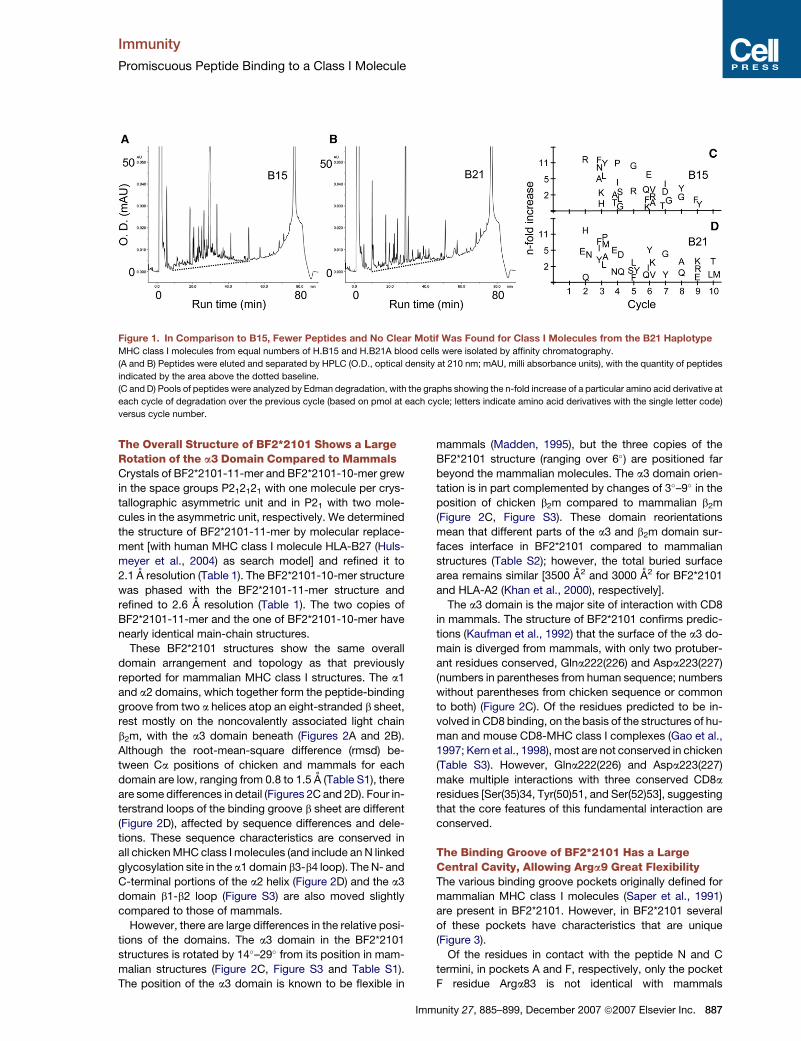
show that, unlike MHC class I molecules from the B4,
B12, and B15 haplotypes, BF2*2101 will not bind oc-
tamers (data not shown). Because octamer peptides
must adopt an extended main-chain conformation to
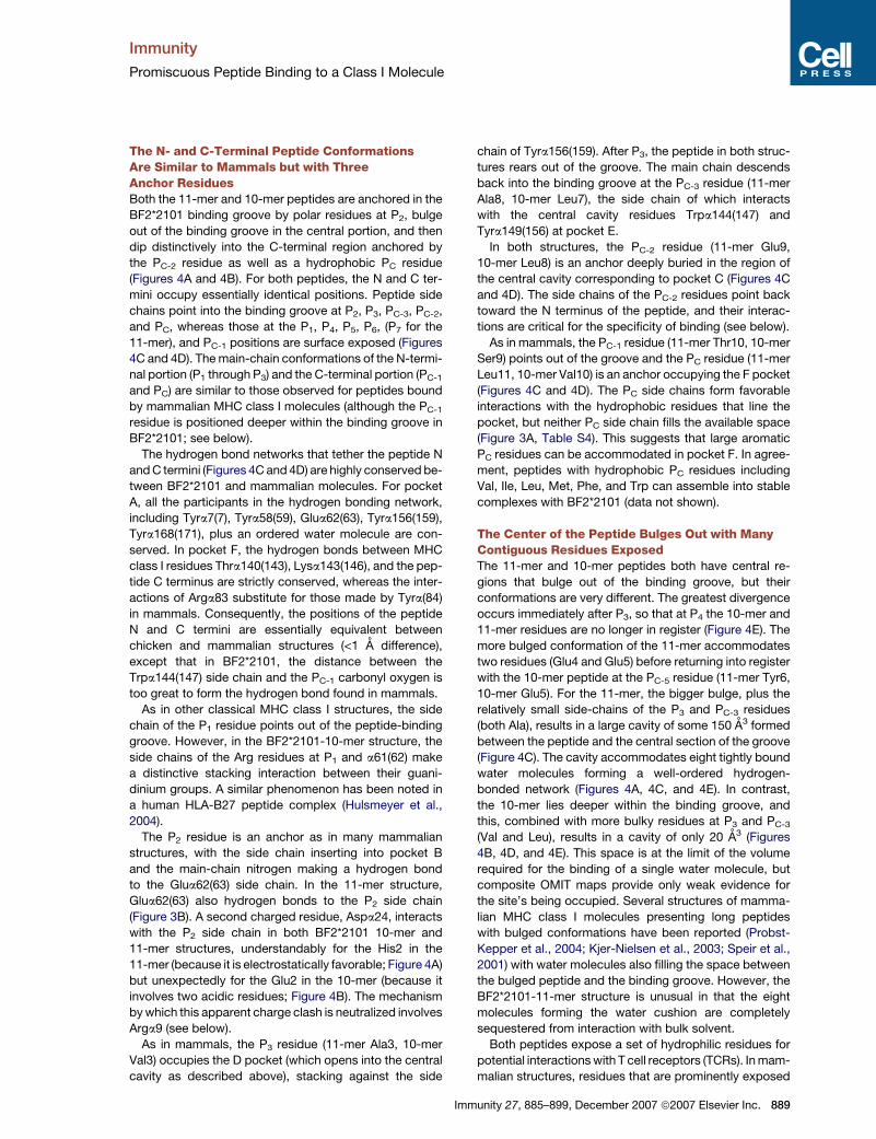
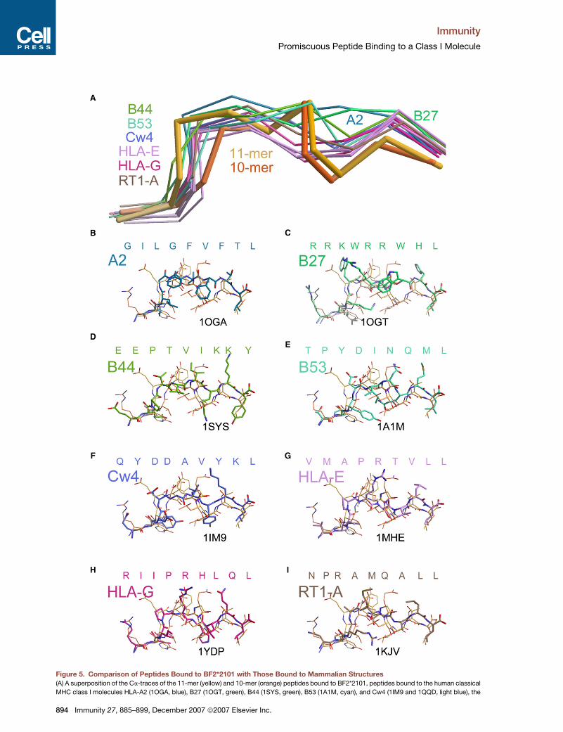
human system (PDB entry 1AKJ) shown in stick representation with green carbons and the equivalent residues in chicken shown with orange carbons.
The close-up view of the red boxed area in (C) shows the superposition of BF2*2101 and HLA-B27 based on a3 domains. In (D), the view is from above
the binding groove. Those a1 and a2 domain loops that show conformational differences between BF2*2101 and HLA-B27 are indicated: b1-b2 and
b3-b4 loops of the a1 domain and b1-b2 and b3-b4 loops of the a2 domain. The main-chain conformations of all of the interstrand loops are well
defined in the BF2*2101 structures (crystallographic B factors < 35 A2 and 46 A2 for noncharged residues and 57 A2 and 50 A2 for charged residues
in the 11-mer and 10-mer, respectively).
munity 27, 885–899, December 2007 ª2007 Elsevier Inc. 891
Immunity
Promiscuous Peptide Binding to a Class I Molecule
892 Immunity 27, 885–899, December 2007 ª2007 Elsevier Inc.
Immunity
Promiscuous Peptide Binding to a Class I Molecule
span between pockets A and F in mammalian MHC class I
molecules (Fremont et al., 1992; Reid et al., 1996; Rudolph
et al., 2001), this result suggests that the helical turn
conformation might be characteristic of BF2*2101 bound
peptides.
The Anchor Residues P2 and PC-2 Do Not BindIndependently, Allowing a NovelPeptide-Binding StrategyAlthough the P2 residue is an anchor inserted in pocket B
in both BF2*2101 structures, the pool sequences show
a broad range of polar and charged resides at this position
(Figure 1, Figures S1 and S2). In the 11-mer, the side chain
of His2 makes an electrostatically favorable interaction
with the side chain of the pocket B residue Aspa24
(Figure 4A). For the 10-mer, there is also a close juxtapo-
sition of the Glu2 and Aspa24 side chains, an apparent
electrostatic clash between two negatively charged resi-
dues (distances between Aspa24 Od1 and P2 Glu O31
and O32 are 2.7 and 3.0 A, respectively; Figure 4B).
Crucially, comparison of the two structures reveals
a mechanism by which the hydrogen-bonding potential
of this portion of the binding groove can be modulated
by changes in the side-chain conformation of Arga9.
In the BF2*2101-11-mer structure, Arga9 interacts with
the negatively charged Glu9, leaving Aspa24 free to inter-
act with His2 (Figures 4A and 4C). In the BF2*2101-10-mer
structure, Arga9 swivels away from the hydrophobic Leu8
to hydrogen bond to the Glu2 (2.3 A between P2 Glu O32
and Arg9 N3) (Figures 4B and 4D). In silico calculations (Li
et al., 2005) indicate that the Glu2 O31 is protonated (Table
S4) and that the three-residue network, Aspa24-Glu2-
Arga9, forms a charge-relay system with no resultant net
charge. Moreover, the reorientation of Arga9 leaves a suit-
ably hydrophobic environment in pocket E for Leu8
(Figure 4D). Thus, conformational flexibility of the Arga9
side chain allows an optional contribution to the hydrogen
bonding and electrostatics of both the B and E pockets.
Taken together, the two structures suggest that peptide
binding to the BF2*2101 molecule requires particular
combinations of amino acids at position P2 and PC-2.
The Binding Groove and Peptide Conformationsof BF2*2101 Are UniqueOur structural analyses reveal that the BF2*2101 binding
groove has an unusually large central cavity, the result of
Im
small residues at a69(70) and a97(99). Comparisons with
mammalian structures show that this distinctive open
groove architecture has striking consequences for
peptide binding. First, the use of the C pocket by a PC-2
anchor, seen in both BF2*2101 structures, is rare
(Figure 5A). Out of nearly 200 structures deposited in the
Protein Data Bank (PDB) (http://www.rcsb.org/pdb/
home/home.do), only 12 structures of six MHC class I
molecules orientate the PC-2 side chain into the binding
groove (Figures 5B–5I). Of these mammalian structures,
only one HLA-B53-nonamer complex orientates the side
chain back along the binding groove, similar to the
BF2*2101 structures (Figure 5E); a second nonamer pep-
tide complexed to HLA-B53 does not have the same con-
formation (Smith et al., 1996). Notably, for the BF2*2101
structures, the PC-2 and PC-1 Ca atoms are positioned as
much as 2.3 A and 1.8 A, respectively, deeper in the bind-
ing groove than they are for any mammalian structure
(Figure 5A). Thus, although the tips of long PC-2 side chains
reach to the bottom of the binding groove in several mam-
malian structures, the BF2*2101 structures are the only
ones for which the entire PC-2 residue is positioned deep
within the binding groove. This position is only possible
because of the spacious nature of the BF2*2101 binding
groove, with the PC-2 main chain being locked into its
unique position (and requisite helical turn conformation)
by hydrogen bonds to Asna76(77) and Hisa111(114)
(Figure 3D).
The second distinctive feature resulting from the open
nature of the BF2*2101 binding groove is the conforma-
tional plasticity of Arga9 and the consequent coupled
variation of the P2 and PC-2 anchor residues. Switches in
side-chain conformation have been reported previously
to be one mechanism by which an MHC class I binding
groove can adapt to bind different peptides (Smith et al.,
1996; Fremont et al., 1992; Madden et al., 1993), but
none involve residue a9. Some interplay in the P2 and P5
anchor residues accommodated in the B and C pockets
has been noted for structures of H-2Kb (Fremont et al.,
1995), a mouse MHC classical class I molecule that, like
BF2*2101, has a small serine residue at position a97(99).
However, in H-2Kb, Vala9 provides little potential for con-
formational plasticity and the effects of different anchor
residue pairings are balanced by changes in bound water
structure. In BF2*2101, the combination of open binding
groove, side-chain flexibility at a9, and counterbalancing
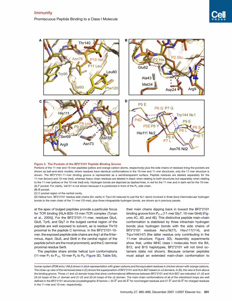
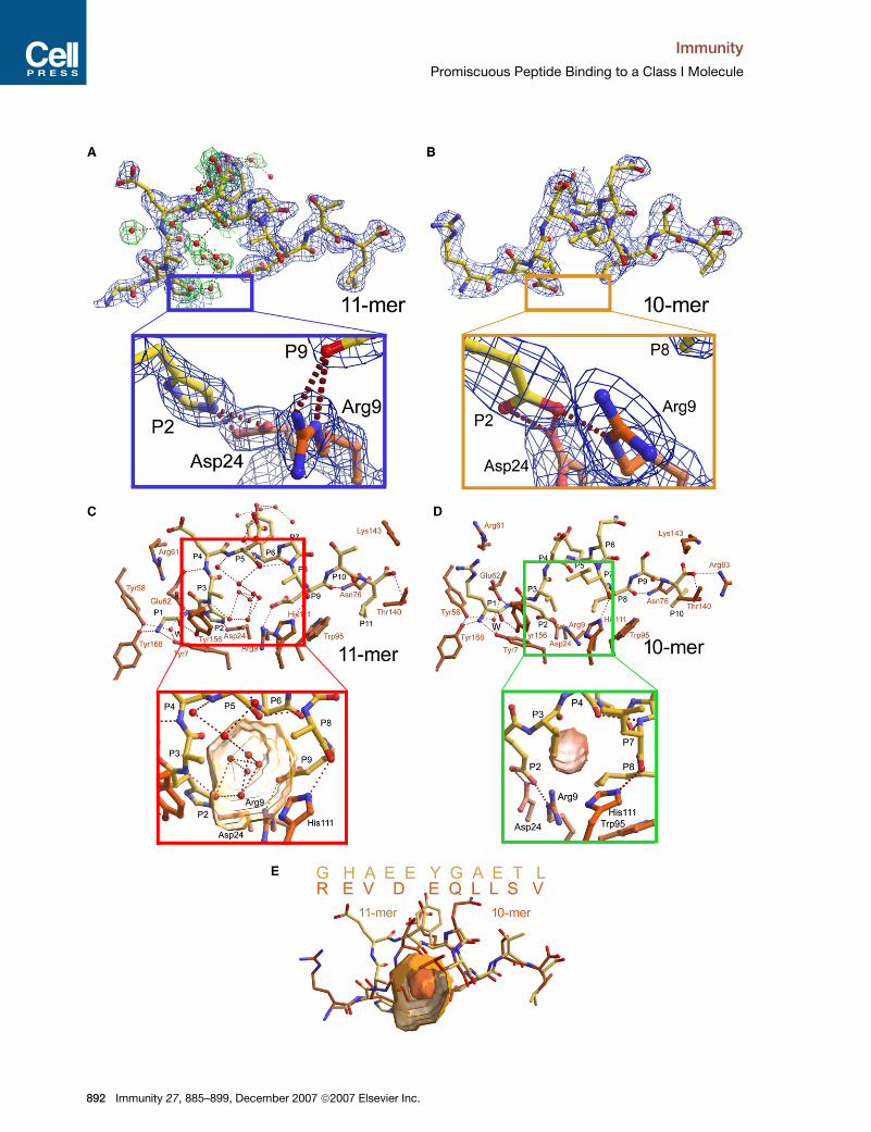
Figure 4. Peptides Bound to BF2*2101
(A and B) The electron density of the 11-mer and 10-mer peptides are shown as composite OMIT maps contoured at 1.0 s in blue for the peptide and in
green for peptide-bound water molecules (both within and outside the binding groove). The blue box shows the details of the electrostatic interactions
at pocket B in the BF2*2101 11-mer, and the yellow box shows the details of the three-residue Aspa24-P2Glu-Arga9 charge-relay system at pocket B
in the BF2*2101 10-mer.
(C and D) The structure of the 11-mer and 10-mer peptides (yellow carbon atoms) are shown with relevant parts of interacting residues from the bind-
ing groove (orange carbon atoms) and bound water molecules (red spheres) buried within the binding groove, one of which being the conserved water
interacting with the N-terminus (W). Hydrogen bonds are depicted as dotted red lines. The red box shows a close-up view of the ‘‘water cushion’’ filling
the central region of the binding groove in the BF2*2101-11-mer structure with the cavity (yellow surface) calculated in the absence of bound waters.
The green box shows a close-up view of the central portion of the 10-mer including the small cavity present beneath the bulge region.
(E) Superposition of the sequences (single letter code) as well as of the structures of the two chicken peptides 11-mer (yellow) and 10-mer (orange) are
shown, with the latter including the cavities depicted in the red and green boxes. Note the two peptides run out of register at P3 and return to register at
PC-5.
munity 27, 885–899, December 2007 ª2007 Elsevier Inc. 893
Immunity
Promiscuous Peptide Binding to a Class I Molecule
Figure 5. Comparison of Peptides Bound to BF2*2101 with Those Bound to Mammalian Structures
(A) A superposition of the Ca-traces of the 11-mer (yellow) and 10-mer (orange) peptides bound to BF2*2101, peptides bound to the human classical
MHC class I molecules HLA-A2 (1OGA, blue), B27 (1OGT, green), B44 (1SYS, green), B53 (1A1M, cyan), and Cw4 (1IM9 and 1QQD, light blue), the
894 Immunity 27, 885–899, December 2007 ª2007 Elsevier Inc.
Immunity
Promiscuous Peptide Binding to a Class I Molecule
charges on Arga9 and Aspa24 provides a particularly
powerful mechanism for charge transfer not previously
observed in MHC molecules.
In order to determine how many MHC class I molecules
might share these features with BF2*2101, we examined
2114 sequences of classical MHC class I molecules
from fish to human, as well as a further 63 nonclassical
class I sequences from mouse, rat, and frog (Table S5).
Very few classical MHC class I sequences have small res-
idues at either a69(70) or a97(99) [5% Ser, 0.5% Gly and
1.8% Ala for a69(70); 4% Ser, 3% Cys and 0.2% Ala for
a97(99)]. Only seven classical sequences (0.3%) have
small residues in both positions: BF2*2101 from chicken,
two from cottontop tamarin, two from sheep, one from
rat, and one from rainbow trout. Moreover, only three non-
classical sequences have small residues in both positions.
Thus, an open central cavity appears to be unusual for
both classical and nonclassical MHC class I molecules.
Strikingly, these analyses show that Arga9 and Aspa24
have not been found outside of chickens. There are six
chicken sequences with Arga9, of which three also have
Aspa24, and ten chicken sequences with Hisa9 and
Aspa24, but of these, only BF2*2101 has small residues
at both a69(70) and a97(99). There are a few classical
sequences outside of chickens with basic residues at a9
or acidic residues at a24 (11% His or Lys at a9; 2.7%
Glu at a24) but none with both in the same sequence.
In rat, there are five nonclassical sequences with both
Lysa9 and Glua24, but none with small residues at
a69(70) and a97(99). Thus, the sequence features allowing
this precise mode of charge transfer seem to be unique to
BF2*2101 among the known classical and nonclassical
MHC class I molecules in vertebrates.
The Peptide-Binding Motif Might Explainthe Strong Resistance of the B21 Haplotypeto Marek’s DiseaseA large number of studies show that the B21 haplotype
confers strong resistance to Marek’s disease, which is
caused by an oncogenic herpesvirus (Bacon, 1987; Bacon
et al., 1987; Briles et al., 1977; Plachy et al., 1992). There
are many candidate genes and little recombination in the
region identified as being responsible for this resistance
(Kaufman et al., 1999a, 1999b; Shiina et al., 2007), so
the gene(s) actually responsible are not known. However,
several studies propose that this resistance is due to the
action of cytotoxic T lymphocytes (CTLs) and/or natural
killer (NK) cells (Garcia-Camacho et al., 2003; Markow-
ski-Grimsrud and Schat, 2003; Omar and Schat, 1996;
Omar and Schat, 1997), both of which implicate an MHC
class I molecule as the target. The distinctive nature of
the BF2*2101 molecule is consistent with these proposals.
To illustrate how the distinctive nature of the BF2*2101
could explain the resistance to Marek’s disease conferred
Im
by the B21 haplotype compared to other well-defined
haplotypes, we determined a preliminary peptide motif
for this molecule by using assembly of the 10-mer and
11-mer peptides substituted at various positions
(Figure S4, Table S6). This is by no means an exhaustive
analysis, and so in fact more peptides might have been
predicted than are specified by this preliminary motif.
However, even at this early stage of analysis, it is clear
that many more peptides from representative Marek’s
disease virus (MDV) genes are predicted to bind the
BF2*2101 molecule than the MHC class I molecules
from haplotypes such as B4, B12, and B15 that do not
confer strong resistance to Marek’s disease (Table 2,
Table S7). On this basis, it is likely that the promiscuous
BF2*2101 molecule would have a greater chance of bind-
ing key protective peptide(s) than the fastidious MHC
class I molecules from the other haplotypes.
DISCUSSION
Here, we report the first structures of an MHC molecule
outside of mammals, the classical class I molecule
BF2*2101 from the chicken MHC haplotype B21. This
molecule has a novel mode of peptide binding that allows
peptides with completely different sequences to be pre-
sented to T lymphocytes. Some malleability of adjacent
pockets in the peptide binding groove, and resultant inter-
dependence of peptide anchors, has been described for
certain mammalian MHC class I molecules. However,
BF2*2101 provides an extreme example of such coupled
variation, with the conformational freedom and charged
nature of the Arga9 side chain allowing very varied combi-
nations of interactions between itself, Aspa24, and pep-
tide residues P2 and PC-2. One of the examples we
describe has an acidic amino acid in the peptide (Glu2 in
the 10-mer) interacting with another acidic amino acid
in the MHC molecule (Aspa24), supported by the basic
amino acid Arga9. Such charge-transfer mechanisms
have been described in enzymes such as hemoglobin,
cytochrome c peroxidase, carboxypeptidase II (Flocco
and Mowbray, 1995), and most recently in the serine-car-
boxyl-type proteinase Kumamolysin (Comellas-Bigler
et al., 2002) but never before in an MHC molecule.
The resulting coupled variation of the peptide residues
binding into pocket B and C also explains why the pool
sequences and individual peptides for MHC class I mole-
cules from the B21 haplotype led to no clear motif. Inter-
dependent pairs of amino acids at P2 and PC-2 meant
these anchor positions could be occupied by amino acids
with very different sizes and chemical properties. There
are several examples in which pool sequences have not
led to consistent motifs or in which single peptides that
bind an MHC class I molecule did not fit the motif (Gad-
dum et al., 1996; Quesnel et al., 1996; Mata et al., 1998;
human nonclassical MHC class I molecules HLA-E (1KPR, 1KTL, 1MHE, and 2ESV, light violet) and HLA-G (1YDP, 2DYP, and 2D31, wine red), and the
rat MHC class I molecule RT1-A (1KJV, brown). The 10-mer and 11-mer peptides are highlighted as thicker lines.
(B–I) The structures of selected peptides are depicted as thick stick models (plus sequence in one letter code) superposed with the 10-mer and
11-mer peptides (thin sticks). Carbon atom colors are as above. Cw4, HLA-E, and HLA-G are represented by 1MI9, 1MEH, and 1YDP, respectively.
munity 27, 885–899, December 2007 ª2007 Elsevier Inc. 895
Immunity
Promiscuous Peptide Binding to a Class I Molecule
Apostolopoulos and Lazoura, 2004). Some of these cases
might also be due to binding mechanisms that involve
coupled variation of the anchor residues. Our data also
suggest caution in the development of peptide prediction
algorithms and peptide modeling projects that might not
take account of such promiscuity, thus failing to predict
functionally important peptides.
The chicken MHC class I molecule BF2*2101 is en-
coded by the B21 haplotype, which is known for having
one of the strongest associations with resistance to an
infectious pathogen, the economically important herpes-
virus that causes Marek’s disease (Briles et al., 1977;
Bacon, 1987; Plachy et al., 1992). It has not been possible
to assign the MHC-determined resistance to Marek’s dis-
ease to a particular gene because of the rarity of recombi-
nation within the chicken MHC (BF/BL region). However,
a number of studies have suggested that the MHC-deter-
mined resistance to Marek’s disease is due at least in part
to the action of CTLs and NK cells (Garcia-Camacho et al.,
2003; Markowski-Grimsrud and Schat, 2003; Omar and
Schat, 1996; Omar and Schat, 1997), both of which impli-
Table 2. Number of Peptides from MDV GenesPredicted to Bind Class I Molecules from Chicken MHCHaplotypes B4, B12, B15, and B21
Haplotype gB pp38 meq ICP4
B4 0-1 0-1 0 1
B12 8-11 0-3 0 9
B15 0-1 0 2-3 2
B21 15-20 0-3 6-11 14-15
The protein sequences from four MDV genes, considered as
likely candidates for protective antigens (Omar and Schat,1996; Omar and Schat, 1997), were analyzed on the basis of
the sequences from the following strains (database entries
or references): gB protein: HVT (Q69408), SB1 (Q69406),and RB1B (P18538); pp38 protein: S1 (Q89483), S2
(D28115), and S3 (Q89873); meq protein: RB1B (M89471),
GA, HPRS16, C12/130, MR36, and MR48 (Barrow and Venu-
gopal, 1999); and ICP4 protein: GA (Q02362), C12/130, MR36,and MR38 (Barrow and Venugopal, 1999). Peptide motifs and
prediction methods were derived from Kaufman et al. (1995);
Wallny et al. (2006), and this paper. The motifs were the following:
B4: x-(D or E)-x-x-(D or E)-x-x-E; B12: x-x-x-x-(V or I)-x-x-V;B15: (K or R)-R-x-x-x-x-x-Y and (K or R)-R-x-x-x-x-x-x-Y;
B21: x-(H, K or R)-x-x-x-x-x-x-(E or D)-x-(A, V, L, I, F or W),
x-(H, K or R)-x-x-x-x-x-(E or D)-x-(A V, L, I, F or W), x-(E orD)-x-x-x-x-x-L-x-(A, V, L, I, F or W) and x-(E or D)-x-x-x-x-x-
x-L-x-(A, V, L, I, F or W). The predominant lengths of peptide
found in single and pool sequences were octamers for B4
and B12, octamers and nonamers for B15, and 10-mers and11-mers for B21. Although longer peptides (nonamers
and longer for B4 and B12, 10-mers and longer for B15, and
12-mers and longer for B21) and shorter peptides (nonamers
for B21) are also found and can also assemble, they are rare.So for comparison’s sake, only the predominant length was
used for the motifs. There are also a number of hydrophobic
residues found at the C terminus of peptides for B4, B12,and B15, but again, they are rare, so for comparison’s sake,
they were not used in the motif.
896 Immunity 27, 885–899, December 2007 ª2007 Elsevier Inc
cate an MHC class I molecule as a target. The data pre-
sented in this paper suggest that BF2*2101 is distinctive
and provide support for the notion that it might be respon-
sible for the strong resistance conferred by this haplotype,
as illustrated by epitope predictions made on the basis of
a preliminary peptide motif. Moreover, given that the B21
haplotype is very common in chickens (Simonsen et al.,
1982), these data might be useful for developing vaccines,
not only to MDV, but to other pathogens, such as avian
influenza.
EXPERIMENTAL PROCEDURES
Isolation of MHC Class I Molecules and Analysis of Peptides
Methods were identical to those of Wallny et al. (2006), including affin-
ity chromatography of class I molecules, peptide isolation and chro-
matography, Edman degradation of peptide pools and individual
peptides, and mass spectrometry (MS/MS) of individual peptides.
For spleen preparations, the capsules were removed, the spleens
were ground up in an equal volume of phosphate-buffered saline
with 10 mM MgCl2, 0.4 mM 4-(2-aminoethyl) benzenesulfonyl fluoride
hydrochloride (AEBSF), and 0.4 mM iodoacetamide on ice with a loose
glass tissue homogenizer, and NP-40 detergent was added to 2.5%
final concentration. The lysate was incubated on ice for 30 min and
centrifuged twice at 4,000 rpm in a Sorvall GSA rotor, twice at
13,500 rpm, and one to three times at 20,000 rpm in a Sorval SLA
600TC rotor, each spin for 30 min at 4�C.
Assembly of Denatured BF2*2101 and b2m with Peptides
and Peptide Libraries
Overall, methods followed those of Dunbar et al. (1998). In brief, com-
plementary DNA (cDNA) was used for amplification of a b2m sequence
encoding the mature protein without a signal sequence but beginning
with a start codon. A cDNA clone of BF2*2101 was used as a source for
amplification of the a1-a2-a3 domains (residues 1–270 of the mature
protein), which was cloned into pBluescript, engineered to have a start
codon at the beginning and a biotinylation sequence followed by a His
tag at the end. Both sequences were cloned into pET22b(+), and pro-
teins were expressed as inclusion bodies in BL21 (lDE3) pLysS
Rosetta cells. Inclusion bodies were isolated and dissolved in solutions
containing urea. Peptides were synthesized with fluorenyl-methoxy-
carbonyl (fMOC) chemistry. The denatured proteins were renatured
in the presence of peptides or peptide libraries, and monomers were
separated from aggregates, heavy chains, b2m, peptides, and smaller
molecules by fast protein liquid chromatography (FPLC) size exclusion
chromatography with a HiLoad 26/60 Superdex 200 column (Phar-
macia).
Sequence Comparisons
MHC class I sequences were sourced as follows: human (IMGT/HLA
database; www.ebi.ac.uk/imgt/hla), nonhuman primates and mon-
keys, cattle, sheep, swine, rat, and bony fish (IPD-MHC databases;
www.ebi.ac.uk/ipd), mouse (Watts et al., 1989), bird (Wallny et al.,
2006; Livant et al., 2004; Lima-Rosa et al., 2004; Hunt and Fulton,
1998; Shiina et al., 1999; Moon et al., 2005), frog (Flajnik et al., 1993;
Flajnik et al., 1999) and shark (Okamura et al., 1997).
Crystallization
Renatured proteins were concentrated to 10 mg/ml in 50mM NaCl,
10mM Tris (pH 8.0) and crystallized by the sitting-drop vapor-diffusion
method. The crystallizations were set up as nanoliter scale drops
(100 nL of protein plus 100 nL of reservoir solution) with a Cartesian
Technologies Microsys MIC4000 (Genomic Technologies) (Walter
et al., 2003).
Crystals grew at room temperature in 0.1 M HEPES (pH 7.0), 100 mM
MgCl2, and 27% polyethylene glycol 3350. The BF2*2101-11-mer
.
Immunity
Promiscuous Peptide Binding to a Class I Molecule
crystals were long thin needles of dimensions 300 3 30 3 30 mm3,
whereas those of BF2*2101-10-mer were larger and multiple, with
the lengths of about 100 mm in all three dimensions. Both crystal types
were soaked briefly in per-fluoropolyether oil (PFPE) before being flash
cooled and maintained at 100�K in a cryostream. Data collection for
the BF2*2101-11-mer was straightforward; however, the BF2*2101-
10-mer required careful selection of suitable portions of crystal and
orientation ranges to minimize problems with multiple overlapping
lattices.
Diffraction data were recorded at station BM14 of the European Syn-
chrotron Radiation Facility (ESRF [Grenoble, France]) with a MARCCD
m225 CCD detector. Diffraction datasets were autoindexed with
Denzo and scaled together with Scalepack (Otwinowski and Minor,
1997) (http://www.hkl-xray.com) (Table 1). The asymmetric unit of
BF2*2101-11-mer contained a single molecule and 47% solvent,
whereas that of BF2*2101-10-mer had two molecules with 61%
solvent.
Structure Determination and Refinement
The structure of BF2*2101-11-mer was determined by molecular
replacement with a human HLA-B27 structure (PDB-entry 1OGT with
peptide coordinates removed) on the webmail interface CaspR
(http://igs-server.cnrs-mrs.fr/Caspr/index.cgi/). The a3 domain re-
quired manual reorientation into initial low-quality 2Fo-Fc electron den-
sity. After further domain-wise rigid-body refinement, the model was
rebuilt (with BF2*2101 sequence) into much improved 2Fo-Fc electron
density and refined by simulated annealing with CNS (Brunger et al.,
1998) (http://cns.csb.yale.edu/). After model building by ARP-wARP
(Morris et al., 2003), the peptide was added. Positional refinement
and individual B factor refinement with bulk solvent scaling and overall
anisotropic B- actor scaling were applied, interspersed with manual
rebuilding with O (http://www.bioxray.dk/�mok/o-files.html). Water
molecules were added with ARP-wARP (based on Fo-Fc peaks of at
least 2.5 s), and refinement was completed with translation, libration,
and screw rotation (TLS) in the REFMAC CCP4 program suite (Collab-
orative Computational Project, 1994; Murshudov et al., 1997; Winn
et al., 2001) (http://www.ccp4.ac.uk). The final structure had good
stereochemistry (Table 1), as assessed by the program PROCHECK
(Laskowski et al., 1993), with Rwork 23.0% and Rfree 28.6%, comprising
residues 1–274 of the heavy chain, 1–99 of the b2m, and 190 water
molecules.
The structure of BF2*2101-10-mer was determined by molecular
replacement with the BF2*2101-11-mer structure as the search model
in MOLREP (Vagin and Teplyakov, 1997). After initial rigid-body refine-
ment and some cycles of restrained refinement with REFMAC, further
modeling was guided by Fo-Fc and 2Fo-Fc electron density maps cal-
culated with REFMAC and by simulated annealing and composite
OMIT maps calculated with CNS. Subsequent refinement in REFMAC
was as described above but with noncrystallographic symmetry (NCS)
restraints applied to the two copies of BF2*2101-10-mer in the
asymmetric unit. The final refined structure had good stereochemistry
(Table 1), as assessed by the program PROCHECK (Laskowski et al.,
1993), with Rwork 23.4% and Rfree 29.5%, comprising residues 1–271
of the heavy chain and residues 1–99 in b2m for both copies, and
129 water molecules.
Structural Analysis and Modeling
Overall and domain-wise superpositions of pairs of structures were
calculated with the program SHP (Stuart et al., 1979). The structures
of the two copies of BF2*2101-10-mer in the crystallographic asym-
metric unit were essentially identical, as was the BF2*2101-11-mer
(Table 1). Superpositions of peptides from mammalian structures
onto the peptides bound to BF2*2101 were done with the program
IMPOSE (R. Esnouf, personal communication), with a ‘‘core set’’
of framework residues (5–13, 21–26, 31–37, 43–47, 54–86, 91–101,
107–123, and 136–175 numbered as in BF2*2101).
Structural figures were prepared with Bobscript (Esnouf, 1999) and
Raster3D (Merritt and Murphy, 1994) (http://trantor.bioc.columbia.
Im
edu/grasp/). Cavity volumes and maps were calculated with the
program VOLUMES (R. Esnouf, personal communication) with a
1.4 A probe radius so that the accessibility of the surface of the mole-
cules to the solvent as well as inside the molecule could be assessed.
Supplemental Data
Four figures and seven tables are available at http://www.immunity.
com/cgi/content/full/27/6/885/DC1/.
ACKNOWLEDGMENTS
We thank Division of Structural Biology (STRUBI) members for assis-
tance and helpful discussions, K. Harlos, R. Esnouf, and staff of the
European Synchrotron Radiation Facility (ESRF) and European Molec-
ular Biology Laboratory (EMBL) in Grenoble for assistance with X-ray
data collection, Susan Lea and Gillian Griffiths for critical reading
and suggestions, and the United Kingdom Medical Research Council
(E.Y.J.), Biotechnology and Biological Sciences Research Council
(BBSRC) (J.K.) and European Union (PeptidEx consortium; grant
code QLK2-CT-2002-000838; J.K.) for funding. We acknowledge
use of crystallization facilities provided by the Medical Research
Council-funded Oxford Protein Production Facility and the EU Inte-
grated Programme (Structural Proteomics in Europe [SPINE], Grant
code QLRT-2001-00988). E.Y.J. is a Cancer Research UK Principal
Research Fellow.
Received: February 16, 2007
Revised: October 10, 2007
Accepted: November 2, 2007
Published online: December 13, 2007
REFERENCES
Apostolopoulos, V., and Lazoura, E. (2004). Noncanonical peptides in
complex with MHC class I. Expert Rev. Vaccines 3, 151–162.
Bacon, L. (1987). Influence of the major histocompatibility complex on
disease resistance and productivity. Poult. Sci. 66, 802–811.
Bacon, L., Ismail, N., and Motta, J. (1987). Allograft and antibody
responses of 15I5-B congenic chickens. Prog. Clin. Biol. Res. 238,
219–233.
Barrow, A., and Venugopal, K. (1999). Molecular characterisation of
very virulent European MDV isolates. Acta Virol. 43, 90–93.
Briles, W., Stone, H., and Cole, R. (1977). Marek’s disease: Effects of B
histocompatibility alloalleles in resistant and susceptible chicken lines.
Science 195, 193–196.
Brunger, A., Adams, P., Clore, G., DeLano, W., Gros, P., Grosse-Kuns-
tleve, R., Jiang, J., Kuszewski, J., Nilges, M., Pannu, N., et al. (1998).
Crystallography & NMR system: A new software suite for macromolec-
ular structure determination. Acta Crystallogr. D Biol. Crystallogr. 54,
905–921.
Collaborative Computational Project (1994). The CCP4 suite: Pro-
grams for protein crystallography. Acta Crystallogr. D Biol. Crystallogr.
50, 760–763.
Comellas-Bigler, M., Fuentes-Prior, P., Maskos, K., Huber, R., Oyama,
H., Uchida, K., Dunn, B., Oda, K., and Bode, W. (2002). The 1.4 a crystal
structure of kumamolysin: A thermostable serine-carboxyl-type
proteinase. Structure 10, 865–876.
Dunbar, P., Ogg, G., Chen, J., Rust, N., van der Bruggen, P., and
Cerundolo, V. (1998). Direct isolation, phenotyping and cloning of
low-frequency antigen-specific cytotoxic T lymphocytes from periph-
eral blood. Curr. Biol. 8, 413–416.
Esnouf, R. (1999). Further additions to MolScript version 1.4, including
reading and contouring of electron-density maps. Acta Crystallogr.
D Biol. Crystallogr. 55, 938–940.
munity 27, 885–899, December 2007 ª2007 Elsevier Inc. 897
Immunity
Promiscuous Peptide Binding to a Class I Molecule
Flajnik, M., and Kasahara, M. (2001). Comparative genomics of the
MHC: Glimpses into the evolution of the adaptive immune system.
Immunity 15, 351–362.
Flajnik, M., Kasahara, M., Shum, B., Salter-Cid, L., Taylor, E., and Du
Pasquier, L. (1993). A novel type of class I gene organization in verte-
brates: A large family of non-MHC-linked class I genes is expressed at
the RNA level in the amphibian Xenopus. EMBO J. 12, 4385–4396.
Flajnik, M., Ohta, Y., Greenberg, A., Salter-Cid, L., Carrizosa, A., Du
Pasquier, L., and Kasahara, M. (1999). Two ancient allelic lineages at
the single classical class I locus in the Xenopus MHC. J. Immunol.
163, 3826–3833.
Flocco, M., and Mowbray, S. (1995). Strange bedfellows: interactions
between acidic side-chains in proteins. J. Mol. Biol. 254, 96–105.
Fremont, D., Matsumura, M., Stura, E., Peterson, P., and Wilson, I.
(1992). Crystal structures of two viral peptides in complex with murine
MHC class I H-2Kb. Science 257, 919–927.
Fremont, D., Stura, E., Matsumura, M., Peterson, P., and Wilson, I.
(1995). Crystal structure of an H-2Kb-ovalbumin peptide complex
reveals the interplay of primary and secondary anchor positions in
the major histocompatibility complex binding groove. Proc. Natl.
Acad. Sci. USA 92, 2479–2483.
Gaddum, R., Willis, A., and Ellis, S. (1996). Peptide motifs from three
cattle MHC (BoLA) class I antigens. Immunogenetics 43, 238–239.
Gao, G., Tormo, J., Gerth, U., Wyer, J., McMichael, A., Stuart, D., Bell,
J., Jones, E., and Jakobsen, B. (1997). Crystal structure of the complex
between human CD8alpha(alpha) and HLA-A2. Nature 387, 630–634.
Garcia-Camacho, L., Schat, K., Brooks, R., and Bounous, D.I. (2003).
Early cell-mediated immune responses to Marek’s disease virus in two
chicken lines with defined major histocompatibility complex antigens.
Vet. Immunol. Immunopathol. 95, 145–153.
Guillemot, F., Billault, A., Pourquie, O., Behar, G., Chausse, A., Zoorob,
R., Kreibich, G., and Auffray, C. (1988). A molecular map of the chicken
major histocompatibility complex: The class II beta genes are closely
linked to the class I genes and the nucleolar organizer. EMBO J. 7,
2775–2785.
Hansen, M., Van Zandt, J., and Law, G. (1967). Differences in suscep-
tibility to Marek’s disease in chickens carrying two different B locus
blood group alleles. Poult. Sci. 46, 1268.
Hulsmeyer, M., Fiorillo, M., Bettosini, F., Sorrentino, R., Saenger, W.,
Ziegler, A., and Uchanska-Ziegler, B. (2004). Dual HLA-B27 subtype-
dependent conformation of a self-peptide. J. Exp. Med. 199, 271–281.
Hunt, H., and Fulton, J. (1998). Analysis of polymorphisms in the major
expressed class I locus (B-FIV) of the chicken. Immunogenetics 47,
456–467.
Hutt, F., and Cole, R. (1947). Genetic control of lymphomatosis in the
fowl. Science 106, 379–384.
Kaufman, J. (1999). Co-evolving genes in MHC haplotypes: The ‘‘rule’’
for nonmammalian vertebrates? Immunogenetics 50, 228–236.
Kaufman, J., Andersen, R., Avila, D., Engberg, J., Lambris, J., Salo-
monsen, J., Welinder, K., and Skjødt, K. (1992). Different features of
the MHC class I heterodimer have evolved at different rates: Chicken
B-F and b2-microglobulin sequences reveal invariant surface residues.
J. Immunol. 148, 1532–1546.
Kaufman, J., Salomonsen, J., and Flajnik, M. (1994). Evolutionary
conservation of MHC class I and class II molecules–different yet the
same. Semin. Immunol. 6, 411–424.
Kaufman, J., Volk, H., and Wallny, H. (1995). A ‘‘minimal essential Mhc’’
and an ‘‘unrecognized Mhc’’: Two extremes in selection for polymor-
phism. Immunol. Rev. 143, 63–88.
Kaufman, J., Milne, S., Gobel, T., Walker, B., Jacob, J., Auffray, C.,
Zoorob, R., and Beck, S. (1999a). The chicken B locus is a minimal
essential major histocompatibility complex. Nature 401, 923–925.
898 Immunity 27, 885–899, December 2007 ª2007 Elsevier Inc
Kaufman, J., Jacob, J., Shaw, I., Walker, B., Milne, S., Beck, S., and
Salomonsen, J. (1999b). Gene organisation determines evolution of
function in the chicken MHC. Immunol. Rev. 167, 101–117.
Kelley, J., Walter, L., and Trowsdale, J. (2005). Comparative genomics
of major histocompatibility complexes. Immunogenetics 56, 683–695.
Kern, P., Teng, M., Smolyar, A., Liu, J., Liu, J., Hussey, R., Spoerl, R.,
Chang, H., Reinherz, E., and Wang, J. (1998). Structural basis of CD8
coreceptor function revealed by crystallographic analysis of a murine
CD8alphaalpha ectodomain fragment in complex with H-2Kb.
Immunity 9, 519–530.
Khan, A., Baker, B., Ghosh, P., Biddison, W., and Wiley, D.C. (2000).
The structure and stability of an HLA-A*0201/octameric tax peptide
complex with an empty conserved peptide-N-terminal binding site.
J. Immunol. 164, 6398–6405.
Kjer-Nielsen, L., Clements, C., Purcell, A., Brooks, A., Whisstock, J.,
Burrows, S., McCluskey, J., and Rossjohn, J. (2003). A structural basis
for the selection of dominant alpha beta T cell receptors in antiviral
immunity. Immunity 18, 53–64.
Laskowski, R., MacArthur, M., Moss, D., and Thornton, J. (1993).
PROCHECK: A program to check the stereochemical quality of protein
structures. J. Appl. Cryst. 26, 283–291.
Li, H., Robertson, A., and Jensen, J. (2005). Very fast empirical predic-
tion and interpretation of protein pKa values. Proteins 61, 704–721.
Lima-Rosa, C., Canal, C., Streck, A., Freitas, L., Delgado-Canedo, A.,
Bonatto, S., and Salzano, F. (2004). B-F DNA sequence variability in
Brazilian (blue-egg Caipira) chickens. Anim. Genet. 35, 278–284.
Livant, E., Brigati, J., and Ewald, S. (2004). Diversity and locus speci-
ficity of chicken MHC B class I sequences. Anim. Genet. 35, 18–27.
Madden, D. (1995). The three-dimensional structure of peptide-MHC
complexes. Annu. Rev. Immunol. 13, 587–622.
Madden, D., Garboczi, D., and Wiley, D. (1993). The antigenic identity
of peptide-MHC complexes: A comparison of the conformations of five
viral peptides presented by HLA-A2. Cell 75, 693–708.
Markowski-Grimsrud, C., and Schat, K. (2003). Infection with chicken
anaemia virus impairs the generation of pathogen-specific cytotoxic
T lymphocytes. Immunology 109, 283–294.
Mata, M., Travers, P., Liu, Q., Frankel, F., and Paterson, Y. (1998). The
MHC class I-restricted immune response to HIV-gag in BALB/c mice
selects a single epitope that does not have a predictable MHC-binding
motif. J. Immunol. 161, 2985–2993.
Matsumura, M., Fremont, D., Peterson, P., and Wilson, I. (1992).
Emerging principles for the recognition of peptide antigens by MHC
class I molecules. Science 257, 27–34.
Merritt, E., and Murphy, M. (1994). Raster3D Version 2.0. A program for
photorealistic molecular graphics. Acta Crystallogr. D Biol. Crystallogr.
50, 869–873.
Moon, D., Veniamin, S., Parks-Dely, J., and Magor, K. (2005). The MHC
of the duck (Anas platyrhynchos) contains five differentially expressed
class I genes. J. Immunol. 175, 6702–6712.
Morris, R., Perrakis, A., and Lamzin, V. (2003). ARP/wARP and
automatic interpretation of protein electron density maps. Methods
Enzymol. 374, 229–244.
Murshudov, G., Vagin, A., and Dodson, E. (1997). Refinement of
macromolecular structures by the maximum-likelihood method. Acta
Crystallogr. D Biol. Crystallogr. 53, 240–255.
Okamura, K., Ototake, M., Nakanishi, T., Kurosawa, Y., and Hashi-
moto, K. (1997). The most primitive vertebrates with jaws possess
highly polymorphic MHC class I genes comparable to those of
humans. Immunity 7, 777–790.
Omar, A., and Schat, K. (1996). Syngeneic Marek’s disease virus
(MDV)-specific cell-mediated immune responses against immediate
early, late and unique MHC proteins. Virology 222, 87–99.
Omar, A., and Schat, K. (1997). Characterization of Marek’s disease
herpesvirus-specific cytotoxic T lymphocytes in chickens inoculated
.
Immunity
Promiscuous Peptide Binding to a Class I Molecule
with a non-oncogenic vaccine strain of MDV. Immunology 90,
579–585.
Otwinowski, Z., and Minor, W. (1997). Processing of X-ray diffraction
data collected in oscillation mode. Methods Enzymol. 276, 307–326.
Plachy, J., Pink, J., and Hala, K. (1992). Biology of the chicken MHC
(B complex). Crit. Rev. Immunol. 12, 47–79.
Probst-Kepper, M., Hecht, H., Herrmann, H., Janke, V., Ocklenburg,
F., Klempnauer, J., van den Eynde, B., and Weiss, S. (2004). Confor-
mational restraints and flexibility of 14-meric peptides in complex
with HLA-B*3501. J. Immunol. 173, 5610–5616.
Quesnel, A., Hsu, S., Delmas, A., Steward, M., Trudelle, Y., and Abas-
tado, J. (1996). Efficient binding to the MHC class I K(d) molecule of
synthetic peptides in which the anchoring position 2 does not fit the
consensus motif. FEBS Lett. 387, 42–46.
Rammensee, H. (1995). Chemistry of peptides associated with MHC
class I and class II molecules. Curr. Opin. Immunol. 7, 85–96.
Rammensee, H., Friede, T., and Stevanoviic, S. (1995). MHC ligands
and peptide motifs: First listing. Immunogenetics 41, 178–228.
Reid, S., McAdam, S., Smith, K., Klenerman, P., O’Callaghan, C.,
Harlos, K., Jakobsen, B., McMichael, A., Bell, J., Stuart, D., and Jones,
E. (1996). Antagonist HIV-1 Gag peptides induce structural changes in
HLA B8. J. Exp. Med. 184, 2279–2286.
Rudolph, M., Speir, J., Brunmark, A., Mattsson, N., Jackson, M.,
Peterson, P., Teyton, L., and Wilson, I. (2001). The crystal structures
of K(bm1) and K(bm8) reveal that subtle changes in the peptide envi-
ronment impact thermostability and alloreactivity. Immunity 14,
231–242.
Saper, M., Bjorkman, P., and Wiley, D. (1991). Refined structure of the
human histocompatibility antigen HLA-A2 at 2.6 A resolution. J. Mol.
Biol. 219, 277–319.
Shiina, T., Oka, A., Imanishi, T., Hanzawa, K., Gojobori, T., Watanabe,
S., and Inoko, H. (1999). Multiple class I loci expressed by the quail
Mhc. Immunogenetics 49, 456–460.
Shiina, T., Briles, W., Goto, R., Hosomichi, K., Yanaqiya, K., Shimizu,
S., Inoko, H., and Miller, M. (2007). Extended gene map reveals tripar-
tite motif, C-type lectin and Ig superfamily type genes within a subre-
gion of the chicken MHC-B affecting infectious disease. J. Immunol.
178, 7162–7172.
Simonsen, M., Crone, M., Koch, C., and Hala, K. (1982). The MHC
haplotypes of the chicken. Immunogenetics 16, 513–532.
Im
Smith, K., Reid, S., Harlos, K., McMichael, A., Stuart, D., Bell, J., and
Jones, E. (1996). Bound water structure and polymorphic amino acids
act together to allow the binding of different peptides to MHC class I
HLA-B53. Immunity 4, 215–228.
Speir, J., Stevens, J., Joly, E., Butcher, G., and Wilson, I. (2001). Two
different, highly exposed, bulged structures for an unusually long
peptide bound to rat MHC class I RT1-Aa. Immunity 14, 81–92.
Stern, L., and Wiley, D. (1994). Antigenic peptide binding by class I and
class II histocompatibility proteins. Structure 2, 245–251.
Stuart, D., Levine, M., Muirhead, H., and Stammers, D. (1979). Crystal
structure of cat muscle pyruvate kinase at a resolution of 2.6 A. J. Mol.
Biol. 134, 109–142.
Tynan, F., Burrows, S., Buckle, A., Clements, C., Borg, N., Miles, J.,
Beddoe, T., Whisstock, J., Wilce, M., Silins, S., et al. (2005). T cell
receptor recognition of a ‘super-bulged’ major histocompatibility
complex class I-bound peptide. Nat. Immunol. 6, 1114–1122.
Vagin, A., and Teplyakov, A. (1997). MOLREP: An automated program
for molecular replacement. J. Appl. Cryst. 30, 1022–1025.
Wallny, H., Avila, D., Hunt, L., Powell, T., Riegert, P., Salomonsen, J.,
Skjodt, K., Vainio, O., Vilbois, F., Wiles, M., and Kaufman, J. (2006).
Peptide motifs of the single dominantly expressed class I molecule
explain the striking MHC-determined response to Rous sarcoma virus
in chickens. Proc. Natl. Acad. Sci. USA 103, 1434–1439.
Walter, T., Diprose, J., Brown, J., Pickford, M., Owens, R., Stuart, D.,
and Harlos, K. (2003). A procedure for setting up high-throughput
nanolitre crystallization experiments. I. Protocol design and validation.
J. Appl. Cryst. 36, 308–314.
Watts, S., Wheeler, C., Morse, R., and Goodenow, R. (1989). Amino
acid comparison of the class I antigens of mouse major histocompat-
ibility complex. Immunogenetics 30, 390–392.
Wilson, I., and Fremont, D. (1993). Structural analysis of MHC class I
molecules with bound peptide antigens. Semin. Immunol. 5, 75–80.
Winn, M., Isupov, M., and Murshudov, G. (2001). Use of TLS parame-
ters to model anisotropic displacements in macromolecular refine-
ment. Acta Crystallogr. D Biol. Crystallogr. 57, 122–133.
Accession Numbers
Coordinates and structure factors have been deposited in the Protein
Data Bank under accession codes 3BEV (BF2*2101-11-mer) and
3BEW (BF2*2101-10-mer).
munity 27, 885–899, December 2007 ª2007 Elsevier Inc. 899