Studies of lincosamide formation complete the biosynthetic pathway for lincomycin A Shao-An Wang a,1 , Chia-I Lin a,1 , Jiawei Zhang a , Richiro Ushimaru a,2 , Eita Sasaki a,3 , and Hung-wen Liu a,b,4 a Department of Chemistry, The University of Texas at Austin, Austin, TX 78712; and b Division of Chemical Biology & Medicinal Chemistry, College of Pharmacy, The University of Texas at Austin, Austin, TX 78712 Edited by Chi-Huey Wong, Academia Sinica, Taipei, Taiwan, and approved August 20, 2020 (received for review May 10, 2020) The structure of lincomycin A consists of the unusual eight-carbon thiosugar core methyllincosamide (MTL) decorated with a pendent N-methylprolinyl moiety. Previous studies on MTL biosynthesis have suggested GDP-D-erythro-α-D-gluco-octose and GDP-D-α-D-lincosamide as key intermediates in the pathway. However, the enzyme-catalyzed reactions resulting in the conversion of GDP-D-erythro-α-D-gluco- octose to GDP-D-α-D-lincosamide have not yet been elucidated. Herein, a biosynthetic subpathway involving the activities of four enzymes—LmbM, LmbL, CcbZ, and CcbS (the LmbZ and LmbS equivalents in the closely related celesticetin pathway)—is reported. These enzymes catalyze the previously unknown bio- synthetic steps including 6-epimerization, 6,8-dehydration, 4-epimerization, and 6-transamination that convert GDP-D-erythro- α-D-gluco-octose to GDP-D-α-D-lincosamide. Identification of these re- actions completes the description of the entire lincomycin biosyn- thetic pathway. This work is significant since it not only resolves the missing link in octose core assembly of a thiosugar-containing natural product but also showcases the sophistication in catalytic logic of enzymes involved in carbohydrate transformations. lincomycin | biosynthesis | lincosamide | celesticetin | thiosugar L incomycins (1 and 2) (1–6), Bu-2545 (3) (7, 8), desalicetin (4), and celesticetin (5) (9, 10) are lincosamide-type antibiotics with activity against Gram-positive bacteria (Fig. 1A). Linco- mycin A in particular can block bacterial protein synthesis by binding to the peptidyltransferase domain of the 50S ribosomal subunit due to its structural resemblance to the 3′-end of L-Pro-Met-transfer RNA (tRNA) and deacetylated tRNA (11, 12). Lincomycins have been used clinically to treat bacterial in- fections in patients who cannot use penicillin, cephalosporin, and macrolide antibiotics (13). The structures of lincosamide-type antibiotics are character- ized by an atypical thiooctose core (alkylthiolincosamide, 6) decorated with a pendant alkylproline moiety. These unique structural features and their biosynthesis have recently drawn the interest of natural product chemists (14–18). The genes required for lincomycin A biosynthesis (lmb cluster) have been isolated and sequenced in Streptomyces lincolnensis strains 78–11 (19) and American Type Culture Collection (ATCC) 25466 (20). The biosynthetic gene cluster (ccb cluster) for celesticetin has also been identified and is publicly available. Both clusters are highly homologous (Fig. 1B). Previous studies have shown that the octose backbone of 1 is constructed via a trans-aldol reaction catalyzed by LmbR in which D-ribose 5-phosphate (7) serves as the C 5 acceptor, and either D-fructose 6-phosphate (8) or D- sedoheptulose 7-phosphate (9) serves as the C 3 donor (15). This is followed by 1,2-tautomerization of the resulting adduct mediated by LmbN to give the octose 8-phosphate 10 (15–21). Subsequent transformations catalyzed by LmbP, LmbK, and LmbO lead to the key intermediate, GDP-D-erythro-α-D-gluco- octose (11) (Fig. 2A) (16). In a separate effort, Zhao et al. (17) demonstrated that sulfur incorporation is initiated by the LmbT-catalyzed substitution of GDP in 12 with ergothioneine (EGT) (13) to yield 14. The en- suing N 6 -amidation that produces 15 is mediated by LmbC, LmbN, and LmbD (19, 22–30). As shown in Fig. 2A, the final maturation steps include displacement of EGT in 15 with mycothiol (MSH) (16) catalyzed by LmbV to yield 17, N-methylation of proline in 17 catalyzed by LmbJ along with LmbE-catalyzed hy- drolysis of the MSH moiety to give 18 (17), elimination of pyruvate and ammonium from 18 catalyzed by the pyridoxal 5′-phosphate (PLP)-dependent LmbF to generate 19 (31–33), and S-methylation of 19 catalyzed by LmbG to complete the assembly of lincomycin A (1). An analogous pathway is believed to be operant in celesticetin biosynthesis. Thus, the complete biosynthetic pathway of lincomy- cin formation is essentially fully established with the exception of the subpathway responsible for the conversion of GDP-octose (11) to GDP-D-α-D-lincosamide (12). It was hypothesized that the conversion of 11 to 12 would require a minimum of three reactions as shown in Fig. 2B, namely, C4 epimerization, C6-C8 dehydration, and C6 trans- amination. Among the few genes left uncharacterized in the lmb cluster (Fig. 1B), the lmbS gene, which is annotated to encode a PLP-dependent transaminase of the DegT/DnrJ/EryC1/StrS family (SI Appendix, Table S1, 62% identity [I]/73% similarity [S]), was hypothesized to be responsible for the C6 transamination Significance Lincomycin A is an antibiotic used clinically in the treatment of Gram-positive bacterial infections. Its biosynthesis has attrac- ted much attention due to its unique sulfur-containing thio- octose core. Despite significant progress in our understanding of lincomycin biosynthesis, the mechanism by which GDP-D- erythro-α-D-gluco-octose maturates to GDP-D-α-D-lincosamide remains obscure. Herein, the long-sought missing link is established to consist of two epimerizations: a 6,8-dehydration and a transamination reaction catalyzed by four enzymes. Furthermore, unlike other epimerases that function regiospe- cifically, a single enzyme is found to catalyze epimerization at two different loci. Also, the dehydration is shown to be an α,γ-dehydration catalyzed by two enzymes. This study thus completes the description of the lincomycin biosynthetic pathway and highlights the complex mechanistic subtleties of unusual sugar biosynthesis. Author contributions: S.-A.W., C.-I.L., and H.-w.L. designed research; S.-A.W., C.-I.L., J.Z., R.U., and E.S. performed research; S.-A.W., C.-I.L., J.Z., R.U., E.S., and H.-w.L. analyzed data; and S.-A.W., C.-I.L., and H.-w.L. wrote the paper. The authors declare no competing interest. This article is a PNAS Direct Submission. Published under the PNAS license. 1 S.-A.W. and C.-I.L. contributed equally to this work. 2 Present address: Laboratory of Natural Products Chemistry, Graduate School of Pharma- ceutical Sciences, The University of Tokyo, 113-0033 Tokyo, Japan. 3 Present address: Department of Applied Biological Chemistry, Graduate School of Agri- cultural and Life Sciences, The University of Tokyo, 113-8657 Tokyo, Japan. 4 To whom correspondence may be addressed. Email: [email protected]. This article contains supporting information online at https://www.pnas.org/lookup/suppl/ doi:10.1073/pnas.2009306117/-/DCSupplemental. First published September 21, 2020. 24794–24801 | PNAS | October 6, 2020 | vol. 117 | no. 40 www.pnas.org/cgi/doi/10.1073/pnas.2009306117 Downloaded by guest on December 16, 2021
Transcript
Studies of lincosamide formation complete thebiosynthetic pathway for lincomycin AShao-An Wanga,1, Chia-I Lina,1, Jiawei Zhanga
, Richiro Ushimarua,2, Eita Sasakia,3, and Hung-wen Liua,b,4
aDepartment of Chemistry, The University of Texas at Austin, Austin, TX 78712; and bDivision of Chemical Biology & Medicinal Chemistry, College ofPharmacy, The University of Texas at Austin, Austin, TX 78712
Edited by Chi-Huey Wong, Academia Sinica, Taipei, Taiwan, and approved August 20, 2020 (received for review May 10, 2020)
The structure of lincomycin A consists of the unusual eight-carbonthiosugar core methyllincosamide (MTL) decorated with a pendentN-methylprolinyl moiety. Previous studies on MTL biosynthesis havesuggested GDP-D-erythro-α-D-gluco-octose and GDP-D-α-D-lincosamideas key intermediates in the pathway. However, the enzyme-catalyzedreactions resulting in the conversion of GDP-D-erythro-α-D-gluco-octose to GDP-D-α-D-lincosamide have not yet been elucidated.Herein, a biosynthetic subpathway involving the activities of fourenzymes—LmbM, LmbL, CcbZ, and CcbS (the LmbZ and LmbSequivalents in the closely related celesticetin pathway)—isreported. These enzymes catalyze the previously unknown bio-synthetic steps including 6-epimerization, 6,8-dehydration,4-epimerization, and 6-transamination that convert GDP-D-erythro-α-D-gluco-octose to GDP-D-α-D-lincosamide. Identification of these re-actions completes the description of the entire lincomycin biosyn-thetic pathway. This work is significant since it not only resolvesthe missing link in octose core assembly of a thiosugar-containingnatural product but also showcases the sophistication in catalyticlogic of enzymes involved in carbohydrate transformations.
Lincomycins (1 and 2) (1–6), Bu-2545 (3) (7, 8), desalicetin (4),and celesticetin (5) (9, 10) are lincosamide-type antibiotics
with activity against Gram-positive bacteria (Fig. 1A). Linco-mycin A in particular can block bacterial protein synthesis bybinding to the peptidyltransferase domain of the 50S ribosomalsubunit due to its structural resemblance to the 3′-end ofL-Pro-Met-transfer RNA (tRNA) and deacetylated tRNA (11,12). Lincomycins have been used clinically to treat bacterial in-fections in patients who cannot use penicillin, cephalosporin, andmacrolide antibiotics (13).The structures of lincosamide-type antibiotics are character-
ized by an atypical thiooctose core (alkylthiolincosamide, 6)decorated with a pendant alkylproline moiety. These uniquestructural features and their biosynthesis have recently drawn theinterest of natural product chemists (14–18). The genes requiredfor lincomycin A biosynthesis (lmb cluster) have been isolatedand sequenced in Streptomyces lincolnensis strains 78–11 (19) andAmerican Type Culture Collection (ATCC) 25466 (20). Thebiosynthetic gene cluster (ccb cluster) for celesticetin has alsobeen identified and is publicly available. Both clusters are highlyhomologous (Fig. 1B). Previous studies have shown that theoctose backbone of 1 is constructed via a trans-aldol reactioncatalyzed by LmbR in which D-ribose 5-phosphate (7) serves asthe C5 acceptor, and either D-fructose 6-phosphate (8) or D-sedoheptulose 7-phosphate (9) serves as the C3 donor (15).This is followed by 1,2-tautomerization of the resulting adductmediated by LmbN to give the octose 8-phosphate 10 (15–21).Subsequent transformations catalyzed by LmbP, LmbK, andLmbO lead to the key intermediate, GDP-D-erythro-α-D-gluco-octose (11) (Fig. 2A) (16).In a separate effort, Zhao et al. (17) demonstrated that sulfur
incorporation is initiated by the LmbT-catalyzed substitution ofGDP in 12 with ergothioneine (EGT) (13) to yield 14. The en-suing N6-amidation that produces 15 is mediated by LmbC,
LmbN, and LmbD (19, 22–30). As shown in Fig. 2A, the finalmaturation steps include displacement of EGT in 15 with mycothiol(MSH) (16) catalyzed by LmbV to yield 17, N-methylation ofproline in 17 catalyzed by LmbJ along with LmbE-catalyzed hy-drolysis of the MSH moiety to give 18 (17), elimination of pyruvateand ammonium from 18 catalyzed by the pyridoxal 5′-phosphate(PLP)-dependent LmbF to generate 19 (31–33), and S-methylationof 19 catalyzed by LmbG to complete the assembly of lincomycin A(1). An analogous pathway is believed to be operant in celesticetinbiosynthesis. Thus, the complete biosynthetic pathway of lincomy-cin formation is essentially fully established with the exception ofthe subpathway responsible for the conversion of GDP-octose (11)to GDP-D-α-D-lincosamide (12).It was hypothesized that the conversion of 11 to 12 would
require a minimum of three reactions as shown in Fig. 2B,namely, C4 epimerization, C6-C8 dehydration, and C6 trans-amination. Among the few genes left uncharacterized in the lmbcluster (Fig. 1B), the lmbS gene, which is annotated to encode aPLP-dependent transaminase of the DegT/DnrJ/EryC1/StrSfamily (SI Appendix, Table S1, 62% identity [I]/73% similarity [S]),was hypothesized to be responsible for the C6 transamination
Significance
Lincomycin A is an antibiotic used clinically in the treatment ofGram-positive bacterial infections. Its biosynthesis has attrac-ted much attention due to its unique sulfur-containing thio-octose core. Despite significant progress in our understandingof lincomycin biosynthesis, the mechanism by which GDP-D-erythro-α-D-gluco-octose maturates to GDP-D-α-D-lincosamideremains obscure. Herein, the long-sought missing link isestablished to consist of two epimerizations: a 6,8-dehydrationand a transamination reaction catalyzed by four enzymes.Furthermore, unlike other epimerases that function regiospe-cifically, a single enzyme is found to catalyze epimerization attwo different loci. Also, the dehydration is shown to be anα,γ-dehydration catalyzed by two enzymes. This study thuscompletes the description of the lincomycin biosyntheticpathway and highlights the complex mechanistic subtleties ofunusual sugar biosynthesis.
Author contributions: S.-A.W., C.-I.L., and H.-w.L. designed research; S.-A.W., C.-I.L., J.Z.,R.U., and E.S. performed research; S.-A.W., C.-I.L., J.Z., R.U., E.S., and H.-w.L. analyzeddata; and S.-A.W., C.-I.L., and H.-w.L. wrote the paper.
The authors declare no competing interest.
This article is a PNAS Direct Submission.
Published under the PNAS license.1S.-A.W. and C.-I.L. contributed equally to this work.2Present address: Laboratory of Natural Products Chemistry, Graduate School of Pharma-ceutical Sciences, The University of Tokyo, 113-0033 Tokyo, Japan.
3Present address: Department of Applied Biological Chemistry, Graduate School of Agri-cultural and Life Sciences, The University of Tokyo, 113-8657 Tokyo, Japan.
(e.g., 20 → 21 or 23 → 12). The lmbL and lmbZ genes displaysequence homology to UDP-D-glucose/GDP-D-mannose6-dehydrogenase (52%[I]/62%[S]) and members of the Gfo/Idh/MocA family of NAD(P)-dependent oxidoreductase (58%[I]/66%[S]) (SI Appendix, Table S1), respectively, and couldthus be involved in the dehydration of the C6-C8 side chain(e.g., 11 → 20 or 22 → 23), which may proceed via the couplingof C6 oxidation with C8 deoxygenation reactions. Finally, thelmbM gene resembles that encoding NAD+-dependent UDP-D-glucose 4-epimerase (32%[I]/47%[S]) (34–37) (SI Appendix, TableS1) and may thus encode the corresponding 4-epimerase in thelincomycin A pathway (e.g., 11 → 22, 20 → 23, 21 → 12).As inferred above, in this pathway, dehydration of the C6-C8
side chain is hypothesized to involve two sequential redox reac-tions beginning with the dehydrogenation of C6–OH that leadsto lowing the pKa of C7–H in order to facilitate the eliminationof the C8 hydroxyl group. This is followed by reduction of theresulting enol intermediate to complete the C8 deoxygenation.Because the resulting 6-oxo intermediate (20 or 23) would be theprecursor to C6 transamination, dehydration should occur earlyin the conversion of 11 to 12. In contrast, epimerization of C4may take place at any stage during the transformation (Fig. 2B).To gain insight into the maturation process of the lincosamidecore, in vitro experiments were carried out to investigate thecatalytic functions of LmbM, LmbL, CcbZ, and CcbS (the lattertwo being homologs of LmbZ and LmbS). Interestingly, theseenzymes that utilize such deceptively simple chemistry haveevolved a catalytic cycle with a more complex mechanism thanoriginally surmised. Overall, the results reported herein not onlyresolve the missing link in octose core assembly and therebycomplete the entire lincomycin biosynthetic pathway, but alsoshowcase the intricacy of carbohydrate conversions in naturalproduct biosynthesis.
Results and DiscussionThe genes lmbL and lmbM were heterologously expressed inEscherichia coli, and LmbL and LmbM were purified asC-His6–tagged proteins in order to test the proposed pathway (SIAppendix, Table S2, and Fig. 1). The gene products CcbZ andCcbS from the celesticetin biosynthetic gene cluster are homol-ogous to LmbZ and LmbS, respectively (SI Appendix, Table S1)(19, 20). CcbZ and CcbS were thus prepared in lieu of LmbZ and
LmbS (SI Appendix, Fig. S1) because the latter could only beobtained as inclusion bodies when lmbZ and lmbS were over-expressed in E. coli. Compound 11, which was synthesized in aprevious work (16), was incubated separately with LmbM, LmbL,and CcbZ to determine which enzyme catalyzes the first trans-formation of 11 in the pathway (Fig. 2B). Excess NAD+ wasroutinely added to assay mixtures to ensure a sufficient supply ofNAD+ for the proposed enzyme-catalyzed reactions. The reac-tions involved mixing 100 μM 11 and 50 μM NAD+ with 2.5 μMof each enzyme alone or a 1:1 molar mixture of two enzymes indifferent combinations in 100 mM Tris buffer (pH 8.0) at roomtemperature for 30 min or 1 h. After incubation, the enzymeswere removed by centrifugal filtration using YM-10 filters. Thefiltrate was then analyzed by High-Performance Liquid Chroma-tography (HPLC) using a Dionex CarboPac PA1 analytical column(Materials and Methods). As shown in Fig. 3A, consumption of
A B1 B2 C D E F G IH J K L M N Z P O S R Q T V W X Y U
IH J 1 K L M N Z F E D C P O S R Q T V 2 3 4 5
Lincomycin A biosynthetic gene cluster (EU124663, 38217 bp)
Fig. 1. (A) Structures of lincosamide antibiotics. (B) Biosynthetic gene clus-ters of lincomycin A (1) and celesticetin (5). Homologous genes found in bothclusters (lmb and ccb) are shown in gray. Genes in color are the focus of thisstudy. All of the white genes represent ORFs that are not directly necessaryfor the biosynthesis of the octose core.
D-Ribose 5-phosphate (7)
=O3PO H
O
OH
OH
OH
orLmbRLmbN
D-Fructose 6-phosphate (8)
=O3PO
O
OH
OH
OH
OH
=O3POOH
OH
OHOH
O
OH
D-Sedoheptulose 7-phosphate (9)
O
HO
HOHO
OH
HO OH
OGDP
GDP-octose (11)
O
HO
OH
HO
MeH2N OH
OGDP12
O
HO
OH
HO
MeH2N OH
14
S
NNH
NMe3
CO2
+
_
LmbT
O
HO
OH
HO
MeHN OH
18S
O
NMe
CO2
NH3
_
+
O
HO
OH
HO
MeHN OH
15
S
NNH
NMe3
CO2
+
_
O
HO
OH
HO
MeHN OH
19 SH
O
NMe
O
HO
OH
HO
MeHN OH
S
O
NMe
MeLincomycin A (1)
O
HNMe
LmbJLmbE
LmbCLmbNLmbD
LmbF
LmbG
?
6 7
8
6
4
8
4
O
HO
OH
HO
MeHN OH
17S
O
HNMe
LmbV
O
HN
HOHO
OH
OH
OHO
HOOH
OH
O
AcHN
Me
Me
MeEGT (13)
Ergothioneine (EGT, 13)
Mycothiol (MSH, 16)
MSH (16)
O
HO
HOHO
OPO3=
HO OH
OH
8
LmbP LmbKLmbO
10
6
12
O
HO
OH
HO
Me
H2N OH
OGDP23
O
HO
OH
HO
Me
O OH
OGDP22
O
HO
OH
HO
HO OH
OGDP
20
O
HO
HOHO
MeO OH
OGDP21
O
HO
HOHO
MeH2N OH
OGDP
O
HO
HOHO
OHHO
OH
OGDP11
6 7
8
4
OH
LmbL
CcbZor
LmbM LmbM LmbM
CcbS
L-GluLmbZor
LmbS
CcbS
L-Glu
or
LmbSLmbL
CcbZor
LmbZ
12
1
A
B
Fig. 2. (A) Enzymes involved in the biosynthesis of lincomycin A (1). (B)Possible reaction sequences of enzymatic conversion of 11 to 12.
Wang et al. PNAS | October 6, 2020 | vol. 117 | no. 40 | 24795
11 with concomitant formation of a new product was observedonly in the presence of LmbM (Fig. 3A, trace 2).Production of this product was also noted when LmbM was
incubated along with LmbL and/or CcbZ (Fig. 3A, traces 5 and6). The latter results suggested that neither LmbL nor CcbZ cancatalyze consumption of the LmbM product. The LmbM productis an isomer of 11, since both compounds have the same mo-lecular weight (calculated [calcd] for C18H29N5O18P2 [M−H]−:664.0910; observed [obsd]: 664.0923 for LmbM product; and664.1078 for 11). However, this product did not coelute with aprepared standard of GDP-D-erythro-α-D-galacto-octose (22) (SIAppendix, S2.3) upon HPLC analysis (SI Appendix, Fig. S2).Further analysis by NMR revealed that the isolated LmbMproduct retains the α-D-gluco-pyranose skeleton with a couplingconstant of J1,2 = 3.0 Hz between H1 and H2, and a set of large
coupling constants of 9.6 Hz for H2/H3, H3/H4, and H4/H5consistent with diaxial arrangements of the latter C–H bonds (SIAppendix, Fig. S3). These results indicated that LmbM catalyzeseither a C6 or C7 epimerization of 11 (Fig. 4) as the first step inthe conversion of 11 to 12 rather than the anticipated C4 epi-merization according to gene annotation of LmbM.Accordingly, the LmbM product was hypothesized to be 24a or
24b (Fig. 4). However, the H6, H7, and H8 signals of 24 (a or b)overlap among themselves and with others in the NMR spectrumand cannot be fully distinguished. To resolve these signals, theC6- and C7-deuterated isotopologues of 11 (i.e., compounds [6-2H]-11 and [7-2H]-11, respectively) were synthesized (SI Ap-pendix, S2.7 and S2.8) and incubated with LmbM as describedabove, and the resulting products were characterized by 1HNMR (SI Appendix, Fig. S4). Retention of the deuterium label at
0 5 10 15 20 25 30Retention Time (min)
1: 11 + No Enzyme, 1 h
7: 11 + LmbL/CcbZ, 1 h
2: 11 + LmbM, 30 min
3: 11 + LmbL, 30 min
4: 11 + CcbZ, 30 min
5: 11 + LmbM/LmbL, 1 h
6: 11 + LmbM/CcbZ, 1 h
GMPNAD+
24
11
11
11
11
24
24
0 5 10 15 20 25 30Retention Time (min)
NAD+
24a
1: 24a + No Enzyme
2: 24a + LmbL, 1 h
3: 24a + CcbZ, 1 h
4: 24a + LmbL/CcbZ, 1 h20
5 10 15 20 25
3: 11 + LmbM/LmbL/CcbZ, 4 h
4: trace 3 + CcbS + L-Glu, 1 h
1: 20 + LmbL/CcbZ, 4 h
2: trace 1 + CcbS + L-Glu, 1 h
5: Synthetic standard of 12
20
23
12
6: 20 + LmbM, 1 h
20
23
Retention Time (min)
23
12
7: 12 + CcbS + α-KG, 2 h20
A
B
C
Fig. 3. (A) HPLC analysis of LmbM, LmbL, and CcbZ reactions using GDP-octose (11) as the substrate (product 24 was later determined to be 24a). (B) HPLCanalysis of LmbL and CcbZ reactions using 24a as substrate. All reaction mixtures contain NAD+. (C) HPLC analysis of CcbS activity on 20 and 23 generated from11 through LmbM/LmbL/CcbZ catalysis (traces 1 to 4), LmbM activity on 20 (trace 6), and the reverse transamination reaction catalyzed by CcbS using 12 as thesubstrate (trace 7). Reaction mixtures in traces 1 to 4 and 6 contain NAD+.
24796 | www.pnas.org/cgi/doi/10.1073/pnas.2009306117 Wang et al.
C6 and C7 in the products derived from [6-2H]-11 and [7-2H]-11,respectively, was noted after the LmbM-catalyzed isomerization(SI Appendix, Figs. S5 and S10). This allowed assignment of thecoupling constants J5,6, J6,7, J7,8a, and J7,8b of the LmbM productto values of ∼0, ∼0, 3.5, and 6.5 Hz, respectively (SI Appendix,Figs. S4 and S5). In parallel, methyl α-D-gluco-octopyranosides25, 26, and 27 were also synthesized (SI Appendix, S2.4–S2.6) asmodel analogs for comparison with the LmbM product. The J5,6coupling constants in 25, 26, and 27 were found to be 2.5, 4.0,and ∼0 Hz, respectively (SI Appendix, Fig. S3). Because J5,6 is∼2.4 to 4.0 Hz in 11, 25, and 26 (6R stereochemistry) versusroughly 0 Hz in 27 (6S stereochemistry), the observed value ofJ5,6 = ∼0 Hz in the LmbM product suggested 6S stereochemistryand assignment as the C6-epimerized octose 24a.Since compound 24a generated by LmbM is the first enzy-
matic product from 11, it should be the substrate for the nextstep in the pathway to 12. As shown in Fig. 3B, while 24a is inertto LmbL or CcbZ alone, it could be metabolized by a 1:1 molarmixture of LmbL and CcbZ to yield a product that has an HPLCretention time of ∼22.0 min (Fig. 3B, trace 4). This compoundwas determined to be 20 because reduction with NaBD4 resultedin formation of (6R)-[6-2H]-28 as the major reduced productbased on NMR and mass spectrometry analysis (SI Appendix,Figs. S6–S8). Although LmbL and CcbZ together are capable ofcatalyzing dehydration of 24a, the same is not true for 11(Fig. 3A, trace 7). These findings ruled out a direct transfor-mation of 11 to 20 and underscored the importance of theLmbM-catalyzed epimerization of 11 to 24a in the pathway(Fig. 4).Incubation of 20 with CcbS, PLP, and L-glutamate (Fig. 3C,
trace 2) did not lead to the anticipated transamination (Materialsand Methods). This is inconsistent with a route involving directturnover of 20 to 21, but instead suggests a model in which 23 islikely the substrate for CcbS (Figs. 2B and 4). It was also notedthat successful conversion of 11 to 12 was achieved when 11 wasfirst treated with a mixture of LmbM, LmbL, and CcbZ (1:1:1molar ratio) followed by the addition of CcbS (Fig. 3C, traces 3and 4). The identity of the overall enzymatic product was con-firmed to be GDP-D-α-D-lincosamide (12) by comparing it with
the synthesized standard (SI Appendix, S2.2). Thus, transforma-tion of 11 to 23 is possible with only LmbM, LmbL, and CcbZ,implying that LmbM catalyzes the epimerization not only of C6in 11 but also of C4 in 20. This is consistent with the observationthat 23 could be produced during the incubation of 20 withLmbM alone (Fig. 3C, trace 6). To further confirm that 23 is theimmediate precursor to 12, the CcbS-catalyzed transaminationreaction was run in the biosynthetic reverse direction using theprepared standard of 12 as the substrate. Incubation of 12 withCcbS, PLP, and α-ketoglutarate showed the appearance of aproduct peak (Fig. 3C, trace 7) that shared the same retentiontime and molecular weight as 23 (calcd m/z for C18H27O17N5P2
−
[M−H]−: 646.0804, found 646.0760). This is consistent with thefinding that LmbM, LmbL, and CcbZ catalyze the conversion of11 to 23 as the immediate precursor to 12. Taken together, thecollective results strongly suggest that the transformation of 11 to12 proceeds in the sequence of 11 → 24a → 20 → 23 → 12 (Figs.2B and 4 and SI Appendix, Fig. S8).
Reaction Catalyzed by LmbM. LmbM is related to two well-studiedepimerases, namely, ADP-L-glycero-D-manno-heptose-6-epimer-ase (AGME) (21%[I]/33%[S]) (38–40) and UDP-D-galactose4-epimerase (GALE) (32% [I]/47%[S]) (34–37) (SI Appendix,Table S3). AGME catalyzes the interconversion between ADP-D-glycero-β-D-manno-heptose 30 and ADP-L-glycero-β-D-manno-heptose 31 during the biosynthesis of lipopolysaccharides andheptose antibiotics and thus operates as a C6 epimerase (Fig. 5)(38–40). In contrast, GALE is a C4 epimerase that catalyzes theinterconversion of UDP-α-D-glucose 32 and UDP-α-D-galactose33 in the Leloir pathway of galactose metabolism (34–36). Se-quence analysis shows that all three enzymes have an NAD+-binding motif GxxGxxG characteristic of members of the short-chain dehydrogenase/reductase family (37) and a YxxxK motifbelieved to be important for interactions with the 4-hydroxylgroup of the NDP-pyranose sugar substrate (SI Appendix, Fig.S9) (41–43).The structures of AGME and GALE are highly similar despite
differences in their regioselectivity (39, 41–43). LmbM, GALE,and AGME thus represent three related epimerases that utilize atightly bound NAD+ cofactor to catalyze epimerization of the C4or C6 positions. Release of NAD+/NADH was indeed observedwhen purified LmbM was denatured (SI Appendix, Fig. S12). Thereactions catalyzed by GALE and AGME have been establishedto be initiated by oxidation of C4-OH or C6-OH of the respec-tive substrate to yield a keto-sugar intermediate with concomi-tant reduction of the bound NAD+. This is followed by aconformational change via bond rotation to expose the opposite
12
O
HO
OH
HO
Me
H2N OH
OGDP23
O
HO
OH
HO
Me
O OH
OGDP22
O
HO
OH
HO
HO OH
OGDP
20
O
HO
HOHO
MeO OH
OGDP21
O
HO
HOHO
MeH2N OH
OGDP
O
HO
HOHO
OHHO OH
OGDP11
67
8
4
OH
LmbL
LmbM LmbM
L-Glu
CcbZ CcbS
CcbS
L-Glu
LmbM LmbL CcbZ
O
HO
HOHO
MeHO OH
OGDP
D
NaBD4
O
HO
HOHO
OHHO OH
OGDP24a
6 7
8
4 O
HO
HOHO
OHHO OH
OGDP
6 7
8
4
or
24b
O
HO
HOHO
OHHO OH
OGDP
D
O
HO
HOHO
OHHO OH
OGDP
D
[6-2H]-11
6 7
major(6S)-[6-2H]-29
minor
O
HO
HOHO
OHHO OH
OMe25
67
8
5 O
HO
HOHO
OHHO
OH
OMe26
67
8
O
HO
HOHO
OHHO
OH
OMe27
67
8
55
(6R)-[6-2H]-28
O
HO
HOHO
MeHO OH
OGDP
D
[7-2H]-11
Fig. 4. The enzymatic conversion of 11 to 12 (D = 2H shown in structures).
O
HO
HOHO
OHHO
OH
OGDP11
6 4
O
HO
HOHO
OHHO
OH
OGDP24a
6
4
OHOHO
HOOH
OADP
30
6
31
20
O
HO
HOHO
MeO
OH
OGDP
23
O
HO
OH
HO
Me
O OH
OGDP
O
HO
HOHO
OH
OUDP32
4
33
O
HO
OH
HO
OH
OUDP
4
LmbM AGME(HldD) LmbM GALE
OH
OHOHO
HOOH
OADP
6 OH
HHA B C D
Fig. 5. (A) LmbM-catalyzed 6-epimerization of 11 to 24a. (B) AGME-catalyzed 6-epimerization of ADP-D-glycero-β-D-manno-heptose (30) to 31.(C) LmbM-catalyzed 4-epimerization of 20 to 23. (D) GALE-catalyzed4-epimerization between UDP-α-D-glucose (32) and UDP-α-D-galactose (33).
Wang et al. PNAS | October 6, 2020 | vol. 117 | no. 40 | 24797
face of the keto group to the NADH factor to facilitate hydridetransfer with inversion of the stereochemistry at C4 or C6 in thecorresponding product (35, 36). A similar mechanism is alsoexpected for the LmbM-catalyzed reactions. LmbM is unique,however, in that it exhibits both C4 and C6 epimerase activitiesdepending on the substrate. Interestingly, the reaction of GALErequires a flipping over of the pyranose ring along the anomericC-O-P bond (35, 41–43), whereas that of AGME requires rota-tion of the C6 carbonyl about the C5-C6 bond (40). The fact thatLmbM can catalyze epimerization at both C4 (GALE-like ac-tivity) and C6 (AGME-like activity) is rather unusual given thedramatic differences in the reorientations required for a directoxidation/reduction mechanism.
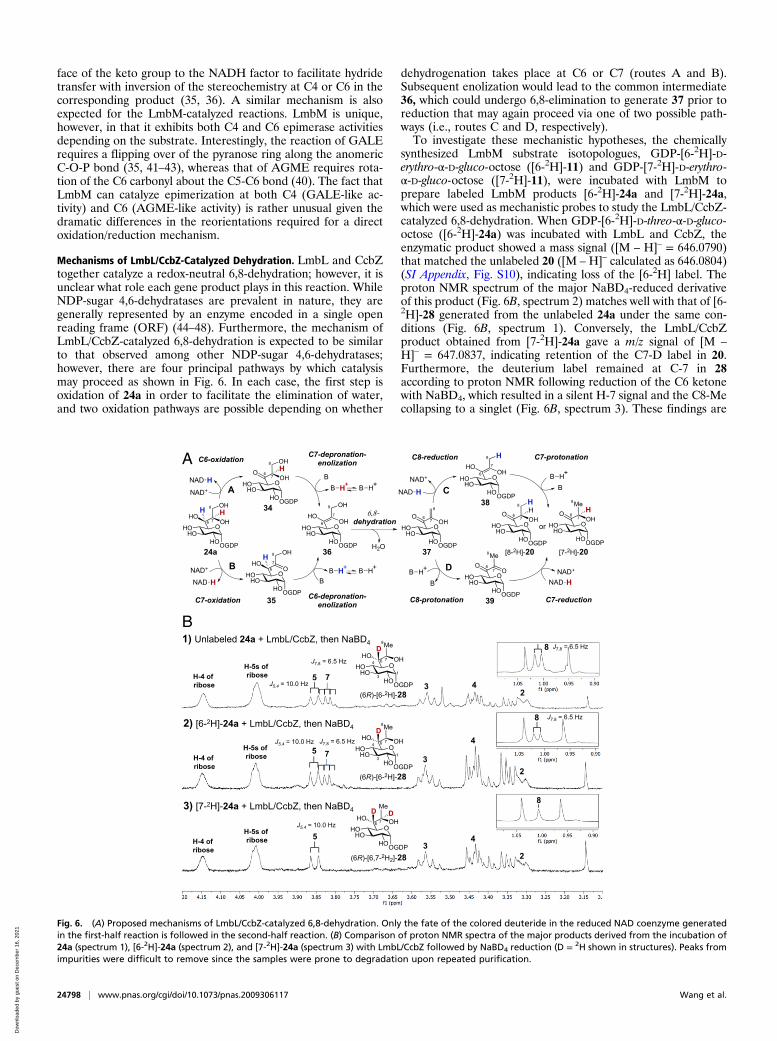
Mechanisms of LmbL/CcbZ-Catalyzed Dehydration. LmbL and CcbZtogether catalyze a redox-neutral 6,8-dehydration; however, it isunclear what role each gene product plays in this reaction. WhileNDP-sugar 4,6-dehydratases are prevalent in nature, they aregenerally represented by an enzyme encoded in a single openreading frame (ORF) (44–48). Furthermore, the mechanism ofLmbL/CcbZ-catalyzed 6,8-dehydration is expected to be similarto that observed among other NDP-sugar 4,6-dehydratases;however, there are four principal pathways by which catalysismay proceed as shown in Fig. 6. In each case, the first step isoxidation of 24a in order to facilitate the elimination of water,and two oxidation pathways are possible depending on whether
dehydrogenation takes place at C6 or C7 (routes A and B).Subsequent enolization would lead to the common intermediate36, which could undergo 6,8-elimination to generate 37 prior toreduction that may again proceed via one of two possible path-ways (i.e., routes C and D, respectively).To investigate these mechanistic hypotheses, the chemically
synthesized LmbM substrate isotopologues, GDP-[6-2H]-D-erythro-α-D-gluco-octose ([6-2H]-11) and GDP-[7-2H]-D-erythro-α-D-gluco-octose ([7-2H]-11), were incubated with LmbM toprepare labeled LmbM products [6-2H]-24a and [7-2H]-24a,which were used as mechanistic probes to study the LmbL/CcbZ-catalyzed 6,8-dehydration. When GDP-[6-2H]-D-threo-α-D-gluco-octose ([6-2H]-24a) was incubated with LmbL and CcbZ, theenzymatic product showed a mass signal ([M – H]– = 646.0790)that matched the unlabeled 20 ([M – H]– calculated as 646.0804)(SI Appendix, Fig. S10), indicating loss of the [6-2H] label. Theproton NMR spectrum of the major NaBD4-reduced derivativeof this product (Fig. 6B, spectrum 2) matches well with that of [6-2H]-28 generated from the unlabeled 24a under the same con-ditions (Fig. 6B, spectrum 1). Conversely, the LmbL/CcbZproduct obtained from [7-2H]-24a gave a m/z signal of [M –
H]– = 647.0837, indicating retention of the C7-D label in 20.Furthermore, the deuterium label remained at C-7 in 28according to proton NMR following reduction of the C6 ketonewith NaBD4, which resulted in a silent H-7 signal and the C8-Mecollapsing to a singlet (Fig. 6B, spectrum 3). These findings are
-
A
B
Fig. 6. (A) Proposed mechanisms of LmbL/CcbZ-catalyzed 6,8-dehydration. Only the fate of the colored deuteride in the reduced NAD coenzyme generatedin the first-half reaction is followed in the second-half reaction. (B) Comparison of proton NMR spectra of the major products derived from the incubation of24a (spectrum 1), [6-2H]-24a (spectrum 2), and [7-2H]-24a (spectrum 3) with LmbL/CcbZ followed by NaBD4 reduction (D = 2H shown in structures). Peaks fromimpurities were difficult to remove since the samples were prone to degradation upon repeated purification.
24798 | www.pnas.org/cgi/doi/10.1073/pnas.2009306117 Wang et al.
consistent with a mechanism in which the 6,8-dehydration isinitiated by dehydrogenation of the C7-hydroxyl by the active-site NAD+ cofactor, elimination of water, and C7 reduction bythe reduced active-site NADH cofactor to complete the catalyticcycle (24a→ 35→ 36→ 37→ 39→ 20, route B followed by routeD in Fig. 6A).Since the lmbL and lmbZ gene products display sequence
homology to UDP-D-glucose/GDP-D-mannose 6-dehydrogenaseand NAD(P)-dependent oxidoreductase, respectively, eitherLmbL or CcbZ could be directly responsible for the redox re-actions underlying the conversion of 24a to 20. However, se-quence alignment of LmbL, CcbL, LmbZ, and CcbZ with knownNDP-hexose 4,6-dehydratases (SI Appendix, Fig. S11) revealedthe absence of a YxxxK motif in these enzymes, which is con-sistent with no LmbL/CcbZ activity on the pyranose core of 24a.Moreover, LmbL and CcbL lack the GxxGxxG NAD+-bindingmotif that is conserved in all NDP-hexose 4,6-dehydratases,while both LmbZ and CcbZ contain the uncommon GxxWxxGmotif at the N terminus that may still serve to bind an NAD+
cofactor. To test this hypothesis, LmbL and CcbZ were sepa-rately denatured, and the respective supernatants were analyzedby liquid chromatography–mass spectrometry for the presence ofreleased cofactor. As expected, no NAD+/NADH was found tobe discharged from the denatured LmbL, whereas CcbZ wasfound to release NAD+/NADH (SI Appendix, Fig. S12). Theseobservations imply that CcbZ may be the catalytic componentdirectly responsible for dehydrogenation of the C7-hydroxylgroup in 24a and subsequent C7 reduction of the putativeintermediate 39.Initial dehydrogenation at the C7 position of 24a catalyzed by
CcbZ presumably necessitates tautomerization to an enediolintermediate such as 36 prior to a 1,4-dehydration (36 → 37) asshown in Fig. 7A. Since LmbL is also required for the 6,8-de-hydration reaction, it may play a role in tautomerization anddehydration (35 → 36 → 37 → 39). This stands in contrast to themore direct 1,2-dehydration (42 → 43) that has generally beensuggested for the NDP-sugar 4,6-dehydratases (Fig. 7B) (44–48).
In the case of the 4,6-dehydratases, deprotonation at C5 of 42leads to an α-carbanion in conjugation with the adjacent carbonyl atC4 effectively forming an enolate intermediate (not shown) duringwhat amounts to an E1cb-type dehydration. However, in view ofthe mechanism proposed for LmbL/CcbZ (Fig. 7A), deprotonationat C5 catalyzed by NDP-sugar 4,6-dehydatases could alternativelyresult in a tautomerization reaction (42 → 44) prior to a 1,4-dehydration (44→ 43) (Fig. 7B). In contrast, tautomerization to theenediol intermediate 36 appears to be necessary in the catalyticcycle of LmbL/CcbZ because 35 has no abstractable α-proton atC7. Thus, the dehydration catalyzed by LmbL/CcbZ represents a1,4-elimination that has not been previously reported.
ConclusionIn summary, four enzymes—LmbM, LmbL, CcbZ (LmbZequivalent), and CcbS (LmbS equivalent)—have been shown tocatalyze the conversion of 11 to GDP-D-α-D-lincosamide (12)during the biosynthesis of lincomycin. This pathway involves C6-epimerization of 11 to 24a catalyzed by LmbM, dehydration of24a to 20 catalyzed by LmbL/CcbZ (i.e., LmbL/LmbZ), C4-epimerization of 20 to 23 also catalyzed by LmbM, and finallythe CcbS (i.e., LmbS) catalyzed transamination of 23 to 12 asshown in Fig. 4. There are several features of this pathway thatare of particular interest and hence distinguish it from otherbiosynthetic/metabolic pathways involving the epimerization andα,γ-dehydration of carbohydrate molecules. First, LmbM cata-lyzes epimerization at either the C6 or C4 position depending onthe structural features of its GDP-octose substrate. This unusualcatalytic property separates it from the related epimerasesAGME and GALE, which appear to be much more regiospecific(38–42). Interestingly, both the C6- as well as the C4-epimerizationreactions catalyzed by LmbM (as well as AGME and GALE) in-volve hydride abstraction from the substrate and return back to theresulting oxidized intermediate at the same site to effect the changein stereochemistry. Thus, while the regiochemistry of the LmbM-catalyzed reaction appears to be substrate specific, the overallhydride transfer remains faithfully “site” specific. Furthermore,
A
B
Fig. 7. (A) Established pathway for the conversion of 11 to 12 in the lincomycin biosynthesis. Reactions catalyzed by LmbM, CcbZ, and LmbL for the con-version of 24a to 20 are highlighted. The labeled carbons (C6, C7, and C8) in 35 to 39 are coplanar with C7 which has a sp2 configuration. The NAD+ coenzymein the active site of CcbZ is likely located at the si face of the above plane (see 35, 39). (B) Reaction catalyzed by CDP-α-D-glucose 4,6-dehydratase. The labeledcarbons (C4, C5, and C6) in 43 and 44 are also expected to be coplanar.
Wang et al. PNAS | October 6, 2020 | vol. 117 | no. 40 | 24799
although C4-epimerization during lincomycin biosynthesis is re-quired for achieving the D-galacto-pyranose configuration observedin the final octose core, the intermediary C6-epimerziation is alsonecessary to facilitate the subsequent dehydration of 24a to 20catalyzed by LmbL/CcbZ.Also of interest is the observation that the subsequent
α,γ-dehydration reaction (24a → 20, Fig. 7A) requires two geneproducts (LmbL and CcbZ) for activity rather than one as istypical of NDP-sugar α,γ-dehydratases (i.e., 4,6-dehydratases)(44–48). While the exact roles played by each of these compo-nents in the dehydration reaction remain to be fully elucidated,only CcbZ is expected to be directly responsible for the under-lying redox reactions that involve NAD+-mediated hydridetransfer from and return to the C7 alcohol/ketone. The LmbL/CcbZ-catalyzed reaction is thus mechanistically unique as itproceeds via dehydrogenation of the hydroxyl group at theβ-carbon (C7) rather than at the α-carbon (C6) as is commonlynoted (e.g., 40 → 42, Fig. 7B). This finding distinguishes LmbL/CcbZ from the NDP-sugar 4,6-dehydratases such as CDP-α-D-glucose 4,6-dehydratase (49, 50), which involve net hydridetransfer from the α-position to the γ-position (C4 to C6) duringthe dehydration of 40 to 41 (Fig. 7B). Thus, while these resultsraise questions regarding the detailed chemistry of the NDP-sugar dehydratases and epimerases in general, they have finallycompleted the description of the lincomycin biosynthetic path-way and serve to highlight the complex mechanistic subtletiesassociated with the biosynthesis of atypical carbohydrate naturalproducts.
Materials and MethodsMaterials and Bacterial Strains. The bacterial strains of Streptomyces lincol-nensis NRRL ISP-5355 (identical to ATCC 25466) and Streptomyces caelestisNRRL-2418 were obtained from the Agricultural Research Service CultureCollection of the National Center for Agricultural Utilization Research. E. coliDH5α, acquired from Bethesda Research Laboratories, was used for routinecloning experiments. The protein overexpression host E. coli BL21 star (DE3)was obtained from Invitrogen. Vectors for protein overexpression werepurchased from Novagen. All chemicals and reagents were purchased fromSigma-Aldrich Chemical or Fisher Scientific and were used without furtherpurification. Oligonucleotide primers were prepared by Integrated DNATechnologies. Kits for DNA gel extraction and spin minipreps were pur-chased from Qiagen. PureLink Genomic DNA Mini Kit was obtained fromInvitrogen. Thermococcus kodakaraensis (KOD) DNA polymerase was pur-chased from Novagen. A QuikChange site-directed mutagenesis kit wasobtained from Stratagene (later acquired by Agilent). Enzymes and molec-ular weight standards used for the cloning experiments were obtained fromNew England Biolabs. Reagents for SDS polyacrylamide gel electrophoresis(SDS/PAGE) were purchased from Bio-Rad, except the protein molecularweight markers, which were obtained from Invitrogen. Growth mediumcomponents were acquired from Becton Dickinson. Sterile syringe filters areproducts of Fisher Scientific. Amicon YM-10 ultrafiltration membranes werebought from Millipore. The analytical and semipreparative CarboPac PA1HPLCy (HPLC) columns were obtained from Dionex. Analytical C-18 HPLCcolumns were products of Varian. Semipreparative C-18 HPLC columns werepurchased from Fisher Scientific.
General Cloning and Expression of Enzymes. Standard genetic manipulationsof E. coli were performed as described by Sambrook and Russell (51). DNAsequencing was performed at the core facility of the Institute of Cellular andMolecular Biology, The University of Texas at Austin. DNA concentrationswere measured using a NanoDrop ND-1000 UV-vis instrument from ThermoFisher Scientific. Target genes lmbL, lmbM, ccbS, and ccbZ were amplifiedfrom their corresponding genomic DNA isolated from S. lincolnensis and S.caelestic using designed primer pairs, and they were cloned into pET24b(+),pET28b(+), and pET-MalE vectors (SI Appendix, Table S2). The resultingplasmids were used to transform E. coli BL21 Star (DE3) cells. The desiredenzymes were overexpressed and purified from E. coli according to thefollowing procedure. The overnight culture grown at 37 °C in 10 mL of Luriabroth medium containing kanamycin (30 μg/mL) was used to inoculate 1 L ofthe same medium in a 100-fold dilution. These cultures were incubated at37 °C with 200 rpm shaking until OD600 reached 0.5. Protein expression wasinduced by the addition of isopropyl β-D-1-thiogalactopyranoside to a final
concentration of 0.1 mM (adjusted to 50 μM for maltose-binding protein[MBP]-fused CcbS). After overnight incubation at 18 °C with 125 × g shaking,the cells were harvested by centrifugation at 4,500 × g for 15 min, resus-pended in 20 mL of 50 mM Tris(hydroxymethyl)-aminomethane (Tris) buffer(pH 8.0) containing 300 mM NaCl, 10 mM imidazole, and glycerol (10%, vol/vol), and disrupted by sonication. For CcbS isolation, excess PLP (1 mM) wasadded to the lysis buffer to aid the folding process of CcbS. Cell debris wasremoved by centrifugation at 20,000 × g for 20 min, and the supernatantwas mixed by slow agitation with nickel-nitrilotriacetic acid (Ni-NTA) resinfor 2 h at 4 °C. The slurry was transferred to a column and washed with100 mL of 50 mM Tris buffer (pH 8.0) containing 300 mM NaCl, 20 mM im-idazole, and glycerol (10%, vol/vol). The protein was eluted with 25 mL of50 mM Tris buffer (pH 8.0) containing 300 mM NaCl, 250 mM imidazole, andglycerol (10%, vol/vol). The pooled protein fractions were dialyzed threetimes against 1 L of 50 mM Tris buffer (pH 8.0) containing 300 mM NaCl and15% glycerol prior to storage at −80 °C. The CcbS protein without His6-tagwas obtained by in vitro tobacco etch virus (TEV) protease cleavage of theMBP from the MBP-CcbS fusion protein. Specifically, 5% (vol/vol) His6-taggedTEV protease was added to the solution containing purified MBP-CcbS tocleave the His10-MBP. The digestion was carried out for 24 h during dialysis.The protein mixture was then filtered through a pad of Ni-NTA resin twiceto remove His-tagged MBP and TEV. The Ni-NTA pad was further washedwith a two-column volume of 50 mM Tris buffer (pH 8.0) containing 300 mMNaCl, 20 mM imidazole, and glycerol (10%, vol/vol), and all protein con-taining filtrates were combined and concentrated with an Amicon ultra-15centrifugal filter unit with a 10-kDa cutoff prior to storage at −80 °C. Themolecular mass and purity of all purified enzymes were determined by SDS/PAGE analysis (SI Appendix, Fig. S1).
Chemical Synthesis. The chemical synthesis and structures of 11, 12, 22, [6-2H]-11, and [7-2H]-11 are described in SI Appendix, S2.1–S2.3, S2.7, and S2.8.
General HPLC Elution Conditions. Purification of GDP-octoses and HPLCanalysis of the enzymatic products was performed using a Dionex CarboPacPA1 analytical column (1 mL/min flow rate) or a Dionex CarboPac PA1semipreparative column (4 mL/min flow rate) with UV absorbance detectionat 254 nm. Gradient elution was performed under a two-solvent system withH2O as solvent A and 1.0 M NH4OAc(aq) as solvent B under the followingconditions: 0 to 2 min of 10% solvent B, 2 to 10 min of 10 to 50% solvent B,10 to 25 min of 50 to 90% solvent B, 25 to 27 min of 90% solvent B, and 27 to30 min of 90 to 10% solvent B.
General Screening for Enzymatic Activity. Enzymatic activity of a specificsubstrate was assayed by mixing 100 μM of the compound being tested and50 μM NAD+ with 2.5 μM enzyme or combination of enzymes in 100 mM Trisbuffer (pH 8.0) at room temperature for 30 min or 1 h. After incubation, theenzymes were removed by centrifugal filtration using YM-10 filters. Thefiltrate was analyzed by HPLC using a Dionex CarboPac PA1 analyticalcolumn.
CcbS Activity Assay. The putative substrate was incubated with CcbS protein(33 μM), L-glutamate (2 mM), and PLP (66 μM) in 100 mM Tris buffer (pH 8.0)at room temperature for 1 h. After the removal of proteins by centrifugalfiltration using YM-10 filters, the filtrate was analyzed by HPLC using aDionex CarboPac PA1 analytical column.
Reverse Transamination of GDP-D-α-D-Lincosamide (12) by CcbS. Synthetic GDP-D-α-D-lincosamide 12 (66 μM) (SI Appendix, S2.2) was incubated with CcbS (33μM), α-ketoglutarate (2 mM), and PLP (66 μM) in 100 mM Tris buffer (pH 8.0)at room temperature for 2 h. After incubation, the enzymes were removedby centrifugal filtration using YM-10 filters. The filtrate was analyzed byHPLC using a Dionex CarboPac PA1 analytical column.
Reduction of Enzymatic Reaction Products with NaBD4 or NaBH4. After incu-bation, the enzymes were removed by centrifugal filtration using YM-10filters. The filtrate was then incubated with 5 mM NaBD4 or NaBH4 inddH2O for 30 min, and the reaction was quenched with acetone. Theresulting mixture was analyzed by HPLC using a Dionex CarboPac PA1 an-alytical column, and the products were purified using a Dionex CarboPacPA1 semipreparative column if necessary.
Data Availability. All data associated with these studies are included in themain text or SI Appendix.
24800 | www.pnas.org/cgi/doi/10.1073/pnas.2009306117 Wang et al.
ACKNOWLEDGMENTS. Portions of the paper were developed from thethesis of C.-I.L. (2015) and the thesis of S.-A.W. (2019), The University ofTexas at Austin. This work was supported by grants from the NIH
(GM035906) and the Welch Foundation (F-1511). The Bruker AVANCE III500 NMR at The University of Texas at Austin was supported by the NSF (1S10 OD021508-01).
1. D. J. Mason, A. Dietz, C. DeBoer, Lincomycin, a new antibiotic. I. Discovery and bio-logical properties. Antimicrob. Agents Chemother. 1962, 554–559 (1962).
2. R. R. Herr, M. E. Bergy, Lincomycin, a new antibiotic. II. Isolation and characterization.Antimicrob. Agents Chemother. 1962, 560–564 (1963).
3. C. Lewis, H. W. Clapp, J. E. Grady, In vitro and in vivo evaluation of lincomycin, a newantibiotic. Antimicrob. Agents Chemother. 1962, 570–582 (1963).
4. D. J. Mason, C. Lewis, Biological activity of the lincomycin-related antibiotics. Anti-microb. Agents Chemother. 10, 7–12 (1964).
5. H. Hoeksema et al., Chemical studies on lincomycin. I. The structure of lincomycin.J. Am. Chem. Soc. 86, 4223–4224 (1964).
6. F. Reusser, Effect of lincomycin and clindamycin on peptide chain initiation. Anti-microb. Agents Chemother. 7, 32–37 (1975).
7. M. Hanada, M. Tsunakawa, K. Tomita, H. Tsukiura, H. Kawaguchi, Antibiotic Bu-2545,a new member of the celesticetin-lincomycin class. J. Antibiot. (Tokyo) 33, 751–753(1980).
8. S. Toda, S. Nakagawa, T. Naito, H. Kawaguchi, Structure of antibiotic Bu-2545, a newmember of the celesticetin-lincomycin class. J. Antibiot. (Tokyo) 34, 596–599 (1981).
9. H. Hoeksema, G. F. Crum, W. H. DeVries, Isolation and purification of celesticetin.Antibiot. Annu. 1955, 837–841 (1954).
10. H. Hoeksema, V. Celesticetin, Celesticetin. V. The structure of celesticetin. J. Am.Chem. Soc. 90, 755–757 (1968).
11. F. Schlünzen et al., Structural basis for the interaction of antibiotics with the peptidyltransferase centre in eubacteria. Nature 413, 814–821 (2001).
12. T. Tenson, M. Lovmar, M. Ehrenberg, The mechanism of action of macrolides, linco-samides and streptogramin B reveals the nascent peptide exit path in the ribosome.J. Mol. Biol. 330, 1005–1014 (2003).
13. S. Padberg, Anti-Infective Agents: Drug During Pregnancy and Lactation, (Elsevier,Amsterdam, ed. 3, 2015), Vol. chap. 2, pp. 115–176.
14. C.-I. Lin, R. M. McCarty, H.-w. Liu, The biosynthesis of nitrogen-, sulfur-, and high-carbon chain-containing sugars. Chem. Soc. Rev. 42, 4377–4407 (2013).
15. E. Sasaki, C.-I. Lin, K.-Y. Lin, H.-w. Liu, Construction of the octose 8-phosphate inter-mediate in lincomycin A biosynthesis: Characterization of the reactions catalyzed byLmbR and LmbN. J. Am. Chem. Soc. 134, 17432–17435 (2012).
16. C.-I. Lin, E. Sasaki, A. Zhong, H.-w. Liu, In vitro characterization of LmbK and LmbO:Identification of GDP-D-erythro-α-D-gluco-octose as a key intermediate in lincomycinA biosynthesis. J. Am. Chem. Soc. 136, 906–909 (2014).
17. Q. Zhao, M. Wang, D. Xu, Q. Zhang, W. Liu, Metabolic coupling of two small-moleculethiols programs the biosynthesis of lincomycin A. Nature 518, 115–119 (2015).
18. D. Zhang, Z. Tang, W. Liu, Biosynthesis of lincosamide antibiotics: Reactions associatedwith degradation and detoxification pathways play a constructive role. Acc. Chem.Res. 51, 1496–1506 (2018).
19. U. Peschke, H. Schmidt, H.-Z. Zhang, W. Piepersberg, Molecular characterization ofthe lincomycin-production gene cluster of Streptomyces lincolnensis 78-11. Mol. Mi-crobiol. 16, 1137–1156 (1995).
20. M. Koberská et al., Sequence analysis and heterologous expression of the lincomycinbiosynthetic cluster of the type strain Streptomyces lincolnensis ATCC 25466. FoliaMicrobiol. (Praha) 53, 395–401 (2008).
21. N. M. Brahme et al., Biosynthesis of the lincomycins. 2. Studies using stable isotopeson the biosynthesis of methylthiolincosaminide moiety of lincomycin A. J. Am. Chem.Soc. 106, 7878–7883 (1984).
22. N. M. Brahme et al., Biosynthesis of the lincomycins. 1. Studies using stable isotopeson the biosynthesis of the propyl-and ethyl-L-hygric acid moieties of lincomycins Aand B. J. Am. Chem. Soc. 106, 7873–7878 (1984).
23. J. Novotná et al., l-3,4-Dihydroxyphenyl alanine-extradiol cleavage is followed byintramolecular cyclization in lincomycin biosynthesis. Eur. J. Biochem. 271, 3678–3683(2004).
24. J. Novotna et al., Lincomycin biosynthesis involves a tyrosine hydroxylating hemeprotein of an unusual enzyme family. PLoS One 8, e79974 (2013).
25. L. Najmanová et al., Characterization of N-demethyllincosamide methyltransferasesLmbJ and CcbJ. ChemBioChem 14, 2259–2262 (2013).
26. J. Bauer et al., Structure and possible mechanism of the CcbJ methyltransferase fromStreptomyces caelestis. Acta Crystallogr. D Biol. Crystallogr. 70, 943–957 (2014).
27. P. Jiraskova et al., New concept of the biosynthesis of 4-alkyl-L-proline precursors oflincomycin, hormaomycin, and pyrrolobenzodiazepines: Could a γ-glutamyltransferasecleave the C–C bond? Front. Microbiol. 7, 276 (2016).
28. G. Zhong, Q. Zhao, Q. Zhang, W. Liu, 4-Alkyl-L-(dehydro)proline biosynthesis in actino-bacteria involves N-terminal nucleophile-hydrolase activity of γ-Glutamyltranspeptidasehomolog for C-C bond cleavage. Nat. Commun. 8, 1–10 (2017).
29. Z. Kamenik et al., C-C bond cleavage in biosynthesis of 4-alkyl-L-proline precursors oflincomycin and anthramycin cannot precede C-methylation. Nat. Commun. 9, 3167(2018).
30. G. Zhong, H. Chen, W. Liu, Reply to “C–C bond cleavage in biosynthesis of 4-alkyl-L-prolineprecursors of lincomycin and anthramycin cannot precede C-methylation”. Nat. Commun.9, 1–3 (2018).
31. Z. Kamenik et al., Deacetylation of mycothiol-derived “waste product” triggers thelast biosynthetic steps of lincosamide antibiotics. Chem. Sci. (Camb.) 7, 430–435(2016).
32. M. Wang, Q. Zhao, Q. Zhang, W. Liu, Differences in PLP-dependent cysteinyl pro-cessing lead to diverse S -functionalization of lincosamide antibiotics. J. Am. Chem.Soc. 138, 6348–6351 (2016).
33. R. Ushimaru, C.-I. Lin, E. Sasaki, H.-w. Liu, Characterization of enzymes catalyzingtransformations of cysteine S-conjugated intermediates in the lincosamide biosyn-thetic pathway. ChemBioChem 17, 1606–1611 (2016).
34. L. F. Leloir, The enzymatic transformation of uridine diphosphate glucose into a ga-lactose derivative. Arch. Biochem. Biophys. 33, 186–190 (1951).
35. P. A. Frey, “Complex pyridine nucleotide-dependent transformations” in PyridineNucleotide Coenzymes, D. Dolphin, R. Poulson, O. Avramovic, Eds. (Wiley-Interscience,1987), Part B, pp. 461–511.
36. H. M. Holden, I. Rayment, J. B. Thoden, Structure and function of enzymes of theLeloir pathway for galactose metabolism. J. Biol. Chem. 278, 43885–43888 (2003).
37. K. L. Kavanagh, H. Jörnvall, B. Persson, U. Oppermann, Medium- and short-chaindehydrogenase/reductase gene and protein families: The SDR superfamily: Func-tional and structural diversity within a family of metabolic and regulatory enzymes.Cell. Mol. Life Sci. 65, 3895–3906 (2008).
38. L. Ding, B. L. Seto, S. A. Ahmed, W. G. Coleman Jr., Purification and properties of theEscherichia coli K-12 NAD-dependent nucleotide diphosphosugar epimerase,ADP-L-glycero-D-mannoheptose 6-epimerase. J. Biol. Chem. 269, 24384–24390 (1994).
39. A. M. Deacon, Y. S. Ni, W. G. Coleman Jr., S. E. Ealick, The crystal structure ofADP-L-glycero-D-mannoheptose 6-epimerase: Catalysis with a twist. Structure 8,453–462 (2000).
40. J. A. Read, R. A. Ahmed, J. P. Morrison, W. G. Coleman Jr., M. E. Tanner, The mech-anism of the reaction catalyzed by ADP-β-L-glycero-D-manno-heptose 6-epimerase.J. Am. Chem. Soc. 126, 8878–8879 (2004).
41. J. B. Thoden, T. M. Wohlers, J. L. Fridovich-Keil, H. M. Holden, Crystallographic evi-dence for Tyr 157 functioning as the active site base in human UDP-galactose4-epimerase. Biochemistry 39, 5691–5701 (2000).
42. J. B. Thoden, P. A. Frey, H. M. Holden, Molecular structure of the NADH/UDP-glucoseabortive complex of UDP-galactose 4-epimerase from Escherichia coli: Implications forthe catalytic mechanism. Biochemistry 35, 5137–5144 (1996).
43. Y. Liu et al., Mechanistic roles of tyrosine 149 and serine 124 in UDP-galactose4-epimerase from Escherichia coli. Biochemistry 36, 10675–10684 (1997).
44. H.-w. Liu, J. S. Thorson, Pathways and mechanisms in the biogenesis of novel deox-ysugars by bacteria. Annu. Rev. Microbiol. 48, 223–256 (1994).
45. G. Samuel, P. Reeves, Biosynthesis of O-antigens: Genes and pathways involved innucleotide sugar precursor synthesis and O-antigen assembly. Carbohydr. Res. 338,2503–2519 (2003).
46. X. M. He, H.-w. Liu, Formation of unusual sugars: Mechanistic studies and biosyntheticapplications. Annu. Rev. Biochem. 71, 701–754 (2002).
47. C. J. Thibodeaux, C. E. Melançon, H.-w. Liu, Unusual sugar biosynthesis and naturalproduct glycodiversification. Nature 446, 1008–1016 (2007).
48. C. J. Thibodeaux, C. E. Melançon III, H.-w. Liu, Natural-product sugar biosynthesis andenzymatic glycodiversification. Angew. Chem. Int. Ed. Engl. 47, 9814–9859 (2008).
49. Y. Yu, R. N. Russell, J. S. Thorson, L. D. Liu, H.-w. Liu, Mechanistic studies of the bio-synthesis of 3,6-dideoxyhexoses in Yersinia pseudotuberculosis. Purification and ste-reochemical analysis of CDP-D-glucose oxidoreductase. J. Biol. Chem. 267, 5868–5875(1992).
50. C.-T. Chang, X. H. Chen, H.-w. Liu, CDP-6-deoxy-6,6-difluoro-D-glucose, a mechanism-based inhibitor for CDP-D-glucose 4,6-dehydratase. J. Am. Chem. Soc. 120, 9698–9699(1998).
51. J. Sambrook, D. W. Russell, Molecular Cloning: A Laboratory Mannual, (Cold SpringHarbor Laboratory Press, ed. 3, 2001).
Wang et al. PNAS | October 6, 2020 | vol. 117 | no. 40 | 24801