1 STUDIES ON BIODEGRADATION OF NITROPHENOL ISOMERS BY MIXED BACTERIAL CULTURES A thesis submitted to the UNIVERSITY OF MYSORE for the Degree of DOCTOR OF PHILOSOPHY IN BIOTECHNOLOGY By SHABANA BASHEER Fermentation Technology & Bioengineering Department Central Food Technological Research Institute Mysore-570 013, India October - 2003
Transcript
1
STUDIES ON BIODEGRADATION OF NITROPHENOL ISOMERS BY MIXED BACTERIAL
CULTURES
A thesis submitted to the UNIVERSITY OF MYSORE
for the Degree of DOCTOR OF PHILOSOPHY
IN BIOTECHNOLOGY
By SHABANA BASHEER
Fermentation Technology & Bioengineering Department Central Food Technological Research Institute
Mysore-570 013, India October - 2003
2
DECLARATION
I hereby declare that the thesis entitled "Studies on biodegradation of nitrophenol isomers by mixed bacterial cultures " submitted for the degree of Doctor
of Philosophy in Biotechnology to the University of Mysore is the result of work carried
out by me under the guidance of Dr. S. Divakar in the Department of Fermentation
Technology and Bioengineering, Central Food Technological Research Institute,
Mysore during the period November 1998 to October 2003.
I further declare that the results of the work have not been submitted for the
award of any degree, diploma or fellowship.
Date : 29-10-2003 Shabana Basheer
Mysore
3
Dr S. DIVAKAR
Scientist
Fermentation Technology & Bioengineering Dept.
29th October 2003
CERTIFICATE
I hereby declare that the thesis entitled "Studies on biodegradation of nitrophenol isomers by mixed bacterial cultures" submitted by Ms. Shabana
Basheer for the degree of Doctor of Philosophy in Biotechnology of the University of
Mysore is the result of research work carried out by her at the Department of
Fermentation Technology and Bioengineering, CFTRI, Mysore under my guidance
during the period from November 1998 to October 2003
Dr. S. Divakar
Guide
4
To my sister… Rahmathunnisa S.A.
5
ACKNOWLEDGEMENTS I think if anyone honestly reflects on who we are, how we got there, what we think we
might do well, and so forth, we discover a debt to others. This page is specifically
designed to note my appreciation of those people who stand out most notably in my
mind as to contributing towards my thesis.
Words are inadequate to express my gratitude to Dr. Divakar S., Scientist, Fermentation Technology and Bioengineering, CFTRI, my guide and mentor, for his
rock solid support and constructive criticism. I can only hope that a small part of his
chemical intuition, breadth of knowledge and depth of understanding has rubbed off on
me
I thank Dr. Kunhi A.A.M., Scientist (Retd), Food Microbiolgy, CFTRI, my erstwhile
supervisor, for taking me as a Research Assistant and kindling interest and leading me
towards Doctorate studies
I wish to sincerely thank Dr. Prakash V., Director, CFTRI, for providing infrastructure,
lending moral support and for always being there to see me through my difficulties
I thank Dr. N.G. Karanth, Head, FTBE, for his support and encouragement
I am indebted to Dr. M.C. Mishra, Scientist, FTBE, for the inumerable favours,
foremost being free access to the lab, his moral support and affection
I owe a debt of gratitude to the staff at Dept. of FTBE- Mr. N.P. Ghildyal, Dr. S.G. Prafulla, Dr.M.S. Thakur, Dr. Avinash S., Mr. Eugene Raj, Mr. Varadaraj and others
who made my Doctorate studies possible with their overwhelming and unforgettable
support
6
I specially thank Dr.M.K. Gowthaman, Scientist, CLRI, for just being himself and
keeping me sane and positive in my moments of madness
I thank Dr. M.C. Varadaraj, Head, HRD, for characterizing the bacterial cultures and for
being so kind
I appreciate Mr. M.R. Radhakantha, COA, for his encouragement to move ahead
I thank Mr. Akmal Pasha and staff, FICP, for their help
I have been fortunate to have some of the best and most supporting friends at FTBE. The cheerfulness and happiness brought in by them shall always be cherished
I thank the Staff and my colleagues at FM, for their co-operation
I deeply appreciate the technical and research assistance accorded by the Central Intruments Facility & Services, FOSTIS, CFTRI, and Sophisticated Intruments Facilty, Indian Institute of Science, Bangalore
I thank Mr. Abdul Khayoum, for all his help in formatting the thesis.
I am thankful to the Security Staff, CFTRI for providing me security
I wish to thank the Council of Scientific and Industrial Research, Govt. of India for
awarding me a Senior Research Fellowship
I gratefully thank my Dee, family and friends, who stood by me with love, forgiveness
and lots of patience
I also appreciate many others whom I have never met but whose published work
inspired me
7
I have saved the best for the last and the best compliments are for my parents, Mr. S.A. Basheer and Mrs. Tajunnisa, whom I feel God sent to make me strong and took them
away to make me stronger
I close by thanking Almighty God for these people and my thesis. Whatever good may
come of this work, the credit belongs to Him. After all, He already knows how microbes
work
Shabana Basheer
8
CONTENTS
Particulars Page No.
CHAPTER 1: INTRODUCTION 1
1.1 NITROAROMATIC COMPOUNDS 1
1.2 REVIEW OF LITERATURE 2
1.2.1 Biodegradation of nitroaromatic compounds 3
1.2.2 Microbial mineralization of nitroaromatic compounds 12
1.2.2.1 An initial oxygenation reaction yielding nitrite 13
1.2.2.2 Reductive transformation reaction 13
1.2.2.3 Complete reductive removal of the nitro group by
the formation of a hydride-Meisenheimer complex
16
1.2.2.4 Degradation of nitroaromatics via partial
reduction and replacement reactions
16
1.2.3 Anaerobic degradation of nitroaromatic compounds 17
CHAPTER 6: NUCLEAR MAGNETIC RESONANCE SPECTROSCOPIC STUDIES OF THE MICROBIAL DEGRADATION OF MONONITROPHENOL ISOMERS
91
6.1 INTRODUCTION 91
6.2 RESULTS 92
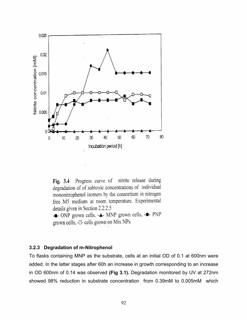
6.2.1 Degradation by the microbial consortium 93
6.2.1.1 o-Nitrophenol 93
6.2.1.2 m-Nitrophenol 93
6.2.1.3 p-Nitrophenol 94
6.2.2 Degradation by the bacterial isolate [SNP-8] 95
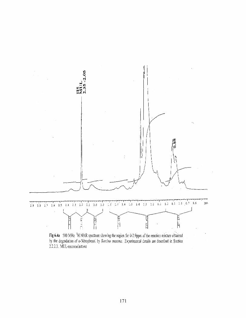
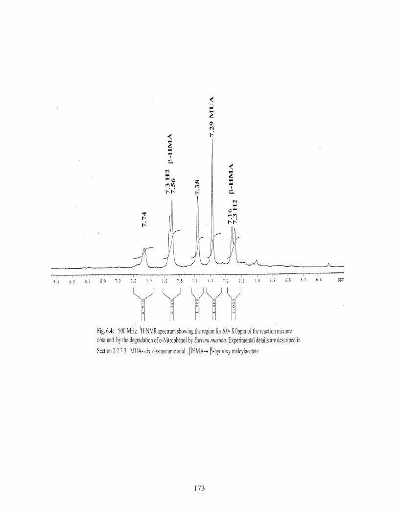
6.2.2.1 o-Nitrophenol 95
6.2.2.2 m-Nitrophenol 96
17
6.2.2.3 p-Nitrophenol 96
6.3 DISCUSSION 97
CHAPTER 7: CONCLUSIONS 101
SCOPE 105
SUMMARY 107
REFERENCES 112
18
LIST OF FIGURES Fig No.
Title of the figure
CHAPTER 1 1.1 Initial oxgenolytic reaction yielding nitrite and catechol 1.2 Pathway of the removal of the nitro group from nitroaromatics by
initial reduction reactions 1.3 Reduction of nitro groups by one electron and two electron
mechanisms 1.4 Complete reductive removal of nitro group from nitroaromatic
compounds 1.5 Partial reduction and replacement reaction 1.6 Monooxygenase catalyzed initial reaction in biodegradation of
nitroaromatic compounds 1.7 Dioxygenase catalyzed initial reaction in biodegradation of
nitroaromatic compounds
CHAPTER 2 2.1 Spectrophotometric calibration of standard ONP 2.2 Spectrophotometric calibration of standard MNP 2.3 Spectrophotometric calibration of standard PNP 2.4 Calibration of standard ONP by HPLC 2.5 Calibration of standard MNP by HPLC 2.6 Calibration of standard PNP by HPLC 2.7 Spectrophotometric calibration of standard Ammonia 2.8 Spectrophotometric calibration of standard Nitrite
19
CHAPTER 3
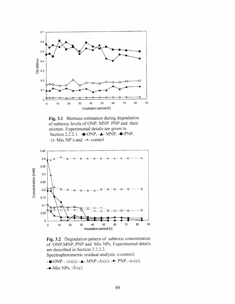
3.1 Biomass estimation during degradation of subtoxic levels of ONP,
MNP, PNP and their mixture
3.2 Degradation pattern of subtoxic concentrations of ONP, MNP,
PNP and Mix NPs
3.3 Degradation pattern of subtoxic concentrations of ONP, MNP,
PNP. Residual substrate analysis by HPLC
3.3a HPLC profile during degradation of subtoxic concentration of
ONP
3.3b HPLC profile during degradation of subtoxic concentration of
MNP
3.3c HPLC profile during degradation of subtoxic concentration of PNP
3.3d HPLC profile during degradation of subtoxic concentration of a
mixture of mononitrophenol isomers
3.4 Progress curve of nitrite release during degradation of subtoxic
concentrations individual mononitrophenol isomers and their
mixture by the consortium
3.5 Progress curve of ammonia production during MNP degradation
by the nitrophenol degrading consortium
3.6 Degradation pattern of a mixture of ONP, MNP and PNP.
Residual substrate analysis by HPLC
3.7 Degradation of 0.1mM mononitrophenols by the consortium
induced with 0.5% sodium acetate
3.8 Degradation of 0.1mM mononitrophenol isomers by the
consortium induced with o-cresol
3.9 Degradation of 0.1mM mononitrophenol isomers by the
20
consortium induced with m-cresol
3.10 Degradation of 0.1mM mononitrophenol isomers by the
consortium induced p-cresol
3.11 Degradation of 0.1mM mononitrophenols by the consortium
induced with 1mM phenol and 0.1mM Mix NPs
3.12 Degradation of 0.1mM mononitrophenol isomers by the
consortium induced with phenol
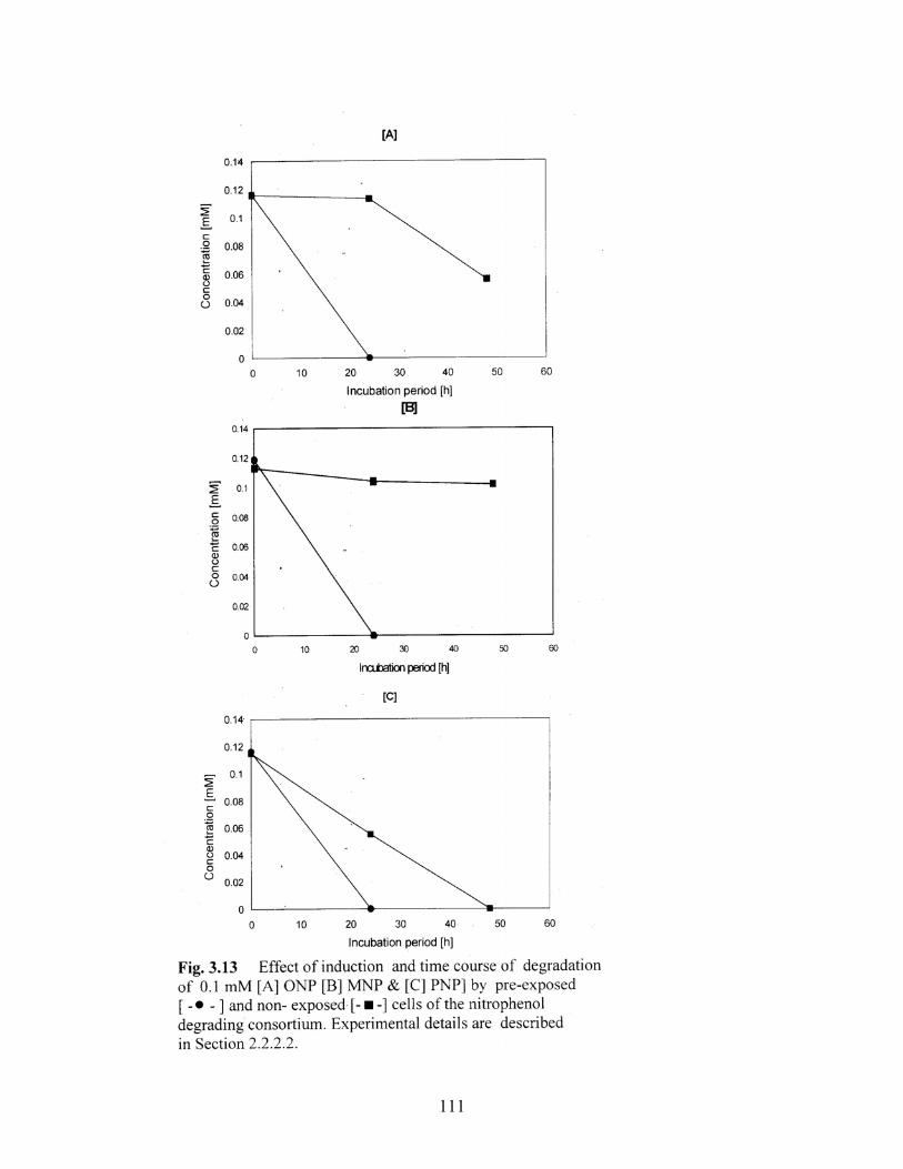
3.13 Effect of induction and time course of degradation of 0.1mM
[a]ONP [b]MNP and [c]PNP by pre-exposed and non-preexposed
cells of the nitrophenol degrading consortium
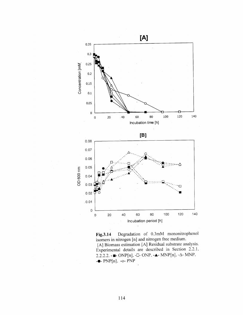
3.14 Degradation of 0.3mM mononitrophenol isomers in nitrogen and
nitrogen free medium
3.15 Degradation pattern of subtoxic to toxic concentrations of ONP by
the nitrophenol degrading consortium
3.16 Degradation pattern of subtoxic to toxic concentrations of MNP by
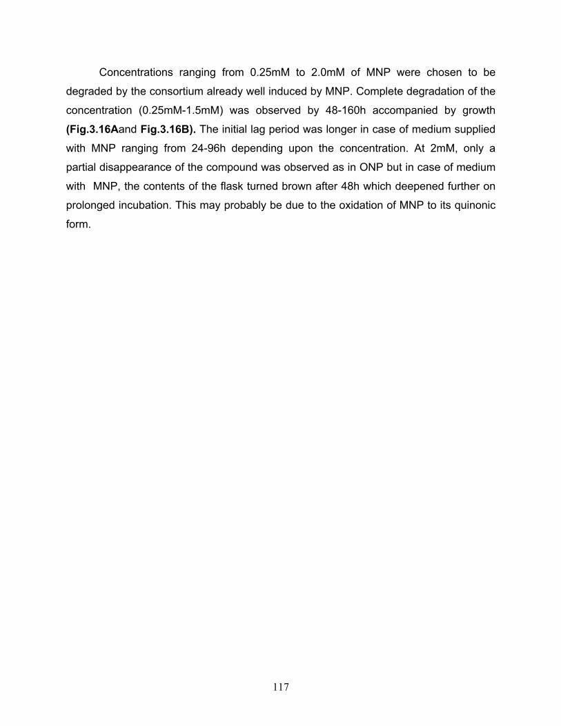
the nitrophenol degrading consortium
3.17 Degradation pattern of subtoxic to toxic concentrations of PNP by
the nitrophenol degrading consortium
CHAPTER 4
4.1 Percent removal of 0.1mM substrate by Bacillus licheniformis
(SNP-1)
4.2 Percent removal of 0.1mM substrate by Xanthomonas maltophila
(SNP-2)
4.3 Percent removal of 0.1mM substrate by Serratia liquefaciens
(SNP-3)
4.4 Percent removal of 0.1mM substrate by Pseudomonas putida
21
(SNP-4)
4.5 Percent removal of 0.1mM substrate by Pseudomonas sp.(SNP-
5)
4.6 Percent removal of 0.1mM substrate by Pseudomonas
alcaligenes (SNP-6)
4.7 Percent removal of 0.1mM substrate by Psuedomonas sp. (SNP-
7)
4.8 Percent removal of 0.1mM substrate by Sarcina maxima (SNP-8)
4.9 Degradation pattern of a mixture of mononitrophenol isomers by
SNP-1
4.10 Degradation pattern of a mixture of mononitrophenol isomers by
SNP-2
4.11 Degradation pattern of a mixture of mononitrophenol isomers by
SNP-3
4.12 Degradation pattern of a mixture of mononitrophenol isomers by
SNP-4
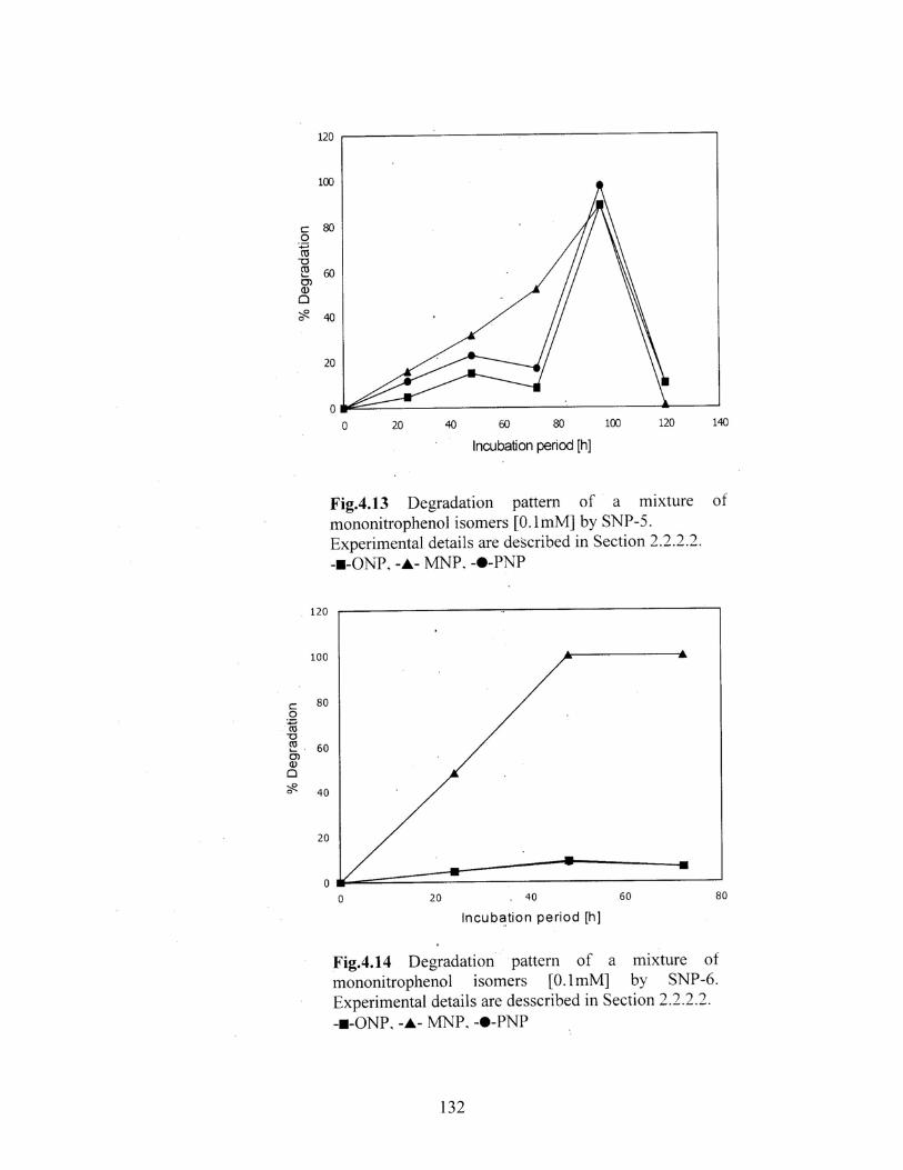
4.13 Degradation pattern of a mixture of mononitrophenol isomers by
SNP-5
4.14 Degradation pattern of a mixture of mononitrophenol isomers by
SNP-6
4.15 Degradation pattern of a mixture of mononitrophenol isomers by
SNP-7
4.16 Degradation pattern of a mixture of mononitrophenol isomers by
SNP-8
4.17 Degradation of 0.1mM substrate by well induced cells of Sarcina
maxima
4.18 Percentage of ONP degradation and nitrite release by Sarcina
22
maxima
4.19 Percentage of MNP degradation and nitrite release by Sarcina
maxima
4.20 Degradation of 0.1mM individual mononitrophenols by Sarcina
maxima. Residual substrate analysis by HPLC
4.21 Percent degradation of [A]0.1mM [B]0.2mM [C]0.5mM of
individual mononitrophenol isomers and their mixture by Sarcina
maxima [SNP-8]
4.22 Degradation of a mixture of mononitrophenol isomers by Sarcina
maxima [SNP-8]. Residual analysis by HPLC
CHAPTER 5
5.1 Biomass estimation during degradation of 0.8mM individual
mononitrophenol isomers
5.2 Degradation pattern during degradation of 0.8mM individual
mononitrophenol isomers by the consortium
5.3 Catechol-1,2-dioxygenase activity as observed in cell free
extracts of the nitrophenol degrading consortium induced with
ONP/MNP/PNP
5.4 Catechol-2,3- dioxygenase activity as observed in cell free
extracts of the nitrophenol degrading consortium induced with
ONP/MNP/PNP
CHAPTER 6
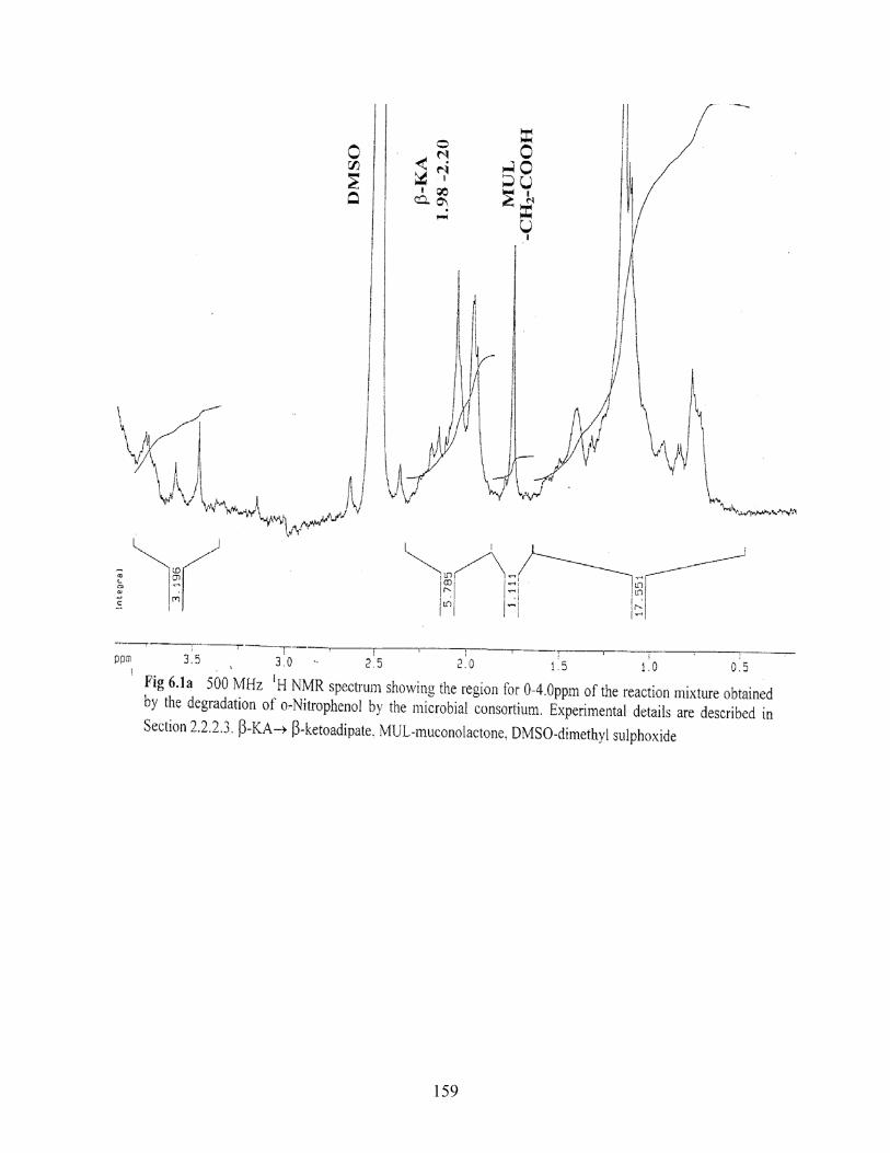
6.1a 500MHz 1H NMR spectrum showing the region for 0-4.0ppm of
the reaction mixture obtained by the degradation of ONP by the
microbial consortium
6.1b 500MHz 1H NMR spectrum showing the region for 6.5-9.5ppm of
the reaction mixture obtained by the degradation of ONP by the
23
microbial consortium
6.2a 500MHz 1H NMR spectrum showing the region for 6.0-8.5ppm of
the reaction mixture obtained by the degradation of MNP by the
microbial consortium
6.2b 500MHz 1H NMR spectrum showing the region for 5.5-9.0ppm of
the reaction mixture obtained by the degradation of MNP by the
microbial consortium
6.3a 500MHz 1H NMR spectrum showing the region for 0.5-5.5ppm of
the reaction mixture obtained by the degradation of PNP by the
microbial consortium
6.3b 500MHz 1H NMR spectrum showing the region for 5.5-11ppm of
the reaction mixture obtained by the degradation of PNP by the
microbial consortium
6.4a 500MHz 1H NMR spectrum showing the region for 0-2.9ppm of
the reaction mixture obtained by the degradation of ONP by
Sarcina maxima
6.4b 500MHz 1H NMR spectrum showing the region for 3.0-6.0ppm of
the reaction mixture obtained by the degradation of ONP by
Sarcina maxima
6.4c 500MHz 1H NMR spectrum showing the region for 6.0-8.0ppm of
the reaction mixture obtained by the degradation of ONP by
Sarcina maxima
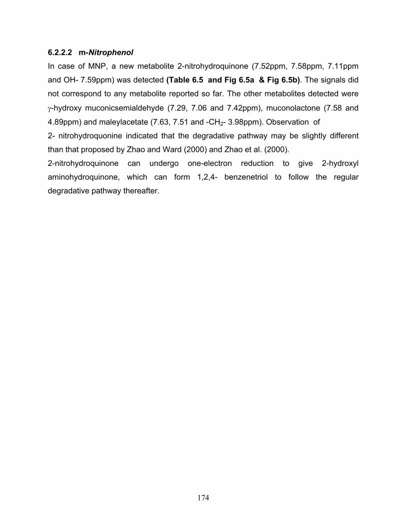
6.5a 500MHz 1H NMR spectrum showing the region for 6.85-7.80ppm
of he reaction mixture obtained by the degradation of MNP by
Sarcina maxima
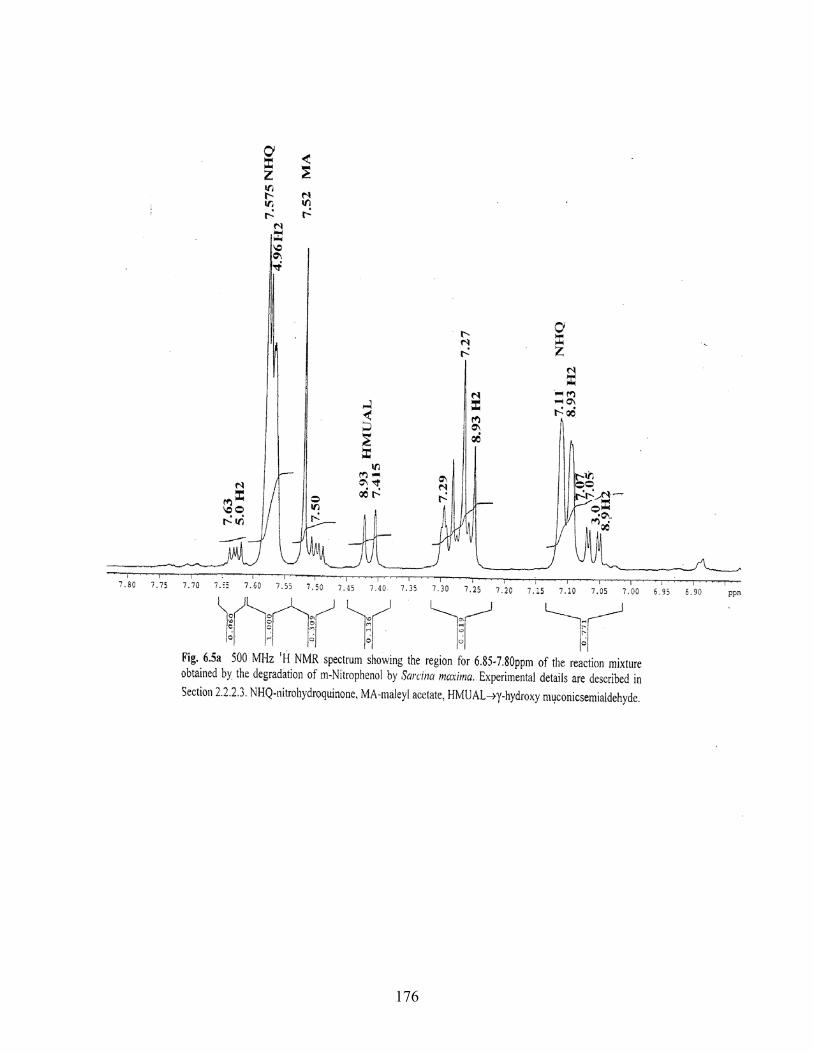
6.5b 500MHz 1H NMR spectrum showing the region for 2.0-5.0ppm of
the reaction mixture obtained by the degradation of MNP by
Sarcina maxima
24
6.6a 500MHz 1H NMR spectrum showing the region for 0.5-4.5ppm of
the reaction mixture obtained by the degradation of PNP by
Sarcina maxima
6.6b 500MHz 1H NMR spectrum showing the region for 6.4-8.5ppm of
the reaction mixture obtained by the degradation of PNP by
Sarcina maxima
6.6c 2D HMQCT spectrum showing the region for 0-10ppm of he
reaction mixture obtained by the degradation of PNP by Sarcina
maxima
6.6d 2D HMQCT spectrum showing the region for 0-5.5ppm of he
reaction mixture obtained by the degradation of ONP by Sarcina
maxima
SCHEMES
6.1 Pathway followed by the [A] Consortium [B] Sarcina maxima for
ONP degradation
6.2 Pathway followed by [A] Consortium [B] Sarcina maxima for MNP
degradation
6.3 Pathway followed by [A] Consortium [B] Sarcina maxima for MNP
degradation
25
LIST OF TABLES Table No.
Title
1.1 Representative bacteria reported to degrade nitroaromatic
compounds.
1.2 Chemical and physical characteristics of ONP, MNP and PNP
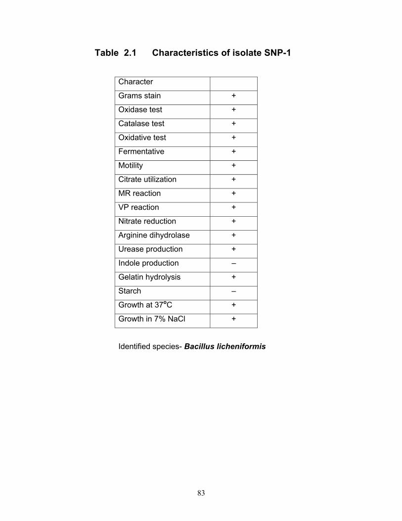
2.1 Characteristics of the bacterial isolate SNP-1
2.2 Characteristics of the bacterial isolates SNP-2, SNP-3, SNP-4,
SNP-5, SNP-6 and SNP-7
2.3 Characteristics of the bacterial isolate SNP-8
6.1 1H NMR data of the degradation of ONP by the consortium
6.2 1H NMR data of the degradation of MNP by the consortium
6.3 1H NMR data of the degradation of PNP by the consortium
6.4 NMR data of the degradation of ONP by Sarcina maxima
6.5 NMR data of the degradation of MNP by Sarcina maxima
6.6 NMR data of the degradation of PNP by Sarcina maxima
Biotechnology encompasses an important science, Bioremediation, which
significantly deals with biotic transformations of consequential pollutants/contaminants.
It offers various options for combating the menace of disturbing ecosystems arising due
to irate xenobiotics. Today we talk in terms of not only pollutant/xenobiotic residues but
also their conjugates and bound forms. Hence both biotic and abiotic transformations of
parent xenobiotics and their fate and consequence in soil, water and air have generated
immense interest. Soil is a major reservoir of microorganisms that plays an important
role in maintaining its fertility. Xenobiotic compounds introduced into soil present
daunting challenges to the soil microflora.
1.1 NITROAROMATIC COMPOUNDS
Nitroaromatic compounds form an important class of xenobiotic compounds. A vast
majority of these compounds detected in the environment are anthropogenic in nature
and nitrosubstituted aromatic compounds are important building blocks for the large
scale synthesis of pesticides, pharmaceuticals, plastics, azo dyes and explosives and
also serve as precursors for the production of aminoaromatic derivatives (Kearney and
Kaufmann, 1976; McCormick et al., 1976; Schwarzenbach et al., 1988). As a
consequence, nitroaromatic compounds have become pollutants in rivers, wastewaters,
groundwater, pesticide treated soils and the urban atmosphere (Golab et al., 1979;
Grosjean, 1985, Keith and Telliard, 1979; Piet and Smeenk, 1985; Zoeteman et al.,
1980). Nitroaromatics are also present in combustion emissions and airborne particulate
matter (Meijers and vander Lur, 1976; Pitts, 1982; Schuetzle, 1983; Tokiwa and
Ohnishi, 1986). Nitrobenzenes, nitrotoluenes, nitrophenols and nitrobenzoates are used
in the manufacture of pesticides, dyes, explosives, polyurethane foams, elastomers and
industrial solvents. Nitrobiphenyls are important plasticizers and wood preservatives
29
(Masse et al., 1985). Chloramphenicol and nitrozepam are example of drugs. HMX,
RDX and TNT have been extensively used as explosives and pose, currently, the most
visible environmental problem (Hartter, 1985). The discharge of nitroaromatic
compounds in wastewater and application as pesticides (Parathion, Dinoseb,
Fenitrothion) have broadened their environmental impact and called for solutions for
redemption of these toxic compounds. Some are highly mutagenic and toxic. Ortho-
nitrophenol (ONP), 2,4-dinitrophenol and para-nitrophenol (PNP) are listed as priority
pollutants by the U.S Environmental Protection Agency (Callahan et al., 1979; Keith and
Telliard, 1979). As the demand for agricultural produce increases, so inevitably does the
need for pesticides. Currently, organophosphate compounds are one of the most widely
used class of pesticides in industrialized countries. High level exposure to these
neurotoxins results in acetylcholine accumulation, which interferes with muscular
responses, leading to the possibility of death. Repeated or prolonged exposure can
cause delayed cholinergic toxicity and neurotoxicity (Tuovinen et al., 1994). Parathion
and Methyl parathion are two popular organophosphate pesticides used for agricultural
crop protection (Kumar et al., 1996). PNP is not only used extensively in manufacturing
processes but is also a major metabolite in the hydrolysis of parathion and methyl
parathion. As a result it can build up in the soil. These compounds may enter industrial
waste streams, where they cause deleterious consequences for the following reasons:
(i) the majority of nitroaromatic compounds are highly toxic to microorganism and may
destabilize the continuos treatment systems by inhibition of growth; (ii) nitro groups and
chloro substituents, reduce the electron density of the aromatic ring and thus impede
electrophilic attack of oxygenases and oxidative degradation of nitroaromatic
compounds; (iii) because of their electrophilic character they are also subject to
reduction of the nitro groups which generate biologically inert azo, azoxy- and polymeric
compounds (McCormick et al., 1976, 1978). Biologically, nitroaromatic compounds
are either simply transformed to dead end products, by several microorganisms, which
many a time prove to be more toxic than the parent compound or they may actually
utilize the nitroaromatic compounds as a carbon and/or nitrogen sources.
30
1.2 REVIEW OF LITERATURE 1.2.1 Biodegradation of nitroaromatic compounds Considerable amount of work has been done on development of treatment systems by
biodegradation. It has been observed that microorganisms have capacity to convert
nitroaromatic compounds into intermediates that can serve as growth substrates.
Populations of microbes able to degrade nitroaromatics or any other compounds can
arise by different means. If the chemical in question is a close analog to an ubiquitous
microbial substrate, native soil microflora may degrade the molecule. Degradative
populations could still arise through natural selection in contaminated environments. In
the former case, biodegradation by in situ microorganisms should always be possible,
while in the latter it might occur only at older spill sites. Because of natural selection
process, it is commonly assumed that a bacterial population in older, more heavily
contaminated spill sites will be more adapted to degradation of the contaminant. Such
organisms may be suitable candidates for use in bioremediation (Crawford, 1995;
Kaake et al., 1994; Marvin-Sikkema and de Bont, 1994). Despite the toxicity of
nitroaromatic compounds, many microorganisms are able to transform or degrade them
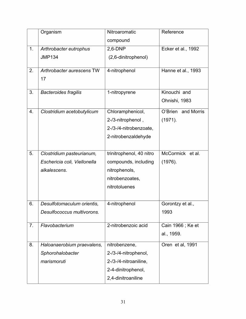
(Table 1.1).
Table 1.1 Representative bacteria reported to degrade nitroaromatic compounds
31
Organism Nitroaromatic
compound
Reference
1. Arthrobacter eutrophus
JMP134
2,6-DNP
(2,6-dinitrophenol)
Ecker et al., 1992
2. Arthrobacter aurescens TW
17 4-nitrophenol Hanne et al., 1993
3. Bacteroides fragilis 1-nitropyrene Kinouchi and
Ohnishi, 1983
4. Clostridium acetobutylicum Chloramphenicol,
2-/3-nitrophenol ,
2-/3-/4-nitrobenzoate,
2-nitrobenzaldehyde
O’Brien and Morris
(1971).
5. Clostridium pasteurianum,
Eschericia coli, Viellonella
alkalescens.
trinitrophenol, 40 nitro
compounds, including
nitrophenols,
nitrobenzoates,
nitrotoluenes
McCormick et al.
(1976).
6. Desulfotomaculum orientis,
Desulfococcus multivorons.
4-nitrophenol Gorontzy et al.,
1993
7. Flavobacterium 2-nitrobenzoic acid Cain 1966 ; Ke et
al., 1959.
8. Haloanaerobium praevalens,
Sphorohalobacter
marismoruti
nitrobenzene,
2-/3-/4-nitrophenol,
2-/3-/4-nitroaniline,
2-4-dinitrophenol,
2,4-dinitroaniline
Oren et al, 1991
32
9. Methanobaterium formicicum 3-nitrophenol,
4-nitrophenol,
2,4-dinitrophenol,
4-nitrobenzoic acid,
4-nitroaniline
Gorontzy et al.,
1993
10. Methanosarcina barkeri 3-nitrophenol,
4-nitrophenol,
2,4-dinitrophenol,
4-nitrobenzoic acid,
4-nitroaniline
Gorontzy et al.,
1993
11. Moraxella sp. 4-nitrophenol Spain et al., 1997
12. Nocardia sp.strainTW12 4-nitrophenol Hanne et al., 1993
13. Nocardia alba 2,4-dinitrophenol Germanier and
Wuhrman , 1963.
14. Pseudomonas putida B2 3-nitrophenol Zeyer et al., 1986
Arthrobacter protophormiae RKJ 100 and Burkholderia cepacia RKJ 200 were reported
by Bhushan et al. (2000) to be using PNP as sole C-, N- and energy source. A PNP
adapted microbial population (from an activated sludge) retained in porous particles
utilized PNP as a sole C-source and degraded PNP releasing nitrite without significant
accumulation of intermediate metabolites (Xing et al., 1999). Ramanathan and
Lalithakumari (1999) observed that Pseudomonas sp. A3, isolated from soil, shown to
degrade methyl parathion (MP) and other pesticides used the aromatic portion (PNP) as
a C- and energy source during hydrolysis of MP. Three Arthrobacter sp. isolated from
parathion contaminated soil could use PNP as C-source (Hanne et al., 1991).
Additional carbon sources and inorganic nutrients have been shown to have a
profound effect on the degradation of nitrophenols. Mohammed et al.,
(1992) isolated from industrial sludge, a strain of Pseudomonas cepacia capable of
using either PNP, DNP, DNOC or NB as its sole N-source but utilized succinic acid as a
primary C-source.. Addition of citrate as a secondary C-source could not improve
bacterial growth of Pseudomonas putida 2NP8 on nitrobenzene but the strain was able
to use ONP and MNP (m-Nitrophenol) as growth substrates (Zhao and Ward, 2000). A
mixed culture comprising of Comomonas testosteronii and Acidovorax delafieldie
showed no increase in rate of growth and degradation of 20mg/l PNP with the addition
of 1% yeast extract (Zhao and Ward, 1999). Zaidi and Mehta (1992) observed that the
addition of glucose, sodium citrate and sodium acetate enhanced the degradation of
PNP by inoculated bacteria. Growth on a second compound may substantially alter the
kinetics of mineralization of low concentration of organic chemicals in loamy soil (Scow
et al., 1989). At a concentration of 10 µg/g soil, phenol slowed the initial rate of
mineralization but increased the final extent of mineralization of 5mg of radio labelled
PNP/g soil, whereas glucose and glutamate had no effect. Glucose stimulated PNP
36
mineralization by Corynebacterium sp., in samples from Beebe Lake and Cayuga Lake
(Zaidi et al., 1989). An acclimated sludge was able to digest ONP in low concentration
and addition of glucose promoted the anaerobic digestion of nitrophenols as well as
upgrade the toxicity tolerance of the sludge. The reaction rate constant increased along
with an increasing nitrophenol concentration (Tseng et al., 1996). Addition of 100 µg/ml
of glucose as a second substrate did not enhance the degradation of 20 µg/ml of PNP in
lake water by Corynebacterium sp. Z4 whereas glucose used at the same concentration
inhibited degradation of 20µg PNP in wastewater by Pseudomonas sp. MS. While
phenol and glucose increased the extent of PNP degradation by Pseudomonas sp. GR,
phenol had no effect on PNP degradation of PNP by Corynebacterium sp. Z4 (Zaidi and
Mehta, 1995). Acclimation time for 2 µg/l PNP degradation increased from 6-12 days in
the presence of 10 mg/l phenol, but lower phenol levels were observed to increase the
acclimation period to 8 days (Wiggins and Alexander, 1988). Mineralization of phenol or
PNP was rapid and Corynebacterium grew extensively in solutions of 5mM and 10mM
phosphate whereas growth was reduced in medium containing high iron concentrations.
Calcium at 5mM but not at 0.5mM inhibited PNP mineralization by Corynebacterium sp.
at a phosphate concentration of 0.2-0.5mM (Robertson and Alexander, 1991). Addition
of phosphate, nitrate or sulfate (at 10mM) decreased the acclimation period for
mineralization of low concentrations of PNP (2mg-2µg/ml) in lake water (Jones and
Alexander, 1988a).
Other factors like inoculum size, substrate concentration, adaptation, varying pH
and temperature conditions have been reported to have profound effect on the rate of
degradation and extent of mineralization of nitrophenols. Pseudomonas putida PNP1
aerobically cultured in a strongly buffered mineral medium at pH 7 and 30ºC was used
for purification of wastewater containing PNP in a continuously working aerobic solid
bed reactor. An optimal load of 270 mg/l/hr was almost completely degraded whereas
loads upto 500 mg/l/hr could be degraded only with an increase in aeration rate (Ray et
al., 1999). A PNP degrading strain PNP1 in ammonium containing mineral medium
grew optimally at pH 7 and at a temperature between 30-35°C and showed
stoichiometric nitrite release. In ammonium free medium the maximum specific growth
37
rate was reduced. Weak inhibition was observed below 40 mg/l whereas acute toxicity
occurred at 600 mg/l (Loeser et al., 1998). Zaidi and Imam (1995) suggested that
bacteria capable of degrading high concentration of toxic chemicals could be isolated
from sites contaminated with high concentration of toxic chemicals. They found that
Pseudomonas putida isolated from a heavily contaminated petrochemical plant in
Gyanilla PR rapidly degraded only high concentration (1-100µg/ml) of PNP, but not low
concentrations (1-10µg/ml). Dramatic detoxification of mononitrophenols occurred at
subtoxic levels by granular sludge in an upflow anaerobic sludge blanket digester
(Donlon et al., 1996). Transformation rate of PNP by pentachlorophenol degrading
Sphingomonas sp. UG30 and Sphingomonas chloramphenolica strains RA2 and ATCC
39723 in mineral salts-glucose medium was dependent on the initial concentration with
the optimum rate at 310µM and inhibition occurring at 1,100 µM or more. An initial lag
was eliminated on pre-exposure of UG30 to PNP (Leung et al., 1997). An indigenously
isolated bacteria isolated from pesticide amended soil utilized PNP as sole C- or N-
sources with the optimal concentration of PNP in the medium being 30 mg/l and a
concentration 60 mg/l being inhibitory (Javanjal and Deopurkar, 1994).
An acclimated sludge was able to anaerobically digest ONP in low concentration
and addition of glucose promoted the digestion of nitrophenols as well as upgraded the
toxicity tolerance of the sludge (Tseng et al., 1996). In an anaerobic biological fluidized
bed used to treat synthetic wastewater containing three types of nitrophenols, PNP was
found to be most toxic of the nitrophenols to methane producing bacteria followed by
MNP and ONP (Tseng and Yang, 1995). Zaidi and Mehta (1994) suggested that the
inoculum size may be important in the success of inoculation to enhance biodegradation
at low concentrations based on their observation that when 10,000 cells/ml of
Corynebacterium species were added to ground water containing 26mg of PNP/ml, it
degraded only 5% in 48h but degraded 70% when inoculum size was increased to
1x10⁵ cells/ml. Nishino and Spain (1993) observed a lag period when Pseudomonas
putida JS444 was treated with 300µg/l PNP. The length of the lag was inversely
proportional to the cell density but was not affected by PNP concentrations over a range
of 15-5000µg/l. Pseudomonas cepacia at a concentration of 330 cells/ml did not
38
mineralize 1.0µg of PNP/ml (lake water) but 80% of PNP was mineralized when the cell
concentration was increased to 33,000 to 360,000 P.cepacia cells/ml (Ramadan et al.,
1990). Similarly higher biomass allowed methanogenic cultures to be less impacted by
nitrophenols (Uberoi and Bhattacharya, 1997). Increased inoculum size from 300,000
to 500,000 cells/ml shortened acclimation period and increased the rate and extent of
mineralization in case of Corynebacterium sp Z-4 mutant whereas a reverse reaction
was observed in case of Pseudomonas putida (Zaidi and Imam, 1996). Most
mononitrophenol degradation studies have been carried out at near room temperature
and around neutral pH conditions. A mixed culture consisting of Comamonas
testesteronii and Acidovorax delafieldii were tested to degrade both nitrophenols and
nitrobenzene in 250ml Erlenmeyer flasks incubated at room temperature with agitation
at 200 rpm (Zhao and Ward, 1993).
Dimkov and Topalova (1994) studied the degradation of phenol, ONP, MNP and
PNP at an optimum pH and temperature of 7.2 and 28oC respectively using 55 culture
isolates from polluted soil. Corynebacterium sp.8/3 grown at 26oC aerobically in mineral
medium at pH 7.2 converted 50 mg/l PNP to 4-nitrocatechol. This conversion was
affected by the pH of the medium in case of encapsulated cells of strain 8/3
(Kotouchkova et al., 1997). A PNP degrading strain PNP1 grew optimally at pH. 7 and
at a temperature between 30-35oC and showed stoichiometric release of nitrite.
(Loeser et al., 1998). The optimum conditions for the biodegradation of nitrobiphenyls to
nitrobenzoic acid and nitrophenol and subsequent degradation of nitrophenol with
release of nitrite were at pH 7.5, 30oC and cell density of 1.5 OD at 590nm (Ali Sadat
and Walia, 1996). An optimum temperature of 25oC and pH 8 were observed by
Horakova and Kotouchkova (1996) for PNP degradation by growing as well as resting
cells of Corynebacterium sp. 8/3.
Effect of acclimation, induction, release of nitrite and CO2, behavioural changes
in degrading organisms, capability of enzymes in degrading related compounds led
several workers to look into the genetic aspects of nitrophenol degradation.
Pseudomonas isolates used compounds such as glucose and fructose as sole C-source
39
as well as methyl parathion and PNP. The degradation of these compounds by the
Pseudomonas isolates was found to be plasmid-encoded (Cortez et al., 1989). The
PNP degrading bacterium harboured a plasmid approximately 36kb in size, while the
methyl parathion-degrading bacterium contained many plasmids. Five soil
Actinomycetes capable of degrading PNP contained large plasmids. Spontaneously
cured variants of one isolate simultaneously lost the ability to degrade PNP. Conjugal
transfer of PNP back into the used strain restored its ability to degrade PNP indicating
that the degradation genes for that isolate were plasmid encoded (Hanne et al., 1991).
A 50-kb plasmid carried the PNP degradation genes in the strain Pseudomonas cepacia
RKJ 2000 which also encoded resistance to inorganic zinc ions (Prakash et al., 1996).
Chauhan et al. (2000) conducted studies on a PNP-derivative and a PNP +
transconjugant which demonstrated that the genes for the 4-nitrocatechol pathway
reside on the plasmid present in Pseudomonas cepacia RKJ200 (now Burkholderia
cepacia). Since both PNP and 4-nitrocatechol are degraded via hydroquinone (HQ)
formation and it was likely that the same set of genes encode further metabolism of HQ
in nitrocatechol (NC) and PNP degradation. Similar studies conducted using
Arthrobacter protophormiae RKJ100 indicated that same genes were probably involved
in the degradation of PNP and NC and investigations revealed a 65,000 bp plasmid
containing genes for the degradation of PNP and NC which has potential applications in
bioremediation and soil decontamination (Chauhan et al., 2000)
1.2.2 Microbial mineralization of Nitroaromatic compounds Several microorganisms have been isolated, which degrade nitroaromatic compounds.
Degradation could occur under both aerobic and anaerobic conditions with or without
enzymes. Presently four mechanisms of microbial mineralization of nitroaromatic
compounds are known.
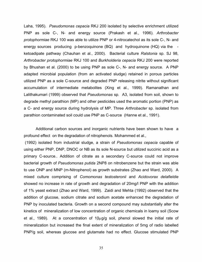
1.2.2.1 An initial oxygenation reaction yielding nitrite Oxidative removal of the nitro group from the aromatic nucleus yielding nitrite has been
demonstrated in various bacteria (Fig 1.1). Some bacteria mineralize these compounds
40
completely but use them as a nitrogen source by oxygenolytic removal of the nitro group
(Bruhn et al., 1987; Dickel and Kanckmuss, 1991). The enzymes responsible for the
removal of the nitro group have been identified. Zeyer and Kochar (1988) isolated and
purified nitrophenol oxygenase from Pseudomonas putida B2 which stoichiometrically
converted ONP to catechol and nitrite. Raymond and Alexander (1971) proposed a
conversion wherein a Flavobacterium converts nitroaromatics to nitrocatechols before
removing the nitro groups as nitrite.
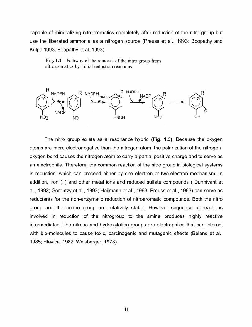
1.2.2.2 Reductive transformation reaction Several microorganisms degrade nitroaromatic compounds by initially reducing the
nitro-substituent to an amino group, which may subsequently be released as ammonia
(Zeyer and Kearney, 1984) (Fig 1.2). The action of nitroreductases has been
demonstrated in cell free systems under both aerobic and anaerobic conditions
(Kinouchi and Ohnishi, 1983; McCormick et al., 1976; Villanaueva, 1964). Schenzle et
al. (1997) found that Ralstonia eutropha JMP134 (Pemberton et al., 1979) converted
MNP using it as its sole source of nitrogen, carbon and energy. The reduction proceeds
via a nitroso and a hydroxylamino group. Theaminoaromatic product is further degraded
in the presence of oxygen by aniline oxygenases to ammonium and catechol which is
further mineralized by ring cleaving enzymes. This pathway is involved in the
degradation of nitrobenzoates, nitrotoluenes and nitrophenols. Some bacteria are not
41
capable of mineralizing nitroaromatics completely after reduction of the nitro group but
use the liberated ammonia as a nitrogen source (Preuss et al., 1993; Boopathy and
Kulpa 1993; Boopathy et al.,1993).
The nitro group exists as a resonance hybrid (Fig. 1.3). Because the oxygen
atoms are more electronegative than the nitrogen atom, the polarization of the nitrogen-
oxygen bond causes the nitrogen atom to carry a partial positive charge and to serve as
an electrophile. Therefore, the common reaction of the nitro group in biological systems
is reduction, which can proceed either by one electron or two-electron mechanism. In
addition, iron (II) and other metal ions and reduced sulfate compounds ( Dunnivant et
al., 1992; Gorontzy et al., 1993; Heijmann et al., 1993; Preuss et al., 1993) can serve as
reductants for the non-enzymatic reduction of nitroaromatic compounds. Both the nitro
group and the amino group are relatively stable. However sequence of reactions
involved in reduction of the nitrogroup to the amine produces highly reactive
intermediates. The nitroso and hydroxylation groups are electrophiles that can interact
with bio-molecules to cause toxic, carcinogenic and mutagenic effects (Beland et al.,
1985; Hlavica, 1982; Weisberger, 1978).
42
The one-electron reduction of the nitro group produces a nitro radical anion,
which can be oxidized to the starting material by molecular oxygen with the concomitant
production of superoxide. The cycle leads to the designation of enzymes that catalyse
one-electron reduction of the nitrogroup as “oxygen sensitive” (Bryant and De Luca,
1991). Enzymes from a variety of sources catalyze one electron reduction of the nitro
group. These include anaerobic bacteria such as Clostridium sp. (Angermaier and
Simon,1983), facultative bacteria such as Eschericia coli (Peterson et al., 1979) and
Enterobacter sp. as well as plants and animals (Bryant and De Luca, 1991).
Reduction of the nitro group by the sequential addition of pairs of electrons is
“oxygen insensitive” because no radicals are produced (Bryant and De Luca, 1991).
Nitroreductases of this type convert nitro groups to either hydroxylamines or amines by
the addition of electron pairs donated by reduced pyridine nucleotides. The electron
pathway goes through the formation of nitroso derivatives, which are difficult to detect
due to their reactivity. Both nitroso and hydroxylated intermediates can react readily
43
with a variety of biological materials including condensation reactions, for example, non-
enzymatic production of azoxy compounds in the presence of oxygen (McCormick et al.,
1976).
The ease of reduction of the aromatic nitro group depends on the nature of other
substituents on the ring and on the reducing potential of the environment. Electron
withdrawing groups activate the molecules for reduction of the nitro group, whereas
electron donating groups make the ring more susceptible to electrophilic attack. In the
case of nitrotoluene, the number of nitro groups increases the probability of reduction
and the probability of electrophilic attack decreases. Therefore, reduction of one nitro
group of TNT is very rapid under a variety a conditions, including those prevalent in
growing cultures of aerobic bacteria. In contrast, reduction of α-amino-4, 6-
dinitrotoluene (ADNT) requires a lower redox potential, and reduction of 2, 4-diamino-6-
dinitrotolune (DANT) requires a redox potential below 200mv (Funk et al., 1993),
because the electron-donating properties of the amino groups lower the electron
deficiency of the molecule.
1.2.2.3 Complete reductive removal of the nitro group by the formation of a hydride-Meisenheimer complex This pathway is characterized by the complete reductive removal of the nitro group as
nitrite and the formation of a hydride-Meisenheimer complex as one of the metabolites
indicating an initiation of nucleophilic attack on the aromatic ring (Fig 1.4) by the hydride
ion. Lenke and Knackmuss (1992) used Rhodococcus erythropolis to utilize picric acid
which was metabolized to form a orange-red hydride-Meisenheimer complex and was
further converted to 2,4,6-trinitro-cyclohexane with concomitant liberation of nitrite.
44
1.2.2.4 Degradation of nitroaromatics via partial reduction and replacement reactions Non-polar nitroaromatic compounds are considered resistant to microbial attack
(Fewson, 1981). This is due in part to the reduction of electron density in the aromatic
ring by the nitro group hindering electrophilic attack by oxygenases and thus preventing
aerobic degradation. The accumulation of ammonia but not nitrite in media in
nitrobenzene grown culture of Pseudomonas alcaligenes JS45 suggested that initial
attack on the nitro group was reductive rather than oxidative leading to formation of
hydroxyl aminobenzene (HAB) requiring 2 mol of NADPH. The HAB undergoes
catalyzed rearrangement analogous to Bamberger rearrangement to form aminophenol
(Nishino and Spain, 1993). This intramolecular Bamberger rearrangement reaction (Fig 1.5), (Bamberger, 1894, Shine, 1967; Sone et al., 1981) resulted in release of ammonia
via ring fission of aminophenol. Implication of this type of rearrangement has been
extensively described by Corbett and Corbett (1995).
45
1.2.3 Anaerobic degradation of nitroaromatic compounds The reactions of nitroaromatic compounds in anaerobic systems almost exclusively
involve the reduction of the nitrogroup. McCormick et al. (1976) clearly demonstrated
that Viellonella alkalescens could reduce TNT and also identified some of the enzymes
involved. Subsequently, a variety of other bacteria have been shown to reduce
aromatic nitro compounds under anaerobic conditions (Angermaier and Simon, 1983;
McCormick et al., 1976, Oren et al., 1991; Rafii et al., 1991 and Schackmann and
Müller, 1991). Boopathy and Kulpa (1993) conducted studies on Desulflovibro sp. strain
B that uses TNT and a variety of other nitroaromatic compounds as the source of
nitrogen for growth and also as the terminal electron acceptor. The nitro compounds
are reduced to the corresponding amines and proposed that the amino groups are
removed from the aromatic ring by a reduction deamination mechanism analogous to
that
described by Schnell and Schink (1991). Preuss et al. (1993) isolated a strain of
Desulfovibrio by selective enrichment with pyruvate as carbon source, sulfate as the
46
terminal electron acceptor, and TNT as the source of nitrogen. The strain fixes
atmospheric nitrogen and can also use ammonia as its nitrogen source. Several strains
of Clostridia have been studied because of their ability to reduce nitroaromatic
compounds (McCormick et al., 1976; Preuss et al., 1993; Rafii et al., 1991). Angermaier
and Simon (1983) provided evidence that hydrogenase and ferrodoxin in Clostridium
kluyveri are responsible for a one-electron reduction of nitroaromatic compounds. Rafii
et al. (1991) characterized oxygen sensitive enzymes from several strains of Clostridium
isolated from human faecal matter. The enzymes reduced 4-nitrobenzoate and several
nitropyrenes to the corresponding amines. Hydrogenase from Clostridium pasteurianum
and carbon monoxide dehydrogenase from Clostridium thermoacticum reduce DANT to
DAHAT when ferrodoxin is included in the reaction mixture (Preuss et al., 1993). The
reduction also took place with reduced ferrodoxin or methyl viologen in the absence of
enzymes suggesting that the enzymes reduce ferrodoxin and not nitroaromatic
compounds. Kaake et al. (1992, 1994) used an anaerobic mixed culture for the
biodegradation of Dinoseb (2-sec-butyl-4,6-dinitrophenol) under methanogenic
conditions with starch as the electron donor. Similar enrichment cultures degraded
RDX (hexahydro-1,3,5-trinitro-1,3,5-triazine) and TNT to non-detectable levels in
contaminated soil (Funk et al., 1993). Culture of Clostridium bifermentans isolated from
other enrichments (Crawford, 1995) degraded both RDX and TNT.
O’Connor et al. (1989) studied the toxicity and anaerobic biodegradability of
substituted phenols under methanogenic conditions using two anaerobic bioassays - the
biochemical methane potential (BMP) and the anaerobic toxicity assay (ATA) to
evaluate the stoichiometric conversion of added substrate carbon to CO2 and CH4. It
was observed that ONP and PNP were completely mineralized. Significant
transformation occurred and aminophenols were detected for higher concentrations.
Mononitrophenols were degraded (Blasco and Castillo, 1992) to a lesser extent than
2,4-DNP under light anaerobiosis with the exception of ONP which was considerably
metabolized in the absence of O2 probably because of anaerobic reduction of the nitro
group. Weak growth and no nitrite excretion were also observed in the presence of
toxic levels of mononitrophenols (0.5mM), thus suggesting the elimination of nitro
47
groups under anaerobiosis. Resting cells of Ralstonia eutropha JMP134 (Schenzle et
al., 1997) metabolized MNP to N-acetylaminohydroquinone under anaerobic conditions.
1.2.4 Biodegradation by fungi The non specific lignolytic system produced by white rot fungus, Phanerochaete
chrysosporium consisting of a complex system of extracellular peroxidases, small
organic molecules and hydrogen peroxide is capable of degrading a wide range of
synthetic chemicals including nitroaromatic compounds. Several groups (Bumpus and
Tatarko, 1994; Fernando et al., 1990; Michels and Gottschalk, 1995; Spiker et al., 1992;
Stahl and Aust, 1993; Valli et al., 1992) have reported degradation and even
mineralization of nitroaromatic compounds by P. chrysosporium. Most fungi can
catalyse the reduction of at least one nitro group of TNT (Parrish, 1977). Mycelia of P.
chrysosporium reduce TNT to a mixture of 2-amino-4.6-dinitrotoluene (Stahl and Aust,
1993). Under lignolytic conditions, the amino compounds disappear and mineralization
can be fairly extensive. In contrast, Valli et al. (1992), suggested that 2,4-dinitrotoluene
is reduced predominantly to 2-amino-4-nitrotoluene by intracellular enzymes. Catalyzed
by manganese peroxidase, it is further converted to 4-nitro-1,2-benzoquinone which is
reduced to 4-nitrocatechol but could provide no strong evidence and suggested that 4-
nitrocatechol could serve as a substrate for oxidative removal of the nitro group.
Michels and Gottschalk (1995) provided strong evidence that under nonlignolytic
condition TNT is reduced to 4-amino-2,6-dinitrotoluene via the corresponding
hydroxylamino intermediate. Hoffrichter et al. (1993) showed cometabolic degradation
of ONP, MNP and PNP by Penicillium sp. B 7/2 growing at the expense of glucose.
1.2.5 Aerobic Biodegradation Bacteria can often derive carbon, nitrogen and energy from degradation of nitroaromatic
substrates. Ability of bacteria to degrade nitrophenols and nitrobenzoates was reported
many years ago. The earliest studies regarding degradation of mononitrophenol
isomers by Baumann and Herter (1877-78) and Meyer (1905) had shown quantitatively
48
in rabbits that o-, m- and p-nitrophenols conjugated in vivo with glucoronic and sulphuric
acid. Meyer was able to detect the reduction of m-and p-isomers. Oettingen (1949)
found PNP to be more toxic than its isomer. Robinson et al. (1951) conducted studies
on the extent of reduction of mononitrophenols in rabbits and found complete
conjugation with glucoronic and sulphuric acids in doses of 0.2 to 0.3g/kg. A number of
bacteria recently have been reported to degrade, aerobically, a wide range of polar and
non-polar nitroaromatic compounds. Such bacteria use a variety of strategies for the
removal of/or transformation of the nitro group aerobically. These include (a)
monooxygenase-catalyzed elimination of the nitro group as nitrite (b) dioxygenase-
catalyzed insertion of two hydroxyl groups with subsequent elimination of the nitro group
as nitrite (c) partial reduction of the nitro group to a hydroxylamine which is the
substrate for rearrangement or hydrolytic reactions and elimination of ammonia and (d)
partial reduction of the aromatic ring to form a Meisenheimer complex and subsequent
elimination of the nitro group as nitrite.
1.2.5.1 Monooxygenase-catalyzed initial reaction
Some of the earliest reports on the biodegradation of nitroaromatic compounds involved
studies of bacteria that can grow on nitrophenols (Simpson and Evans, 1953). They
provided preliminary evidence in 1953 that a strain of Pseudomonas could convert PNP
to hydroquinone. Studies with a partially purified enzyme (Spain et al., 1979) revealed
that a strain of Moraxella degrades PNP by initial oxygenase attack that results in the
release of nitrite and accumulation of hydroquinone requiring 2 moles of NADPH to
oxidize each mole of PNP (Fig 1.6) corresponding to nitro and hydroxyl group.
49
Experiments with 18O2 provided rigorous evidence that the mechanism of the reaction is
a monohydroxylation (Spain et al., 1979) and preliminary evidence suggested that the
enzyme was a flavoprotein monooxygenase. The stoichiometry and hydroquinone
accumulation as the first detectable intermediate suggests that the initial product of the
reaction is 1,4-benzoquinone. However an inducible quinone reductase could not be
easily separated from the membrane bound oxygenase that catalyzed the initial
reaction. The hydroquinone produced served as the substrate for ring fission reaction
catalyzed by a ferrous iron-dependent dioxygenase yielding γ-hydroxy
muconicsemialdehyde which was oxidized to maleyl acetic acid. Catalytic amounts of
NAD+ were required for the conversion of the ring fission product to β-ketodipate via
50
reduction of maleyl acetate in cell extracts because the two reactions recycle the
cofactor. Hanne et al. (1993) proposed a similar pathway using Arthrobacter and a
Nocardia sp. which converted PNP to hydroquinone. Cell extracts grown on PNP also
contained enzymes which converted it to 1,2,4-benzentriol. In contrast Jain et al. (1994)
isolated an Arthrobacter sp. which seemed to degrade PNP to 1,2,4-benzentriol via 4-
nitrocatechol by a monooxygenase catalyzed hydroxylation at the ortho position. This
was suggested by Raymond and Alexander (1951) who confirmed the conversion of
PNP to 4-nitrocatechol (UV, Visible, IR, TLC and GC) by resting cells of a soil bacterium
which was able to use PNP a source of carbon and energy and released stoichiometric
amounts of nitrite. Jain et al. (1994) observed that 1,2,4-benzenetriol was further
oxidized by an ortho ring fission to maleyl acetic acid but the enzyme responsible could
not be detected. However an enzyme that converts PNP to 4-nitrocatechol has been
purified from a strain of Nocardia sp. grown on PNP (Mitra and Vaidyanathan, 1984)
and a similar enzyme activity has been demonstrated in another strain of Nocardia after
growth on phenols (Hanne et al., 1993).
Similarly, monooxygenase catalyzed conversions of ONP were reported by
various researchers. Zeyer and Kearney (1984) isolated and purified an NADPH
dependent monooxygenase that catalyzed the conversion of ONP to catechol with the
concomitant release of nitrite and oxidation of 2 moles of NADPH. Catechol was
subsequently oxidized by 1,2-dioxygenase and was degraded further giving cis, cis-
muconic acid and muconolactone through an ortho cleavage pathway (Zeyer and
Kocher, 1988). Spain et al. (1979) proposed that the enzymatic reaction produces 1,2-
benzoquinone by a mechanism analogous to the reaction catalyzed by PNP
oxygenases. The activity of key enzymes of the pathway, nitrophenol oxygenase,
catechol 1,2-dioxygenase and lactonizing enzymes in cell extracts and catechol 2, 3-
dioxygenase (key enzyme of meta cleavage pathway) were detected thus confirming
previous reports (Zeyer and Kearney, 1984). The ortho-nitrophenol monooxygenase is
unusual among monooxygenases that catalyse the removal of aromatic nitro groups in
that it does not seem to require the participation of a flavin co-factor.
51
1.2.5.2 Dioxygenase-catalyzed initial reaction The catabolism of aromatic hydrocarbons by aerobic bacteria generally requires the
activation of the molecules by the addition of two hydroxyl groups to the ring. The
reactions are catalyzed by dioxygenase enzymes that introduce two atoms of molecular
oxygen on adjacent carbon atoms (Gibson and Subramanian, 1984) (Fig.1.7). In
substituted aromatic compounds, the introduction of the hydroxyl groups can lead to
spontaneous elimination of the substituent and rearomatization of the ring: for example,
toluene dioxygenase catalyzes the elimination of hydroxyl groups from phenol (Spain et
al., 1989). Removal of aromatic nitro groups by dioxygenase enzymes was first reported
as a result of studies on transformation of 2,6-dinitrophenol by Alcaligenes eutrophus
(Ecker et al., 1992). Nitrobenzene, used extensively as the starting material for
synthesis of aniline, is converted to catechol by a dioxygenase as the first step in its
mineralization by a Comomonas sp. isolated from an aerobic waste-treatment plant
(Nishino and Spain, 1995). The inducible nitrobenzene dioxygenase was found to be
non specific and catalyzed the oxidation of a variety of nitrophenols, dinitrobenzene and
nitrotoluenes (Spain, 1995). A Pseudomonas strain isolated from contaminated soil by
selective enrichment grew on 2-nitrotoluene as the sole source of nitrogen and carbon
(Haigler et al., 1994). The catabolic pathway involves an initial dioxygenase attack at
the 2,3 position of the molecule to form 3-methyl catechol and release of nitrite. The 3-
methyl catechol was degraded by a typical meta cleavage pathway. Purification of the
enzymes allowed rigorous proof that the insertion of molecular oxygen and release of
nitrite involves a dioxygenase mechanism, and that the rearomatization of the ring does
not require a separate enzyme. Strains of Pseudomonas and Comomonas were found
to convert 3-nitrobenzoate to protocatechuate by means of a dioxygenase attack at the
3,4 position with subsequent elimination of nitrite (Nadeau and Spain, 1995). Haigler
and Spain (1991) investigated the ability of seven bacterial strains containing toluene
degradative pathways to oxidize nitrobenzene.
52
Cells of Pseudomonas putida F and Pseudomonas sp. strain JS150 converted
nitrobenzene to nitrocatechol in presence of 18O2 suggesting a dioxygenase
mechanism. Pseudomonas mendocina converted nitrobenzene to a mixture of MNP
and PNP. Pseudomonas picketti PK01 converted nitrobenzene to 3-and 4-nitrocatechol
via MNP and PNP which were slowly degraded to unidentified metabolites. They also
observed that nitrobenzene did not serve as an inducer for the enzyme that catalyzed
its oxidation, clearly indicating that nitrobenzene ring is subjected to initial attack by both
mono and dioxygenase enzymes. Mineralization of a nitroaromatic compound via a
dioxygenase initial attack was first reported as a result of studies with Pseudomonas sp.
strain DNT grown on 2,4-DNT by a dioxygenase enzyme that was very similar to that of
naphthalene dioxygenase (Suen et al., 1994). It adds hydroxyl groups to the 4- and 5-
positions on the ring of 2, 4-DNT, and the nitro group is eliminated non-enzymatically as
nitrite (Spanggord et al., 1991). 4-Methyl-5-nitrocatechol (MNC) produced by 2,4-DNT
dioxygenase is the substrate for a monooxygenase that catalyses the replacement of
the nitro group and elimination of nitrite. The constitutive enzyme, partially purified from
cells of Pseudomonas sp. strain DNT, converts MNC to 2-hydroxy-5-methyl-quinone
(Haigler et al., 1994), the reaction mechanism being similar to that described for other
enzymes that catalyze the removal of nitro group from nitrophenols (Spain et al., 1979,
53
Zeyer and Kocher, 1988) and other electron-withdrawing groups from substituted
phenols (Xun et al., 1992) or carboxylic acids (Hussain et al., 1980).
1.2.5.3 Reduction of the aromatic ring The electron withdrawing properties of the nitro group cause the aromatic ring of poly
nitroaromatic compounds to be highly electron deficient and resistant to microbial
attack. Lenke et al. (1992) discovered an alternate mechanism of transformation
involving reduction of the aromatic ring. They isolated strains of Rhodococcus
erythropolis that use 2,4-dinitrophenol as the carbon, energy and nitrogen source. The
isolates released nitrite from 2,4-dinitrophenol with transient accumulation of significant
amount of 4,6-dinitrohexanoate. Presence of enzymes able to catalyse the reduction of
the aromatic ring and accumulation of 4,6-dinitro-hexanoate suggested that the aliphatic
compound was a dead end metabolite.
Resting cells of Rhodococcus erythropolis grown on 2,4-dinitrophenol released
nitrite from picric acid, and spontaneous mutants could use picric acid as the nitrogen
source (Lenke and Knackmuss, 1992). Initial reactions by cells and cell extracts
showed the addition of a hydride ion to the aromatic ring to form a hydride-
Meisenheimer complex. Addition of a second hydride ion led to the eventual formation
of 2,4,6-trinitrocyclo-hexanone which decomposed to form 1,3,5-trinitropentane upon
acidification and extraction. In contrast, protonation of the hydride-Meisenheimer
complex led to the enzyme catalyzed rearomatization of the molecule and elimination of
nitrite, which could be assimilated by bacteria along with 2,4-dinitrophenol generated
during the process. Three reactions of hydride-Meisenheimer complex have been
demonstrated in bacteria. The complex can (a) spontaneously decompose to the
parent compound (b) be reduced to aliphatic compounds or (c) rearomatize by the
addition of a proton and elimination of nitrite.
1.2.5.4 Partial reduction of the nitro group Very early reports on the biodegradation of 2-nitrobenzoate (Cain, 1966; Ke et al., 1959)
and 4-nitrobenzoate (Cartwright and Cain, 1959) provided evidence for the partial
54
reduction of the nitro group and the release of nitrogen and ammonia. Bacteria able to
grow on MNP have been isolated (Germanier and Wuhrman, 1963) and the initial steps
in the degradation pathway were found to be reductive rather than oxidative. A
Pseudomonas putida that grew on ONP and MNP as sole sources of carbon and
nitrogen was isolated from soil (Zeyer and Kearney, 1984) and was found to degrade
ONP and MNP releasing nitrite and ammonium respectively but was unable to degrade
PNP. Enzymes involved in metabolism were found to be inducible. Ralstonia eutropha
strain JMP134 was shown to utilize MNP as the sole source of nitrogen, carbon and
energy at a concentration of <0.5mM, above which growth was inhibited and
accumulation of oxygen sensitive metabolites occurred. The conversion of 4-
hydroxybenzoate to 3,4-dihydroxybenzoate has been identified as a key reaction in the
degradative pathway of 4-nitrobenzoate (Groenewegen and de Bont, 1992) and 4-
nitrotoluene (Haigler and Spain, 1993; Rhys-Williams et al., 1993). This novel
enzymatic reaction leads to simultaneous elimination of ammonia and has also been
observed in the degradation of MNP by Pseudomonas putida B2 (Meulenberg et al.,
1996). Nishino and Spain (1993) identified an enzyme which converts
hydroxylaminobenzene to 2-aminophenol in the degradative pathway of nitrobenzene
by Pseudomonas pseudoalcaligenes JS45. This intramolecular reaction is known as
Bamberger rearrangement (Bamberger, 1894; Shine, 1967; Sone et al., 1981) in which
hydroxyl amino-aromatic compounds rearrange to aminophenols under mildly acidic
conditions. The non-enzymatic rearrangement yields predominantly 4-aminophenol,
whereas the enzyme (hydroxylaminobenzene mutase) directs the production of
predominantly (>99%) 2-aminophenol. The 2-aminophenol thus produced by the initial
steps in the pathway is degraded by a dioxygenase that catalyses the opening at the
1,6-position to produce 2-amino muconicsemialdehyde. The mechanism for degradation
of this compound by Pseudomonas pseudoalcaligenes is not known. But enzymes in
crude extracts from nitrobenzene grown cells catalyse the degradation of the ring-fission
product and release of ammonia requiring NAD and indicating an oxidation of the
aldehyde. The reductive pathway for degradation of nitrobenzene seems much more
complex than the oxidative pathway, requiring one mole of oxygen and one mole of
NADH to convert nitrobenzene to central metabolic intermediates and release ammonia.
55
In contrast, the oxidative pathway requires two moles of oxygen and one mole of NADH
that can be regained if the 2-hydroxy muconicsemialdehyde, undergoes an NAD-
dependent oxidation to oxalocrotonate (Nishino and Spain, 1995). If the isolate is to
use the nitrite released by the oxygenolytic reaction as its nitrogen source, three
additional moles of NADPH would be required for the reduction of nitrite to ammonia.
Hence the more complex reductive pathway for nitrobenzene reduction seems to be
well adapted to exploit the condition of an oxygen-limited ecosystem. Analogous
enzyme catalyzed reactions have been reported in animals (Sternson and Gammans,
1975) and in yeast (Corbett and Corbett, 1981) but not in bacteria. The implications of
the Bamberger like rearrangement in biochemistry have been discussed extensively by
Corbett and Corbett (1995). In contrast to the above, Ralstonia eutropha JMP134
(Schenzle et al., 1997) converted hydroxyl aminobenzene to α-aminophenol and 4-
aminophenol. Correspondingly 3-hydroxyl aminophenol as a metabolite of MNP
underwent an enzyme catalyzed rearrangement to aminohydroquinone which was
acetylated to N-acetylaminohydroquinone under anaerobic conditions. Acetylation of
anilines has been demonstrated to be an important detoxification mechanism by
microorganisms (Bollag and Russel, 1976, Engelhardt et al., 1977; Tweedy et al.,
1970). Schackmann and Müller (1991) described a nitro reducing activity for MNP
generating 3-aminophenol and 3-N-acetylamino-phenol as dead end metabolites by
resting cells of Pseudomonas sp. strain CBS3. Zhao et al. (2000) using Pseudomonas
putida 2NP8 proposed a pathway for MNP degradation and evidence for ammonia
release postulated on hydroxyl aminobenzene transformation wherein 3-hydroxyl
amino-phenol, reduction product produced by MNP nitroreductase is converted possibly
to two intermediates - aminohydroquinone and 4-aminocatechol, via ortho and para-
Bamberger rearrangement respectively. These are oxidized to imines which on
hydrolysis form quinones and subsequently are reduced leading to the formation of
1,2,4-benzenetriol. Meulenberg et al. (1996) identified 1,2,4-benzenetriol as an
intermediate and observed ammonia during nitroreductase-initiated MNP transformation
by Pseudomonas putida B2 but under anaerobic conditions. Schenzle et al. (1997)
observed a Bamberger rearrangement type of conversion of 3-hydroxyl aminophenol to
amino hydroquinone during aerobic conversion of MNP but did not investigate the
56
release of ammonia. It was also observed that MNP grown cells of Pseudomonas
putida produced ammonia, 2-amino-phenol, 4-aminophenol, 4-benzoquinone, N-acetyl-
Sisco Research Laboratories Pvt. Ltd., India 4- Aminoantipyrene, p- Nitrophenol
Loba-Chemie Indoaustranal Co., India Calcium nitrate, o- Nitrophenol
N.R. Chemicals, India m- Nitrophenol
2.1.1 Media The basal mineral medium (M5 medium), with the following composition was used for
enrichment as well as for growing the bacterial consortium. This medium was slightly
modified for degradation and enzymatic studies.
M5 medium : g/l NaH2PO4 0. 792
Na2HPO4.2H2O 2. 563
NH4NO3 0. 25
MgSO4. 7 H2O 0. 2
Ca (NO3)2 0. 1
Trace elements 1 ml.
60
Trace elements solution contained in g/l.
Fe SO4. 2H2O 1. 0
MnSO4. H2O 1. 0
Na2MoO4. 2H2O 0. 25
CuCl2. H2O 0. 25
Conc. H2SO4 5 ml.
pH of the M5 medium was adjusted to 7.5 and was autoclaved for 20 min at
121oC. M5 medium was supplemented with sodium succinate (1%) and yeast extract
(0.5%) when increased cell yields were required.
2.1.2 Culture conditions Growth and degradation experiments were carried out in sterile M5 medium with the
required test substrate supplemented with sodium succinate and yeast extract or
aromatic compounds such as nitrophenols, phenol, cresols or acetate respectively. All
the experiments were conducted in 500ml Erlenmeyer flask maintained on a rotary
shaker (Environ shaker 3597-1, Labline Instruments, USA) and at room temp (28 –
30o°C) in dark conditions.
Nutrient agar medium containing (g/l), peptone, 5.0; beef extract, 3.0; NaCl 5.0;
agar 20 (pH – 7.2) was used for plating, isolation and purification of the individual
strains of the microbial consortium. The individual isolates and the consortium were
routinely maintained on nutrient agar slants and plates. They were also maintained in
liquid M5 medium supplemented with yeast extract and sodium succinate or
mononitrophenol isomers
ONP, MNP and PNP.
61
2.2 ANALYTICAL PROCEDURES 2.2.1 Growth Bacterial growth was monitored turbidometrically. A known volume of the culture broth
was centrifuged at 10,000 x g, the cell pellet obtained was washed thoroughly with M5
medium, suspended in the same volume of M5 and optical density determined at
600nm using a Genesys spectrophotometer.
2.2.2 Estimations 2.2.2.1 Estimation of phenol and o- and m- cresol by colorimetry A modified 4-aminoantipyrene colorimeteric method based on the procedure of Lacost
et al. (1959) was followed.
Materials 4- Aminoantipyrene
Potassium ferricyanide
Borate buffer – 6.2g of boric acid powder and 7.0g potassium chloride were dissolved
in 800ml distilled water. To this 64ml of 1N NaOH was added and solution was made
upto 1L and pH adjusted to 9.2 – 9.4.
Method
To 10ml of diluted sample, 0.5ml of borate buffer, 0.1ml of 1.5% 4-amino-antipyrene
and 0.1ml of 10% potassium ferricyanide solutions were added. The color developed
was measured at 505nm. The results were computed from a standard calibration
prepared using the respective standard compound.
Estimation of p- cresol by HPLC
Residual p-cresol in the culture supernatants was analysed by reverse phase high
performance liquid chromatography (HPLC), (Shimadzu LC– 6 A, Japan) with a C–18
62
column (150 x 4.6mm). The mobile phase used was methanol – water – acetic acid (60
: 35 : 5 by volume). The flow rate was 1ml. min-1 and detection was by UV
absorbance at a wave length of 278 nm.
2.2.2.2 Estimation of nitrophenols and identification of their metabolites in reaction mixtures. Estimation of residual nitrophenol in reaction mixtures was done by spectrophotometry
and chromatographic methods. The identification of metabolites during the course of
nitrophenol degradation was done by NMR spectroscopy.
Estimation of nitrophenols The nitrophenol concentration in reaction mixtures was spectrophoto-metrically
measured in a Genesys spectrophotometer (Spectronic instruments, USA.) using a
known volume (1ml) of the culture filtrate at the following wavelengths.
o – Nitrophenol (ONP) 412nm
m – Nitrophenol (MNP) 272nm
p – Nitrophenol (PNP) 401nm.
For a reaction mixture containing a mixture of all three isomers a wavelength of
272nm was applied or the mixture was monitored at all the three wavelengths.
Nitrophenol concentration was also determined by high pressure liquid chromatography
by monitoring the degradation at a wavelength of 254nm. The absorbance
(spectrophotometric) and peak area values (HPLC) were plotted against concentrations
respectively. From the straight lines obtained, a linear least square analysis was
performed and the slope values obtained were used for measuring the concentration of
compounds in the test solution (correlation coefficient = 0.99).
63
Materials
ONP, MNP, PNP
M5 medium
Acetonitrile
Glacial acetic acid
C18 column
Water Spectrophotometric calibration A stock solution of the individual isomers was prepared in M5 medium and different
concentration of nitrophenols in the range of 2.5–145 µg/ml were photometrically
measured at the respective wavelengths. A standard curve was obtained by plotting the
optical densities against the concentration employed (Fig. 2.1, Fig 2. 2, Fig 2.3).
64
65
66
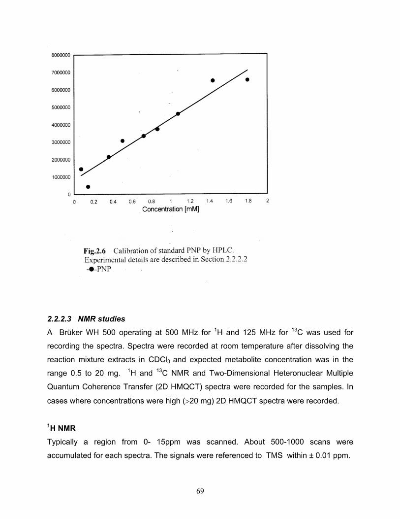
HPLC calibration The HPLC analysis was performed on a 25cm Bondapak C18 column (Shimadzu SPD
– 10A). Different concentrations of ONP, MNP and PNP separately were injected and
eluted with water: acetonitrile (65:35) and 0.1% acetic acid. Solvents were eluted at the
rate of 1ml/min and compounds were monitored at a wavelength of 254nm. Peak areas
were plotted against concentration which gave a calibration curve enabling
quantification of data from reaction mixtures
(Fig. 2.4, Fig 2.5, Fig 2.6)
67
68
69
2.2.2.3 NMR studies
A Brüker WH 500 operating at 500 MHz for 1H and 125 MHz for 13C was used for
recording the spectra. Spectra were recorded at room temperature after dissolving the
reaction mixture extracts in CDCl3 and expected metabolite concentration was in the
range 0.5 to 20 mg. 1H and 13C NMR and Two-Dimensional Heteronuclear Multiple
Quantum Coherence Transfer (2D HMQCT) spectra were recorded for the samples. In
cases where concentrations were high (>20 mg) 2D HMQCT spectra were recorded.
1H NMR Typically a region from 0- 15ppm was scanned. About 500-1000 scans were
accumulated for each spectra. The signals were referenced to TMS within ± 0.01 ppm.
70
2D HMQCT The spectra contained 1H data in one dimension and 13C data in the other dimension.
The carbon signals from carbon atom to which a proton is attached were seen clearly. A
6000 Hz spectral width, 2.7s acquisition time, pulse width 83.2µs and 6µs pulses with a
delay of 1.0µs were employed. Number of scans were 16 for each trace. For 1H, a 8.8
µs pulse was used. A - 1 Hz line broadening was employed.
2.2.2.4 Estimation of ammonia
Ammonia was estimated using Nesslers reagent and quantitative data was obtained by
preparing a calibration curve using standard ammonium solution.
Materials Mercuric iodide
Potassium iodide
Sodium hydroxide
Ammonium chloride
Method Preparation of Nesslers reagent (Standard Methods, APHA, 17th ed., 1985) 100gm HgI2 and 70 KI were dissolved in a small quantity of water and was slowly added
to 160g NaOH dissolved in 500ml water. This mixture was then made up to 1l. The
reagent was stored in rubber- stoppered borosilicate glassware and away from sunlight
to maintain reagent stability for upto a year.
Stock ammonium solution: 3.819g anhydrous NH4Cl at 15oC was dissolved in
ammonia free water and made up to 1000ml. 1ml of this solution contained 1mg N=
1.214mg NH3. 10ml of the stock solution was made upto 1000ml with water, 1ml –
10µgN= 12.14µg NH3.
71
Standard calibration Different aliquots of 5ml sample containing NH3 concentration in the range of 0.24-
8.25µg and 0.1ml Nesslers reagent made up by ammonia free water were measured
spectrophotometrically. The optical densities observed at 400nm were plotted against
concentration employed to get a standard curve
(Fig 2.7).
72
2.2.2.5 Estimation of nitrite (Montgomery and Dymock, 1996) Nitrite release from reaction mixtures was quantified from a standard curve prepared
using a standard nitrite solution.
Materials Sodium nitrite
Sulphanilic acid
Potassium hydrogensulphate
N- (1- Naphthyl) ethylenediamine hydrochloride.
Method Preparation of reagents Reagent A: Sulphanilic acid solution
Potassium hydrogensulphate 27. 2 g/l
Sulphanilic acid 3. 46 g/l.
Reagent B: 0.4% N (1-napthyl) ethylenediamine hydrochloride Standard sodium nitrite solution 1.2325gm of sodium nitrite was dissolved in 250ml freshly distilled water. A standard
solution was made using 5ml of the stock and making it to 500ml with distilled water.
1ml of this solution contained 0.0493 mg of nitrite.
Calibration Different volumes of the standard solution containing nitrite in the range of 0.065–
19.7�g were measured spectrophotometrically. The optical densities observed were
plotted against concentration of NO2 and a standard curve obtained (Fig 2.8). A linear
least square analysis was performed to get the regression data from which nitrite
concentration in the test samples was determined (correlation coefficient = 0.99).
MICROBIAL DEGRADATION OF MONONITROPHENOLS BY A CONSORTIUM
3.1 INTRODUCTION Microbial communities are likely to have an extremely important role to fulfill in the
degradation of simple and complex natural products and environmentally foreign
compounds (xenobiotics) (Gibson, 1984). It is a commonly accepted observation that
often the rate of biodegradation of a particular compound is faster in nature, host of
heterogenous communities, than in pure cultures of organisms isolated from that
environment. Many microbial communities clearly show that relationships between the
populations confer beneficial effects which make the associations more successfull
than any of the individual populations alone (Slater, 1978, 1979, 1979). Microbial
degradation of organic compounds is often investigated in the laboratory by using mixed
culture systems obtained from the environment (Baughman et al., 1980; Boethling and
Alexander, 1979; Larson, 1979; Pritchard et al., 1979).
A microbial consortium was obtained from a contaminated soil sample for
conducting degradation studies of mononitrophenol isomers. The catabolic potential of
this consortium, obtained by enrichment, with nitrophenols was tested in degrading
ONP, MNP and PNP separately and the simultaneous degradation of all the three
isomers was also checked. Effect of pre-exposure to compounds such as phenol and
cresols on the consortium’s capability in degrading nitrophenols, utilization of ONP,
MNP and PNP as sole sources of carbon and nitrogen and the impact of varying
concentrations of these isomers on the consortium’s ability were some of the
experiments conducted.
Gunner and Zuckerman (1968) were among the first to describe the importance
of synergistic metabolic activity between an Arthrobacter sp. and Streptomyces sp.
involved in the degradation of the insecticide, diazinon. Any organic compound in
87
question may act as a carbon source and support the growth of a mixed culture. It is
now firmly established that many microorganisms growing at the expense of one
substrate, may be able to transform a different substrate in a reaction or sequence of
reactions, which are not directly associated with that organism’s energy production,
carbon assimilation and biosynthesis or growth processes (Horvath, 1972; Alexander,
1979) leading to a mechanism of co-metabolism. Parathion, a widely used infecticide
has been found to be degraded by microbial communities dependent upon a co-
metabolic step (Munnecke and Hsieh, 1974, 1975, 1976). Daughton and Hsieh (1977)
observed in a microbial consortium of four Pseudomonads, that, P. stutzeri hydrolyzed
parathion to diethylthiophosphate and PNP. Neither product could separately support
the growth of P. stutzeri. A second species, P. aeruginosa could not grow on parathion
but grew on one of the products, PNP. Thus these two organisms acting together
enabled at least part of parathion molecule to be converted to biomass, a process
initially dependent on a co-metabolic step. Growth of mixed cultures and stable
microbial communities establishes a situation in which organisms with different genetic
background have the opportunity to exchange genetic information. The evolution of
certain classes of microbial community may be an important prerequisite to the
evolution of novel degradative pathways as a result of exchange and rearrangement of
genetic information from different gene pools.
3.2 RESULTS 3.2.1 Degradation conditions The conditions employed for degradation studies are given below. The basal mineral
medium (M5 medium), culture medium and conditions were the same as mentioned
earlier (Section 2.1.1 & 2.1.2). An increased biomass obtained by supplementing the M5
medium devoid of NH4NO3 (nitrogen source) with 1% succinate and 0.5% yeast extract
was employed. Harvested and washed cells were induced with 0.1mM of the substrate
separately for a period of 24h. Thereafter the cells were once again washed thoroughly
and inoculated at OD 600nm of 0.1-0.6 into flasks containing 0.1-0.3mM
mononitrophenol isomers individually in separate flasks, each in triplicates. Growth,
88
residual substrate, ammonia and nitrite estimation were done regularly at an interval of
6h for varying periods of incubation for the three isomers. Growth, ammonia and nitrite
release were monitored spectrophoto-metrically at specified wavelengths (Sections
2.2.1; 2.2.2.4; 2.2.2.5 respectively), whereas residual substrate was analyzed by both
HPLC and spectrophotometry (Section 2.2.2.2)
3.2.2 Degradation of o-Nitrophenol At 0.1mM ONP as substrate, the biomass as measured spectrophoto-metrically showed
an irregular growth curve up to 40h. Thereafter it showed a steady decrease. This
indicated that in the presence of 0.1mM substrate not much growth could be registered.
Although 0.1mM ONP was employed, the ONP concentration estimated
spectrophotometrically at 412nm was very less. The reason for this may probably be
due to the absorption of ONP on to the cell wall. An initial concentration of only 0.07mM
was detected. Within a period of 12h, 67.5% decrease in the substrate concentration
was observed. The control for the same period practically showed very little change in
concentration (Fig 3.1 and 3.2). About 90% degradation of ONP was observed by 54h.
HPLC analysis also showed decrease in ONP concentration after 6h (Fig 3.3 and Fig 3.3a). No ammonia could be detected in the culture filtrate during the course of
degradation but a maximum of 0.009mM nitrite was observed at 6h which remained
constant up to 50h and then declined coinciding with decrease in substrate
concentration and subsequent disappearance (Fig 3.4).
89
90
91
92
3.2.3 Degradation of m-Nitrophenol To flasks containing MNP as the substrate, cells at an initial OD of 0.1 at 600nm were
added. In the latter stages after 60h an increase in growth corresponding to an increase
in OD 600nm of 0.14 was observed (Fig 3.1). Degradation monitored by UV at 272nm
showed 98% reduction in substrate concentration from 0.39mM to 0.005mM which
93
was observed after 12h (Fig 3.2). This was confirmed by HPLC where no substrate
could be detected beyond 12h (Fig 3.3 & Fig 3.3b). Interestingly no nitrite could be
detected in the culture filtrate at any stage during substrate utilization. Ammonia was
detected after 6h (Fig 3.5). A gradual increase in the concentration of ammonia
(0.026mM) was observed from 6h which reached a maximum of 0.068mM at 55h.
Presence of ammonia in the culture filtrate clearly indicated that MNP was degraded via
a reductive pathway unlike ONP which showed the release of nitrite due to an oxidative
pathway.
94
95
3.2.4 Degradation of p-Nitrophenol There was poor growth in cells with PNP as substrate. Cells were inoculated at an initial
OD of 0.57 at 600nm. A gradual decrease in biomass with time was observed (Fig.3.1). However, PNP degradation as monitored spectrophotometrically showed a gradual
decrease up to 36h, after which it was rapid and complete by 48h (Fig 3.2). HPLC data
also consolidated the observation (Fig 3.3 and Fig 3.3c). Nitrite release was observed
as
96
during ONP degradation. Among the three isomers, maximum concentration of nitrite
release (0.02mM) was observed in the culture filtrate supplemented with PNP as shown
in (Fig.3.4). A lag in the nitrite release was also observed up to 12h, thereafter gradually
increasing and reaching a maximum at 42h. No ammonia could be detected at all.
97
98
3.2.5 Simultaneous degradation of ONP, MNP and PNP The concentration of each isomer was taken at 0.1mM amounting to a total of 0.3mM
when a mixture of ONP, MNP and PNP was considered for degradation studies.
Biomass of 0.15 at OD 600nm was considered. There was no significant cell growth
even up to 80h (Fig 3.1). The residual substrate analysis was carried out at 272nm
spectrophotometrically which was a wavelength at which all the nitrophenol isomers
showed absorbance. A 98% reduction in substrate was observed at 18h which
remained constant upto 72h (Fig 3.2). It was not possible to analyze the degradation of
the individual isomers in a mixture by UV spectroscopy. Hence HPLC analysis was
performed. Fig 3.6and Fig 3.3d shows the degradation pattern of the individual
isomers. In a mixture, ONP showed complete degradation at about 24h, maximum
decrease in MNP concentration was observed by 6h and around 98% of PNP was
degraded by 18h Differential rates of degradation by the consortium indicates that MNP
was degraded faster than ONP and PNP. The initial concentration of ONP, MNP and
PNP as analyzed by HPLC were 0.04mM, 0.03mM and 0.21mM respectively. From the
data obtained by HPLC, it was inferred that complete disappearance of ONP, MNP and
PNP occurred at 24h, 6h and 18h respectively. About 0.01mM nitrite was found to be
released from the mixture of nitrophenol isomers employed (Fig.3.4). Detection of
ammonia was not possible in the mixture as the concentration of MNP, the probable
source of ammonia, employed was comparatively less.
99
100
101
The results indicate that the available consortium was capable of degrading
ONP, MNP and PNP separately using them as sole sources of carbon, energy and
nitrogen sources, though remarkable growth was absent due to the early disappearance
of substrate. Several mononitroaromatic compounds, including nitrophenols, are readily
utilized by a variety of microorganisms as carbon, energy and nitrogen sources (Higson,
1992; Marvin-Sikkema and de Bont, 1994; Spain, 1995). The release of nitrite from the
breakdown of ONP and PNP indicates that the consortium was following an oxidative
mechanism to degrade these substrates and release of ammonia from MNP indicating a
reductive mechanism. This clearly proves that the consortium is constituted of different
cultures following different degradative mechanisms, which is an advantage especially
in effluent treatment systems, which usually contain a mixture of compounds. Mixed
cultures have been shown to be the most effective, than single microorganisms in
degrading target molecules (Piper et al., 1996). This consortium was obtained from soil
samples collected from around a phenol utilizing pharmaceutical company.
3.2.6 Effect of pre-exposure to other substrates on the degradation of nitrophenols Majority of the phenol degraders were shown to degrade cresols, and other aromatic
compounds (Babu et al., 1995, Haigler et al., 1992). The present nitrophenol degrading
consortium was tested for its ability to utilize phenolic compounds such as phenol and
the isomers of cresol and also sodium acetate, as carbon sources. The effect of pre-
exposure of the consortium to these compounds on the degradation of the three
isomers was also studied. Similar culture medium and conditions as described in
Sections 2.1.1 and 2.1.2 were employed and the concentration of the phenolic
compounds was maintained at 0.1mM and sodium acetate at 0.5%. The consortium
was incubated for 96h. A lag was observed up to 24h thereafter which growth was
observed. The cells were harvested, washed and freshly reinoculated. The OD 600nm
of the cultures grown with phenol, o-, m- and p-cresol and acetate was 0.45, 0.48, 0.5,
0.6 and 0.78 respectively. Lowest growth was observed with o-cresol. Hanne et al.
(1993) have observed that ONP does not induce PNP degrading pathway of
102
Achromobacter aurescens TW17, but gets induced well with para-substituted aromatics
including 4-chlorocatechol.
The degradation of individual isomers of nitrophenol by the consortium pre-grown
with phenol and cresol isomers, as well as with a combination of phenol (1mM) and all
the isomers of nitrophenol (0.1mM) was studied. Pre-growth of the consortium on these
substrates drastically affected the rates of degradation of all the isomers of nitrophenol,
complete degradation of all the isomers (0.1mM) took not less than 96h (Fig 3.7, Fig 3.8, Fig 3.9, Fig 3.10, Fig 3.11, Fig 3.12). It is possible that the composition of the
consortium changed during the pre-growth on these substrates, due to their preferential
utilization by some members, thus suppressing the growth of the active nitrophenol
degrading members of the consortium. However the effect of the above substrates on
the pathways of degradation of different nitrophenol isomers was varied. Pre-growth on
acetate deprived the consortium of its ability to degrade ONP and affected the
degradation of other isomers (Fig.3.7). There was a lag of 72h and 24h observed in
culture containing PNP and MNP respectively. Very little degradation of ONP was
observed even up to 96h. Although the consortium could utilize o-cresol as a substrate,
it did not induce the ONP degrading pathway, as indicated by very poor degradation
(Fig.3.8). Similarly m-cresol also failed to induce this pathway (Fig.3.9), whereas p-
cresol seems to have induced the pathway of degradation of all these isomers, the
induction of PNP being the best (Fig.3.10). Degradation of MNP was faster than that of
PNP by the consortium grown on m-cresol (Fig.3.9). PNP degradation was the fastest
when cells pre-exposed to induction by PNP in the case of cells pre exposed to p-cresol
, which showed a lag of 48h (Fig.3.10). Pre-growth of the consortium on phenol, in
presence of all three isomers of
nitrophenol affected the induction of PNP pathway (Fig.3.11), although not to the same
extent as with pre-growth on phenol alone (Fig.3.12).
103
104
105
106
107
108
109
Degradative enzymes can be induced by the substrate itself or by compounds similar to
the enzyme substrate (Abril et al., 1989). Hanne et al. (1993) have shown that PNP
degradation by A. aurescens TW17 was induced by PNP, 4-nitrocathechol, 3-methyl-4-
nitropheonol or MNP. Also ONP and nitrobenzene did not induce the PNP pathway,
indicating the strict requirement of a nitrophenol as an inducer, that too with the nitro
group in para or meta position. On the other hand, PNP degradation by Nocardia sp.
strain TW2 was induced by PNP, 4-nitrocatechol, phenol, MNP or p-cresol, but not by 3-
methyl-4-nitrophenol. Pentachlorophenol degrading Sphingomonas sp. UG30 and
Sphingomonas chlorophenolica strains RA2 and ATCC39723 degraded PNP (Leung et
al, 1997).
In the present study, pre-exposure of the consortium to a non-aromatic substrate,
sodium acetate resulted in the retardation of the degradation of nitrophenol isomers
(Fig.3.7). The pattern of growth of the consortium on all the three nitrophenol isomers,
after pre-growth on the above substrates, was similar. There was a fall in biomass as
indicated by a drop in OD 600nm values during the first 24-48h, which later picked up
(Fig.3.8-3.12). This slow increase in growth was probably due to the induction of cells
by respective substrate. A similar phenomenon was also observed by Horakov and
Kotouchkova (1996) with a PNP-degrading bacterium, Corynebacterium sp. strain 8/3.
This strain required pre-incubation with 50mg/l of PNP for 72h, to achieve sufficient
growth, before inoculation into higher concentration of PNP (150 mg/l) and during which
a partial suppression of growth was observed.
3.2.7 Effect of induction on degradation of mono-nitrophenol isomers Adaptation of microorganisms can play a major role in determining biodegradation rate
(Felset et al., 1981; Fournier et al., 1981, Simon et al., 1979; Spain et al., 1980,
Torrtensson et al., 1975). There are three ways by which adaptation can occur upon
exposure of the population to a new substrate: (i) induction of specific enzymes not
present (or present at low levels) in the population before (ii) selection of new metabolic
capabilities produced by genetic changes and (iii) increase in the number of organisms
able to catalyze a particular transformation (Spain et al., 1980). The third type of change
110
often follows one of the first two. Rate constants determined by using adapted
population (Larson, 1979) may not be comparable with those determined by using
unadapted population (Baughman et al., 1980). Bayly and Barbour (1984) reported that
expression of genes which code for degradation of aromatic compounds is often
inducible rather than constitutive.
In the present work the nitrophenol degrading consortium was initially grown
under the same media and culture conditions (Section 2.1.1 & 2.1.2). The cells were
harvested after sufficient biomass production and washed thoroughly. To test whether
ONP, MNP and PNP degradation is inducible in the consortium employed in the present
work, the time required to degrade the substrate by pre-exposed and non-preexposed
cultures was compared. The biomass was divided into two sets for each isomer. To only
one set, the isomers (separately) were added at a concentration of 0.1mM to obtain
exposed cells. The reaction was stopped at 50% decrease in substrate concentration.
The culture was harvested and cells washed. The non-preexposed cells and pre-
exposed cells were incubated at room temperature on the shaker, and with ONP, MNP
and PNP separately in both sets of cells at an initial concentration of 0.1mM. The
change in substrate concentration was monitored spectrophotometrically at their
respective wave lengths (Section 2.2.2.2).
The culture was incubated for 48h. Pre-exposed cells could degrade ONP within
24h (Fig 3.13A) whereas non-preexposed cells showed a reduction of only 51% at 48h.
MNP pre-exposed cells brought about complete degradation of substrate within 24-48h
with only 7.5% reduction at 48h in (Fig 3.13B) medium inoculated with non-preexposed
cells. Interestingly non-preexposed cells were able to bring around 52% reduction in
PNP concentration within 24h, though 100% degradation was observed by pre-exposed
cells within the same period (Fig.3.13C).
111
112
Inducible degradation of ONP and MNP was also reported by Zeyer and Kearney
(1984). It proved that induction was a favourable process for rapid biodegradation of
complex organic compounds though the induction periods varied for each of the
isomers (MNP> ONP> PNP). The initial lag of 24h shown by non pre-exposed cells in
initiating degradation was probably being used by the cells for induction. The absence
of growth in both sets of flasks of all the isomers during test period may be attributed
to presence of toxins or lack of an adequate supply of essential nutrients which may
delay the increase in the numbers of the degrading organisms. A comparatively low
concentration of 0.1mM of the substrate was chosen for induction to avoid the toxicity of
the substrate at high concentration.
Grover (1967) observed that the acclimation period for degradation, in soil, of
herbicide Picloram increased as its concentration increased. Similar observations were
also made by Schenzle et al. (1997); Hanne et al. (1993); Wiggins and Alexander
(1988). Tseng and Yang (1995) used an activated carbon biofilm acclimated with ONP,
MNP and PNP for 2.5yrs in the anaerobic treatment of synthetic wastewater and found
para-nitrophenol to be most toxic of all the mononitrophenols.
3.2.8 Utilization of mononitrophenol isomers as nitrogen and carbon sources In order to confirm the utilization of ONP, MNP and PNP as the sole source of nitrogen,
carbon and energy by the consortium used in the present degradation studies, flasks
containing basal mineral medium with and without an additional nitrogen in the form of
NH4NO3 along with the isomers (individually) were incubated at room temperature on a
shaker. Growth was monitored spectrophotometrically by measurement of turbidity at
600nm. Because of the toxicity of the substrate, an ideal concentration of 0.3mM was
chosen for the test.
As shown in Fig 3.14B, the utilization of ONP co-related with an increase in cell
density up to 60h of incubation. Similar results were obtained in medium supplied with
MNP and PNP. The cell growth observed in the above experiment carried out with
nitrophenol isomers with and without NH4NO3 showed only a marginal difference in
113
biomass, the medium containing NH4NO3 showed slightly higher biomass than the
medium without it. This showed that nitrophenol isomers could be used as C and N
sources.
Degradation of individual isomers also showed the same behaviour. Medium
inoculated with the consortium with or without NH4NO3 showed similar rates of
degradation (Fig.3.14A). The only exception was PNP which showed gradual
degradation in the absence of an external N-source in form of NH4NO3 whereas
degradation was rapid in medium in the presence of NH4NO3.
In all the cases a lag period of 8-12h was observed before any apparent growth.
Bruhn et al. (1987) have shown utilization of nitro-aromatic compounds as nitrogen
sources by bacteria. Similar observations were made by Schenzle et al. (1997) when
they used MNP as a sole source of nitrogen, carbon and energy for a bacterium
Ralstonia eutropha JMP134.
114
115
3.2.9 Degradation of varying concentrations of the three isomers of mononitrophenol The ability of the consortium to degrade various concentrations of the three isomers of
nitrophenol (ONP, MNP and PNP) separately, was tested. The inoculum, for this
purpose was prepared by incremental feeding of the respective isomer, at a rate of
0.07mM/day for several days till sufficient biomass was obtained. This method of
inoculum preparation also helped the culture get acclimated to the substrate. Cells were
added to each flask to obtain an initial OD of 0.1 to 0.25 at 600nm.
Among the different concentration of (0.1-2mM) ONP tested the consortium was
able to degrade up to 1.5mM (Fig.3.15A). A sudden fall in the concentration by 24h was
observed in all cases except in medium containing 1.7mM ONP. This was followed by a
lag, the duration of which depended on the concentration of the substrate. The lag
period ranged from 24h (0.2mM) to 192h (1.5mM). A corresponding lag in the growth
was also observed (Fig.3.15B). 25% of 1mM substrate was degraded by 24h followed
by no apparent change for a period of 96h. Complete disappearance was observed only
after 192h. It took around 264h for the consortium to completely degrade 1.5mM of
ONP. A concentration of 1.7mM of ONP proved to be toxic to the cells as indicated by
the drop in optical density at 600nm.
116
117
Concentrations ranging from 0.25mM to 2.0mM of MNP were chosen to be
degraded by the consortium already well induced by MNP. Complete degradation of the
concentration (0.25mM-1.5mM) was observed by 48-160h accompanied by growth
(Fig.3.16Aand Fig.3.16B). The initial lag period was longer in case of medium supplied
with MNP ranging from 24-96h depending upon the concentration. At 2mM, only a
partial disappearance of the compound was observed as in ONP but in case of medium
with MNP, the contents of the flask turned brown after 48h which deepened further on
prolonged incubation. This may probably be due to the oxidation of MNP to its quinonic
form.
118
119
Figure 3.17A shows the degradation of different concentrations of PNP by the
consortium (0.25-3mM). The degradation of 0.75mM was complete by 24h, 1mM by 48
hr, 1.25mM and 1.5mM by 144 hr. Corresponding growth was observed at
concentrations up to 1.5mM but no growth was observed at higher concentration
(Fig.3.17B). Only partial degradation was observed for higher concentrations and was
highest in case of MNP. Degradation of 1.5mM of MNP took 168h whereas the same
concentration of PNP was degraded by 144h. Degradation of ONP at the same
concentration was much slower taking 264h. In other words, the degradability of the
three isomers at toxic concentrations was in the order PNP> MNP> ONP.
120
121
3.3 DISCUSSION Mixed cultures have been to shown to be the most effective in degrading target
molecules (Pieper et al., 1996). Several studies have reported that natural bacteria
readily degraded mononitrophenols in soil. The mixed culture for the present study was
obtained by enrichment from soil samples collected from around a phenol utilizing
company. The consortium was able to degrade phenol and cresol isomers in addition to
mononitrophenols. The consortium was defined and was found to be constituted of eight
morphologically different bacterial cultures which were able to degrade all the
mononitrophenol isomers separately as well as their mixtures.
Zaidi and Imam (1996) reported the degradation of PNP by six non-indigenous
bacteria capable of degrading phenolic compounds in pure culture when inoculated into
industrial wastewater. Simpson and Evans (1953) isolated strains of microbes from
sewage that could use either ONP or PNP but not both, nitrite was released and the
organisms were induced respectively to form catechol and hydroquinone. The
consortium used in the present study could use
ONP and PNP and released nitrite in the process whereas ammonia was
released during MNP degradation indicating the presence of organisms following
oxidative and/ or reductive mechanisms which is beneficial especially in bioremediation
of sites contaminated with heterogenous wastes. While ONP and PNP was degraded by
an oxidative pathway, MNP was found to be degraded by a reductive pathway. Peres et
al. (1998) reported the degradation of nitrobenzene and aniline by a mixed bacterial
culture that showed two antagonistic activities - reductive and oxidative. The
consortium’s ability to degrade nitrophenol isomers and cresol isomers is in confirmation
of the report that organisms capable of degrading one aromatic compound may degrade
several other related compounds (Haigler et al.,1992; Babu et al., 1995). Zaidi and
Imam (1996) reported degradation of PNP by six non-indigenous bacteria capable of
degrading phenolic compounds in pure cultures when inoculated into industrial
wastewater samples. The consortium used in the present study was defined and found
to have eight bacterial cultures (Section 2.3.2). A mixed bacterial culture was capable of
degrading methyl parathion to its final oxidation products, CO2 and H2O, but only in the
122
presence of a second carbon source such as glucose or yeast extract (Ou Li-Tse and
Anil Sharma, 1989) whereas no additional sources was added to the culture medium
employed in the present degradation studies.
All the degradation studies were conducted in phosphate medium as the addition
of phosphorus has been shown to enhance bacterial decomposition of PNP (Jones and
Alexander, 1988b; Ramadan et al., 1990). Nitrogen in the form of ammonium nitrate
was used only in the initial enrichment of the consortium and degradation studies were
conducted in ammonium free medium to enable the mixed cultures to use
mononitrophenol isomers as the sole source of nitrogen and it was observed that the
absence of an additional N-source hardly influenced the rate of degradation. Similar
effect was observed by Ray et al. (1999) during the aerobic degradation of PNP.
The mineralization of many organic compounds in different environments is
preceded by an acclimation period which is the length of time between the addition of a
compound and the onset of its detectable mineralization (Jones and Alexander, 1988a).
All the experiments during the present study were conducted after 24-48h acclimation
time. Pre-exposed cells degraded all the isomers comparatively faster than non-
exposed cells. A minimum lag of 6-12h for the various concentrations used was
observed. The lag period observed in case of degradation of isomers by non-
preexposed cells may probably be due to the time required for induction of necessary
enzymes. Similar observations were done by Hanne et al. (1993) who found that non-
exposed cells of Arthrobacter aurescens TW17 and Nocardia sp. TW2 took 1-2h longer
to degrade PNP. Substrate concentration of 0.05-0.1mM was used for induction before
initiation of mineralization studies. Higher the concentration of substrate used during
degradation, longer was the induction time required. Wiggins (1988) observed a
reduction in the number of active organisms and an increase in the acclimation period
with increasing substrate levels. Death of cells was not observed even at high
concentrations (1.0-5.0mM) indicating the tolerance of the consortium to changing
concentrations or the toxicity of any intermediary metabolites. No accumulation of either
123
ammonia or nitrite occurred in the culture medium but a decline in their concentration
with progress of time indicated their utilization by the consortium.
124
CHAPTER 4
DEGRADATION STUDIES BY INDIVIDUAL CULTURES OF A NITROPHENOL DEGRADING CONSORTIUM
4.1 INTRODUCTION A consortium could exhibit important, perhaps, novel mechanisms of biodegradation.
Many isolated communities clearly show that relationships between the populations
confer beneficial effects, which make the association more successful than any of the
individual populations alone (Slater, 1978, 1979, 1979). Not ruling out the importance of
microbial consortium studies, resolution of the consortium to its constituent cultures
becomes necessary. The primary aim is to obtain an easily growing microorganism in
sufficient quantities to determine the biodegradation mechanisms by elucidating the
catabolic sequence, purifying and identifying intermediate metabolites, assaying and
characterizing the enzymes involved and determining the factors controlling the
regulations of pathway expression.
Resolution of the individual cultures from the consortium was carried out as
described in Section 2.3.2. About eight cultures were identified to be present in the
consortium and the individual cultures were isolated and characterized (Section 2.3.3)
as Bacillus licheniformis (SNP-1), Xanthomonas maltophila (SNP-2), Serratia
Pseudomonas alcaligenes (SNP-6), Pseudomonas sp. (SNP-7) and Sarcina maxima
(SNP-8). The cultures were maintained on M5 agar plates and also in suspension (M5
medium) supplied with the nitrophenol isomers. Each culture was tested for its
degradation ability with a low concentration of ONP and PNP (0.1mM). All the eight
cultures were individually grown under the same culture conditions as with the
consortium. The cultures were incubated for a period of 72h. The extent of degradation
of the mononitrophenol isomers by the individual isolates and the single culture are
125
shown in terms of percentage with respect to their initial absorbance values
corresponding to 0.1mM concentration at 272nm.
4.2 RESULTS 4.2.1 Degradation of o-Nitrophenol and p-Nitrophenol by individual cultures The substrate reduction was monitored spectrophotometrically. Cultures SNP-1, 2, 3, 4,
7 and 8 brought about 50% reduction in substrate concentration by 72h in case of ONP
(Figs. 4.1 and 4.2, Fig 4.3 and 4.4, Fig 4.7 and 4.8) whereas cultures SNP-5 and SNP-
6 showed only 16% and 26.1% degradation (Figs. 4.5 and 4.6). Cultures when grown in
presence of PNP showed varying degrees of degradation. The percentage of
degradation for SNP-1, 2, 3, 4, 5, 6, 7, 9 were 9.9, 36, 30, 33, 17, 13, 15 and 30%
respectively. Degradation of MNP could not be studied.
126
127
128
129
130
4.2.2 Simultaneous degradation of mononitrophenol isomers by individual cultures An attempt to study the simultaneous degradation of the isomers by individual culture
was carried out. The individual cultures were incubated with 0.1mM of a mixture (each
isomer ~ 0.033mM) of the nitrophenol isomers, and degradation was measured based
on the changes in absorbance at a wavelength of 272nm.
The percentage of degradation by following the changes in absorbance at 272nm
were as follows. Cultures SNP-2, SNP-5 and SNP-6 showed 100%, 97% and 89%
degradation within 48, 72 and 96 hours (Fig.4.10, Fig 4.13 and Fig 4.14) respectively.
They were followed by SNP-3, which showed 50% degradation (Fig 4.11). SNP-8 and
SNP-1, showed 49% and 39% (Fig 4.16 and Fig 4.9) respectively while SNP-4 and
SNP-7 could bring about only 21% and 19% (Fig 4.12 and Fig 4.15) respectively. The
100% increase in absorbancies at 272nm in medium inoculated with culture SNP-2,
SNP-5 and SNP-6 could also imply that these cultures were capable of completely
breaking down MNP to its metabolites. In culture medium that showed 50% increase in
absorbance (SNP-3, SNP-5 and SNP-8) there was a sudden decrease within the next
24h (Fig 4.11, 4.13, 4.16). The increase in the absorbance could be due to the
accumulation of a metabolite that absorbs at 272nm.The cultures when inoculated into
ONP and PNP separately showed a decrease in absorbance at 412nm and an increase
at 401nm respectively. But when the same cultures were inoculated into media
containing a mixture of ONP, MNP and PNP, an alternate increase and decrease in
absorbance values were observed.
131
132
133
134
4.2.2 Catabolic potential of a single culture, Sarcina maxima [SNP-8] The catabolic potential of a single culture SNP-8 identified as Sarcina maxima was
tested as no reports exist regarding mononitrophenol degradation studies using this
culture.
The culture was pre -exposed to ONP, MNP and PNP separately before initiating
the experiments by incremental feeding of substrate from a concentration of 0.05mM-
0.1mM to facilitate induction. The culture medium was as described in Section 2.1.1.
The nitrophenol isomers were supplied as a single source of nitrogen, carbon and
energy at a concentration 0.1mM. All experiments were conducted separately and in
triplicates. Induction process was carried out for 24-48 hrs until 50% reduction in
substrate concentration was observed. For all the experiments with SNP-8, volume of
cells were equated to an optical density of 0.5 at 600nm which showed CFU equivalent
of 2.3x10¹³ /ml.
Sarcina maxima (SNP-8) brought a 67.6% reduction in ONP concentration by
24h that is from an initial concentration 0.071mM to 0.023 mM (Fig.4.17). PNP
degradation (60%) was observed between 48 to 96h bringing down the concentration
from 0.191mM to 0.075mM (Fig.4.17). Degradation of MNP (50%) was observed at
60h after a long lag (Fig.4.17). As observed with the consortium studies, a
stoichiometric release of nitrite was not observed in culture filtrate of ONP or PNP
grown cells but extremely low nitrite was released from ONP induced medium
(Fig.4.18). But stoichiometric amount of ammonia was released from cells in M5
medium supplied with only MNP, as shown in Fig 4.19. HPLC data also showed
35.45%, 70% and 74% decrease in substrate concentration of ONP, MNP and PNP
respectively (Fig.4.20).
Degradation of ONP and PNP accompanied by the release of nitrite and the
release of ammonia from MNP proves that this organism shows both oxidative and
reductive mechanisms which is advantageous especially in bioremediation of industrial
effluents which usually are a heterogeneous mixture.
135
136
137
4.2.3 Degradation of varying concentrations of ONP, MNP and PNP Bacterial isolate, Sarcina maxima (SNP-8) was subjected to exposure of different
concentrations of the substrate to check the toxicity level. The culture was increased in
biomass by growing in 1% succinate and 0.5% yeast extract and later harvested by
centrifugation and washed. The washed cells were induced with 0.05mM-0.1mM
substrate for 48h and then washed and reinoculated into fresh M5 medium with 0.2mM
and 0.5mM substrate separately as sole carbon, nitrogen and energy (CFU/ml -
2.3x10¹³).
138
As observed earlier with 0.1mM concentration of substrate, culture Sarcina
maxima degraded ONP faster compared to other substrates. The percentage of
degradation of 0.2mM substrate in ONP, MNP and PNP supplied medium were 72%,
20% and 7% respectively though (Fig. 4.21B) 0.22mM was not toxic as viable colonies
were obtained on plating the culture upon nutrient agar. The culture was unable to use
other substrates, except ONP as growth substrate. Since the induction period was
longer, 50% reduction in substrate concentration (0.062mM to 0.028mM) was seen at
12h and 72% reduction by 18h, which remained constant up to 36h-42h. A 20%
degradation of MNP was observed after a lag of about 6 days. PNP degradation was
poorest of all, which showed only a 7% change in absorbance (Fig. 4.21B). Culture
filtrate from ONP grown cells was spectrophotometrically analyzed for ammonia and
nitrite. No ammonia was detected but there was a sudden surge of nitrite (though not
stoichiometric) from 48h onwards that is only after 72% of substrate was degraded (Fig. 4.21A) proving the activity of the initial hydroxylating enzyme and the formation of
probably catechol with nitrite production.
Cells induced for a comparitively shorter period when inoculated into medium
containing 0.2mM of a mixture of the substrate brought about 34% degradation by 144h.
But cells induced with 0.1mM of mixture of nitrophenol isomers for around 72h were
capable of showing 49% degradation of 0.5mM mixture by 96h, further confirming the
need for induction. (Fig 4.21C). There was no increase in optical density at 600nm as
the concentration of 0.5mM was high.
139
140
4.2.4 Simultaneous degradation of a mixture of nitrophenols by bacterial isolate, SNP-8 Yeast extract and succinate grown cells of Sarcina maxima were inoculated into M5
medium for induction with a mixture of the nitrophenol isomers. The culture was induced
from 24-72h and cells at CFU - 2.3x10¹¹/ml were used for degradation studies. An
attempt for the first time was made to study the degradative capability of the culture in
M5 medium supplied with a mixture of ONP, MNP and PNP as sole sources of carbon,
nitrogen and energy. The changes in the absorbancies were monitored spectrophoto-
metrically at regular intervals of time. Three different concentrations (0.1mM, 0.3mM
and 0.5mM) of the mixture of the isomers containing equal amounts of ONP, MNP and
PNP were considered.
The culture filtrate obtained from M5 medium with 0.1mM of a mixture of the
isomers was analyzed at three different wavelengths 272nm, 401nm and 412nm for
MNP, PNP and ONP. A gradual increase in absorbance at 272nm was observed from
18h which increased to a 100% at 72h and maintained up to 120h indicating the
accumulation of a metabolite which absorbed at 272nm . But at 401nm and 412nm, a
57% and 58% reduction in substrate concentration was observed respectively. Since
the concentration of each substrate was low in the mixture (0.033mM), there was no
detection of nitrite. No ammonia production was observed. HPLC analysis showed
(Fig. 4.22) more than 50% degradation of all the substrates by 96h.
141
4.3 DISCUSSION Bacteria can oxidatively remove nitro groups from ONP (Suzuki et al., 1991, Zeyer and
Kearney, 1984) MNP (Zeyer and Kearney, 1984) and PNP (Raymond and Alexander,
1971; Spain and Gibson, 1991; Spain et al., 1979). Psuedomonads are one of the
common bacterial genus among the soil microorganisms which have been successfully
employed in degradation studies. Five of the bacterial cultures which were isolated from
the consortium were found to belong to the genus Pseudomonas. The cultures were
able to degrade ONP, MNP and PNP individually though to varying degrees. It was
interesting to note that though all the cultures were able to degrade ONP individually,
142
the duration was longer when the individual isolates were used as a consortium to
degrade the same isomer. This indicated that behavioural changes play an important
role in degradation by mixed culture systems. The ONP, MNP and PNP degrading
bacterial culture for the present study were isolated from phenol contaminated soil and
were identified as Bacillus licheniformis (SNP-1), Xanthomonas maltophila (SNP-2),
Serratia liquefaciens (SNP-3), P. putida (SNP-4), Psuedomonas sp. (SNP-5), P.
alcaligenes (SNP-6), Psuedomonas sp. (SNP-7) and Sarcina maxima (SNP-8). Isolate
Bacillus licheniformis (SNP-1) was a very poor PNP degrader. The other three cultures
capable of degrading 50% ONP and 30% PNP were Xanthomonas maltophila, Serratia
liquefaciens and
P. putida,
Xanthomonas sp. and Pseudomonas sp. isolated from a pesticide disposal site
degraded Parathion in two stages- hydrolysis of parathion to PNP and later further
degradation of PNP (Tchelet et al., 1993). Zablotowicz et al. (1999) isolated a
Sphingomonas sp. strain UG30 from a PCP contaminated soil which could mineralize
both PNP and PCP. The cultures used in the present study were successful in
degrading even a mixture of the mononitrophenol isomers. Few reports exist regarding
the biodegradation of two isomers of nitrophenols. Of the eight cultures, SNP-2, SNP-5
and SNP- 6 (Fig. 4.2, 4.5 and 4.6) brought a 100%, 97%, and 89% degradation of all
the three isomers in a mixture whereas SNP-3, SNP-8 and SNP-1 (a very poor PNP
degrader) showed 50%, 49% and 39% reduction of MNP concentration respectively
(Fig. 4.3, 4.8 and 4.1). Only 21% and 19% of MNP degradation was observed in case
of
SNP-4 (Fig. 4.4) and SNP-7 (Fig. 4.7).
Present degradation studies either by the consortium or the individual cultures
were conducted in medium wherein the mononitrophenol isomers were supplied as sole
sources of nitrogen, carbon and energy. Pseudomonas putida B2 was able to grow on
ONP as the sole source of carbon and nitrogen and converted it to nitrite and catechol
which was further degraded (Zeyer et. al., 1986). A strain of Pseudomonas cepacia,
143
isolated from industrial sludge was capable of using either PNP, 2,4-dinitrophenol
(DNP), 4,6 dinitrocresol or 2-nitrobenzoic acid as its sole N-source, with succinic acid as
primary C-source (Mohammed et al., 1992).
Detailed degradation studies were conducted using an unreported culture
Sarcina maxima (SNP-8) which was able to use ONP, MNP and PNP separately as well
as a mixture. Pseudomonas putida 2NP8 (Zhao and Ward, 2000) utilized ONP with
production of nitrite and MNP with formation of ammonia. In the present study,
accumulation of nitrite during degradation of ONP/PNP indicated an initial
monooxygenase attack and accumulation of ammonia during MNP degradation
suggested an initial reductive reaction. This also suggests the presence of both
oxidative and reductive mechanisms in a single organism. Though the consortium took
a longer time to degrade ONP, SNP- 8 degraded ONP more rapidly than MNP and
PNP. Pre-exposed cells of Sarcina maxima degraded 49% of 0.5mM ONP whereas
non-exposed cells brought about only 34% reduction of 0.2mM substrate further proving
the need for induction (Section 3.2.6). Induced and washed cells degraded more than
50% of around 0.1mM, 0.3mM and 0.5mM of ONP, MNP and PNP accompanied by
varying lag periods before initiation of mineralization. The lag period increased with
increasing substrate concentration. Previously Grover (1967) had observed that the
acclimation period for degradation of the herbicide picloram in soil increased as its
concentration increased. Lag phases ranging from 2-42 days have been reported
followed by faster PNP degradation (Heitkamp et al., 1990). Though there was absence
of growth at high concentration, toxicity was not observed in the present study due to
the high biomass (2.3x10¹³ CFU/ml) involved in degradation studies. The cells on
completion of degradation of concentration as high as 0.5mM were found to be viable
thus ruling out toxicity. Similar observation were made by Uberoi and Bhattacharya
(1997) in methanogenic cultures when supplied with ONP and PNP.
Sarcina maxima could be subjected to longer induction periods to successfully
degrade higher concentration of mononitrophenols and other related aromatic
compounds as evident by their degradation by the consortium. Another possibility in
144
increasing the degradative ability of all the culture of the consortium is to check the
efficiency in the presence of an additional, easily utilizable carbon source or any
inorganic nutrients. For example, addition of phosphorus has been known to enhance
the degradation rate and also minimize the acclimation period in lake water (Jones and
Alexander, 1998b). However this aspect was not tested in the present work. Thus this
study has clearly brought out the efficiency of individual cultures in degrading
mononitrophenol isomers individually or collectively.
145
CHAPTER 5
MICROBIAL ENZYMES IN THE DEGRADATION OF MONONITROPHENOLS BY THE CONSORTIUM
5.1 INTRODUCTION Aerobic microorganisms use diverse biochemical reactions to initiate the degradation of
nitroaromatic compounds. Reactions that attack the nitro substituents can be grouped
into two general categories: either oxidative or reductive (Reiger and Knackmuss,
1995). With mono- or di-nitro substituted aromatic compounds the preferred route for
their initial degradation is hydroxylation carried out by mono- or dioxygenases, a class
of enzymes which catalyze the fixing of oxygen into organic molecules. The end result
of the oxygenase reactions are hydroxyl or carbonyl compounds which are normally
more water soluble than the parent compound and can be broken down by a large
number of microorganisms.
Oxidative reactions of nitroaromatic compounds normally result in replacement of
the nitro group by an -OH group, with nitrite release. Reductive reactions,
predominantly in compounds with greater than two nitro substituents on the aromatic
ring involve the reduction of the nitro-substituent first to nitroso (NO), and then to
hydroxylamino (NHOH) followed by an amino (NH2) derivative prior to further processing
with the release of ammonium ion. Monooxygenases incorporate one atom of the
oxygen molecule into the organic substrate while the second oxygen atom goes to form
water. Dioxygenases incorporate both atoms of the oxygen molecule into the
substrates, except some such as naphthalene dioxygenase which carry out
dioxygenation as well as monooxygenation. These enzymes participate in the oxidative
metabolism of a wide variety of chemicals of pharmaceutical, agricultural and
environmental significance.
5.1.1 Dioxygenases
146
Two types of dioxygenases have been reported -
1. Ring - hydroxylating dioxygenases
2. Ring - cleaving dioxygenases
The present study centered mostly around ring cleaving dioxygenases. These
enzymes have iron as the prosthetic group (in the active site) which participate in
catalysis, hence also referred to as pyrocatechase. These enzymes do not have a co-
factor requirement in contrast to ring-hydroxylating dioxygenases. Dihydroxyl
substituted aromatic compounds (e.g. catechol) or hydroxyl group in para position
relative to the other hydroxyl group (gentisate) can be cleaved by ring cleaving
dioxygenases. Cleavage can occur in the bond joining carbons containing two hydroxyl
groups (ortho or intradiol) or in the bond proximal to the carbon containing one of the
two hydroxyl groups (meta or extradiol).
Ortho cleavage This type of intradiol cleavage occurs between two adjacent hydroxyl groups, for
example, catechol 1,2-dioxygenase also named as Pyrocatechase I. This enzyme
contains a ferric ion (Fe3+) as the prosthetic group. It oxidizes catechol to cis, cis-
muconic acid (Eltis et al., 1993; Gibson, 1993) which is further metabolized to form a
major intermediate, β-ketoadipate, which is fed into the TCA cycle for further
metabolism. Ortho cleaving enzymes have narrow substrate specificity.
Meta cleavage This type of extradiol cleavage occurs proximal to one of the two-carbon containing
hydroxyl groups, example, catechol 2,3-dioxygenase. These enzymes contain a ferrous
ion (Fe 2+) as the prosthetic group. Extradiol cleavage of catechol produces 2-hydroxy
muconicsemialdehyde. This product undergoes further metabolism to form the
key intermediate 2-keto-4-hydroxyvalerate, which is eventually metabolized to form
acetoacetate, pyruvate and acetaldehyde.
5.1.2 Monooxygenases
147
Monooxygenases are more abundant than dioxygenases, more complex in action and
can catalyse several different types of oxygen insertion reactions. This class of
enzymes are also called mixed function oxidases since one atom of oxygen becomes
reduced to water i.e. two reductants (substrate) are needed. Since one of the main
substrate becomes hydroxylated, they are also called hydroxylases. Bacterial
monooxygenases can also hydroxylate aromatic compounds. Monohydroxylation of the
aromatic ring is usually carried out by monooxygenases, for example, in case of phenol,
these enzymes incorporate an additional hydroxyl group to the existing hydroxyl to form
catechol. Some of the monooxygenases are phenol hydroxylase, nitrophenol
oxygenase etc.
5.2 RESULTS 5.2.1 Culture conditions Initially the biomass of the nitrophenol degrading consortium was increased by growing
it in M5 medium supplemented with 1% sodium succinate and 0.5% yeast extract. The
culture was washed thoroughly with M5 medium and the cells were induced with the
three mononitrophenol isomers (o-, m- and p-Nitrophenol) separately at a concentration
of 0.8mM (Fig.5.1). Degradation of the substrate was spectrophotometrically monitored
regularly at wavelengths- 412nm, 272nm and 401nm for ONP, MNP and PNP
respectively (Fig. 5.2). Later the cells were harvested, suspended in a small volume of
the culture filtrate. The cells were broken by sonication (7X15s). The resultant mixture
was centrifuged (12000 rpm) for 30 minutes at 4oC. The supernatant thus obtained was
used as a crude extract to assay for enzyme activity.
148
149
5.2.1.1 Catechol 1,2-dioxygenase (Pyrocatechase I) Pyrocatechase I was assayed according to the method of Nakazawa and Nakazawa
(1970) spectrophotometrically, based on the rate of formation of cis, cis-muconic acid.
The reaction mixture in a final volume of 3 ml contained 2.89 ml of 0.1mM
phosphate buffer at pH 7.5, 0.06 ml of 0.01M catechol and 0.05 ml of the enzyme
(crude extract).
The enzyme kinetics was monitored at an optical density of 260nm which
registers the formation of cis, cis-muconic acid (ccMuA). The optical density was
recorded at 30 seconds interval for 3 minutes at 24oC (Genesys spectrophotometer).
The amount of cis, cis-muconic acid was calculated from the difference in extinction co-
efficients of the reaction mixture. The difference in the molar extinction co-efficients of
catechol and cis, cis-muconic acid equals 16000 at 260nm under the conditions
described. One unit of enzyme activity is defined as the amount which catalyzes the
formation of 1µmol of cis, cis-muconic acid per minute at 24oC. Specific activity of the
enzyme is defined in units as 1µmol of cis, cis-muconic acid formed per minute per mg.
protein.
5.2.1.2 Catechol 2-3 -dioxygenase [metapyrocatechase] Metapyrocatechase was assayed by the method of Nozaki (1970) by measuring the
increase in optical density at 375nm caused by the formation of 2-hydroxy
muconicsemialdehyde.
The reaction mixture in a final volume of 3ml contained 2.8 ml of 0.5mM
phosphate buffer at pH 7.5, 0.1 ml of 0.01M catechol and 0.1 ml crude extract as
enzyme.
The assay was carried out in a UV quartz cell. The reaction was initiated by the
addition of the enzyme at 24oC. The rate of increase in absorbance at 375nm was
followed in a spectrophotometer for 3 minutes. One unit of enzyme is defined as the
150
amount which oxidizes 1µmol of catechol per minute at 24oC. The molar extinction co-
efficient of the product is approximately 4.4 x10⁴ under the assay conditions.
Crude extracts obtained from ONP induced cells showed pronounced catechol
1,2-dioxgenase activity followed by PNP and MNP induced cells and the activity units
were 0.81, 0.25 and 0.15/ml of culture filtrate respectively (Fig. 5.3). Catechol 2,3-
dioxygenase activity was observed though minimal (0.0198 AU/ml) in MNP induced
crude extract (Fig. 5.4) only.
It was not possible to assay other enzymes involved in the degradation pathway
still further. The results indicate that catechol 1,2-diooxygenase and catechol 2,3-
dioxygenase were produced by the constituent cultures of the consortium on induction
with ONP, MNP and PNP.
151
152
153
5.3 DISCUSSION
In the present study catechol dioxygenase activity was observed in crude extracts of
cells induced with ONP, MNP and PNP. Catechol 1,2-dioxygenase activity was
pronounced in cell extracts induced with ONP as the reaction mixture still contained
some residual ONP due to its incomplete degradation even up to 120h at which the
reaction was stopped for ONP MNP and PNP. Residual substrate analysis of MNP and
PNP showed their disappearance by 50h and 96h respectively implying the non-
availability of enough catechol for enzyme activity which may have undergone further
metabolism. Maximum catechol dioxygenase activity therefore could have been
achieved in crude extracts of cells induced with all isomers in the initial hours of
degradation. Catechol 1,2-dioxygenase activity was found to be high towards catechol
and methylated catechols (Zeyer et al., 1986). Psuedomonas putida B2 grown on ONP
catabolized ONP to catechol by nitrophenol oxygenase which was subsequently
degraded via ortho cleavage (Folsom et al., 1993). A similar nitrophenol activity was
observed in the crude extracts used in the present study. An ortho cleavage pathway
indicates the involvement of catechol 1,2-dioxygenase which is an ortho cleaving
enzyme. Crude extracts of Rhodobacter capsulatus B10 and E1F1 converted PNP to 4-
nitrocatechol upon the addition of NADPH and showed catechol 1,2-dioxygenase
activity similar to the activity observed during the present study but without the
requirement of NADPH (Roldon et al., 1997). It is interesting to note that R. capsulatus
did not metabolize PNP in dark and could tolerate concentrations up to 0.5mM which is
in contrast to the behaviour of the mixed culture system used in the present study
wherein all the experiments by the consortium as well as the single isolate Sarcina
maxima were conducted in dark and the consortium showed a concentration tolerance
>0.5mM.
Catechol 2,3-dioxygenase activity could be observed in crude extracts of MNP
JS765 showed high catechol 2,3-dioxygenase activity that was not abolished by heating
to 60oC for 10 minutes. The ring cleavage product had an absorbance maximum at
154
375nm consistent with findings in the present study, which was for γ-hydroxy
muconicsemialdehyde.
155
CHAPTER 6
NUCLEAR MAGNETIC RESONANCE SPECTROSCOPIC STUDIES OF THE MICROBIAL DEGRADATION OF MONONITROPHENOL ISOMERS
6.1 INTRODUCTION Nuclear magnetic resonance (NMR) is a branch of spectroscopy based on the fact that
atomic nuclei oriented by a strong magnetic field absorb electromagnetic radiation at
characteristic frequencies. The parameters that can be measured on the resulting
spectral lines can be interpreted in terms of molecular structure, conformation,
molecular motion and other rate parameters. The usefulness of NMR to the chemist and
biologist stems in large measure from the finding that nuclei of the same element in
different chemical environments give rise to distinct chemically shifted signals. The fact
that proton appears in most organic compounds and in many inorganic ones as well has
resulted in a concentration of effort upon this particular nucleus, termed as 1H NMR
spectroscopy. Another isotope used in NMR studies is carbon-13 which has an
advantage over 1H NMR in that it provides direct observation of molecular backbone of
carbon containing functional groups with no attached protons and of carbon reaction
sites of interest (Farrar & Becker, 1971). The importance of NMR rests on the fact that it
provides a much greater wealth of different clues on question of structure, dynamics
and function than other methods especially in solution.
Both proton and carbon-13 nuclear magnetic resonance spectroscopy was
employed in the present degradation studies using reaction mixtures of the consortium
as well as of a single bacterial culture, Sarcina maxima (SNP-8).
Most reports regarding ONP degradation give proof of an initial ortho cleavage
forming catechol, which is converted to cis, cis- muconic acid through a series of steps
156
following a β-ketoadipate pathway. PNP degradation via production of hydroquinone
and p-benzoquinone with further degradation via
β-ketoadipate pathway has been proposed by Chauhan et al. (2000) using Arthrobacter
protophormiae RKJ100. Kadiyala and Spain (1999) reported the conversion of PNP to
4-nitrocatechol subsequently getting oxidized to 1,2,4-trihydroxybenzene by Bacillus
sphaericus JS905. Initial studies on MNP degradation metabolites were based on the
degradation of a closely related nitroaromatic compound, nitrobenzene. Comparatively,
few reports exist regarding MNP degradation pathway and the identification of
intermediate pathways by NMR studies (Schenzle et al., 1997). Intensive NMR studies
were undertaken as described in this work to decipher the degradation pathways of
ONP, MNP and PNP using a consortium, which was a daunting challenge, and an
unreported culture, Sarcina maxima.
6.2 RESULTS
The samples of nitrophenol isomers after degradation by the microbial
consortium and Sarcina maxima were subjected to intensive 1H NMR and 2D HMQCT
studies. Compounds identified by 1H NMR and 2D HMQCT along with the NMR data for
all the nitrophenol isomers are shown in Table 6.1-6.6. Since sufficient concentration of
nitrophenol isomers were used, isolated residue after evaporation of the extracted
solvent, contained detectable amounts of metabolites to give NMR spectra with a very
good signal-to-noise ratio. Further the coupling pattern of spins were clearly identifiable,
enabling unequivocal assignments of signals in most of the cases. This was also aided
by areas of signals and chemical shift values of the non- exchangeable protons.
6.2.1 Degradation by microbial consortium 6.2.1.1 o-Nitrophenol Unlike MNP and PNP, a large number of metabolites were detected in the culture media
of ONP degraded by the consortium (Table 6.1 and Fig. 6.1a & Fig 6.1b). Residual
157
undegraded ONP was also detected. The metabolites detected were - catechol
(6.58ppm, 7.22ppm and OH- 7.82ppm), cis, cis-muconic acid (7.30ppm, 6.9ppm and
COOH- 4.3ppm), γ-hydroxy muconic-semialdehyde (7.92ppm, 7.06ppm and 7.41ppm),
maleylacetate (7.69ppm, 7.52ppm, and CH2- 4.13ppm) and β-ketoadipate (CH2 at 4.18
ppm and 4.4ppm). The detection of the above mentioned metabolites indicated that the
consortium exhibited the degradation pathway proposed by Zeyer and Kearney (1984)
and Zeyer and Kocher (1988).
158
159
160
161
6.2.1.2 m-Nitrophenol The detection of 4-aminocatechol in the NMR spectra of MNP degraded sample
clearly indicates that it came from 3-hydroxyl aminophenol (Table 6.2 and Fig 6.2a &
Fig 6.2b). Aromatic protons at 6.89, 6.92 and 7.05ppm with a broad signal at 8.5ppm for
the NH2 group confirm the formation of 4-aminocatechol. The formation of 1,2,4-
benzentriol was inferred from the observation of aromatic proton signals at 6.66ppm,
6.62ppm (d, 8.3 Hz) and 7.0 ppm (d, 8.3 Hz) and OH signals at 8.26ppm. β-ketoadipate
detected from its characteristic CH2 signals was the other metabolite. The detection of
above mentioned metabolites indicates that the degradation of MNP is probably by the
pathway proposed by Zhao et al., (2000 ).
162
163
164
165
6.2.1.3 p-Nitrophenol Degradation of PNP by the consortium appears to proceed through the degradative
pathway reported in case of Moraxella sp. (Spain and Gibson, 1991; Spain and
Gibson, 1979) and Arthrobacter sp. (Hanne et al., 1993). The metabolites detected in 1H
NMR spectra are 4-nitrocatechol, maleyl acetate and β-ketoadipate (Table 6.3 and Fig. 6.3a & Fig 6.3b). While maleyl acetate is characterized by the aromatic protons at
7.72ppm and 7.58ppm, β-ketoadipate is characterized by the chemical shift values of 4 -
CH2 groups at 2.1ppm -2.35ppm and 4.1ppm and their characteristic coupling pattern.
Identification of 4-nitrocatechol indicated that the pathway does not proceed through the
conversion of p-benzoquinone to hydroquinone from PNP. Detection of 4-nitrocatechol
also indicated the other metabolites to probably be 1,2,4-benzenetriol, γ- hydroxy
muconicsemialdehyde, maleyl acetate and β-ketoadipate which was confirmed by their
detection in the NMR spectra. The other metabolites could not be identified
convincingly.
166
167
168
169
6.2.2 Degradation by the bacterial isolate (SNP-8) Since Sarcina maxima was found to degrade all the nitrophenol isomers (Chapter-4), an
elaborate degradative study by this organism was carried out with all the three
nitrophenol isomers as substrates. Generally the degradative products by SNP-8
contained a large amount of undegraded nitrophenols in all the three cases. However,
the metabolites detected were in sufficient amounts and a careful examination of NMR
signals indicated that they were from initial metabolites of the degradative pathway
followed in case of all the three nitrophenol isomers. In these cases, two-dimensional
HMQCT spectral data gave unequivocal assignments of several metabolites.
6.2.2.1 o-Nitrophenol
¹H NMR spectra of ONP degraded sample (Table 6.4 and Fig. 6.4a, Fig 6.4b &
Fig 6.4c) indicated the presence of a new metabolite, β-hydroxy maleylacetate
(7.16ppm, 7.56ppm and -CH- 5.85ppm and CH2-2.25ppm). The coupling pattern
indicated the presence of highly coupled -CH group at 5.85ppm. The other metabolites
detected were cis, cis-muconic acid (7.38 and 7.29ppm) and muconolactone (7.74ppm,
5.05ppm,
-CH-O-4.25ppm and -CH2-2.05-2.25ppm). Observation of β-hydroxy maleylacetate
showed that there is no deviation to the general degradative pathway followed for ONP
degradation proposed by Zeyer and Kearney (1984), Zeyer et al., (1986) and Zeyer
and Kocher (1988). Also it confirms that formation of maleylacetate from γ-hydroxy
muconicsemialdehyde should go through the formation of β-hydroxy maleylacetate.
170
171
172
173
174
6.2.2.2 m-Nitrophenol In case of MNP, a new metabolite 2-nitrohydroquinone (7.52ppm, 7.58ppm, 7.11ppm
and OH- 7.59ppm) was detected (Table 6.5 and Fig 6.5a & Fig 6.5b). The signals did
not correspond to any metabolite reported so far. The other metabolites detected were
γ-hydroxy muconicsemialdehyde (7.29, 7.06 and 7.42ppm), muconolactone (7.58 and
4.89ppm) and maleylacetate (7.63, 7.51 and -CH2- 3.98ppm). Observation of
2- nitrohydroquonine indicated that the degradative pathway may be slightly different
than that proposed by Zhao and Ward (2000) and Zhao et al. (2000).
2-nitrohydroquinone can undergo one-electron reduction to give 2-hydroxyl
aminohydroquinone, which can form 1,2,4- benzenetriol to follow the regular
degradative pathway thereafter.
175
176
177
178
6.2.2.3 p-Nitrophenol Besides the signals from undegraded PNP (6.82 and 7.99ppm for aromatic protons and
115ppm and 124ppm for aromatic carbons), the other metabolites detected were
maleylacetate (7.63ppm, 7.51ppm, and CH2 -4.13ppm, carbon 130 and 127ppm), γ-
hydroxy muconicsemialdehyde (7.29ppm, 7.07ppm and 7.41ppm) and β-ketoadipate (-
CH2 at 3.95ppm, 1.95ppm and carbon- 39.0ppm; Table 6.6 and Fig.6.6a, Fig 6.6b, Fig 6.6c, Fig 6.6d). Carbon-13 signals from 2D HMQCT were quite confirmatory in
deducing the aromatic and aliphatic nature of the metabolites clearly. Unlike in the case
of the consortium, 4-nitrocatechol was not detected. The degradation may be expected
to go through p- hydroquinone formation as proposed by Spain and Gibson (1979) and
Spain and Gibson (1991).
179
180
181
182
183
184
6.3 DISCUSSION Due to lack of sufficient concentration of metabolites in the sample, especially in
case of degradation by the consortium, satisfactory 2D HMQCT spectra could not be
detected. In such case only normal 1H NMR spectra were recorded. Identification of
metabolites both in the case of degradation by consortium and Sarcina maxima were
made from the data obtained by one and two dimensional (2D HMQCT) spectra. The
splitting patterns were helpful in identifying some of the metabolites like 2-
(broad singlet 2H), δ 6.90ppm and a low field proton (5H) was split into a triplet by two
ortho couplings. Under anaerobic conditions resting cells of R.eutropha gave a new
metabolite which did not give prominent signals like for 3-hydroxyl aminophenol. The
metabolite displayed a singlet at 2.13ppm typical for an acetyl group in the 1H -NMR
spectrum. Further, three multiplets in the aryl proton region integrating each for one H
appearing at δ <7.0ppm led to the identification of N-acetylaminohydroquinone. Also
hydroxyl aminobenzene, a structural analog of hydroxyl aminophenol was converted to
2-aminophenol and 4- aminophenol but under anaerobic conditions. However in the
present study with the consortium, 4- aminocatechol was identified as the metabolite of
3- hydroxyl aminophenol under aerobic conditions which was further converted to β-
ketoadipate via 1,2,4-benzenetriol. Meulenberg et al., (1996) identified 1,2,4-
benzenetriol as an intermediate of nitro reductase initiated MNP transformation by P.
putida B2 under anaerobic conditions.
A parallel pathway during MNP metabolism by bacterial culture Sarcina maxima
was observed wherein 2-nitrohydroquinone was detected as the initial metabolite
followed by others such as γ-hydroxy muconicsemialdehyde, maleylacetate etc. A
similar pathway was proposed by Zhao et al. (2000) for MNP metabolism based on
studies with analogous hydroxyl aminobenzene which was converted to hydroxyl
aminophenol which was later reduced to aminohydroquinone and 4-aminocatechol.
These products were oxidized to imines and later to 1,2,4- benzenetriol. No imines
could be detected in the present study.
The consortium and the single culture followed two different pathways in the
degradation of PNP also. The consortium degraded PNP to β-ketoadipate via the
formation of 4- nitrocatechol, 1,2,4-benzenetriol and maleylacetate. Earlier, Raymond
and Alexander (1971) had suggested the conversion of PNP to 4-nitrocatechol by a
Flavobacterium species. Jain et al. (1994) suggested 4- nitrocatechol and 1,2,4-
benzenetriol to be involved in the degradative pathway of PNP metabolism. However in
the reaction mixture of Sarcina maxima, no nitrocatechol was detected but the NMR
signals showed the presence of
186
γ-hydroxy muconicsemialdehyde, maleylacetate and β-ketoadipate. Therefore in
Sarcina maxima, PNP degradation is proposed to go through hydroquinone formation
as proposed by Spain and Gibson (1991 ) during their studies on PNP degradation by a
Moraxella sp. where they reported the conversion of hydroquinone to 1,2,4-
benzenetriol and maleylacetate in the absence of NADPH. All the degradation studies
during the present work were carried out in the absence of any co-factors.
Based on intensive NMR studies conducted during the present work, Scheme 6.1, Scheme 6.2 and Scheme 6.3 show the proposed pathways followed by ONP, MNP
and PNP during their degradation by the consortium and the bacterial culture Sarcina
maxima.
The microbial consortium employed is a group eight microorganisms probably
present in different proportions. Also each potential organism may degrade the three
nitrophenol isomers to different extents. The degradative action may also be synergetic
aided by different microorganisms at various stages.
Identification of new metabolites indicating new pathways followed by the
constituent organisms of a mixed bacterial culture system explains the effectiveness of
NMR spectroscopy in biodegradation studies.
187
188
189
190
CHAPTER 7 CONCLUSIONS
The mixed bacterial culture used in the present study was obtained from a soil sample
polluted with phenolic compounds and was found to consist of eight different bacterial
cultures. The consortium on enrichment and long periods of acclimation was found to
degrade all the mononitrophenol isomers (ONP, MNP and PNP) individually as well
their mixture, though, to varying degrees after a minimum lag of 6-12h. The rate of
MNP degradation being generally the fastest followed by PNP and ONP. Simultaneous
degradation also showed rapid disappearance of MNP. Nitrite was released during the
degradation of ONP and PNP indicating an oxidative mechanism whereas production
of ammonia during MNP breakdown indicated a reductive pathway. This type of activity
was observed during degradation of mononitrophenol isomers by the consortium and a
single bacterial isolate. Release of nitrite is probably a defense mechanism against the
toxicity of the compound. Production of ammonia from nitroaromatic compounds avoids
the production of potentially toxic amines in the environment. Toxic concentrations (>
0.5mM) were tolerated by the consortium and successful degradation ranging between
0.5mM-5mM were recorded using the consortium as evident by the viability of the cells
and the identification of metabolites. The lag period increased with increasing substrate
concentration. At very low concentrations, the long acclimation periods may be the
result of slow growth of the mineralizing organisms. However at high concentrations, the
toxicity of the compound may reduce the number of active organisms and increase the
acclimation period and this may also be accounted for the apparent low growth
observed throughout the experimental study. The presence of toxic intermediates, their
accumulation or even an inadequate supply of essential nutrients may account for the
delay in growth. Induction experiments revealed that pre-exposed cells brought about
early substrate disappearance which was a favourable process for rapid
biodegradation.
191
Strong dioxygenase activity was observed in cell extracts of the consortium
induced with mononitrophenol isomers. Monooxygenase activity was also observed in
partially degraded crude cell extracts of ONP and PNP induced cells. Pronounced
catechol 1,2-dioxygenase activity in ONP and PNP induced cell extracts clearly
indicated an ortho cleavage of the substrates. In contrast MNP induced cell extracts
showed only catechol 2,3-dioxygenase activity which is an enzyme involved in meta
cleavage. The ring cleaving enzyme had an absorbance maxima at 375nm consistent
with hydroxy muconicsemialdehyde. None of the enzymes involved seemed to require
any additional co-enzymes or manganese ions as no such compounds were added
during the course of study.
Resolution of the consortium revealed the presence of eight bacterial culture
which were characterized and identified as-Bacillus licheniformis (SNP-1),