Page 1
TitleStudies on Liposome Membrane Design forSelective Adsorption of Amino Acids and ItsApplication
Author(s) 石上, 喬晃
Citation
Issue Date
Text Version ETD
URL https://doi.org/10.18910/55887
DOI 10.18910/55887
rights
Page 2
Studies on Liposome Membrane Design for Selective
Adsorption of Amino Acids and Its Application
TAKAAKI ISHIGAMI
MARCH 2016
Page 4
Studies on Liposome Membrane Design for Selective
Adsorption of Amino Acids and Its Application
A dissertation submitted to
THE GRADUATE SCHOOL OF ENGINEERING SCIENCE
OSAKA UNIVERSITY
in partial fulfillment of the requirements for the degree of
DOCTOR OF PHILOSOPHY IN ENGINEERING
BY
TAKAAKI ISHIGAMI
MARCH 2016
Page 5
PREFACE
This dissertation work was conducted under the supervision of Professor Hiroshi
Umakoshi at Division of Chemical Engineering, Graduate School of Engineering Science,
Osaka University from 2009 to 2016.
The objective of this thesis is to establish the methodology to design the liposome
membranes for the selective adsorption of amino acids and its application. The selective
adsorption of amino acids on liposome membranes and its mechanism are investigated,
especially focusing on the surface property of liposome membranes, in order to understand
the key factors for efficient molecular recognition.
The author hopes that this research would contribute to the design of the liposome
membrane for the application of efficient separation processes. The methodology established
in this study is expected to contribute to the understanding of the function induced in
self-assembled interfaces and its application.
Takaaki Ishigami
Division of Chemical Engineering
Graduate School of Engineering Science
Osaka University
Toyonaka, Osaka, 560-8531, Japan
Page 6
Summary
Self-assembly system is known to exhibit the high molecular selectivity with variation of
its configuration. Actually, the function of recognition and regulation of biological molecules
can be induced by the self-assembled liposome membranes, which form ordered interfaces. In
this study, the selective adsorption of amino acids is investigated by liposome membranes in
order to establish the design of liposome membranes for the efficient molecular recognition
systems.
In chapter 1, the partition behaviors of amino acids are compared among different kinds of
systems including phospholipid assemblies. The selective adsorption of L-tryptophan (Trp) is
observed on liposome membranes, despite of non-selective Trp partition in emulsion systems,
indicating the importance of both highly-ordered membranes and hydrophilic interface for
selective adsorption. In addition, the liposome membranes can show the molecular
recognition at the surface hydrophilic region. It is thus required to investigate the detail
characterization of membranes, and to understand the mechanism of selective adsorption in
liposome membranes.
In chapter 2, the liposome membrane property during adsorption is evaluated based on the
combination of several analyses. The adsorption of amino acids is assumed to progress in the
surface region of liposome membranes together with the variation of membrane property. It is
thus suggested that the detailed understanding of the membrane surface property can
contribute to design of the liposome membrane for the efficient selective adsorption.
In chapter 3, the surface property and the adsorption behavior of histidine (His) are
investigated in the liposomes containing cholesterol. The enhancement of His adsorption is
associated with the correlation diagram of two surface properties such as surface polarity and
surface fluidity, indicating that the liposomes with high surface hydrophilicity or the domain
formation in heterogeneous liposomes can induce the higher efficiency for adsorption. These
findings can propose the strategy of the liposome membrane design for efficient molecular
recognition.
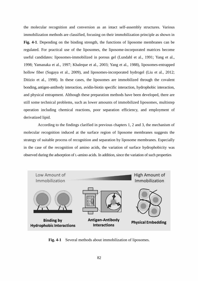
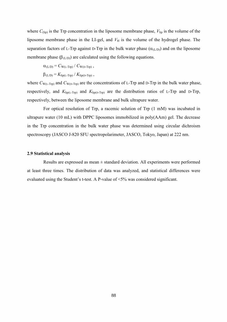
In chapter 4, the application of liposome membranes is examined by the case studies such
as the polycondensation reaction and separation process. The oligomerization of His was
enhanced by the adsorption of L-His on liposome membranes. In addition, the immobilized
liposome membranes embedding in hydrogels showed the chiral resolution of Trp. These
results are expected to contribute to the design strategy of efficient process by using liposome
membranes.
Based on the findings in this study, the selective adsorption of amino acids induced by
liposome membranes is evaluated by the understanding of adsorption mechanism and of the
effect of surface property of liposome membranes, proposing the design of liposome
membranes for the induction of efficient molecular recognition function.
Page 7
i
Contents
General Introduction 1
Chapter 1
Comparison of Separation of Amino Acids in Solvent-Water-Lipid System and
in Liposome Membrane System
13
1. Introduction 13
2. Materials and Methods 17
3. Results and Discussion
3.1 Partitioning of Tryptophan in Solvent-Water System Modified
Amphiphilic Phospholipids
20
3.2 Adsorption of Tryptophan and Histidine in Liposome Membrane
System and Its Chiral Selectivity
22
3.3 Adsorption Behavior of Other Amino Acids or Propranolol in Liposome
Membranes
24
3.4 Chiral-Selective Adsorption of Racemic Tryptophan or Histidine 26
3.5 A Plausible Model for Chiral Selectivity Based on Adsorption Isotherms 28
4. Summary 31
Chapter 2
Mechanism for the Selective Adsorption on Liposome Membranes Based on
Physicochemical Properties
33
1. Introduction 33
2. Materials and Methods 37
3. Results and Discussion
3.1 Analysis of Surface Hydrophobicity of Liposome Membranes by Using
Fluorescent Probe, ANS
40
3.2 Evaluation of Bound Water in Surface of Liposome Membranes by
Dielectric Dispersion Analysis
42
3.3 Observation of Bindings of Tryptophan or Histidine in Liposomes by
Page 8
ii
Resonance Raman Spectroscopy Analysis 43
3.4 Thermodynamic Analysis for Adsorption in Liposome Membranes 45
3.4.1 Evaluation of Phase Transition by DSC Analysis 45
3.4.2 Discussion of Adsorption in Relation between Enthalpy and Entropy 47
3.5 Adsorption Mechanism in Liposome Membranes 49
4. Summary 52
Chapter 3
Evaluation of Surface Properties of Cholesterol-Containing Binary and Ternary
Liposomes to Regulate Molecular Recognition and Design of Liposome
Membranes
54
1. Introduction 54
2. Materials and Methods 58
3. Results and Discussion
3.1 Effect of Mixing Cholesterol for Interior Membrane Properties 61
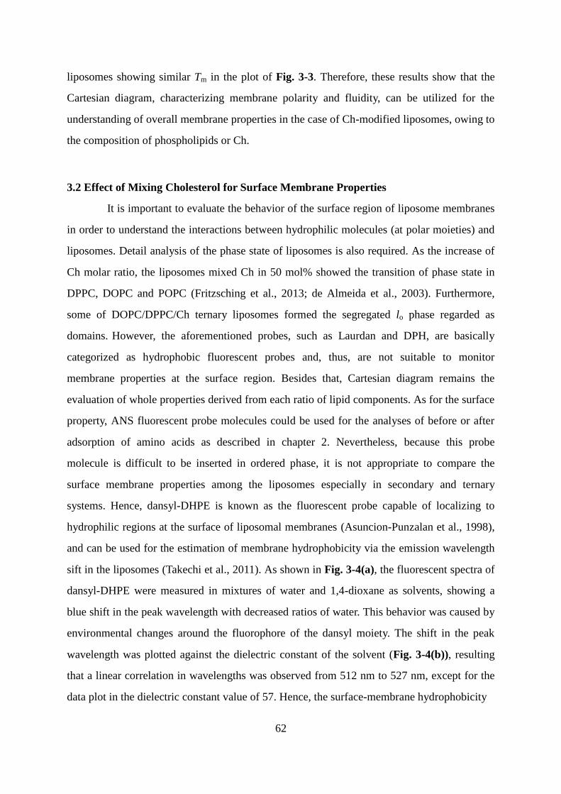
3.2 Effect of Mixing Cholesterol for Surface Membrane Properties 62
3.3 Relation between Membrane Properties and Chiral Recognition of
Histidine in Binary or Ternary Liposomes
66
3.4 Variation of Liposome Membrane Properties Induced by Adsorption of
L-Histidine
67
3.5 Design of DOPC/DPPC/Ch Ternary Liposomes to Induce Molecular
Recognition Function
69
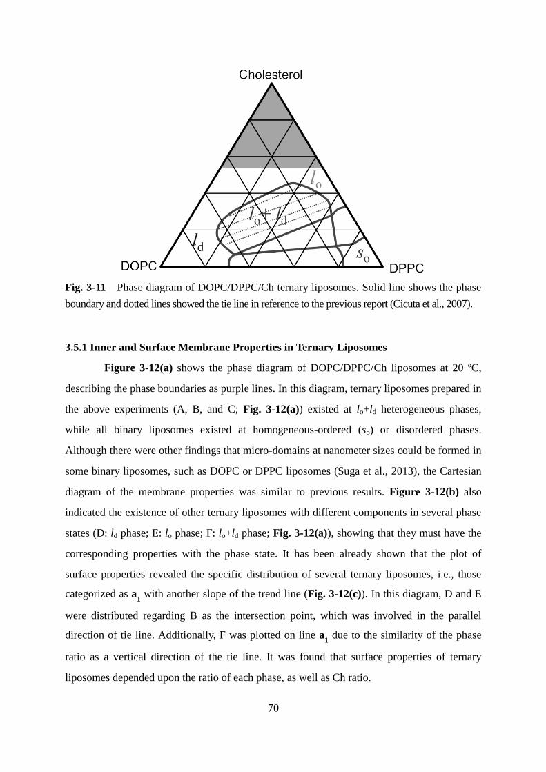
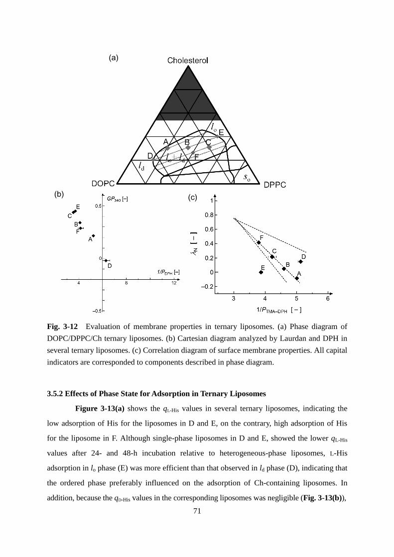
3.5.1 Inner and Surface Membrane Properties in Ternary Liposomes 70
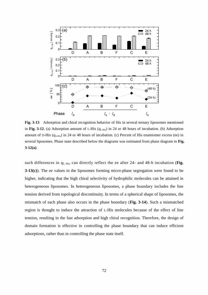
3.5.2 Effects of Phase State for Adsorption in Ternary Liposomes 71
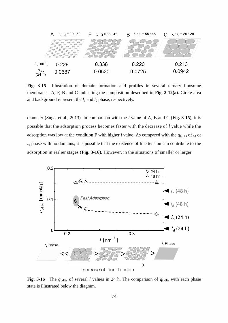
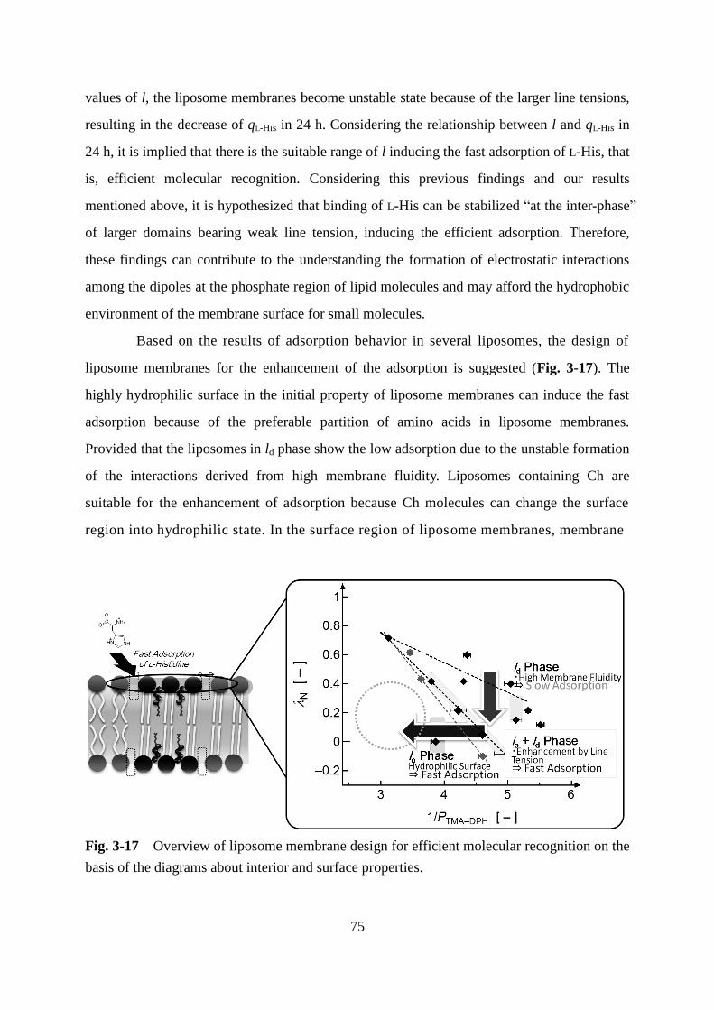
3.5.3 Effects of Domain Edge in Adsorption and Chiral Recognition 73
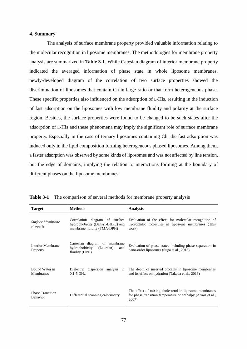
4. Summary 77
Chapter 4
Application for Separation and Conversion Process by Liposomal Membrane
System
81
Page 9
iii
1. Introduction 81
2. Materials and Methods 84
3. Results and Discussion
3.1 Scheme for Application of Liposome Membranes Using Designed
Membranes
89
3.2 Oligomerization of Histidine on Liposome Membranes 92
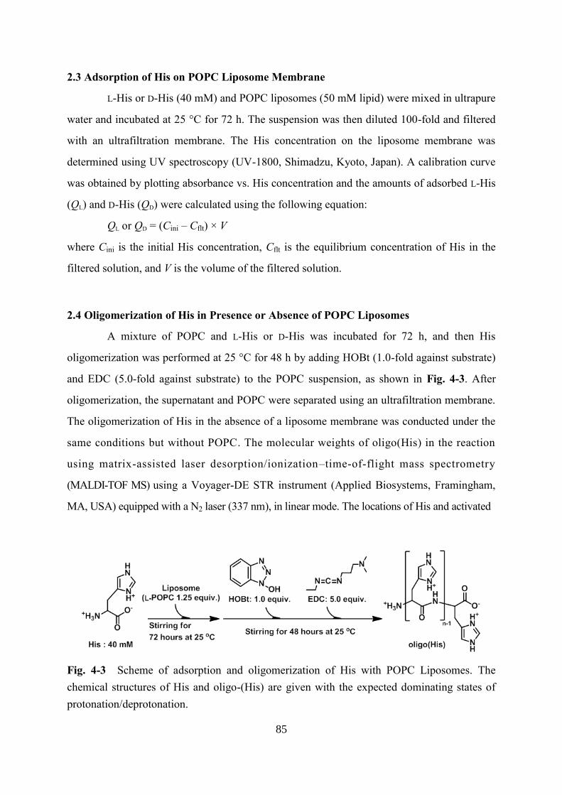
3.2.1 Adsorption and Condensation of Histidine on Liposome Membranes 92
3.2.2 Polymerization Degree of Adsorbed L- or D-Histidine 93
3.2.3 Mechanism of Inducing Reaction on Liposome Membranes 94
3.3 Preparation of Liposome-Immobilized Hydrogels (LI-gel) for Utilizing
Liposomes as a Device of Separation Process
95
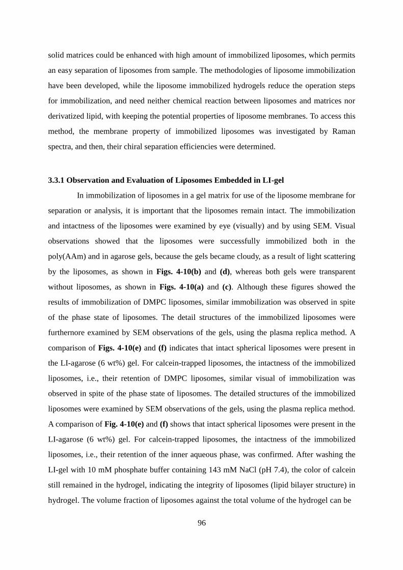
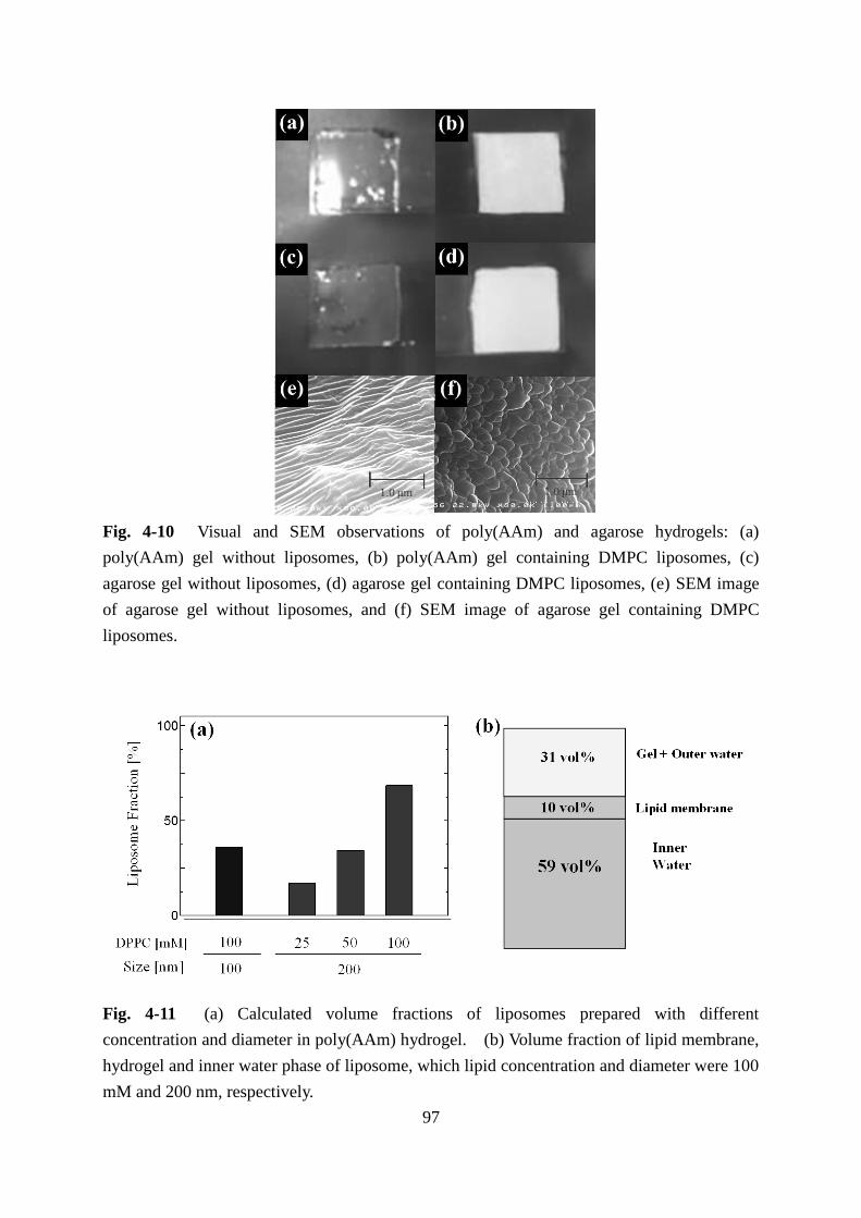
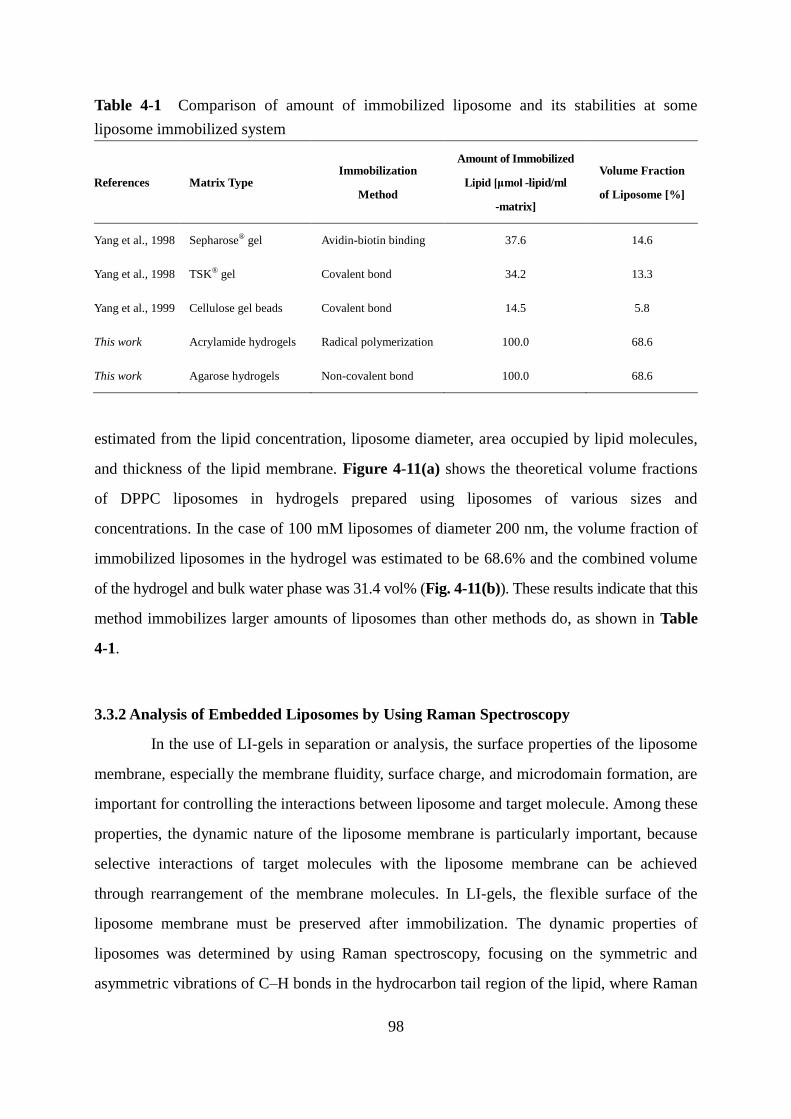
3.3.1 Observation and Evaluation of Liposomes Embedded in LI-gel 96
3.3.2 Analysis of Embedded Liposomes by Using Raman Spectroscopy 98
3.3.3 Optical Resolution of Tryptophan in LI-gel 100
4. Summary 104
General Conclusions 106
Suggestions for Future Works 109
Nomenclatures 111
List of Abbreviations 112
References 114
List of Publications 128
Acknowledgement 132
Page 10
1
General Introduction
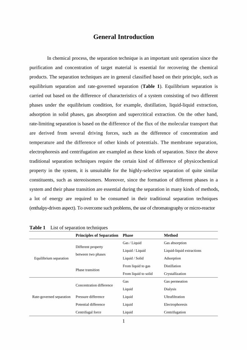
In chemical process, the separation technique is an important unit operation since the
purification and concentration of target material is essential for recovering the chemical
products. The separation techniques are in general classified based on their principle, such as
equilibrium separation and rate-governed separation (Table 1). Equilibrium separation is
carried out based on the difference of characteristics of a system consisting of two different
phases under the equilibrium condition, for example, distillation, liquid-liquid extraction,
adsorption in solid phases, gas absorption and supercritical extraction. On the other hand,
rate-limiting separation is based on the difference of the flux of the molecular transport that
are derived from several driving forces, such as the difference of concentration and
temperature and the difference of other kinds of potentials. The membrane separation,
electrophoresis and centrifugation are exampled as these kinds of separation. Since the above
traditional separation techniques require the certain kind of difference of physicochemical
property in the system, it is unsuitable for the highly-selective separation of quite similar
constituents, such as stereoisomers. Moreover, since the formation of different phases in a
system and their phase transition are essential during the separation in many kinds of methods,
a lot of energy are required to be consumed in their traditional separation techniques
(enthalpy-driven aspect). To overcome such problems, the use of chromatography or micro-reactor
Table 1 List of separation techniques
Principles of Separation Phase Method
Equilibrium separation
Different property
between two phases
Gas / Liquid Gas absorption
Liquid / Liquid Liquid-liquid extractions
Liquid / Solid Adsorption
Phase transition From liquid to gas Distillation
From liquid to solid Crystallization
Rate-governed separation
Concentration difference Gas Gas permeation
Liquid Dialysis
Pressure difference Liquid Ultrafiltration
Potential difference Liquid Electrophoresis
Centrifugal force Liquid Centrifugation
Page 11
2
process has been proposed. Although these methods developed the performance of separation
by means of the increase of the number of theoretical plates or the introduction of
stereospecific structure, the low efficiency of separation was remained as an inevitable
problem in terms of the cost.
Chiral isomers are often obtained in the chemical synthesis of the chemicals that
have asymmetric carbons in their structure and are formed by identical composition but
non-superposable mirror image configurations, which shows reverse optical rotatory (Pasteur
et al., 1848). Many kinds of chiral isomers sometimes show the different effects for the living
systems, i.e., the side effect of teratogenic was found by only in one side of thalidomide
enantiomer. Hence, the separation of one enantiomer from the other is important techniques
for fine chemical products. Typical methods of separating chiral molecules are shown in
Table 2. Although chiral column chromatography is in general utilized as the analytical
methods (Kaida et al., 1994; Yang et al., 1993), the cost of equipment in its use is quite high
because of the requirement of constructing chiral stationary phase and its maintenance. While
the crystallization methods can be carried out in lower cost (Wu et al., 2012; Martín et al.,
2007; Takahashi et al., 2002), there are some difficulties in the optimization of the operational
condition to obtain pure crystal of one side of enantiomers and to maintain the crystal quality
during a long-term process in pharmaceutical industry. In the case of some rate-governed
separations through the asymmetrically specific binding with proteins (Ghanem et al., 2004),
low efficiency in the recovery yield is still remained. While chiral selective synthesis techniques
Table 2 Chiral separation methods by several chiral selector
Reference Selector Method
Kaida et al., 1994 Polysaccharide carbamates Chromatography
Yang et al., 1993 Human serum albumin Chromatography
Gumí et al., 2005 N-Hexadecyl-L-hydroxyproline Capillary electrophoresis
Wu et al., 2012 Enantiomeric tartaric acid Separation of diastereomer salt
Martín et al., 2007 Methylbenzylamine Salt formation in supercritical carbon dioxide
Takahashi et al., 2002 Crystal of NBMe3 Preferential enrichment
Ghanem et al., 2004 Lipase enzyme Dynamic kinetic resolution
List et al., 2000 L-Proline catalyst Synthesis of enantiomer
Page 12
3
have been also developed by the design of catalysts (List et al., 2000), the cost of catalyst is
still high.
Amino acid is one of the basic biological molecules, which are known as the building
block to construct proteins. From the viewpoint as for the physicochemical property, amino
acids are known to be regarded as zwitterionic molecules that possess both amine and
carboxyl group at the -carbon, together with other functions called as “side chains” that can
be classified as about 20 species (Fig. 1). Based on the characteristics of side chains, the
tertiary structures are constructed when the amino acids are polymerized to peptides or
proteins. In the practical use, amino acid monomers are produced for the nutrient or seasoning.
In addition, dipeptides such as carnosine are treated as the drug for recovery from exhaustion.
One of the important features of amino acids is forming chiral compounds as L-form and
D-form. Among these enantiomers, it is known that there is the difference in their biological
function, such as the sense of taste. In biological system, amino acids in only L-form among
Fig. 1 Chemical structures of amino acids.
Page 13
4
their enantiomers are selected as the building block of the biomacromolecules or the key
material of their derivatives. Besides, biopolymers such as proteins and DNA also exhibit
homochirality. The origin of, reason for, and properties of homochirality have been still
attracting many researchers and have been widely investigated (Bada et al., 1995). On the
other hand, previous study revealed the asymmetric autocatalysis in the enantiomeric
amplification (Kawasaki et al., 2009). Based on these findings, it is also possible that the
chiral amplification is induced by utilizing the self-organizing system that consists of
biological system.
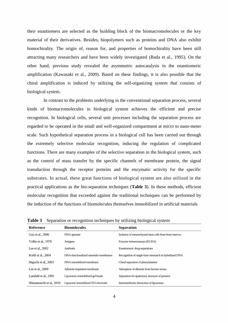
In contrast to the problems underlying in the conventional separation process, several
kinds of biomacromolecules in biological system achieves the efficient and precise
recognition. In biological cells, several unit processes including the separation process are
regarded to be operated in the small and well-organized compartment at micro to nano-meter
scale. Such hypothetical separation process in a biological cell has been carried out through
the extremely selective molecular recognition, inducing the regulation of complicated
functions. There are many examples of the selective separation in the biological system, such
as the control of mass transfer by the specific channels of membrane protein, the signal
transduction through the receptor proteins and the enzymatic activity for the specific
substrates. In actual, these great functions of biological system are also utilized in the
practical applications as the bio-separation techniques (Table 3). In these methods, efficient
molecular recognition that exceeded against the traditional techniques can be performed by
the induction of the functions of biomolecules themselves immobilized in artificial materials
Table 3 Separation or recognition techniques by utilizing biological system
Reference Biomolecules Separation
Guo et al., 2006 DNA aptamer Isolation of mesenchymal stem cells from bone marrow
Voller et al., 1978 Antigens Enzyme immunoassays (ELISA)
Lee et al., 2002 Antibody Enantiomeric drug separations
Kohli et al., 2004 DNA-functionalized nanotube membranes Recognition of single-base mismatch in hybridized DNA
Higuchi et al., 2003 DNA immobilized membrane Chiral separation of phenylalanine
Lee et al., 2009 Albumin imprinted membrane Adsorption of albumin from bovine serum
Lundahl et al., 1991 Liposomes immobilized gel beads Separation for quaternary structure of proteins
Shimanouchi et al., 2010 Liposome immobilized ITO-electrode Intermembrane interaction of liposomes
Page 14
5
(Voller et al., 1978; Lee et al., 2002; Kohli et al., 2004; Higuchi et al., 2003). However,
there is a problem in the cost of preparing such systems owing to the difficulty in the
maintenance of the structure and function of biomolecules during their use. Therefore, the
understanding of the insight about the biological system and its application are expected to be
applied to the efficient design of artificial separation system for practical use, that is,
“bio-inspired system”.
Recently, self-assembled materials have attracted many interests of researchers in
various research fields. Depending on the property derived from chemical structure and/or
surrounding environment, some kinds of amphiphilic molecules can be automatically
assembled to form the supramolecular structures. Amphiphilic molecules, for example, can
form the self-assembly by several driving forces, such as electrostatic interactions and van der
Waals interactions in hydrophobic regions. Apart from the typical polymers formed by
covalent bonds, such self-assembly structures can show dynamic property despite of the
increment of entropy, which is regarded as fluctuation in non-equilibrium state. These
phenomena are described as the theory of dissipative systems (Prigogine et al., 1967), which
contributes to research about the birth of living system. According to the biomolecules,
various organisms such as cells is formed by the assemblies, called as “self-organization”
system. Microtubule which is one of the organelle is constructed by the self-organization of
tubulin proteins, forming the long and rigid fibers. Hence, they are enabled to transport
proteins in the cell. As for the other example, the protein of actin can organize to filament
structure and form the actomyosin complex with another protein of myosin, inducing the
active movement of muscle fiber due to the flexible organization (Geeves et al., 2005). In
more microscopic view, proteins or DNA is formed by the conformation of polymers of
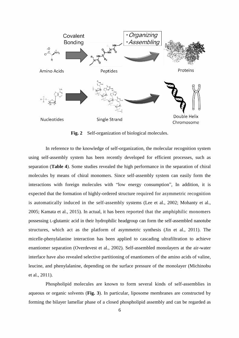
amino acids or nucleic acids (Fig. 2). The appropriate self-organization of such structures
induces several functions by regulating the dynamic changes of its conformation. Furthermore,
cell membranes, which exist in the boundary of cells, are composed by amphiphilic
phospholipids. The dynamic changes of cell membranes such as endocytosis are induced by the
rearrangement of phospholipid components of outer or inner membranes (Farge, 1995). These
dynamic regulations of assembled states may possibly play important role for the flexible
recognition of target materials.
Page 15
6
Fig. 2 Self-organization of biological molecules.
In reference to the knowledge of self-organization, the molecular recognition system
using self-assembly system has been recently developed for efficient processes, such as
separation (Table 4). Some studies revealed the high performance in the separation of chiral
molecules by means of chiral monomers. Since self-assembly system can easily form the
interactions with foreign molecules with “low energy consumption”, In addition, it is
expected that the formation of highly-ordered structure required for asymmetric recognition
is automatically induced in the self-assembly systems (Lee et al., 2002; Mohanty et al.,
2005; Kamata et al., 2015). In actual, it has been reported that the amphiphilic monomers
possessing L-glutamic acid in their hydrophilic headgroup can form the self-assembled nanotube
structures, which act as the platform of asymmetric synthesis (Jin et al., 2011). The
micelle-phenylalanine interaction has been applied to cascading ultrafiltration to achieve
enantiomer separation (Overdevest et al., 2002). Self-assembled monolayers at the air-water
interface have also revealed selective partitioning of enantiomers of the amino acids of valine,
leucine, and phenylalanine, depending on the surface pressure of the monolayer (Michinobu
et al., 2011).
Phospholipid molecules are known to form several kinds of self-assemblies in
aqueous or organic solvents (Fig. 3). In particular, liposome membranes are constructed by
forming the bilayer lamellar phase of a closed phospholipid assembly and can be regarded as
Page 16
7
Table 4 Self-assembly systems formed by artificial monomers
Reference Monomer and its application
Lee et al., 2002 Bio-nanotube membranes for enantiomeric drug separations
Ziserman et al., 2011 Helical nanotube formation from self-assembly of amphiphiles
Eliseev et al., 1994 Molecular recognition of some biomolecules by aminocyclodextrins
Mohanty et al., 2005 SDLV vesicles as pseudo-stationary phase for enantiomer separation
Liu et al., 2015 Lyotropic liquid crystals for extractant of biomolecules
Jin et al., 2011 Catalytic reaction by nanotube containing bola-amphiphilic amino acid
Kameta et al., 2015 Enantiomer-sensitive vesicle formation by fluorescent glycolipid amphiphiles
Makino et al., 2012 Vesicle formation from poly-L-lactic acid utilized in drug carrier
Fig. 3 Self-assembled structures derived from phospholipids.
the model of biomembranes. Moreover, these membranes also have the ordered alignment of
phospholipids, inducing the increasing anisotropy of several steroid molecules. This feature is
expected to contribute to the formation of stereospecific interactions. Furthermore, it is
known that the liposome membranes are characterized by the degree of lateral diffusion as the
membrane fluidity. Based on the thermodynamic analyses, the phase transition can be
observed in specific temperatures, where the endothermic transition of enthalpy is induced
with dynamic changes. Hence, the interaction with guest molecules may induce the change of
assembled states of phospholipids in liposome membranes with easily formation. From
another point of view, micelles or liposomes in aqueous phase can include the hydrophobic
regions, resulting that it can induce the several reactions of hydrophobic substrates even in the
Page 17
8
aqueous phase (Table 5). In this case, this approach for reactions is useful due to the reduction
of solvent and the regulation of reaction rate by the assembled states. In addition, the reverse
micelles may also develop the infinitely-connected structure to give the fiber network in some
kinds of solvent conditions, resulting in the formation of the organogels containing emulsion
phases (Scartazzini et al., 1986). This finding means that phospholipids can form the specific
materials that can include the hierarchical assembly in their material structure. The several
findings about the phospholipid assemblies indicate the emergence of functions and materials
with varying systems dynamically (Walde et al., 2014).
Based on the above findings, the development of liposome membrane systems is
important for the efficient separation techniques with high molecular recognition. In the
previous investigation, Liposomes have been developed as a functional platform for
recognition of biomolecules: proteins and enzymes (Umakoshi et al., 2010), amyloid fibrils
(Shimanouchi et al., 2013), and single-stranded RNA (Suga et al., 2013). These recognition
events were significantly affected by the liposomal membrane properties, such as fluidity and
polarity. The phospholipid used to make the liposomes is usually consisted of L-enantiomers;
thus, the liposomal membranes potentially possessing chiral specificity. It is therefore
expected that not only chemical properties of amphiphilic molecules, but also the
physicochemical properties of self-assembled membranes are possible clues to develop a
novel, flexible recognition site (“Lock”) on the membranes. Based on the characterization of
physicochemical properties of membranes, an ideal membrane surface for molecular
recognition can be designed. Furthermore, in the surface region of liposome membranes, it is
possible to form the highly-ordered alignment of charged functions and to induce the domain
Table 5 List of reactions induced in hydrophobic region of self-assembly
Reference Assembly Reaction
Manabe et al., 2002 Emulsion droplets formed by DBSA Several dehydrative conversion
Monti et al., 2002 CTAB and Mn-derived amphiphiles Epoxidation reactions
Peng et al., 2003 SDS micelles Aldol reaction catalyzed by L-proline
Mase et al., 2005 Assembly of catalyst with the reactants Direct asymmetric aldol reactions
Otto et al., 1998 SDS, CTAB micelles or vesicles Diels–Alder reactions
Li et al., 2012 Micellar assembly of amphiphilic catalysts Asymmetric reduction of aliphatic ketones
Page 18
9
formation by plural components. As for the recognition of RNA in liposome membranes, the
domain size is regarded as the key factor of the design of recognition (Suga et al., 2013).
The final purpose of this thesis is to establish the methodology to design the
liposome membranes for the selective adsorption of chiral small molecules such as amino
acids by utilizing the liposome membranes as the platform of recognition and conversion
processes. To design the molecular recognition, it is required to understand the membrane
property in several aspects and to investigate the mechanism of bindings between liposome
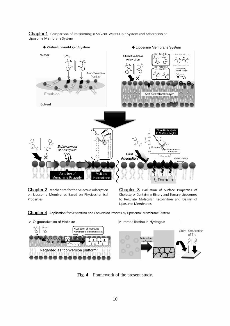
membranes and guest molecules. The framework and flow chart of the present study are
schematically shown in Figs. 4 and 5, respectively.
In chapter 1, the partitioning and adsorption behavior of amino acids were
investigated in two kinds of self-assembled states of phospholipids, e.g., emulsion layer at the
water-solvent interface or liposome membranes. In the liquid-liquid extraction system, the
partitioning of the tryptophan (Trp) enantiomer in emulsion layer of phospholipids was
investigated in different ratio of chloroform/methanol mixture as the solvent. In addition, the
partitioning behavior of the Trp in liposome membranes was investigated by adsorption of L-
or D-form amino acids dissolved in water phase. Based on the adsorption behaviors of several
amino acids possessing hydrophilic or hydrophobic side chains and the adsorption isotherms,
chiral recognition function by L-enantiomer of phospholipids was induced by the formation
of highly-ordered assembly such as lamellar phase.
In chapter 2, the variation of liposome membrane property was investigated by
employing the adsorption of amino acids as targets. Time course of surface hydrophobicity
was first evaluated by 8-anilino-1-naphthalenesulfonic acid (ANS) fluorescent probes during
the adsorption process of amino acids. This analysis for membrane surface property was also
examined by analyzing dielectric dispersion spectra at the higher frequency region. As the
evidence of multiple interactions, direct observation of adsorbed Trp or histidine (His)
molecules was also carried out by Raman spectroscopic analysis. In addition, thermodynamic
analysis for this adsorption was measured by differential scanning calorimetry (DSC), comparing
the interaction behavior by the evaluating compensation of enthalpy and entropy. Each
measurement mentioned above was conducted in both enantiomers of amino acids in order to
discuss the mechanism of chiral recognition of the liposome membranes.
Page 19
10
Fig. 4 Framework of the present study.
Page 20
11
Fig. 5 Flow chart of the present study.
Page 21
12
In chapter 3, the effect for chiral recognition of His is investigated by using
liposomes of several components contained cholesterol (Ch). At First, the methodology of
evaluating the surface property of the liposome membranes is developed by the combination
of two fluorescent probes that can be localized at the surface edge of the membrane. By
means of the comparison of Cartesian diagram for interior property, it is expected to evaluate
the liposome membrane property in detail. According to the relation between such properties
and the adsorption of L- or D-form of His, the effect of mixed Ch in liposome membranes is
discussed. The transition of membrane property after the adsorption is also investigated to
understand the behavior of liposome membranes. Furthermore, the membrane properties of
ternary liposomes are observed based on their phase state, and then, the relation with chiral
recognition of His are investigated. By comparing the domain states relating to the line
tension, the contribution of domain is considered for understanding the effect of existing
domain “edges”.
In chapter 4, based on the molecular recognition in liposome membranes described in
chapters 1, 2, and 3, the application of liposome membrane systems is demonstrated in the
conversion reaction and optical resolution. The promotion of oligomerization of amino acids
was investigated by partition in liposome membranes with molecular recognition of L-His. In
this reaction, the behavior of conversed substrates was considered in reference to previous
reports about the aqueous reactions (Kunishima et al., 2005). As for the application for
separation process, continuous process is examined by liposomes accumulated in the
ultrafilter. Besides, the immobilization of liposomes is developed by the embedding in several
hydrogels, which is analyzed by the direct observation and the Raman spectroscopy.
Adsorption behavior and chiral separation of Trp are evaluated in prepared liposome
immobilized hydrogel (LI-gel).
The results obtained in this work are summarized in the General Conclusions section.
Suggestions for Future Works are described as extension of the present thesis.
Page 22
13
Chapter 1
Comparison of Separation of Amino Acids in Solvent-Water-Lipid
System and in Liposome Membrane System
1. Introduction
Extraction is one of the separation methods based on the partitioning behavior of the
molecules among the immiscible two-phase system. The operational conditions of this
process have been usually optimized by the following factors: (i) physicochemical property of
two solvent system, (ii) characteristics of the target material, and (iii) property of the interface
formed in solvent-water two-phase system. Two-phase extraction process has a merit of low
energy consumption because of no requirement of phase transition unlike the distillation. On
the other hand, experimental knowledge is required in each separation process to optimize the
separation condition. Therefore, the synthetic ligands that can specifically interact with target
molecules (so called as “extractants”) are usually applied in the extraction in order to achieve
highly-selective separations (Fig. 1-1). Calix[4]arene derivatives, for example, have been
used as the extractants of Chromium (VI) with higher selectivity, where the extraction can be
controlled by varying pH of the aqueous phase (Ediz et al., 2004). In other cases, the
extraction processes of Am (III) in organic phase extraction have been investigated by using the
extractant, N,N,N’N’-tetraoctyl-3-oxapentanediamide (Panja, et al., 2012). Chiral host
molecules, such as hydrophilic--cyclodextrin (Wang et al., 2014) and L-tartaric acid
derivatives (Ren et al. 2014), have been also used to perform the chiral recognition for
pantoprazole and ibuprofens, respectively.
In recent years, there have been some reports on the usage of surfactants as extractant
in liquid–liquid extraction of the silica refinary (Kusaka et al., 1998), and in biomass recovery
(Pursell et al., 2009). In general, the surfactant molecules are known to be distributed at the
liquid-liquid interface due to their amphiphilic nature and, after that, they can enhance the
emulsification of one liquid phase into another phase. These behaviors or both partitioning
and emulsification have been reported to depend on the structure of surfactant, such as charges,
Page 23
14
Fig. 1-1 Schematic illustration of extraction using extractants.
hydrophilicity of headgroups and length of acyl chains. In addition, the surfactants and some
amphiphilic molecules can form several kinds of self-assembly structures, depending on their
geometry and solvent environment. In particular, micelles and lamellar structures are typical
forms of amphiphilic surfactant or lipid (Fig. 1-2). Owing to the dynamic equilibrium nature
(including their deformation under various temperatures or concentrations), the micelles can
control the partitioning of micelle-constituting molecules and also guest molecules, resulting
in the less stability of the host-guest complex. In contrast to the micelle, the lamellar
structure forms relatively stable bimolecular phase at the high-ordered state in horizontal
direction. Lamellar membranes can thus orientate the materials such as proteins (White, 1999)
and several sterols (Biruss, et al. 2007).
Phospholipid molecules are common amphiphilic molecules in L-enantiomer derived
from biological system. Therefore, the lamellar membranes formed by phospholipids can
partition several biological molecules in the hydrophobic interior and hydrophilic surface.
Besides, the biological molecules can be recomposed in phospholipid membranes by the
molecular order. Based on these features, there have been several researches focusing on the
up- or down-regulation of function of gene expression machinery (Bui et al., 2008), and the
conformational change of RNA (Suga, et al., 2011), peptide (Tuan, et al., 2008) and enzyme
(Umakoshi, et al., 2012; Suga, et al., 2015) on the phospholipid membranes. In addition, the
enantiospecific interaction of the amino acid dimers has been observed in the phospholipid
Page 24
15
Fig. 1-2 Illustration of micelle and lamellar about specific features.
bilayers. At the phospholipid liposomal or micellar surface, 1H NMR analysis indicated that
dipeptide enantiomers at different conformations were interacted with their surface, in which
the distance and dihedral angle of Trp-Trp differed between the enantiomers (Cruciani, et al.,
2006; Bombelli, et al., 2004). The possible of chiral recognition by the phospholipid
membranes are also reported by using some liposomes (Yamada, et al., 2006; uniono et al.,
2011; Pathirana et al., 1992). It is expected that the use of the highly-ordered structure of
phospholipid membrane could provide us some benefits on the chiral selectivity in the
recovery of biomolecules.
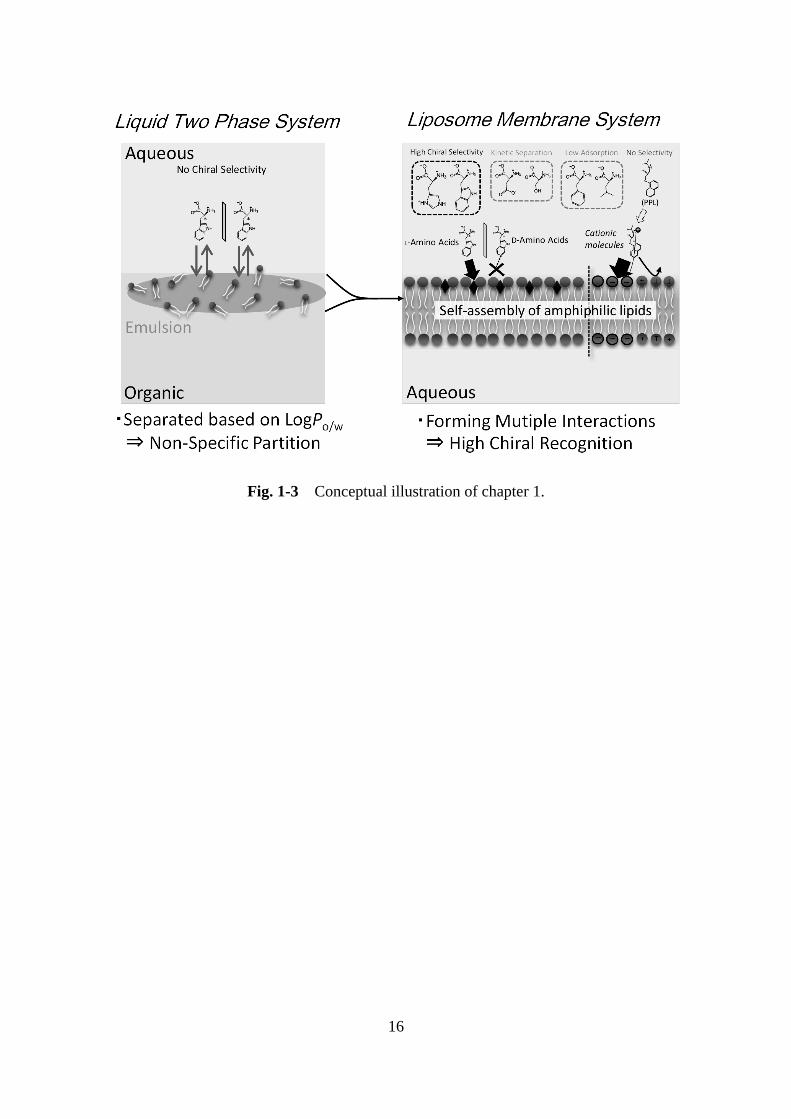
In this chapter, the partitioning and adsorption behavior of amino acids were
investigated in two kinds of self-assembled states of phospholipids, e.g., emulsion layer at the
water-solvent interface or liposome membranes (Fig. 1-3). In the liquid-liquid extraction
system, the partitioning behavior of the tryptophan (Trp) enantiomer in emulsion layer of
phospholipids was investigated in different ratio of chloroform/methanol mixture as the
solvent. In addition, the partitioning behavior of the Trp in liposome membranes was
investigated by adsorption of L- or D-form amino acids dissolved in water phase. Based on the
adsorption behaviors of several amino acids possessing hydrophilic or hydrophobic side
chains and the adsorption isotherms, the interaction model in liposome membranes was
finally proposed.
Page 25
16
Fig. 1-3 Conceptual illustration of chapter 1.
Page 26
17
2. Materials and Methods
2.1 Materials
A zwitterionic phospholipid 1,2-dipalmitoyl-sn-glycero-3-phosphocholine (DPPC;
carbon number/unsaturated bond = 16:0), and negatively-charged phospholipids
1,2-dimyristoyl-sn-glycero-3-phosphatidic acid (DMPA; 14:0),
1,2-dimyristoyl-sn-glycero-3-phospho-L-serine (DMPS; 14:0) were purchased from Avanti
Polar Lipids, Inc. (Alabaster, AL, USA). L-Trp, D-Trp and other amino acids were purchased
from Peptide Institute (Suita, Osaka, Japan). All the amino acids were over 98% purity of
enantiomers. Chloroform, methanol and other chemicals were purchased from Wako Pure
Chemical Industry Ltd. (Osaka, Japan) and used without further purification.
2.2 Liposome Preparation
A solution of phospholipids in chloroform was dried in a round-bottom flask by
rotary evaporation under vacuum. The resulting lipid films were dissolved in chloroform and
the solvent was evaporated twice. The lipid thin film was kept under high vacuum for at least
3 h, and then hydrated with ultrapure water at room temperature. The vesicle suspension was
frozen at -80 °C and then thawed at 50 °C to enhance the transformation of small vesicles into
larger multilamellar vesicles (MLVs). This freeze-thaw cycle was repeated five times. MLVs
were used to prepare large unilamellar vesicles (LUVs) by extruding the MLV suspension 11
times through two layers of polycarbonate membrane with mean pore diameters of 100 nm
using an extruding device (Liposofast; Avestin Inc., Ottawa, Canada).
2.3 Evaluation of Partition Behavior of L-Trp in Solvent-Water System
With regard to solvent-water systems, chloroform and its mixtures with methanol
were used as solvent phases. First, DPPC was dissolved in solvent phase in 27 mM of
concentration. In the case of chloroform/methanol mixtures, the ratio of methanol (xmet) varied
from 0.05 up to 0.25. Secondly, L-Trp or D-Trp was dissolved in aqueous phase as 3 mM
solution. Then, the distribution ratio (D) of Trp at each enantiomer (DL-Trp for L-Trp and
DD-Trp for D-Trp) was calculated from the decrease of Trp in aqueous phase by following
equation:
Page 27
18
D = (Cini – Ceq) / Cini,
where Cini and Ceq represent the initial and equilibrium concentration of Trp in aqueous phase.
The Trp concentration in water phase was measured by UV absorbance of 280 nm, based on
their calibration curves.
2.4 Evaluation of Adsorption Behavior of L-Trp in Liposome Membrane System
The liposome suspensions (lipid: 4.5 mM) were mixed with L-Trp or D-Trp, and
other amino acids (0.5 mM). They were incubated at 25 oC for 48 hours, to be equilibrium of
adsorption. After the incubation, liposomes and adsorbed amino acids were separated by
ultrafiltration membrane with the molecular cut of 50,000 Da (USY-5; Toyo Roshi Kaisha,
Ltd., Tokyo, Japan). The concentration of filtered amino acids (Cflt) was measured by the
absorbance by using UV spectrometer (UV-1800; Shimadzu, Kyoto, Japan), and by the
fluorescence of fluorescamine (Ex: 390 nm, Em: 475 nm)
(Stein et al., 1973) by
spectrofluorometer (FP-8500; JASCO, Tokyo, Japan). The concentration of adsorbed amino
acids (Cads) and adsorbed amount of amino acids per lipid amount (q) were calculated by
using following equations:
Cads = Cini – Cflt
q = Cads / clip ,
where Cflt represent the concentration of amino acids in leakage of ultrafiltration and clip
represents the concentration of lipid (liposomes). The adsorption isotherms were evaluated by
the plot of q versus Cflt in 48 hours incubation (regarded as equilibrium concentration) at the
same lipid concentration (4.5 mM). Especially, Langmuir isotherms were described by
following equations:
q = qmax K Cflt / (1 + K Cflt) ,
where qmax and K represent the maximum of q and a binding constant. The correlation for
Langmuir isotherms and qmax and K values were estimated by the plot of Cflt versus Cflt/qmax.
From the Cads of L-amino acids and D-amino acids, separation parameter (SL/D) was calculated
by following equations:
SL/D = Cads (L form) / Cads (D form) ,
Page 28
19
where Cads (L form) and Cads (D form) represent the Cads values of L-amino acids and that of D-amino
acids, respectively.
2.5 Circular Dichroism Spectroscopy Analysis of Racemic Amino Acids
In order to analyze the concentration ratio of L- and D-amino acids, circular
dichroism (CD) spectra were measured by JASCO J-820 SFU spectropolarimeter (JASCO,
Tokyo, Japan). The CD spectrum from 300 nm to 200 nm was measured with a quartz cell
(0.1 cm path length) at a scan speed of 100 nm per minute and a width of 2 nm. Three scans
excluding water background signals were obtained at 25 ºC, and the data was calculated as
molar ellipticities. In the case of racemic solutions, the initial concentrations of total amino
acids were 1.0 mM. The racemic mixtures of L- and D-Trp or L- and D-His were incubated
with DPPC liposome (4.5 mM) for 48 hour at 25 ºC, and the liposomes and adsorbed amino
acids were removed by filtration shown in above.
2.6 Statistical analysis
Results are expressed as mean ± standard deviation. All experiments were performed
at least three times. The distribution of data was analyzed, and statistical differences were
evaluated using the Student’s t-test. A P-value of <5% was considered significant.
Page 29
20
3. Results and Discussion
3.1 Partitioning of Tryptophan in Solvent-Water System Modified Amphiphilic
Phospholipids
Although Trp is known as a hydrophobic amino acid due to the hydrophobic side
chains (Tanford, 1962), it has a difficulty in its partitioning to organic solvent (Fig. 1-4(a))
because of its relatively hydrophilic nature that can be described in the negative value of
LogP (-2.2). In order to investigate the partitioning of Trp, the effect of additives, such as the
amphiphilic phospholipid (DPPC) and methanol, was first evaluated in the water-chloroform
extraction system. When chloroform (dielectric constant = 5) was used as the organic phase,
a thin cloudy layer was formed in chloroform phase on mixing the two phases (Fig. 1-4(b))
due to the emulsification mediated by DPPC molecules. This was probably caused by the
formation of phospholipid aggregates at the interface on the solvent phase. When methanol
(= 33) was added to the chloroform phase, the formation of such emulsion phases was
enhanced with an increase in the ratio of methanol (Fig. 1-4(c)).
The partitioning behaviors of L- and D-Trp in water-DPPC-solvent phase are shown
in Fig. 1-5. The distribution ratio of Trp, DTrp, was determined by measuring the Trp
concentration in water phase, which can be therefore regarded as an indicator of the Trp
Fig. 1-4 Schematic illustration of the partitioning behavior of L- or D-tryptophan (Trp) in
chloroform-water system with phospholipids. (a) Water-chloroform extraction system. (b) Formation
of emulsion after mixing as the cloudy layer. (c) Increasing thickness of cloudy layer by addition of
methanol. (d) Liposomes prepared by thin film of phospholipids in the aqueous phase.
Page 30
21
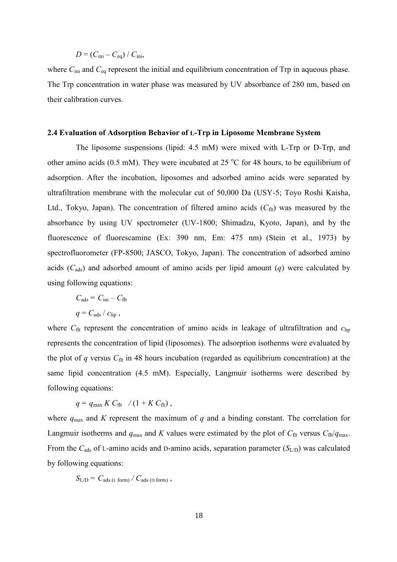
partitioning in the water/organic two-phase systems (including the emulsion layer). The DTrp
value was found to become larger as the increase of methanol ratio with expanding the
emulsion phase, while the DTrp value was small (approximately 0.2) at the lower methanol
ratio (xmet = 0 ~ 0.1). Finally, the DTrp value reached a plateau at a methanol ratio above 0.2.
Incidentally, at methanol ratios over 0.25, the emulsion phase disappeared on mixing due to
the reduction in the difference in the dielectric constant between the two phases. This behavior
indicated that the DTrp value did not always depend on the dielectric constant of the organic
solvent because the variation of the values was slight in available range of forming emulsion
(Fig. 1-5). It is thus considered that the enhancement in the formation of the emulsion layer is
promoted by the efficient interaction of DPPC and Trp molecules. It is thus suggested that the
partitioning behavior of Trp was enhanced through the strong binding between the
phospholipid and Trp molecules in the emulsion layer containing lipid aggregation. The chiral
selectivity of Trp partitioning was also investigated by determining the ratio of the DTrp value
of L-Trp to that of D-Trp (DL-Trp/DD-Trp). As the xmet increased, the DL-Trp/DD-Trp value reached
to approximately 1 (xmet = 0.15), while the DD-Trp became slightly larger at lower methanol
ratio. From the viewpoint of actual recovery of amino acids, the partitioning behaviors were
Fig. 1-5 Distribution ratio of L- or D-Trp (DTrp) and its chiral selectivity (DL-Trp/DD-Trp) in
different ratio of methanol in chloroform solvent (xmet). Dielectric constant () of solvent were
shown as the black keys and line.
Page 31
22
thus improved with phospholipids and methanol. The distribution ratio of Trp can be
predicted by 0.006 at the interface of water and organic solvent system based on the LogP
value of Trp (-2.2). On the other hand, the DTrp value increased to approximately 0.2 by the
addition of phospholipids that can induce the formation of emulsion phase. Furthermore, the
increase of in organic phase was also found to induce the increase of the DTrp up to 0.7. It
was thus found that the specificity of enantiomers in partitioning Trp was not observed in the
liquid-liquid two phase systems with the DPPC extractant.
3.2 Adsorption of Tryptophan and Histidine in Liposome Membrane System and Its
Chiral Selectivity
DPPC liposome bilayer membranes have ordered structures owing to molecular
alignment and can also be characterized as gradient polarity layers at nano-meter scale as
shown in Fig. 1-4(d). As a preliminary experiment, Trp dissolved in the aqueous phase was
partitioned to the liposome membranes by mixing them for 48 hours. In this liposome
membrane system, the yield of L-Trp recovery, calculated by its partitioning behavior from
the aqueous phase to the phospholipid phase, was found to be high, while that of D-Trp was
Fig. 1-6 DL-Trp/DD-Trp and the yield of Trp (ratio of concentration between in water and in
lipid phase) at the solvent of chloroform, chloroform mixed methanol and liposome
membranes corresponding to Fig. 1-4.
Page 32
23
extremely low (Fig. 1-6). As for the chiral selectivity, the liposome system showed an
extremely high value (~104) as compared with other solvent extraction systems. It is thought
that the liposome membrane could provide a suitable environment for the partitioning of the
hydrophilic Trp, where some interactions (i.e. electrostatic interaction, and hydrogen bond)
neighboring to the chiral carbon of the lipid molecules could be related to its chiral
recognition. For further investigation to the liposome membranes, the partitioning behavior of
Trp and another amino acids (His) were investigated by analyzing the adsorption behaviors
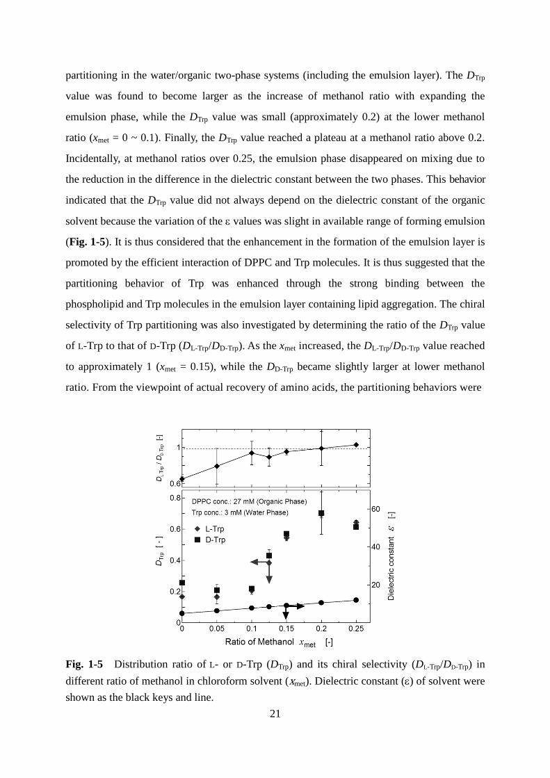
from aqueous solution. Figure 1-7(a) shows the time course for the adsorption of Trp on
DPPC liposomes. The adsorbed amounts of L-Trp gradually increased after 16-hour incubation,
Fig. 1-7 Time course of adsorbed concentration (Cads) of L or D-forms of amino acids. (a)
Cads of L-Trp (filled) and D-Trp (open). (b) Cads of L-His (filled) and D-His (open).
and reached to be an equilibrium after 48 hours, where almost all of the amino acids adsorbed on
Page 33
24
DPPC liposomes. On the other hand, negligible amounts of D-Trp and D-His adsorbed on the
DPPC liposomes even at 48 hours. As a result, extremely high chiral selectivity in the Trp
adsorption on the DPPC liposome was observed at the final stage of the adsorption process,
while such behaviors were not found in the solvent-water system modified with DPPC (Fig.
1-6). As shown in Fig. 1-7(b), similar tendencies for the time course of the adsorption and for
the chiral selectivity were observed in the case of His. It was also found that the adsorption
kinetics of both L-Trp and L-His were sigmoidal with a lag time (no adsorption from 0 to 16
hours). This result was also imply that the binding of L-amino acids on the liposome structure
can be very weak at the initial stage of adsorption, considering the previous findings on the
adsorption in supported lipid bilayer (Sarangi, et al., 2012). This is because the amino acids
preferably exist in the aqueous phase rather than in liposome membranes, judging from the
negative values of LogP. It is thought that the membrane property could be varied after the
accumulation of amino acids on the membranes at the initial stage: such varied membrane
properties could recruit the additional L-Trp partitioned to the membrane at the latter step. In
comparison with the adsorption kinetics in L-Trp and L-His, it is assumed that there could be
the similar steps to promote L-His adsorption.
3.3 Adsorption Behavior of Other Amino Acids or Propranolol in Liposome Membranes
Natural amino acids possess unique surface characteristics, depending on their side
chains, such as hydrophobic and hydrophilic. In order to investigate the effect of side chain
property in the chiral selective adsorption on liposomes, the Cads values for 10 kinds of amino
acids were analyzed after 48 hours incubation (Fig. 1-8). For almost all amino acids (except
for Ser), the adsorption of the L-form was dominant, indicating that DPPC liposomes could be
widely applied to recognize the chirality of amino acids. The chiral selectivity (SL/D) was
calculated from the ratio of Cads of L- and D-amino acids. The highest SL/D was obtained with
Trp and His (SL/D >1000). Relatively high SL/D was also observed in the case of Tyr and Pro
(SL/D >100), owing to the hydrogen donors or acceptors in their side chains, indicating that the
formation of hydrogen bonds provided from the aromatic structure could play a crucial role in
their high L-amino acid selectivity. In contrast, amino acids possessing hydrophobic (non-polar)
Page 34
25
Fig. 1-8 Adsorbed concentration (Cads) of L- or D-forms of 10 amino acids on DPPC
liposomes. Amino acids is listed by the hydrophobicity (Tanford, 1962) from left to right.
Separation parameter (SL/D) of each amino acid is described above the corresponding bars. In
all samples, the incubation temperature was 25 ºC.
Fig. 1-9 Time course of Cads of L-Asp (filled) and D-Asp (open) in DPPC liposomes.
side chains (Leu and Val) showed lower L-selectivity. Moreover, no L-selectivity was
observed for Ser. These results indicate that the hydrophobicity of the side chains might not
Page 35
26
be an important factor for inducing asymmetric recognition. The contribution of small side
chains is also negligible in stereospecific recognition due to the difficulties in forming
certain binding to phospholipid. Interestingly, Asp showed high chiral selectivity in adsorption
kinetics at 0-40 hours, although the low SL/D values were obtained after 48 hours (Fig. 1-9). It
is possibly explained that Asp molecules (even in D-form) easily form the hydrogen bonds
with phospholipids due to possessing the polar and small side chains, resulting in decreasing
L-selectivity in long time incubation.
3.4 Chiral-Selective Adsorption of Racemic Tryptophan or Histidine
In many case of chiral separation methods, enantiomer excess (ee) from racemic state
is evaluated as the efficiency of chiral selectivity. As mentioned above, the chiral recognitive
adsorption was observed only in enantio-pure solution, thus, the performance of chiral
recognition of liposome membranes was evaluated by studying the adsorption of racemic Trp
or His in CD spectroscopic analysis.
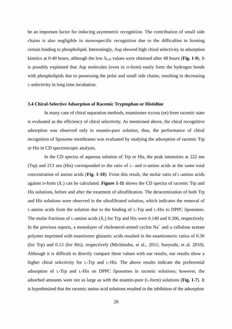
In the CD spectra of aqueous solution of Trp or His, the peak intensities at 222 nm
(Trp) and 213 nm (His) corresponded to the ratio of L- and D-amino acids at the same total
concentration of amino acids (Fig. 1-10). From this result, the molar ratio of L-amino acids
against D-form (XL) can be calculated. Figure 1-11 shows the CD spectra of racemic Trp and
His solutions, before and after the treatment of ultrafiltration. The deracemization of both Trp
and His solutions were observed in the ultrafiltrated solution, which indicates the removal of
L-amino acids from the solution due to the binding of L-Trp and L-His to DPPC liposomes.
The molar fractions of L-amino acids (XL) for Trp and His were 0.140 and 0.306, respectively.
In the previous reports, a monolayer of cholesterol-armed cyclen Na+ and a cellulose acetate
polymer imprinted with enantiomer glutamic acids resulted in the enantiomeric ratios of 0.36
(for Trp) and 0.13 (for His), respectively (Michinobu, et al., 2011; Sueyoshi, et al. 2010).
Although it is difficult to directly compare these values with our results, our results show a
higher chiral selectivity for L-Trp and L-His. The above results indicate the preferential
adsorption of L-Trp and L-His on DPPC liposomes in racemic solutions; however, the
adsorbed amounts were not as large as with the enantio-pure (L-form) solutions (Fig. 1-7). It
is hypothesized that the racemic amino acid solutions resulted in the inhibition of the adsorption
Page 36
27
Fig. 1-10 (a) CD spectra of Trp solutions including L- and D-forms at different ratios (XL =
1.0 - 0). Inset shows the plot of XL versus 222. (b) CD spectra of His solutions including L and
D forms at different ratios (XL = 1.0 - 0). Inset shows the plot of XL versus 213. The total
concentrations of amino acids are 1.0 mM.
Fig. 1-11 CD spectroscopic analysis of racemic amino acid solutions before or after
adsorption in DPPC liposomes. (a) CD spectra of initial racemic (dotted line) and filtered
(solid line) solutions of Trp. The inset shows the XL calculated by 222. (b) CD spectra of
initial racemic (dotted line) and filtered (solid line) solutions of His. The inset shows the XL
calculated by 213. In all samples, the incubation time and temperature were 48 h and 25 ºC,
respectively.
200 220 240 260 280 300–30000
–20000
–10000
0
10000
20000
30000
0 0.5 1
–20000
0
20000
Wavelength [ nm ]
1.00.80.6
0.50.40.20
XL
222
[ d
eg
cm
2 /
dm
ol ]
Mo
lar
elli
pticity
L–Trp ratio ( XL )
222
y = 42604x – 19244
R2 = 0.9989
(a)
200 220 240 260 280 300
–10000
–5000
0
5000
10000
0 0.5 1
–8000
0
8000
Wavelength [ nm ]
1.00.80.6
0.50.40.20
L–His ratio ( XL )
213
[ d
eg
cm
2/
dm
ol ]
Mo
lar
elli
pticity
XL
213
y = 15325x – 7537
R2 = 0.9980
(b)XL = 1.0
XL = 0
XL = 1.0
XL = 0
200 210 220 230 240 250–30000
–20000
–10000
0
10000
20000
Wavelength [ nm ]
Mo
lar
ellip
ticity [ d
eg
cm
2 / d
mo
l ]
Racemic Trp
Ultrafiltration
200 210 220 230 240 250
–6000
–3000
0
3000
Wavelength [ nm ]
[ d
eg
cm
2/ d
mo
l ]
Mo
lar
ellip
ticity
Racemic His
Ultrafiltration
(a) (b)
222 XL
(-) DPPC -134.31 0.500
(+) DPPC(48 h)
-16291 0.065
213 XL
(-) DPPC 228 0.500
(+) DPPC(48 h)
-2038 0.265
Page 37
28
of L-forms. Although D-form Trp and His showed little adsorptions on DPPC liposomes, there
still exists a possibility that D-form amino acids weakly interact with DPPC membranes,
resulting in the occupancy of the amino acid binding site of L-forms. In the case of the
enatio-pure samples, the interactive moieties (carboxyl group, amine group, side chain) in
L-form amino acid molecules could fully interact with DPPC liposome, while those in
D-forms did partially, and then, resulted in the enantioselective adsorption.
3.5 A Plausible Model for Chiral Selectivity Based on Adsorption Isotherms
The analysis of adsorption isotherms is known to be effective to investigate the
adsorption type for several adsorbent. It could be analyzed by investigating the adsorption
behavior in various initial concentrations of adsorbent and target molecules. In accordance
with the profile of fitting curves, the adsorption type, such as Henry, Langmuir, Freundlich
type, and so on, can be assessed. The adsorptive behaviors of L-amino acids on liposomal
membranes can be, in general, regarded as those of guest molecules onto host materials (e.g.,
molecular imprinted membrane), according to the previous report for the molecular imprinted
membrane (Yoshikawa et al., 2003).
To estimate the adsorption type of Trp and His, the adsorbed amount (q) versus the
equilibrium concentration of amino acids (Cflt) was plotted at against the different molar ratios
of amino acids and DPPC (Fig. 1-12). A linear correlation between equilibrium and adsorbed
concentrations, as revealed by the Langmuir plot (Fig. 1-13), suggesting that the adsorption
isotherms of L-Trp and L-His with DPPC liposomes are Langmuir type. It has been reported
that the adsorption of L-arginine on 1,2-dimyristoyl-sn-glycero-3-phosphocholine (DMPC)
or 1,2-dimyristoyl-sn-glycero-3-phosphoethanolamine (DMPE) liposomes was also correlated
with Langmuir isotherms (Bouchet et al., 2010). The other report also revealed that the
adsorption of propranolol (PPL) could be correlated with Langmuir isotherms in the case of
the negatively-charged liposomes (Kubo et al., 1986). In the case of D-amino acids, reliable
correlations were not obtained for Cflt vs. q, possibly due to their negligible adsorption on
DPPC. According to general theory, a monolayer adsorption to the surface site is known as
Langmuir model. These results therefore suggest that the membrane structure of DPPC
liposome forms a “uniform adsorption site” for L-Trp and L-His molecules due to the rearrangement
Page 38
29
Fig. 1-12 Evaluation of adsorption isotherms of L-Trp (filled circles), L-His (filled triangles),
D-Trp (open circles), D-His (open triangles) on DPPC liposomes (4.5 mM). The fitting curve
of Langmuir isotherms are shown by dotted line.
Fig. 1-13 Langmuir plot of L-Trp (Circles) and L-His (Triangles) on DPPC liposomes.
Linear relationships between Cflt and Cflt/q were obtained both for L-Trp (R2
= 0.9999) and for
L-His (R2 = 0.9995).
of phospholipid molecules, to decrease the binding free energy to achieve a local minimum.
Page 39
30
From the fitting of Langmuir equation, the binding constants for L-Trp and L-His to
DPPC liposomes were calculated as 39.6 and 42.3 mM-1
, respectively. These values suggest a
slightly strong binding of L-Trp to DPPC liposomes compared with the case of L-His. The
ratio of DPPC per adsorbed L-Trp and L-His was about 3.9 and 8.8, respectively, indicating
that the chiral recognition sites were composed of multiple DPPC molecules. In the other case
of PPL adsorption in negatively-charged liposomes, those values became approximately 1,
indicating that the strong binding between opposite charged molecules provided one-to-one
interaction, resulting in non-chiral selectivity due to the lack of stereospecific formation of
interactions by plural phospholipids. The plausible interaction model is shown in Fig. 1-14. It
was therefore investigated whether the self-assembled membrane structure of the DPPC
liposomes showed a higher chiral recognition for L-Trp and L-His.
Fig. 1-14 The plausible adsorption model of each molecule on phospholipid assemblies.
Page 40
31
4. Summary
The assembly structures formed by phospholipids were shown to play an important
role to promote their interaction with target molecules. In the liquid-liquid interface, the
emulsion phase formed by phospholipids became expanded by the decrease of dielectric
constant of the solvent, resulting in the increase of the partitioning of Trp from aqueous phase
to organic phase. Because amphiphilic molecules (e.g. phospholipids) are known to decrease
the interfacial tension, the formation of emulsion layers was promoted by the lipid
aggregation in the organic phase. In the case of Trp partitioning, it is considered that
electrostatic interactions were induced between phospholipids and Trp molecules owing to
their zwitterionic group. Although it is indicated that the certain degree of phospholipids can
be useful as the extractant to improve the recovery yield, the chiral selectivity lies in low level
because of the formation of “disordered” aggregation. On the other hand, highly-selective
recognition of L-Trp and L-His was observed in their partitioning in liposome membranes by
using the same phospholipid DPPC. In comparison with above results, it was found that the
chiral recognition function by L-enantiomer of phospholipids was induced by the formation of
highly-ordered assembly bilayer membrane in gel phase. The comparison of partition
behavior in solvent-water system and liposomes membranes system are shown in Fig. 1-15.
This chiral recognition of liposomes was also induced in racemic amino acid solution.
Although the chiral selective adsorption in liposome membranes was also observed
in other amino acids in part, its efficiency depended on the chemical structure of side chain.
Amino acids possessing polar side chain showed high chiral selectivity, wherein those with
hydrophobic side chains or with low-molecular weight side chains remained lower adsorption
amounts and lower chiral selectivity. From these results, it is considered that the hydrogen
bonds were very important to form chiral selective adsorption rather than electrostatic
interactions, which strongly affected on the direction and the distance. This assumption could
be regarded as one of the factors to increase the adsorption of L-amino acids, although it
requires very long time as compared with general adsorption strategies. From other
viewpoints, it was shown that the adsorption of opposite-charged systems, such a
negatively-charged liposome and a positively-charged PPL molecule while no adsorption of
PPL occurred in the zwitterionic liposomes. Because amino acids and PPL have hydrophilic
moieties, their bindings were possibly carried out at the surface region around the headgroup
Page 41
32
Fig. 1-15 The overview in the comparison of the molecular recognition in phospholipid
assemblies.
of phospholipids. As for the molar ratio adsorbent against phospholipid, L-Trp and L-His were
calculated as over 4, while that of PPL was approximately 1, speculating the interactions with
plural phospholipids. From these results, it is suggested that chiral molecules is adsorbed to
the liposome surface which forms the binding site regarded as the “lock-and-key” model.
Based on the investigation in this chapter, it is obvious that detail analysis for
the physicochemical properties of the membrane is required for chiral recognition
function in order to understand the mechanism. The variations in the membrane properties
during amino acid adsorption were investigated in the following chapter (chapter 2). Besides,
because the adsorption by using liposomes required very long time, this methodology might be
unsuitable for the practical application for industrial processes. To overcome such
disadvantages, the design of lipid composition of liposomes is an important strategy to
improve the adsorption behavior as well as the selection of target molecules. Design of the
liposome membranes for more effective chiral recognition, and the development of analyzed
method is described in chapter 3.
Page 42
33
Chapter 2
Mechanism for the Selective Adsorption on Liposome Membranes
Based on Physicochemical Properties
1. Introduction
It is important to analyze the interaction of the molecules for the deeper
understanding of its mechanism. There have been several kinds of previous reports on the
evaluation of inter-molecular interactions (Table 2-1). Fluorescent probe molecules are
generally used for the evaluation of the specific interaction or environmental property based
on the degree of Stokes shift. The fluorescent prove method has been usually applied not only
the detection of calcium ions or reactive oxygen species (Minta et al., 1989; Umezawa et al.,
1999), but also characterization of lipid membranes, such as polarity (Parasassi et al., 1991),
protonation degree (Zuidam et al., 1997) and membrane fluidity (Lentz, 1989). From
another aspect, fluorescence resonance energy transfer has been also developed for the
investigation of membrane fusion or peptide localization by energy transfer among the two
fluorescent probes that can be closely interacted (Düzgünes et al., 1987; Persson, et al., 2004).
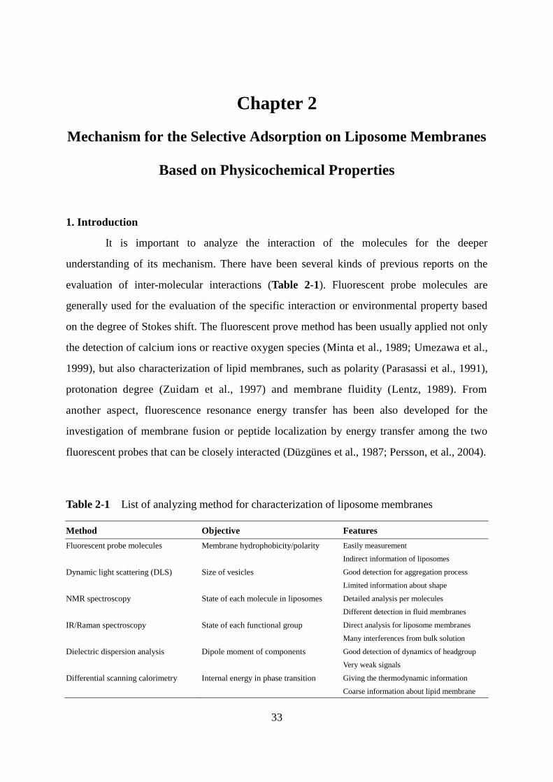
Table 2-1 List of analyzing method for characterization of liposome membranes
Method Objective Features
Fluorescent probe molecules Membrane hydrophobicity/polarity Easily measurement
Indirect information of liposomes
Dynamic light scattering (DLS) Size of vesicles Good detection for aggregation process
Limited information about shape
NMR spectroscopy State of each molecule in liposomes Detailed analysis per molecules
Different detection in fluid membranes
IR/Raman spectroscopy State of each functional group Direct analysis for liposome membranes
Many interferences from bulk solution
Dielectric dispersion analysis Dipole moment of components Good detection of dynamics of headgroup
Very weak signals
Differential scanning calorimetry Internal energy in phase transition Giving the thermodynamic information
Coarse information about lipid membrane
Page 43
34
Although the fluorescent probes can be employed in the characterization of liposome
membranes because of its comparative ease, the obtained results are not derived from the
membrane itself and can provide us only indirect information. Thus, the combination of
different kinds of the analyses can be important for interpretation of information of the
properties of liposome membranes.
Infrared spectroscopy (IR) and Raman spectroscopy analysis are useful tools for the
direct detection of the liposome membrane properties. IR and Raman spectra are gained by
the analyses of penetrating light and scattering light, respectively. In the IR spectra, the energy
absorbance derived from each molecular vibration mode is observed as the peaks, which can
be applied for evaluation of the interactions as each functional group. However, it is difficult
to measure weak signals in a dispersion sample such as liposome suspension because of the
obstruction from the bending vibration of water molecules. On the other hand, Raman spectra
are obtained by observing difference of wavelength between incident and scattering light.
Raman spectra are complement with IR spectra, and there have been advantages in the
measurement of liposome suspension using confocal Raman spectroscopy. Recently, the
enhanced Raman spectra induced by surface plasmon of nanoparticles was developed for the
detection of liposome membranes to overcome the disadvantage about feeble scattering light
(Suga et al., 2015). Incidentally, some electrochemical approaches are also available to
analyze the characteristics of liposome membranes, such as surface charge and hydrophilicity.
Zeta potential of liposomes provides the information about the surface charge and,
furthermore, dielectric dispersion analysis (DDA) is also effective methodology of measuring
the liposome membrane properties. In this measurement, assigned frequency becomes higher
from center to surface of liposome membranes because of hydrophilicity. Actually, the
rotation of headgroup or acyl chain moieties can be evaluated by dielectric dispersion spectra
(Shimanouchi et al., 2014; Hayashi et al., 2013).
By using the methods recommended above, the interactions of phospholipid
membranes with foreign molecules have been evaluated. In the case of hydrophobic
molecules such as cholesterol, the alignment in lipid assembly and the accumulation in
hydrophobic core can be observed by the measurements of membrane polarity and fluidity
evaluated by fluorescent probes (M'Baye et al., 2008; Suga et al., 2013). On the contrary,
hydrophilic molecules with smaller molecular weight or hydrophilic polymers are interacting
Page 44
35
with the headgroup regions of liposome membranes. Arginine, which is one of the amino
acids, is adsorbed on the surface of some zwitterionic liposomes as well as negatively-charged
ones (Bouchet et al., 2010). This adsorption induced the increase of membrane polarity and
fluidity, indicating the effect in inner region of liposomes. From another viewpoint, the
phospholipid monolayer membranes formed by PE phospholipids have reported to adsorb the
tartaric acids at the headgroup region of the membrane (Petelska et al., 2002). Furthermore,
adsorption of poly-L-lysine on anionic liposomes induces the marked changes of the phase
transition temperature, depending on the polymerization degree of poly-L-lysine (Schwieger
et al., 2007). These results were considered to be caused by the effect that these polymers
could interact only in surface charged region small molecular weight and, in the case of larger
polymers, the insertion into interior of membranes occurs by means of secondary structure of
-helix.
Furthermore, thermodynamic properties of the liposome membranes are helpful to
evaluate the adsorption of guest molecules on the liposome membrane. Differential scanning
calorimetry (DSC) can evaluate the variation of internal energy by measuring the change of
temperature with heating constantly. In measurement of liposome membranes, a dramatic
response appears around the phase transition temperature, which provides the thermodynamic
effect due to forming interactions. In addition, isothermal titration calorimetry (ITC) can also
evaluate the thermodynamic behavior of molecular adsorption such as difference of enthalpy
and entropy, which is used for assignment of ligand of ciguatoxin fragment from
thermodynamic aspects (Ui et al., 2008). Furthermore, the relation between enthalpy and
entropy is also important for understanding the several host-guest interactions. In general, the
change of enthalpy shows compensation to that of entropy, enabled to assume the degree of
conformational changes and the dehydration from the slopes and the intercept of regression
line (Rekharsky et al., 2007). As for the assembly of phospholipids, these relations are
considered similar in the case of micellar formation (Sugihara et al., 1999), implying that it
could be a good way of investigating the interactions of liposomes and amino acids.
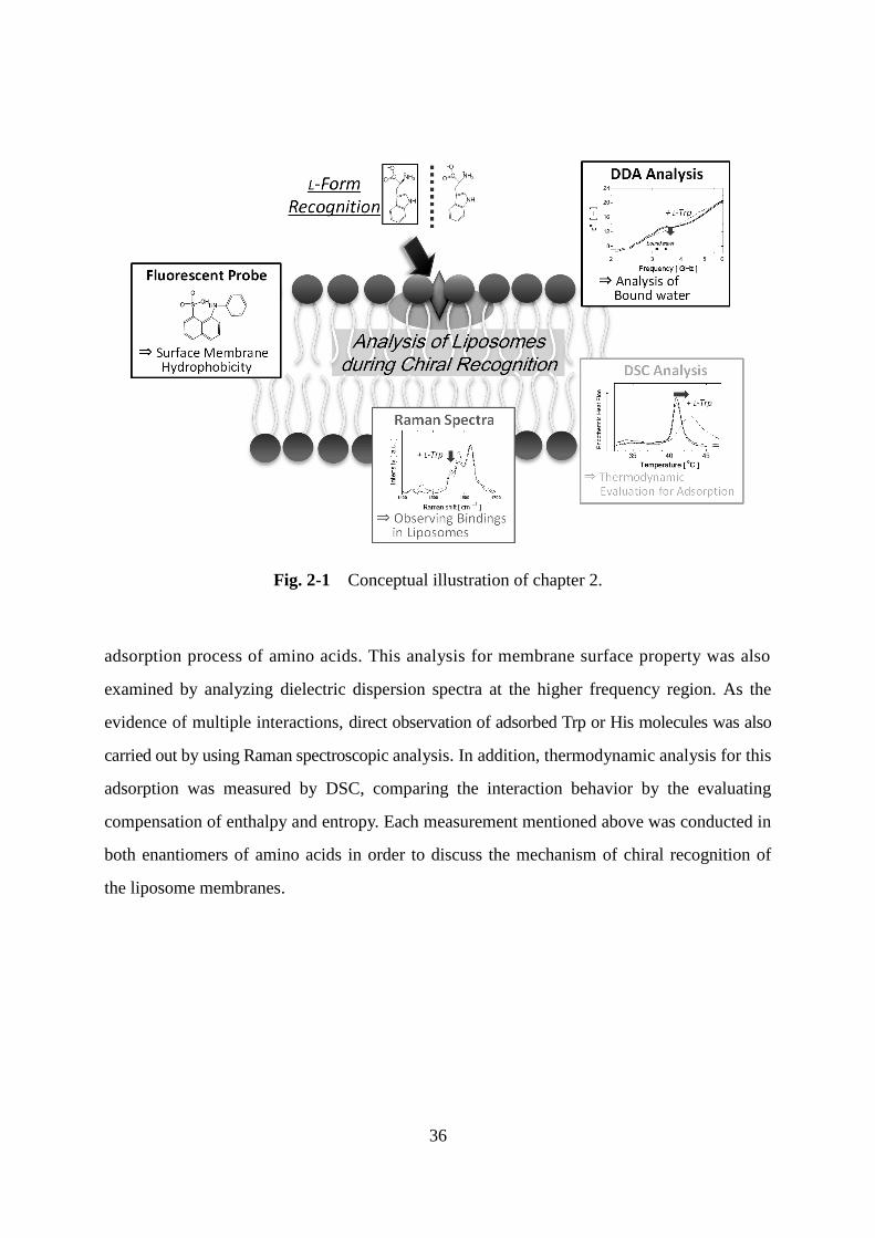
In this chapter, the variation of liposome membrane property was investigated by
employing the enantioselective adsorption of amino acids as case study. Time course of the
surface hydrophobicity was first evaluated by using ANS fluorescent probes during the
Page 45
36
Fig. 2-1 Conceptual illustration of chapter 2.
adsorption process of amino acids. This analysis for membrane surface property was also
examined by analyzing dielectric dispersion spectra at the higher frequency region. As the
evidence of multiple interactions, direct observation of adsorbed Trp or His molecules was also
carried out by using Raman spectroscopic analysis. In addition, thermodynamic analysis for this
adsorption was measured by DSC, comparing the interaction behavior by the evaluating
compensation of enthalpy and entropy. Each measurement mentioned above was conducted in
both enantiomers of amino acids in order to discuss the mechanism of chiral recognition of
the liposome membranes.
Page 46
37
2. Materials and Methods
2.1 Materials
1,2-Dipalmitoyl-sn-glycero-3-phosphocholine (DPPC) was purchased from Avanti
Polar Lipids, Inc. (Alabaster, AL, USA). L-Trp, D-Trp, L-His and D-His amino acids were
purchased from Peptide Institute (Suita, Osaka, Japan). All amino acid reagents were over
98% purity of enantiomers. The fluorescent probe, 8-anilino-1-naphthalenesulfonic acid
(ANS), was purchased from Sigma Aldrich (St. Louis, MO, USA). 1,4-Dioxane and other
chemicals were purchased from Wako Pure Chemical Industry Ltd. (Osaka, Japan) and were
used without further purification.
2.2 Liposome Preparation
A solution of phospholipids in chloroform was dried in a round-bottom flask by
rotary evaporation under vacuum. The resulting lipid films were dissolved in chloroform and
the solvent evaporated twice. The lipid thin film was kept under high vacuum for at least 3 h,
and then hydrated with distilled water at room temperature. The vesicle suspension was
frozen at -80 °C and then thawed at 50 °C to enhance the transformation of small vesicles into
larger multilamellar vesicles (MLVs). This freeze-thaw cycle was repeated five times. MLVs
were used to prepare large unilamellar vesicles (LUVs) by extruding the MLV suspension 11
times through two layers of polycarbonate membrane with mean pore diameters of 100 nm
using an extruding device (Liposofast; Avestin Inc., Ottawa, Canada). Liposomes with
different compositions were also prepared by using the same method.
2.3 Characterization of Fluorescent Probes
The fluorescent probes 8-anilinonaphthalene-1-sulfonic acid (ANS) was excited at
350 nm respectively. Fluorescent spectra in water/dioxane solutions were monitored using
FP-6500 or FP-8500; JASCO, Tokyo, Japan).
2.4 Hydrophobicity Analysis of the Membrane Surface by ANS
The local hydrophobicity of the liposomal membrane was characterized by using an
environmentally sensitive probe; ANS (Kachel et al., 1998). ANS dissolved in ethanol were
Page 47
38
added to the liposome suspension or the pre-incubated mixture of liposome and amino acids.
The final concentrations of lipid, ANS, and amino acids were 100 M, 1 M, and 10 M,
respectively. The fluorescence spectra of ANS were measured by using a fluorescent
spectrometer after incubation for 30 minutes. ANS was excited at 350 nm, and the emission
spectra were measured from 375 nm to 600 nm.
2.5 Dielectric Dispersion Analysis (DDA) for Analysis of Bound-Water at Liposome
Surface
The dielectric loss (’’) was measured at the frequency range from 1.0 GHz and 6.0
GHz, by using a network analyzer (Keysight Technologies, PNA-X N5245A, 10 MHz to 50
GHz). The measurements were performed at 25 ºC and the concentrations of lipid or amino
acids of liposome suspension were 100 mM and 15 mM, respectively.
2.6 DSC Analysis of Liposomal Membranes
A differential scanning calorimeter (DSC-60; Shimadzu, Kyoto, Japan) was used for
calorimetric measurements of liposomes. Liposome suspensions (100 mM) with or without
amino acids (15 mM) were incubated for 48 hours at 25 ºC before DSC measurements. The
20 l sample solution was sealed in an alumina hermetic pan. Thermograms were obtained
with a heating and cooling rate of 2 ºC/min between 25 ºC and 50 ºC. There were no
significant differences between the thermograms in heating and in cooling processes for one
sample. At least three cycles of heating/cooling were repeated in each experiment, and the
accumulated data was used for the calculation of the enthalpy variation (H) and transition
temperature (Tm) values.
2.7 Raman Spectroscopy Analysis of Trp and His
UV resonance Raman spectra of Trp and His were measured by a confocal Raman
microscope (LabRAM HR-800; HORIBA, Ltd., Kyoto, Japan) at a excitation wavelength of
266 nm, with laser power of 50 mW. In addition, Raman spectra of liposomes were measured
by using a confocal Raman microscope (LabRAM HR-800; HORIBA, Ltd., Kyoto, Japan) at
a wavelength of 532 nm, with laser power of 100 mW and a total data accumulation time of
Page 48
39
30 s. For each sample, the background signal of the solution was removed, and then the
baseline was corrected.
2.8 Statistical Analysis
Results are expressed as mean ± standard deviation. All experiments were performed
at least three times. The distribution of data was analyzed, and statistical differences were
evaluated using the Student’s t-test. A P-value of <5% was considered significant.
Page 49
40
3. Results and Discussion
3.1 Analysis of Surface Hydrophobicity of Liposome Membranes by Using Fluorescent
Probe, ANS
The properties of liposomal membranes during amino acid adsorption were
characterized by using the fluorescence probe, 8-anilino-1-naphthalenesulfonic acid (ANS).
ANS is known as a polar environment-sensitive probe (Slavík, 1982), which has been
extensively applied to the analysis of membrane hydrophobicity. In addition, the localization
in surface region of liposome membranes was also considered by the quench of xanthene dye
probes attached in phospholipids by covalent bond (Kachel et al., 1998). Figure 2-2(a) shows
the fluorescent spectra of in the mixture of water and 1,4-dioxane. As the increase of dielectric
constant, blue shifts of the spectra and the increase of intensity were observed. In reference to
previous report about dielectric constant of the water and 1,4-dioxane mixtures (Critchpield et
al., 1953), the relation of dielectric constant and ANS fluorescence was estimated by
calculating the fluorescence intensity ratios (I474/I518) of ANS (Fig. 2-2(b)). Except for the plot
in the lowest dielectric constant values, a linear correlation was obtained, indicating the
I474/I518 of ANS fluorescence can monitor the hydrophobicity in the surface region of
liposomes.
Figure 2-3 shows the time course of the I474/I518 of ANS in the DPPC liposome with
L- or D-forms of Trp and His. In the presence of L-Trp, the I474/I518 values increased after 20
hours of incubation, while the opposite tendencies were observed in the presence of L-His
(Fig. 2-3(a)). These results imply that the adsorption of amino acids induces the changes in
the hydrophobicity of the surface region of liposome membranes. The adsorption of L-Trp
resulted in the exclusion of water molecules from the DPPC liposomes, whereas L-His
induced the hydration of the membrane surface, possibly because His is less hydrophilic than
Trp (Tanford, 1962). As for the kinetics of surface hydrophobicity, these changes are
corresponding to the adsorption behaviors of amino acids (Fig. 1-6). It is possible that the
change of surface property is one of the essential factors for increasing adsorption. Conversely,
there were no significant changes of I474/I518 values in the presence of D-Trp and D-His, even
after 48 hours (Fig. 2-3(b)), indicating the importance of the change of surface
hydrophobicity for the enantioselective adsorption, that is to say, chiral recognition.
Page 50
41
Fig. 2-2 Fluorescent spectra of ANS in water/1,4-dioxane. (a) Fluorescence spectra of ANS
at the excitation of 350 nm. (b) The ratio of fluorescence intensity (I474/I518) of ANS in the
different dielectric constant.
Fig. 2-3 Time course of the relative fluorescence intensity of ANS with amino acids. The
ratio of fluorescence intensities at 474 nm and 518 nm (I474/I518) are measured in the presence
of (a) L-Trp (filled circles) and L-His (filled triangles), (b) D-Trp (open circles) and D-His
(open triangles). In all samples, the concentrations of amino acid and DPPC were 0.1 mM and
1.0 mM, and the incubation temperature was 25 ºC before measurements.
400 450 500 550 600
0
40
80
120
Wavelength [ nm ]
Flu
ore
sce
nce
In
ten
sity o
f A
NS
[ a
.u.
]1,4–Dioxane ratio
0%10%20%30%40%50%
60%70%80%90%100%
0%
100%
90%
0 20 40 60 80
0.5
1
1.5
2
2.5
Dielectric constant [ – ]
( I 4
74 / I
518 )
Ra
tio
of flu
ore
sce
nce
in
tensity
(a) (b)
0 20 40
1.5
2
2.5
Incubation time [ h ]
Rela
tive flu
ore
scence in
tensity
( I 4
74 / I
51
8 )
[ -
]
DPPC + L-Trp
DPPC + L-His
0 20 40
1.5
2
2.5
Incubation time [ h ]
Rela
tive flu
ore
scence in
tensity
( I 4
74 / I
51
8 )
[ -
]
DPPC + D-Trp
DPPC + D-His
(a) (b)
Page 51
42
3.2 Evaluation of Bound Water in Surface of Liposome Membranes by Dielectric
Dispersion Analysis
To investigate the hydration states of liposome membranes, dielectric dispersion
analysis (DDA) is a useful tool because this is the direct analysis of bound water molecules.
According to the previous studies (Noda et al., 2006; Takada et al., 2012), the peaks of
dielectric loss around 0.1-5 GHz is assigned to bound water in liposome membranes, towards
that around 20 GHz means the bulk water. Figure 2-4 shows the dielectric spectra of DPPC
liposomes with or without L- or D-forms of Trp and His. In this figure, the mixing of L-Trp
induced the suppression of the shoulder peak from bound water unlike the mixing of D-Trp.
The suppression of this shoulder peaks is corresponding to diminishing the dielectric
relaxation by insertion of cholesterols or proteins (Takada et al., 2012). It is thus considered
that the adsorption of L-Trp resulted in the exclusion of water molecules from the membrane
structure of the DPPC liposomes, while L-His induced the hydration of the membrane surface.
Considering this result and ANS measurement, it is suggested that the chiral recognition of
amino acids induced the changes in the membrane surface properties.
Fig. 2-4 Dielectric dispersion analysis of DPPC liposome membranes with amino acids. (a)
Dielectric dispersion spectra of DPPC mixed L-form (dashed line) or D-forms (dotted line) of
Trp. (b) Dielectric dispersion spectra of DPPC mixed L-form (dashed line) or D-forms (dotted
line) of His. The dielectric loss (’’) were analyzed in frequencies of 2-6 GHz. All samples
were incubated by 25 oC in 48 hours before measurements.
8
12
16
20
24Lipid: DPPC
Lipid conc.: 100 mMTrp conc.: 15 mM
: DPPC + L–Trp
Frequency [ GHz ]
'
' [ – ]
3 4 5 6
: DPPC: DPPC + D–Trp
2
bound water 8
12
16
20
24Lipid: DPPC
Lipid conc.: 100 mMHis conc.: 15 mM
: DPPC + L–His
Frequency [ GHz ]
'
' [ – ]
3 4 5 6
: DPPC: DPPC + D–His
2
bound water
(b)(a)
Page 52
43
3.3 Observation of Bindings of Tryptophan or Histidine in Liposomes by Resonance
Raman Spectroscopy Analysis
A possible model for interaction of amino acids by liposome membranes was
investigated based on UV resonance Raman spectroscopic analysis. UV resonance Raman
signals of Trp and His can be distinguished based on their constituents (Fig. 2-5),; for Trp, the
peaks at 1467, 1550, 1576, and 1616 cm−1
were assigned as N1-H bending, C2-C3 or C3-C
stretching, respectively (Hirakawa et al., 1978; Miura et al., 1989; Wei et al., 2007); for His,
the peaks at 1498, 1576 and 1632 cm-1
were assigned as stretching mode of the imidazole ring,
C1-C2 stretching mode, and C4-N5 stretching mode, respectively (Marques et al., 2013).
Figures 2-6(a) and (b) showed the UV resonance Raman spectra of L-Trp and D-Trp in the
presence of DPPC liposomes after 48 hours of incubation. In the presence of DPPC liposomes,
the peak intensities at 1467, 1550, 1576 cm−1
of L-Trp decreased, while no significant changes
was observed in the case of D-Trp. A decrease in the peak intensities of L-His was also observed,
but not in the case of D-His (Figs. 2-6(d) and (e)). These results suggest that L-Trp and L-His
interacted with the DPPC liposomes. In addition, the decrease in the peak intensities was
proportional to the lipid concentration (Figs. 2-6(c), (f)). The Raman peak intensity of Trp is
sensitive to environmental hydrophobicity (Nagatomo et al., 2013). It has been reported that
the adsorption of cysteine ethyl ester on DPPC liposomal membranes resulted in a decrease in
its Raman peak intensities, in which the interaction between the phospholipids and amino acid
side chains could be induced by the replacement of water molecules, with a consequent
weakening of the vibrational force constants (Arias et al., 2015). Furthermore, nucleobases,
Fig. 2-5 Peak assignments of side chains of amino acids. (a) Assignment of Trp. (b) Assignment
and His. Atom number about carbon and nitrogen were described in chemical formula.
Page 53
44
Fig. 2-6 UV resonance Raman spectroscopic analysis of Trp and His with DPPC liposomes.
Raman spectra of each amino acids are measured without (dotted line) or with DPPC
liposomes (solid line): (a) L-Trp, (b) D-Trp, (d) L-His and (e) D-His. (c) Relative Raman peak
intensity of Trp (1467cm-1
) with DPPC liposomes. The similar analysis for His (1498 cm-1
)
was shown in (f). All samples were incubated by 25 ºC in 48 hours before measurements.
which are hydrophobic moieties in RNA molecules, can interact with the liposome
membranes via hydrophobic and hydrogen bonding interactions (Suga et al., 2013). The logP
values of the indole ring (Trp) and imidazole ring (His) are 2.1 and -0.1, respectively (cf.
amino acid back bone (Gly): -3.9)), and the hydrogen bond donors and acceptors are
accumulated at the hydrophobic-hydrophilic interface of membranes. Taken together, these
data suggest that the side chains of L-amino acids interacted with the hydrophobic regions of
the liposome membranes, in which hydrogen bonds could be formed between L-amino acids
Page 54
45
and DPPC molecules.
3.4 Thermodynamic Analysis for Adsorption in Liposome Membranes
Based on the above results of amino acids, the “surface region” of liposomes was
found to be significant in the adsorption of amino acids. To discuss the adsorption mechanism,
thermodynamic analysis of adsorption energy is useful for understanding the behavior of
liposomes as the adsorbent of amino acids.
3.4.1 Evaluation of Phase Transition by DSC Analysis
The phase transition of the DPPC liposomes was analyzed by DSC, and
pre-transition and main transition peaks were observed at 34.69 ºC and at 41.09 ºC,
respectively. The effect in phase transition is investigated by mixing L- or D-forms of Trp and
His (Fig. 2-7). The main phase transition temperature (Tm) slightly increased by the addition
of L-Trp or L-His, whereas no change was induced by D-amino acids (Figs. 2-7(a) and (b)).
Such a variation of DPPC liposomes in Tm is corresponding to the previous report about the
incorporation of cationic lipid, owing to the electrostatic interaction between lipid molecules
(Troutier et al., 2005). In the case of anionic liposomes, Tm was increased by the addition of
poly(L-lysine) or poly(L-arginine), owing to the stabilization of the gel phase via the
restriction of electrostatic repulsion between neighboring phospholipid molecules (Schwieger
et al., 2007; Schwieger et al., 2009). Considering our results, the slight increases in the Tm
values indicate that the electrostatic interactions between L-amino acids and liposome
membranes induced the stabilization of the membrane packing. The adsorption of L-amino
acids is also suggested by the suppression of the pre-transitions to a ripple gel phase only in
the presence of L-amino acids. The enthalpy (H) was also calculated for DPPC liposomes
incubated with the amino acids (Fig. 2-7(c)). The H values slightly increased with the
addition of L-amino acids, but no significant increase was observed in the presence of
D-amino acids. Furthermore, the increase in H values was proportional to the amino acids
concentration (Fig. 2-8). These results suggest that such variations in Tm and H correspond to
the adsorption of the L-enantiomers on DPPC liposome membranes. It has been suggested that
these endothermic changes could be related to the stabilization of acyl chain packing via interactions
Page 55
46
Fig.2-7 DSC analyses of DPPC liposome membranes with or without amino acids. The
thermograms of pure DPPC liposomes are shown as black lines. The main transition of DPPC
liposomes (41.09 ºC) is shown as dotted line. (a)DSC thermograms of DPPC liposomes mixed
with L-Trp (lowers) or D-Trp (uppers) at the different concentrations. (b)DSC thermograms of
DPPC liposomes mixed with L-His (lowers) or D-His (uppers) at the different concentrations.
(c)Table of phase transition temperatures (Tm) and enthalpies (H). All samples were
incubated by 25 ºC in 48 hours before measurements.
with L-amino acids (Zhao et al., 2007; Marques et al., 2013). Therefore, it was investigated
whether the enantioselective adsorption of L-Trp and L-His on DPPC liposomes induced the
membrane to become more ordered, in which electrostatic and van der Waals interactions are
possible driving forces of their interaction.
Page 56
47
Fig. 2-8 Variations of the main transition enthalpy (H) in different concentrations of (a) L-
or D-forms of Trp and (b) L- or D-forms of His, respectively. All samples are incubated by 25
ºC in 48 hours before measurements.
3.4.2 Discussion of Adsorption in Relation between Enthalpy and Entropy
The compensation analysis between enthalpy and entropy is important to evaluate the
adsorption of amino acids on liposome membranes. Since the entropy changes (H) are a
state function, it can be estimated from the DSC analysis of phase transition behavior. In
addition, entropy changes (TS) can be calculated from the free energy changes obtained by
the distribution constant of amino acids (Klip) as the following equations:
TS = H + RTm ln Klip
Klip = Cflt Vlip / {(Cini – Cflt) V}
where Cini and Cflt represent the concentration of amino acids in the initial solution and the
ultrafiltrated dolution, V and Vlip represent the volume of the bulk solution phase and the
liposome membrane phase, R means the gas constant. In the case of D-Trp or D-His,
compensation relationship could not be discussed because of the positive value of free energy
changes, which is caused by no adsorption. On the other hand, the adsorption of L-Trp or
L-His showed the linear correlation of enthalpy changes and entropy changes (Fig. 2-9),
indicating the compensation relationship. These plots lay in the upper-left area of diagram
similarly in the case of the ligand bindings (Muralidhara et al., 2007) and distribution of water
Page 57
48
in reverse micelle (Khougaz et al., 1997), implying that thermal changes by adsorption were
relatively large compared to the structural change of whole system. As a result, these
adsorptions were regarded as the high affinity of binding.
To compare with general examples of interactions, the slopes () and intercepts
(TS0) of plot of enthalpy changes and entropy changes were summarized in Table 2-2. When
the TS0 values are positive, the entropy-driven adsorption can be applicable in general.
Almost all the cases (including this study) can be regarded as the entropy-driven adsorption.
As compared with other cases, the value is relatively small in this study. From this result,
the energy required in the structural change of the liposome membrane is assumed to be the
Fig. 2-9 The relation between enthalpy changes and entropy changes in several systems. In
addition to the L-amino acid adsorptions, the profile of solvent extraction (1), ligand bindings
(2) and distribution of water in reverse micelle (3) were shown. Dotted line represents the
proportional line whose slope and intercept are 1 and 0 kJ/mol, respectively. The detailed
plots of adsorption of L-Trp and L-His were shown in the inset.
Page 58
49
Table 2-2 List of thermodynamic parameters in several types of interactions
Reference Interaction type [-] TS0 [kJ mol-1
]
Bojesen et al., 2003 Fatty acid binding to BSA from water 1.4 68.8
Fatty acid binding to BSA from hexane 1.01 0.13
Khougaz et al., 1997 Distribution of water in reverse micelle 1.13 17.6
Kojima et al., 2001 Distribution of carboxylic acids in solvent-water system 1.05 -3.16
Dimerization of carboxylic acids 0.91 20
Haroun et al., 2005 Binding Trp in chiral-recognitive ligand 1.02 22
Kinkel et al., 1981 Solvent extraction between phosphate buffer and
2,2,4-trimethylpentane 0.21 13.9
Nagatoishi et al., 2009 Ligand binding to TATA-box 0.856 53.3
Muralidhara et al., 2007 Allosteric binding to Cytochrome P450eryF 1.76 46.8
Richieri et al., 1997 Fatty acid (PA) bindings by Mutant proteins 0.864 -1.2
This work Adsorption of L-Trp on DPPC liposomes 0.416 14.5
Adsorption of L-His on DPPC liposomes 0.459 10.4
smaller than that of solvation or ligand bindings, which implies that the energy required in
the adsorption and the relating change of hydrophobicity at the surface region are not so
drastic as compared with that in the structural changes of liposomes: thus, the membrane
fusion or disruption are hardly occurring. From the thermodynamic investigations, the
liposome membranes were shown to act as the host adsorbents that recruit the guest molecules
in entropy-driven manner.
3.5 Adsorption Mechanism in Liposome Membranes
Based on the above results, a plausible mechanism of chiral recognition is suggested
as schematically shown in Fig. 2-10. In the early stage of adsorption, the adsorption amount
of L-amino acids was still retained at the lower level till 12 hours of incubation, indicating that
the relatively weak interactions were induced by the contribution of electrostatic interactions
(Fig. 2-10). On the contrary, PPL adsorption in opposite charged-liposomes was saturated in
short-times incubation. The contributions of electrostatic interactions are thought to be very
important to improve the final recovery yield in the adsorption. The adsorption amount of
L-Trp obtained was lower in acidic and basic pH conditions, wherein the L-Trp surface was
positively- and negatively-charged, respectively (data not shown). During their interaction
Page 59
50
Fig. 2-10 Schematic illustration of the mechanism of chiral recognition on liposomal
membrane.
process, the membrane property, such as hydrophobicity, was varied, especially at the surface
region of the membranes. These variations were well corresponding to the increase of the
adsorbed amount of L-amino acids, indicating that the adsorption can be promoted by the
variation of the surface membrane properties. It is therefore implied that the surface property
of liposome membranes transition to appropriate states for high adsorption. In the case of the
adsorption of amino acids, it is also suggested that there are several steps in the adsorption
mechanism as the variation of surface property induce the enhancement of the binding
between amino acids and liposome membranes. At the step of variation in surface property,
the liposome membranes are considered to exhibit the chiral recognition function, judging
from the results of remarkable increase of the L-enantiomer. Finally, the adsorption of
L-amino acids was saturated, resulting in the binding of plural phospholipids and one amino
acid molecule, which means that the assembled state at the membrane surface is quite
important for the chiral recognition. In addition, the possibility of more than three kinds of
interactions was also evidenced by Raman spectroscopy analysis. Because the concept of
three-points interaction is important for chiral recognition (Davankov, 1997), the
suprastructure of liposome membranes may effectively induce such multiple interactions for
chiral recognition. Furthermore, DSC thermograms revealed the increase of the phase
transition temperature for the DPPC liposomes adsorbed with of L-amino acids, indicating
that the recognized amino acid can be a member of the assembled membranes. In the
viewpoint of adsorption energy, the slight conformational change is considered to be induced,
and then the chiral recognitive interaction is gradually carried out to achieve a “local
Page 60
51
minimum” of free energy. Based on the aforementioned adsorption mechanism, the high
recognition function is induced on the liposome membranes. Considering the results in
chapter 1, such adsorption steps are possibly suggested in the case of the adsorption of other
amino acids, as shown in Fig. 2-11.
Fig. 2-11 List of adsorption behavior in several amino acids (picked up) and propranolol.
Page 61
52
4. Summary
The chiral recognition of amino acids on the liposome membranes was found to be
induced through the interactions especially at the surface region. The fluorescent probe
analysis using surface-localized fluorescent probe, ANS, indicated the specific changes of
hydrophobicity by mixing of L-Trp and L-His after 20 hours incubation, while no changes by
mixing of D-Trp or D-His. The variations were also observed in dielectric dispersion spectra as
the shift of the relaxation of peaks relating to “bound water” at the surface region of
liposomes, indicating that the effect in membrane property can be corresponding to the amino
acid adsorption. It is therefore important to form the interactions between the side chain of
amino acids and lipid headgroups, which is evidenced by the decrease of peak intensity in
resonance Raman spectra. In addition, the increase of the phase transition temperature
occurred in liposomes that adsorbed L-Trp and L-His, implying the decrease of electrostatic
dispersion in headgroup region of liposomes. Considering the relation of enthalpy and entropy
changes, it is thought that this adsorption induces dehydration with slight effect of
conformational changes in phospholipid assembly.
According to the above results, the mechanism of chiral recognition can be explained
as “step-by-step” adsorption. In the first step, the partitions derived from electrostatic
interactions are dominant. In the case of amino acids, stereospecific interactions are induced
by the formation of multiple interactions. After that, the increase of adsorption was induced
by the rearrangement of surface assembly as the change of membrane property. The effect on
bound water existing in the membrane surface region is possible key factors for the binding of
amino acids. Finally, multiple bindings were formed between plural phospholipids and one
amino acid, according to the results about adsorption isotherms investigated in chapter 1. In
the energetic viewpoint, it is considered that the molecular adsorption can be induced with
gradually transfer of the total free energy to a local minimum in organizing the self-assembled
membrane composed with phospholipid and “adsorbed” amino acid.
Aforementioned mechanism required longer time to complete adsorption process,
which is one of the disadvantages for the practical application for industrial processes. Toward
this, it is possible that the design of liposome membranes focusing on initial hydrophobicity
or fluidity controls the adsorption behavior. Because such chiral recognition occurred in
surface region of liposome membranes, the specific characterization for surface property is
Page 62
53
important to design the chiral recognition function. The investigation of detailed surface
property in liposomes for efficient chiral recognition will describe in the following chapter 3.
Page 63
54
Chapter 3
Evaluation of Surface Properties of Cholesterol-Containing
Binary and Ternary Liposomes to Regulate Molecular
Recognition and Design of Liposome Membranes
1. Introduction
According to the results in the previous chapter, the analysis of the surface property
of liposome membranes is important in order to design the optimal liposome membranes for
recognition of small molecules. As mentioned in chapter 2, the fluorescent probe analysis can
provide the membrane property in specific region of liposome membranes. Actually, it has
been investigated that the dielectric constants of liposome membranes decrease with the
sigmoid curve from surface to interior (Cevc, 1990). Thus, the location of fluorescent probes
in liposome membranes depends on the polarity of the probe molecule: such behaviors can be
investigated by the quencher bound to phospholipids in designated location (Kachel et al.,
1998; Jurkiewicz et al., 2006; Kaiser et al., 1998; Asuncion-Punzalan et al., 1998). The
locations of several fluorescent probes were summarized in Fig. 3-1. In contrast to ANS,
Laurdan and DPH are stably localized in inner regions, and they indicate the whole property
of liposome membranes. Among the surface-localized fluorescent probes, the detail
localizationcan be analyzed by the distinction of fluorophore location. That is why the
combination of multiple fluorescent probes is good way to evaluate the changes in the
membrane properties in various regions. In addition, such a technique is also applied for
evaluating the phase state of liposomes. Suga et al. have developed the evaluation method for
the phase state of liposome membranes by Cartesian diagram evaluated by plotting the
membrane polarity and fluidity derived from Laurdan and DPH, respectively (Suga et al.,
2013).
Cholesterol (Ch) is known to regulate the membrane property because of the steroid
skeletons which induce high order in liposome membranes. Besides, Ch also induces the increase
Page 64
55
Fig. 3-1 Summary of the depth of fluorescent probes in liposome membranes.
of membrane packing via the hydrogen bonds between hydroxyl groups of Ch and
headgroups of phospholipids. As a result, disordered liposome membranes show the decrease
of membrane fluidity by mixing Ch. On the contrary, the fluidity of quite rigid liposome
membranes increased in mixing Ch by disturbing the membrane packing. Actually, the
liposomes containing Ch resulted in the specific effects via the adsorption of some peptides
(Shigematsu et al., 2007; Gopal et al., 2013). Furthermore, heterogeneous liposomes
containing Ch indicate the regions of low fluidity, such as microdomains or “rafts” (Scolari et
al., 2010; Björkbom et al., 2007).
Microdomains mean the separated phases in liposome membranes, which are mainly
induced by the difference of lateral diffusion in each phase and hydrophobic interactions.
Such domains actually play significant roles in the regulation of protein and enzyme functions
in the cell membranes (Litt et al., 2009; Anderson et al., 2002; Carozzi et al., 2002). In giant
unilamellar vesicles (GUV), domains can be formed in micrometer order, and are directly
observed in microscopic images by fluorescent labeling (Veatch et al., 2003; Stottrup et al.,
2004; Cicuta et al., 2007). In addition, the membrane property of domains has been evaluated
by thermodynamic procedures (Svetlovics et al., 2012; Garidel et al., 2000), Raman, and
nuclear magnetic resonance spectroscopy (de Lange et al., 2007; Davis et al., 2014). On the
other hand, the analysis of domains for small liposomes (nanometer order) is difficult due to
the limit of measurable scale in optical microscope, nevertheless, domain formation in
liposome membranes can be analyzed by excimer/monomer ratios of pyrene probes and their
Page 65
56
derivatives even in nano-order (Lagane et al., 2002; Arrais et al., 2007). The estimation of
nanodomain formation (<5 nm) has been also carried out by the fluorescent quench method
using TEMPO probe (Suga et al., 2013). Moreover, the line tension is generated in the edge of
domains due to the boundary of different phases. Actually, the previous report revealed that
the HIV fusion peptide preferentially targeted to lo-ld boundary regions and promoted full
fusion at the interface between ordered and disordered lipids (Yang et al., 2015). As the
increase of domain size, the contributions of such a line tension is thought to decrease
(Tolpekina et al., 2004). Therefore, it is possible that the detailed investigation of the phase
behaviors in heterogeneous liposomes contributes to understanding about the formation of
interactions in liposome membranes.
In this chapter, the effect for chiral recognition of His is investigated by using the
liposomes with multiple components contained Ch (Fig. 3-2). At first, the methodology for
evaluating the surface property of the liposome membranes was developed by the
combination of two types of fluorescent probes that can be localized at the surface edge of the
membrane. By means of the comparison of Cartesian diagram for interior membrane
properties, it is expected to evaluate the liposome membrane property in details. According to
the relation between such properties and the adsorption of L- or D-form of His, the effect of
mixed Ch in liposome membranes is discussed. The variation in the membrane property after
the His adsorption was also investigated to understand the behavior of liposome membranes.
Furthermore, the membrane properties of ternary liposomes were evaluated based on their
phase behaviors, and then, the relation with chiral recognition of His was investigated. By
comparing the domain states relating to the line tension, the contribution of domain is
considered for understanding the effect of existing domain “edges”
Page 66
57
Fig. 3-2 Conceptual illustration of chapter 3.
Page 67
58
2. Materials and Methods
2.1 Materials
Several phospholipids such as 1,2-dipalmitoyl-sn-glycero-3-phosphocholine (DPPC),
1-palmitoyl-2-oleoyl-sn-glycero-3-phosphocholine (POPC) and 1,2-dioleoyl-sn-glycero-3-
phosphocholine (DOPC), were purchased from Avanti Polar Lipids, Inc. (Alabaster, AL,
USA). A fluorescent probe, N-(5-dimethylaminonaphthalene-1-sulfonyl)-1,2-dihexadecanoyl-
sn-glycero-3-phosphoethanolamine (Dansyl-DHPE) was also purchased from Avanti Polar
Lipids, Inc., and 1,6-diphenyl-1,3,5-hexatriene (DPH), (1-(4-trimethylammoniumphenyl)-6-
phenyl-1,3,5-hexatriene (TMA-DPH), 6-lauroyl-2-dimethylaminonaphthalene (Laurdan) were
obtained from Sigma Aldrich (St. Louis, MO, USA). Cholesterol (Ch) was purchased from
Wako Pure Chemical Industry Ltd. (Osaka, Japan), and L or D-form of histidine (His) were
purchased from Peptide Institute (Suita, Osaka, Japan). Other chemicals were purchased from
Wako Pure Chemical Industry Ltd. (Osaka, Japan) and were used without further purification.
2.2 Liposome Preparation
A solution of phospholipids in chloroform was dried in a round-bottom flask by
rotary evaporation under vacuum. The resulting lipid films were dissolved in chloroform and
the solvent evaporated twice. The lipid thin film was kept under high vacuum for at least 3 h,
and then hydrated with ultrapure water at room temperature. The vesicle suspension was
frozen at -80 °C and then thawed at 50 °C to enhance the transformation of small vesicles into
larger multilamellar vesicles (MLVs). This freeze-thaw cycle was repeated five times. MLVs
were used to prepare large unilamellar vesicles (LUVs) by extruding the MLV suspension 11
times through two layers of polycarbonate membrane with mean pore diameters of 100 nm
using an extruding device (Liposofast; Avestin Inc., Ottawa, Canada). Liposomes with
different compositions were also prepared by using the same method.
2.3 Evaluation of Membrane Properties by Fluorescent Probes
Membrane properties of liposomes can be characterized in Cartesian diagram by the
plot of the membrane fluidity versus polarity evaluated by DPH and Laurdan, respectively
(Suga et al., 2013). The fluidity in the interior of the liposome membrane was evaluated by
Page 68
59
measuring the fluorescence anisotropy of the DPH incorporated in the vesicles using the
fluorescence spectrophotometer FP-6500 (JASCO, Tokyo, Japan). 10 μL of 100 μM DPH in
ethanol was added into 1 mL of liposome suspension (lipid: 0.25 mM). The samples were
excited with vertically polarized light (360 nm), and emission intensities both perpendicular
(I⊥) (0°, 0°) and parallel (I∥) (0°, 90°) to the excited light were recorded at 430 nm. The
polarization (P) of DPH was then calculated by using the following equations (Hayashi et al.,
2011):
P = (I∥- GI⊥) / (I∥ + GI⊥) ,
G = i⊥ / i∥ ,
where i⊥ and i∥ are the emission intensities perpendicular to the horizontally polarized light
(90°, 0°) and parallel to the horizontally polarized light (90°, 90°), and G is the correction
factor. The membrane fluidities were evaluated based on the reciprocal of polarization, 1/P.
The membrane fluidities were measured at room temperature. The fluorescent probe Laurdan
is sensitive to the polarity around itself, which allows the membrane polarity of liposomes to
be determined. Laurdan emission spectra exhibit a red shift caused by dielectric relaxation.
Thus, emission spectra were calculated by measuring the general polarization (GP340) for each
emission wavelength as follows (Parasassi et al., 1991):
GP340 = (I440 − I490) / (I440 + I490) .
Laurdan excited with 340 nm light at 20 ºC. The fluorescent spectrum of each sample was
normalized. The total concentrations of amphiphilic phospholipid and Laurdan in the test
solution were 1000 and 10 μM, respectively.
Besides, the surface membrane property could be characterized by the similar
diagram analyzed by partially-hydrophilic molecular probes. TMA-DPH could be used for
characterization of the surface membrane fluidity (1/PTMA-DPH) by analyzing anisotropy with
the same manner to DPH. As for surface membrane polarity, dansyl-DHPE was used as a
probe molecule: it was mixed with a liposome suspension in final concentrations of lipid and
dansyl-DHPE were 100 and 1.0 μM, respectively. The fluorescence spectra were analyzed by
the excitation light (336 nm) for observing the wavelength of maximum fluorescence.
Page 69
60
2.4 Measurement of Adsorption to Liposome Membranes
The liposome suspensions (Lipid: 3.0 mM) were mixed with L-histidine (His) (0.5
mM) and then they were incubated at 20 ºC for 24 or 48 hours. After incubation, liposomes
and adsorbed L-His were separated by ultrafiltration with the 50,000 Da of molecular weight
cut off (USY-5; Toyo Roshi Kaisha, Ltd., Tokyo, Japan). After filtration, the leaked
concentration (Cflt) of L-His was measured by the absorbance of UV spectrometer (UV-1800;
Shimadzu, Kyoto, Japan). The adsorbed concentration (Cads) and adsorption amount of L-His
(qL-His) or D-His (qD-His) in several liposome membranes were calculated by following
equations:
Cads = Cini - Cflt
qL-His or D-His = Cads / clip ,
where Cini and clip represent an initial concentration of adsorbates and liposomes, respectively.
To evaluate the selectivity of His enantiomer, percent based enantiomer excess (ee) were
calculated by following equation:
ee = (Cads(L-His) - Cads(D-His)) / Cini .
2.5 Statistical Analysis
Results are expressed as mean ± standard deviation. All experiments were performed
at least three times. The distribution of data was analyzed, and statistical differences were
evaluated using the Student’s t-test. A P-value of <5% was considered significant.
Page 70
61
3. Results and Discussion
3.1 Effect of Mixing Cholesterol for Interior Membrane Properties
The properties of the interior of liposome membranes were first evaluated based on
the previous report (Suga et al., 2013). As shown in Fig. 3-3, the general polarity (GP340) of
Laurdan and the fluidity evaluated by DPH (1/PDPH) for several liposomes containing Ch were
plotted in a Cartesian diagram, indicating the regression of the plotting data in a single line.
POPC (Tm: −5 ºC) and DOPC (Tm: −20 ºC) liposomes were in the disordered (ld) phases, and
an increase in both properties in the upper-left of the diagram along with increased Ch ratios,
while DPPC (Tm: 41ºC) liposomes were in the solid-ordered (so) phases, and only a slight
increase in both properties were observed. These results agree with the previous findings
showing that Ch induced the phase transition of liposomes from ld to liquid-ordered (lo) phase
caused by membrane orientation (Arrais et al., 2007; Walde et al., 2014). In the case of ternary
DOPC/DPPC/Ch liposomes, their surface properties were similar to those of liposomes with
corresponding Ch concentrations, indicating that there was no clear difference among
Fig. 3-3 Cartesian diagram of membrane polarity (GP340) and fluidity (1/PDPH) for binary
and ternary liposomes containing Ch. Numbered points are defined in the inset.
Page 71
62
liposomes showing similar Tm in the plot of Fig. 3-3. Therefore, these results show that the
Cartesian diagram, characterizing membrane polarity and fluidity, can be utilized for the
understanding of overall membrane properties in the case of Ch-modified liposomes, owing to
the composition of phospholipids or Ch.
3.2 Effect of Mixing Cholesterol for Surface Membrane Properties
It is important to evaluate the behavior of the surface region of liposome membranes
in order to understand the interactions between hydrophilic molecules (at polar moieties) and
liposomes. Detail analysis of the phase state of liposomes is also required. As the increase of
Ch molar ratio, the liposomes mixed Ch in 50 mol% showed the transition of phase state in
DPPC, DOPC and POPC (Fritzsching et al., 2013; de Almeida et al., 2003). Furthermore,
some of DOPC/DPPC/Ch ternary liposomes formed the segregated lo phase regarded as
domains. However, the aforementioned probes, such as Laurdan and DPH, are basically
categorized as hydrophobic fluorescent probes and, thus, are not suitable to monitor
membrane properties at the surface region. Besides that, Cartesian diagram remains the
evaluation of whole properties derived from each ratio of lipid components. As for the surface
property, ANS fluorescent probe molecules could be used for the analyses of before or after
adsorption of amino acids as described in chapter 2. Nevertheless, because this probe
molecule is difficult to be inserted in ordered phase, it is not appropriate to compare the
surface membrane properties among the liposomes especially in secondary and ternary
systems. Hence, dansyl-DHPE is known as the fluorescent probe capable of localizing to
hydrophilic regions at the surface of liposomal membranes (Asuncion-Punzalan et al., 1998),
and can be used for the estimation of membrane hydrophobicity via the emission wavelength
sift in the liposomes (Takechi et al., 2011). As shown in Fig. 3-4(a), the fluorescent spectra of
dansyl-DHPE were measured in mixtures of water and 1,4-dioxane as solvents, showing a
blue shift in the peak wavelength with decreased ratios of water. This behavior was caused by
environmental changes around the fluorophore of the dansyl moiety. The shift in the peak
wavelength was plotted against the dielectric constant of the solvent (Fig. 3-4(b)), resulting
that a linear correlation in wavelengths was observed from 512 nm to 527 nm, except for the
data plot in the dielectric constant value of 57. Hence, the surface-membrane hydrophobicity
Page 72
63
Fig. 3-4 Peak wavelength analysis of dansyl-DHPE surrounding in solvent or liposome
membranes. (a) Fluorescent spectra of dansyl-DHPE (Ex.: 336 nm) in several ratio of
water/1,4-dioxane solvent. (b) Peak wavelengths versus dielectric constant. Dielectric
constant values were calculated the ratio of water (ε = 80.1) and 1,4-dioxane (ε = 2.2). (c)
Peak wavelength of dansyl-DHPE inserted in several liposome membranes. Numbered points
are correlated in the inset of Fig. 3-3.
can be evaluated by the normalized value of the surface hydrophobicity (N) by using the
following equation:
N = (- 1 ) / (0 - 1 )
where 0 and 1 represent the fiducial peak wavelengths in hydrophobic (512 nm) and
hydrophilic (527 nm) condition, respectively.
Based on these preliminary results, the peak wavelength of dansyl-DHPE
fluorescence in the liposome was measured (Fig. 3-4(c)). The peak wavelength of highly
ordered DPPC liposomes was found to be lower as compared with those in disordered
membranes, such as DOPC and POPC, indicating that the DPPC liposome membrane was
displaying a hydrophobic (dehydrated) surface. With the increase in Ch ratio, DOPC/Ch and
DPPC/Ch liposomes displayed a red shift in the fluorescence spectrum, while no shifts were
observed in the binary POPC/Ch liposomes. In the case of ternary liposomes, the wavelength
of dansyl-DHPE spectra remained to be similar to those of hydrophilic surfaces, implying that
the profile of surface hydrophobicity was not exactly characterized by the GP340 measured by
using Laurdan.
With reference to the Cartesian diagram (Fig. 3-3), Fig. 3-5 shows the evaluation of
the properties at the surface of various liposomes by the plot of N versus TMA-DPH fluidity
15 30 45 60500
510
520
530
Peak w
avele
ngth
[ n
m ]
Dielectric constant [ – ]
r2 = 0.9696
(512)
(527)
450 500 550 6000
20
40
60
Flu
ore
sce
nt
inte
nsity [
a.u
. ]
Wavelength [ nm ]
Dioxane ratio
0.3
0.4
0.5
0.6
0.7
0.8
0.9
515
520
525
530
535
Pe
ak w
ave
len
gth
[ n
m ]
1 2 3 4 5 6 7 8 9 10 11 12
Liposome composition
(a) (b) (c)
Page 73
64
Fig. 3-5 Correlation diagram evaluated by surface membrane fluidity (1/PTMA-DPH) and
surface membrane hydrophobicity (N) in several binary and ternary liposomes containing Ch.
Numbered points are defined in the inset. Dotted line a0 represents the trend in pure or binary
liposomes containing 30% of Ch; dotted line a1 and a2 also represent that in 50% Ch ratio
liposomes and ternary liposomes, respectively.
(1/PTMA-DPH). In the case of liposomes with single components (DOPC, POPC, and DPPC)
or those with 30% Ch, the data were clustered on the right-downward trend line (a0),
indicating that the surface hydrophobicity decreased with increased surface fluidity. However,
in the case of binary liposomes at 50% Ch, the data were clustered in another trend line (a2)
that demonstrated lower value of its slope, resulting in the discrimination of transitioned
phase caused by Ch. The variation of these parameters was also plotted against the Ch ratio in
order to discuss the features of membrane properties (Fig. 3-6). The N value of various
liposomes, except for POPC, decreased depending upon the Ch ratio, with the 1/PTMA-DPH
values indicating only a slight decrease, while GP340 and 1/PDPH reached values similar to
those observed with DPPC liposomes. As increasing the Ch ratio, the liposome membranes
change in accessible surface capable of permitting water-molecule invasion (Stein et al.,
2015), resulting in membrane variation toward hydrophilicity only at the surface regions of
liposomes modified with Ch. The change in N became larger than that of 1/PTMA-DPH, since
4 6
–0.2
0
0.2
0.4
0.6
0.8
1
1/PTMA–DPH [ – ]
N [ –
]
3
2
1
4
5
6
12
11
10
7
8
9
1. DOPC2. POPC3. DPPC
6. DPPC/Ch(7/3)
8. POPC/Ch(5/5)
4. DOPC/Ch(7/3)
7. DOPC/Ch(5/5)
5. POPC/Ch(7/3)
9. DPPC/Ch(5/5)
10. DOPC/DPPC/Ch(5/2/3)
11. DOPC/DPPC/Ch(3.5/3.5/3)12. DOPC/DPPC/Ch(2/5/3)
a0
a2 a1
10. DOPC/DPPC/Ch(5/2/3)
Surface
Hydrophobicity
Surface
Membrane Fluidity
Hydrophobic
Hydrophilic
Ordered and
Hydrophilic Surface
Low High
Page 74
65
Fig. 3-6 Transition of several membrane properties by increase of Ch molar ratio in three
liposomes. Composition of liposomes represented below each bar.
the water exclusion from the inner membrane to the surface region can be induced by
insertion of Ch molecules (M’Baye et al., 2008). From these results, it is considered that
membrane properties derived from fluorescent probes located at the surface region can be
utilized for the evaluation of intrinsic properties of the liposome surface.
The above characterization method was also applied to ternary liposomes. Although
the Ch ratio was unchanged and kept at 30%, the data were found to be clustered on another
trend line (a1). It has been reported that liposomes at these components could form the
heterogeneous surfaces of lo and ld phases (Cicuta et al., 2007), resulting in hydrophilic
liposome surfaces. Although previous methods evaluating membrane properties provided
general information relating to phase separation, the proposed method for determining
membrane properties at surface regions can discriminate between heterogeneous or Ch-rich
liposomes. Based on these results, it is expected that variations in membrane properties before
and after adsorption of hydrophilic guest molecules, such as amino acids, will be capable of
more detailed study.
Page 75
66
3.3 Relation between Membrane Properties and Chiral Recognition of Histidine in
Binary or Ternary Liposomes
Highly-selective adsorption of L-His molecules was induced in a DPPC liposome
formed in the ordered (so) phase, where the surface membrane hydrophobicity decreased
during the adsorption of L-His and the adsorption behavior of His was affected by initial
membrane properties. The His adsorption behavior was studied by selecting various
liposomes classified in different categories (a1, a
3, and a
2; Fig. 3-5). Figure 3-7(a) shows the
Fig. 3-7 Adsorption and chiral recognition behavior in several liposomes. (a) Adsorption
amount of L-His (qL-His) in 24 or 48 hours of incubation. (b) Adsorption amount of D-His
(qD-His) in 24 or 48 hours of incubation. (c) Percent of His enantiomer excess (ee) in several
liposomes. Closed and open keys show ee values in 24 and 48 hours of incubation. (d) Time
course of qL-His in the three kinds of components categorized in a0 (dotted line), a2 (dashed
line) and a1 (solid line). (e) Time course of qL-His in the three kinds of components categorized
in a0 (dotted line), a2 (dashed line) and a1 (solid line). a0, a2 and a1 represent the trend lines
described in Fig. 3-5.
Page 76
67
adsorption levels of L-His (qL-His) in binary and ternary liposomes containing Ch. In the case
of several liposomes categorized in a0, except for DPPC, the qL-His value increased after 24
and 48 hours incubations, while the N value decreased. Specifically, the liposome of
DOPC/Ch 7/3 indicated the larger qL-His after a 24- and 48-h of incubation in contrast to DPPC
liposomes, resulting in more efficient adsorption as compared to results in previous chapter 1.
This result was due to the promotion of the insertion of L-His molecules, resulting in high
levels of surface fluidity and hydrophilicity. However, higher qL-His values over 24 h were
obtained in several liposomes categorized as a2, despite the indications of lower
surface-membrane fluidities. In comparison with the adsorption to liposomes categorized as
a0, this result indicates that the liposomes categorized along the trend line with lower slope
can have potentials capable of increasing qL-His. The conditions enabling fast and efficient
adsorption can be estimated by the evaluation of surface-membrane properties, suggesting
that the changes in membrane properties toward hydrophilic surfaces could be an important
step in L-His adsorption as mentioned in chapter 2. Furthermore, high qL-His values observed
over 24 h were obtained in all liposomes containing ternary components categorized in a1,
indicating that the heterogeneous phase preferably influenced L-His adsorption in phase
kinetics. To investigate the chiral recognition function of the above liposomes, the qD-His value
was also shown in Fig. 3-7(b). In all liposomes, the qD-His value remained at level <0.01, even
after a 48-h incubation, resulting in the high enantiomer excess (Fig. 3-7(c)).
3.4 Variation of Liposome Membrane Properties Induced by Adsorption of L-Histidine
In chapter 2, the changes of membrane properties by adsorption of L-amino acids
were investigated by several procedures. To compare these changes among several phases of
liposomes, the above evaluation is conducted after adsorption of L-His.
As shown in Fig. 3-8(a), the variation of surface properties of the whole membrane
was not significant before and after L-His adsorption in all components. This result may imply
that the adsorption of L-His molecules had less effect on properties of the inner region of
liposomes as mentioned in previous results involving dielectric dispersion spectra or
anionic-probe hydrophobicity due to the difficulty of insertion of highly polar molecules.
However, the surface-membrane properties were changed in the fixed areas of 0–0.4 in N and
Page 77
68
Fig. 3-8 Transition of membrane properties by the adsorption of L-His. (a) Cartesian
diagram analyzed by Laurdan and DPH before or after adsorption of L-His. (b) Correlation
diagram of surface membrane properties before or after adsorption of L-His. The dotted circle
showed the area of plots after adsorption. In both diagrams, numbered plots before and after
adsorption of L-His are defined in the inset, and the plots after adsorption are described in
primed numbers.
2.8–3.8 in 1/PTMA-DPH after L-His adsorption, even in the initial surface properties (Fig.
3-8(b)). This result revealed that L-His binding at the surface region of liposome membranes
could induce the convergence of surface-membrane properties despite the initial phase state.
Therefore, our results supported the previously-suggested hypothesis that L-His adsorption
occurred in two steps (insertion and enhancement) during the simultaneous change of
membrane property. Furthermore, it is considered that the variation of surface hydrophobicity
is more effective than that of surface fluidity because qL-His indicates larger values in liposome
membranes which show the N in the fixed area of 0-0.4. This suggestion can imply the
adsorption model that the surface hydrophobicity of liposome membranes is regulated by the
slight partition of amino acids at first, and then, the effective “trapping” of amino acids results
in the increase of adsorption with decrease of surface membrane fluidity (Fig. 3-9). As for the
phase state of liposome membranes, high ee value after 24 hand 48 h was obtained in ternary
liposomes with separated lo phases as dispersed microdomains on the membrane surface (Fig.
3-10).
(a) (b)
4 8 12
–0.5
0
0.5
1/PDPH [ – ]
GP340 [ – ]
1. DOPC
2. DPPC
4. DPPC/Ch(7/3)
6. DPPC/Ch(5/5)
3. DOPC/Ch(7/3)
5. DOPC/Ch(5/5)
7. DOPC/DPPC/Ch(3.5/3.5/3)
1
2
46
3
5
7
2' 4'6'
5'
7'
1'
3'
4 6
–0.2
0
0.2
0.4
0.6
0.8
1
1/PTMA–DPH [ – ]
N
[ –
]
2
1
3
4
7
5
6
1. DOPC2. DPPC
4. DPPC/Ch(7/3)3. DOPC/Ch(7/3)
5. DOPC/Ch(5/5)6. DPPC/Ch(5/5)7. DOPC/DPPC/Ch(3.5/3.5/3)
2'
1'
4'
3'6'
5'
7'
Page 78
69
Fig. 3-9 Adsorption steps described in the diagrams of interior and surface membrane
properties.
Fig. 3-10 Conceptual illustration of fast L-His adsorption in Ch containing phase.
3.5 Design of DOPC/DPPC/Ch Ternary Liposomes to Induce Molecular Recognition
Function
Based on the above results, detailed investigation of ternary liposomes can play an
important role for the understanding of molecular recognition function of liposome surface.
The method for possible design of the liposome membranes with heterogeneous ones was
further investigated based on the phase diagram of the ternary components membrane. In
reference to the previous report (Cicuta et al., 2007), a phase diagram of DOPC/DPPC/Ch
ternary liposomes is shown in Fig. 3-11. The detailed investigations is conducted focusing on
the lo + ld phases and its boundary.
Page 79
70
Fig. 3-11 Phase diagram of DOPC/DPPC/Ch ternary liposomes. Solid line shows the phase
boundary and dotted lines showed the tie line in reference to the previous report (Cicuta et al., 2007).
3.5.1 Inner and Surface Membrane Properties in Ternary Liposomes
Figure 3-12(a) shows the phase diagram of DOPC/DPPC/Ch liposomes at 20 ºC,
describing the phase boundaries as purple lines. In this diagram, ternary liposomes prepared in
the above experiments (A, B, and C; Fig. 3-12(a)) existed at lo+ld heterogeneous phases,
while all binary liposomes existed at homogeneous-ordered (so) or disordered phases.
Although there were other findings that micro-domains at nanometer sizes could be formed in
some binary liposomes, such as DOPC or DPPC liposomes (Suga et al., 2013), the Cartesian
diagram of the membrane properties was similar to previous results. Figure 3-12(b) also
indicated the existence of other ternary liposomes with different components in several phase
states (D: ld phase; E: lo phase; F: lo+ld phase; Fig. 3-12(a)), showing that they must have the
corresponding properties with the phase state. It has been already shown that the plot of
surface properties revealed the specific distribution of several ternary liposomes, i.e., those
categorized as a1 with another slope of the trend line (Fig. 3-12(c)). In this diagram, D and E
were distributed regarding B as the intersection point, which was involved in the parallel
direction of tie line. Additionally, F was plotted on line a1 due to the similarity of the phase
ratio as a vertical direction of the tie line. It was found that surface properties of ternary
liposomes depended upon the ratio of each phase, as well as Ch ratio.
Page 80
71
Fig. 3-12 Evaluation of membrane properties in ternary liposomes. (a) Phase diagram of
DOPC/DPPC/Ch ternary liposomes. (b) Cartesian diagram analyzed by Laurdan and DPH in
several ternary liposomes. (c) Correlation diagram of surface membrane properties. All capital
indicators are corresponded to components described in phase diagram.
3.5.2 Effects of Phase State for Adsorption in Ternary Liposomes
Figure 3-13(a) shows the qL-His values in several ternary liposomes, indicating the
low adsorption of His for the liposomes in D and E, on the contrary, high adsorption of His
for the liposome in F. Although single-phase liposomes in D and E, showed the lower qL-His
values after 24- and 48-h incubation relative to heterogeneous-phase liposomes, L-His
adsorption in lo phase (E) was more efficient than that observed in ld phase (D), indicating that
the ordered phase preferably influenced on the adsorption of Ch-containing liposomes. In
addition, because the qD-His values in the corresponding liposomes was negligible (Fig. 3-13(b)),
(a)
(b) (c)
Page 81
72
Fig. 3-13 Adsorption and chiral recognition behavior of His in several ternary liposomes mentioned
in Fig. 3-12. (a) Adsorption amount of L-His (qL-His) in 24 or 48 hours of incubation. (b) Adsorption
amount of D-His (qD-His) in 24 or 48 hours of incubation. (c) Percent of His enantiomer excess (ee) in
several liposomes. Phase state described below the diagrams was estimated from phase diagram in Fig.
3-12(a).
such differences in qL-His can directly reflect the ee after 24- and 48-h incubation (Fig.
3-13(c)). The ee values in the liposomes forming micro-phase segregation were found to be
higher, indicating that the high chiral selectivity of hydrophilic molecules can be attained in
heterogeneous liposomes. In heterogeneous liposomes, a phase boundary includes the line
tension derived from topological discontinuity. In terms of a spherical shape of liposomes, the
mismatch of each phase also occurs in the phase boundary (Fig. 3-14). Such a mismatched
region is thought to induce the attraction of L-His molecules because of the effect of line
tension, resulting in the fast adsorption and high chiral recognition. Therefore, the design of
domain formation is effective in controlling the phase boundary that can induce efficient
adsorptions, rather than in controlling the phase state itself.
(a)
(b)
(c)
Page 82
73
Fig. 3-14 Conceptual illustration of domain boundary formed by separated phases in
liposome membranes.
3.5.3 Effects of Domain Edge in Adsorption and Chiral Recognition
In terms of phase state, heterogeneous liposomes commonly indicated high qL-His
value after 24 h incubation. As for the micro-domain formation, the diameters of the
micro-domain on prepared heterogeneous liposomes (ddomain) were estimated by the
fluorescent resonance energy transfer (FRET) between 1-myristoyl-2-[12-[(5-dimethylamino-
1-naphthalenesulfonyl)amino]dodecanoyl]-snglycero-3-phosphocholine (DAN-PC) and
dehydroergosterol (DHE) in the previous report (Brown et al., 2007). Therefore, the domain
length per liposome perimeter (Ldomain) could be calculated from following equation:
Ldomain = 4 × Xlo × (dlip / ddomain),
where dlip and Xlo represent a diameter of liposomes (100 nm) and an area ratio of lo phase
estimated by tie-lines in Fig. 3-12(a), respectively. The above equation can further be
transformed into the following equations:
l = Ldomain / dlip ,
that show the specific boundary length (l) regarded as the total domain length per liposome
surface area. Considering this equation, l can be regarded as the parameter analogized to the
specific surface area with the decrease of one order. The estimation of l is supported by the
reported results that domain size may increase as the ratio of lo phase in liposomes of 100 nm
Page 83
74
Fig. 3-15 Illustration of domain formation and profiles in several ternary liposome
membranes. A, F, B and C indicating the composition described in Fig. 3-12(a). Circle area
and background represent the lo and ld phase, respectively.
diameter (Suga, et al., 2013). In comparison with the l value of A, B and C (Fig. 3-15), it is
possible that the adsorption process becomes faster with the decrease of l value while the
adsorption was low at the condition F with higher l value. As compared with the qL-His of ld or
lo phase with no domains, it is possible that the existence of line tension can contribute to the
adsorption in earlier stages (Fig. 3-16). However, in the situations of smaller or larger
Fig. 3-16 The qL-His of several l values in 24 h. The comparison of qL-His with each phase
state is illustrated below the diagram.
Page 84
75
values of l, the liposome membranes become unstable state because of the larger line tensions,
resulting in the decrease of qL-His in 24 h. Considering the relationship between l and qL-His in
24 h, it is implied that there is the suitable range of l inducing the fast adsorption of L-His, that
is, efficient molecular recognition. Considering this previous findings and our results
mentioned above, it is hypothesized that binding of L-His can be stabilized “at the inter-phase”
of larger domains bearing weak line tension, inducing the efficient adsorption. Therefore,
these findings can contribute to the understanding the formation of electrostatic interactions
among the dipoles at the phosphate region of lipid molecules and may afford the hydrophobic
environment of the membrane surface for small molecules.
Based on the results of adsorption behavior in several liposomes, the design of
liposome membranes for the enhancement of the adsorption is suggested (Fig. 3-17). The
highly hydrophilic surface in the initial property of liposome membranes can induce the fast
adsorption because of the preferable partition of amino acids in liposome membranes.
Provided that the liposomes in ld phase show the low adsorption due to the unstable formation
of the interactions derived from high membrane fluidity. Liposomes containing Ch are
suitable for the enhancement of adsorption because Ch molecules can change the surface
region into hydrophilic state. In the surface region of liposome membranes, membrane
Fig. 3-17 Overview of liposome membrane design for efficient molecular recognition on the
basis of the diagrams about interior and surface properties.
Page 85
76
fluidity decrease as the increase of adsorption of amino acids. It is considered that such
variation may be derived from the formation of interactions with the rearrangement of
phospholipid assemblies, resulting in the adsorption formed by plural phospholipids with high
molecular recognition. In the case of liposome membranes forming domains, faster adsorption
is induced by the effect of line tension in addition to the specific surface property in the
boundary of domains. Since the appropriate domain size is implied in the above result, the
length of domain in liposome membranes should be considered in the design of liposomes for
efficient adsorption.
Page 86
77
4. Summary
The analysis of surface membrane property provided valuable information relating to
the molecular recognition in liposome membranes. The methodologies for membrane property
analysis are summarized in Table 3-1. While Catesian diagram of interior membrane property
indicated the averaged information of phase state in whole liposome membranes,
newly-developed diagram of the correlation of two surface properties showed the
discrimination of liposomes that contain Ch in large ratio or that form heterogeneous phase.
These specific properties also influenced on the adsorption of L-His, resulting in the induction
of fast adsorption on the liposomes with low membrane fluidity and polarity at the surface
region. Besides, the surface properties were found to be changed to be such states after the
adsorption of L-His and these phenomena may imply the significant role of surface membrane
property. Especially in the case of ternary liposomes containing Ch, the fast adsorption was
induced only in the lipid composition forming heterogeneous phased liposomes. Among them,
a faster adsorption was observed by some kinds of liposomes and was not affected by line tension,
but the edge of domains, implying the relation to interactions forming at the boundary of
different phases on the liposome membranes.
Table 3-1 The comparison of several methods for membrane property analysis
Target Methods Analysis
Surface Membrane
Property
Correlation diagram of surface
hydrophobicity (Dansyl-DHPE) and
membrane fluidity (TMA-DPH)
Evaluation of the effect for molecular recognition of
hydrophilic molecules in liposome membranes (This
work)
Interior Membrane
Property
Cartesian diagram of membrane
hydrophobicity (Laurdan) and
fluidity (DPH)
Evaluation of phase states including phase separation in
nano-order liposomes (Suga et al., 2013)
Bound Water in
Membranes
Dielectric dispersion analysis in
0.1-5 GHz
The depth of inserted proteins in liposome membranes
and its effect on hydration (Takada et al., 2013)
Phase Transition
Behavior Differential scanning calorimetry
The effect of mixing cholesterol in liposome membranes
for phase transition temperature or enthalpy (Arrais et al.,
2007)
Page 87
78
The scheme of liposome membrane design for the molecular recognition is shown in
Fig. 3-18. The adsorption and chiral selectivity of several amino acids was observed on the
liposome membranes with the relation in the property of its side chains, assuming the high
molecular recognition through the multiple interactions with phospholipid molecules. Such a
process that the guest molecule form interactions with host can be specifically discriminated
from the non-selective adsorption, for example, the case of the PPL adsorption governed by
electrostatic interactions with opposite-charged phospholipids. As for the mechanism of
selective adsorption of amino acids in liposome membranes, the variation of membrane
property, especially at the surface region, is suggested during the progress of the adsorption
process, implying the possibility of the rearrangement of assembled states. The above
plausible model is also involved in the result that no chiral selective partition of Trp is
observed in the emulsion phase formed by solvent-water-lipid system. Although the chiral
selective adsorption of amino acids requires the long-time incubation due to the stepwise
adsorption, the regulation of surface property of liposome membranes, such as mixing of Ch,
shows the possibility of inducing the faster adsorption. Furthermore, the enhancement of
adsorption is also observed in the liposome membranes possessing segregated phases, which
were characterized based on the relationship between the phase state and the surface property
of liposome membranes. The effect of forming domains is notable in the fast adsorption of
L-His. In spite of the positive effect of existing domains, it is assumed that the contribution of
line tension induces the negative effect in the adsorption of L-His. While the phase boundaries
of liposome membranes affect the adsorption via the specific assembly, such as the lateral
mismatch of phospholipids and Ch molecules, it is possible that large line tension induces the
instability of L-His bindings. Based on the comprehensive consideration of findings in chapter
1, 2 and 3, consequently, a scheme to design the liposome membranes for the selective
recognition of various target molecules can finally be proposed (Fig. 3-18).
The mechanism of the molecular recognition induced by the liposome membranes is
suggested based on several aspects of investigations. This molecular recognition is important
in the application of liposomes for optical resolution (chiral separation) processes. The design
of continuous process is reasonable because such a molecular recognition induces the
dynamic behavior in adsorbed molecules and membrane property itself. It is therefore
expected that the liposome membrane with adsorbed amino acids can be also applied for the
Page 88
79
Fig. 3-18 Scheme of design of the molecular recognition.
Page 89
80
conversion process in aqueous solution. Despite the advantages of the liposome membrane for
the chiral recognition, the liposomes suspended in aqueous phase are regarded as unstable, as
compared to the functional ligands immobilized in the solid surface. Hence, it is required to
develop the techniques of immobilizing liposomes to fully utilize its functions. The case
studies as an extension of the proposed strategy on membrane design will be described in the
following chapter 4.
Page 90
81
Chapter 4
Application for Separation and Conversion Process by Liposomal
Membrane System
1. Introduction
There are several reports on the use of liposome membranes as a platform of
chemical conversion. In such reports, the main purpose of using the liposomes is to solubilize
the hydrophobic substrates in water phase and to induce asymmetric catalyst function by
utilizing the ordered chiral environment of the membranes. The previously-reported results
revealed the possible functions of liposome membranes as the platform of molecular
recognition. In addition to the recognition function, the specific hydrolysis for L-form
enantiomer has been performed in lipid vesicles (Ueoka et al., 1986). It is generally known
that the “recognition” of substrate on the active site of the enzyme is the most important step
for the effective catalytic reaction because it varies the energy profiles of the reactant system
and, as a result, the reduction of activation energy. It is therefore considered that the molecular
recognition function of liposome membranes could be extended to the effective chemical
reaction process on their surface, with a similar way of enzyme strategy.
One of possible extensions to the chemical conversion is the homochiral
polymerization of amino acids on the membranes. In biological system, amino acids form
peptides and proteins through their polycondensation in the ribosomal apparatus. In this case,
it is known that all of such products are constructed by L-amino acids. Based on the results
obtained in previous chapters, it is considered that the selective adsorption of L-amino acids
could be carried out in liposome membranes. It is also expected that the appropriate design of
the liposome membranes could provide us the improvement of the effective polymerization of
amino acids in liposome membranes in reference to the effect of enhancing reaction in the
interface of lipid assemblies.
From the viewpoint of practical application, the immobilization of liposome
membranes in various carriers is required to effectively utilize the “membrane platform” for
Page 91
82
the molecular recognition and conversion as an intact self-assembly structures. Various
immobilization methods are classified, focusing on their immobilization principle as shown in
Fig. 4-1. Depending on the binding strength, the functions of liposome membranes can be
regulated. For practical use of the liposomes, the liposome-incorporated matrices become
useful candidates: liposomes-immobilized in porous gel (Lundahl et al., 1991; Yang et al.,
1998; Yamanaka et al., 1997; Khaleque et al., 2003; Yang et al., 1988), liposomes-entrapped
hollow fiber (Sugaya et al., 2009), and liposomes-incorporated hydrogel (Liu et al., 2012;
Ditizio et al., 1998). In these cases, the liposomes are immobilized through the covalent
bonding, antigen-antibody interaction, avidin-biotin specific interaction, hydrophobic interaction,
and physical entrapment. Although these preparation methods have been developed, there are
still some technical problems, such as lower amounts of immobilized liposomes, multistep
operation including chemical reactions, poor separation efficiency, and employment of
derivatized lipid.
According to the findings clarified in previous chapters 1, 2 and 3, the mechanism of
molecular recognition induced at the surface region of liposome membranes suggests the
strategy of suitable process of recognition and separation by liposome membranes. Especially
in the case of the recognition of amino acids, the variation of surface hydrophobicity was
observed during the adsorption of L-amino acids. In addition, since the variation of such properties
Fig. 4-1 Several methods about immobilization of liposomes.
Page 92
83
was assumed to remain the slight effect in surface regions according to the thermodynamic
analysis, it is possible that the liposome membranes can preferably promote the reaction in
hydrophilic regions. Meanwhile, to achieve the utilization of the above liposomal function,
the moderate effects for surface property or bound water are important for the immobilization
of liposomes. The necessity of this condition is also indicated by the results that the
interactions in high molecular recognition are relatively weak. Consequently, direct
embedding methods is suitable for the application of liposomes for molecular recognition, and
then, the development of adsorption amount is required for the practical use.
In this chapter, the application of liposome membrane systems is demonstrated in the
conversion reaction and optical resolution, based on the molecular recognition of the liposome
membranes described in chapter 1, 2, and 3, (Fig. 4-2). The promotion of oligomerization of
amino acids was investigated by partition in liposome membranes with molecular recognition
of L-His. In this reaction, the behavior of conversed substrates was considered in reference to
previous reports about the aqueous reactions (Kunishima et al., 2005). As for the application
for separation process, a continuous process is examined by liposomes accumulated in the
ultrafilter. Besides, the immobilization of liposomes is developed by the embedding in several
hydrogels, which is analyzed by the direct observation and the Raman spectroscopy.
Adsorption behavior and chiral separation of Trp were evaluated in prepared liposome
immobilized hydrogel (LI-gel).
Fig. 4-2 Conceptual illustration of this chapter.
Page 93
84
2. Materials and Methods
2.1 Materials
Several phospholipids, such as 1,2-dipalmitoyl-sn-glycero-3-phosphocholine (DPPC),
1-palmitoyl-2-oleoyl-sn-glycero-3-phosphocholine (POPC) and 1,2-dioleoyl-sn-glycero-3-
phosphocholine (DOPC) were purchased from Avanti Polar Lipids, Inc. (Alabaster, AL,
USA). Amino acids, such as L-Trp, D-Trp, L-His and D-His, were purchased from Peptide
Institute (Suita, Osaka, Japan). All amino acid reagents were over 98% purity of enantiomers.
1-Hydroxybenzotriazole (HOBt) and 1-ethyl-3-(3-dimethylaminopropyl)carbodiimide
hydrochloride (EDC) were purchased from Wako Pure Chemical Industries, Ltd. (Osaka,
Japan). For the preparation of LI-gel, acrylamide (AAm), N,N'-methylenebisacrylamide,
ammonium persulfate, calcein, poly(oxyethylene p-t-octylphenyl ether) (Triton X-100), and
other chemicals were also purchased from Wako Pure Chemicals Industries, Ltd. (Osaka,
Japan). Agarose was purchased from Bio-Rad Laboratories (Hercules, CA, USA). All
chemicals were used without purification. Ultrafiltration membranes (molecular weight cut
off: 50 kDa) were purchased from Toyo Roshi Kaisha, Ltd. Ultrapure water was produced
using a Direct-Q 3 UV system (Merck, Darmstadt, Germany).
2.2 Liposome preparation
Liposomes were prepared by using a freeze–thaw extrusion method. Briefly, a
chloroform solution of lipids was dried in a round-bottomed flask under vacuum with a rotary
evaporator to prepare a lipid thin film. The thin film was hydrated with ultrapure water at
room temperature to prepare a vesicle suspension. The vesicle suspension was frozen at
−80 °C and thawed at 50 °C to enhance the transformation of small vesicles to large
multilamellar vesicles (MLVs); this freeze–thaw cycle was performed five times. The MLVs
were used to prepare smaller unilamellar vesicles by extruding the MLV suspension 11 times
through two layers of polycarbonate membranes, with mean pore diameters of 100 or 200 nm,
using an extruding device (Liposofast; Avestine Inc., Ottawa, ON, Canada). The obtained
unilamellar vesicles were concentrated by centrifugation at 135,000 ×g for 2 h at 4 °C. In
preparation of calcein-entrapped POPC liposomes, a calcein solution (1 mM) was added in
the hydration of thin lipid film.
Page 94
85
2.3 Adsorption of His on POPC Liposome Membrane
L-His or D-His (40 mM) and POPC liposomes (50 mM lipid) were mixed in ultrapure
water and incubated at 25 °C for 72 h. The suspension was then diluted 100-fold and filtered
with an ultrafiltration membrane. The His concentration on the liposome membrane was
determined using UV spectroscopy (UV-1800, Shimadzu, Kyoto, Japan). A calibration curve
was obtained by plotting absorbance vs. His concentration and the amounts of adsorbed L-His
(QL) and D-His (QD) were calculated using the following equation:
QL or QD = (Cini – Cflt) × V
where Cini is the initial His concentration, Cflt is the equilibrium concentration of His in the
filtered solution, and V is the volume of the filtered solution.
2.4 Oligomerization of His in Presence or Absence of POPC Liposomes
A mixture of POPC and L-His or D-His was incubated for 72 h, and then His
oligomerization was performed at 25 °C for 48 h by adding HOBt (1.0-fold against substrate)
and EDC (5.0-fold against substrate) to the POPC suspension, as shown in Fig. 4-3. After
oligomerization, the supernatant and POPC were separated using an ultrafiltration membrane.
The oligomerization of His in the absence of a liposome membrane was conducted under the
same conditions but without POPC. The molecular weights of oligo(His) in the reaction
using matrix-assisted laser desorption/ionization–time-of-flight mass spectrometry
(MALDI-TOF MS) using a Voyager-DE STR instrument (Applied Biosystems, Framingham,
MA, USA) equipped with a N2 laser (337 nm), in linear mode. The locations of His and activated
Fig. 4-3 Scheme of adsorption and oligomerization of His with POPC Liposomes. The
chemical structures of His and oligo-(His) are given with the expected dominating states of
protonation/deprotonation.
Page 95
86
intermediates in the POPC membrane were predicted from the logP values, which were
calculated using ChemBioDraw 12.0.2 (CambridgeSoft Corporation, MA, USA). It is
assumed for estimation of logP values that the carboxyl group of His is deprotonated and one
of nitrogen atom at imidazoyl group would be protonated.
2.5 Preparation of LI-gel
Liposomes of diameter 100 or 200 nm were used to prepare LI-gels. An LI-gel based
on poly(AAm) was prepared as follows. Liposomes (lipid: 100 mM) and AAm (1.15 M) were
mixed in ultrapure water and degassed for 1 h. This solution was heated at 50 °C and
polymerized overnight by the addition of ammonium persulfate (0.48 mM) and
tetramethylethylenediamine (2.0 mM). An LI-agarose gel containing 6.0 wt% agarose and
liposomes (lipid: 100 mM) was prepared by heating and cooling a mixture of agarose and
liposomes. Prepared LI-gel was cut into a rectangle-shape of 20 × 20 mm with 0.75 mm
thickness.
2.6 Scanning Electron Microscopy (SEM) Observations Using Plasma Replica Method
The sample structures were examined using the plasma replica method, as previously
reported (Iida et al., 2005). A small amount of agarose gel and liposomes immobilized in
agarose gel were injected into a cellophane-tape cylinder of diameter 2 mm. The sample was
vitrified by dipping in liquid nitrogen (−80 °C). The quenched sample was cracked under
vacuum with a cutter, and immediately replicated by plasma polymerization of methane and
ethylene (NL-OP50SF, Laser Techno Co., Ltd., Nagoya, Japan). After osmium coating to
make it electrically conductive, the replica was examined using SEM (S-5000, Hitachi, Ltd.,
Tokyo, Japan).
2.7 Investigation of Dynamic Properties Using Raman Spectroscopy
DMPC liposomes were used to determine the dynamic properties of liposomes in a
poly(AAm) gel using Raman spectroscopy. The phase transition temperature of DMPC
liposomes is 23 °C. The Raman spectra of the liposomes were recorded using a confocal
Raman microscope (LabRAM HR-800, Horiba, Ltd., Kyoto, Japan) at 532 nm (YAG, 50
Page 96
87
mW), with a 600 grooves/mm grating and a total data accumulation time of 30 s. For each
sample, the background signal of the solution was removed and then the baseline was
corrected. The packing density of the lipid membrane (Huang et al., 1983), R, was determined
using the following equation:
R = I2880 / I2850,
where I2880 and I2850 are the peak intensities at 2880 and 2850 cm−1
, respectively.
2.8 Adsorption of Trp on Liposome Membrane in the Hydrogel
The amount of Trp adsorbed on the liposome membrane in the hydrogel was
determined, using 100 mM DPPC liposomes of diameter 200 nm immobilized in a
poly(AAm) gel, by incubation in ultrapure water (10 mL) containing L-Trp or D-Trp (0.15
mM) for 48 h at room temperature under stirring. The concentration of non-adsorbed Trp was
determined using ultraviolet spectroscopy, based on a calibration curve at 280 nm. The
distribution ratio of Trp was determined by assuming that three phases are present, i.e., a lipid
membrane phase, hydrogel phase, and bulk water phase. For the hydrogel without liposomes,
the distribution constant (KZ) of Trp between the hydrogel and bulk ultrapure water is
calculated using the following equations:
KZ = CH / CW ,
Cini VW = CW VW + CH VH .
These equations are combined to give
KZ = (Cini – CW ) VW / (CW VH) ,
where CW is the Trp concentration in the bulk water phase, CH is the Trp concentration in the
hydrogel phase, Cini is the initial Trp concentration, VW is the volume of the bulk ultrapure
water phase, and VH is the volume of the hydrogel phase. In the case of a hydrogel containing
liposomes, the distribution constant (Klip) of Trp between the liposome membrane and bulk
ultrapure water is calculated using the following equations:
Klip = C(lip) / CW ,
Cini VW = CW VW + CH VH + Clip Vlip .
These equations are combined to give
Klip = (Cini VW / CW – KZ VH – VW) / Vlip ,
Page 97
88
where C(lip) is the Trp concentration in the liposome membrane phase, Vlip is the volume of the
liposome membrane phase in the LI-gel, and VH is the volume of the hydrogel phase. The
separation factors of L-Trp against D-Trp in the bulk water phase ((L/D)) and on the liposome
membrane phase ((L/D)) are calculated using the following equations.
(L/D) = CW(L-Trp) / CW(D-Trp) ,
(L/D) = Klip(L-Trp) / Klip(D-Trp) ,
where CW(L-Trp) and CW(D-Trp) are the concentrations of L-Trp and D-Trp in the bulk water phase,
respectively, and Klip(L-Trp) and Klip(D-Trp) are the distribution ratios of L-Trp and D-Trp,
respectively, between the liposome membrane and bulk ultrapure water.
For optical resolution of Trp, a racemic solution of Trp (1 mM) was incubated in
ultrapure water (10 mL) with DPPC liposomes immobilized in poly(AAm) gel. The decrease
in the Trp concentration in the bulk water phase was determined using circular dichroism
spectroscopy (JASCO J-820 SFU spectropolarimeter, JASCO, Tokyo, Japan) at 222 nm.
2.9 Statistical analysis
Results are expressed as mean ± standard deviation. All experiments were performed
at least three times. The distribution of data was analyzed, and statistical differences were
evaluated using the Student’s t-test. A P-value of <5% was considered significant.
Page 98
89
3. Results and Discussion
3.1 Scheme for Application of Liposome Membranes Using Designed Membranes
The design scheme of the application of liposome membranes is shown in Fig. 4-4.
The appropriate selection on the characteristics of the target materials is required to utilize the
hydrophobic region of the membrane and highly ordered structure constructed in liposome
membranes. The effective suggestions are expected in the application of liposome membranes
Fig.4-4 Design scheme for the application of liposome membranes.
Page 99
90
by means of utilizing the findings about the mechanism of molecular recognition and about
the surface properties of liposome membranes.
As for the selective adsorption of several hydrophilic molecules described in the
previous chapters, some advantages are expected by using the liposome membranes as the
platform of reactions. In the process of polycondansation of amino acids, the polarity of the
molecule of reaction intermediate is known to be varied during the reaction. In spite of the
reaction conducted in aqueous solution, the LogP values of intermediates indicate the positive
values, which means less hydrophobic nature of intermediates. By means of utilization of
liposome membranes, the formation of such intermediates is considered to enhance their
localization at the hydrophobic region of the membrane, as a result, promoting the
polycondensation reaction (Fig. 4-5). The design of selective conversion is also expected by
the specific interactions against the chemical structures of target molecules, such as the
accumulation of ionic function by the electrostatic interactions, and as the organized
alignment of ring moieties. Thus, the membrane property is thought to be important for the
control of polycondensation reactions. In previous chapters, it was revealed that the formation
of domains and the concomitant line tension could be key factors to promote the adsorption
of L-His. In addition, the surface fluidity and hydrophobicity are considered to be involved
in the formation of intermediates because of the transition of polarity to be hydrophobic.
Toward the change of characteristics in conversion process, liposome membranes may induce
the rearrangement of the lipid ordering in membrane surface based on the findings that the surface
property changes during adsorption with several steps.
Fig. 4-5 Variation of polarity of molecules during their oligomerization process.
Page 100
91
Considering direct embedding of liposomes, the hierarchical structure of the
liposome immobilized materials is suggested as shown in Fig. 4-6. Such materials are
expected to show the effective function in molecular recognition and conversion, because the
interactions between phospholipids and amino acids are stereospecific in assembled structures.
Hence, the performance of prepared devices can be predicted by that of liposome membranes.
In particular, the performance of this device in the recognition of hydrophilic molecules can
be controlled by means of the evaluation of surface property of liposomes. On the other hand,
the effect of immobilization in liposome membranes can be estimated via the evaluation of
membrane characteristics and of adsorption behavior. In reference to the effect of
poly(L-lysine) in anionic liposome membranes (Schwieger et al., 2007), the embedding in
hydrogels derived from hydrophilic monomer can affect the surface property of liposome
membranes, which mean the possibility of the effect of interactions with amino acids.
Therefore, the design in surface hydrophobicity or line tension is required for the effective
performance of the device.
Fig. 4-6 Conceptual illustration of hydrogel matrix immobilizing liposomes.
Page 101
92
3.2 Oligomerization of Histidine on Liposome Membranes
To demonstrate the polycondensation of the His adsorbed in liposome membranes,
the adsorption behavior of L- or D-form of His was investigated in the high concentration of
amino acid as compared with previous experiments. In these conditions, the polycondensation
was examined with water soluble initiators. In order to invade reactants easily, liposomes
were prepared by POPC phospholipids that form ld phase in room temperature.
3.2.1 Adsorption and Condensation of Histidine on Liposome Membranes
Figure 4-7 shows the amount of L- or D-form of His adsorbed on the POPC
liposomes after incubation for 72 h was 5.0 × 10⁻⁶
mol. In contrast, negligible amount of
D-His were adsorbed on the POPC liposomes. The total volume of POPC liposome
membranes in the sample solution was 5.9 vol%, wherein the adsorbed L-His was condensed.
From these results, the concentration of L-His on or within the POPC liposome bilayer
membrane (Con membrane) after incubation for 72 h was estimated to be 84.3 mM, using the
following equations:
Cads (on membrane) = nads / Vlip
Vlip = (4π/3) × ( rout3 - rin
3 )
where Vliposome is the volume of POPC liposomes (100 nm diameter), nads is the amount of His
Fig. 4-7 Time-course of adsorption of L-His (Diamonds) or D-His (Circles) on POPC
liposomes.
Page 102
93
adsorbed on the POPC liposome membrane (5.0 × 10⁻⁶
mol), and rin (~46 nm) and rout (~50
nm) are the inner and outer radius, respectively, of the POPC liposomes. Although the
adsorption of His in POPC liposomes required long time of incubation, the result also
showed high selectivity of L-His as shown in chapter 3. Furthermore, the concentration of
His on the POPC membrane was twice as large as that in the bulk solution, i.e., POPC
liposomes enantioselectively concentrated L-His on the liposome membrane. These specific
properties of the POPC liposomes suggest that they could be used for the specific
oligomerization of L-His.
3.2.2 Polymerization Degree of Adsorbed L- or D-Histidine
The oligomerization of L/D-His in the presence and absence of POPC liposomes were
determined by using MALDI-TOF MS. Figures 4-8(a), (b) and (c) show that the
oligomerization degrees of oligo(L-His) in the reaction solution including POPC liposomes
reached 13 mer. After the reaction solution was treated with ultrafiltration to remove the
Fig. 4-8 MALDI-TOF mass spectra of oligo(L-His) and oligo(D-His). (a), (d); in reaction
solution containing POPC liposomes, (b), (e); in supernatant, (c), (f); in absence of POPC
liposomes.
Page 103
94
liposome-binding oligomer, the oligomerization degree of oligo(L-His) in the supernatant
reached 5 mer. The oligomerization degree in the absence of POPC reached 8 mer. These
results indicate that POPC liposomes can promote the oligomerization of L-His and enable
longer peptide production. Similar effects of the liposomes have been seen in the previous
findings of the oligomerization of Trp in the presence of POPC liposomes (Hitz et al., 2001).
In contrast, the oligomerization degrees of oligo(D-His) in the reaction solution, in the
supernatant, and in the absence of POPC reached 10 mer, 7 mer and 9 mer, respectively, as
shown in Figs. 4-8(d), (e) and (f). The lack of significant differences among the
oligomerization degrees shows that the POPC liposomes did not greatly affect the
oligomerization of D-His, because D-His has no specific interactions with the POPC
liposomes and is not adsorbed on them.
3.2.3 Mechanism of Inducing Reaction on Liposome Membranes
The following theoretical model of L-His oligomerization with POPC liposomes was
employed to understand the details of the liposomal effect. A theoretical study of the
polycondensation of amino acids (Orgel et al., 1998) showed that the average chain length
𝑛 depends on the balance between the rate of chain elongation and the rate of hydrolysis, as
shown in following equations:
n̅ = √𝛬 / 𝑘h
= ke Cmono
where is the elongation rate, kh is the hydrolysis rate constant, ke is the elongation rate
constant, and Cmono is the concentration of the activated monomer. A longer polymer chain can
where is the elongation rate, kh is the hydrolysis rate constant, ke is the elongation rate
constant, and Cmono is the concentration of the activated monomer. A longer polymer chain can
be therefore obtained by using a sufficient concentration of reactants, suppression of
hydrolysis, and removal of water from the reaction field. In this oligomerization system with
liposomes, the emergent properties of the liposomes, such as enantioselectivity, hydrophobic
environments, and fluidity (Walde et al., 2014), are favorable for the formation of longer
peptide chain; L-His is enantioselectively concentrated and the fluidity enables molecular
interactions, in contrast to solid-phase reactions (Ferris et al., 1996). A hydrophobic environment
Page 104
95
Fig. 4-9 Predicted locations of His monomer and activated monomer in POPC liposome
membrane. Please note that the pKa values of the acidic groups present in the molecules may
change in the vicinity of the liposome membrane. The logP calculations were made for the
chemical structures given.
prevents hydrolysis (Kunishima et al., 2005) and removes the produced water. The use of the
liposome membrane system can therefore enhance to synthesize of L-form peptides with
longer chain lengths than the D-form peptide synthesis. The L-His concentration increased
from 40 mM in the bulk solution to 84.3 mM in the POPC membrane area. Furthermore, the
estimated logP values of the monomer and activated intermediates suggest that the activated
intermediates are located in the center of the bilayer and protected from hydrolysis, as shown
in Fig. 4-9.
The superior properties of the liposomes in our oligomerization system, such as
enantiomer selectivity, a hydrophobic environment, and fluidity, therefore enable efficient
oligomerization of L-amino acids rather than D-amino acids. The system has potential
applications in homochiral oligomerization from racemic monomers.
3.3 Preparation of Liposome-Immobilized Hydrogels (LI-gel) for Utilizing Liposomes as
a Device of Separation Process
In order to apply the function of liposomes for separation, the condensed liposomes
were immobilized within hydrogels. By adopting this method, the stability of liposomes in
Page 105
96
solid matrices could be enhanced with high amount of immobilized liposomes, which permits
an easy separation of liposomes from sample. The methodologies of liposome immobilization
have been developed, while the liposome immobilized hydrogels reduce the operation steps
for immobilization, and need neither chemical reaction between liposomes and matrices nor
derivatized lipid, with keeping the potential properties of liposome membranes. To access this
method, the membrane property of immobilized liposomes was investigated by Raman
spectra, and then, their chiral separation efficiencies were determined.
3.3.1 Observation and Evaluation of Liposomes Embedded in LI-gel
In immobilization of liposomes in a gel matrix for use of the liposome membrane for
separation or analysis, it is important that the liposomes remain intact. The immobilization
and intactness of the liposomes were examined by eye (visually) and by using SEM. Visual
observations showed that the liposomes were successfully immobilized both in the
poly(AAm) and in agarose gels, because the gels became cloudy, as a result of light scattering
by the liposomes, as shown in Figs. 4-10(b) and (d), whereas both gels were transparent
without liposomes, as shown in Figs. 4-10(a) and (c). Although these figures showed the
results of immobilization of DMPC liposomes, similar immobilization was observed in spite
of the phase state of liposomes. The detail structures of the immobilized liposomes were
furthernore examined by SEM observations of the gels, using the plasma replica method. A
comparison of Figs. 4-10(e) and (f) indicates that intact spherical liposomes were present in
the LI-agarose (6 wt%) gel. For calcein-trapped liposomes, the intactness of the immobilized
liposomes, i.e., their retention of DMPC liposomes, similar visual of immobilization was
observed in spite of the phase state of liposomes. The detailed structures of the immobilized
liposomes were examined by SEM observations of the gels, using the plasma replica method.
A comparison of Fig. 4-10(e) and (f) shows that intact spherical liposomes were present in the
LI-agarose (6 wt%) gel. For calcein-trapped liposomes, the intactness of the immobilized
liposomes, i.e., their retention of the inner aqueous phase, was confirmed. After washing the
LI-gel with 10 mM phosphate buffer containing 143 mM NaCl (pH 7.4), the color of calcein
still remained in the hydrogel, indicating the integrity of liposomes (lipid bilayer structure) in
hydrogel. The volume fraction of liposomes against the total volume of the hydrogel can be
Page 106
97
Fig. 4-10 Visual and SEM observations of poly(AAm) and agarose hydrogels: (a)
poly(AAm) gel without liposomes, (b) poly(AAm) gel containing DMPC liposomes, (c)
agarose gel without liposomes, (d) agarose gel containing DMPC liposomes, (e) SEM image
of agarose gel without liposomes, and (f) SEM image of agarose gel containing DMPC
liposomes.
Fig. 4-11 (a) Calculated volume fractions of liposomes prepared with different
concentration and diameter in poly(AAm) hydrogel. (b) Volume fraction of lipid membrane,
hydrogel and inner water phase of liposome, which lipid concentration and diameter were 100
mM and 200 nm, respectively.
Page 107
98
Table 4-1 Comparison of amount of immobilized liposome and its stabilities at some
liposome immobilized system
References Matrix Type Immobilization
Method
Amount of Immobilized
Lipid [μmol -lipid/ml
-matrix]
Volume Fraction
of Liposome [%]
Yang et al., 1998 Sepharose® gel Avidin-biotin binding 37.6 14.6
Yang et al., 1998 TSK® gel Covalent bond 34.2 13.3
Yang et al., 1999 Cellulose gel beads Covalent bond 14.5 5.8
This work Acrylamide hydrogels Radical polymerization 100.0 68.6
This work Agarose hydrogels Non-covalent bond 100.0 68.6
estimated from the lipid concentration, liposome diameter, area occupied by lipid molecules,
and thickness of the lipid membrane. Figure 4-11(a) shows the theoretical volume fractions
of DPPC liposomes in hydrogels prepared using liposomes of various sizes and
concentrations. In the case of 100 mM liposomes of diameter 200 nm, the volume fraction of
immobilized liposomes in the hydrogel was estimated to be 68.6% and the combined volume
of the hydrogel and bulk water phase was 31.4 vol% (Fig. 4-11(b)). These results indicate that this
method immobilizes larger amounts of liposomes than other methods do, as shown in Table
4-1.
3.3.2 Analysis of Embedded Liposomes by Using Raman Spectroscopy
In the use of LI-gels in separation or analysis, the surface properties of the liposome
membrane, especially the membrane fluidity, surface charge, and microdomain formation, are
important for controlling the interactions between liposome and target molecule. Among these
properties, the dynamic nature of the liposome membrane is particularly important, because
selective interactions of target molecules with the liposome membrane can be achieved
through rearrangement of the membrane molecules. In LI-gels, the flexible surface of the
liposome membrane must be preserved after immobilization. The dynamic properties of
liposomes was determined by using Raman spectroscopy, focusing on the symmetric and
asymmetric vibrations of C–H bonds in the hydrocarbon tail region of the lipid, where Raman
Page 108
99
peaks at 2850 and 2880 cm−1
are assigned to CH2 symmetric and asymmetric oscillations,
respectively. Figure 4-12(a) shows the Raman shifts of DMPC liposomes immobilized in
poly(AAm) gel at various temperatures (15–35 °C). The results show that the intensity of the
Raman peak at 2880 cm−1
decreased with increasing temperature, and reached to the
minimum value above 30 °C. This is because the asymmetric vibration becomes similar to the
symmetric one as a result of enhancement of lipid molecular motion at higher temperatures
and in a disordered phase. The hydrocarbon-packing density inside the liposome membrane
was determined from the ratio of I2880 to I2850 (Huang et al., 1983). The DMPC liposomes in
aqueous solution and the poly(AAm) LI-gel showed similar phase transition behaviors, where
the phase transition from gel (ordered) phase to liquid-crystalline (disordered) phase was
found at around 23 °C, while the packing densities of DMPC in chloroform solution were
constant independent to temperatures, as shown in Fig. 4-12(b). The reversible changes in the
packing densities were confirmed by performing heating and cooling cycles between 15 and
35 °C. Figure 4-13 shows that the packing densities of the LI-gel could be controlled based
on the successive temperature shifts, and those values in gel phases (15 °C) or those values in
liquid-crystalline phases (35 °C) remained at similar levels (constant). These results suggest
that polymer chains do not interfere with the dynamic behaviors of liposomes, indicating the
Fig. 4-12 Raman spectroscopic analysis of LI-gels: (a) DMPC liposomes immobilized in
poly(AAm) hydrogels at various temperatures, (b) temperature dependence of packing density
of DMPC liposome membrane immobilized in poly(AAm) hydrogel.
Page 109
100
Fig. 4-13 Reversibility of packing density of DMPC liposome membranes.
liposomes immobilized in a hydrogel possess their original dynamic properties that are necessary
for their emergent functions (e.g. selective adsorption of amino acids).
3.3.3 Optical Resolution of Tryptophan in LI-gel
The adsorption behaviors of L-Trp and D-Trp on DPPC liposomes immobilized in
poly(AAm) gel were investigated to determine whether LI-gels have this function. Figure
4-14(a) shows the time courses of Trp adsorption on poly(AAm) gels with or without
liposomes. In the absence of liposomes, little amounts of L-Trp and D-Trp were entrapped (or
adsorbed) in the poly(AAm) gel. In contrast, L-Trp adsorption on the LI-gel increased and
reached a plateau at approximately 0.009 mM (99% of the initial concentration) after
incubation for 20 h, but D-Trp adsorption was negligible. These results suggest that the chiral
selective separation of amino acids can be achieved using the LI-gel. In Fig. 4-14(a), a time
lag before the adsorption occurring was observed, which is similar to the case of free DPPC
liposomes in chapter 1. After incubation for 27 h, the amount of L-Trp adsorbed on liposomes
in the poly(AAm) hydrogel turned to be greater than that on bulk liposomes at the same lipid
concentration, as shown in Fig. 4-14(b). One possible reason for this enhancement is the
concentration difference between Trp in bulk water and the hydrogel, which accelerates the
Page 110
101
Fig. 4-14 (a) Time course of Trp concentration in bulk water phase coexisting with
poly(AAm) LI-gel. (b) The adsorbed concentration of L-Trp (Cini-Cw) in liposome suspension
or LI-gel.
adsorption. Another is the hydrophobic environment around the liposomes in the poly(AAm)
hydrogel. In this case, a dehydration of the liposome membrane surface was induced by
strong binding of water molecules to the poly(AAm) side chains (Sekine et al., 2014). The Trp
concentration difference and liposome dehydration cause faster adsorption of Trp in the LI-gel
in the early stage.
The separation efficiency was estimated for the distribution ratios of L-Trp and D-Trp
between the lipid membrane and bulk water phases. The separation factors of L-Trp against
D-Trp on the liposome membrane phase ((L/D)of L-Trp and D-Trp were calculated as shown
in Fig. 4-15. The distribution ratio of L-Trp and (L/D) for the LI-gel was higher than that
for the liposome-free hydrogel. The separation factors of L-Trp against D-Trp in the bulk
water phase ((L/D)for the current and other separation methods are summarized in Table 4-2.
The data show that the separation factors achieved using the method described here
are higher than those obtained using other methods such as protein-captured polymer
membranes (Yong et al., 2010) and -cyclodextrin glutaraldehyde crosslinked membranes
(Singh et al., 2012). The selective adsorption of L-Trp in a racemic solution was
investigated. Figure 4-16(a) shows the time course of the Trp concentration in the racemic
solution; the Trp concentration gradually decreased after 20 h, and was reduced to the half of
the total Trp concentration in the racemic solution after incubation for 40 h. Circular dichroism
Page 111
102
Fig. 4-15 Distribution ratios (Klip) and separation factors ((L/D)) of L-Trp and D-Trp in
liposome membrane phase.
Table 4-2 Comparison of separation factors achieved using several methods
References Materials Separation Factor ((L/D))
Yong et al., 2010 Bovine serum albumin captured polymer membranes 3.8
Singh et al., 2012 -CD glutaraldehyde cross-linked membranes 5.8
This work Poly(AAm) hydrogel 0.77
This work Liposomes immobilized poly(AAm) hydrogel 27 h 15
52 h 32
Fig. 4-16 Chiral separation of racemic Trp solution by LI-gel: (a) time course of racemic
Trp concentration (CW) and (b) circular dichroism spectrum of bulk water phase with LI-gel.
(a)
[ m
M]
0 20 400
0.5
1
Time [hr]
Lipid: DPPC
Trp conc.: 0.95 mM
Lipid conc.: 100 mM
poly(AAm): 8wt%
Initial Conc. ( )
(b)
Wavelength [nm]
Mo
lar
elli
pticity
θ
[ d
eg
cm
2/d
mo
l]
200 210 220 230 240 250
–20000
0
200000 hr
24 hr
36 hr
48 hr
Pure L-Trp
Pure D-Trp
Half of
Racemic Trp
0 h
48 h
𝐾lip
[-
]
■ L-Trp □ D-Trp
𝛽(L
D
)
[-]
(𝐾Z)
Page 112
103
spectroscopy clearly shows that mainly D-Trp remained in the chamber, because of the selective
adsorption of L-Trp, after incubation for 46 h (Fig. 4-16(b)). It is therefore concluded that
LI-gels can be used as novel optical resolution agents for amino acids.
Page 113
104
4. Summary
In this chapter, some possible extensions of the design scheme described in previous
chapter and described by selecting (i) homochiral oligomerization on designed membranes
and (ii) optical resolution using designed membranes embedded in hydrogel, by selecting
amino acid recognition as a core phenomenon. The developments of chiral resolution and
oligomerization for amino acids are induced by utilizing the functions of liposome
membranes to selectively accumulate the molecules at the membrane surface and to induce
the chiral recognition.
By means of the adsorption of amino acids on liposome membranes, the
oligomerization of L-His was found to be promoted as compared with that in bulk solution. In
the case of selective adsorption of L-His, the hypothetical concentration increased in the
liposome membranes. This situation induced the longer oligomers than bulk L-His in
polycondensation conducted by water-soluble initiators such as EDC and HOBt. The effect of
the adsorption into liposome membranes is revealed by no elongation of D-His
oligomerization in the presence of the liposomes. With regards to the reaction process of
oligomerization, there are some relatively hydrophobic intermediates. Hence, the localization
of reactants in liposome membranes leads to the protection of such intermediates against the
hydrolysis.
The above results indicated that the promotion of conversion reaction is conducted in
surface region of liposome membranes. Therefore, the design of more efficient reaction is
expected by the analysis of surface membrane property developed in chapter 3. It is possible
that mixing Ch in liposome membranes may induce the interference of conversion reaction by
hydrolysis. However, the heterogeneous liposomes possibly induce the accumulation or
orientation of reactants due to the specific assembled states around phase boundaries, which
results in the promotion of conversion reaction in liposome membranes. In addition, the above
oligomerization is examined by His molecule that possesses hydrophilic side chain. Thus, the
evaluation of the surface membrane property is expected to contribute to the supposition of
the effect of liposome membranes for the conversion of other molecules such as Trp that have
hydrophobic side chain.
The immobilization of liposomes was examined by the embedding in poly(AAm)
and agarose hydrogels. In these hydrogels, it was observed that DPPC liposomes embedded in
Page 114
105
high concentration, which was monitored by the microscopy and SEM images. The
immobilization of liposomes encapsulating calcein was also observed, indicating that the
vesicle structure was still maintained during immobilization in hydrogels. As for the
membrane property of liposomes, the packing density of immobilized liposome membranes
was characterized by Raman spectra. The embedded DMPC liposome membranes showed the
similar phase transition temperature to the DMPC liposomes in water, and the phase
transitions were reversibly, implying that the effect of interactions with the branch of
poly(AAm) was not so large. In the case of investigation of Trp adsorption in hydrogel
prepared above, high selectivity for L-forms was shown after 27 hours of incubation. The
adsorption performance of this method is at least equal to or more than other separation
membranes. The optical resolution of racemic Trp was also demonstrated in the hydrogels
immobilized liposomes.
According to the L-Trp adsorption in LI-gel, the incubation time for adsorption
became shorter than the bulk condition with same liposomes, which means that the adsorption
steps suggested in chapter 2 is induced efficiently. The enhancement of adsorption was also
observed by the mixing of Ch based on the results in chapter 3, indicating that the
understanding of the surface property plays an important role in the design of liposome
membranes for the selective adsorption of amino acids. Although the use of fluorescent
probes is very difficult to apply for the characterization of the solid materials embedded
liposomes, LI-gel systems are expected to be utilized by designing and easily-characterization
of the liposomes membrane properties optimized for separation processes. This examination
treated DPPC as the liposome components, nevertheless, the mixing of Ch may be available
for the design of LI-gel in the case of the negligible interference of polymerization.
Furthermore, the above results about selective adsorption of Trp show the great significance
in the development for various polar molecules including amino acids.
As a summary of this chapter, it is found that liposome membranes, as a
self-assembly system, can induce the selective adsorption of amino acids, suggesting its
plausible mechanism and the membrane design for performing its functions. Based on the
case studies focusing on the separation and conversion processes, the application of liposome
membranes is expected to contribute for the development of practical separation techniques.
Page 115
106
General Conclusions
The method to design the liposome membranes for selective adsorption of amino
acids was established in order to develop innovative separation processes. Based on the
previous findings of the recognition functions that can be performed by some biological
molecules, the utilization of liposome membranes is expected to be extended for the
separation process that can perform the prominent recognition of amino acids. From the
viewpoint of the self-assembly system, the variation of the surface properties of liposome
membranes, together with their physicochemical state, was observed during the
above-described recognition step and can also play an important role to elucidate the highly
selective adsorption. A scheme for the liposome membrane design was proposed based on the
obtained results on the characterization of the liposome membrane, its adsorption function and
their relationship. Consequently, some extensions of the proposed scheme were examined
through two kinds of case studies.
In chapter 1, the selective interaction of target molecules was examined by some
assembled structures of phospholipids. In a solvent-water system, emulsion layer formed by
phospholipids at the interface in the solvent phase became expanded by the decrease of
dielectric constant of solvent, resulting in the increase of the partitioning of Trp from aqueous
phase to organic phase with low enantiomer discrimination in the formation of “disordered”
aggregation. On the other hand, highly selective recognition of L-Trp and L-His was observed
in their partitioning in the liposome membrane prepared by the same phospholipids; DPPC.
Such a difference between the solvent-water system and the liposome membrane system can
be caused by the formation of highly-ordered assembly, wherein the certain orientations of the
membrane-constituting lipid molecules and of the guest molecule interacting at the
hydrophilic-hydrophobic interface were organized. In addition, the liposome membranes also
induced the discrimination of charge in guest molecules and the enantioselective adsorption in
racemic solution. Considering the assumption of monolayer adsorption by the adsorption
isotherms, it is proposed that some recognition sites for L-amino acid could be formed on the
liposome membranes.
In chapter 2, the variation of liposome membrane property was investigated by
Page 116
107
employing the adsorption of amino acids as targets. In the case of selective adsorption of
L-amino acids, the formation of interactions from side chain of amino acids was evidenced by
the decrease of peak intensity in resonance Raman spectra. In addition, the variation of
surface hydrophobicity of liposome membranes was observed by fluorescent spectra of ANS
fluorescent probes and, also, by dielectric dispersion analysis. Therefore, the selective
adsorption could be carried out by accompanying with the subtle changes in the surface
property on the liposome membranes. This assumption was supported by the thermodynamic
analysis i.e. the relation of enthalpy and entropy in L-amino acid adsorption. These findings
suggest the mechanism of step-by-step adsorption in the surface region of liposome
membranes. It was thus found that the detailed evaluation of surface property of liposome
membranes could be important to design the recognition performance for L-amino acids.
In Chapter 3, the design of liposomes containing Ch was investigated based on the
surface membrane property analysis and His adsorption. The methodology of evaluating the
surface property of the liposome membranes was developed with the combination of two
fluorescent probes that can be localized at the surface edge of the membrane. The diagram
obtained by this methodology enabled to characterize the variation by selective adsorption of
L-His, indicating the convergence of the surface property by adsorption of L-His. Furthermore,
in the lipid composition forming heterogeneous phase, the fast adsorption of L-His was shown
to correspond with the increase of the line tension derived from domain boundary. These
results indicate that the understanding of the surface property leads to the important
contribution in the design of liposome membranes for the efficient and selective adsorption
for amino acids.
The above findings revealed that the liposome membranes could perform high
selective adsorption of L-amino acids. The design of electrostatic interactions in hydrophilic
region could control the adsorption behaviors of amino acids, resulting in the selectivity of
chiral molecules. The highly ordered structures of phospholipid assemblies could play a
beneficial role in inducing the stereochemical selectivity through multiple interactions. In
addition, the design of membrane property induced by surrounding adsorbed molecules was
also important for enhancement of adsorption process. The formation of interactions in
adsorption was induced together with the changes of membrane property and of assembled
states in surface region of liposome membranes. It is thus considered that such changes could
Page 117
108
be promoted by the design of initial states of liposome membranes, resulting in the induction
of fast adsorption. Besides, the flexibility on the heterogeneously-segregated liposome
membranes could provide suitable environments for the adsorption in the energetic aspect.
According to the above findings, the strategy of liposome membrane design for efficient
performance of chiral separation was proposed as utilizing the function of liposomes, i.e.,
asymmetric recognition of amino acids.
In Chapter 4, some extensions of the design scheme of the liposome membrane have
been investigated through two case studies, such as the conversion reaction followed by the
amino acid recognition, and optical resolution of racemic solution of amino acids by using the
liposome immobilizing hydrogel. The adsorption of L-His molecules could induce their
condensation and localization in hydrophobic region of the liposome membranes. This
behavior was utilized for the efficient oligomerization of L-His assisted in liposome
membranes, resulting in the elongation of poly(L-His), but not poly(D-His). It is thus proposed
that the liposome membranes could be utilized as the platform of prominent conversion
process of hydrophilic molecules due to the combination with its function of selective
adsorption. Furthermore, a development of immobilization method of liposomes can
contribute to the practical use of the functions of liposome membranes. The embedding of
liposomes in hydrogels could achieve the immobilization in high concentration with
unimpaired property of liposome membranes. Based on the results about the chiral separation
by liposome-immobilized hydrogels, the expansion of the selective adsorption of amino acids
is expected by designing the processes utilizing liposome membranes.
The scheme of the design of liposome membranes for molecular recognition was
thus established based on the analysis of surface membrane property, and on the mechanism
of adsorption of target molecules. These findings are expected to contribute to the approach
for the separation processes and for the unit process in practical use such as fine chemicals.
Page 118
109
Suggestions for Future Works
To expand the findings obtained in this work, the following studies are recommended
as future work.
(1) Further Investigation on the Design of Molecular Recognition by Regulating
External Condition
In order to design the property of liposome membranes, the external condition such
as temperature, pH, salt concentration, are important factors. For the practical use of liposome
membranes, the design and characterization of liposome membranes through such conditions
is valuable because of the easier handling and regulation. From the viewpoint of surface
property of liposome membranes, the addition of the other molecules such as small organic
acids or metal-affinity ligands can be used to assist the molecular recognition. In addition to
the effect of membrane property, the formation of clusters with target molecules is expected to
affect the regulation of molecular recognition. For the development of recognition processes
by the above methods, it is useful to employ the methodology of evaluating surface
membrane property proposed in this study.
(2) Extension of Selective Adsorption of Liposome Membranes for Various Processes
In this study, the application for separation or conversion processes was examined by
employing some case studies. From these findings, it is expected to apply for various
processes. On liposome membranes, the condensation of amino acids could be induced with
high chiral selectivity. This result is considered to be developed for the chiral crystallization
of amino acids on the liposome membranes. Furthermore, because the location of inserted
molecules can be regulated by the design of liposome membranes, the effective production
such as self-reproduction can be induced by not only controlling the localization of reactants
but also the dynamic changes of assembled structures derived from alignment of product.
Page 119
110
(3) Expansion of Target Molecules for Asymmetric Recognition of Liposome Membranes
The molecular recognition function of liposome membranes was performed by
employing small hydrophilic molecules such as amino acids in this study. In actual, the
liposome membranes are considered to be applied for molecular recognition in the wide range
of target molecules because of the recognition functions for several biological molecules.
According to the chiral selectivity, the application of liposome membranes for hydrophobic
chiral molecules is important in addition to hydrophilic molecules. It is possible to achieve
chiral recognition by the features of liposome membranes with ordered structures in acyl
chains and stacked rings derived from cholesterols. In this case, the design strategy of
liposome membranes suggested in this study can be applied by the several evaluations for
both interior and exterior membrane properties.
(4) Approach of Inversed Stereochemistry by Utilization of D-Liposomes
In biological system, all kinds of phospholipids are produced in only the L-form
enantiomer. A novel understanding can be therefore obtained about the chiral recognition in
liposome membranes by means of the investigation in liposomes formed by D-phospholipids.
In reference to this study and previous findings, self-assembly systems are expected to
perform chiral recognition function based on the ordered structures of the components. It is
thus required to consider the effect of the chirality of the phospholipid molecules for chiral
recognition induced by liposome membranes. Such a finding possibly contributes to excellent
control such as chiral switching.
Page 120
111
Nomenclatures
Cads = concentration of adsorbent on liposome membranes [mM]
Cads (on membrane) = concentration of adsorbent per liposome membrane volume [mM]
D = distribution ratio in emulsion phase [-]
ee = enantiomer excess [-]
G = correction factor [-]
GP340 = general polarization calculated at exciting light at 340 nm [-]
I474 / I518 = fluorescence intensity of ANS [-]
K = binding constant [mM-1
]
KZ = distribution constant in hydrogel phase [-]
Klip = distribution constant on liposome membranes [-]
l = specific boundary length [nm-1
]
Ldomain = domain length per liposome perimeter [-]
1/P = membrane fluidity [-]
P = fluorescence polarization of probes embedding in membranes [-]
q = amount of adsorbent per lipid amount [mmol/g]
Q = amount of adsorbent on liposome membranes [-]
R = packing density of lipid membrane [-]
SL/D = separation parameter [-]
XL = molar ratio of L-amino acids against D-form [-]
Xlo = area ratio of lo phase [-]
(L/D) = separation factor in the bulk water phase [-]
(L/D) = separation factor on the liposome membrane phase [-]
N = normalized surface hydrophobicity [-]
Page 121
112
List of Abbreviations
AAm Acrylamide
ANS 8-Anilino-1-naphthalenesulfonic acid
Asp Aspartic acid
BSA Bovine serum albumin
CD Circular dichroism
CTAB Cetyltrimethylammonium bromide
Ch Cholesterol
Cys Cysteine
DBSA Dodecylbenzenesulfonic acid
DDA Dielectric dispersion analysis
DLS Dynamic light scattering
DMPA 1,2-Dimyristoyl-sn-glycero-3-phosphate
DMPC 1,2-Dimyristoyl-sn-glycero-3-phosphocholine
DMPE 1,2-Dimyristoyl-sn-glycero-3-phosphoethanolamine
DMPS 1,2-Dimyristoyl-sn-glycero-3-phospho-L-serine
DOPC 1,2-Dioleoyl-sn-glycero-3-phosphocholine
DPH 1,6-Diphenyl-1,3,5-hexatriene
DPPC 1,2-Dipalmitoyl-sn-glycero-3-phosphocholine
DSC Differential scanning calorimetry
Dansyl-DHPE N-(5-Dimethylaminonaphthalene-1-sulfonyl)-1,2-dihexadecanoyl-
sn-glycero-3-phosphoethanolamine
EDC 1-Ethyl-3-(3-dimethylaminopropyl)carbodiimide hydrochloride
ELISA Enzyme-linked immunosorbent assay
Em Emission wavelength
Ex Excitation wavelength
GUV Giant unilamellar vesicle
HIV Human immunodeficiency virus
1H NMR Proton nuclear magnetic resonance
Page 122
113
HOBt 1-Hydroxybenzotriazole
His Histidine
IR Infrared resonance
ITC Isothermal titration calorimetry
ITO Indium tin oxide
ld Liquid disordered
LI-gel Liposome-immobilized hydrogels
lo Liquid ordered
LUV Large unilamellar vesicle
Laurdan 6-Lauroyl-2-dimethylamino naphthalene
Leu Leucine
MALDI-TOF MS Matrix-assisted laser desorption/ionization–time-of-flight mass spectrometry
MLV Multilamellar vesicle
POPC 1-Palmitoyl-2-oleoyl-sn-glycero-3-phosphocholine
PPL Propranolol
Phe Phenylalanine
Pro Proline
SDS Sodium dodecyl sulfate
SEM Scanning electron microscopy
Ser Serine
so Solid ordered
TEMPO (2,2,6,6-Tetramethylpiperidin-1-yl)oxyl
TMA-DPH 1-(4-Trimethylammoniumphenyl)-6-phenyl-1,3,5-hexatriene
TNS 6-(p-Toluidino)naphthalene-2-sulfonate
Tm Phase transition temperature
Triton X-100 Poly(oxyethylene p-t-octylphenyl ether)
Trp Tryptophan
Trp-Trp Ditryptophan
Tyr Tyrosine
UV Ultraviolet-visible
Val Valine
Page 123
114
References
Anderson, R.G.W.; Jacobson, K. Cell Biology: a Role for Lipid Shells in Targeting Proteins
to Caveolae, Rafts, and Other Lipid Domains. Science 2002, 296, 1821-1825.
Arias, J.M.; Tuttolomondo, M.E.; Díaz, S.B.; Altabef, A.B. FTIR and Raman Analysis of
L-Cysteine Ethyl Ester HCl Interaction with Dipalmitoylphosphatidylcholine in
Anhydrous and Hydrated States. J. Raman Spectrosc. 2015, 46, 369-376.
Arrais, D.; Martins, J. Bilayer Polarity and Its Thermal Dependency in the ℓo and ℓd Phases of
Binary Phosphatidylcholine/Cholesterol Mixtures. Biochim. Biophys. Acta, Biomembr.
2007, 1768, 2914-2922.
Asuncion-Punzalan, E.; Kachel, K.; London, E. Groups with Polar Characteristics Can Locate
at Both Shallow and Deep Locations in Membranes: The Behavior of Dansyl and Related
Probes. Biochemistry 1998, 37, 4603-4611.
Bada, J. L. Origins of Homochirality. Nature 1995, 374, 594-595.
Batenjany, M. M.; Wang, Z. Q.; Huang, C. -H.; Levin, I. W. Bilayer Packing Characteristics
of Mixed Chain Phospholipid Derivatives: Raman Spectroscopic and Differential
Scanning Calorimetric Studies of 1-Stearoyl-2-Capryl-sn-Glycero-3-Phosphocholine
(C(18): C(10)PC) and 1-Stearoyl-2-Capryl-sn-Glycero-3-Phospho-N-
Trimethylpropanolamine (C(18): C(10) TMPC). Biochim. Biophys. Acta Biomembr. 1994,
1192, 205−214.
Biruss, B.; Dietl, R.; Valenta, C. The Influence of Selected Steroid Hormones on the
Physicochemical Behaviour of DPPC Liposomes. Chem. Phys. Lipids 2007, 148, 84-90.
Björkbom, A.; Ramstedt, B.; Slotte, J.P. Phosphatidylcholine and Sphingomyelin Containing
an Elaidoyl Fatty Acid Can Form Cholesterol-Rich Lateral Domains in Bilayer
Membranes. Biochim. Biophys. Acta, Biomembr. 2007, 1768, 1839-1847.
Bojesen, E.; Bojesen, I. N. Albumin Binding of Long-Chain Fatty Acids: Thermodynamics
and Kinetics. J. Phys. Chem. 1996, 100, 17981-17985.
Bombelli, C.; Borocci, S.; Lupi, F.; Mancini, G.; Mannina, L.; Segre, A. L.; Viel, S. Chiral
Recognition of Dipeptides in a Biomembrane Model. J. Am. Chem. Soc. 2004, 126,
13354-13362.
Page 124
115
Bouchet, A.; Lairion, F.; Disalvo, E. A. Role of Guanidinium Group in The Insertion of
L-Arginine in DMPE and DMPC Lipid Interphases. Biochim. Biophys. Acta, Biomembr.
2010, 1798, 616-623.
Brown, A.C.; Towles, K.B.; Wrenn, S.P. Measuring Raft Size as a Function of Membrane
Composition in PC-Based Systems: Part II - Ternary Systems. Langmuir 2007, 23,
11188-11196.
Bui, H. T.; Umakoshi, H.; Ngo, K. X.; Nishida, M.; Shimanouchi, T.; Kuboi, R. Liposome
Membrane Itself can Affect Gene Expression in the Escherichia Coli Cell-Free
Translation System. Langmuir 2008, 24, 10537-10542.
Carozzi, A.J.; Roy, S.; Morrow, I.C.; Pol, A.; Wyse, B.; Clyde-Smith, J.; Prior, I.A.; Nixon,
S.J.; Hancock, J.F.; Parton, R.G. Inhibition of Lipid Raft-Dependent Signaling by a
Dystrophy-Associated Mutant of Caveolin-3. J. Biol. Chem. 2002, 277, 17944–17949.
Cevc, G. Membrane Electrostatics. Biochim. Biophys. Acta 1990, 1031, 311-382.
Cicuta, P.; Keller, S.L.; Veatch, S.L. Diffusion of Liquid Domains in Lipid Bilayer
Membranes. J. Phys. Chem. B 2007, 111, 3328–3331.
Critchpield, F. E.; Gibson Jr., J. A.; Hall, J. L. Dielectric Constant for the Dioxane-Water
System from 20 to 35 °C. J. Am. Chem. Soc., 1953, 75, 1991-1992.
Cruciani, O.; Borocci, S.; Lamanna, R.; Mancini, G.; Segre, A. L. Chiral Recognition of
Dipeptides in Phosphatidylcholine Aggregates. Tetrahedron: Asymmetry 2006, 17,
2731-2737.
Davankov, V. A. The Nature of Chiral Recognition: Is it a Three-Point Interaction? Chirality
1997, 9, 99-102.
Davis, J.H.; Schmidt, M.L. Critical Behaviour in DOPC/DPPC/Cholesterol Mixtures: Static
2H NMR Line Shapes Near the Critical Point. Biophys. J. 2014, 106, 1970-1978.
de Almeida, R.F.M.; Fedorov, A.; Prieto, M. Sphingomyelin/Phosphatidylcholine/Cholesterol
Phase Diagram: Boundaries and Composition of Lipid Rafts. Biophys. J. 2003, 85,
2406-2416.
de Lange, M. J. L.; Bonn, M.; Müller, M. Direct Measurement of Phase Coexistence in
DPPC/Cholesterol Vesicles Using Raman Spectroscopy. Chem. Phys. Lipids 2007, 146,
76−84.
Page 125
116
DiTizio, V.; Ferguson, G. W.; Mittelman, M. C.; Khoury, A. E.; Bruce, A. W.; DiCosmo, F.
A Liposomal Hydrogel for the Prevention of Bacterial Adhesion to Catheters.
Biomaterials 1998, 19, 1877-1884.
Düzgüneş, N.; Allen, T. M.; Fedor, J.; Papahadjopoulos, D. Lipid Mixing during Membrane
Aggregation and Fusion: Why Fusion Assays Disagree. Biochemistry 1987, 26,
8435-8442.
Ediz, O.; Tabakci, M.; Memon, S.; Yilmaz, M.; Roundhill, D. M. A Convenient Approach
towards the Synthesis of a “Proton Switchable” Chromium(VI) Extractant Based on
Calix[4]arene. Supramol. Chem. 2004, 16, 199-204.
Eliseev, A. V.; Schneider, H. -J. Molecular Recognition of Nucleotides, Nucleosides, and
Sugars by Aminocyclodextrins. J. Am. Chem. Soc. 1994, 116, 6081-6088.
Farge, E. Increased Vesicle Endocytosis Due to an Increase in the Plasma Membrane
Phosphatidylserine Concentration. Biophys. J. 1995, 69, 2501-2506.
Ferris, J. P.; Hill, A. R. Jr.; Liu, R.; Orgel, L. E. Synthesis of Long Prebiotic Oligomers on
Mineral Surfaces. Nature 1996, 381, 59-61.
Fritzsching, K.J.; Kim, J.; Holland, G.P. Probing Lipid-Cholesterol Interactions in
DOPC/eSM/Chol and DOPC/DPPC/Chol Model Lipid Rafts with DSC and 13
C
Solid-State NMR. Biochim. Biophys. Acta, Biomembr. 2013, 1828, 1889-1898.
Garidel, P.; Johann, C.; Blume, A. Thermodynamics of Lipid Organization and Domain
Formation in Phospholipid Bilayers. J. Liposome Res. 2000, 10, 131-158.
Geeves, M. A.; Holmes, K. C. The Molecular Mechanism of Muscle Contraction. Adv.
Protein Chem. 2005, 71, 161-193.
Ghanema, A.; Aboul-Enein, H. Y. Lipase-Mediated Chiral Resolution of Racemates in
Gopal, R.; Lee, J. K.; Lee, J. H.; Kim, Y. G.; Oh, G. C.; Seo, C. H.; Park, Y. Effect of
Repetitive Lysine-Tryptophan Motifs on the Eukaryotic Membrane. Int. J. Mol. Sci. 2013,
14, 2190-2202.
Gumí, T.; Valiente, M.; Palet, C. Elucidation of SR-Propranolol Transport Rate and
Enantioselectivity Through Chiral Activated Membranes. J. Membr. Sci. 2005, 256,
150-157.
Page 126
117
Guo, K. -T.; Schäfer, R.; Paul, A.; Gerber, A.; Ziemer, G.; Wendel, H. P. A New Technique
for the Isolation and Surface Immobilization of Mesenchymal Stem Cells from Whole
Bone Marrow Using High-Specific DNA Aptamers. Stem Cells 2006, 24, 2220-2231.
Haroun, M.; Ravelet, C.; Ravel, A.; Grosset, C.; Villet, A.; Peyrin, E. Thermodynamic Origin
of the Chiral Recognition of Tryptophan on Teicoplanin and Teicoplanin Aglycone
Stationary Phases. J. Sep. Sci. 2005, 28, 409–420.
Hayashi, K.; Shimanouchi, T.; Kato, K.; Miyazaki, T.; Nakamura, A.; Umakoshi, H. Span80
Vesicles have a More Fluid, Flexible and "Wet" Surface Than Phospholipid Liposomes.
Colloids Surf. B 2011, 87, 28-35.
Higuchi, A.; Higuchi, Y.; Furuta, K.; Yoon, B. O.; Hara, M.; Maniwa, S.; Saitoh, M.; Sanui,
K. Chiral Separation of Phenylalanine by Ultrafiltration through Immobilized DNA
Membranes. J. Membr. Sci. 2003, 221, 207-218.
Hirakawa, A. Y.; Nishimura, Y.; Matsumoto, T.; Nakanishi, M.; Tsuboi, M. Characterization
of a Few Raman Lines of Tryptophan. J. Raman Spectrosc. 1978, 7, 282-287.
Hitz, T.; Blocher, M.; Walde, P.; Luisi, P. L. Stereoselectivity Aspects in the Condensation of
Racemic NCA-Amino Acids in the Presence and Absence of Liposomes. Macromolecules
2001, 34, 2443-2449.
Huang, C.; Mason, J. T.; Levin, I. W. Raman Spectroscopic Study of Saturated Mixed-Chain
Phosphatidylcholine Multilamellar Dispersions. Biochemistry 1983, 22, 2775-2780.
Iida, S.; Shoji, T.; Obatake, N.; Sato, H.; Ohgaki, K. Formation of BaSO4 Whiskers in
Microemulsion. J. Chem. Eng., Jpn. 2005, 38, 357-359.
Jin, Q.; Zhang, L.; Cao, H.; Wang, T.; Zhu, X.; Jiang, J.; Liu, M. Self-Assembly of Copper(II)
Ion-Mediated Nanotube and Its Supramolecular Chiral Catalytic Behavior. Langmuir
2011, 27, 13847–13853.
Jurkiewicz, P. Olzùyńska, A.; Langner, M.; Hof, M. Headgroup Hydration and Mobility of
DOTAP/DOPC Bilayers: A Fluorescence Solvent Relaxation Study. Langmuir 2006, 22,
8741-8749.
Kachel, K.; Asuncion-Punzalan, E; London, E. The Location of Fluorescence Probes with
Charged Groups in Model Membranes. Biochim. Biophys. Acta, Biomembr. 1998, 1374,
63-76.
Page 127
118
Kaida, Y.; Okamoto, Y. Optical Resolution by High-Performance Liquid Chromatography on
Benzylcarbamates of Cellulose and Amylase. J. Chromatogr. 1993, 641, 267-278.
Kaiser, R. D.; London, E. Location of Diphenylhexatriene (DPH) and Its Derivatives within
Membranes: Comparison of Different Fluorescence Quenching Analyses of Membrane
Depth. Biochemistry 1998, 37, 8180-8190.
Kameta, N.; Masuda, M.; Shimizu, T. Qualitative/Chiral Sensing of Amino Acids by
Naked-Eye Fluorescence Change Based on Morphological Transformation and
Hierarchizing in Supramolecular Assemblies of Pyrene-Conjugated Glycolipids. Chem.
Commun. 2015, 51, 11104-11107.
Kawasaki, T.; Matsumura, Y.; Tsutsumi, T.; Suzuki, K.; Ito, M.; Soai, K. Asymmetric
Autocatalysis Triggered by Carbon Isotope (13
C/12
C) Chirality. Science 2009, 234,
492-495.
Khaleque, M. A.; Okumura, Y.; Yabushita, S.; Mitani, M. Liposome Immobilization on
Polymer Gel Particles by in situ Formation of Covalent Linkages. Chem. Lett. 2003, 32,
416-417.
Khougaz, K.; Gao, Z.; Eisenberg, A. Distribution of Water in Solutions of Reverse Micelles
of Sodium Bis[2-ethylhexyl] Sulfosuccinate and Block Ionomers in Toluene. Langmuir
1997, 13, 623-631.
Kinkel, J. F. M.; Tomlinson, E.; Smit, P. Thermodynamics and Extrathermodynamics of
Organic Solute Liquid-Liquid Distribution between Water and 2,2,4-Trimethylpentane.
Int. J. Pharm. 1981, 9, 121-136.
Kohli, P.; Harrell, C. C.; Cao, Z.; Gasparac, R; Tan, W.; Martin, C. R. DNA-Functionalized
Nanotube Membranes with Single-Base Mismatch Selectivity. Science 2004, 305,
984-986.
Kojima, I.; Ogawa, M. Thermodynamics of the Distribution of Some Carboxylic Acids
between Organic Solvents and a Perchlorate Solution. Anal. Sci. 2001, 17, 417-420.
Kubo, M.; Gardner, M. F.; Hostetler, K. Y. Binding of Propranolol and Gentamicin to Small
Unilamellar Phospholipid Vesicles. Contribution of Ionic and Hydrophobic Forces.
Biochem. Pharmacol. 1986, 35, 3761-3765.
Page 128
119
Kunishima, M.; Yamamoto, K.; Watanabe, Y.; Hioki, K.; Tani, S. Unusual Rate Enhancement
of Bimolecular Dehydrocondensation to Form Amides at the Interface of Micelles of Fatty
Acid Salts. Angew. Chem. Int. Ed. 2005, 44, 7254-7257.
Kusaka, E.; Kamata, Y.; Fukunaka, Y.; Nakahiro, Y. Effect of Hydrolysed Metal Cations on
the Liquid–Liquid Extraction of Silica Fines with Cetyltrimethylammonium Chloride.
Colloid Surf. A 1998, 139, 155-162.
Lagane, B.; Mazères, S.; Grimellec, C.L.; Cézanne, L.; Lopez, A. Lateral Distribution of
Cholesterol in Membranes Probed by Means of a Pyrene-Labelled Cholesterol: Effects of
Acyl Chain Unsaturation. Biophys. Chem. 2002, 95, 7-22.
Lee, M. -H.; Thomas, J. L.; Tasi, S. -B.; Liu, B. -D. Lin, H. -Y. Formation and Recognition
Characteristics of Albumin-Imprinted Poly(Ethylene-co-Vinyl-Alcohol) Membranes. J.
Nanosci. Nanotechnol. 2009, 9, 3469-3477.
Lee, S. B.; Mitchell, D. T.; Trofin, L.; Nevanen, T. K.; Söderlund, H.; Martin, C. R.
Antibody-Based Bio-Nanotube Membranes for Enantiomeric Drug Separations. Science
2002, 296, 2198-2200.
Lentz, B. R. Membrane "Fluidity" as Detected by Diphenylhexatriene Probes. Chem. Phys.
Lipids 1989, 50, 171-190.
Li, J.; Tang, Y.; Wang, Q.; Li, X.; Cun, L.; Zhang, X.; Zhu, J.; Li, L.; Deng, J. Chiral
Surfactant-Type Catalyst for Asymmetric Reduction of Aliphatic Ketones in Water. J. Am.
Chem. Soc. 2012, 134, 18522-18525.
List, B.; Lerner, R. A.; Barbas, C. F. Proline-Catalyzed Direct Asymmetric Aldol Reactions. J.
Am. Chem. Soc. 2000, 122, 2395-2396.
Litt, J.; Padala, C.; Asuri, P.; Vutukuru, S.; Athmakuri, K.; Kumar, S.; Dordick, J.; Kane, R.S.
Enhancing Protein Stability by Adsorption onto Raftlike Lipid Domains. J. Am. Chem.
Soc. 2009, 131, 7107-7111.
Liu, X.; Yang, Q.; Bao, Z.; Su, B.; Zhang, Z.; Ren, Q.; Yang, Y., Xing, H. Nonaqueous
Lyotropic Ionic Liquid Crystals: Preparation, Characterization, and Application in
Extraction. Chem. Eur. J. 2015, 21, 9150 – 9156
Liu, Y.; Li, Z.; Liang, D. Behaviors of Liposomes in a Thermo-Responsive Poly(N-
isopropylacrylamide) Hydrogel. Soft Matter 2012, 8, 4517-4523.
Page 129
120
Lundahl, P.; Yang, Q.; Liposome Chromatography: Liposomes Immobilized in Gel Beads as
a Stationary Phase for Aqueous Column Chromatography. J. Chromatogr. 1991, 544,
283-304.
M'Baye, G.; Mély, Y.; Duportail, G.; Klymchenko, A. S. Liquid Ordered and Gel Phases of
Lipid Bilayers : Fluorescent Probes Reveal Close Fluidity but Different Hydration.
Biophys. J. 2008, 95, 1217-1225.
Makino, A.; Hara, E.; Hara, I.; Yamahara, R.; Kurihara, K.; Ozeki, E.; Yamamoto, F.; Kimura,
S. Control of in vivo Blood Clearance Time of Polymeric Micelle by Stereochemistry of
Amphiphilic Polydepsipeptides. J. Controlled Release 2012, 161, 821–825.
Manabe, K.; Iimura, S.; Sun, X. -M.; Kobayashi, S. Dehydration Reactions in Water.
Brønsted Acid-Surfactant-Combined Catalyst for Ester, Ether, Thioether, and Dithioacetal
Formation in Water. J. Am. Chem. Soc. 2002, 124, 11971-11978.
Marques, A.V.; Marengo T. P. J.; Marques, S.; Brum, T.; Harte, E.; Rodrigues, M.O.; D'Oca,
M. G. M.; Da Silva, P. A.; Pohlmann, A. R.; Alves, I. D.; De Lima, V. R. Isoniazid
Interaction with Phosphatidylcholine-Based Membranes. J. Mol. Struct. 2013, 1051,
237-243.
Martín, A.; Cocero, M. J. Separation of Enantiomers by Diastereomeric Salt Formation and
Precipitation in Supercritical Carbon Dioxide: Application to the Resolution of Mandelic
Acid. J. Supercrit. Fluids 2007, 40, 67-73.
Mase, N.; Nakai, Y.; Ohara, N.; Yoda, H.; Takabe, K.; Tanaka, F.; Barbas III , C. F.
Organocatalytic Direct Asymmetric Aldol Reactions in Water. J. Am. Chem. Soc. 2006,
128, 734-735.
Michinobu, T.; Shinoda, S.; Nakanishi, T.; Hill, J. P.; Fujii, K.; Player, T. N.; Tsukube, H.;
Ariga, K. Langmuir Monolayers of a Cholesterol-Armed Cyclen Complex That Can
Control Enantioselectivity of Amino Acid Rrecognition by Surface Pressure. Phys. Chem.
Chem. Phys. 2011, 13, 4895-4900.
Minta, A.; Kao, J. P. Y.; Tsien, R. Y.; Fluorescent Indicators for Cytosolic Calcium Based on
Rhodamine and Fluorescein Chromatography. J. Biol. Chem. 1989, 264, 8171-8178.
Miura, T.; Takeuchi, H.; Harada, I. Tryptophan Raman Bands Sensitive to Hydrogen Bonding
and Side-Chain Conformation. J. Raman Spectrosc. 1989, 20, 667-671.
Page 130
121
Mohanty, A.; Dey, J. Vesicles as Pseudostationary Phase for Enantiomer Separation by
Capillary Electrophoresis. J. Chromatogr. A 2005, 1070, 185-192.
Monti, D.; Pastorini, A.; Mancini, G.; Borocci, S.; Tagliatesta, P. Structural Effects on the
NaOCl Epoxidation of Styrene in Micellar Media Catalysed by Amphiphilised
Mn(III)metalloporphyrins. J. Mol. Catal. A: Chem. 2002, 179, 125–131.
Muralidhara, B. K.; Negi, S. S.; Halpert, J. R. Dissecting the Thermodynamics and
Cooperativity of Ligand Binding in Cytochrome P450eryF. J. Am. Chem. Soc. 2007, 129,
2015-2024.
Nagatoishi, S.; Tanaka, Y.; Kudou, M.; Tsumoto, K. The Interaction of Hyperthermophilic
TATA-Box Binding Protein with Single-Stranded DNA is Entropically Favorable and
Exhibits a Large Negative Heat Capacity Change at High Salt Concentration. Mol. Biosys.
2009, 5, 957-961.
Nagatomo, S.; Nagai, M.; Ogura, T.; Kitagawa, T. Near-UV Circular Dichroism and UV
Resonance Raman Spectra of Tryptophan Residues as a Structural Marker of Proteins. J.
Chem. Phys. B 2013, 117, 9343-9353.
Nakagawa, H.; Onoda, M.; Masuoka, Y.; Yamada, K. Effect of Phosphatidylcholine Vesicle
Size on Chirality Induction and Chiral Discrimination. Chirality 2006, 18, 212-216.
Noda, M.; Shimanouchi, T.; Suzuki, H.; Okuyama, M.; Kuboi, R. Analysis of Complex
Permittivity of Liposome for Its Biochemical Dynamics up to 30 GHz Range. IEEE
MTT-S Int. Microwave Symp. Dig. 2006, 1983-1986.
Orgel, L. E. Polymerization on the Rocks: Theoretical Introduction. Origins Life Evol.
Biospheres 1998, 28, 227-234.
Otto, S.; Engberts, J. B. F. N.; Kwak, J. C. T. Million-Fold Acceleration of a Diels-Alder
Reaction due to Combined Lewis Acid and Micellar Catalysis in Water. J. Am. Chem. Soc.
1998, 120, 9517-9525.
Overdevest, P. E. M.; Hoenders, M. H. J.; van't Riet, K.; van der Padt, A.; Keurentjes, J. T. F.
Enantiomer Separation in a Cascaded Micellar-Enhanced Ultrafiltration System. AIChE J.
2002, 48, 1917-1926.
Panja, S.; Mohapatra, P. K.; Tripathi, S. C.; Gandhi, P. M.; Janardan, P. Role of Organic
Diluents on Am(III) Extraction and Transport Behaviour Using
Page 131
122
N,N,N’,N’-Tetraoctyl-3-Oxapentanediamide as the Extractant. J. Membr. Sci. 2012, 403,
71-77.
Parasassi, T.; De Stasio, G.; Ravagnan, G.; Rusch, R. M.; Gratton, E. Quantitation of Lipid
Phases in Phospholipid Vesicles by the Generalized Polarization of Laurdan Fluorescence.
Biophys. J. 1991, 60, 179−189.
Pasteur, L. Recherches sur les Relations qui Peuvent Exister Entre la Forme Crystalline, la
Composition Chimique et le Sens de la Polarisation Rotatoire. Ann. Chim. Phys. 1848, 24,
442-459.
Pathiran, S.; Neely, W. C.; Myers, M. J.; Vodyanoy, V. Chiral Recognition of Odorants (+)-
and (-)-Carvone by Phospholipid Monolayers. J. Am. Chem. Soc. 1992, 114, 1404-1405.
Peng, Y. -Y.; Ding, Q. -P.; Li, Z.; Wang, P. G.; Cheng, J. -P. Proline Catalyzed Aldol
Reactions in Aqueous Micelles: An Environmentally Friendly Reaction System.
Tetrahedron Letters 2003, 44, 3871–3875.
Persson, D.; Thorén, P. E.; Esbjörner, E. K.; Goksör, M.; Lincoln, P.; Nordén, B. Vesicle
Size-Dependent Translocation of Penetratin Analogs Across Lipid Membranes. Biochim.
Biophys. Acta, Biomembr. 2004, 1665, 142-155.
Petelska, A. D.; Figaszewski, Z. A. Interfacial Tension of Bilayer Lipid Membrane Formed
from Phosphatidylethanolamine. Biochim. Biophys. Acta, Biomembr. 2002, 1567, 79-86.
Prigogine, I.; Lefever, R. Symmetry Breaking Instabilities in Dissipative Systems. II J. Chem.
Phys. 1968, 48, 1695-1700.
Pursell, M. R.; Mendes, M. A.; Stuckey, D. C. Interactions between Surfactants and Biomass
during Liquid–Liquid Extraction. Biotechnol. Prog. 2009, 25, 1686-1694.
Rekharsky, M. V.; Mori, T.; Yang, C.; Ko, Y. H.; Selvapalam, N.; Kim, H.; Sobransingh, D.;
Kaifer, A. E.; Liu, S.; Isaacs, L.; Chen, W.; Moghaddam, S.; Gilson, M. K.; Kim, K.;
Inoue, Y. A Synthetic Host-Guest System Achieves Avidin-Biotin Affinity by
Overcoming Enthalpy– Entropy Compensation. Proc. Natl. Acad. Sci. U.S.A. 2007,
104,20737-20742.
Richieri, G. V.; Low, P. J.; Ogata, R. T.; Kleinfeld, A. M. Mutants of Rat Intestinal Fatty
Acid-Binding Protein Illustrate the Critical Role Played by Enthalpy-Entropy
Compensation in Ligand Binding. J. Biol. Chem. 1997, 272, 16737-16740.
Page 132
123
Santaella, C.; Vierling, P. Molecular Order and Mobility within Liposomal Membrane Made
from Highly Fluorinated Phospholipids. Chem. Phys. Lipids 1995, 77, 173-177.
Sarangi, N. K.; Patnaik, A. L-Tryptophan-Induced Electron Transport across Supported Lipid
Bilayers: An Alkyl-Chain Tilt-Angle, and Bilayer-Symmetry Dependence.
ChemPhysChem 2012, 13, 4258-4270.
Scartazzin, R.; Luisi, P. L. Organogels from Lecithins. J. Phys. Chem. 1988, 92, 829-833
Schwieger, C.; Blume A. Interaction of Poly(L-Lysines) with Negatively Charged
Membranes: An FT-IR and DSC Study. Eur. Biophys. J. 2007, 36, 437-450.
Schwieger, C.; Blume, A. Interaction of Poly(l-Arginine) with Negatively Charged DPPG
Membranes: Calorimetric and Monolayer Studies. Biomacromolecules 2009, 10,
2152-2161.
Scolari, S.; Müller, K.; Bittman, R.; Herrmann, A.; Müller, P. Interaction of Mammalian
Seminal Plasma Protein PDC-109 with Cholesterol: Implications for a Putative CRAC
Domain. Biochemistry 2010, 49, 9027-9031.
Sekine, Y.; Takagi, H.; Sudo, S.; Kajiwara, Y.; Fukazawa, H. Dependence of Structure of
Polymer Side Chain on Water Structure in Hydrogels. Polymer 2014, 55, 6320-6324.
Shigematsu, D.; Matsutani, M.; Furuya, T.; Kiyota, T.; Lee, S.; Sugihara, G.; Yamashita, S.
Roles of Peptide–Peptide Charge Interaction and Lipid Phase Separation in Helix–Helix
Association in Lipid Bilayer. Biochim. Biophys. Acta, Biomembr. 2007, 1768, 2914-2922.
Shimanouchi, T.; Nishiyama, K.; Hiroiwa, A.; Vu H. T.; Kitaura, N.; Umakoshi, H.; Kuboi, R.
Growth Behavior of Aβ Protofibrils on Liposome Membranes and Their Membrane
Perturbation Effect. Biochem. Eng. J. 2013, 71, 81-88.
Shimanouchi, T.; Oyama, E.; Vu, H. T.; Ishii, H.; Umakoshi, H.; Kuboi, R. Monitoring of
Membrane Damages by Dialysis Treatment: Study with Membrane Chip Analysis.
Desalin. Water Treat. 2010, 17, 45-51.
Shimanouchi, T.; Yoshimoto, N.; Hiroiwa, A.; Nishiyama, K.; Hayashi, K.; Umakoshi, H.
Relationship between the Mobility of Phosphocholine Headgroup and the
Protein-Liposome Interaction: A Dielectric Spectroscopic Study. Colloids Surf. B 2014,
116, 343-350.
Page 133
124
Singh, K.; Ingole, P. G.; Bajaj, H. C.; Gupta, H. Preparation, Characterization and Application
of -Cyclodextrin-Glutaraldehyde Crosslinked Membrane for the Enantiomeric
Separation of Amino Acids. Desalination 2012, 298, 13-21.
Slavík, J. Anilinonaphthalene Sulfonate as a Probe of Membrane Composition and Function.
Biochim. Biophys. Acta 1982, 694, 1-25.
Stein, S.; Böhlen, P.; Stone, J.; Dairman, W.; Udenfriend, S. Amino Acid Analysis with
Fluorescamine at the Picomole Level. Arch. Biochem. Biophys. 1973, 155, 203-212.
Stein, S.; Böhlen, P.; Stone, J.; Dairman, W.; Udenfriend, S. Effects of Stretching Speed on
Mechanical Rupture of Phospholipid/Cholesterol Bilayers: Molecular Dynamics
Simulation. Sci. Rep. 2015, 5, 15369.
Stottrup, B. L.; Veatch, S. L.; Keller, S. L. Nonequilibrium Behavior in Supported Lipid
Membranes Containing Cholesterol. Biophys. J. 2004, 86, 2942–2950.
Sueyoshi, Y.; Fukushima, C.; Yoshikawa, M. Molecularly Imprinted Nanofiber Membranes
from Cellulose Acetate Aimed for Chiral Separation. J. Membr. Sci. 2010, 357, 90-97.
Suga, K.; Umakoshi, H. Detection of Nano-Sized Ordered Domains in DOPC/DPPC and
DOPC/Ch Binary Lipid Mixture Systems of Large Unilamellar Vesicles Using a TEMPO
Quenching Method. Langmuir, 2013, 29, 4830–4838.
Suga, K.; Hamasaki, A.; Chinzaka, J.; Umakoshi, H. Liposomes Modified with Cardiolipin
can Act as a Platform to Regulate the Potential Flux of NADP+-Dependent Isocitrate
Dehydrogenase. Metabolic Eng. Commun. 2016, 3, 8-14.
Suga, K.; Tanabe, T.; Tomita, H.; Shimanouchi, T.; Umakoshi, H. Conformational Change of
Single-Stranded RNAs Induced by Liposome Binding, Nucl. Acids Res. 2011, 39,
8891-8900.
Suga, K.; Tanabe, T.; Umakoshi, H. Heterogeneous Cationic Liposomes Modified with
3b-{N-[(N',N'-dimethylamino)ethyl]carbamoyl}cholesterol Can Induce Partial
Conformational Changes in Messenger RNA and Regulate Translation in An Escherichia
Coli Cell-Free Translation System. Langmuir 2013, 29, 1899-1907.
Suga, K.; Yoshida, T.; Ishii, H.; Okamoto, Y.; Nagao, D.; Konno, M.; Umakoshi, H.
Membrane Surface-Enhanced Raman Spectroscopy (MSERS) for Sensitive Detection of
Molecular Behaviour of Lipid Assemblies. Anal. Chem. 2015, 87, 4772-4780.
Page 134
125
Sugaya, H.; Umakoshi, H.; Tohtake, Y.; Shimanouchi, T.; Kuboi, R. Characterization of
Hollow Fiber Immobilized Liposome Membrane by Using Aqueous Two-Phase
Partitioning Systems. Solv. Extr. Res. Dev., Jpn. 2009, 16, 103-109.
Sugihara, G.; Hisatomi, M. Enthalpy–Entropy Compensation Phenomenon Observed for
Different Surfactants in Aqueous Solution. J. Colloid Interf. Sci. 1999, 219, 31-36.
Svetlovics, J.A.; Wheaten, S.A. Almeida, P.F. Phase Separation and Fluctuations in Mixtures
of a Saturated and an Unsaturated Phospholipid. Biophys. J. 2012, 102, 2526-2535.
Takada, K; Yamashita, K.; Noda, M.; Shimanouchi, T.; Umakoshi, H. A New Biosensing by
Dielectric Dispersion Analysis of Interaction between Lipid Membrane of Liposome and
Target Biomolecules up to 20 GHz Range. Proc. of IEEE Sens. 2012, 6411071.
Takahashi, H.; Tamura, R.; Fujimoto, D.; Lepp, Z.; Kobayashi, K.; Ushio, T. Preferential
Enrichment: Full Crystallographic Analysis of the Unusual Phenomenon in the Mixed
Crystals' Version. Chirality 2001, 14, 541-547.
Takechi, Y.; Yoshii, H.; Tanaka, M.; Kawakami, T.; Aimoto, S.; Saito, H. Physicochemical
Mechanism for the Enhanced Ability of Lipid Membrane Penetration of Polyarginine.
Langmuuir 2011, 27, 7099-7107.
Tanford, C. Contribution of Hydrophobic Interactions to The Stability of The Globular
Conformation of Proteins. J. Am. Chem. Soc. 1962, 84, 4240-4247.
Tolpekina, T.V.; den Otter, W.K.; Briels, W.J. Simulations of Stable Pores in Membranes:
System Size Dependence and Line Tension. J. Chem. Phys. 2004, 121, 8014-8020.
Troutier, A. -L.; Véron, L.; Delair, T.; Pichot, C.; Ladavière, C. New Insights into
Self-Organization of a Model Lipid Mixture and Quantification of Its Adsorption on
Spherical Polymer Particles. Langmuir 2005, 21, 9901-9910.
Tuan, L. Q.; Umakoshi, H.; Shimanouchi, T.; Kuboi, R. Liposome-Recruited Activity of
Oxidized and Fragmented Superoxide Dismutase, Langmuir 2008, 24, 350-354.
Ueoka, R.; Matsumoto, Y.; Yoshino, T.; Watanabe, N.; Omura, K.; Murakami, Y.
Remarkable Steric Control for the Enantioselective Cleavage of Amino Acid Esters. Chem.
Lett. 1986, 15, 1743-1746.
Ui, M; Tanaka, Y.; Tsumuraya, T.; Fujii, I.; Inoue, M.; Hirama, M.; Tsumoto, K. How Protein
Recognizes Ladder-like Polycyclic Ethers. Interactions between Ciguatoxin (CTX3C)
Fragments and its Specific Antibody 10C9. J. Biol. Chem. 2008, 283, 19440-19447.
Page 135
126
Umakoshi, H.; Morimoto, K.; Yasuda, N.; Shimanouchi, T.; Kuboi, R. Development of
Liposome-Based Mimics of Superoxide Dismutase and Peroxidase Based on the
"LIPOzyme" Concept. J. Biotechnol. 2010, 147, 59-63.
Umakoshi, H.; Nishida, A. Modulation of Yeast Hexokinase on Bio-Inspired Membranes.
Biochem. Eng. J., 2012, 69, 138–143.
Umezawa, N.; Tanaka, K.; Urano, Y.; Kikuchi, K.; Higuchi, T.; Nagano, T. Novel
Fluorescent Probes for Singlet Oxygen. Angew. Chem. Int. Ed. 1999, 38, 2899-2901.
Veatch, S.L.; Keller, S.L. Separation of Liquid Phases in Giant Vesicles of Ternary Mixtures
of Phospholipids and Cholesterol. Biophys. J. 2003, 85, 3074-3083.
Voller, A.; Bartlett, A.; Bidwell, D. E. Enzyme Immunoassays with Special Reference to
ELISA Techniques. J. Clin. Pathol. 1978, 31, 507-520.
Walde, P.; Umakoshi, H.; Stano, P.; Mavelli, F. Emergent Properties Arising from the
Assembly of Amphiphiles, Chem. Comm., 2014, 50, 10177-10197.
Wang, Y.; Tang, K.; Zhang, P.; Zhou, J.; Huang, Y.; Wen, P.; Sun, G. Continuous Separation
of Pantoprazole Enantiomers by Biphasic Recognition Chiral Extraction in Centrifugal
Contactor Separators. Org. Process Res. Dev., 2015, 19, 1082–1087.
Wei, C.; Song, B.; Yuan, J.; Feng, Z.; Jia, G.; Li, C. Luminescence and Raman Spectroscopic
Studies on The Damage of Tryptophan, Histidine and Carnosine by Singlet Oxygen. J.
Photochem. Photobiol., A 2007, 189, 39-45.
White, S. H.; Wimley, W. C. Membrane Protein Folding and Stability: Physical Principles.
Annu. Rev. Biophys. Biomol. Struct. 1999, 28, 319–365.
Wu, H.; Jones, A. G. Crystallization, Dehydration, and Phase Transformations of
Diastereomeric Salts: L-Ephedrine and D-Tartaric Acid. Chem. Eng. Technol. 2012, 35,
1031–1038.
Yamanaka, S. A.; Charych, D. H.; Loy, D. A.; Sasaki, D. Y. Solid Phase Immobilization of
Optically Responsive Liposomes in Sol-Gel Materials for Chemical and Biological
Sensing. Langmuir 1997, 13, 5049-5053.
Yang, J.; Hage, D. S. Characterization of the Binding and Chiral Separation of D- and
L-Tryptophan on a High-Performance Immobilized Human Serum Albumin Column. J.
Chromatorgr. 1993, 645, 241-250.
Page 136
127
Yang, Q.; Liu, X.; Ajiki, S.; Hara, M.; Lundahl, P.;, Miyake, J. Avidin-Biotin Immobilization
of Unilamellar Liposomes in Gel Beads for Chromatographic Analysis of
Drug-Membrane Partitioning. J. Chromatogr. B 1998, 707, 131-141.
Yang, Q.; Liu, X.; Yoshimoto, M.; Kuboi, R.; Miyake, J. Covalent Immobilization of
Unilamellar Liposomes in Gel Beads for Chromatography. Anal. Biochem. 1999, 268,
354-362.
Yang, Q.; Wallsten, M.; Lundahl, P. Immobilization of Phospholipid Vesicles and
Protein-Lipid Vesicles Containing Red Cell Membrane Proteins on Octyl Derivatives of
Large-Pore Gels. Biochim. Biophys. Acta 1988, 938, 243-256.
Yang, S. -T.; Kiessling, V.; Simmons, J. A.; White, J. M.; Tamm, L. K. HIV Gp41–Mediated
Membrane Fusion Occurs at Edges of Cholesterol-Rich Lipid Domains. Nat. Chem. Biol.
2015, 11, 424-431.
Yong, J. K. L.; Xiao, Y.; Chung. T. -S. The Facile Synthesis of an Aldehyde-Containing Graft
Copolymer Membrane for Covalent Protein Capture with Retention of Protein
Functionality. J. Chromatogr. A 2010, 1217, 1904-1911.
Yoshikawa, M.; Izumi, J. -I. Chiral Recognition Sites Converted from Tetrapeptide
Derivatives Adopting Racemates as Print Molecules. Macromol. Biosci. 2003, 3, 487-498.
Zhao, L.; Feng, S.-S.; Kocherginsky, N.; Kostetski, I. DSC and EPR Investigations on Effects
of Cholesterol Component on Molecular Interactions between Paclitaxel and Phospholipid
within Lipid Bilayer Membrane. Int. J. Pharm. 2007, 338, 258-266.
Ziserman, L.; Lee, H. -Y.; Raghavan, S. R.; Mor, A.; Danino, D. Unraveling the Mechanism
of Nanotube Formation by Chiral Self-Assembly of Amphiphiles. J. Am. Chem. Soc. 2011,
133, 2511–2517
Zuidam, N. J.; Barenholz, Y. Electrostatic Parameters of Cationic Liposomes Commonly
Used for Gene Delivery as Determined by 4-Heptadecyl-7-Hydroxycoumarin. Biochim.
Biophys. Acta, Biomembr. 1997, 1329, 211-222.
Zunino, M. P.; Turina, A. V.; Zygadlo, J. A.; Perillo, M. A. Stereoselective Effects of Monoterpenes
on the Microviscosity and Curvature of Model Membranes Assessed by DPH Steady-State
Fluorescence Anisotropy and Light Scattering Analysis. Chirality 2011, 23, 867-877.
Page 137
128
List of Publications
[Papers]
1. Takaaki Ishigami, Hiroshi Umakoshi, Comparison of Partitioning Behaviors of L-/D-Trp
in Solvent-Water System and Liposome Membrane System. Solvent Extr. Res. Dev., Jpn.,
2013, 20, 213-217.
2. Takaaki Ishigami, Keishi Suga, Hiroshi Umakoshi, Chiral Recognition of L-amino Acid
on Liposome Prepared by L-Phospholipid. ACS App. Mater. Interfaces 2015, 7,
21065-21072.
3. Takaaki Ishigami, Kazuma Sugita, Keishi Suga, Yukihiro Okamoto, Hiroshi Umakoshi,
High Performance Optical Resolution with Liposome Immobilized Hydrogel. Colloid
Surf. B 2015, 136, 256-261.
4. Takaaki Ishigami, Yoshinori Kaneko, Keishi Suga, Yukihiro Okamoto, Hiroshi Umakoshi,
Homochiral Oligomerization of L-Histidine in the Presence of Liposome Membranes.
Colloid Polym. Sci. 2015, 293, 3649-3653.
[Related Papers]
1. Hye Sung Cho, Junsoo Kim, Keishi Suga, Takaaki Ishigami, Hyunchul Park, Jung Won
Bang, Soonmin Seo, Mansoo Choi, Pahn-Shick Chang, Hiroshi Umakoshi, Ho-Sup Jung,
Kahp-Yang Suh, Microfluidic Platforms with Monolithically Integrated Hierarchical
Apertures for the Facile and Rapid Formation of Cargo-Carrying Vesicles. Lab Chip
2015, 15, 373-377.
2. Masanori Hirose, Takaaki Ishigami, Keishi Suga, Hiroshi Umakoshi, Use Liposome
Membrane as a Platform of L-Pro Catalyzed Michael Addition Reaction of
trans--Nitrostyrene and Acetone. Langmuir 2015, 31, 12968-12974.
[International Conference (Proceeding)]
1. Hiroshi Umakoshi, Takaaki Ishigami, Chiral Recognition of L-/D-Amino Acid by
Liposome, 9th
International Conference on Separation Science and Technology
(ICSST11), Jeju, Korea, November, (2011)
Page 138
129
[International Conference / Symposium]
1. Takaaki Ishigami, Hiroshi Umakoshi, Chiral Recognition of L-/D- Amino Acids by
Liposome Membrane, JST Japan-India Joint Workshop on “Biomedical Research”,
Tokyo, Japan, February (2012)
2. Takaaki Ishigami, Hiroshi Umakoshi, Asymmetric Recognition and Synthesis on
L-Liposome Membrane, The 7th Conference of Aseanian Membrane Society, Busan,
Korea, Jury (2012)
3. Takaaki Ishigami, Kazuma Sugita, Keishi Suga, Hiroshi Umakoshi, Chiral Recognition
of Amino Acids by "Polymer Membrane" Immobilizing "Liposome Membrane", The 8th
Conference of Aseanian Membrane Society, Xi’an, China, Jury (2013)
4. Takaaki Ishigami, Hiroshi Umakoshi, Use Liposome Membrane as a Platform of
Asymmetric Recognition and Conversion, Next Symposium “Membranome” for
“Bio-Inspired Chemical Engineering”, Osaka, Japan, September (2013)
5. Takaaki Ishigami, Hiroshi Umakoshi, Analysis of Chiral Recognition Induced by
Liposome Membrane; Effects of Surface Polarity at Initial Adsorption Step, Next
Symposium “Membranome” for “Bio-Inspired Chemical Engineering”, Osaka, Japan,
September (2013)
6. Takaaki Ishigami, Hiroshi Umakoshi, Asymmetric Recognition of L-/D-Amino Acid by
Liposome Membrane, 2013 AIChE Annual Meeting, San Francisco, U.S.A., November
(2013)
7. Hiroshi Umakoshi, Takaaki Ishigami, Optical Resolution of Racemic Compounds by
W/O/W Emulsion with Nano-Hydrophobic Interface (Liposome), 20th Regional Symp.
on Chemical Engineering, Bohol, Philippine, November (2013)
8. Takaaki Ishigami, Keishi Suga, Yukihiro Okamoto, Hiroshi Umakoshi, Roles of
Physicochemical Properties of "Nano-Platform on Membrane" in Asymmetric
Recognition Function of Liposome Membrane, 2014 AIChE Annual Meeting, Atlanta,
U.S.A., November (2014)
Page 139
130
9. Takaaki Ishigami, Keishi Suga, Hiroshi Umakoshi, Characterization of Selective
Adsorption Behavior of L-Amino Acid on Liposome Membrane, 10th Int'l Conference on
Separation Science and Technology, Nara, Japan, October (2014)
10. Takaaki Ishigami, Kazuma Sugita, Keishi Suga, Yukihiro Okamoto, Hiroshi Umakoshi,
Characterization of Selective Adsorption Behavior of L-Amino Acid on Liposome
Membrane, The 20th Symposium of Young Asian Biochemical Engineers' Community,
Chiayi, Taiwan, July (2014)
11. Takaaki Ishigami, Yoshinori Kaneko, Keishi Suga, Yukihiro Okamoto, Hiroshi
Umakoshi, Recognition of L-Amino Acid at Liposome Membrane Surface and Its
Application for Homochiral Polymerization, 2015 AIChE Annual Meeting, Salt Lake City,
U.S.A., November (2015)
[Patent]
1. Hiroshi Umakoshi, Takaaki Ishigami, Kazuma Sugita, Toshinori Shimanouchi, New
Optical Resolution Method, JP 5743595,July 1st (2015)
[Article / Review]
1. Takaaki Ishigami, Development of New Optical Resolution Method Using Asymmetric
Recognition Function of Liposome (in Japanese),Bunri Gijutsu, 41, 310-311 (2011)
2. Takaaki Ishigami, Characterization of Selective Adsorption Behavior of L-Amino Acid
on Liposome Membrane (in Japanese), Newsletter of SCEJ Biotechnology Division, 38,
23-24 (2015)
3. Takaaki Ishigami, Characterization of Selective Adsorption Behaviors of L-Amino Acids
Using Designed Liposome Membrane (in Japanese), Bunri Gijutsu, 43, 296-297 (2013)
4. Hiroshi Umakoshi, Keishi Suga, Takaaki Ishigami, Kazuma Sugita, Yukihiro Okamoto,
Membranome: New Chemical Engineering Utilizing Self-Assembly Membrane as
“Platform” (in Japanese), Newsletter of Low Temperature Center, Osaka University, 161,
1-4 (2014)
Page 140
131
5. Hiroshi Umakoshi, Keishi Suga, Takaaki Ishigami, Yukihiro Okamoto, Separation
Engineering Based on Self-Assembly Membrane, in “Seeds of Separation Engineering
and Implementation of Licensed Technology (eds by Society of Separation Process and
Engineering, Japan)”, 118-124 (2015)
Page 141
132
Acknowledgements
The author is greatly indebted to Prof. Dr. Hiroshi Umakoshi (Division of Chemical Engineering,
Graduate School of Engineering Science, Osaka University), for his excellent guidance and helpful
advice and supports throughout this work. The author is thankful to Prof. Dr. Masahito Taya, Prof. Dr.
Norikazu Nishiyama, Assoc. Prof. Dr. Yukihiro Okamoto (Division of Chemical Engineering,
Graduate School of Engineering Science, Osaka University) for a number of valuable comments and
suggestions during the completion of this thesis. The author would like to offer my special thanks to
Assoc. Prof. Dr. Toshinori Shimanouchi (Division of Environmental Science, Graduate School of
Environmental and Life Science, Okayama University) and Assist. Prof. Dr. Keishi Suga (Division of
Chemical Engineering, Graduate School of Engineering Science, Osaka University) for his valuable
comments, supports, helpful advices and discussion throughout this work. The author would like to
express one’s thankfulness to Ms. Keiko Fukumoto for her kind support during this work.
The author would like to show my greatest appreciation to Prof. Dr. R. Kuboi, Prof. Dr. K. Ohgaki,
Prof. Dr. Y. Inoue, (Honored Professor of Osaka University). The author is thankful to Prof. Dr. K.
Jitsukawa, Prof. Dr. Y. Okano, Prof. Dr. M. Nakano, Prof. Dr. T. Hirai, Prof. Dr. N. Matsubayashi,
and all the staff of Division of Chemical Engineering, Graduate School of Engineering Science, Osaka
University for their kind cooperation during my research.
The author wishes to thank for Prof. Dr. P. Walde (Institute for Polymer, ETH, Zurich), Prof. Dr.
B. Higgins (Department of Chemical Engineering and Materials Science, University of California,
Davis), Prof. Dr. P. Alexandridis (Department of Chemical Engineering, University at Buffalo), Prof.
Dr. Ho-Sup Jung (Department of Mechanical and Aerospace Engineering, Seoul National University),
Prof. Dr. S. Ichikawa (Graduate School of Life and Environmental Sciences, University of Tsukuba),
Prof. Dr. H. Nakamura (Department of Chemical Engineering, National Institute of Technology, Nara
College), Prof. Dr. M. Konno (Department of Applied Chemistry, Tohoku University) for their
comments and suggestions during this work. The author is grateful for the advice given by Assoc. Prof.
Dr. A. Heyden (Department of Chemical Engineering, University of South Carolina), Assoc. Prof. Dr.
K. Kato (Department of Materials Science and Biotechnology, Graduate School of Science and
Engineering, Ehime University), Assoc. Prof. Dr. K. Shiomori (Department of Applied Chemistry,
University of Miyazaki), Assoc. Prof. Dr. D. Nagao (Department of Applied Chemistry, Tohoku
University), Assoc. Prof. Dr. M. Yoshimoto (Department of Applied Molecular Bioscience,
Yamaguchi University), Assoc. Prof. Dr. S. Morita (Department of Material Science, Wakayama
National College of Technology), Accist. Prof. Dr. N. Yoshimoto (Graduate School of Medicine,
Yamaguchi University), Assist. Prof. Dr. H. Ishii (Tohoku University), Assist. Prof. Dr. K. Hayashi
(National Institute of Technology), Dr. V. T. Huong (Hanoi National University of Education), Dr. H.
Sugaya (Toray Industries, inc.), Dr. Y. Yamada (Kao corporation).
The author is particularly grateful for the assistance given by Kazuma Sugita and Yoshinori
Kaneko. Special thanks are given to following colleagues for their experimental collaboration: E.
Oyama, N. Shimauchi, R. Onishi, T. Tanabe, P. Kiattisak, N. Kitaura, Y. Manno, A. Nishida, H.
Tomita, S. Tanaka, T. Tatsui, T. Yokoi, H. Mohammed, S. Fujiwara, J. Chinzaka, T. Hinoyama, S.
Kawa, N. Maruyama, N. Oketani, F. Iwasaki, M. Hirose, T. Bando, T. Katsura, M. Kiriishi, D. Kondo,
Y. Takaya, B. T. Tham, K. Goshima, A. Hamasaki, M. Kota, S. Taguchi, Y. Tsujimoto, T. Yoshida, M.
S. Chern, C. Lishi, Y. Kishi, K. Akizaki, Y. Higashie, T. Ikeda, R. Matsuba, Y. Mine, N. Watanabe, A.
Tauchi, K. Midogochi, Y. Otsuka, Y. Shinozuka, S. Sugisaki, D. Wada, C. Tran, J. Han and all the
member in Bio-Inspired Chemical Engineering Laboratory.
The author would like to thank his parents Hiroichi Ishigami and Kaori Ishigami and his brothers
Yoshihiko Ishigami, Hiroshi Ishigami and Mamoru Ishigami for their continuous encouragements and
kind support throughout this work.
The author gratefully acknowledges the financial support of this work by the fellowship of the
Japan Society for the Promotion of Science (JSPS). I would also like to express my gratitude to the
Tiger Scholarship Foundation for their financial support.