STUDIES ON THE CENTRIFUGAL FRACTIONATION OF LEUKOCYTE GRANULES, ESPECIALLY ON THE ISO- LATION OF A PURE FRACTION OF NEUTROPHILIC GRANULES AND ON THE FINE STRUCTURE OF NEUTROPHILIC GRANULES AS OBSERVED UNDER THE ELECTRON MICROSCOPE HIROSHI TAKA HASH! . lst Department of Internal Medicine Nagoya University School of Medicine (Director: Prof. Susumu Hibino) Functionally active mature neutrophile leukocytes, generally are found to contain a few mitochondria, in extremely small number, either by supravital staining with Janus green, or by smear preparat ion and Altmann's staining process, and even with the electron microscope, a very few mitochondria may be observed. On t he other hand, abundant neutrophilic granules may be found in neutrophile leukocytes. Up to now, the proposition, as to whether the energy metaboilsm of neutro- phile leukocytes is inherent in neoplastic respiration 1 > or in embryonic gly- colysis, has remained unsolved. Recently, the metabolism of neutrophile leuko- cytes has been said to be inherent in aerobic glycolysis; 2 > therefore, neutro- philic granules might play some important role in the mechanism of this peculiar metabolism of neutrophils , differing from that of the other cells in the body. Further, some studies 3 > •> 5 > have been made on eosinophilic granules, and to- date they have been isolated and analysed for chemical composition, but no research has yet been reported on neutrophilic granules, and the chemical components and the function or functions of the neutrophilic granules thus remain practically unknown. Finally, in order to investigate the properties of neutrophilic granules, the isolation of these gra nules was attempted. The possibility of fractionating the homogenate of leukocytes by differential cen- trifugation and of obtaining such frations as, nuclei, eosinophilic granules, mitochondria, neutrophilic granules, and microsomes, has already been reported in previous papers. 6 l 7 > 8 > At that time it was demonstrated by observation of the various fractions with an electron microscope, that almost pure neutro- philic granules could be obtained if the fract ionat ing technique could be im- proved. Hence, in t his paper, a detailed account of the fractionation and technique of isolation, and of the fine structure of the isolated granules, will be reported. Recei ved for publication August 10, 1959.
Transcript
STUDIES ON THE CENTRIFUGAL FRACTIONATION OF LEUKOCYTE GRANULES, ESPECIALLY ON THE ISOLATION OF A PURE FRACTION OF NEUTROPHILIC
GRANULES AND ON THE FINE STRUCTURE OF NEUTROPHILIC GRANULES AS OBSERVED
UNDER THE ELECTRON MICROSCOPE
HIROSHI TAKA HASH!
.lst Department of Internal Medicine Nagoya University School of Medicine (Director: Prof. Susumu Hibino)
Functionally active mature neutrophile leukocytes, generally are found to contain a few mitochondria, in extremely small number, either by supravital staining with Janus green, or by smear preparation and Altmann's staining process, and even with the electron microscope, a very few mitochondria may be observed. On the other hand, abundant neutrophilic granules may be found in neutrophile leukocytes.
Up to now, the proposition, as to whether the energy metaboilsm of neutrophile leukocytes is inherent in neoplastic respiration 1> or in embryonic glycolysis, has remained unsolved. Recently, the metabolism of neutrophile leukocytes has been said to be inherent in aerobic glycolysis; 2> therefore, neutrophilic granules might play some important role in the mechanism of this peculiar metabolism of neutrophils, differing from that of the other cells in the body. Further, some studies 3> •> 5> have been made on eosinophilic granules, and to-date they have been isolated and analysed for chemical composition, but no research has yet been reported on neutrophilic granules, and the chemical components and the function or functions of the neutrophilic granules thus remain practically unknown. Finally, in order to investigate the properties of neutrophilic granules, the isolation of these granules was attempted. The possibility of fractionating the homogenate of leukocytes by differential centrifugation and of obtaining such frations as, nuclei, eosinophilic granules, mitochondria, neutrophilic granules, and microsomes, has already been reported in previous papers.6l 7> 8> At that time it was demonstrated by observation of the various fractions with an electron microscope, that almost pure neutrophilic granules could be obtained if the fractionating technique could be improved. Hence, in t his paper, a detailed account of the fractionation and technique of isolation, and of the fine structure of the isolated granules, will be reported.
Received for publication August 10, 1959.
2 H. TAKAHASHI
I. Technique for Collecting Neutrophile Leukocytes
A reliable technique for obtaining pure samples of neutrophilic granules, is
first to collect highly pure neutrophile leukocytes, for when other kinds of
leukocytes, which contain mitochondria but do not contain neutrophilic granules,
are present, they will not only decrease the yield of neutrophilic granules, but
also will interfere with the purification of these granules because of an increase
in the total amount of mitochondria present in the fraction.
In the following experiments, by using large quantities of horse blood, the
author successfully collected neutrophile leukocytes in a highly pure state, and
in adequate amounts for further examination. The procedures are as follow.
MATERIAL AND METHOD
5 000 cern of horse blood with the anticoagulant EDTA ( 1% in a physiologi
cal saline solution) added in a ratio of 10:1 was allowed to stand for about
60 minutes in order to precipitate the red coupuscles, after which the super
natant plasma (approximately 2 000 cern) was centrifuged at 2 000 r.p.m. (800
g) for 7 minutes in order to obtain neutrophile leukocytes (impure). These
leukocytes were mixed with small amounts of erythrocytes, lymphocytes and
platelets, with most of the last being retained in the supernatant plasma. The
sediment was resuspended in a platelet free plasma (50-80 cern) prepared
separately by high speed centrifugation. This suspension was centrifuged at
1000 r.p.m. (600 g) for 10 minutes in order to develop a neutrophile leukocyte
layer. This layer appeared at the upper portion of the sediment, while an
erythrocyte layer appeared at the bottom of the sediment, the plateles and
lymphocytes still remaining in the supernatant plasma. The neutrophile leuko
cyte layer was separated and resuspended in the platelet free plasma and pre
cipitated by low speed centrifugation for further refinement of the neutrophile
leukocytes; however, a certain amount of neutrophile leukocytes still were
trapped in the erythrocyte layer. Hence, this layer was suspended once more
in the platelet free plasma for low centrifugation in order to obtain additional
neutrophile leukocytes which then were added to the neutrophile leukocytes
already collected. The aggregate of neutrophile leukocytes was resuspended
and centrifuged twice with a 0.25 M surose solution to purge the remaining
platelets, lymphocytes and blood plasma constituents.
RESULTS
The yield of neutrophile leukocytes was 48-66%. The ratio of the count
of collected neutrophile leukocytes to the count of total leukocytes in the 5 000
cern of horse blood was between 48-66%. Purity of the leukocytes: Mingling of red cells with the leukocytes was 2
or less in 100. Purity of the neutrophile leukocytes: Of the total (number of) leukocytes,
the percentage of neutrophile leukocytes was around 90%.
Approximately 5% of the platelets were found mingled with the neutrophile leukocytes.
Function of the neutrophile leukocytes: The wandering ability of the neutro-
ISOLATION OF NEU'rROPHILIC GRANULES 3
phile leukocytes was not very pronounced even though pseudopodia were seen through a phase contrast microscope.
The phagocytic activity of the neutrophile leukocytes showed some deterioration when examined by Tanabe's method 9l with the use of Indian ink.
Supravital staining of the neutrophile leukocytes with Janus green and neutral red was entirely normal.
Observation employing on electron microscope: Some of the neutrophlile leukocytes were observed to be in a rather degenerate state but they could not be distinctly separated from those of fixation artifact.
DISCUSSION
To date, various methods of collecting leukocytes have been attempted; however it has been extremely difficult to isolate pure neutrophile leukocytes. For the isolation of leukocytes, the following methods have been attempted and tested with varying degrees of success.
1) Floatation method: This method utilizes the difference in the specific gravities of leukocytes and red blood cells. By regulating the specific gravity of the suspension medium to a density midway between the leukocytes and erythrocytes it becomes possible to float the leukocytes in the supernatant and precipitate the erythrocytes in the sediment. A modification of this method is to use piling layers of two media, different in densities, and to collect the leukocytes in the limiting face of the two media.
Medium: As medium, the following substances have been used. Glucose 10 >
solution, Albumin solution 11 l 12l, Gum Acacia solution.13> 14 l 15ll6l
Another floatation method developed by Wagner 171 (1950) utilized the normally high rate of erythrocyte sedimentation in horse blood to retain the leukocytes floating in the supernatant plasma.
Tullis,18 > J. L. developed a special kind of centrifugal fractionation apparatus which automatically separated the leukocytes by modifying the differences in their sizes and densities by centrifugal force.
2) Erythrocytes Aggregation -Method: In order to facilitate the aggregation of red cells and to accelerate the
sedimentation rate by inducing a rouleaux formation, the following substances have been added to blood;
Fibrinogen 19l 20) (Minor and Burnett 1948), Dextran 21 >22> (Minor and Burnett 1952), , Polyvinylpyrolidon 23l (Robineax and Lebrun-Page 1950), Phytohemagglutinine 24l 25> (Li and Osgood 1949).
Miyazaki, E., Maruyama, S. and Matsuno, A.26l adopted the agglutination reaction of red blood cells in the plasma obtained from blood belonging to different blood groups.
While, Skoog, W. A. and Beck, W. SY> (1956) made comparative studies of the Fibrinogen method, the Dextran method and the Phytohemagglutinine method.
4 H. TAkAHASHI
3) Hemolysis Method: The method of hemolysing the red corpuscles to obtain leukocytes.
Szilard, P.28l (1925) and Philippu, A. }.291 (1956) hemolysed red corpuscles
by adding a mixture of acetate and tartarate to various bloods and Singer,
T. P., Silberbach. I. and Schwarz, S.30l ( 1947) accomplished this process by
using gramicidine and lysolecithine. Behrens, M. and Manti, H. R. 31 l ( 1954) separated eosinophile leukocytes by
centrifugation of blood mixed with a saline solution, aceton, carbon tetra
chloride, kerosin and ether.
4) Ascites collection method:
The method of collecting leukocytes from an animals peritoneal cavity into
which either a saline,32 l 33 l glucose,3' l glycogen 35l or gum arabic 36l solution was
injected in order to induce the propagation of leukocytes.
All the above mentioned methods were investigated in order to determine
the one most suitable for the collection of leukocytes; however, none of the
methods proved very satisfactory as it was difficult to obtain large quantities
of leukocytes and still more difficult to collect neutrophile leukocytes of high
purity. In a previous investigation, 51 11 SJ it was ascertained that leukemic
bloods have an accelerated rate of erythrocytes sedimentation, thus after allow
ing these bloods to stand the erythrocytes easily precipitated, while the leuko·
cytes floated in the supernatant plasma. By treating these bloods with low
speed centrifugation it was possible to collect large quantities of the leukemic
leukocytes. In order to isolate pure neutrophilic granules, it was necessary
first to collect pure neutrophile leukocytes since they alone contain extremely,
few mitochondria and can be considered to be a most adequate material for
protecting the separated neutrophilic granules from contamination by mito
chondria. For this purpose leukemic leukocytes are not suitable. Among the
larger domestic animals capable of supplying enough blood for the demands of
these experiments the horse was selected because horse blood has a naturally
high sedimentation rate of erythrocytes. Bovine blood, on the contrary, has
a very slow sedimentation rate of erythrocytes while the sedimentation rate
of pig blood stands just between these two. Previously, Wagner 17 J (1950) used
horse blood to collect leukocytes, but he did not work on the second step,
namely to purify the leukocytes be collected and to separate the pure neutro
phile leukocytes from them. In the present study, the following procedures were devised. After pre
cipitating the erythrocytes, the leukocytes were collected from the supernatant
plasma by low speed centrifugation. However, these leukocytes still contained
lymphocytes, monocytes, platelets and small amounts of erythrocytes; therefore,
it was necessary to purify the leukocytes and this was accomplished to a con·
siderable degree. The leukocytes that had been collected were resuspended in
a ·platelet free plasma, (after low speed centrifugation most of the platelets
may be found floating in the plasma, but after high speed centrifugation all
the platelets are precipitated and a platelet free plasma may be obtained) and
were recentrifuged using low speed centrifugation. The erythrocytes and
ISOLATION OF NEUTROPHILIC GRANULES 5
leukocytes formed the sediment, the erythrocytes forming the lower layer to the sediment, the leukocyte forming the upper layer, while the lymphocytes and platelets remained in the supernatant. In this manner purified leukocytes were collected. In order to obtain leukocytes of a high degree of purity this operation was repeated 2 or 3 times; however, it was found that the monocytes were hard to separate.
After the addition of either dextran, polyvinylpyrolidon, gum arabic or bactophytohemagglutinine to bloods with low erythrocytes sedimentation rate in order to increase the sedimentation rate, and after following the above procedure as a secondary purifying operation, the results never reached a desirable stage and the erythrocytes could not be removed. Further, the addition of the above mentioned organic substances to the blood often makes it impossible to investigate the chemical components of the leukocytes and their cytoplasmic granules through the use of analytical techniques. Under the present circumstance, with the exception of horse blood, the collection of large quantities of purified neutrophile leukocytes was not successful.
BRIEF SUMMARY
Using horse blood which has a high erythrocyte sedimentation rate, neutrophile leukocytes in a pure state and in large amounts were collected by the use of a two step method.
First, the leukocytes which remained floating in the plasma obtained from horse blood that had been allowed to stand for a short time in order to precipitate most of erythrocytes, were centrifuged at low speed for approximately 7 minutes and the leukocytes which were found along with the erythrocytes in the sediment (the platelets remaining in the supernatant plasma) were collected.
Next, this sediment (leukocytes mixed with erythrocytes) was resuspended in a small amount of the platelet free plasma (obtained by high speed centrifugation) and recentrifuged at low speed to separate the leukocytes; the leukocytes forming a layer above the erythrocyte layer in the sediment, while the lymphocytes and platelets which mingled in the first sediment remained in the supernatant. In this manner purified neutrophile leukocytes were obtained.
II. Fractionation of Pure Neutrophilic Granules
After suspending the pure neutrophile leukocytes. which had been collected, in a 0.25 M sucrose solution, the suspension was homogenized and fractionated. Resuspension operations were added to the centrifugal fractionation technique. All the operations were performed at lowered temperature (0°- 4° C). The qualitative difference among the various parts of each sediment was carefully studied. The fractionation schedule is shown in Fig. 1.
10 000 r.p.m. 10 min. I _______ ___________________ L ___ _ ______ __ I
Sediment 0.25 M Sucrose 10 cern Supernatant
I Sediment
~F~a~~~~~~- ~
I ------- ----~--------1 I Centrifugation i
10000 'tm. 10 min . . _ _1
I
Supernatant L _ _ ___ __ I -··--- r -
centrifugation 14 000 r.p.m. 10 min.
I I
Supernatant I
Sediment 0.25 M Sucrose 5 cern
I Centrifugation
14 000 r .p.m . 10 min.
--·----··-··-·--·--·-·- I I I
Sediment 0.25 M Sucrose 5 cern Supernatant
I c~t,nuiatioo J I 14 000 r.p.m. 10 min. t
~ --------------- !_ __________ ____________ ~ I Sediment Supernatant I
'-~~~~~~_[ 1""'-------~- -·--·--·---·---- -
F inal Supernatant
FIG. 1
7
8 H. TAKAHASHI
MATERIAL AND METHOD
After dividing the neutrophile leukocytes which were collected as described in the preceding part, into two equal parts, according to previous reports,6 ' 7' 8'
one part was stained supravitally with pinacyanole and neutral red, while the other part was retained unstained. Next, each part was suspended in 4 volumes of 0.25 M sucrose solution and homogenized in a Potter-Elvehjem's glass homogenizer at 3 000 r.p.m. for 2 minutes respectively. Finally both homogenates were simultaneously centrifugally fractionated.
Using the fractions obtained from the supravitally stained leukocytes, the distribution of the various cytoplasmic granules in each fraction was studied by observing the coloration of each fraction.
The fractions obtained from the unstained leukocytes were used to produce ultra-thin sections necessary for electron microscopy, and for chemical analysis.
In order to produce separation of the nuclei the homogenates were centrifuged at 1000 r.p.m. (600 g) for 5 minutes. The separated supernatants were retained for further fractionation. Then the sediments were resuspended in 4 volumes of a 0.25 M sucrose solution and rehomogenized for 30 seconds. The homogenates then were centrifuged again at 1000 r.p.m. (600 g) for 5 minutes. The supernatants were added to the supernatants of the first fractionation. Further when necessary, the sediments were centrifuged further at 2 000 r.p.m. for 5 minutes in order to increase the separation of the supernatant as much as possible which increases the yield of all the granular components with the exception of the nucleL The sediments from the above fractionation were the nuclear fractions. The isolation of the nuclear fraction after homogenization was performed as quickly as possible, otherwise the nuclear substance changes to a gel- like state.
The combined supernatants were then centrifugally fractionated as follows: The supernatants were centrifuged at 4 000 r.p.m. (1 000 g) for 10 minutes. The supernatants were retained while the sediments were resuspended in 10 cern of a 0.25 M sucrose solution and centrifuged again at the same speed ( 4 000 r.p.m.) for the same period ( 10 m). The sediments obtained from this centrifugation were classified as fraction I, and the supernatants were added to the supernatants which had been retained from the preceding centrifugation.
These supernatants were then centrifuged at 6 000 r.p.m. (2 500 g) for 10 minutes. After separation and retention of the supernatants, the sediments were resuspended in 5 cern of a 0.25 M sucrose solution and recentrifuged at 6 000 r.p.m. (2 500 g) for 10 minutes. Again the supernatants were separated and retained and the above procedure was repeated on the sediments. The sediments were classified as fraction II. The supernatants were added to the other two batches of supernatants which already had been collected and these supernatants were centrifuged at 8 000 r.p.m. ( 4 600 g) for 15 minutes. The resuspension and recentrifugation was repeated on the sediments twice, the final sediments being classified as fraction III.
Just as in the above, the combined supernatants were centrifuged at 10 000 r.p.m. (7000 g) for 10 minutes. The procedure on the sediments was repeated
ISOLATION OF NEUTROPHILIC GRANULES 9
with the exception that they were resuspended only once in 10 cern of a 0.25 M sucrose solution, the final sediments being classified as fraction IV.
Again, just as in the above, the combined supernatants were centrifuged at 14000 r.p.m. (14000 g) for 10 minutes. The same procedure was repeated on the sediments as for fraction II, the final sediments being classified as fraction V while the combined supernatants became the final supernatants.
RESULTS
Observations on the Fractions from Leukocytes Previously Supravitally Stained with Pinacyanole and Neutral Red:
The nuclei, eosinophilic granules, mitochondria and neutrophilic granules of neutrophile leukocytes supravitally stained with pinacyanole and neutral red, are colored salmon pink, brilliant orange yellow, blue and red respectively. The nuclear fraction consisted of isolated nuclei stained salmon pink mixed with some broken nuclei, a small amount of blue stained granules of irregular shape and some unbroken cells.
Fraction I for the most part consisted of large blue granules of mitochondria and fragments of the nuclei. In some cases a few nuclei were found included in the fraction.
Fraction II was mainly blue in color even to the naked eye. Through the microscope it consisted of relatively large blue mitochondira granules. In some parts of the fraction, large granules of brillant orange color were found. These are considered to be eosinopnilic granules.
Fraction III was a mixture of blue granules and red granules; more of the red granules.
Fraction IV contained a homogeneous group of red granules and few blue granules.
Fraction V was composed almost entirely of homogeneous red granules with extremly few blue granules being present.
Electron Microscopic View of the Fractions from the Unstained Leukocytes: Fraction I consisted of broken nuclei, cells that showed sings of rupture,
mitochondria and a small number of neutrophilic granules. Fraction II for the most part contained mitochondria with a double mem
brane and a crista formation toward its internal cavity, mixed with a few neutrophilic granules.
Fraction III was a mixture of mitochondria and neutrophilic granules. Fraction IV consisted of a greater amount of neutrophilic granules and a
lesser amount of mitochondria. The neutrophilic granules were spherical or oval shaped particles, their interior structure and membrane formation not being clear in many cases; however, some of the granules had a dark globular part in their center, considered to be relatively young granules almost progranula. Watanabe 37> has said that neutrophilic granules present three forms, namely, A, B and C; however, further investgation is necessary to substantiate this theory.
Fraction V consisted almost completely of neutrophilic granules, mito-
10 H. T AKAI-IASHI
chondria and vesicles, considered as coming from the endoplasmic reticulum,
only occasionally were observed. A number of blocks were made from various parts of this fraction and
observed through the electron microscope. It can be stated that neutrophilic granules were collected in an almost pure state, for almost no difference could
be observed in the blocks prepared from the central and surrounding parts of
this fraction.
DISCUSSION
The various techniques of centrifugal fractionation of cell homogenates
which have been used up to the time this paper was written are shown in
Table 1. In every investigation liver cells were used as "material". Leukocytes
containing special granules were never investigated; however, the present in
vestigation deals with leukocytes. In the last report/' in just what serial frac
tions obtained from the leukocyte homogenate by centrifugal fractionation, the
nuclei, eosinophilic granules, mitochondria, neutrophilic granules and microsomes were located was discussed. In order to study the distribution of enzymatic activities away to the various fractions, the specific activities of each fraction were estimated. In addition it was also necessary to ascertain the
total activity with relation to the recovery of the distributed activities in order to calculate the percentage of the activity (the activity of any one fraction
against the total activity) in each fraction. For this, the fractionation pro
cedure has to be made as rapidly as possible to keep the denaturation of the
enzymes at a minimum. In this experiment, however, the primary purpose
was to obtain a fraction containing pure neutrophilic granules.
1) Technique of Homogenization As medium, in order to compare the results of this experiment with the
results in the previous report,6> 7> 8' a 0.25 M sucrose solution similar to that used in the previous report was chosen. Four times the volume of this solution was added to the neutrophile leukocytes that has been collected (wet volume about 10 cern) to produce a suspension whose volume was approximately 50 cern.
At the time of homogenization, the suspension was kept as concentrated as possible, and the nuclear breakage 46> was held to a minimum; however, the
greater the concentration of the homogenate the more difficult becomes the
isolation of the nuclei. Since the resuspension operations were performed during the fractionation
process, the dilution of the suspension thus was increased and this tended to aid the more complete isolation of the granules.
During homogenization a Potter-Elvehjem's 47 > glass homogenizer having a
capacity of 50 cern was used. Thus it was possible to homogenize the entire suspension at the same time and this tended to hold the nuclear breakage to
a mm1mum. Accordingly the yield of the granular component was better for broken nuclei tend to wrap the granular component and thus decrease the yield. The method of homogenizing the cells with quarts sand in a mortar 48> was not
very satisfactory due to the difficulty in separating the sand from the homo·
Mat
eria
l
Med
ium
--------,,_~~==--
~__:___::::___ __
_
Ben
sley
as)
1934
I
Cla
ud
e 19
4139
)
1944
40J
TA
BL
E
1
Bra
chet
41J
j H
ogeb
oom
,42J
et
I S
chn
eid
er
Ch
antr
enn
e an
d P
alla
de
I 19
47
I 19
48
-----~Liver-and----~ ------·--~----
·-·
Liv
er
Tu
mo
r of
L
iver
L
iver
R
at L
euk
emia
Iso
ton
ic
Sal
ine
Alk
alin
e Is
oto
nic
S
alin
e
Iso
ton
ic
Sal
ine
0.88
M
Su
cro
se
D
D
44
) !
Nov
ikof
f 43
J e
uve,
i
Po
db
er'
1 P
ress
man
, 1
Ry
an a
nd
Noe
I Gi
~net
to,
I 19
5<!
Wat
hg
ux
an
d
~ JA
pp
lem
ans
1955
, ---
--
I I
' I
Liv
er
Liv
er
i I
Wit
ter,
45J
Wat
son
an
d
Co
tto
ne
1955
Liv
er
-0.2
5 M
-
···~-·
0.44
M
0.88
M
Su
cro
se
Su
cro
se
I S
ucr
ose
+
I
+
0.00
1 M
I
Cit
rate
v
erse
ne
pH
6.2
--
-··
·--
----
--·-
--
f ~-~-
... :
--
----
-Po
tter-
~-----
-··
~--~--~
·--~
··-·-
··-·
-
H.0~?ge·
Mo
rtar
I
~s ~
r o
r j'
Qu
arts
san
d
Elv
ehje
m's
I H
om
og
eniz
er
!' H
om
og
eniz
er
Ho
mo
gen
izer
m
za w
n
1 o
r ar
Ho
mo
gen
izer
I 2
00
0 r
.p.m
. I
30
00
I
I I
Nu
clea
r i
3'
2 50
0 g
' 2~
;P.m
. 60
0 g
594
g 10
000
g-m
in.
675
g F
ract
ion
I
2 00
0 1~;p
.m.
1-2
0'
, ( 1
46
0 g
) I
10'
1
10'
1 6
000
g-m
in.
20'
I I
'
I H
igh
er
I 18
000
g
9 00
0 m
[
2 40
0 g
r 5
180
g I
Mit
och
on
dri
al
Sp
eed
fo
r a
1.5
H
5~;P
· ·
1 2
0'
! 10
' 33
000
g-m
in.
13 0
00 g
F
ract
ion
I
Lo
ng
er
(Lar
g.er
)
( 57
00
)
1 (
1-5
ux
) i
22
00
0 g
25
0000
g-m
in.
10'
Per
iod
1
Gra
nu
les
g 1
0.3-
0.5
u J
10'
i 1
I --
~------~~-------~-----
--I
: 18
000
g
I I
·~
'I I
2.5
H
55
00
0
27
70
0 g
I
Mic
ros?
me
I (m
icro
som
e)
3;;p
.m.
41 0
00 g
10
' 16
000
000
g-m
in.
Fra
ctw
n
1
Cla
ud
e ( 1
01 0
00
) 2.
0 H
11
1 00
0 g
'I 65
000
g
g 69
' I
I 2.
0 H
I
I ---~----~~----~-----
w
0 ~ i-J 0 z 0 >:
rj z tiJ c: i-J
::0
0 "1:1
::r::
H t-<
0 Q
::0 > z c: t-<
tiJ
UJ .....
.....
12 H. TAKAHASHI
genate. Further, although this method required a large amount of washing, thus diluting the homogenate, the yield of granular components was relatively small, because the nuclei were apt to break and wrap the granular component and sand; therefore, this method was not used. The first homogenization was performed at 3 000 r.p .. m. for 2 minutes and if the homogenization was not complete, the same operation was repeated with the sediment obtained after the centrifugation of the first homogenate.
2) Separation of the Nuclear Fraction The conditions for homogenization were chosen with care so as to disrupt
the cells' membranes sufficiently to cause separation of the granular component and at the same time to avoid tearing the nuclear membrane. Broken nuclei become gel-like and wrap the granular substance of the cells, thus decreasing the yield remarkably. Dounce, A. L. et al.491 501511 reported that the gel-like state outcome of the nuclear fraction was sure proof that the nuclear component was not denaturated, but that this nuclear substance which was freed from a nuclear membrane with the passage of time, eventually developed a gel-like state. Although special care was taken to prevent the destruction of the nuclei, the breakage of a few could not be avoided. In addition, right after homogenization, the nuclear fraction was separated as quickly as possible.
3) The Refrigerated High Speed Ceutrifuge In the previous report, a Sakuma's old model centrifuge was used; however,
for the a forementioned centrifuged fractionation a new model S.M.C. 50 B. centrifuge was employed. In this centrifuge the slanting angles of the centrifuge tube of the angular rotor are different, the slant of the centrifuge tube in the new model centrifuge being steeper than that in the old one, so that when the new model runs at the same speed as the older model the effect of the centrifugal precipitation becomes different, and the difference in the centrifugal force between the meniscus and the bottom of the centrifuge tube of the new model becomes smaller and is considered more favorable for differential centrifugal fractionation. In addition the capacity of the centrifuge tube in the old model is 8 ml while that of the new model is 15 ml which facilitates each operation, thus greatly decreasing the time necessary to complete the entire centrifugal fractionation process.
4) The Resuspension Operation during Centrifugal Fractionation In the previous experiment, leukocytes which had been collected from horse
blood were homogenized, and several fractions were obtained by the process of differential centrifugtian. This took several hours for the entire operation, from start to finish. In order to determine the distribution of the chemical components in each fraction, the time necessary to complete this process has to be reduced to a minimum, in order to prevent the deterioration of the chemical components, especially that of the enzymes. For this reason, the more complicated resuspension operation had to be omitted, for this operation considerably delays the entire procedure.
The resuspension operation with a 0.25 M sucrose solution has several func-
!SOLATION OF N'EUTROPWLIC GR ANULES 13
ions, i.e. not only to remove the soluble components or more minute particles rom the surface of the granules to which they had become attached but also o decrease the viscosity which increases with the amount of the soluble comtonents present; therefore, in order to facilitate the movement of the particles, .nd furthermore, to disperse temporarily aggregated and consequently inaptropriately precipitated particles, the resuspension operation is very desirable .s these particles after dispersion will be precipitated in their proper fractions. ~he centrifugal force at the bottom of the centrifuge tube is stronger than .t any other part of the tube; therefore, relatively small particles tend to be leposited here. As a result, the granules in the sediment are heterogenous. ~Y repeatedly resuspending the sediment this obstacle could be decreased. ~urther, the supernatants after recentrifugation of the resuspended sediments 11ere added to the supernatants obtained from the previous operation and the :ombined supernatants were centrifuged in the following operation; thus thinnng the supernatant and increasing its volume.
5) The Sediments during Differential Centrifugal Fractionation Within the sediments during any stage of centrifugation, larger sized parti
:les having a relatively heavier specific gravity have a tendency to precitate :omparatively more rapidly thus, from which part of the seminent the specimen hould be extracted as a fraction should · be carefully considered. A specimen aken from the central part of the sediment was compared with others taken rom the surrounding parts of the sediment by an electron microscope and rom these observations, it was confirmed that with the aforementioned techtique, the neutrophilic granules fraction contained hardly any visible contanination due to mitochondria. This pure yield depended entirely upon the act that prior to this step of fractionation the mitochondria were completely Jrecipitated in the previous sedimentation of the mitochondrial fraction.
6) Reason jor Obtaining a Pure Fraction of Neutrophilic Granules The reasons why the neutrophilic granules fraction could be obtained in a
tighly pure form even as observed under the electron microscope can be listed ts follows :
a) It became possible to collect neutrophile leukocytes of high purity and n a sufficient amount. Mature neutrophile leukocytes containing an abundance >f neutrophilic granules but with very few mitochondria and endoplasmic ·eticulum are most appropriate as material.
b 1 A quick separation of the nuclei could be performed before the nuclei >egan to disintegrate. Contamination of the nuclear substance decreases the rield of the neutrophilic granules by absorbing them, in addition it interferes vith the separation of each kind of granule by affecting the medium with a mclear soluble substance.
c) The dilution of the cell homogenate was designed to increase gradually. f a suspension of the cell homogenate is concentrated, smaller sized particles 1aving a lower specific gravity will precipitate much quicker because of the ormation of temporary aggregates. The gradually diluted suspensions offset :his difficulty.
14 H. TAKAHASHI
d) It became possible to decrease the difference of the centrifugal force between the bottom and the surface of the liquid in the centrifuge tube which allows the comparatively larger particles to precipitate effectively by having the tubes in the angular rotor steeply arranged.
e) It was possible to satisfactorily separate the mitochonria and neutrophilic granules even though the difference in size and specific gravity between them is very small, by the resupension operation.
BRIEF SUMMARY
Neutrophile leukocytes which had been collected in a highly pure state were homogenized and the particulate substances of these cells were fractionated by using the differetial centrifugation process.
Through this process neutrophilic granules could be successfully isolated in a highly pure state. The homogenizations were performed in a Potter· Elvehjem's glass homogenizer, after suspending the neutrophile leukocytes in 4 times their volume of 0.25 M sucrose solution, to a degree sufficient to disrupt the cell membranes but insufficient to destroy their nuclei. If the nuclei were destroyed the yields of the granular components would be decreased. The separation of the nuclei from the homogenate was accomplished as quickly as possible. In the event that this procedure was prolonged the nuclear substance changed to a gel-like state and this resulted in a decrease of the granular components. For the differential centrifugation process an angular type rotor was used in which the centrifuge tubes having capacities of 15 cern are steeply arranged (at 45° as against horizontal). It is believed that the steeper the tube, the more effective is the differential centrifugal fractionation. Sediments of each of the operations during the differential centrifugation were resuspended once or twice with a 0.25 M sucrose solution in order to further refine the fraction and these resuspensions were submitted to subsequent fractionation in order to increase the yield of each subsequent fraction. Thus, by this integrated procedure a fraction of pure neutrophilic granules could be obtained. The extremely high purity of the isolated neutrophilic granules was very clearly demonstrated by observing the sediment containing these granules by an electron microscope.
The ultrastructure of the neutrophilic granules also was explained.
CONCLUSION
Up to the present, it was not unusual to read and hear reports on the results of a chemical analysis made on eosinophilic granules; however, as yet, none has been made on isolated neutrophilic granules. Even in Hensley-ClaudeSchneider's technique of isolating particulate fractions from a cell homogenate by using the differential centrifugation method, leukocytes have never been used as a "Material". Modifying this technique, the isolation of nuclei, mito· chondria, neutrophilic granules and microsomes from leukocytes was accom· pl1shed, and further, at the same time the isolation of neutrophilic granules in an almost pure form, confirmed after observing these granules under the
lSOLATION OF NEUTROPHILIC GRANULES 15
electron microscope, also was accomplished. Therefore, the details of this technique, the electron microscopic findings on the isolated neutrophilic granules and some important technical factors suspected to have led to the success in isolating these pure granules are reported and discussed.
REFERENCES
1. WARBURG, U. The Metabolism of Tumors. London: Constable and Co. Ltd., 1950. 2. BECK, W. S. AND W. N. VALENTINE. Cancer Res. 13: 309, 1953. 3. PETRY, E. Wien. klin. Wschr. 21: 1360, 1908; Biochem. Z. 38: 92, 1912; Muench. med.
Wschr. S 1892, 1912. 4. MUELLER, H. Wien. klin. Wsch. S 1025, 1913. 5. NEUMAN, A. Folia Haemat. 36: 95, 248, 462, 1928. 6. HIBINO, S., K. TAKIKAWA AND T. ITO. Rinsho no Nippon 1: 111, 1955 (Japanese);
TAKIKAWA, K., T. ITO, T. YOSHIDA, H. KONDO AND I. MIYATA. Acta Haemat. 18: 179, 1957.
7. TAKIKAWA, K. Acta Haem. ]ap. 21 Suppl.: 387, 1958 (Japanese). 8. YOSHIDA, T. Nagoya ]. med. Sci. in Press. 9. TANABE, T. Acta Haem. ]ap. 2: 351, 1938 (Japanese).
10. MAUPIN, B. AND R. CARRY. Sang 23: 336, 1952. 11. VALEE, B. L., W. L. HUGHES AND ]. G. GIBSON. Blood Spec. 1: 82, 1947. 12. AGRAFF, B. W., B. L. VALEE AND D. F. WAUGH. Blood 9: 804, 1954. 13. SPEAR, F. Blood 3: 1055, 1948. 14. KIMURA, E. jap. ]. Physiol. 3: 25, 1952. 15. KIMURA, E., I. HANAWA AND I. ENOMOTO. j. Osaka City Med. Center 3: 324, 1954
(Japanese). 16. ENOMOTO, I. ]. Osaka City Med. Center 5: 221, 1956 (Japanese). 17. WAGNER, R. Arch. Biochem. Biophys. 26: 123, 1950. 18. TULLIS, J. L. Blood 7: 891, 1952. 19. MINOR, A. H. AND L. BURNETT. Blood 3: 799, 1948. 20. BUCKLEY, E . S., M. ]. POWELL AND ]. G. GIBSON. ]. Lab. Clin. Med. 36: 29, 1950; Fed.
Proc. 8: 18, 1949. 21. MINOR, A. H. AND L. BURNETT. Blood 7: 693, 1952. 22. DAUSSEL, ]., A. NENNA AND H. BRECY. Blood 9: 696, 1954. 23. ROBINEAX, R. AND ]. LEBRUN·PAGE. Sang 21: 658, 1950. 24. LI, J. AND E. OSGOOD. Blood 4: 670, 1949. 25. SAKURATANI, M. AND]. lTAGAKI. Hokkaido ] . Med. Sci. 29: 61, 1954 (Japanese). 26. MIYAZAKI, E., T. MARUYAMA AND S. MATSUNO. ]. Sapporo Univ. Med. 2: 212, 1951
(Japanese).
27. SKOOG, W. A. AND W. S. BECK. Blood 11 : 436, 1956. 28. SZILARD, P. Pjiueg. Arch. ges. Physiol. 211: 597, 1925. 29. PHILIPPU, A. ]. Blood 11: 1041, 1956.
30. SINGER, T. P., I. SILBERBACH AND S. SCHWARZ. Blood Spec. l Morph. Haem. 88, 1947. 31. BEHRENS, M. AND H. R. MANTI. Experimentia 10: 315, 1954. 32. DE HAAN. Abderhaldens Handbuch Biolog.Arbeitsmethoden 4: 965, 1927. 33. MUDD, S., B. LUCKE, M. MCCUCHEON AND M. STRUMIA. f. Exp. Med. 49: 779, 1929. 34. KONOBE, T. Acta Haem. fap. 21: 736, 1958 (Japanese). 35. SUTER, E. f . Exp. Med. 97: 235, 1953. 36. YAGI, K. Seitai no Kagaku 8: 344, 1957 (Japanese). 37. WATANABE, Y. Acta Haem. fap. 19: 327, 1956 (Japanese) . 38. BENSLEY, R. R. AND N. HOERR. Anat. Rec. 60: 449, 1934.
16 H. TAKAHASHI
39. CLAUDE, A. Cold Spr. Harb. Syrnp. Quant. Bioi. 9: 236, 1941. 40. CLAUDE, A. f. Exp. Med. 80: 19, 1944. 41. CHANTRENNE, H. Biochrn. Biophys. Acta. 1: 437, 1947. 42. HOGEBOOM, G. H., W. C. SCHNEIDER AND G. E. PALLADE. f. Bioi. Chern. 172 : 619,
1948. 43. NOVIKOFF, A. B., E. PODBER, J. RYAN AND E. NOE. ]. Histochern. Cytochern. 1: 27,
1953. 44. DE DUVE, C., B. C. PRESSMAN, R. GIANETTO, R. WATTIGUX AND F . APPLEMANS.
Biochern. ]. 60: 604, 1955. 45. WITTER, R. F., M. L. WATSON AND M.A. COTTONE. ]. Biophys. Biochem. Cytol. 1:
127, 1955. 46. DOUNCE, A. L., R. F. WITTER, K. J. MONTY, S. PATE AND M . A. COTTONE. ]. Bio-
chem. Biophys. Cytol. 1: 139, 1955. 47. POTTER, V. R. AND C. A. ELVEHJEM. f. Bioi. Chern. 114: 495, 1936. 48. SLATER, E. C. Nature 166: 982, 1960. 49. DOUNCE, A. L. Science 110: 442, 1949. 50. DOUNCE, A. L. The Nucleic Acid II. P. 96, N ew York: Acad. Press, 1955. 51. DOUNCE, A. L., S. R. TISHKOFF, S. R. BURNETT AND R. M. FREER. ]. Gen. Physiol.
33: 629, 1950.
EXPLANATION OF FIGURES
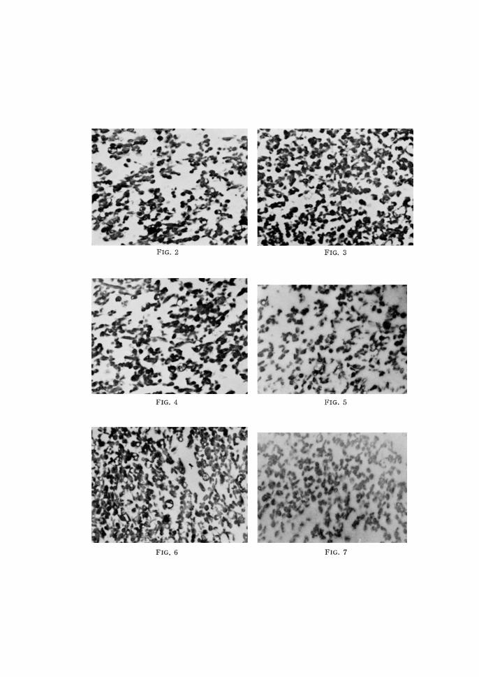
FIG. 2-7. Electron microscopical figures of neutrophilic granules isolated by centrifugal fractionation.