ORTHODONTICS Orthodontic records Assist.Lec.Kasem A. University of Babylon Faculty of Dentistry 4 th stage 1 Diagnostic (Orthodontic) records: Clinical orthodontic records are used primarily for diagnosis, monitoring of growth and development, and are a medico-legal requirement. They provide an accurate representation of the patient prior to orthodontic treatment, demonstrate treatment progress and allow communication between orthodontists, other healthcare professionals and the patient. Records also play an important role in research and clinical audit. It is essential that accurate clinical records are taken before commencing orthodontic treatment. Study models Impressions showing all the erupted teeth, full depth of the palate and good soft tissue extension are needed. These can be taken in alginate for study models and poured in dental stone. The study models provide a good help to examine the teeth from the facial and lingual during articulation, in addition to the possibility of the space analysis on the study models and the size selection of the orthodontic bands. The study model also used for modulation due to treatment and explanation of that to the patients. Accurate digital study casts are also now available, which have the advantages of occupying no physical storage space and having no deterioration over time, enabling indefinite storage. Clinical photographs Good clinical photographs form an essential part of the clinical record. The following views should be taken: • Intraoral, taken with the occlusal plane horizontal: • Frontal occlusion • Buccal occlusion (left and right) • Maxillary dentition • Mandibular dentition. • Extraoral, taken against neutral background in natural head posture: • Full facial frontal • Full facial frontal smiling • Facial three-quarters • Facial profile.
Transcript
ORTHODONTICS Orthodontic records Assist.Lec.Kasem A.
University of Babylon Faculty of Dentistry 4th stage
1
Diagnostic (Orthodontic) records:
Clinical orthodontic records are used primarily for diagnosis, monitoring
of growth and development, and are a medico-legal requirement. They
provide an accurate representation of the patient prior to orthodontic
treatment, demonstrate treatment progress and allow communication
between orthodontists, other healthcare professionals and the patient.
Records also play an important role in research and clinical audit. It is
essential that accurate clinical records are taken before commencing
orthodontic treatment.
Study models
Impressions showing all the erupted teeth, full depth of the palate and
good soft tissue extension are needed. These can be taken in alginate for
study models and poured in dental stone. The study models provide a good help
to examine the teeth from the facial and lingual during articulation, in addition to the
possibility of the space analysis on the study models and the size selection of the
orthodontic bands. The study model also used for modulation due to treatment and
explanation of that to the patients. Accurate digital study casts are also now
available, which have the advantages of occupying no physical storage
space and having no deterioration over time, enabling indefinite storage.
Clinical photographs
Good clinical photographs form an essential part of the clinical record.
The following views should be taken:
• Intraoral, taken with the occlusal plane horizontal:
• Frontal occlusion • Buccal occlusion (left and right)
• Maxillary dentition • Mandibular dentition.
• Extraoral, taken against neutral background in natural head posture:
• Full facial frontal • Full facial frontal smiling
• Facial three-quarters • Facial profile.
ORTHODONTICS Orthodontic records Assist.Lec.Kasem A.
University of Babylon Faculty of Dentistry 4th stage
2
Cephalometrics
A cephalometric lateral skull radiograph is a specialized view of the
facial skeleton and cranial base from the lateral aspect, with the head
position at a specific distance from the film. This method aims to study a
various component of the face and relate them to the cranium to see
whether there is a balance and harmony between these components or
not.
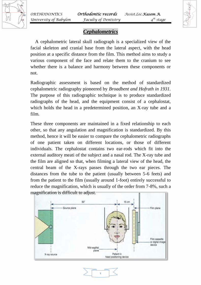
Radiographic assessment is based on the method of standardized
cephalometric radiography pioneered by Broadbent and Hofrath in 1931.
The purpose of this radiographic technique is to produce standardized
radiographs of the head, and the equipment consist of a cephalostat,
which holds the head in a predetermined position, an X-ray tube and a
film.
These three components are maintained in a fixed relationship to each
other, so that any angulation and magnification is standardized. By this
method, hence it will be easier to compare the cephalometric radiographs
of one patient taken on different locations, or those of different
individuals. The cephalostat contains two ear-rods which fit into the
external auditory meati of the subject and a nasal rod. The X-ray tube and
the film are aligned so that, when filming a lateral view of the head, the
central beam of the X-rays passes through the two ear pieces. The
distances from the tube to the patient (usually between 5-6 feets) and
from the patient to the film (usually around 1-foot) entirely successful to
reduce the magnification, which is usually of the order from 7-8%, such a
magnification is difficult to adjust.
ORTHODONTICS Orthodontic records Assist.Lec.Kasem A.
University of Babylon Faculty of Dentistry 4th stage
3
Indications for cephalometric evaluation:
A)An aid to diagnosis
It is possible to carry out successful orthodontic treatment without taking
a cephalometric radiograph, particularly in class I malocclusions.
However, the information that cephalometric analysis yields is helpful in
assessing the probable etiology of a malocclusion and in planning the
treatment. Therefore, a lateral cephalometric radiograph is best limited to
patients with a skeletal discrepancy and/or where anteroposterior
movement of the incisors is planned.
In a small proportion of patients, it may be helpful to monitor growth to
aid the planning and timing of treatment by taking serial cephalometric
radiographs. Also it is often helpful in the accurate localization of
unerupted displaced teeth and other pathology.
B) A pretreatment record:
A lateral cephalometric radiograph is useful in providing a baseline
record prior to the placement of the appliances, particularly where
movement of the upper and lower incisors is planned.
C) Monitoring the progress of treatment:
In the management of sever malocclusions, where tooth movement is
occuring in all three planes of space (for example treatment involving
functional appliances, or upper and lower fixed appliances), it is common
to take a lateral cephalometric radiograph during treatment to monitor
anchorage requirements and incisor inclinations. Also it is useful in
monitoring the movement of unerupted teeth and is the most accurate
view for assessing root resorption if this occurs during treatment.
D)Research purposes:
A great deal of information has been obtained about growth and
development by longitudinal studies which involved taking serial
cephalometric radiographs from birth to the late teens or beyond.
ORTHODONTICS Orthodontic records Assist.Lec.Kasem A.
University of Babylon Faculty of Dentistry 4th stage
4
In the cephalometric assessment, certain carefully defined points are identified
and linear and angular measurements are made from these points.
The points location and measurements have been performed simply by
tracing outlines on the skull radiograph and measuring by hand, but
systems are now available for computer analysis of skeletal form after the
manual plotting of co-ordinates on the radiograph. More recently methods
of automatic scanning of radiographs are being investigated, to avoid the
need for manual point location, which is the source of most of the errors
in cephalometric methods.
Some of the most commonly used cephalometric points and lines on
the lateral skull radiograph.
Cephalometric points
Porion: The highest point on the margin of the external auditory
meatus.
Articulare: The point of intersection of the outlines of the posterior
border of the mandible and the inferior border of the temporal
bone.
Bolton: The highest point in the concavity of the fossa behind the
occipital condyle.
Basion: The lowest point on the anterior margin of the foramin
magnum in the mid-line.
Pterygomaxillary point: The lowest point of the outline of the
pterygomaxillary fissure.
Sella: The center of the shadow of sella turcica.
A B
ORTHODONTICS Orthodontic records Assist.Lec.Kasem A.
University of Babylon Faculty of Dentistry 4th stage
5
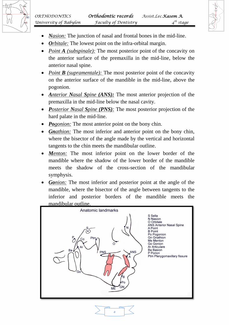
Nasion: The junction of nasal and frontal bones in the mid-line.
Orbitale: The lowest point on the infra-orbital margin.
Point A (subspinale): The most posterior point of the concavity on
the anterior surface of the premaxilla in the mid-line, below the
anterior nasal spine.
Point B (supramentale): The most posterior point of the concavity
on the anterior surface of the mandible in the mid-line, above the
pogonion.
Anterior Nasal Spine (ANS): The most anterior projection of the
premaxilla in the mid-line below the nasal cavity.
Posterior Nasal Spine (PNS): The most posterior projection of the
hard palate in the mid-line.
Pogonion: The most anterior point on the bony chin.
Gnathion: The most inferior and anterior point on the bony chin,
where the bisector of the angle made by the vertical and horizontal
tangents to the chin meets the mandibular outline.
Menton: The most inferior point on the lower border of the
mandible where the shadow of the lower border of the mandible
meets the shadow of the cross-section of the mandibular
symphysis.
Gonion: The most inferior and posterior point at the angle of the
mandible, where the bisector of the angle between tangents to the
inferior and posterior borders of the mandible meets the
mandibular outline.
ORTHODONTICS Orthodontic records Assist.Lec.Kasem A.
University of Babylon Faculty of Dentistry 4th stage
6
It will be appreciated that some of these points are in the mid-sagittal
plane, and are therefore single points, while others, such as orbitale and
gonion, are bilateral points. On a true lateral radiograph the bilateral
points should be superimposed, and should therefore appear as a single
point, but owing to facial asymmetries this is not always the case. If any
bilateral point appear separately on the radiograph, it is conventional to
accept a point half-way between the two as being the correct position.
The significance of certain points should also be appreciated. The lines
Nasion-Sella and Sella-Basion broadly represent the slope of the center of
the anterior and middle cranial bases, respectively. Point A and point B
also represent the anterior surfaces of the tooth-supporting dental bases of
the maxilla and mandible, respectively. The Pterygomaxillary point at the
posterior surface of the maxillary antrum, represents the posterior end of
the tooth bearing area of the maxilla. Other points are joined to form
cephalometric lines, which are used for measurement and alignment of
the head in growth and other studies.
Cephalometric lines
It has been conventional for many years to refer to the various reference
lines constructed by joining points on lateral skull radiographs as
cephalometric planes. However, since a plane has two dimensions while a
line drown on a radiograph has only one dimension, it is more accurate to
refer to cephalometric lines. The following is a descriptive list of the
more commonly used reference lines. There are many different systems
of cephalometric analysis, and other reference lines are sometimes used.
Frankfort line: The line joining the orbitale and porion. This is
accepted by convention as the horizontal line of the head, and is
commonly used for orientation of the head in clinical and
radiographic assessments.
Maxillary line: The line joining the anterior nasal spine and the
posterior nasal spine. This line is sometimes used instead of the
frankfort line in radiographic assessment, particularly as the
frakfort line is sometimes difficult to locate accurately on a
radiograph. However, the maxillary line does not bear any fixed
ORTHODONTICS Orthodontic records Assist.Lec.Kasem A.
University of Babylon Faculty of Dentistry 4th stage
7
relationship to the frankfort line, and the two lines are not usually
parallel. The maxillary line cannot of course, be assessed clinically.
Mandibular line: The line joining gonion and menton. It
represents the line of the lower border of the mandible, and can
roughly be assessed clinically.
Sella-Nasion line: The line joining the center of the sella turcica
and the nasion. It is used to represent the anterior cranial base, to
which the position of the jaws and teeth are often related in
cephalometric analysis.
Facial line: The line joining nasion and pogonion. The angular
relationship between the facial line and the frankfort line is used as
a measurement of mandibular prognathism.
Occlusal line: The line from the mid-point between the tips of the
upper and lower incisors to the anterior contact between the upper
and lower first molars in occlusion.
Bolton line: The line joining the bolton point and the nasion.
Y-axis: The line from sella to gnathion.
De-Coster line: The outline of the internal surface of the anterior
cranial base from the anterior border of the sella turcica to the
endocranial surface of the frontal bone.
Three further reference lines are sometimes used, particularly in
treatment planning, to assess the aesthetic qualities of the facial
profile and tooth position:
ORTHODONTICS Orthodontic records Assist.Lec.Kasem A.
University of Babylon Faculty of Dentistry 4th stage
8
The A-pogonion line: Is a line joining A point and the pogonion. It
is claimed that in ideal skeletal and occlusal form the incisal tips of
the lower central incisors should lie on or near to this line. It is a
useful guide in treatment planning, but it must be remembered that
this line does not necessarily represent a position of stability for the
lower incisors, particularly if the skeletal relationship in the sagittal
plane is not ideal.
The aesthetic line: Is a line joining the most anterior points of the
tip of the nose and the chin. It is claimed that, for good aesthetic
profile qualities, the anterior border of both upper and lower lips
should lie on or close to this line.
The Holdaway line: Is a line joining the anterior borders of the
upper lip and the chin. Again, it is claimed that for good aesthetic
profile qualities, the anterior border of the lower lip should lie on
or close to this line.
It will be appreciated that the last two lines mentioned above have soft
tissue landmarks and relate to lip position. They are therefore subject to
soft tissue variation, such as lip muscle thickness and tone, and although
they will be modified by variation in the skeletal relationship, and in
tooth position although with a relatively smaller effect. Furthermore, they
are related to aesthetic qualities, and beauty is in the eye of the beholder.
Cephalometric norms for Caucasians (Eastman
standard)
Measurement Mean value (in degree)
SNA 81 +3
SNB 78 +3
ANB 3 +2
U Inc to Mx Pl 109 +6
L Inc to Mn Pl 93 +6
IIA 135 +10
MMPA 27 +4
ORTHODONTICS Orthodontic records Assist.Lec.Kasem A.
University of Babylon Faculty of Dentistry 4th stage
9
Other Diagnostic records:
Orthopantomographs(OPG)
Exactly after the Second World War, Pattero of Finland turned to body
section radiography or tomography (laminagraphy) for better
radiographic visualization of craniofacial structures. It is possible to
obtain radiographic image of a particular area, or a layer within 3
dimensional subject by carefully synchronizing the movement of the
source of radiation and the film.
With both the X-ray beam source and the film cassette rotating,
everything else is thrown out of focus except the precise level of
structures of interest. The parts that in focus will appear in sharp details,
while the intervening structures are blured out. The exposure factors are
( 80-86 KVP with 15-20 milliampers and short exposure time about 15
second, depending on the machine type).
Up to date OPG, as soon as the patients symptoms are associated with
the mandibular joints and/or the maxillary sinuses you have no option
other than extending the scope of X-ray investigation to these regions.
Such applications are beyond the capabilities of conventional panoramic
X-rays unit.
ORTHODONTICS Orthodontic records Assist.Lec.Kasem A.
University of Babylon Faculty of Dentistry 4th stage
10
Advantages of OPG
1)Comfort, since no film will be inserted inside the patients mouth. and
the total time needed to perform the X-ray not exceed 1.5 min.
2) It is more easy for the operator when he has uncooperative patients,
children patient with gagging reflex and trismus (lock jaw).
3) Least total radiation ( 5.06 rad) in comparison with periapical X-ray.
Disadvantages
1)It cannot give precise information on periodontal membrane. The
intaoral radiograph provide more accurate information about the .....
2) The lower incisors region is not properly reproduced due to some
overlapping as a result of shifting in the axis of rotation. Additionally
their inclination is usually slanted with the crown mesially inclined.
Uses of OPG
A...For growth and development studies: Delayed tooth eruption,
abnormality in eruption path, abnormal resorption, supernumerary teeth,
systs, congenitally absent teeth, ankylosis, prolong retention, density of
the bone, axial inclination, inadequate space of the clinical entities that
concerning to the general practitioner or orthodontist, and distant from
apices to the mandibular plane.
B...The Tempromandibular joint: The OPG provides a sharp and
accurate profile view of the condyle and the articular eminence of the
articular fossa itself.
C...Sinuses and mastoid region : The importance of maxillary sinuses is
very recognized by orthodontist since a reduced sinuses size related to
mouth breather and collapse of maxillary segments.
D...Mandibular morphology: The OPG gives a clear picture about the
bony mass of the mandible, the extent of the alveolar bone, height and
width of the ramus.
ORTHODONTICS Orthodontic records Assist.Lec.Kasem A.
University of Babylon Faculty of Dentistry 4th stage
11
E...Space adequacy: The OPG of great benefit during the serial
extraction procedure which require the removal of some deciduous teeth
followed by some permanent teeth (usually the first premolars) and this
require knowledge abouit the stages of root formation of the teeth.
F...Investigation of facial asymmetries and swelling.
G...Suspected fractures of the mandible and maxilla.
Three-dimensional imaging
Plain film and cephalometric radiography are invaluable
for accurate diagnosis and treatment
planning, but they only provide a two
dimensional image of a three-
dimensional structure, with all the
associated errors of projection,
anatomical superimposition, landmark
identification, measurement and interpretation.
Cone-beam computed tomography
Imaging of the hard tissues composing the jaws and dentition using
conventional computed tomography (CT) is largely impractical, due to the high
radiation dosage, lack of resolution and significant cost. The introduction of
cone-beam computed tomography (CBCT) has resulted in the dosage being
reduced and the resolution significantly improved, with its adaption and
refinement for imaging of the teeth and jaws now providing a useful three-