British Iournal of Haemalology. 1993, 84, 608-614 Subcellular fractionation studies indicate an intracellular localization for human monocyte specific esterase (MSE) DIVYEN PATEL, NIGEL M. HOOPER AND COLIN STEPHEN SCOTT Haematological Malignancy Diagnostic Service, Cookridge Hospital, Leeds, and Department of Biocheniistry and Molecular Biology, University of Leeds, Leeds Received 6 October 1992; accepted for publication 10 March 1993 Summary. Human monocyte-specificesterase (MSE)is one of the few haemopoietic cell enzymes that show absolute lineage restriction. Although the function of MSE has yet to be deduced, its potential role in tumour cell killing has stimulated particular interest. Knowledge of subcellular localization of MSE is fundamental to understanding its function and. in this context, it is widely believed that MSE is expressed as a plasma membrane ectoenzyme; a contention that is largely based upon experiments which examined fixed cells by ultrastructural cytochemistry. However, as recent Carboxylesterases are hydrolytic enzymes that are found in high concentrations in a wide range of tissues and organisms. Within the multi-lineage system of haemopoietic cells, esterase species that hydrolyse non-physiological substrates such as cr-naphthyl acetate or butyrate, have been shown by extensive electrophoretic analyses (Scott et al, 1984b; Scott & Drexler, 1989) to comprise a number of isoenzyme groups that are heterogeneous with regards to their cellular ex- pression and distribution. Although many of these esterase isoforms are not lineage-specific, one particular carboxyles- terase species (monocyte-specific esterase; MSE) appears to be almost unique in that its synthesis and expression is restricted to myeloid haemopoietic cells of monocytic/macrophage type (Drexler et al, 1991; Scott et al, 1992b). Early investigations which combined azo-dye staining techniques with light and electron microscopy suggested that MSE was a plasma membrane ectoenzyme (Monahan et al. 1981; Bozdech & Bainton. 1981). However, doubts regard- ing this conclusion have resulted from more recent studies which show that ‘mature’ monocyte/macrophage-specific carboxylesterases lack a hydrophobic membrane anchoring domain and have a COOH terminal tetrapeptide signal analogous to that displayed by proteins normally retained within the lumen of the endoplasmic reticulum (ER) (Munger et al, 1991;Zschunke et al, 1991: Scott et al, 1992b).IfMSEis Correspondence: Dr C. S. Scott, Haematological Malignancy Diag- nostic Service, Cookridge Hospital, Leeds LSI 6 6QB. molecular studies of human MSE indicate, a number of inconsistencies between its structure and a membrane localization, we applied the techniques of phase separation in the non-ionic detergent Triton X-114 and differential centri- fugation to further investigate whether this particular ester- ase species is membrane-bound or associated with an intracellular organelle. These studies provide strong evidence that MSE is in fact a soluble intracellular enzyme that is almost certainly located within the lumen of the endoplasmic reticulum. indeed a plasma membrane ectoenzyme then its cellular function is likely to differ quite considerably from that of an intracellular enzyme which may participate either in the post-synthetic processing of protein, lipid or carbohydrate moieties prior to their transfer to the Golgi apparatus, or as part of the human detoxification systems (if located in the smooth ER) as is the case for the cytochrome P450 cascade. In this context, it is reasoned that because MSE is one of the very few leucocyte enzymes that has an absolute lineage association, then its presence in such cells will almost certainly reflect a distinct function. Consequently, as the subcellular localization of MSE is of fundamental importance in determining what this function might be, we applied the techniques of phase separation in the non-ionic detergent Triton X-114 (Bordier, 1981) and differential centrifugation to investigate whether this particular esterase species is membrane-bound or associated with an intracellular orga- nelle. MATERIALS AND METHODS Cells. Normal human peripheral blood monocytes were isolated from healthy volunteer donors to a purity of > 90% by differential sedimentation techniques. Briefly, this involved the preliminary removal of erythrocytes from EDTA- anticoagulated whole blood by inducing rouleaux formation with 6% w/v dextran in 0.9% saline, and subsequent density sedimentation of the leucocyte-rich supernatant using Nyco- 608
Transcript
British Iournal of Haemalology. 1993, 84, 608-614
Subcellular fractionation studies indicate an intracellular localization for human monocyte specific esterase (MSE)
DIVYEN PATEL, NIGEL M. HOOPER AND COLIN STEPHEN SCOTT Haematological Malignancy Diagnostic Service, Cookridge Hospital, Leeds, and Department of Biocheniistry and Molecular Biology, University of Leeds, Leeds
Received 6 October 1992; accepted for publication 10 March 1993
Summary. Human monocyte-specific esterase (MSE) is one of the few haemopoietic cell enzymes that show absolute lineage restriction. Although the function of MSE has yet to be deduced, its potential role in tumour cell killing has stimulated particular interest. Knowledge of subcellular localization of MSE is fundamental to understanding its function and. in this context, it is widely believed that MSE is expressed as a plasma membrane ectoenzyme; a contention that is largely based upon experiments which examined fixed cells by ultrastructural cytochemistry. However, as recent
Carboxylesterases are hydrolytic enzymes that are found in high concentrations in a wide range of tissues and organisms. Within the multi-lineage system of haemopoietic cells, esterase species that hydrolyse non-physiological substrates such as cr-naphthyl acetate or butyrate, have been shown by extensive electrophoretic analyses (Scott et al, 1984b; Scott & Drexler, 1989) to comprise a number of isoenzyme groups that are heterogeneous with regards to their cellular ex- pression and distribution. Although many of these esterase isoforms are not lineage-specific, one particular carboxyles- terase species (monocyte-specific esterase; MSE) appears to be almost unique in that its synthesis and expression is restricted to myeloid haemopoietic cells of monocytic/macrophage type (Drexler et al, 1991; Scott et al, 1992b).
Early investigations which combined azo-dye staining techniques with light and electron microscopy suggested that MSE was a plasma membrane ectoenzyme (Monahan et al. 1981; Bozdech & Bainton. 1981). However, doubts regard- ing this conclusion have resulted from more recent studies which show that ‘mature’ monocyte/macrophage-specific carboxylesterases lack a hydrophobic membrane anchoring domain and have a COOH terminal tetrapeptide signal analogous to that displayed by proteins normally retained within the lumen of the endoplasmic reticulum (ER) (Munger et al, 1991; Zschunke et al, 1991: Scott et al, 1992b). IfMSEis
Correspondence: Dr C. S. Scott, Haematological Malignancy Diag- nostic Service, Cookridge Hospital, Leeds LSI 6 6QB.
molecular studies of human MSE indicate, a number of inconsistencies between its structure and a membrane localization, we applied the techniques of phase separation in the non-ionic detergent Triton X-114 and differential centri- fugation to further investigate whether this particular ester- ase species is membrane-bound or associated with an intracellular organelle. These studies provide strong evidence that MSE is in fact a soluble intracellular enzyme that is almost certainly located within the lumen of the endoplasmic reticulum.
indeed a plasma membrane ectoenzyme then its cellular function is likely to differ quite considerably from that of an intracellular enzyme which may participate either in the post-synthetic processing of protein, lipid or carbohydrate moieties prior to their transfer to the Golgi apparatus, or as part of the human detoxification systems (if located in the smooth ER) as is the case for the cytochrome P450 cascade. In this context, it is reasoned that because MSE is one of the very few leucocyte enzymes that has an absolute lineage association, then its presence in such cells will almost certainly reflect a distinct function. Consequently, as the subcellular localization of MSE is of fundamental importance in determining what this function might be, we applied the techniques of phase separation in the non-ionic detergent Triton X-114 (Bordier, 1981) and differential centrifugation to investigate whether this particular esterase species is membrane-bound or associated with an intracellular orga- nelle.
MATERIALS AND METHODS Cells. Normal human peripheral blood monocytes were
isolated from healthy volunteer donors to a purity of > 90% by differential sedimentation techniques. Briefly, this involved the preliminary removal of erythrocytes from EDTA- anticoagulated whole blood by inducing rouleaux formation with 6% w/v dextran in 0.9% saline, and subsequent density sedimentation of the leucocyte-rich supernatant using Nyco-
608
MSE Subcellular Localization 609 Table I. Subcellular localization of monocyte-specific esterase (MSE) as assessed by dfierential centrifugatioq.*
denz (SG 1.068; Nycomed). Two separate ‘pools’ of normal monocytes were examined in this study, with each compris- ing a mixture of fractionated cells from three individuals. In addition, cultured immortalized human monocytic led-ae- mia U937 cells, known to express high concentrations of MSE. were also analysed. These cells were maintained in RPMI 1640 medium supplemented with 5% heat-inactivated fetal calf serum (Gibco) in a 5% C 0 2 humidified atmosphere at 3 7°C and taken from continuous culture as required.
Cellular disruption procedure. All operations were carried out at 4°C unless otherwise stated. Normal blood monocytes or U937 cells (107-40i cells) were suspended in 2 ml of 10 mM TrislHCI buffer pH 7.4 containing 20 mM calcium chloride (included to prevent cell membrane fractions from becoming attached to the nuclei) and placed in a PARR cell disruption bomb. The vessel was pressurized to 800 psi and this was maintained for 10 min to allow nitrogen to dissolve and reach equilibrium within the cells. At this time, the nitrogen-pressurized cells were collected through a discharge valve with a cellular disruption resulting at the moment of decompression. The disrupted sample was then centrifuged at 600 g for 10 min and the supernatant (containing subcellu- lar organelles, fractured membrane components and soluble proteins) removed for subsequent processing. The pelleted material, containing intact cells and nuclei, was discarded.
Subcellular fractionation. Following cellular disruption, sub- cellular components were further isolated by a series of centrifugation steps (Shibko & Tappel, 1965). The cell homogenate was centrifuged at 6000 g for 8 min to pellet lysosomes and mitochondria. The supernatant from this stage was removed and centrifuged at 40 000 g for 30 min to give a microsomal pellet. The resulting supernatant from this step was centrifuged at 100000 g for 90 min to give a light microsomal/ribosomal pellet and a final soluble fraction. Pelletted material at each centrifugation step was washed, re- centrifuged and then solubilized in 10 mM Tris/HCl pH 7.5 containing 1 .Ox Triton X-100. Esterase activities of each fraction were determined by quantitative ffuorimetric ace- tate/butyrate esterase assays (Scott et a/, 1992a) and protein concentrations were determined by the bicinchoninic acid method (Pierce Chemical Co.). The samples were additionally analysed by isolectric focusing (IEF) for the presence of MSE isoenzymes (Scott et a/ , 1984a, b) by electrophoresing 2 5-50 pl sample aliquots for 1400 V on PI range 3.5-9.5 IEF gels (Pharmacia). Gels were subsequently stained for a-naphthyl acetate esterase (ANAE) by standard histochemical pro- cedures and the resulting ANAE isoenzyme profiles were examined to assess the relative staining intensities of MSE and non-MSE isoenzyme species.
lsolatiori of soluble and membrane-associated proteins. The cell homogenates were centrifuged at 100 000 g for 90 min at 4°C. The resulting supernatant (containing soluble proteins) was removed, and the pellet (containing subcellular orga- nelles and plasma membrane components) resuspended in 200 pI 10 mM Tris/HCl pH 7.5. This was re-centrifuged at 100 000g for 90 min, with the supernatant from this second centrifugation being mixed with the supernatant from the previous step, and the pellet being resuspended in 200 pl 10 mM Tris/HCl pH 7.5 buffer containing 1% Triton X-100. All
* Normal blood monocyte and U93 7 cell homogenates prepared by PARR cell bomb disruption and removal of debris by low-speed centrifugation (600 g for 10 min). Subsequent centrifugation stages (6000. 40000 and 100000 g ) were carried out as described in Materials and Methods and each fraction was assayed for acetate/ butyrate esterase (AcE/ButE) activities by fluorimetry (expressed as U/mg fraction protein), and isoelectric focusing (IEF) for the presence of MSE. For the purposes of this analysis the staining intensities of MSE components were graded from - (complete absence) to + + + (strong expression).
the supernatants and pellets obtained from this process were then analysed quantitatively for acetate/butyrate esterase and protein concentrations, and qualitatively by IEF to determine MSE isoenzyme expression.
Phase separation in Triton X - 2 24. The cell homogenates obtained from disrupted cells were mixed with pre-condensed Triton X-114 (Sigma) to a final concentration of 2.0% (Bordier. 1981). The sample was kept on ice for 5 min and subsequently incubated at 30°C for 3 min and centrifuged at 3000 g for 10 min. At this time the sample resolved into two distinct (aqueous and detergent-rich) phases which were separated and stored for further analysis. All fractions were analysed for esterase activities, protein concentration and MSE isoenzyme expression as detailed above.
Preparation of rough (RER) and smooth (SER) endoplasmic reticulum fractions. Normal monocyte and U937 cells were suspended in 0.25 M sucrose solution and disrupted using a PARR nitrogen bomb prior to fractionation as described by Gram (1 9 74). The cellular homogenate was centrifuged at 10 000 g for 20 min and the supernatant removed and mixed
610 D. Patel, N. M. Hooper and C. S. Scott
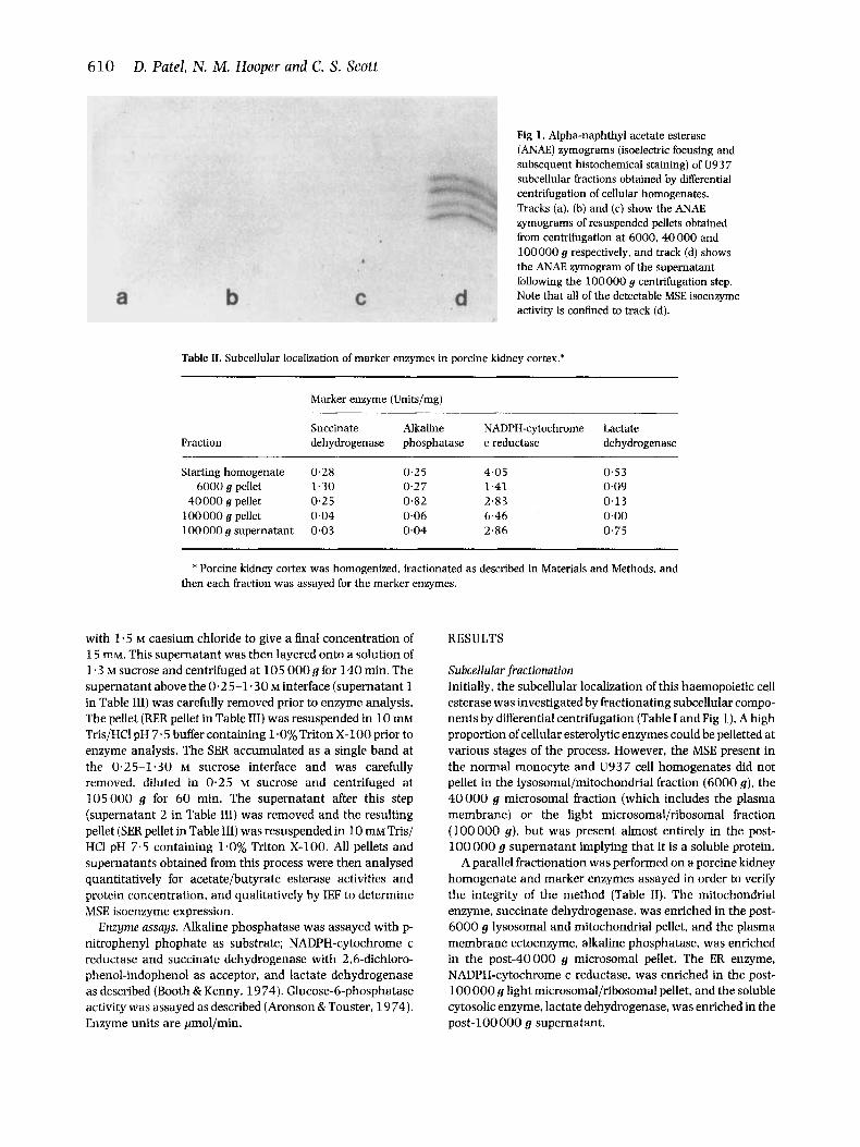
Fig 1. Alpha-naphthyl acetate esterase (ANAE) zymograms (isoelectric focusing and subsequent histochemical staining) of U93 7 subcellular fractions obtained by differential centrifugation of cellular homogenates. Tracks (a), (b) and (c) show the ANAE zymograms of resuspended pellets obtained from centrifugation at 6000, 40000 and 100000 g respectively, and track (d) shows the ANAE zymogram of the supernatant following the 100000 g centrifugation step. Note that all of the detectable MSE isoenzyme activity is confined to track (d).
Table 11. Subcellular localization of marker enzymes in porcine kidney cortex.*
Fraction
Marker enzyme (Units/mg)
Succinate Alkaline NADPH-cytochrome Lactate dehydrogenase phosphatase c reductase dehydrogenase
40000 g pellet 0.25 0.82 2.83 100 000 g pellet 0.04 0.06 6.46 100000 g supernatant 0.03 0.04 2.86
0.53 0.09 0.13 0.00 0.75
* Porcine kidney cortex was homogenized, fractionated as described in Materials and Methods, and then each fraction was assayed for the marker enzymes.
with 1.5 M caesium chloride to give a final concentration of 1 5 mM. This supernatant was then layered onto a solution of 1.3 M sucrose and centrifuged at 105 000 g for 140 min. The supernatant above the 0.25-1.30 M interface (supernatant 1 in Table 111) was carefully removed prior to enzyme analysis. The pellet (RER pellet in Table 111) was resuspended in 10 mM Tris/HCI pH 7.5 buffer containing l.O%Triton X-100 prior to enzyme analysis. The SER accumulated as a single band at the 0.25-1.30 M sucrose interface and was carefully removed, diluted in 0.25 M sucrose and centrifuged at 105000 g for 60 min. The supernatant after this step (supernatant 2 in Table 111) was removed and the resulting pellet (SER pellet in Table 111) was resuspended in 10 mM Tris/ HC1 pH 7.5 containing 1.0% Triton X-100. All pellets and supernatants obtained from this process were then analysed quantitatively for acetate/butyrate esterase activities and protein concentration, and qualitatively by IEF to determine MSE isoenzyme expression.
Enzyme assays. Alkaline phosphatase was assayed with p- nitrophenyl phophate as substrate; NADPH-cytochrome c reductase and succinate dehydrogenase with 2,6-dichloro- phenol-indophenol as acceptor, and lactate dehydrogenase as described (Booth & Kenny. 19 74). Glucose-6-phosphatase activity was assayed as described (Aronson & Touster, 19 74). Enzyme units are jcmol/min.
RESULTS
Subcellular fractionation Initially, the subcellular localization of this haemopoietic cell esterase was investigated by fractionating subcellular compo- nents by differential centrifugation (Table I and Fig 1). A high proportion of cellular esterolytic enzymes could be pelletted at various stages of the process. However, the MSE present in the normal monocyte and U937 cell homogenates did not pellet in the lysosomal/mitochondrial fraction (6000 g), the 40 000 g microsomal fraction (which includes the plasma membrane) or the light microsomal/ribosomal fraction (100000 9). but was present almost entirely in the post- 100 000 g supernatant implying that it is a soluble protein.
A parallel fractionation was performed on a porcine kidney homogenate and marker enzymes assayed in order to verify the integrity of the method (Table 11). The mitochondrial enzyme, succinate dehydrogenase, was enriched in the post- 6000 g lysosomal and mitochondrial pellet, and the plasma membrane ectoenzyme, alkaline phosphatase, was enriched in the post-40000 g microsomal pellet. The ER enzyme, NADPH-cytochrome c reductase. was enriched in the post- 100000 g light microsomal/ribosomal pellet, and the soluble cytosolic enzyme, lactate dehydrogenase, was enriched in the post-100000 g supernatant.
MSE Subcellular Localization 61 1 Table 111. Investigation of the membrane association of MSE in normal blood monocytes and U937 cells.*
* Results shown are for two separate experiments. Normal blood monocytes and U937 cells were disrupted in a PARR cell bomb and debris removed by low-speed centrifugation (600 g for 10 min). The supernatant was then processed by (a) ultracentrifugation at 100000 y or (b) Triton X-114 phase separation as described in Materials and Methods.
t Acetate (ACE) and butyrate (ButE) esterase activities given as arbitrary units, defined by fluorimetry (Scott ef d, 1992b), per rng fraction protein.
$Analysis of cell fractions for monocyte-specific esterase (MSE) by isoeletric focusing (IEF).
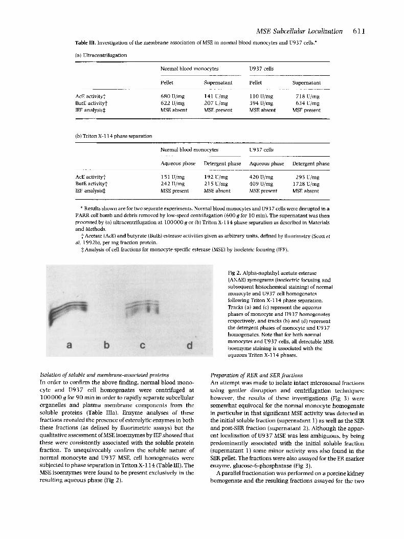
Fig 2. Alpha-naphthyl acetate esterase (ANAE) zymograms (isoelectric focusing and subsequent histochemical staining) of normal monocyte and U937 cell homogenates following Triton X-114 phase separation. Tracks (a) and (c) represent the aqueous phases of monocyte and U937 homogenates respectively, and tracks (b) and (d) represent the detergent phases of monocyte and U9 3 7 homogenates. Note that for both normal monocytes and U937 cells, all detectable MSE isoenzyme staining is associated with the aqueous Triton X-114 phases.
Isolation of soluble and membrane-associated proteins In order to confirm the above finding, normal blood mono- cyte and U937 cell homogenates were centrifuged at 100000 g for 90 min in order to rapidly separate subcellular organelles and plasma membrane components from the soluble proteins (Table IIIa). Enzyme analyses of these fractions revealed the presence of esterolytic enzymes in both these fractions (as defined by fluorimetric assays) but the qualitative assessment of MSE isoenzymes by IF,F showed that these were consistently associated with the soluble protein fraction. To unequivocably confirm the soluble nature of normal monocyte and U937 MSE, cell homogenates were subjected to phase separation in Triton X-114 (Table 111). The MSE isoenzymes were found to be present exclusively in the resulting aqueous phase (Fig 2).
Preparation of RER and SER fractions An attempt was made to isolate intact microsomal fractions using gentler disruption and centrifugation techniques: however, the results of these investigations (Fig 3) were somewhat equivocal for the normal monocyte homogenate in particular in that significant MSE activity was detected in the initial soluble fraction (supernatant 1) as well as the SER and post-SER fraction (supernatant 2). Although the appar- ent localization of U93 7 MSE was less ambiguous, by being predominantly associated with the initial soluble fraction (supernatant 1) some minor activity was also found in the SER pellet. The fractions were also assayed for the ER marker enzyme, glucose-6-phosphatase (Fig 3).
A parallel fractionation was performed on a porcine kidney homogenate and the resulting fractions assayed for the two
Mixed with 1.5M caesium chloride, layered onto 1.3M sucrose and centrifuged at 105 OOOg for 140 min
s!mEM Suoernatanto ACE 22 U/mg ButE. 60 U/mg
EF MSE ++
ACE 42 U/mg Bu@. 42 U/mg
IEF MSE ++ G6-P: 0.36 G6-P 0.44
iuhldkt Suoernatanto ACE: 80 U/mg ButE: 253 U/mg
ACE: 293 U/mg ButE: 280 U/mg
G6-P: 0.73 G6-P 1.20 IEF: MSE + IEF MSE -
Acetate and butyrate esterase activities for each fraction determined by fluonmetry and expressed as U/mg fraction protein. Glucose-6-phosphatase (G6-P) activity expressed as poVmin/mg fractim protein. IEF analyses Indicate the staining intensities of MSE components graded from - (complete absence) to ++ (moderate staining)
Fig 3. Fractionation of smooth (SER) and rough endoplasmic reticulum (RER) from normal blood monocytes and U937 cells: Relationships with monocyte-specific esterase (MSE) localization."
ER marker enzymes, glucose-6-phosphatase and NADPH- cytochrome c reductase. The distribution of these two enzymes were 0.24 and 16.92 Units/mg respectively in the RER pellet, 0.50 and 58.77 Units/mg in the initial soluble fraction (supernatant l), 0.54 and 28.69 Units/mg in the SER pellet. and 3 . 3 3 and 1 5 0 4 Units/mg in the post-SER fraction (supernatant 2).
DISCUSSION
Using the combined techniques of zone electrophoresis and histochemical staining (Hunter & Markert, 19 5 7), multiple isoenzyme forms of carboxylesterases have been shown to occur in human liver, brain, muscle, kidney, red cells, leucocytes and plasma. With particular reference to human haemopoietic elements, isoelectrophoretic analyses have convincingly demonstrated that cells developing along the monocytic/macrophage axis specifically synthesize a group of carboxylesterase (EC 3.1.1.1) isoenzymes that are widely referred to as monocyte-specific esterases (MSE). When analysed by analytical focusing (IEF) techniques, MSE com- prises a series of four to six individual isoforms within a narrow PI (isoelectric point) range of 5.5-6.2 (Drexler et al,
1991; Scott et al, 1992b). Studies of these isoenzymes have provided important insights into the patterns of myeloid cell differentiation and, in addition, has resolved many of the uncertainties regarding the esterase cytochemical staining patterns of leukaemic cells (Scott & Drexler, 1989; Drexler et al, 1991).
Whereas the physiological roles of some esterases, such as acetylcholinesterases, cholinesterase and juvenile hormone esterase, are clearly understood, the function(s) of MSE remains unresolved. Speculated functions include detoxifica- tion and metabolic processing of drugs, facilitation of diape- desis and migration through tissues, and a role in pinocytosis, inflammation, growth regulation and tumour cell killing (reviewed in Drexler et al, 1991). Of these, the potential role of MSE in tumour cell cytotoxicity has generated the greatest interest (Newcombe, 1992), and indeed there does appear to be some relationship (albeit tenuous) between monocyte MSE deficiency and an increased frequency of malignant disease (Markey et al, 1987, 1990). Obviously, the subcellular localization of MSE is clearly of fundamental importance in determining its physiological function.
While recent studies of the purified enzyme (Saboori & Newcombe. 1990; Scott et al, 1992a, b) have clarified the
MSE Subcellular Localization 61 3
molecular and biochemical characteristics of human MSE, little has been reported on the subcellular localization of this enzyme. Early investigations which combined azo-dye stain- ing techniques with light and electron microscopy suggested that MSE was a plasma membrane ectoenzyme (Monahan e t al, 1981: Bozdech & Bainton. 1981). However, doubts regarding this conclusion have resulted recently from the determination of the complete amino acid sequence of MSE following isolation and sequencing of its cDNA (Munger et al. 1991; Zschunke et al, 1991). Apart from the hydrophic N- terminal signal sequence, which is absent in the mature protein (Scott et al, 1992b), there is no other region of consecutive hydrophobic amino acids in the deduced sequence which could possibly act as a transmembrane anchoring domain. In addition, the amino acid sequence of MSE lacks any of the consensus sequences for modification by alternative membrane anchoring structures such as an N- terminal myristic acid or a C-terminal glycosylphosphatidyli- nositol or polyisoprenoid moiety (Chow et al, 1992; Hooper & Turner, 1992).
In this present study, we attempted to determine the subcellular localization of MSE in normal and leukaemic cells by fractionating cell homogenates into various organelle fractions by differential centrifugation (Tables I and 111). However, MSE in both the normal and leukaemic cells was present almost entirely in the post-100000 g supernatant, implying that it is a freely soluble protein. To confirm this result, we subjected cell homogenates to phase separation in the non-ionic detergent Triton X-114. Temperature-induced phase separation in Triton X-114 is a powerful technique for identifying integral membrane proteins and separating them from soluble, hydrophilic proteins (Bordier, 198 1; Hooper & Turner, 1988). -At 3OoC the detergent separates into two distinct phases: a detergent-rich phase containing integral membrane proteins which possess a hydrophobic anchoring domain, and an aqueous phase containing hydrophilic soluble or peripheral membrane proteins. Thus, the observa- tion that MSE was recovered exclusively in the aqueous phase after phase separation of cell homogenates in Triton X-114 (Table 11%) strongly indicates that the mature protein lacks a hydrophobic anchoring domain and is not associated with the hydrophobic interior of the lipid bilayer.
The presence of an N-terminal signal peptide predicted by the cDNA for MSE (Munger et al, 1991) indicates that the protein is almost certainly directed to the lumen of the ER during its synthesis. Further evidence that MSE is transported to the ER comes from the observation that the mature protein contains a high mannose-type glycan moiety which is not further modified (Scott et al, 1992a) and in vitro translation experiments (Harano et al, 1988). In addition, human MSE has a COOH-terminal tetrapeptide (HIEL) (Munger et al. 1991: Zschunke et al, 1991) which is analogous to the KDEL motif displayed by proteins normally retained within the lumen of the ER (Pelham, 1989). Recent mutagenesis studies have shown that it is the two C-terminal residues (glutamic acid and leucine) of this motif which are most critical for the retention of a protein in the ER (Andres et al, 1990). In the light of these observations, we attempted to localize MSE in microsomes derived from the RER and SER (Fig 3) . Although
some MSE activity was present in the RER and SER fractions derived from normal monocytes and in the SER fraction from U937 cells, the majority of MSE activity from both cell types was present in the soluble fraction. Interpretation of these results, is, however, influenced by the practical problems associated with maintaining RER and SER integrity during subcellular fractionation. One possible explanation for these findings is that a significant proportion of MSE is indeed present within the lumen of the ER in intact cells but, because of its soluble nature as ascertained above, MSE is lost into the non-ER supernatants through organelle disruption or leak- age during microsome formation. This contention is sup- ported to some extent by the observation that significant activities of the ER marker enzymes, glucose-6-phosphatase and NADPH-cytochrome c reductase, were also found in the non-ER supernatants of both the normal monocytes and U937 cells (Fig 3) as well as the porcine kidney.
A comparison of the reported sequence with human and non-human liver carboxylesterases reveals a high degree of homology (Mungeretd, 1991; Zschunke et al. 1991; Robbi et a!, 1990; Korza & Ozols, 1988). These liver esterases also possess C-terminal tetrapeptide sequences, HXEL, which appear to retain the proteins in the lumen of the ER (Robbi 81 Beaufay, 1983; Harano et al, 1988; Korza & Ozols, 1988; Robbi et al, 1990). Furthermore, the liver microsomal carboxylesterases show similarities in molecular weight, subunit association, presence of high mannose-type sugars and, to some extent, substrate and inhibitor reactivities to carboxylesterases isolated from monocyte/macrophage cells (Robbi & Beaufay, 1983, 1987), suggesting that these esterases are closely related and possibly share the same subcellular location and physiological role(s).
The apparent discrepancy between the conclusions of this study (i.e. soluble intracellular localization within the lumen of the ER) and those obtained by electron microscopy (i.e. plasma membrane localization) (Monahan et a / , 1981; Bozdech & Bainton, 1981) may be resolved by the explana- tion proposed by Zschunke et aZ(1992) who suggested that ‘resting’ monocytes may indeed express intracellular MSE but that, following appropriate stimulation, there is a rapid transfer to the plasma membrane. MSE does not, however, appear to be secreted as the examination of sera from patients with acute leukaemias of monocytic type, in which the blast cells strongly express MSE, do not reveal the presence of soluble serum MSE (personal observations).
ACKNOWLEDGMENTS
We thank the Yorkshire Cancer Research Campaign and the Wellcome Trust for their financial support. We are also grateful to S. Y. Oppong for the marker enzyme assays.
REFERENCES
Andres. D.A., Dickerson. I.M. & Dixon, J.E. (1990) Variants of the carboxyl-terminal KDEL sequence direct intracellular retention. Journal of Biological Chemistry, 265, 5952-5956.
Aronson. N.N. & Touter, 0. (1974) Isolation of rat liver plasma
614 D. Patel, N. M . Hooper and C. S. Scott membrane fragments in isotonic sucrose. Methods in Enzymology, 31, 90-102.
Booth, A.G. & Kenny, A.J. (1974) A rapid method for the preparation of microvilli from rabbit kidney. Biochemical Journal, 142, 575- 581.
Bordier, C. (1 981) Phase separation of integral membrane proteins in Triton X-114 solution. Journal of Biological Chemistry. 256.1604- 1607.
Bozdech. M.J. & Bainton. D.F. (1981) Identification of a-naphthyl butyrate esterase as a plasma membrane ectoenzyme of monocytes and as a discrete intracellular membrane-bound organelle in lymphocytes. Journal of Experimental Medicine, 153, 182-195.
Chow, M., Der, C.J. & Buss, J.E. (1992) Structure and biological effects of lipid modifications on proteins. Current Opinion in Cell Biology. 4,
Drexler. H.G.. Gignac, S.M.. Patel, D. & Scott, C.S. (1991) Lineage- specific monocyte esterase: cytochemical, isoenzymatic and bio- chemicalfeatures. Areview. LRukemiaandLymphoma, 4,295-312.
Gram, T.E. (1974) Separation of hepatic smooth and rough micro- somes associated with drug-metabolizing enzymes. Methods in Enzymology. 31, 225-237.
Harano, T.. Miyata, T.. Lee, S.. Aoyagi, H. & Omura, T. (1988) Biosynthesis and localization of rat liver microsomal carboxyl- esterase E l . Journal of Biochemistry. 103, 149-1 55.
Hooper. N.M. & Turner, A.J. (1988) Ectoenzymes of the kidney microvillar membrane. Differential solubilization by detergents can predict a glycosyl-phosphatidylinositol membrane anchor. Biochemical Journal. 250, 865-869.
Hooper. N.M. & Turner, A.J. (eds) (1992) Lipid Modification of Proteins: a practical approach. IRL Press. Oxford.
Hunter, R.L. & Markert, C.L. (1957) Histochemical demonstration of enzymes separated by zone electrophoresis in starch gels. Science,
Korza, G. & Ozols, J. (1988) Complete covalent structure of 60-kDa esterase isolated from 2,3,7,8-tetrachlorodibenzo-p-dioxin- induced rabbit liver microsomes. Journal of Biological Chemistry,
Markey, G.M., McCormick, J.A. & Morris, T.C.M. (1990) Monocyte esterase deficiency in malignant neoplasia. Journal of Clinicnl
Markey, G.M.. Morris, T.C.M. & Alexander, M.D. (1987) Monocyte esterase? A factor involved in the pathogenesis of lymphoprolifera- tive neoplasia. Leukemia, 1, 236-2 39.
Monahan. R.A.. Dvorak, H.F. &Dvorak, A.M. (1981) Ultrastructural localisation of non-specific esterase activity in guinea pig and human monocytes, macrophages and lymphocytes. Blood. 58, 1089-1099.
Munger, J.S., Shi. G.P.. Mark, E.A., Chin, D.T., Gerad, C. &Chapman, H.A. (1991) A serine esterase released by human alveolar
629-636.
125, 1294-1301.
263, 3486-3488.
Pathology, 45, 282-286.
macrophages is closely related to liver microsomal carboxylester- ases. Journal of Biological Chemistry, 266, 18832-18838.
Newcombe. D.S. ( I 992) Immune surveillance. organophosphorus exposure and lymphomagenesis. Lanret. 339, 539-541.
Pelham, H.R.B. (1989) Control of protein exit from the endoplasmic reticulum. Annual Review of Cell Biology., 5. 1-23.
Robbi, M. & Beaufay, H. (1983) Purification and characterization of various esterases from rat liver. European Journal of Biochemistry.
Robbi. M. & Beaufay. H. (1987) Biosynthesis of rat liver PI-6.1 esterase, a carboxylesterase of the cisternal space of the endoplas- mic reticulum. Biochemical Journal, 248, 545-5 50.
Robbi, M., Beaufay, N. &Octave, J.-N. (1990) Nucleotide sequence of cDNA coding for rat liver PI 6.1 esterase (ES-lo), a carboxylester- ase located in the lumen of the endoplasmic reticulum. Biochemical Journal. 269, 451-458.
Saboori, A.M. & Newcombe, D.S. (1990) Human monocyte carboxyl- esterase: purification and kinetics. Journal of Biological Chemistry.
Scott, C.S. & Drexler. H.G. (1989) Isoenzyme studies of normal and leukaemic haemopoietic cells. Leukaemia Cytochemistry and Diagno- sis: principles and practice (ed. by C. S. Scott), pp. 295-341. Ellis Horwood. Chichester.
Scott, C.S.. Hough, D.. Bynoe, A.G., Jones, D.B. & Roberts, B.E. (1 984a) Fractionation and further characterization of granulocy- tic and monocytic a-naphthyl acetate (ANAE) esterases. Journal of Histochemistry and Cytochemistry, 32, 5 79-584.
Scott, C.S.. Linch. D.C., Bynoe. A.G., Allen, C., Hogg, N.. Ainley. M.J.. Hough. D. &Roberts, B.E. (1984b) Electrophoretic and cytochemi- cal characterization of alpha-naphthyl acetate esterases in acute myeloid leukemia: relationships with membrane receptor and monocyte-specific antigen expression. Blood, 63, 5 79-58 7.
Scott, C.S.. Patel, D. & Debray, H. (1992a) Analysis of the lectin column binding characteristics of myeloid cell esterases: Evidence for distinct differences in glycosidic structures. Clinical Chemistry and Enzymology Communications, 4,415-427.
Scott, C.S.. Patel. D. & Keen, J. (1992b) Purification of human monocyte-specific esterase (MSE): molecular and kinetic charac- teristics. British lourrial of Haematology. 81, 470-479.
Shibko, S. & Tappell. A.L. (1 965) Rat kidney lysosomes: isolation and properties. Biochemical Journal. 95, 731-741.
Zschunke, F., Salrnassi, A., Kreipe, H., Buck, F.. Parwaresch. M.R. & Radzun. H.J. (199 1) cDNA cloning and characterisation of human monocyte/macrophage serine esterase-1. Blood, 78, 506-512.
Zschunke, F.. Salmassi, A,. Kreipe. H.. Parwaresch, M.R. & Radzun, H.J. (1992) Heterogenous expression and putative structure of human monocyte/macrophage serine esterase 1. Research in Immunology, 143, 125-128.