The development of immunotoxins and lectin conjugates as ‘magic bullets’ that would direct a drug to specific target tumour cells bore enormous promise in the 1970s but did not bring about the change that was hoped for. Targeted drug delivery remains a considerable challenge1,2. Currently, the pharmaceutical industry relies on general molecular parameters such as the molecule’s size, its ability to partition into hydrophobic solvents and its capacity to participate in hydrogen bonding3 to ensure bulk delivery to the systemic circulation, ideally through the oral route4. This delivery is typically achieved at the expense of specificity in targeting the drug to the subcellular site of action.
Upon reaching the systemic circulation and subsequently the target organ or tissue, the drug binds to its target molecule, provided the target is localized at the plasma membrane. However, if the target is localized in intracellular compartments, the drug–target interaction could be impeded owing to the intracellular sequestration of the target. In this context, the bioavailable drug at the tissue of interest might not be able to inhibit or modulate its target. Currently, drug delivery to sub cell ular compartments is achieved by designing or identifying membranepermeant drugs, which diffuse through intestinal and target cell membranes to pervade the entire cell. However, the diffuse presence of the drug might lead to nonspecific interactions. These issues of subcellular availability and accessibility of a target molecule to a drug are of crucial importance in drug delivery, and alternative approaches are now being pursued to address this issue1 (TABLE 1).
Cells constantly renew their constituents and traffic them to their respective locations. Therefore, understanding the cellular machinery itself could shed light on how specific drug targeting can be achieved. In the case of
eukaryotic cells, newly synthesized proteins destined for various intracellular organelles contain sorting signals. These molecular ‘zip codes’ are recognized by the sorting machinery that targets the proteins to their respective compartments5. Similar signals participate in retrieving proteins from the plasma membrane and sending them to intracellular compartments, for either degradation or recycling, through endocytosis. The sorting machineries involved in biosynthetic and endocytic trafficking use a range of adaptors, retrieval proteins, coat proteins, Rab GTPases and soluble Nethylmaleimidesensitive factor accessory protein receptor (SNARE) proteins to ensure precise targeting to distinct organelles. Viruses and toxins, the host targets of which are localized in specific intracell ular compartments, use host trafficking machinery to gain access to these targets6.
Even at the plasma membrane, proteins and lipids are differentially sorted to different domains of the plasma membrane. For example, lipid rafts, which are dynamic cholesterol–sphingolipid assemblies, have key roles in signalling and pathogenesis7 (BOX 1). Proteins also show preferential partitioning into these domains, and so raftophilic molecules are potentially of interest for targeting drugs to lipidraftpreferring proteins. Other membrane domains such as Rab domains8 or ceramide domains9 that are present in intracellular compartments play crucial parts in various disease processes and are also of interest for targeted inhibition. Here, we review the sorting mechanisms that are essential for drug targeting to these compartments and consider potential traffickingbased targeting strategies that determine drug activity at specific subcellular sites. Like a letter sent in the post, drugs or molecules (the ‘message’) could be targeted by using specific sorting moieties (the ‘address’) to deliver the drug to the appropriate cellular location.
*Systems and Cell Biology of Neurodegeneration, Division of Psychiatry Research, University of Zurich, 8008, Zurich, Switzerland. ‡Max Planck Institute of Molecular Cell Biology and Genetics, Pfotenhauerstrasse 108, 01307, Dresden, Germany. §Department of Chemistry, Bergstrasse 66, 01069 Dresden University of Technology, Dresden, Germany.e-mails: [email protected]; [email protected]; [email protected]:10.1038/nrd2897
Subcellular targeting strategies for drug design and deliveryLawrence Rajendran*‡, Hans-Joachim Knölker§ and Kai Simons‡
Abstract | Many drug targets are localized to particular subcellular compartments, yet current drug design strategies are focused on bioavailability and tissue targeting and rarely address drug delivery to specific intracellular compartments. Insights into how the cell traffics its constituents to these different cellular locations could improve drug design. In this Review, we explore the fundamentals of membrane trafficking and subcellular organization, as well as strategies used by pathogens to appropriate these mechanisms and the implications for drug design and delivery.
REVIEWS
NATURE REVIEwS | Drug Discovery VOLUmE 9 | jANUARy 2010 | 29
Targeting to the plasma membraneThe plasma membrane is an important site for cellular signalling events, and many proteins of therapeutic interest are localized in this compartment10,11. Targeting drugs to these membraneembedded proteins does not require substantial modification, as extracellular availability alone should facilitate drug–target interaction. However, there is considerable evidence that the efficiency of these drugs could be enhanced by modifications that increase their affinity for the plasma membrane. For example, the membrane affinity of peptide hormones determines their biological activity12,13. membrane anchoring, through either lipid or protein conjugation, increases the concentration of the drug at the target membrane and confines the drug to subdomains therein, thereby increasing the
effective concentration at the membrane and decreasing the halfmaximal inhibitory concentration (IC50) of the compound. membrane anchoring also reduces the dimensionality of a drug, increases the halflife of the com pound and/or enables efficient inhibition of conformationspecific events at the membrane13–15. The reaction rates of interaction between two membrane anchored molecules are enhanced if the anchored molecules are confined to a subregion of the membrane (such as a lipid raft domain) at which the target is localized. This limits the diffusion of the molecule in the confined area and thereby would also increase the diffusionlimited interaction15. we briefly describe some examples in which membrane anchoring of inhibitors to plasma membrane proteins was successfully achieved.
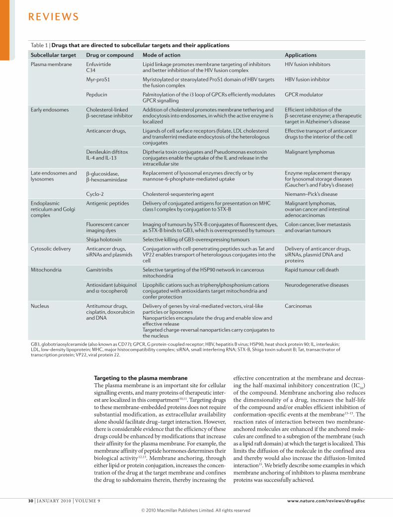
Table 1 | Drugs that are directed to subcellular targets and their applications
subcellular target Drug or compound Mode of action Applications
Plasma membrane Enfuvirtide C34
Lipid linkage promotes membrane targeting of inhibitors and better inhibition of the HIV fusion complex
HIV fusion inhibitors
Myr-proS1 Myristoylated or stearoylated ProS1 domain of HBV targets the fusion complex
HBV fusion inhibitor
Pepducin Palmitoylation of the i3 loop of GPCRs efficiently modulates GPCR signalling
GPCR modulator
Early endosomes Cholesterol-linked β-secretase inhibitor
Addition of cholesterol promotes membrane tethering and endocytosis into endosomes, in which the active enzyme is localized
Efficient inhibition of the β-secretase enzyme; a therapeutic target in Alzheimer’s disease
Anticancer drugs, Ligands of cell surface receptors (folate, LDL cholesterol and transferrin) mediate endocytosis of the heterologous conjugates
Effective transport of anticancer drugs to the interior of the cell
Denileukin diftitox IL-4 and IL-13
Diptheria toxin conjugates and Pseudomonas exotoxin conjugates enable the uptake of the IL and release in the intracellular site
Malignant lymphomas
Late endosomes and lysosomes
β-glucosidase, β-hexosaminidase
Replacement of lysosomal enzymes directly or by mannose-6-phosphate-mediated uptake
Enzyme replacement therapy for lysosomal storage diseases (Gaucher’s and Fabry’s disease)
Antigenic peptides Delivery of conjugated antigens for presentation on MHC class I complex by conjugation to STX-B
Malignant lymphomas, ovarian cancer and intestinal adenocarcinomas
Fluorescent cancer imaging dyes
Imaging of tumours by STX-B conjugates of fluorescent dyes, as STX-B binds to GB3, which is overexpressed by tumours
Colon cancer, liver metastasis and ovarian tumours
Shiga holotoxin Selective killing of GB3-overexpressing tumours
Cytosolic delivery Anticancer drugs, siRNAs and plasmids
Conjugation with cell-penetrating peptides such as Tat and VP22 enables transport of heterologous conjugates into the cell
Delivery of anticancer drugs, siRNAs, plasmid DNA and proteins
Mitochondria Gamitrinibs Selective targeting of the HSP90 network in cancerous mitochondria
Rapid tumour cell death
Antioxidant (ubiquinol and a-tocopherol)
Lipophilic cations such as triphenylphosphonium cations conjugated with antioxidants target mitochondria and confer protection
Neurodegenerative diseases
Nucleus Antitumour drugs, cisplatin, doxorubicin and DNA
Delivery of genes by viral-mediated vectors, viral-like particles or liposomes Nanoparticles encapsulate the drug and enable slow and effective release Targeted charge-reversal nanoparticles carry conjugates to the nucleus
Carcinomas
GB3, globotriaosylceramide (also known as CD77); GPCR, G protein-coupled receptor; HBV, hepatitis B virus; HSP90, heat shock protein 90; IL, interleukin; LDL, low-density lipoprotein; MHC, major histocompatibility complex; siRNA, small interfering RNA; STX-B, Shiga toxin subunit B; Tat, transactivator of transcription protein; VP22, viral protein 22.
R E V I E W S
30 | jANUARy 2010 | VOLUmE 9 www.nature.com/reviews/drugdisc
EndosomeA membrane-bound vesicle that is formed by the invagination of the plasma membrane during endocytosis.
Clathrin triskelionA clathrin structure that consists of three heavy chains and three light chains that weave together to form three ‘legs’ radiating from a central point. The heavy chains form the backbone whereas the light chains are involved in the formation of clathrin lattices.
Targeting HIV by membrane-anchored inhibitors. HIV1 hijacks raft domains at the plasma membrane to enter T cells. The envelope protein of HIV1 is composed of two proteins, glycoprotein 120 (gp120) and gp41, which are essential for virus fusion with either the host cell plasma membrane or endosomes. Enfuvirtide (Fuzeon/T20; Roche/Trimeris), a 36mer synthetic peptide fusion inhibitor derived from the gp41 region overlapping with its carboxyterminal heptad region16,17, inhibited the conformational change that is essential for HIV fusion and thereby inhibited HIV entry in vivo. However, large amounts of the inhibitor are needed to bring about inhibition. membrane anchoring of enfuvirtide peptides by conjugation to a transmembrane domain of the human lowaffinity nerve growth factor inhibited HIV infection with 100fold higher efficiency than the soluble counterpart18. Apart from the reduction in dimensionality of the peptide, membraneanchored enfuvirtide interacted preferentially with the prefusion conformation of gp41, further increasing efficiency19. This example provides a proof of principle for increasing drug efficacy by adding a membranetargeting tag an inhibitor of virus entry.
membrane anchoring of a different HIV fusion inhibitor with a lipid anchor (C34Chol) also demonstrated enhanced potency20. Cholesterol anchoring facilitated specific targeting to raft domains, where HIV fusion occurred, demonstrating the therapeutic power of reducing the compound’s dimensionality by membrane anchoring and raft targeting. Although HIV fusion is presumed to occur at the plasma membrane, a new study using highresolution time lapse imaging suggests that the fusion of HIV occurs in the endosomes21. Lipidlinked inhibitors can act at the plasma membrane but they can also be endocytosed and thus inhibit endosomeassociated processes22,23.
Inhibition of hepatitis B virus (HBV) entry. Similar to inhibition of HIV entry, lipidated membraneanchored peptides derived from surface glycoproteins exhibited inhibitory effects against HBV infection24. The envelope of HBV consists of large, middle and small proteins. Peptides derived from the proS1 domain of these proteins, when myristoylated or stearoylated, inhibited HBV entry efficiently. Acylation also increased their membrane partitioning and intracellular staining25. when the acylation of the peptides was modified to stearoylation, the inhibitory activity improved dramatically and was observed at picomolar concentrations. Lipidated preS1 peptides efficiently inhibited HBV infection in vivo.
Modulation of G protein-coupled receptor (GPCR) signalling. Owing to their involvement in many disease processes, GPCRs are important drug targets26. Ligandinduced conformational changes of transmembrane domain 3 (Tm3) and Tm6 of GPCRs initiate downstream signalling, and the third intracellular loop (i3 loop) is essential for the coupling between the receptor and the G protein27. Soluble peptides corresponding to the loop regions have been shown in vitro to modulate G protein activation but failed to act in vivo, perhaps owing to their
inability to access the cytosolic side of the membraneassociated GPCR. Attaching a palmitate group to the i3 peptide sequence enabled the modified peptide (termed pepducin) to cross lipid bilayers and efficiently inhibit receptor activation28. The nonpalmitoylated peptide neither crossed the membrane nor inhibited GPCR signalling. Such pepducins have resulted in the production of potent prophylactics against thrombotic complications associated with stroke29. Although different GPCRs differ in function and protein sequences, their mechanism of activation is highly conserved. Therefore, targeting the i3 loop of any GPCR with the corresponding pepducins might be a general strategy for inhibiting GPCR signalling.
Intracellular membrane traffickingmany drug targets are localized to intracellular compartments such as the cytosol, endosomes, lysosomes, the Golgi complex, the endoplasmic reticulum (ER), mitochondria and the nucleus6,30,31. In certain cases, the drug target is distributed in several cellular compartments but resides in an active conformation in only one compartment32. Targeting drugs to these locations is a challenge, but the normal cellular machinery is able to overcome such challenges by making use of sorting signals33.
An increasing number of endocytic pathways are being defined (FIG. 1). This heterogeneity in endocytic mechanisms ensures that different cargoes are internalized to their specific locations and are subjected to different interactions during this process34. we briefly review these various routes to gain a better understanding of their potential for drug targeting.
Clathrin-mediated endocytosis. This endocytic route engages mainly receptor–ligand complexes35 (FIG. 2). Upon binding of the nutrient or ligand to its cognate receptor, sorting motifs in the cytoplasmic tail of the transmembrane receptors engage adaptor proteins, such as adaptor protein 2, which interact with clathrin triskelions to initiate the formation of clathrincoated pits36,37. The invaginated pits are released into the cytoplasm as vesicles, aided by a small GTPase called dynamin that facilitates the fission process36. After delivery to early (or sorting) endosomes, the endocytosed cargo is recycled, sorted for degradation or delivered to the Golgi complex (FIG. 2).
Following the release of ligands from internalized receptor–ligand complexes in early endosomes, proteins such as transferrin receptors are recycled to the plasma membrane by one of two distinct recycling routes. One route involves RAB4 in direct sorting of the cargo from the endosomes to the plasma membrane38. By contrast, RAB11 regulates the sorting of the cargo from early endosomes through the perinuclear recycling compartment (FIG. 2). Control of recycling versus degradation is crucial as deregulation of this process could lead to cancer39. Contrary to the previously accepted tenet that endocytosis downregulates signalling, recent work suggests that endosomes contain signallingactive molecules that ultimately lead to nuclear signalling40–42 and encourages investigation into targeted inhibition in these endosomes43 for therapeutic purposes. For example, effective signalling from
R E V I E W S
NATURE REVIEwS | Drug Discovery VOLUmE 9 | jANUARy 2010 | 31
ProdrugA drug that is designed to release the active moiety only upon certain activating conditions.
Transition state inhibitorInhibitors that are designed to mimic the transition state of a substrate molecule in the enzyme–substrate catalytic reaction. Such inhibitors do not undergo catalysis and inhibit the enzyme at the substrate-binding site.
epidermal growth factor to the nucleus has been found to be mediated by a subset of APPL1 (adapter protein containing PH domain, PTB domain and leucine zipper motif 1)positive early endosomes44. Similarly, sustained nuclear signalling by signal transducer and activator of transcription 3 requires the trafficking of mET from the plasma membrane to the perinuclear endosomal compartment45. SARA (Smad anchor for receptor activation) endosomes engaged in transforming growth factorβ signalling are crucial for morphogen gradient formation and asymmetrical cell division46,47. In the case of the δopioid receptors, signalling molecules such as βadrenergic receptor kinase 1 (also known as GRK2) have been shown to associate with the receptor in endosomes only after
endocytosis48. Targeted inhibition of these endosomes43 could be an effective strategy for the treatment of cancer and related diseases.
Raft-mediated endocytic routes. many proteins, lipids, viruses49,50 and toxins are internalized by routes that are nonclathrin mediated51,52. Cholesterol plays a crucial part in these modes of internalization, whereas dynamin is essential for only some cargoes. Cholera toxin, viruses, bacterial toxins and plant toxins are internalized by this route to the ER. Internalization of interleukin2 receptor subunitβ is regulated by dynamin, rasrelated C3 botulinum toxin substrate 1 (RAC1), PAK1, PAK2 and RhoA GTPases53,54. By contrast, glycophosphatidylinositol (GPI)anchored proteins are internalized by a raftdependent pinocytic route termed the GEEC (GPIanchored proteinenriched early endosomal compartment) pathway55. This route is strictly dependent on cell division cycle 42 (CDC42) but is dynaminindependent and bypasses the sorting to early endosomes by using long invaginations from the surface (in contrast to the small vesicular endocytic carriers of the caveolar and clathrin pathways)56 (FIG. 1).
In some cases, there is no clear distinction between the clathrin and raftmediated internalization routes, and there seems to be some interplay between these two routes. Tetanus toxin and amyloid precursor protein require a cholesteroldependent preclustering process before they can be internalized through the clathrindependent route57,58 (FIG. 1). This heterogeneity in the endosomal system allows for specific targeting of drugs to various compartments.
Targeting to intracellular compartmentsTargeting to early endosomes. Early endosomes, which have lower pH than the extracellular environment, serve as sorting stations for endocytosed proteins. The unique pH environment of the endosomes regulates the activity of endosomespecific enzymes and is used by pHdependent poreforming toxins to disrupt the endosomal membrane59,60. The low pH of the endosomal lumen also allows the design of pHdependent prodrugs61 against therapeutic targets in diseases such as cancer and Alzheimer’s disease.
The importance of early endosomal targeting was demonstrated in the case of βsecretase23, the ratelimiting enzyme in the formation of the neurotoxic amyloidβ peptide62. βsecretase contains a cytoplasmic sorting motif that directs the enzyme into early endosomes. Although present at the cell surface, the enzyme is not active until internalized into early endosomes, in which the endosomal pH (~6) is optimal for its activity32. Therefore, most transition state inhibitors against βsecretase that are active in cellfree assays but do not penetrate into the endosomal lumen fail to act in cells and in vivo. moreover, βsecretase partitions into raft domains, which seems to be essential for its catalytic activity. Both endosomal localization and raft partitioning modulate enzymatic activity63. As a proof of principle, effective βsecretase inhibitors that exploit both of these properties have been constructed32,63 (FIG. 3).
Box 1 | Lipid rafts in pathogenesis
Lipid rafts are dynamic nano-assemblies in cell membranes that are enriched in cholesterol and sphingolipids7. Physicochemical differences in membrane lipids produce lateral heterogeneity in the membrane. The formation of these domains and the partitioning of proteins into them are dynamic processes144, which makes direct visualization of these domains by conventional microscopic means difficult. However, higher-order cross-linking of either the raft lipids or proteins leads to stable raft clustering and the formation of domains that are easier to visualize7. Such a clustering process modifies the properties of raft domains, leading to the budding of these domains. This raft-induced budding is often used by viruses and pathogens to gain entry into the cell by triggering clustering of raft components145. In some cases, budding aids the release of the virus from the cell146.
Pathogens use lipid and protein components in the rafts as receptors for gaining entry into the cell6. The protective antigen of anthrax toxin binds to cellular glyco-phosphatidylinositol-anchored proteins to enter the cell, whereas cholera and Shiga-like toxins use gangliosides6. HIV uses CD4 and chemokine receptors as entry receptors, both of which are partitioned in lipid rafts147. Host cell rafts assist HIV entry and also the assembly and subsequent release of the virus148. This results in viral envelopes that are enriched in raft lipids145. Therapies are aimed at multiple targets to inhibit raft formation or assembly as a means to inhibit HIV infection149. Raft-targeting of HIV entry inhibitors by cholesterol modification of fusion inhibitors shows promise20. Ectopic application of raft-disrupting agents (disrafters) has been successfully used in preventing vaginal HIV infection in transgenic mice150. Disrafters could be used in microbicide gels151. Identifying disrafters that are specific for rafts characterized by particular proteins poses a challenge, but these findings encourage further exploration152.
In the case of influenza, the virus uses raft lipids to release itself from the cell146,153. The envelope components associate with raft lipids after biosynthesis and become apically sorted in epithelial cells. Once these proteins reach the apical surface, the host raft lipids provide a platform for virus budding to occur. The processes that involve raft lipids are potential targets for raft-directed inhibition146,153,154.
Parasites such as Plasmodium use host raft domains in the erythrocyte for their entry155. Therapies based on raft targeting are being investigated for infectious diseases caused by protozoans156,157.
Amyloidogenic peptides also rely on raft domains for pathogenicity. In the case of the production of β-amyloid peptide, the amyloidogenic secretases cleave amyloid precursor protein (APP) in raft domains to release the peptide63,158. The conversion of the cellular prion protein to the scrapie version occurs in detergent-resistant domains159. Internalization of APP and cellular prion protein, which plays a crucial part in the production of the amyloidogenic peptides, has been shown to occur in a unique clathrin- and raft-dependent endocytic route. Additionally, both prions and β-amyloid peptides use raft domains for their oligomerization159. In the case of mast cells, signalling mediated by F
Cε receptor 1 occurs in lipid rafts160. Upon binding the
immunoglobulin E–antigen complex, the receptor undergoes oligomerization in rafts and mediates signalling from these domains. Raft partitioning of the receptor is crucial in regulating and integrating signal progression and for the subsequent degranulation160. Treatment with disrafting molecules, such as small-chain ceramides, inhibits mast cell degranulation161.
R E V I E W S
32 | jANUARy 2010 | VOLUmE 9 www.nature.com/reviews/drugdisc
Tetanus toxin,anthrax toxin,PRPC, IL-2receptor and APP
Mycobacteriumleprae, Mycobacterium tuberculae and Listeriamonocytogenes
Cholera toxin, GPI-anchoredproteins andSV40
Cholera toxin, ricin, GPI-anchoredproteins and MHC class I
Golgi complexEarly endosomesLate endosomes
PtdIns(3,5)P2
Intraluminalvesicles
PtdIns(3,4,5)P3
BioavailabilityThe extent to and rate at which the drug enters the systemic circulation.
Cholesterol linking led to membrane anchoring and enabled the inhibitor to be localized to raft domains, which are enriched for βsecretase. This modification reduced βsecretase activity more efficiently than its soluble counterpart in vitro and in vivo. The success of this strategy encourages further examination of lipid modifications to optimize drug delivery. However, these studies20,23,25 were proofofprinciple studies, and several issues need to be addressed before proceeding to clinical studies. Lipid modifications could cause the drugs to be nonspecifically adsorbed onto the membranes at the injection site and might therefore reduce the bioavailability of the compound. Cholesterolmodified drugs might be trafficked to the liver for detoxification and also affect the bioavailability of the drug. One other issue with targeted compounds is their stability. Future work should address these issues.
Several anticancer drugs have been targeted to endosomes by conjugating drugs to ligands that are internalized to endosomes through receptormediated
interactions. Ligands such as transferrin70,64,65, folate66 and lowdensity lipoproteins67 have been exploited for drug targeting, as these ligands show high affinity for their cognate receptors. As these receptors are often overexpressed in malignant tumours, conjugation of drugs or dyes to the ligands of these receptors aids specific tumour targeting for therapy or imaging68. However, the level of overexpression that is required to see differences between the tumour and normal tissue remains unclear.
Ironloaded transferrin binds to the transferrin receptor (TFR) — a transmembrane protein that is present in almost all cells — and is sorted to early endosomes, in which the ligand is released from its receptor. Release enables the delivery of transferrin (or transferrinconjugated drugs) to the early endosomal lumen and sorting of the receptor to recycling compartments. Antitumour drugs such as doxorubicin and cisplatin have been coupled to transferrin and showed more cytotoxic potency than unconjugated drugs69,70.
Figure 1 | Heterogeneity of internalization pathways. Cargoes such as transferrin (Tf) and low-density lipoprotein (LDL) are internalized through pits coated with clathrin (depicted as orange strips) that are pinched off into the cell employing the GTPase dynamin (shown as pink circles). Other cargoes such as anthrax toxin, cellular prion protein (PRPC), amyloid precursor protein (APP) and tetanus toxin are internalized through clathrin-coated pits but require cholesterol (shown as blue bars) for their endocytosis. Cholera toxin, glycophosphatidylinositol (GPI)-anchored proteins and some viruses are internalized through raft-mediated endocytic pathways. Larger particles, bacteria and viruses are internalized through phagocytic or macropinocytic pathways. Phagocytic internalization occurs with the stimulation of phagocytic receptors, which leads to the reorganization of membrane in an actin-dependent manner to form pseudopods. The pseudopods extend to engulf the particulate matter. Engulfed material in phagosomes undergoes fusion with lysosomes, a process that is inhibited by several intracellular pathogens. Cholera toxin and GPI-anchored proteins could also be internalized through ADP-ribosylation factor 6 (ARF6)-mediated GPI-anchored protein-enriched early endosomal compartment (GEEC) pathways that use long invaginations for transporting the cargoes into the cell. IL-2, interleukin-2; MHC, major histocompatibility complex; PtdIns(3,5)P
The folate receptor is also of interest for cancer therapy as it is upregulated in many epithelial cancers68,71. Folate receptors are GPIanchored and internalized by a nonclathrin, nondynamin, CDC42dependent raft pathway56. Folate (and folate analogues such as petorate) have high receptor affinity (dissociation constant in the range 10–10 m), which enables efficient binding of folate conjugates to the receptors66. Folate receptortargeted delivery of liposomal doxorubicin to folate receptorexpressing cells increased the cellular uptake and cytotoxicity of doxorubicin72. Polyethylene glycol–folate conjugates of thioctic acid carrying gold nanoparticles were used against ovarian cancer cells and were also faithfully targeted to the endosomal compartment73. Other ligands such as vitamin B12 and low density lipoproteinlike nanoparticles have also been used as drug conjugates67. In some cases, conjugates directed to TFR are shown to be not only targeted to endosomes but also imported into the brain. These results suggest that TFRs could be used to deliver drugs targeting the central nervous system74–76. Their broader use remains to be demonstrated.
Targeting to late endosomes and lysosomes. Lysosomes contain ~40 different hydrolytic enzymes that mediate controlled intracellular degradation of macromolecules. These organelles are of particular interest for the design and delivery of pHdependent prodrugs and for devising strategies to inhibit degradation in these compartments.
Genetic deficiency in lysosomal hydrolases or proteins involved in the efflux of metabolites causes lysosomal storage diseases77 in which accumulation of undigested metabolites often results in neurological symptoms. These diseases also arise owing to mutations that lead to defective localization or trafficking of lysosomal hydrolases to lysosomes from the ER or Golgi complex31. In many cases, enzyme replacement therapy remains the most successful and viable option for patients with lysosomal storage diseases78,79. These enzymes are normally targeted to lysosomes but in some cases the expression of certain surface receptors, such as the mannose6phosphate receptors80, has been found to upregulate the delivery of the exogenous enzyme to macrophages in patients with Gaucher’s disease. Dexamethasone, which is known to upregulate mannose6phosphate receptors, can be administered with the recombinant enzyme to enhance the uptake of the enzyme in macrophages81.
Niemann–Pick disease type A and Niemann–Pick disease type B are caused by defective sphingomyelinase activity in lysosomes, whereas Niemann–Pick disease type C is caused by the loss of the polytopic transporter Niemann–Pick C1 protein that leads to accumulation of cholesterol in late endosomes and lysosomes82. Defective cholesterol trafficking has been implicated in Niemann–Pick’s disease and a single injection of the cholesterolsequestering agent 2hydroxypropylβcyclodextrin (also known as cyclo) ameliorated the severity of the disease in a mouse model83.
Drug targeting to the ER and Golgi complex. Various clathrinindependent pathways traffic toxins such as Shiga or cholera toxins into the Golgi and ER
Figure 2 | clathrin-mediated endocytic pathway. Proteins that are internalized through pathways mediated by clathrin (depicted as orange strips) and dynamin (shown as pink circles) are first sorted to early endosomes, which are characterized by the presence of the Rab GTPase RAB5. RAB5 participates in the fusion of early endosomes and the switch between RAB5 and RAB7 mediates the conversion of these endosomes to late endosomes. It is thought that, in many cases, an intermediate organelle called the multivesicular body (MVB; also known as the endosomal carrier vesicle) mediates the cargo transfer. Recent work has shown that ceramide is involved in the biogenesis of MVBs, which distinguishes these vesicles from late endosomes. Lipids and proteins sorted to the intraluminal vesicles (shown as blue circles) of MVBs can be released into the extracellular space by fusion of the MVBs with the plasma membrane as exosomes. Exosomes are implicated in various processes including antigen presentation, signalling and release of pathogenic peptides, such as prions and β-amyloid pepides. These pathogenic processes could be inhibited by specifically directing the inhibitors to the exosome-specific MVBs. Proteins destined for degradation, such as the epidermal growth factor receptors, are sorted from late endosomes to lysosomes, although the exact sorting mechanism is unclear. The low pH of lysosomes facilitates the activation of enzymes that are responsible for cargo degradation; this is a key consideration for drug design and delivery of pH-sensitive molecules. Proteins and lipids can also be directly recycled back from early endosomes to the plasma membrane by RAB4 GTPase. Recycling of some proteins to the cell surface occurs through recycling endosomes, marked by RAB11. Cargo transfer from endosomes to the Golgi complex is carried out by Rab proteins, such as RAB9 in the case of late endosomes. Intracellular organelles are also marked by the presence of unique phosphoinositide phosphates — namely, phosphatidylinositol-4,5-bisphosphate (PtdIns(3,5)P
2) and phosphatidylinositol-3,4,5-trisphosphate
(PtdIns(3,4,5)P3) (shown as coloured arcs on the endosomes).
R E V I E W S
34 | jANUARy 2010 | VOLUmE 9 www.nature.com/reviews/drugdisc
Small interfering RNA(siRNA). Small stretches of RNA, usually 21–25 nucleotides long, that bind to mRNA and target it for degradation, thereby silencing gene expression.
compartments51,84 (FIG. 4). Conjugating drugs to such toxins would therefore target them to the ER and eventually to the cytosol (discussed below). As peptide loading onto major histocompatibility complex I (mHC I) during antigen presentation occurs in the ER, peptides conjugated to Shiga toxin reach the ER through retrograde transport. Shiga toxin conjugates were also efficiently used for crosspresentation by mHC I and mHC II in dendritic cells84, thereby conferring increased immunity against viral antigens. Interestingly, the receptor glyco lipid for Shiga toxin B, globotriaosylceramide (GB3; also known as CD77), is overexpressed in many tumours. This makes GB3 a biomarker for detection of these carci nomas and presents an opportunity to target toxin conjugates of anticancer drugs and tracers used in tumour imaging to the tumour85,86. Topoisomerase inhibitors have successfully reached the ER when conjugated to Shiga toxin B subunit. The same strategy has been used to deliver the imino sugar Nbutyldeoxynojiromycin to the ER, in which it inhibited the Nglycosylation of tyrosinase in melanoma cells87.
Targeting to the cytosolThe cytoplasm hosts various metabolic, signalling and pathogenic processes that are targets for several diseases. Viruses deliver their genome to the cytosol by fusion of their envelopes at the plasma membrane or in endosomes. Posttranscriptional control mechanisms, such as those mediated by microRNA and small interfering RNA (siRNA), occur in the cytoplasm, and therapeutic siRNAs
have to be targeted to this subcellular compartment (BOX 2). To target drugs to the cytosol and avoid lysosomal degradation, several strategies have been devised. These involve the use of cellpenetrating peptides (CPP) that permeate through the plasma membrane bilayer and release the drug directly into the cytosol; pHresponsive carriers that dispense the drug into the cytosol from the endosomes; and endosomedisrupting agents that aid in the release of drugs into the cytosol (FIG. 4).
CPPs that act directly at the plasma membrane. Direct transport of cargo across the plasma membrane can be achieved by conjugating molecules to CPPs88. The discovery that the HIV transactivator of transcription (Tat) protein can traverse cellular membranes and gain access to the nucleus stimulated considerable interest in exploiting this molecule for targeted drug delivery89. Several other proteins, including the transcription factor Antennapedia and the VP22 protein from herpes simplex virus90, were also found to penetrate membranes. The peptidic regions responsible for cell penetration are either amphipathic (model amphipathic peptides or transportan) or argininecontaining stretches (Tat, VP22 or penetratin), usually ~30 amino acids long88. It is not clear how these peptides (and their conjugates of diverse chemical nature) cross the membrane at neutral pH, although interaction of cationic peptides with membrane phospholipids is thought to trigger a conformational change that allows their insertion into the membrane91. The mechanisms underlying the subsequent translocation through the lipophilic bilayers are also unclear. Emerging studies suggest that, although some peptides can traverse the membrane through lipid interactions, conjugates of CPPs are mainly internalized by endocytosis92–94. The uptake of labelled polyarginine occurred at spatially restricted membrane domains involving clathrin and dynamin95. In agreement with these findings, membrane anchoring of similar peptides through stearoylation or cholesterol modification increased the efficiency of cytosolic delivery by shifting the mode of internalization to endocytosis95,96. As CPP conjugates are internalized through endosomes and not directly to the cytosol, pHsensitive drugs are likely to lose activity before reaching the cytosol. The direct translocation of the CPPs from the plasma membrane to the cytosol is evidently lost after conjugation and, despite enormous efforts, this delivery strategy is only successful in exceptional cases.
Peptide-mediated escape from early endosomes and the ER–Golgi network. To release the contents of endosomes into the cytosol, two strategies could be envisioned: fusion of a membraneencapsulated particle or enveloped virus with the endosomal membrane, enabling the contents to be emptied into the cytosol (fusogenic mechanism); or disruption the endosomal membrane by an internalized particle, causing the endosome to discharge its toxic components into the cytosol (endoosmolytic mechanism)97. Viruses such as influenza use fusion mechanisms employing haemagglutinin protein. Under acidic pH conditions, haemagglutinin acquires
Figure 3 | endosomal targeting of β-secretase inhibitors. β-secretase (shown as orange bars) is sorted from the plasma membrane to the early endosomes, in which it acquires an active conformation through endocytosis mediated by lipid rafts (shown as orange blocks) and clathrin (shown as orange strips). Activity of the enzyme is also dependent on its localization to lipid rafts. In early endosomal rafts, β-secretase cleaves amyloid precursor protein (APP), which initiates the production of the toxic β-amyloid peptide. As active β-secretase (when in an open conformation) is found only in early endosomes, transition state inhibitors that are not membrane permeant (shown as purple circles) are unable to gain access to these endosomes (a). Cholesterol linking enables membrane anchoring of the inhibitors (purple circles attached to the membrane), and subsequent membrane trafficking transports them to the endosomes to inhibit the endosomal raft-bound β-secretase (b).
R E V I E W S
NATURE REVIEwS | Drug Discovery VOLUmE 9 | jANUARy 2010 | 35
a conformation that is able to insert into and fuse with the endosomal membrane, allowing the viral nucleocapsid to be released in the cytosol98. The aminoterminal region of the haemagglutinin protein of influenza contains a fusogenic peptide sequence, and several synthetic peptides have been designed to mimic the properties of these fusogenic sequences, including peptides containing GALA99 or KALA sequences, which are used in the cytosolic delivery of conjugated drugs. In contrast to the fusogenic influenza mechanisms, adenoviruses and bacterial toxins use the endosomal membrane disruption mechanism100. Toxins such as diphtheria toxin or anthrax toxin are internalized into endosomes and
undergo a conformational change that allows them to insert into the limiting membrane of early endosomes and escape the endosomes by rupturing the endosomal membrane98. Interestingly, recent evidence challenges these views and presents an alternative model of a fusion and release mechanism. In the case of anthrax toxin, the protective antigen is not directly released into the cytosol from endosomes, but is first delivered into the lumen of the intraluminal vesicles of the endosomes. Fusion of these intraluminal vesicles with the limiting membrane of the endosomes releases the internalized toxin into the cytosol (a process termed ‘backfusion’)101 (FIG. 4). This mechanism is proposed to further protect the toxin from
Figure 4 | strategies to target drugs to different cellular compartments. Drugs conjugated to toxins such as diphtheria toxin are internalized through endocytosis mediated by clathrin (shown as orange strips) and are targeted to early endosomes. From here, the conjugated toxins are released into the cytosol through endosomal disruption. For some cargoes, such as anthrax or vesicular stomatitis virus (VSV), the release into the cytosol occurs through back-fusion of the intraluminal vesicles with the limiting membrane of the endosomes. Such release protects the conjugates or the toxins from the harsh degradative conditions of the endosomal lumen. Adenoviruses escape from endosomes at either the early or late endosomal stages, depending on the virus subtype. Once released into the cytosol, they are transported to the nucleus by motor-driven transport. Early endosomal transport of drug conjugates could be achieved by anchoring the drugs to cholesterol (shown as green circles). Cholesterol anchoring would target the drugs to lipid rafts (shown as blue bars) in the membrane. Cell-permeant peptides could directly traverse the plasma membrane and therefore localize to the cytosol. Gamitrinibs that contain cyclic guanidinium moieties target mitochondria whereas peptoids localize to the cytosol and nucleus. Cholera or Shiga toxins are sorted from the plasma membrane to the Golgi complex through the pentameric B subunit of the toxin. From there, the toxins are retrogradely sorted to the endoplasmic reticulum, in which the A subunit of the toxin is released into the cytosol. Drugs are conjugated to the B subunits of the toxins. PtdIns(3,4,5)P
3,
phosphatidylinositol-3,4,5-trisphosphate.
Nature Reviews | Drug Discovery
Earlyendosomes
Lateendosomes
Golgicomplex
Drug-conjugated cholera orShiga toxins
Diphtheria toxin, influenza, anthrax toxin and VSV
Viruses
Endoplasmic reticulum
Nucleus
Membrane anchoring
Cholesterol-modified drug
Lipd rafts Cell
permeablepeptides
Anthraxtoxin and VSV
Adenovirus
Peptoids
Gaminitribs
Diptheria toxinHaemagglutinin
A subunitB subunit
PtdIns(3,4,5)P3
R E V I E W S
36 | jANUARy 2010 | VOLUmE 9 www.nature.com/reviews/drugdisc
Singlet oxygenA form of molecular oxygen that is a reactive oxygen species and less stable than the normal triplet oxygen.
the harsh conditions in multivesicular bodies (mVBs), and a similar mechanism has been shown to mediate endosomal release of the nucleocapsid of vesicular stoma titis virus102. Polycationic polymers, such as polyhydroxyethyl aspartamide conjugated to histidine, also have pHdependent endoosmolytic properties. Such artificial polymers have been used to target doxorubicin to the nucleus by first releasing it into the cytosol from endosomes103 for targeted cancer therapy.
Proteins can also be released to the cytoplasm from the ER. As part of the cell’s ‘quality control’ checks, misfolded proteins are exported from the ER to the cytosol for degradation. This process is exploited by toxins such as cholera toxin to target their toxic A subunit to the cytosol. Immunotoxins or toxin conjugates are also used to traffic molecules to the cytosol84,104 (FIG. 4).
membrane anchoring of pHsensitive probes has recently been shown to create novel prodrugs that increase the efficiency of cytosolic release105. membrane anchoring of a pHdependent lytic dodecapeptide and a fluorophore (5carboxyfluorescein) can be achieved by linkage to sterols. In the case of the fluorophore, a disulphidecontaining linker was used. The internalized fluorophore was confined to the endosomes as the endosomal lumen is predominantly in the oxidative state. However, following coincubation with the cholesterolanchored pHdependent lytic peptide, the lytic peptide disrupted the endosomal membrane, allowing leakage of glutathione from the cytosol into endosomes. This influx reduced the disulphide bond in the cholesterylanchored fluorophore, enabling the release of the fluorophore into the cytosol. Inhibition of endosomal acidification by bafilomycin inhibited the release from the endosome105. Such prodrugs could be used to evade lysosomal degradation and to increase the efficiency of cytosolic release of molecules such as siRNAs106–109.
By choosing appropriate lipid anchors and exploiting the large chemical space allowed in the linker region, organellespecific targeting of inhibitors or prodrugs can be achieved (BOX 3).
Endosomal disruption and release of the luminal contents into the cytosol can also be triggered by photochemical internalization (PCI)110. PCI is based on the lightinduced rupture of the endosomal membrane loaded with photosensitizing molecules. These photosensitizers are targeted to endosomes and, after they are internalized with the drug, photochemical illumination induces endosomal rupture through production of singlet oxygen and triggers the release of the internalized drug into the cytoplasm. However, this novel method has its limitations. The singlet oxygen that is produced could damage the conjugate itself, particularly in case of oligonucleotides, limiting the use of this technology.
Targeting to the nucleusThe nucleus contains various anticancer targets, ranging from proteins involved in DNA replication to nuclear receptors. DNA viruses have to deliver their genetic material into the host nucleus after gaining access to the cytosol, and studying virus entry has enabled the development of viralmediated gene delivery111. One of the
commonly used viruses for gene delivery is adenovirus100 which, after endocytosis and rupture of the endosome, uses motordriven translocation along microtubules to reach the nucleus. Binding of the nuclear localization signal in the viral capsid proteins to the nuclear pore is thought to mediate the entry of the virus.
However, some viruses, such as simian vacuolating virus 40 (SV40), mediate their entry into the nucleus by retrogradely trafficking from the plasma membrane to the ER followed by transfer to the nucleus112. Adenoviruses, adenoassociated viruses, retroviruses and lentiviruses have been used as delivery vehicles for gene therapy. However, owing to toxicity, immunogenicity and lack of specificity, these delivery methods are being replaced by specific viral machinery that lacks pathogenic components.
Viral and non-viral vectors for nuclear delivery. Small peptide regions from viruses that show nuclear translocation activity, such as the KKKRKV peptide derived from SV40 large T antigen, are used for the nuclear delivery of conjugated DNA. To avoid complications associated with viralmediated delivery, nonviral vectors or chemical formulations that mimic the properties of the viruses have been designed to mediate gene transfer. These molecular formulations are often composed of lipids that form vesicles encapsulating the DNA molecule, such as the DNA molecules packaged into cationic liposomes, termed lipoplexes113. Polyplexes, by contrast, contain DNA in polymeric complexes comprised of polylysine, spermine or the dendrimer polymers, such as polyethylimine97,114. These polymers are internalized by various endocytic mechanisms, and incorporation of endoosmolytic agents aid the cytosolic release of these conjugates115. Coadministration of peptides containing nuclear localization signals, covalent coupling or incorporation into lipid formulations are used to enhance nuclear delivery of conjugates. Finally, nuclear proteins such as histones and protamine that are highly enriched in basic amino acids can be used in gene delivery100.
Delivery using nanoparticles. Nanoparticles are colloidal particles that are used as alternative delivery devices to liposomes or viral vectors116–118. Nanomedicine, a new field that is rapidly progressing, has shown promise in delivering drugs, some of which have already made it to the market4,119. In nanoparticles or nanocarriers, the drug or prodrug of interest is dissolved or encapsulated in the particulate matter120. These particles, either as suspensions or particulates, enhance stability and drug dissolution rate. Encapsulating with appropriate uptake signals might enable cellular uptake and specific targeting to subcellular destinations121. whereas the smaller particulates could be taken up by fluidphase endocytosis, the larger ones might be phagocytosed and targeted to phagosomes. Another issue in the subcellular delivery of nanoparticles is the release into the cytosol. Exploiting the lower endosomal pH compared with the cytosol, pHsensitive polymers could be designed to enable the release of the drugs from the particulate material.
R E V I E W S
NATURE REVIEwS | Drug Discovery VOLUmE 9 | jANUARy 2010 | 37
Blood–brain barrierA semi-permeable cellular structure consisting of endothelial cells that allows selective passage of some molecules but prevents the passage of others.
Drugs that could be specifically targeted to mitochondrial proteins are therefore of therapeutic interest. CPPs derived from mitochondrial proteins, such as cytochrome oxidase subunits and gene associated with retinoicinterferoninduced mortality (Grim) proteins, have been shown to target heterologous proteins, peptides and small molecules specifically in mitochondria124. Lipophilic cationic conjugates of anticancer agents target mitochondria to trigger cell death in target tissues125–127. A novel class of aromatic cationic Szeto–Schiller peptides selectively partition into the inner mitochondrial membrane and confer mitoprotection through their antioxidative properties128. These peptides readily cross the blood–brain barrier and reduce the production of mitochondrial reactive oxygen species, and are showing promise for the treatment of neurodegenerative diseases. Recently, a novel class of compounds termed gamitrinibs (so called because they are geldanamycin mitochondrial matrix inhibitors) was shown to exclusively target mitochondrial heat shock protein 90 (HSP90) in tumour cells129. In mitochondria, HSP90 plays an active part in many tumours, and inhibitors of HSP90 such as geldanamycin have been shown to have antitumour properties130. As these inhibitors also show inhibitory activity to HSP90 and other heat shock proteins located in the cytosol, specific targeting of these inhibitors to mitochondria could increase efficacy and reduce toxicity. Gamitrinibs contain geldanamycin as the active molecule, linked through an amidecontaining linker to cyclic guanidinium moieties, which act as mitochondrial targeting signals. Only those containing three to four units enabled efficient intracellular uptake and mitochondrial targeting in a cellular context (FIG. 4). Surprisingly, no mitochondrial toxicity in nontumour cells was reported, making gamitrinibs attractive novel antitumour drugs130.
Compartmentalization of RNA and lipidsIn addition to proteins, lipids and RNA molecules have been shown to be localized to distinct subcellular locations131. Lipids such as phosphoinositol phospholipids (PIPs) exhibit polarized localization in the epithelial cells, and different PIPs are localized in different organelles (FIG. 2). These lipids play an important part in cell polarity, migration and in pathological conditions131–133. Sphingolipids such as ceramides are selectively recruited to raft domains in the membrane and initiate apoptotic signals9. Ceramide recruitment to early endosomes is important for the formation of intraluminal vesicles of the exosomal subset of mVBs134. whether sitespecific modulation of these lipids can be achieved by subcellular targeting approaches is yet to be experimentally demonstrated.
Spatially restricted protein synthesis occurs by recruiting mRNA to subcellular compartments135. Recent studies have shown that a large number of mRNAs localize to different subcellular sites, such as centrosomes, apical or basolateral domains, spindle poles or axon dendritic compartments136. These studies also suggest that mRNA localization is crucial for the formation of functionally distinct compartments135. Recent studies show that, in addition to mRNAs, regulatory RNAs such as micro RNAs (miRNAs) are also localized in spatially distinct sites in
Nuclear delivery through peptoids. Peptoids are a novel class of peptidomimetics that penetrate cells mainly through a backbone containing five or six glycine residues122. Two kinds of peptoids have been designed, one with amino side chains and the other with guanidinium side chains122. Both types are positively charged but are sorted differently. The aminomodified peptoid is targeted to the cytoplasm whereas the peptoid with the guanidinium side chain is trafficked to the nucleus. This indicates that the nature of the side chain determines the uptake kinetics and destination of the peptoid, potentially facilitating the design of drugs that target distinct compartments.
Drug targeting to mitochondriaDrugs reaching the cytosol need to be further targeted if their targets reside in membranebound compartments that are not accessible from the surface by endocytic routes. mitochondria serve as hotspots for targeted therapy both in host cells and in parasites. In host cells, mitochondrial proteins such as B cell lymphoma family proteins serve as anticancer targets. Inhibiting the electron transfer chain or the redox system in parasite mitochondria is a successful antimicrobial approach123. Furthermore, dysfunction in mitochondria has been observed in several neurodegenerative diseases such as Parkinson’s disease, Alzheimer’s disease and amyotrophic lateral sclerosis.
Box 2 | RNAs and lipid modifications
Cytosolic drug targeting without major losses to endolysosomes is desired when designing small interfering RNA (siRNA) conjugates for therapy109. To promote delivery, RNAs are either encapsulated in lipid vesicles or conjugated to membrane-penetrating peptides. Stable nucleic acid–lipid particles (SNALPs) or interfering nanoparticles carrying siRNAs have been designed to deliver siRNAs to target organs in vivo. For example, SNALPs containing siRNAs targeting hepatitis B virus (HBV) RNA effectively inhibit the replication of HBV162. Similarly, by coupling with N-acetyl-galactosamine and disulphide-linked endo-osmolytic polyvinylether, which facilitates hepatocyte targeting, endocytic uptake and cytoplasmic release of siRNAs through endo-osmolysis was achieved107. This technology, termed dynamic polyconjugation, is used to effectively silence the genes encoding low-density lipoprotein (LDL) and peroxisome proliferator-activated repetor-α in mouse liver. Recently, β1,3-d-glucan-encapsulated siRNA particles (GERPs) were used as efficient oral delivery vehicles for silencing genes in mouse macrophages163.
Targeting can also be achieved by covalent attachment of siRNA to cholesterol and other lipids108. Recently, siRNAs against apolipoprotein B (APOB) were modified with cholesterol and were shown to reduce APOB mRNA levels164. This reduction occurred several fold more efficiently when cholesterol–siRNAs were associated with high-density lipoprotein (HDL) particles. The resultant decrease in the levels of plasma LDL and serum cholesterol underscores the possibility of such modifications for therapeutic use. Other lipophilic conjugates such as fatty acids and bile acids have also been shown to aid the delivery of siRNAs. siRNAs conjugated with long-chain fatty acids such as stearoyl (C18) and docosanyl (C22) silenced the APOB gene more efficiently than their shorter counterparts such as lauroyl (C12) and myristoyl (C14) chains. Higher lipophilicity, which is conferred by the longer saturated chain lengths, seems to determine the affinity of these conjugates for HDL and subsequent efficiency of gene silencing164. As different lipid anchors partition into different lipoproteins, this property could also be exploited to transport conjugates to different target organs. Despite these advances, this promising drug strategy requires improved tissue delivery in vivo to become more generally applicable.
R E V I E W S
38 | jANUARy 2010 | VOLUmE 9 www.nature.com/reviews/drugdisc
Box 3 | Designing tripartite inhibitors for subcellular targeting
Covalent linkage of lipids to drugs could be achieved in several ways depending on the nature of the drug and the lipid. Modification should preserve drug function and should not affect the partitioning and trafficking of the lipid. Drugs are often conjugated to a lipid through a linker, giving rise to ‘tripartite’ molecules (see the figure).
The pharmacophore (the ‘message’)The main part in the tripartite molecule is the active ingredient (the drug) against a target molecule that is localized in a particular subcellular space. The pharmacophore could also be designed as a prodrug that exposes the active moiety in the appropriate environment. The pharmacophore could include drugs in combination with imaging dyes or moieties that couple the pharmacophore to the linker.
The lipid anchor (the ‘address’)The lipid tails anchored to the drug determine their sorting in subcellular compartments. Lipids differ in their membrane-partioning properties and in their sorting behaviour165. For example, cholesterol is more readily taken up in biological membranes than long-chain fatty acids. However, it is trafficked mainly through lysosomes, in which it can be degraded, affecting the stability of the linked molecule. By carefully choosing various lipids as anchors, it could be possible to sort the conjugates to different subcellular and submembrane compartments such as lipid rafts34,166. As lipid-raft domains contain several drug targets, lipids that are enriched in these regions (such as cholesterol and sphingolipids) could be used to target drugs to these domains. Cholesterol and sterol analogues such as ergosterol, stigmasterol, dihydrocholesterol, sphingolipids, gangliosides, globosides, ceramides and sulphatides could be used as lipid anchors167,168. For targeting drugs to other submembrane compartments, phospholipids, unsaturated fatty acids and glycerophospholipids could be used. The fatty-acid composition and chain length of globotriaosylceramide (GB3), the receptor glycolipid for Shiga toxin B, have been shown to regulate sorting of the toxin to the Golgi complex34. Particular fatty-acyl moieties have also been reported to mediate the retrograde trafficking of two classes of verotoxins169,170. Sphingomyelins that vary in their fatty-acyl chain lengths are sorted to different compartments in the cell: long-chain, ordered-domain-preferring (or raft-partitioning) sphingolipids are targeted to late endosomal compartments in a cholesterol-dependent manner, whereas short-chain, disordered-domain-preferring lipids are recycled more effectively independently of cholesterol levels171. These findings highlight the importance of selecting the appropriate lipid chains to modulate the kinetics of internalization and directing of drugs to various subcellular destinations.
Important considerations when selecting a lipid anchor include: a high level of partitioning into biological membranes; high micellar concentrations; easy transport into the bloodstream by lipoproteins or lipid-binding proteins; low non-specific adsorption to the tissues at the site of injection; and faithful targeting of the conjugates to the respective subcellular destinations.
The linkerA linker, which is usually an oligomer, is introduced to connect the active pharmacophore to the lipid anchor. Direct conjugation of the drug to the lipid molecule might hinder the interaction of the drug with its intended target by causing steric hindrance or by placing the drug too close to the lipid bilayer. Oligo(ethylene glycol) or oligoamides can be used as backbone structures for linkers105,172. The linker lengths could be adjusted to optimize accessibility to the target molecule. In some cases, the optimal length of the linker could be designed by analysing the drug–target interaction site and the distance of this interaction site from the bilayer. Linkers also provide space to introduce additional modifications, such as sites for enzymatic cleavage, pH-dependent cleavage moieties, disulphide bonds to modulate glutathione-dependent reduction and consequently the liberation of linked molecules. Such linkers are crucial for the design of lipid-linked prodrugs105.
Nature Reviews | Drug Discovery
Linker
Address
OO
OO n
O
NH
O
NH2
O
O
Message
Message
H2NHN
NH
HN
NH
COOH
O
O O
NH2
O
OH O
O
HN
NH
HN
NH
O
COOH
O
OAddressLinker
R E V I E W S
NATURE REVIEwS | Drug Discovery VOLUmE 9 | jANUARy 2010 | 39
1. Gershell, L. J. & Atkins, J. H. A brief history of novel drug discovery technologies. Nature Rev. Drug Discov. 2, 321–327 (2003).This article gives an excellent timeline on the development of various technologies in drug discovery.
2. Langer, R. Drug delivery. Drugs on target. Science 293, 58–59 (2001).A well written primer on the need for targeted drug delivery to reduce adverse effects.
3. Lipinski, C. A., Lombardo, F., Dominy, B. W. & Feeney, P. J. Experimental and computational approaches to estimate solubility and permeability in drug discovery and development settings. Adv. Drug Deliv. Rev. 46, 3–26 (2001).An excellent reference for determining the bioavailability of drugs.
4. Rosen, H. & Abribat, T. The rise and rise of drug delivery. Nature Rev. Drug Discov. 4, 381–385 (2005).
5. Blobel, G. Protein targeting (Nobel lecture). Chembiochem 1, 86–102 (2000).
6. Gruenberg, J. & van der Goot, F. G. Mechanisms of pathogen entry through the endosomal compartments. Nature Rev. Mol. Cell Biol. 7, 495–504 (2006).
7. Simons, K. & Ikonen, E. Functional rafts in cell membranes. Nature 387, 569–572 (1997).The first detailed description of lipid rafts in cellular membranes and the implications for cellular signalling.
8. Zerial, M. & McBride, H. Rab proteins as membrane organizers. Nature Rev. Mol. Cell Biol. 2, 107–117 (2001).An important reference on the subcellular compartmentalization and the role of Rab GTPases in membrane trafficking.
9. Grassme, H. et al. CD95 signaling via ceramide-rich membrane rafts. J. Biol. Chem. 276, 20589–20596 (2001).
10. Heldin, C. H. Dimerization of cell surface receptors in signal transduction. Cell 80, 213–223 (1995).
11. Jordan, B. A. & Devi, L. A. G-protein-coupled receptor heterodimerization modulates receptor function. Nature 399, 697–700 (1999).
12. Schwyzer, R. Membrane-assisted molecular mechanism of neurokinin receptor subtype selection. EMBO J. 6, 2255–2259 (1987).
13. Schwyzer, R. How do peptides interact with lipid membranes and how does this affect their biological activity? Braz. J. Med. Biol. Res. 25, 1077–1089 (1992).
14. Kholodenko, B. N., Brown, G. C. & Hoek, J. B. Diffusion control of protein phosphorylation in signal transduction pathways. Biochem. J. 350, 901–907 (2000).
15. Kholodenko, B. N., Hoek, J. B. & Westerhoff, H. V. Why cytoplasmic signalling proteins should be recruited to cell membranes. Trends Cell Biol. 10, 173–178 (2000).A brilliant description of the reduction-in- dimensionality principle and its implications for cellular signalling. It details the importance of the membrane association of acylated proteins upon signalling and their recruitment to lipid rafts.
16. Este, J. A. & Telenti, A. HIV entry inhibitors. Lancet 370, 81–88 (2007).
17. Reeves, J. D. & Piefer, A. J. Emerging drug targets for antiretroviral therapy. Drugs 65, 1747–1766 (2005).
18. Hildinger, M. et al. Membrane-anchored peptide inhibits human immunodeficiency virus entry. J. Virol. 75, 3038–3042 (2001).The first study to show that a membrane- anchored fusion inhibitor is superior to its soluble counterpart. The implications for the reduction-in-dimensionality principle are discussed.
19. Melikyan, G. B., Egelhofer, M. & von Laer, D. Membrane-anchored inhibitory peptides capture human immunodeficiency virus type 1 gp41 conformations that engage the target membrane prior to fusion. J. Virol. 80, 3249–3258 (2006).
20. Ingallinella, P. et al. Addition of a cholesterol group to an HIV-1 peptide fusion inhibitor dramatically increases its antiviral potency. Proc. Natl Acad. Sci. USA 106, 5801–5806 (2009).
21. Miyauchi, K., Kim, Y., Latinovic, O., Morozov, V. & Melikyan, G. B. HIV enters cells via endocytosis and dynamin-dependent fusion with endosomes. Cell 137, 433–444 (2009).
22. Boonyarattanakalin, S., Hu, J., Dykstra-Rummel, S. A., August, A. & Peterson, B. R. Endocytic delivery of vancomycin mediated by a synthetic cell surface receptor: rescue of bacterially infected mammalian cells and tissue targeting in vivo. J. Am. Chem. Soc. 129, 268–269 (2007).
23. Rajendran, L. et al. Efficient inhibition of the Alzheimer’s disease beta-secretase by membrane targeting. Science 320, 520–523 (2008).This work showed that an inactive inhibitor of β-secretase was rendered active by membrane anchoring through lipid-conjugation and subsequent endosomal targeting.
24. Gripon, P., Cannie, I. & Urban, S. Efficient inhibition of hepatitis B virus infection by acylated peptides derived from the large viral surface protein. J. Virol. 79, 1613–1622 (2005).
25. Petersen, J. et al. Prevention of hepatitis B virus infection in vivo by entry inhibitors derived from the large envelope protein. Nature Biotech. 26, 335–341 (2008).
26. George, S. R., O’Dowd, B. F. & Lee, S. P. G-protein-coupled receptor oligomerization and its potential for drug discovery. Nature Rev. Drug Discov. 1, 808–820 (2002).
27. Gether, U. Uncovering molecular mechanisms involved in activation of G protein-coupled receptors. Endocr. Rev. 21, 90–113 (2000).
28. Covic, L., Gresser, A. L., Talavera, J., Swift, S. & Kuliopulos, A. Activation and inhibition of G protein-coupled receptors by cell-penetrating membrane-tethered peptides. Proc. Natl Acad. Sci. USA 99, 643–648 (2002).
29. Covic, L., Misra, M., Badar, J., Singh, C. & Kuliopulos, A. Pepducin-based intervention of thrombin-receptor signaling and systemic platelet activation. Nature Med. 8, 1161–1165 (2002).
30. Sheff, D. Endosomes as a route for drug delivery in the real world. Adv. Drug Deliv. Rev. 56, 927–930 (2004).
31. Pagano, R. E., Puri, V., Dominguez, M. & Marks, D. L. Membrane traffic in sphingolipid storage diseases. Traffic 1, 807–815 (2000).
32. Rajendran, L. et al. Alzheimer’s disease β-amyloid peptides are released in association with exosomes. Proc. Natl Acad. Sci. USA 103, 11172–11177 (2006).
33. Sorkin, A. Cargo recognition during clathrin-mediated endocytosis: a team effort. Curr. Opin. Cell Biol. 16, 392–399 (2004).
34. Maxfield, F. R. & McGraw, T. E. Endocytic recycling. Nature Rev. Mol. Cell Biol. 5, 121–132 (2004).
35. Conner, S. D. & Schmid, S. L. Regulated portals of entry into the cell. Nature 422, 37–44 (2003).
36. Schmid, S. L. Clathrin-coated vesicle formation and protein sorting: an integrated process. Annu. Rev. Biochem. 66, 511–548 (1997).
37. Mukherjee, S., Ghosh, R. N. & Maxfield, F. R. Endocytosis. Physiol. Rev. 77, 759–803 (1997).
38. Sonnichsen, B., De Renzis, S., Nielsen, E., Rietdorf, J. & Zerial, M. Distinct membrane domains on endosomes in the recycling pathway visualized by multicolor imaging of Rab4, Rab5, and Rab11. J. Cell Biol. 149, 901–914 (2000).
39. Han, W., Zhang, T., Yu, H., Foulke, J. G. & Tang, C. K. Hypophosphorylation of residue Y1045 leads to defective downregulation of EGFRvIII. Cancer Biol. Ther. 5, 1361–1368 (2006).
40. Miaczynska, M., Pelkmans, L. & Zerial, M. Not just a sink: endosomes in control of signal transduction. Curr. Opin. Cell Biol. 16, 400–406 (2004).
41. Sorkin, A. & Von Zastrow, M. Signal transduction and endocytosis: close encounters of many kinds. Nature Rev. Mol. Cell Biol. 3, 600–614 (2002).
42. Schenck, A. et al. The endosomal protein Appl1 mediates Akt substrate specificity and cell survival in vertebrate development. Cell 133, 486–497 (2008).
43. von Zastrow, M. & Sorkin, A. Signaling on the endocytic pathway. Curr. Opin. Cell Biol. 19, 436–445 (2007).
44. Miaczynska, M. et al. APPL proteins link Rab5 to nuclear signal transduction via an endosomal compartment. Cell 116, 445–456 (2004).
45. Kermorgant, S. & Parker, P. J. Receptor trafficking controls weak signal delivery: a strategy used by c-Met for STAT3 nuclear accumulation. J. Cell Biol. 182, 855–863 (2008).
46. Bokel, C., Schwabedissen, A., Entchev, E., Renaud, O. & Gonzalez-Gaitan, M. Sara endosomes and the maintenance of Dpp signaling levels across mitosis. Science 314, 1135–1139 (2006).
47. Coumailleau, F., Furthauer, M., Knoblich, J. A. & Gonzalez-Gaitan, M. Directional Delta and Notch trafficking in Sara endosomes during asymmetric cell division. Nature 458, 1051–1055 (2009).
48. Schulz, R., Wehmeyer, A. & Schulz, K. Opioid receptor types selectively cointernalize with G protein-coupled receptor kinases 2 and 3. J. Pharmacol. Exp. Ther. 300, 376–384 (2002).
49. Smith, A. E. & Helenius, A. How viruses enter animal cells. Science 304, 237–242 (2004).
the cell. Using expression profiling of miRNAs enriched in synaptosomes, miRNA138 was found to negatively regulate dendritic spine size in rat hippocampal neurons137. Both miRNAs and siRNAs are now known to require membrane trafficking through functional mVBs138,139. mRNA silencing through siRNAs is increasingly being used for disease intervention, and targeting therapeutic RNA interference drugs to these compartments might enhance their efficiency140.
OutlookDrug delivery remains a considerable challenge in effective drug design. In this context, an understanding of how cells traffic their constituents to the correct location inside or outside the cell could provide valuable information. Cell biological strategies that take
advantage of cellular distribution systems have so far not had the role in drug development that they deserve. Usually, drugs are designed to overcome the cellular barriers that impede access to their targets. The consequences are often increased toxicity and therapeutic failure. Can we improve drug specificity by increasing its concentration in the cellular compartment in which the drug exerts its action? This is an important field to be explored. Ongoing studies that are trying to exploit cellular machineries for specific delivery are showing promise; these are examining transport in extracellular vehicles such as exosomes and lipoproteins, the crossing of the blood–brain barrier by transcytotic routes and the addition of targeting tags and lipid moieties to facilitate cellular delivery141–143. There are a myriad of possibilities yet to be tried.
R E V I E W S
40 | jANUARy 2010 | VOLUmE 9 www.nature.com/reviews/drugdisc
50. Marsh, M. & Helenius, A. Virus entry: open sesame. Cell 124, 729–740 (2006).
51. Sandvig, K. & van Deurs, B. Membrane traffic exploited by protein toxins. Annu. Rev. Cell Dev. Biol. 18, 1–24 (2002).
52. Mercer, J. & Helenius, A. Vaccinia virus uses macropinocytosis and apoptotic mimicry to enter host cells. Science 320, 531–535 (2008).
53. Lamaze, C. et al. Interleukin 2 receptors and detergent-resistant membrane domains define a clathrin-independent endocytic pathway. Mol. Cell 7, 661–671 (2001).
54. Grassart, A., Dujeancourt, A., Lazarow, P. B., Dautry-Varsat, A. & Sauvonnet, N. Clathrin-independent endocytosis used by the IL-2 receptor is regulated by Rac1, Pak1 and Pak2. EMBO Rep. 9, 356–362 (2008).
55. Mayor, S. & Pagano, R. E. Pathways of clathrin-independent endocytosis. Nature Rev. Mol. Cell Biol. 8, 603–612 (2007).An updated review that focuses mainly on the clathrin-independent endocytic routes and discusses newly identified pathways and cargoes.
56. Sabharanjak, S., Sharma, P., Parton, R. G. & Mayor, S. GPI-anchored proteins are delivered to recycling endosomes via a distinct cdc42-regulated, clathrin-independent pinocytic pathway. Dev. Cell 2, 411–423 (2002).
57. Deinhardt, K., Berninghausen, O., Willison, H. J., Hopkins, C. R. & Schiavo, G. Tetanus toxin is internalized by a sequential clathrin-dependent mechanism initiated within lipid microdomains and independent of epsin1. J. Cell Biol. 174, 459–471 (2006).
58. Schneider, A. et al. Flotillin-dependent clustering of the amyloid precursor protein regulates its endocytosis and amyloidogenic processing in neurons. J. Neurosci. 28, 2874–2882 (2008).
59. Cabiaux, V. pH-sensitive toxins: interactions with membrane bilayers and application to drug delivery. Adv. Drug Deliv. Rev. 56, 987–997 (2004).
60. Caron, N. J., Quenneville, S. P. & Tremblay, J. P. Endosome disruption enhances the functional nuclear delivery of Tat-fusion proteins. Biochem. Biophys. Res. Commun. 319, 12–20 (2004).
61. Asokan, A. & Cho, M. J. Exploitation of intracellular pH gradients in the cellular delivery of macromolecules. J. Pharm. Sci. 91, 903–913 (2002).
62. Vassar, R. Beta-secretase (BACE) as a drug target for Alzheimer’s disease. Adv. Drug Deliv. Rev. 54, 1589–1602 (2002).
63. Ehehalt, R., Keller, P., Haass, C., Thiele, C. & Simons, K. Amyloidogenic processing of the Alzheimer beta-amyloid precursor protein depends on lipid rafts. J. Cell Biol. 160, 113–123 (2003).
64. Ippoliti, R. et al. A chimeric saporin-transferrin conjugate compared to ricin toxin: role of the carrier in intracellular transport and toxicity. FASEB J. 9, 1220–1225 (1995).
65. Wellhoner, H. H., Neville, D. M. Jr, Srinivasachar, K. & Erdmann, G. Uptake and concentration of bioactive macromolecules by K562 cells via the transferrin cycle utilizing an acid-labile transferrin conjugate. J. Biol. Chem. 266, 4309–4314 (1991).
66. Leamon, C. P. & Reddy, J. A. Folate-targeted chemo-therapy. Adv. Drug Deliv. Rev. 56, 1127–1141 (2004).
67. Kader, A., Davis, P. J., Kara, M. & Liu, H. Drug targeting using low density lipoprotein (LDL): physicochemical factors affecting drug loading into LDL particles. J. Control Release 55, 231–243 (1998).
68. Hilgenbrink, A. R. & Low, P. S. Folate receptor-mediated drug targeting: from therapeutics to diagnostics. J. Pharm. Sci. 94, 2135–2146 (2005).
69. Lubgan, D., Jozwiak, Z., Grabenbauer, G. G. & Distel, L. V. Doxorubicin-transferrin conjugate selectively overcomes multidrug resistance in leukaemia cells. Cell. Mol. Biol. Lett. 14, 113–127 (2009).
70. Singh, M., Atwal, H. & Micetich, R. Transferrin directed delivery of adriamycin to human cells. Anticancer Res. 18, 1423–1427 (1998).
71. Zhao, X., Li, H. & Lee, R. J. Targeted drug delivery via folate receptors. Expert Opin. Drug Deliv. 5, 309–319 (2008).
72. Gupta, Y., Jain, A., Jain, P. & Jain, S. K. Design and development of folate appended liposomes for enhanced delivery of 5-FU to tumor cells. J. Drug Target 15, 231–240 (2007).
73. Dixit, V., Van den Bossche, J., Sherman, D. M., Thompson, D. H. & Andres, R. P. Synthesis and grafting of thioctic acid-PEG-folate conjugates onto Au nanoparticles for selective targeting of folate receptor-positive tumor cells. Bioconjug. Chem. 17, 603–609 (2006).
74. Beduneau, A. et al. Brain targeting using novel lipid nanovectors. J. Control Release 126, 44–49 (2008).
75. Gupta, Y., Jain, A. & Jain, S. K. Transferrin-conjugated solid lipid nanoparticles for enhanced delivery of quinine dihydrochloride to the brain. J. Pharm. Pharmacol. 59, 935–940 (2007).
76. Ulbrich, K., Hekmatara, T., Herbert, E. & Kreuter, J. Transferrin- and transferrin-receptor-antibody-modified nanoparticles enable drug delivery across the blood-brain barrier (BBB). Eur. J. Pharm. Biopharm. 71, 251–256 (2009).
77. Futerman, A. H. & van Meer, G. The cell biology of lysosomal storage disorders. Nature Rev. Mol. Cell Biol. 5, 554–565 (2004).A comprehensive review on the biochemistry, genetics and cell biology of lysosomal storage disease and various treatment strategies.
78. Grabowski, G. A. & Hopkin, R. J. Enzyme therapy for lysosomal storage disease: principles, practice, and prospects. Annu. Rev. Genomics Hum. Genet. 4, 403–436 (2003).
79. Desnick, R. J., Thorpe, S. R. & Fiddler, M. B. Toward enzyme therapy for lysosomal storage diseases. Physiol. Rev. 56, 57–99 (1976).
80. Sly, W. S. et al. Enzyme therapy in mannose receptor-null mucopolysaccharidosis VII mice defines roles for the mannose 6-phosphate and mannose receptors. Proc. Natl Acad. Sci. USA 103, 15172–15177 (2006).
81. Zhu, Y., Li, X., Schuchman, E. H., Desnick, R. J. & Cheng, S. H. Dexamethasone-mediated up-regulation of the mannose receptor improves the delivery of recombinant glucocerebrosidase to Gaucher macrophages. J. Pharmacol. Exp. Ther. 308, 705–711 (2004).
82. Mukherjee, S. & Maxfield, F. R. Lipid and cholesterol trafficking in NPC. Biochim. Biophys. Acta 1685, 28–37 (2004).
83. Liu, B. et al. Reversal of defective lysosomal transport in NPC disease ameliorates liver dysfunction and neurodegeneration in the npc1-/- mouse. Proc. Natl Acad. Sci. USA 106, 2377–2382 (2009).
84. Johannes, L. & Decaudin, D. Protein toxins: intracellular trafficking for targeted therapy. Gene Ther. 12, 1360–1368 (2005).
85. Gariepy, J. The use of Shiga-like toxin 1 in cancer therapy. Crit. Rev. Oncol. Hematol. 39, 99–106 (2001).
86. Furukawa, K. et al. Expression of the Gb3/CD77 synthase gene in megakaryoblastic leukemia cells: implication in the sensitivity to verotoxins. J. Biol. Chem. 277, 11247–11254 (2002).
87. Sillence, D. J. et al. Glucosylceramide modulates membrane traffic along the endocytic pathway. J. Lipid Res. 43, 1837–1845 (2002).
88. Gupta, B., Levchenko, T. S. & Torchilin, V. P. Intracellular delivery of large molecules and small particles by cell-penetrating proteins and peptides. Adv. Drug Deliv. Rev. 57, 637–651 (2005).
89. Frankel, A. D. & Pabo, C. O. Cellular uptake of the tat protein from human immunodeficiency virus. Cell 55, 1189–1193 (1988).One of the first reports on the cell-penetrating activity of Tat proteins.
90. Elliott, G. & O’Hare, P. Intercellular trafficking and protein delivery by a herpesvirus structural protein. Cell 88, 223–233 (1997).
91. Wender, P. A. et al. The design, synthesis, and evaluation of molecules that enable or enhance cellular uptake: peptoid molecular transporters. Proc. Natl Acad. Sci. USA 97, 13003–13008 (2000).
92. Duchardt, F., Fotin-Mleczek, M., Schwarz, H., Fischer, R. & Brock, R. A comprehensive model for the cellular uptake of cationic cell-penetrating peptides. Traffic 8, 848–866 (2007).
93. Chen, B., Liu, Q., Zhang, Y., Xu, L. & Fang, X. Transmembrane delivery of the cell-penetrating peptide conjugated semiconductor quantum dots. Langmuir 24, 11866–11871 (2008).
94. Holowka, E. P., Sun, V. Z., Kamei, D. T. & Deming, T. J. Polyarginine segments in block copolypeptides drive both vesicular assembly and intracellular delivery. Nature Mater. 6, 52–57 (2007).
95. Khalil, I. A. et al. Mechanism of improved gene transfer by the N-terminal stearylation of octaarginine: enhanced cellular association by hydrophobic core formation. Gene Ther. 11, 636–644 (2004).
96. Futaki, S. et al. Stearylated arginine-rich peptides: a new class of transfection systems. Bioconjug. Chem. 12, 1005–1011 (2001).
97. Murthy, N., Robichaud, J. R., Tirrell, D. A., Stayton, P. S. & Hoffman, A. S. The design and synthesis of polymers for eukaryotic membrane disruption. J. Control Release 61, 137–143 (1999).
98. Mastrobattista, E. et al. Functional characterization of an endosome-disruptive peptide and its application in cytosolic delivery of immunoliposome-entrapped proteins. J. Biol. Chem. 277, 27135–27143 (2002).
99. Li, W., Nicol, F. & Szoka, F. C. Jr. GALA: a designed synthetic pH-responsive amphipathic peptide with applications in drug and gene delivery. Adv. Drug Deliv. Rev. 56, 967–985 (2004).
100. Leopold, P. L. & Crystal, R. G. Intracellular trafficking of adenovirus: many means to many ends. Adv. Drug Deliv. Rev. 59, 810–821 (2007).
101. Abrami, L., Lindsay, M., Parton, R. G., Leppla, S. H. & van der Goot, F. G. Membrane insertion of anthrax protective antigen and cytoplasmic delivery of lethal factor occur at different stages of the endocytic pathway. J. Cell Biol. 166, 645–651 (2004).
102. Le Blanc, I. et al. Endosome-to-cytosol transport of viral nucleocapsids. Nature Cell Biol. 7, 653–664 (2005).
103. Duncan, R. Designing polymer conjugates as lysosomotropic nanomedicines. Biochem. Soc. Trans. 35, 56–60 (2007).
104. Kreitman, R. J. Recombinant toxins for the treatment of cancer. Curr. Opin. Mol. Ther. 5, 44–51 (2003).
105. Sun, Q., Cai, S. & Peterson, B. R. Selective disruption of early/recycling endosomes: release of disulfide-linked cargo mediated by a N.-alkyl-3beta- cholesterylamine-capped peptide. J. Am. Chem. Soc. 130, 10064–10065 (2008).
106. Whitehead, K. A., Langer, R. & Anderson, D. G. Knocking down barriers: advances in siRNA delivery. Nature Rev. Drug Discov. 8, 129–138 (2009).
107. Rozema, D. B. et al. Dynamic PolyConjugates for targeted in vivo delivery of siRNA to hepatocytes. Proc. Natl Acad. Sci. USA 104, 12982–12987 (2007).
108. Akinc, A. et al. A combinatorial library of lipid-like materials for delivery of RNAi therapeutics. Nature Biotech. 26, 561–569 (2008).An excellent article describing the use of lipid-conjugated RNAi molecules for therapeutic purposes.
109. de Fougerolles, A., Vornlocher, H. P., Maraganore, J. & Lieberman, J. Interfering with disease: a progress report on siRNA-based therapeutics. Nature Rev. Drug Discov. 6, 443–453 (2007).
110. Berg, K. et al. Photochemical internalization: a novel technology for delivery of macromolecules into cytosol. Cancer Res. 59, 1180–1183 (1999).
111. Kamiya, H., Tsuchiya, H., Yamazaki, J. & Harashima, H. Intracellular trafficking and transgene expression of viral and non-viral gene vectors. Adv. Drug Deliv. Rev. 52, 153–164 (2001).
112. Nabi, I. R. & Le, P. U. Caveolae/raft-dependent endocytosis. J. Cell Biol. 161, 673–677 (2003).
113. Li, S. & Huang, L. Nonviral gene therapy: promises and challenges. Gene Ther. 7, 31–34 (2000).
114. Goula, D. et al. Polyethylenimine-based intravenous delivery of transgenes to mouse lung. Gene Ther. 5, 1291–1295 (1998).
115. Budker, V., Gurevich, V., Hagstrom, J. E., Bortzov, F. & Wolff, J. A. pH-sensitive, cationic liposomes: a new synthetic virus-like vector. Nature Biotech. 14, 760–764 (1996).
116. Farokhzad, O. C. & Langer, R. Nanomedicine: developing smarter therapeutic and diagnostic modalities. Adv. Drug Deliv. Rev. 58, 1456–1459 (2006).
117. Yu, D. et al. The specific delivery of proteins to human liver cells by engineered bio-nanocapsules. FEBS J. 272, 3651–3660 (2005).
118. Yamada, T. et al. Nanoparticles for the delivery of genes and drugs to human hepatocytes. Nature Biotech. 21, 885–890 (2003).
119. Farokhzad, O. C. & Langer, R. Impact of nanotechnology on drug delivery. ACS Nano 3, 16–20 (2009).
120. Peer, D. et al. Nanocarriers as an emerging platform for cancer therapy. Nature Nanotech. 2, 751–760 (2007).
121. Nagaoka, T. et al. Characterization of bio-nanocapsule as a transfer vector targeting human hepatocyte carcinoma by disulfide linkage modification. J. Control Release 118, 348–356 (2007).
R E V I E W S
NATURE REVIEwS | Drug Discovery VOLUmE 9 | jANUARy 2010 | 41
122. Schroder, T. et al. Peptoidic amino- and guanidinium-carrier systems: targeted drug delivery into the cell cytosol or the nucleus. J. Med. Chem. 51, 376–379 (2008).
123. Sen, N. & Majumder, H. K. Mitochondrion of protozoan parasite emerges as potent therapeutic target: exciting drugs are on the horizon. Curr. Pharm. Des. 14, 839–846 (2008).
124. Muratovska, A., Lightowlers, R. N., Taylor, R. W., Wilce, J. A. & Murphy, M. P. Targeting large molecules to mitochondria. Adv. Drug Deliv. Rev. 49, 189–198 (2001).
125. Pilkington, G. J., Parker, K. & Murray, S. A. Approaches to mitochondrially mediated cancer therapy. Semin. Cancer Biol. 18, 226–235 (2008).
126. Debatin, K. M., Poncet, D. & Kroemer, G. Chemotherapy: targeting the mitochondrial cell death pathway. Oncogene 21, 8786–8803 (2002).
127. Murphy, M. P. Targeting lipophilic cations to mitochondria. Biochim. Biophys. Acta 1777, 1028–1031 (2008).
128. Szeto, H. H. Development of mitochondria-targeted aromatic-cationic peptides for neurodegenerative diseases. Ann. NY Acad. Sci. 1147, 112–121 (2008).
129. Kang, B. H. et al. Combinatorial drug design targeting multiple cancer signaling networks controlled by mitochondrial Hsp90. J. Clin. Invest. 119, 454–464 (2009).
130. Kang, B. H. et al. Regulation of tumor cell mitochondrial homeostasis by an organelle-specific Hsp90 chaperone network. Cell 131, 257–270 (2007).References 129 and 130 describe the mitochon-drion-specific HSP90 protein network and the design of a drug aimed at inhibiting this organelle-specific network.
131. Di Paolo, G. & De Camilli, P. Phosphoinositides in cell regulation and membrane dynamics. Nature 443, 651–657 (2006).An excellent review on the role of PIPs in cellular signalling and intracellular trafficking.
132. Wymann, M. P. & Schneiter, R. Lipid signalling in disease. Nature Rev. Mol. Cell Biol. 9, 162–176 (2008).
133. Hammond, G. R. & Schiavo, G. Polyphosphoinositol lipids: under-PPInning synaptic function in health and disease. Dev. Neurobiol. 67, 1232–1247 (2007).
134. Trajkovic, K. et al. Ceramide triggers budding of exosome vesicles into multivesicular endosomes. Science 319, 1244–1247 (2008).
135. Lecuyer, E. et al. Global analysis of mRNA localization reveals a prominent role in organizing cellular architecture and function. Cell 131, 174–187 (2007).
136. Besse, F. & Ephrussi, A. Translational control of localized mRNAs: restricting protein synthesis in space and time. Nature Rev. Mol. Cell Biol. 9, 971–980 (2008).
137. Siegel, G. et al. A functional screen implicates microRNA-138-dependent regulation of the depalmitoylation enzyme APT1 in dendritic spine morphogenesis. Nature Cell Biol. 11, 705–716 (2009).
138. Gibbings, D. J., Ciaudo, C., Erhardt, M. & Voinnet, O. Multivesicular bodies associate with components of miRNA effector complexes and modulate miRNA activity. Nature Cell Biol. 11, 1143–1149 (2009).
139. Lee, Y. S. et al. Silencing by small RNAs is linked to endosomal trafficking. Nature Cell Biol. 11, 1150–1156 (2009).References 137–139 demonstrate that regulatory RNAs are compartmentalized in the cell and require membrane trafficking components for their function.
140. Miyashiro, K. Y., Bell, T. J., Sul, J. Y. & Eberwine, J. Subcellular neuropharmacology: the importance of intracellular targeting. Trends Pharmacol. Sci. 30, 203–211 (2009).
141. Panakova, D., Sprong, H., Marois, E., Thiele, C. & Eaton, S. Lipoprotein particles are required for Hedgehog and Wingless signalling. Nature 435, 58–65 (2005).
142. Fevrier, B., Vilette, D., Laude, H. & Raposo, G. Exosomes: a bubble ride for prions? Traffic 6, 10–17 (2005).
143. Spencer, B. J. & Verma, I. M. Targeted delivery of proteins across the blood-brain barrier. Proc. Natl Acad. Sci. USA 104, 7594–7599 (2007).This study reports lentivirus-mediated gene delivery into the brain using the LDL receptor- binding domain of APOB.
144. Eggeling, C. et al. Direct observation of the nanoscale dynamics of membrane lipids in a living cell. Nature 457, 1159–1162 (2009).
145. Fantini, J., Garmy, N., Mahfoud, R. & Yahi, N. Lipid rafts: structure, function and role in, HIV, Alzheimer’s and prion diseases. Expert Rev. Mol. Med. 4, 1–22 (2002).
146. Nayak, D. P., Hui, E. K. & Barman, S. Assembly and budding of influenza virus. Virus Res. 106, 147–165 (2004).
147. Popik, W., Alce, T. M. & Au, W. C. Human immunodeficiency virus type 1 uses lipid raft-colocalized CD4 and chemokine receptors for productive entry into CD4+ T cells. J. Virol. 76, 4709–4722 (2002).
148. Manes, S. et al. Membrane raft microdomains mediate lateral assemblies required for HIV-1 infection. EMBO Rep. 1, 190–196 (2000).
149. Nguyen, D. H. & Taub, D. D. Targeting lipids to prevent HIV infection. Mol. Interv. 4, 318–320 (2004).
150. Khanna, K. V. et al. Vaginal transmission of cell-associated HIV-1 in the mouse is blocked by a topical, membrane-modifying agent. J. Clin. Invest. 109, 205–211 (2002).
151. Cranage, M. et al. Prevention of SIV rectal transmission and priming of T cell responses in macaques after local pre-exposure application of tenofovir gel. PLoS Med. 5, e157; discussion e157 (2008).
152. Verma, S. P. HIV: a raft-targeting approach for prevention and therapy using plant-derived compounds (review). Curr. Drug Targets. 10, 51–59 (2009).
153. Scheiffele, P., Rietveld, A., Wilk, T. & Simons, K. Influenza viruses select ordered lipid domains during budding from the plasma membrane. J. Biol. Chem. 274, 2038–2044 (1999).
154. Ali, A., Avalos, R. T., Ponimaskin, E. & Nayak, D. P. Influenza virus assembly: effect of influenza virus glycoproteins on the membrane association of M1 protein. J. Virol. 74, 8709–8719 (2000).
155. Murphy, S. C. et al. Lipid rafts and malaria parasite infection of erythrocytes. Mol. Membr. Biol. 23, 81–88 (2006).
156. Samuel, B. U. et al. The role of cholesterol and glycosylphosphatidylinositol-anchored proteins of erythrocyte rafts in regulating raft protein content and malarial infection. J. Biol. Chem. 276, 29319–29329 (2001).
157. Ferguson, M. A. Glycosylphosphatidylinositol biosynthesis validated as a drug target for African sleeping sickness. Proc. Natl Acad. Sci. USA 97, 10673–10675 (2000).
158. Cheng, H. et al. Mechanisms of Disease: new therapeutic strategies for Alzheimer’s disease-targeting APP processing in lipid rafts. Nature Clin. Pract. Neurol. 3, 374–382 (2007).
159. Hooper, N. M. Roles of proteolysis and lipid rafts in the processing of the amyloid precursor protein and prion protein. Biochem. Soc. Trans. 33, 335–338 (2005).
160. Holowka, D. et al. Lipid segregation and IgE receptor signaling: a decade of progress. Biochim. Biophys. Acta 1746, 252–259 (2005).
161. Gidwani, A., Brown, H. A., Holowka, D. & Baird, B. Disruption of lipid order by short-chain ceramides correlates with inhibition of phospholipase D and downstream signaling by FceRI. J. Cell Sci. 116, 3177–3187 (2003).
162. Morrissey, D. V. et al. Potent and persistent in vivo anti-HBV activity of chemically modified siRNAs. Nature Biotech. 23, 1002–1007 (2005).
163. Aouadi, M. et al. Orally delivered siRNA targeting macrophage Map4k4 suppresses systemic inflammation. Nature 458, 1180–1184 (2009).
164. Wolfrum, C. et al. Mechanisms and optimization of in vivo delivery of lipophilic siRNAs. Nature Biotech. 25, 1149–1157 (2007).
165. Mukherjee, S., Soe, T. T. & Maxfield, F. R. Endocytic sorting of lipid analogues differing solely in the chemistry of their hydrophobic tails. J. Cell Biol. 144, 1271–1284 (1999).
166. van Meer, G. Cellular lipidomics. EMBO J. 24, 3159–3165 (2005).
167. Fivaz, M. et al. Differential sorting and fate of endocytosed GPI-anchored proteins. EMBO J. 21, 3989–4000 (2002).
168. van Meer, G. & Sprong, H. Membrane lipids and vesicular traffic. Curr. Opin. Cell Biol. 16, 373–378 (2004).
169. Pellizzari, A., Pang, H. & Lingwood, C. A. Binding of verocytotoxin 1 to its receptor is influenced by differences in receptor fatty acid content. Biochemistry 31, 1363–1370 (1992).
170. Kiarash, A., Boyd, B. & Lingwood, C. A. Glycosphingolipid receptor function is modified by fatty acid content. Verotoxin 1 and verotoxin 2c preferentially recognize different globotriaosyl ceramide fatty acid homologues. J. Biol. Chem. 269, 11138–11146 (1994).
171. Koivusalo, M., Jansen, M., Somerharju, P. & Ikonen, E. Endocytic trafficking of sphingomyelin depends on its acyl chain length. Mol. Biol. Cell 18, 5113–5123 (2007).This work illustrates the utility of different sphingomyelin species for targeting drugs to different subcellular compartments.
172. Boonyarattanakalin, S., Martin, S. E., Dykstra, S. A. & Peterson, B. R. Synthetic mimics of small Mammalian cell surface receptors. J. Am. Chem. Soc. 126, 16379–16386 (2004).
AcknowledgementsWe thank M. Zerial, I. Levental, J. Ries, V. Kotelianski, C. Schroeder, Z. Maliga, G. Kalyanaraman and G. Jennings for their insightful comments on the work. We acknowledge the funding support from the Swiss National Foundation (NCCR Neuro) and a grant from Alzheimer Forschungs Initiative e.V to L.R.
Competing interests statementThe authors declare competing financial interests: see web version for details.
DATABASESOMIM: http://www.ncbi.nlm.nih.gov/omimAlzheimer’s disease | amyotrophic lateral sclerosis | Niemann–Pick disease type A | Niemann–Pick disease type B | Niemann–Pick disease type C | Parkinson’s diseaseUniProtKB: http://www.uniprot.orgCDC42 | HSP90 | PAK1 | PAK2 | RAB4 | RAB11 | RAC1
FURTHER INFORMATIONLawrence Rajendran’s homepage: http://www.neuroscience.ethz.ch/research/aging_disorders/rajendranTarget P: http://www.cbs.dtu.dk/services/TargetP/Nanoparticles: http://www.nanoparticles.org/
All links Are AcTive in THe online pDf
R E V I E W S
42 | jANUARy 2010 | VOLUmE 9 www.nature.com/reviews/drugdisc