Cite this article: Zygogiannis K, Aartman IHA, Parsa A, van der Stelt PF, Wismeijer D (2017) Implant Size Selection and Location of Anatomical Structures Prior to Implant Placement to Retain Mandibular Overdentures: Panoramic Radiographs vs Cone Beam Computed Tomography. JSM Dent 5(1): 1081.
Central
*Corresponding authorKostas Zygogiannis, Department of Oral Implantology and Prosthodontics Academic Center for Dentistry Amsterdam (ACTA), Gustav Mahlerlaan 3004, 1081 LA Amsterdam, The Netherlands, Tel: 31(0)20 59 80833; Email:
Implant Size Selection and Location of Anatomical Structures Prior to Implant Placement to Retain Mandibular Overdentures: Panoramic Radiographs vs Cone Beam Computed TomographyKostas Zygogiannis1*, Irene HA Aartman2, AzinParsa3, Paul F. van der Stelt3, and Daniel Wismeijer1
1Department of Oral Implantology and Prosthodontics, Academic Center for Dentistry Amsterdam (ACTA), The Netherlands2Department of Social Dentistry and Behavioral Sciences, Academic Center for Dentistry Amsterdam (ACTA), The Netherlands3Department of General and Specialized Dentistry, Section Oral and Maxillofacial Radiology, Academic Center for Dentistry Amsterdam (ACTA), The Netherlands
Abstract
Objectives: We investigated the level of agreement amongst clinicians on the size of the implants planned to retain a two-implant mandibular overdenture based on panoramic radiographs and on Cone Beam Compute Tomography (CBCT) scans as well as their confidence in accurately locating various anatomical landmarks in the anterior mandible.
Materials and methods: To select the appropriate diameter and length of implants planned to retain an overdenture, panoramic radiographs and the associated putty replicas of the edentulous mandibles of 15 patients were given to a group of 20 dentists and radiologists. The assessors were also asked how accurately they could locate seven anatomical landmarks in the anterior mandible using a 5-point Likert scale. One week later, the same exercises were performed on the basis of the information provided by CBCT scans.
Results: The agreement between the selected implant length and diameter based on the panoramic radiographs and on the CBCT scans was poor. The length differed in approximately 50% of the cases (Cohen’s kappa = 0.26), and the diameter in 30% of the cases (Cohen’s kappa = 0.07 and 0.09). The assessors’ confidence in accurately locating anatomical structures, such as the incisive canal was significantly higher when CBCT scans were available (p<0.001).
Conclusions: Preoperative selection of the appropriate implant size for an overdenture may be enhanced by examining cross-sectional images. Based on the additional information provided by CBCT scans, narrower and shorter implants were selected by the clinicians. The availability of CBCT scans seemed to improve the visualization of important anatomical landmarks in the anterior mandible.
ABBREVIATIONSCBCT: Cone Beam Computed Tomography
INTRODUCTIONIt is generally accepted that an implant overdenture retained
by two implants provides a better outcome in terms of functional benefits and quality of life for edentulous patients who are dissatisfied with their mandibular conventional dentures [1]. Although it has been assumed that the mandibular symphysis is safe and predictable for implant placement, clinicians should be aware of potential complications during implant placement in this region [2].
Before placing implants to retain an implant overdenture, various imaging techniques can aid in treatment planning in the edentulous mandible. The most common image used for preoperative implant planning in the edentulous anterior mandible is the panoramic radiograph. However, a disadvantage of using panoramic radiographs for the preoperative planning of implants is that they provide a limited, two-dimensional evaluation of the relevant anatomical structures. While panoramic radiographs may show the height of bone that is present, this can be misleading for knife-edge ridges [3 ]. Furthermore, such images often fail to detect important structures, such as the anterior loop and the mandibular incisive canal. The latest in particular is often neglected during treatment planning of implants in the anterior
Zygogiannis et al. (2017)Email:
JSM Dent 5(1): 1081 (2017) 2/6
Central
mandible, despite the fact that its existence in a large number of mandibles is well documented [4]. Another disadvantage that can be expected in panoramic radiographs is image magnification and this should be considered when determining the implant size that is most suitable for this region.
These limitations can be overcome to a large degree by using more advanced imaging techniques, such as cone beam computed tomography (CBCT). This approach can facilitate more reliably treatment planning prior to implant placement in the edentulous mandible, especially in cases in which severe resorption has occurred or the shape of the alveolar ridge cannot be accurately estimated during the clinical examination. Kutuk et al., reported that neurosensory disturbances, such as neuropathic pain caused by perforation of the incisive canal and nerve during implant placement, could be avoided by using CBCT as part of the preoperative planning for implant surgery in the mandibular anterior area [5]. This is because cross-sectional images could greatly enhance the visibility of the incisive canal and its course in the anterior mandible [6].
Authorities commenting on the use of CBCT for implant site assessment in edentulous patients provide equivocal statements. According to the consensus workshop organized by the European Association for Osseointegration, the radiological information required when planning for implant surgery can initially be obtained with conventional radiographs [7]. The decision to proceed to cross-sectional imaging should be based on the clearly identified need for more information and the clinical and surgical requirements and judgment of the clinician.
A systematic review that assessed the diagnostic and therapeutic impact of conventional and cross-sectional imaging was unable to identify studies that were solely related to the placement of implants in the anterior edentulous mandible [8]. It is therefore clinically relevant to evaluate the diagnostic value of cross-sectional imaging as part of the treatment planning prior to fabricating mandibular overdentures that are retained by two implants placed in the interforaminal region. Because of the lack of uniformity of the guidelines, it is desirable to gain a better understanding of the clinicians’ decision-making process when prescribing imaging procedures and this could be helpful information to make the guidelines less equivocal.
Therefore, the aims of this study were as follows:
1) to evaluate the clinical outcome of the implant size planned on the basis of a panoramic radiograph and on the basis of a CBCT scan,
2) to assess the confidence of clinicians in accurately locating anatomical landmarks in the anterior mandible using panoramic imaging and CBCT.
MATERIALS AND METHODS CBCT scans and panoramic radiographs of fifteen patients
were available as part of a clinical study in which mandibular overdentures retained by two implants were fabricated. Thirteen of the fifteen CBCT scans had been made using the Accuitomo apparatus (3D Accuitomo 170, Morita, Kyoto, Japan; exposure settings of 9.0/17.5 seconds, 90 kV, 5 mA, FOV 10 cm x 10 cm, resolution 0.25 mm isotropic voxel size), while the other two
scans had been made using the NewTom apparatus (NewTom 5G, QR, Verona, Italy, exposure settings of 3.6-4.0 seconds, 110 kV, 2.36-3.48 Ma, FOV 8 cm x 8 cm, and resolution 0.3 mm isotropic voxel size). The panoramic radiographs and CBCT scans were performed within 12 weeks before implant placement. For the panoramic radiographs, digital images were acquired, using the same device (Planmeca, Helsinki, Finland, exposure settings of 18 seconds, 65 kV and 6 mA). In addition, impressions of the fitting surfaces of the mandibular dentures were fabricated using putty impression material to replicate the form and the shape of the mandibular alveolar ridge in order to simulate the clinical situation.
Twenty assessors participated in this study. The group consisted of three radiologists employed in a university-affiliated clinic and seventeen clinicians (twelve clinicians with considerable experience in implant dentistry, working both in a private practice and in an academic institute, and five clinicians who were attending postgraduate education in implant dentistry).
Initially, all 17 clinicians were asked to select the diameter and length of the implants they deemed to be the most appropriate to be placed in the anterior mandible based on the information provided by the panoramic radiographs and the putty replicas of the anterior mandible. The implants would be used to retain a two implant-retained mandibular overdenture. The fifteen panoramic radiographs were presented to the clinicians on a 21-inch monitor screen. The brightness and contrast settings were optimized in advance. The observers were aware of the magnification factor of the panoramic radiographs prior to the study. “Boxes” representing implants of various lengths (6, 8, 10, and 12 mm) and diameters (3.3 and 4.1 mm) were available at the top of the screen, which the assessors could select, drag and drop onto the panoramic radiograph. However, free choice of the appropriate size of implants by clinicians was also possible for each individual case. The clinicians could also inspect the putty replicas of the mandible. A week later, in addition to the previous information, the corresponding CBCT data with their own viewer software of the same sample of patients were also given to each clinician for evaluation. They were asked to select again the appropriate implant length and diameter, this time on the basis of the extra information provided by the CBCT scans.
Furthermore, to gain insight into the decision-making process with the use of the CBCT, the 17 clinicians completed a questionnaire. The questions pertained to the imaging technique they use in their everyday clinical practice when planning implants in the edentulous jaw to retain mandibular overdentures, as well as to the factors that might influence their decision to prescribe a CBCT scan. Several options were presented to the clinicians, and they were asked to select those that were applicable to them (see table 1).
In addition, all 20 assessors (the 17 clinicians as well as the three radiologists) were asked to locate various anatomical landmarks related to implant placement in the anterior mandible based on the information provided by the panoramic radiographs alone or in combination with the CBCT scans. They were asked to identify the following anatomical structures: 1) the superior and 2) inferior alveolar border of the mandible, 3) inferior alveolar
Zygogiannis et al. (2017)Email:
JSM Dent 5(1): 1081 (2017) 3/6
Central
nerve, 4) mental foramen, 5) presence of an anterior loop, 6) incisive canal and 7) lingual foramen. Next, they were asked to indicate on a Likert-type scale (1 strongly disagree, 2 disagree, 3 neither agree nor disagree, 4 agree and 5 strongly agree) their level of confidence in accurately locating these anatomical landmarks, on the basis of the information provided only by the panoramic radiographs. One week later, CBCT scans of the same patients were given to the same assessors and the procedure described above was repeated.
Statistical analysis
The implant size the assessors selected on the basis of the information provided by the panoramic radiographs was compared with the implant size they selected on the basis of the CBCT scans. Agreement in choosing the length and diameter of the implants between the two imaging techniques was determined by calculating the percentage of agreement and the Cohen’s kappa value. ANOVA for repeated measures was used to assess the difference in the confidence scores of the seven anatomical landmarks. The interaction effect with assessor type was also determined. ANOVA for repeated measures was also used to analyze the differences between the panoramic radiographs and CBCT scans with respect to the confidence of the assessors in accurately locating the seven landmarks Statistical analysis was performed using IBM SPSS Statistics for Windows version 23 (Armonk, NY: IBM Corp.)
RESULTSThe mean age of the twelve clinicians with a high level of
experience in the field of implant dentistry was 50.5 years old (SD=7.9). They had an average of 26.6 (SD=6.9) years of clinical experience. The vast majority of them (82.4%) had received postgraduate education at an academic university center. The mean age of the five clinicians who were attending postgraduate education in implant dentistry was 34.8 years old (SD=9.5), with an average of 10.8 years (SD= 8.6) working as qualified dentists. In addition, three radiologists completed the assessment questionnaire. All of them were staff members at an academic dental institute.
Agreement in the implant size between panoramic radiographs and CBCT scans
Table (2) shows the percentage agreement and Cohen’s
kappa between the chosen implant sizes based on the panoramic radiographs versus those based on the CBCT scans. The chosen implant diameters were 3.3 or 4.1 mm except for one clinician who chose in eight cases an implant diameter of 3.75 mm on the CBCT scans. When the preoperative planning was based on the panoramic radiographs the chosen implant lengths were 8, 10 and 12 mm, while when the planning was based on the information provided by the CBCT scans in two cases an implant length of 6 mm was chosen by ten clinicians.
Although the percentages of agreement for the diameter were higher than those for the chosen lengths, the accompanying Cohen’s kappa values were lower (Cohen’s kappa over 0.75 indicates an excellent level of agreement, 0.40 to 0.75 a fair to good, and below 0.40 poor).9 All Cohen’s kappa values indicated very poor agreement for both the entire group and for the experienced clinicians and postgraduate students separately.
Confidence in accurately locating anatomical landmarks
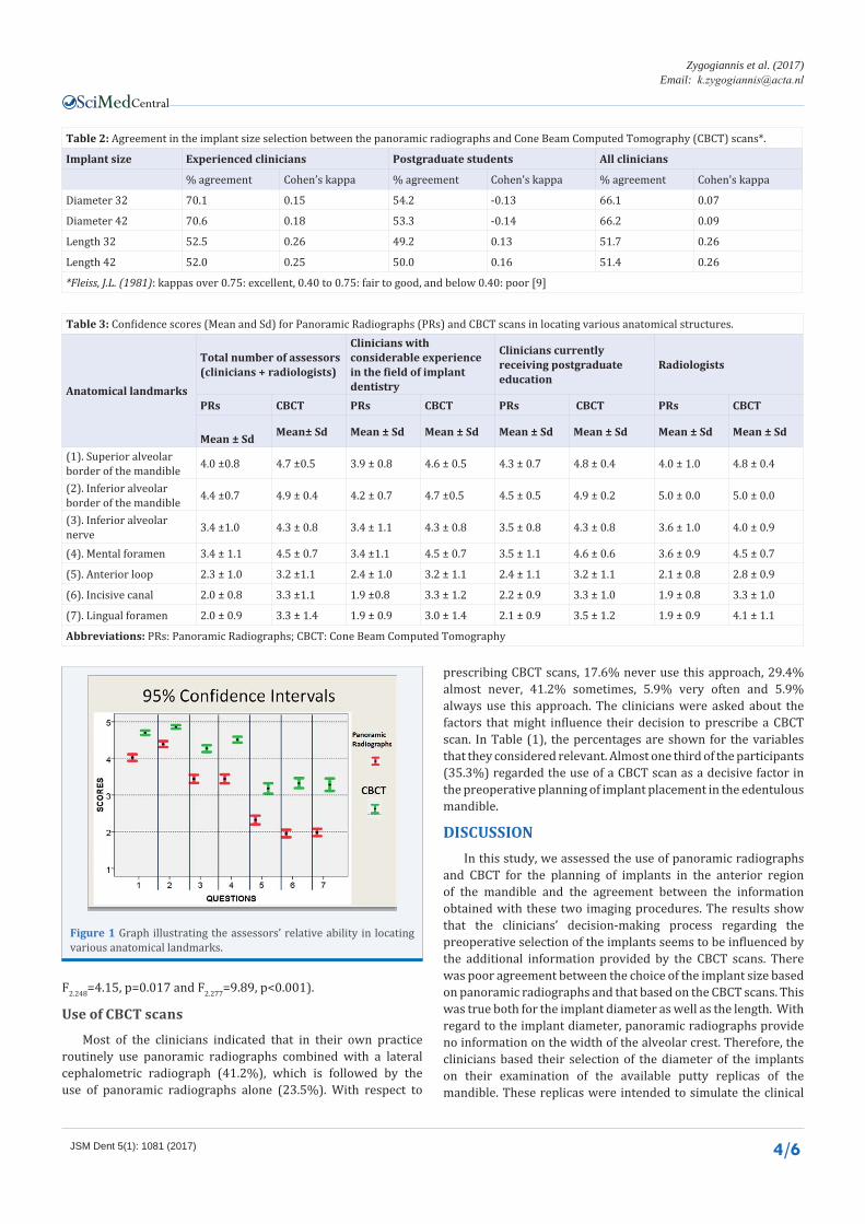
The questionnaire about the location of anatomical landmarks was completed by both the clinicians and the radiologists. The mean and SD values of the questions about their confidence in the accuracy of locating the anatomical landmarks by the two imaging techniques are given in Table (3). The assessors’ relative abilities to locate the various anatomical landmarks are also illustrated in Figure (1). There was a statistically significant difference in the confidence of locating the seven anatomical landmarks using the panoramic radiographs (F6.286=274.3, p<0.001) and CBCT scans (F6.244=97.09, p<0.001). Post hoc pairwise comparisons indicated that only the scores for the inferior alveolar nerve,mental foramen, incisive canal and lingual foramen landmarks were not statistically significantly different from each other for the confidence when using the panoramic radiographs. When using the CBCT scans, the scores of all landmarks differed from each other. The interaction effect with the assessor type was statistically significant for both the panoramic radiographs and CBCT scans (F12.574=5.73, p<0.001 and F12.490=2.85, p=0.001).
For all landmarks, the confidence was higher using the CBCT scans than using the panoramic radiographs (all p values <0.001). Only for the inferior border of the mandible, the incisive canal and the lingual foramen there was a statistically significant interaction effect with the assessor type (F2.279=10.04, p<0.001;
Table 1: Factors influencing the prescription of CBCT scans.
Factors influencing the prescription of Cone Beam Computed Tomography (CBCT) scans % yes % no
Placement of more than 2 implants 29.4 70.6
Anatomical considerations based on clinical examinationLingual undercuts 47.1 52.9Knife-edged crestal ridge 35.3 64.7
Anatomical considerations based on initial radiographic examination
Availability of CBCT scan equipment 5.9 94.1Do you consider the use of a CBCT scan as a decisive factor in the preoperative planning of implant placement in the edentulous mandible? 35.3 64.7
Figure 1 Graph illustrating the assessors’ relative ability in locating various anatomical landmarks.
F2.248=4.15, p=0.017 and F2.277=9.89, p<0.001).
Use of CBCT scans
Most of the clinicians indicated that in their own practice routinely use panoramic radiographs combined with a lateral cephalometric radiograph (41.2%), which is followed by the use of panoramic radiographs alone (23.5%). With respect to
prescribing CBCT scans, 17.6% never use this approach, 29.4% almost never, 41.2% sometimes, 5.9% very often and 5.9% always use this approach. The clinicians were asked about the factors that might influence their decision to prescribe a CBCT scan. In Table (1), the percentages are shown for the variables that they considered relevant. Almost one third of the participants (35.3%) regarded the use of a CBCT scan as a decisive factor in the preoperative planning of implant placement in the edentulous mandible.
DISCUSSIONIn this study, we assessed the use of panoramic radiographs
and CBCT for the planning of implants in the anterior region of the mandible and the agreement between the information obtained with these two imaging procedures. The results show that the clinicians’ decision-making process regarding the preoperative selection of the implants seems to be influenced by the additional information provided by the CBCT scans. There was poor agreement between the choice of the implant size based on panoramic radiographs and that based on the CBCT scans. This was true both for the implant diameter as well as the length. With regard to the implant diameter, panoramic radiographs provide no information on the width of the alveolar crest. Therefore, the clinicians based their selection of the diameter of the implants on their examination of the available putty replicas of the mandible. These replicas were intended to simulate the clinical
Zygogiannis et al. (2017)Email:
JSM Dent 5(1): 1081 (2017) 5/6
Central
examination, which includes palpation of the lingual part of the mandible. However, it should be emphasized that the information obtained using the putty replicas was limited and this is not a true simulation of the clinical examination. The clinicians’ selection on the basis of the putty replicas was compared with that made using the CBCT images. The percentage agreement for the 17 assessors between the initial scores and CBCT scores was 66%. It was interesting to note that, in general, the implants selected on the basis of the CBCT images were narrower than those planned on the basis of the putty replicas. Consequently, complications are likely to follow during the surgical procedure, especially when implants are placed in resorbed or knife-edged residual ridges [10]. The clinicians’ level of experience was also related to their choice of implant diameter. The percentage agreement between the two methods was significantly higher in the group of experienced dentists than in the group of postgraduate students (70% vs. 50%). These results may indicate that a more experienced dentist is more likely to make an accurate preoperative assessment on the basis of the clinical examination only.
With respect to the length rather than the width of the implant, we found an even higher deviation per clinician between the two diagnostic methods. In approximately 50% of the cases, the use of cross-sectional images led to the selection of a different implant length than that selected on the basis of the panoramic radiographs. It was particularly clear that when the experienced dentists used the CBCT scans, there was a trend towards the selection of shorter implants. For example, after evaluating the panoramic radiographs, the dentists selected an implant length of 12 mm (which was the longest implant in our study) in 53.7% of the cases. In contrast, when CBCT scans were available, they selected a 12mm implant in only 34.5% of the cases. Similarly, when the panoramic radiographs were available, the postgraduate students selected a 12mm implant length in 25% of the cases; when they assessed the CBCT images, none of them selected this implant size. In severely resorbed mandibles, in which the maximum available bone height has to be used, it is even more critical to accurately select the correct length of the implants planned for placement in the anterior edentulous mandible. Schropp et al., showed that the length of the implants inserted in the mandible changed in 56% of the cases when the information of tomograms was used [11].
With regard to the assessors’ confidence in locating the various anatomical landmarks, additional information from the CBCT scan increased their level of confidence. While the assessors could locate the upper and lower borders of the anterior mandible, inferior alveolar nerve and mental foramen with a high degree of confidence, they found it more difficult to locate the anterior loop, incisive canal and lingual foramina. This indicates the limitations of assessing these three structures on panoramic radiographs. Similar outcomes were obtained by Reddy et al, who argued that 3-D reconstructions and cross sections allow clinicians to locate various anatomical structures more accurately, assisting them to develop an appropriate preoperative treatment plan [10]. It should be noted that the observers were asked about their confidence in locating the various anatomical structures. The outcome does not tell how accurate their observations were.
More specifically, when only a panoramic radiograph was evaluated, it was difficult for all assessors to accurately locate the incisive canal. Conversely, the confidence level increased considerably (from 1.95 to 3.32) when the assessors were asked to locate the incisive canal on the CBCT scan. However, their confidence levels never reached the highest possible score. The results were not greatly influenced by the assessors’ level of clinical or radiological experience. It was interesting to note that when the clinicians were asked whether the presence of the incisive canal would guide their decision to prescribe a CBCT scan, only 17.6% of them gave a positive answer. Dentists might generally be unaware of the importance of this anatomical structure, or they tend to underestimate the risk of implant placement in this region. The limitations of a panoramic radiograph with regard to detecting the route of the mandibular incisive canal was emphasized in another study, in which only 15% of the panoramic radiographs showed the route [12]. By contrast, CBCT can be used to identify predictably the incisive canal, indicating the high preoperative value of this imaging technique for surgical procedures in the anterior mandible [6,13].
The results of the questionnaire about the referral criteria for CBCT showed that the decision of the clinicians to prescribe a CBCT might be influenced by the presence of lingual undercuts or knife-edged ridges, when they are identified during the clinical examination. In addition, almost half of the clinicians (47.1%) considered the importance of knowing more precisely the presence and route of the anterior loop as a reason to prescribe a CBCT scan. A very small percentage of the clinicians (11.8%) would prescribe a CBCT scan on a regular basis. In our study, the availability of CBCT equipment did not influence the clinicians’ decision on whether to obtain a CBCT scan. This is a positive finding because it has been shown that the choice of radiographic examinations may be influenced by the availability of equipment and resources rather than the clinical need [14,15]. It is interesting to note that only one third of the participants regarded the information provided by CBCT scans as a decisive factor in the preoperative planning of implant placement in the edentulous mandible. Among them, the vast majority of the postgraduate students (80%) did not consider CBCT as an influential component of the preoperative treatment planning. The clinical implications might be that there is a possibility that the clinician would fail to obtain a comprehensive knowledge of oral-bone anatomy prior to implant placement, which may increase the risk of surgical complications, especially when they possess a limited amount of clinical experience.
In the present study, there was a tendency to overestimate the amount of bone available for optimal implant placement when the clinicians relied only on the information provided by the (simulated) clinical examination and the panoramic radiographs. This was true for both the vertical height of alveolar bone as well as the buccolingual width. The additional information provided by the CBCT scans, therefore, might change the initial implant size as this is selected on basis of the panoramic radiographs. Selection of the appropriate implant size may reduce the risk of surgical complications such as perforation of the lingual cortical plate. In addition, it can optimize the final inclination of the implants, especially in the bucco-lingual direction, allowing for a more favorable prosthodontic design. It can also warrantee
Zygogiannis et al. (2017)Email:
JSM Dent 5(1): 1081 (2017) 6/6
Central
that the implants are surrounded by sufficient amount of bone preventing exposure of the implant threads.
The question whether the use of CBCT is necessary for the preoperative implant planning was addressed in a recently published study by Jensen et al. [16]. They assessed the influence of two diagnostic imaging techniques (panoramic radiograph and CBCT) in treatment planning prior to implant placement in the severely resorbed posterior mandible to support removable partial dentures. It was concluded that panoramic radiography in combination with clinical examination can provide -in the vast majority of the cases -sufficient preoperative information. However, in cases of knife-edged ridges, limited bone height, or if the course of the mandibular canal is unclear,a CBCT scan might be justified. According to the guidelines of the European Association for Osseointegration, only if the combination of clinical examination and conventional radiographic examination fail to provide sufficient information for a reliable implant treatment planning, CBCT is justified [7]. The results of our study suggest that the initial imaging assessment by means of panoramic radiography more often provides sufficient information prior to implant placement in the edentulous anterior mandible. CBCT, however, might assist in selecting the most appropriate implant size, especially in specific situations such as resorbed or knife-edged residual ridges, or presence of lingual undercuts. Furthermore, cross-sectional tomography may allow the visualization of anatomical structures such as the anterior loop and the incisive canal with increased reliability and confidence, which in turn might reduce the risk of surgical complications.
CONCLUSIONIn our study, the type of radiographic technique used for
preoperative planning of implants in the anterior edentulous mandible seemed to influence the selection of the size of the implants. When data provided by the CBCT scans was added, narrower and shorter implants were selected.
Only a minority of the participants considered the availability of CBCT scans as a decisive factor for the preoperative treatment plan to provide implant-retained mandibular overdentures, avoiding regular utilization of such imaging techniques. The additional information provided by the CBCT scans appeared to improve clinicians’ confidence in locating important anatomical landmarks in the anterior mandible.
Mandibular two implant-supported overdentures as the first choice standard of care for edentulous patients--the York Consensus Statement. Br Dent J. 2009; 207:185-186.
2. Kraut RA. Interactive CT diagnostics, planning and preparation for dental implants. Implant Dent. 1998; 7: 19-25.
3. Bouserhal C, Jacobs R, Quirynen M, van Steenberghe D. Imaging technique selection for the preoperative planning of oral implants: a review of the literature. Clin Implant Dent Relat Res. 2002; 4:156-172.
4. Mraiwa N, Jacobs R, Moerman P, Lambrichts I, van Steenberghe D, Quirynen M. Presence and course of the incisive canal in the human mandibular interforaminal region: two-dimensional imaging versus anatomical observations. Surg Radiol Anat. 2003; 25: 416-423
5. Kütük N, Demirbaş AE, Gönen ZB, Topan C, Kiliç E, Etöz OA, et al. Anterior mandibular zone safe for implants. J Craniofac Surg. 2013; 24: 405-408.
6. Makris N, Stamatakis H, Syriopoulos K, Tsiklakis K, van der Stelt PF. Evaluation of the visibility and the course of the mandibular incisive canal and the lingual foramen using cone-beam computed tomography. Clin Oral Implants Res. 2010; 21: 766-771.
7. Harris D, Horner K, Gröndahl K, Jacobs R, Helmrot E, Benic G et al. E.A.O. guidelines for the use of diagnostic imaging in implant dentistry 2011. A consensus workshop organized by the European Association for Osseointegration at the Medical University of Warsaw. Clin Oral Implants Res. 2012; 23:1243-1253.
8. Shelley AM, Glenny AM, Goodwin M, Brunton P, Horner K. Conventional radiography and cross-sectional imaging when planning dental implants in the anterior edentulous mandible to support an overdenture: a systematic review. Dentomaxillofac Radiol. 2014; 43: 20130321.
9. Fleiss JL. Statistical methods for rates and proportions (2nd ed.). New York. John Wiley. 1981.
10. Reddy MS, Mayfield-Donahoo T, Vanderven FJ, Jeffcoat MK. A comparison of the diagnostic advantages of panoramic radiography and computed tomography scanning for placement of root form dental implants. Clin Oral Implants Res. 1994; 5: 229-238
11. Schropp L, Wenzel A, Kostopoulos L. Impact of conventional tomography on prediction of the appropriate implant size. Oral Surg Oral Med Oral Pathol Oral Radiol Endod. 2001; 92: 458-463.
12. Jacobs R, Mraiwa N, Van Steenberghe D, Sanderink G, Quirynen M. Appearance of the mandibular incisive canal on panoramic radiographs. Surg Radiol Anat. 2004; 26: 329-33.
13. Juodzbalys G, Wang HL, Sabalys G. Anatomy of Mandibular Vital Structures. Part II: Mandibular Incisive Canal, Mental Foramen and Associated Neurovascular Bundles in Relation with Dental Implantology. J Oral Maxillofac Res. 2010; 1: 3.
14. Ekestubbe A. Conventional spiral and low-dose computed mandibular tomography for dental implant planning. Swed Dent J Suppl. 1999; 138: 1-82.
15. Schropp L, Stavropoulos A, Gotfredsen E, Wenzel A. Calibration of radiographs by a reference metal ball affects preoperative selection of implant size. Clin Oral Investig. 2009; 13: 375-381.
16. Jensen C, Raghoebar GM, Meijer HJ, Schepers R, Cune MS. Two Diagnostic Procedures in Planning Dental Implants to Support a Mandibular Free-Ending Removable Partial Denture. Clin Implant Dent RelatRes. 2016; 18: 678-685.
Zygogiannis K, Aartman IHA, Parsa A, van der Stelt PF, Wismeijer D (2017) Implant Size Selection and Location of Anatomical Structures Prior to Implant Place-ment to Retain Mandibular Overdentures: Panoramic Radiographs vs Cone Beam Computed Tomography. JSM Dent 5(1): 1081.